Prevalence of Mycoplasma gallisepticum in the Ten licensed ...
81
Islamic University-Gaza Deanship of Graduate Studies Faculty of Science Biological Sciences Master Program Medical Technology ŗƒƆƚŪƗŒ ŗŶƆœŞƃŒ ŖŨŹ œƒƄŶƃŒ ŘœŪŒŧťƃŒ ŖťœƆŵ ƅƍƄŶƃŒ ŗƒƄƂ ŗƒřœƒšƃŒ ƅƍƄŶƃŒ ŧƒřŪŞœƆ ŝƆœƈŧŕ ŗƒŕűƃŒ ¾ƒƃœšřƃŒ Prevalence of Mycoplasma gallisepticum in the Ten licensed Hatcheries in Gaza strip, Palestine by Mahmoud A. Almanama Supervisors Dr. Abdelraouf A. Elmanama Ph. D Microbiology Dr. Basim M. Ayesh Ph. D Molecular biochemistry A Thesis Submitted in Partial Fulfillment of the Requirements for the Degree of Master of Biological Sciences/ Medical Technology 2011
Transcript of Prevalence of Mycoplasma gallisepticum in the Ten licensed ...

Islamic University-Gaza Deanship of Graduate Studies
Faculty of Science
Biological Sciences Master Program
Medical Technology
ŗƒƆƚŪƗŒŗŶƆœŞƃŒŖŨŹ
œƒƄŶƃŒŘœŪŒŧťƃŒŖťœƆŵ ƅƍƄŶƃŒŗƒƄƂ
ŗƒřœƒšƃŒƅƍƄŶƃŒŧƒřŪŞœƆŝƆœƈŧŕ ŗƒŕűƃŒfrac34ƒƃœšřƃŒ
Prevalence of Mycoplasma gallisepticum in the
Ten licensed Hatcheries in Gaza strip Palestine
by
Mahmoud A Almanama
Supervisors
Dr Abdelraouf A Elmanama
Ph D Microbiology
Dr Basim M Ayesh
Ph D Molecular biochemistry
A Thesis Submitted in Partial Fulfillment of the Requirements for the
Degree of Master of Biological Sciences Medical Technology
2011
id9040000 pdfMachine by Broadgun Software - a great PDF writer - a great PDF creator - httpwwwpdfmachinecom httpwwwbroadguncom
i
Dedication
To the Ministry of Agriculture - Palestine
ii
Acknowledgement
I would like to express my sincere gratitude and appreciation to my
supervisors Dr Abdelraouf Elmanama and Dr Basim Ayesh for giving me
experience meticulous valuable advice and support faithful guidance and
close supervision Thanks are extended to the biological science department
staff I wish to express my deepest gratitude to my family friends and
colleagues
iii
Table of contents
Dedication i Acknowledgment ii Table of contents iii List of tables vi List of figures vii List of abbreviation viii Abstract ix Arabic Abstract x Chapter 1 Introduction 1 11 Overview 1 12 Objective 3 121 General objective 3 122 Specific objective 3 13 Significance 3 Chapter 2 Literature Review 4 21 Study area 4 22 Hatcheries 5 211 Hatchery management 6 23 Chicken egg 6 231 Egg weigh 6 232 Egg structural and characteristics 7 233 Maternal antibodies 8 24 Mycolplasma 9 241 History 9 242 MG organism 9 243 Taxonomy of MG 10 244 MG structural and characteristics 10 2441 Antigenic structure and toxins 12 245 Epidemiology 14 246 Morbidity and mortality 14 247 Transmission 15 248 Incubation period 15 249 Clinical signs 16 2410 Economic impact 17 25 Diagnostic technique 18 251 Differential diagnosis 19 26 Prevention and control 19 261 Immunity and mucosa immune system 19 262 Vaccination 20 263 Medication 20 264 Hazards of residual drugs in poultry products 21 Chapter 3 Materials and Methods 22 31 Materials 22 311 Equipments 22 312 Kits reagents and disposables 22
iv
32 Methods 23 321 Sampling 23 3211 Samples size calculation 23 3212 Study population 23 3213 Samples collection 23 3214 Samples transport and storage 23 3215 Samples processing 23 322 Rapid slide agglutination test 24 3221 Test principle 24 3222 Test procedure 24 323 Real time polymerase chain reaction technique 25 3231 Pooling of samples 25 3232 DNA extraction procedure 26 3233 DNA amplification and interpretation 27 324 Data collection 28 325 Data analysis 28 Chapter 4 Results 29 41 Distribution of hatchability in Gaza strip governorates 29 42 Samples source 29 43 Eggs weight 30 44 Veterinary medical certificate 30 441 Israel veterinary medical certificate 30 442 PNA veterinary medical certificate 30 45 Data for the presence of MG specific antibodies 30 451 Prevalence of MG specific antibodies 30 452 Prevalence of MG antibodies according to the eggs weight 31 453 Prevalence of MG antibodies according to the eggs source 31 46 Data for the prevalence of MG-DNA 32 461 Pooling samples 33 462 Prevalence of MG-DNA 34 4621 Prevalence of MG-DNA according to the eggs weight 35 4622 Prevalence a of MG-DNA according to the eggs source 35 47 Comparison between RSA test and real time PCR technique 36 48 Questionnaire 36 481 Knowledge of MG 36 482 Location designed biosafety attitudes and behavior in hatcheries 37 Chapter 5 Discussion 39 51 Hatchability 39 52 Samples selection 40 53 Prevalence of MG 40 531 Data for the prevalence of MG specific antibodies 43 5311 Prevalence of MG antibodies according to the eggs weight 43 5312 Prevalence of MG antibodies according to the eggs source 44 532 Data for the prevalence of MG-DNA 44 5321 Prevalence of MG-DNA according to eggs weight 44 5322 Prevalence of MG-DNA according to eggs source 45 54 Questionnaire 45
v
Chapter 6 Conclusions and Recommendations 47 61 Conclusions 47 62 Recommendations 48 621 Recommendations from the study 48 622 Recommendations from the literature 48 References 49 Annexes 59 Annex 1 Arabic version questionnaire 59 Annex 2 An English version questionnaire 61 Annex 3 Israel veterinary medical certificate 63 Annex 4 PNA veterinary medical certificate 68 Annex 5 Possible corrective measures for the ongoing infection with MG 69
vi
List of tables
4 Hatcheries and poultry capacity in Gaza strip (21) Table 5 Eggs source and quantities (22) Table 6 Major functions for incubation and eggs hatching (23) Table 7 Eggs weight categories (24) Table 8 Physicochemical constants of yolk and albumen (25) Table 10 Taxonomy of MG (26) Table 28 Tube components for DNA amplification (31) Table 28 Qualitative assay interpretation (32) Table 30 Samples distribution according to eggs weight (41) Table 31 Prevalence of MG specific antibodies according to eggs weight (42) Table 32 Prevalence of MG specific antibodies according to eggs source (43) Table 35 Prevalence of MG-DNA according to eggs weight (44) Table 36 The results by two methods (45) Table 36 Knowledge of MG in hatcheries (46) Table 3738 Location design biosafety behavior and attitudesin hatcheries (47) Table 4142 Prevalence of MG in different countries (51) Table
vii
List of figures
5 Al Ghefary hatchery- Gaza strip (21) Figure 7 Egg structure (22) Figure 9 A hypothetical MG scheme cell (23) Figure 11 Mycoplasma cell membrane (24) Figure 11 Origin of replication (25) Figure 14 MG interaction with CEF cells (26) Figure 16 Severe airsacculitis (27) Figure 24 Samples processing (31) Figure 25 Rapid slide agglutination test (32) Figure 29 Hatchability in August 2010 (41) Figure 29 Samples distribution according to source (42) Figure 31 Prevalence of MG specific antibodies (43) Figure 32 MG-DNA was detected in the sample under the test condition (44) Figure 33 MG-DNA was not detected in the sample under the test condition (45) Figure 33 Results of initial real time PCR experiment from pooled samples (46) Figure 34 Representative results of the same non-pooled samples (47) Figure 34 Prevalence of MG-DNA (48) Figure 35 Prevalence of MG-DNA according to eggs source (49) Figure
viii
List of abbreviations
Adenosine triphosphateATP Base pair Bp Carotenogenic A CrtnA Center for disease control and prevention CDC Chicken embryo fibroblasts CEF Chronic respiratory disease CRD Coding DNA sequence CDS Cytokine response modifier A CrmA Deoxyribonucleic acid DNA Enzyme linked immunosorbent assay ELISA Gene-targeted sequencing GTS Growth inhibition GI Haemagglutination inhibition HI Human epithelial Hela Immunoglobulin A IgA Immunoglobulin G IgG Immunoglobulin M IgM Immunoglobulin Y IgY Infectious bronchitis IB Kilobase pairs Kb Kilodalton KDa Monoclonal antibodies MAb Mycoplasma gallisepticum MG Mycoplasma gallisepticum cytadherence membrane 1 Mgcl Mycoplasma gallisepticum cytadherence membrane 2 Mgc2 National Poultry Improvement Plan NPIP Newcastle disease ND Office International Epizootie OIE Palestinian national authority PNA Phase-variable putative adhesion protein PvpA Phosphate buffer saline PBS Power of hydrogen PH Proteins 52 P52 Proteins 67 P67 Proteins of Mycoplasma gallisepticum A PMGA Random amplified polymorphic DNA RAPD Rapid slide agglutination RSA Real time-Polymerase chain reaction Real time-PCR Ribosomal ribonucleic acid RRNA Rounds per minute Rpm Variable lipoprotein hemagglutininVlhA World health organizationWHO
ix
Abstract
Prevalence of Mycoplasma gallisepticum in the ten licensed Hatcheries
in Gaza strip Palestine
Avian mycoplasmas are pathogens of different avian species especially poultry MG
is the most important mycoplasma Vertical transmission can occur in eggs laid by
naturally infected hens or horizontally within the hatcheries resulting in rapid spread
of the disease throughout the flock It is common in many parts of the world as the
primary agent of chronic respiratory disease and sinusitis causing important
economic losses in the poultry industry and increase the risk to the overall health of
human
The study was conducted in ten licensed local hatcheries which hatch more than 16
million of one old day chicks yearly for more than 1200 poultry breeding farms in
Gaza strip governorates The study aimed to determine the prevalence of MG
infection in representative samples of 390 non-incubated imported fertilized eggs
collected systematically randomly in August 2010 from Gaza strip hatcheries In
addition the diagnostic value of rapid slide agglutination (RSA) test and real time
polymerase chain reaction (real time PCR) technique was compared and the current
control procedure was investigated
The results showed that the prevalence of MG specific antibodies by using RSA test
was (366) while the prevalence of MG-DNA by using real time PCR technique
was (62) In addition the imported eggs which were used for hatching process
were not handled in the best-case specifications and quality control measures were
limited or absent A significant correlation between the eggs weight and the
prevalence of MG specific antibodies and between the eggs weight and the
prevalence of MG-DNA were found The poultry industry in Gaza strip is facing the
infection of MG in the imported fertilized eggs and the visual observation and
certification assessment are not enough for prevention of MG transmission The free
MG egg sources bio-safety measures knowledge and protection programs could
be reduce the rate of infection
Key words Mycoplasma gallisepticum chronic respiratory disease hatcheries
Gaza strip
x
ŚšŕƃŒŭŤƄƆ
ƑżƅƂřŕƒŪƒƃœŹœƆŨƚŕƍƂƒœƆƃŒŗƆƍśŧŞŧœŬřƈŒƃŒŧŬŶŖŖŨŹųœűſƑżŗŮŤŧƆƃŒŘœŤŧŽƆƇƒűŪƄż
ũƏraquoraquoƔųƅŔŕraquoraquoƈŪƜŗƏƄƔŕƈũƏraquoraquoƔųƅŔŵŔƏraquoraquoƊŌŶraquoraquoƔƈŠƅřraquoraquoŲŃũĆƈąƈƇƔŝŔũraquoraquoŠƓraquoraquoƍƉŠŔƏŧraquoraquoƅŔřraquoraquoŰŕŦ
ƓƍƏřƔƈƍŌŕƍũŝƄŌƇƄśŗƔŬƔƅŕŻŕƈŪƜŗƏƄƔŕƈƅŔũŗśŸśƏŰraquoƈƅŔŚŕraquoƎƈƗŔiquestƜraquoŦƉraquoƈĻŕƔraquoŬŌũiquestƂśƊśƑraquoƅŏřŗŕűƔŗƅŔŚŕraquoŦũſƈƅŔiquestŦŔŧĻŕƔƂžŌƏŶraquoƔųƂƅŔiquestraquoƄƓraquožűũraquoƈƆƅŶƔũraquoŬũŕŮraquośƊŔƃraquoƅŨƉraquoŷşśraquoƊƔƅƉŏ
ƇƅŕraquoŸƅŔʼnŕraquoţƊŌŶraquoƔƈŠƓžřŸœŕŮƇƄśŗƔŬƔƅŕŻŕƈŪƜŗƏƄƔŕƈƅŔƏŖŕraquoƎśƅŔűũraquoƈƅūƔœũraquoƅŔŖŗŬraquoƈƅŔƓraquoƍƉƈŪƈƅŔƓŬſƊśƅŔŪŕƎŠƅŔřƔſƊƗŔŖƏƔŠƅŔŖŕƎśƅŔƏċřƔŧŕŰraquośƁŔĄũœŕŬraquoŦƑraquoƅŏƒŧŎraquoƔƒŨraquoƅŔũƏraquoƔųƅŔƓraquož
ƉŠŔƏŧƅŔŵŕųƁƓžĻŔŧŠċŘũƔŗƄƉŕŬƊƚƅřƈŕŸƅŔřţŰƅŔƑƆŷŶƁŔƏƅŔũųŦƅŔƑƅŏřžŕŲŏ
ŚŕŦũſƈŘũŮŸƅŔƓžřŬŔũŧƅŔŚƔũŠŌŧƁƏŘŪraquoŻŵŕraquoųƁŚŕraquoŴžŕţƈƓraquožĻŕƔƆţƈřŰŦũƈiquestraquoƈŸśƓraquośƅŔƏƉƈũŝƄŌŞŕśƊŏƑƆŷƉƏƔƆƈĻŕraquoƈƏƔŕraquoƎƊƈĊŧţŔƏƅŔąũƈŷċŚƏƄśƄƉraquoƈĄũraquoŝƄƗċřraquoŷũŪƈ
ƅŔƏřraquoƈƏŝũŠŗƐƏŧraquoŸƅŔũŕŮraquośƊŔŧraquoƔŧţśƅƏŘŪraquoŻŃŵŕraquoųƁĊŚŕraquoŴžŕţƈƓraquožƉŠŔƏŧƅŔřƔŗũśƑƆŷiquestƈŸśƓśƉraquoƈřraquoƊƏƄƈřƔœŔƏŮŷřƊƔŷŶƈŠƇśƇƄśŗƔŬƔƅŕŻŕƈŪƜŗƏƄƔŕƈƅŔřŗŰraquoŦƈŘŧũƏśŬraquoƈřŲraquoƔŗũraquoƔŻ
ūųŬraquoŻŌũƎraquoŮƓžŘŪŻŵŕųƁŚŕŦũſƈƉƈřƊŲţƈŚŔʼnŔũraquoŠŏƉraquoƈƀraquoƂţśƅŔƏŖraquoƊŕŠƉraquoƈřƊũŕƂƈƏřƔƅŕţƅŔřŗŕƁũƅŔƉraquoƈŘũraquoƈƆŗƅŔiquestraquoŷŕſśřƆŬraquoƆŬřraquoƔƊƂśƏŶƔũŬraquoƅŔƉŪƜśƅŔũŕŗśŦŔƉƈiquestƄƅşœŕśƊƅŔ
ũŦŊŖƊŕŠ
řraquoraquoŬŔũŧƅŔƋŨraquoraquoƍşœŕraquoraquośƊŚũraquoraquoƎŴŌŕraquoraquoƈŪƜŗƏƄƔŕƈƆƅřŰraquoraquoŰŦƈƅŔřraquoraquoƔŷŕƊƈƅŔŚŔŧŕŲraquoraquoƈƅŔŧƏraquoraquoŠƏřŗŬraquoraquoƊƉŌŚƊŕƄŶƔũŬƅŔƉŪƜśƅŔũŕŗśŦŔƇŔŧŦśŬŕŗƇƄśŗƔŬƔƅŕŻϯϲϲŕraquoƈƊƔŗűƈŕraquoţƅŔŧƏraquoŠƏřŗŬraquoƊƒƏƏraquoƊƅŔ
ƊƂśƇŔŧŦśŬŕŗŘũraquoƈƆŗƅŔiquestŷŕſśřƆŬƆŬřƔŚraquoƊŕƄřŗŬraquoƊŗƓraquožƇŧŦśŬraquoƈƅŔŧũƏśŬraquoƈƅŔűƔraquoŗƅŔƉŌƏŘŧƏraquoŠƏƈũraquoƔŻƏŌŘŧƏŧraquoţƈŘŧƏraquoŠƅŔřraquoŗƁŔũƈũƔƔŕraquoŸƈƉŌƏƌśŕſŰŔƏƈiquestŲžŌƓžūƔƅŖŕŰŦƙŔřƔƆƈŷ
ƆŷřŬŔũŧƅŔŚŧƄŌƏƑƉƔraquoŗƏřraquoƈƏŝũŠƆƅřraquoƔŷŕƊƈƅŔŚŔŧŕŲraquoƈƅŔƏűƔraquoŗƅŔƉŪƏƉƔraquoŗřƔƏƊŸƈřƁƜŷŧƏŠƏƅŔƉŪƏŕţƅŔƏűƔŗŕƎƅƒƏƏƊƅŔűƈ
řraquoƈƏŝũŠŗřŗŕraquoŰƙŔƌŠŔƏśŘŪŻŵŕųƁƓžƉŠŔƏŧƅŔřŷŕƊŰƉŏűƔraquoŗƅŔƓraquožƇƄśŗƔŬraquoƔƅŕŻŕraquoƈŪƜŗƏƄƔŕƈƅŔ
ŔƉŔƏŖŰŦƈƅŔŧũƏśŬƈƅŔűũraquoƈƅŔiquestŕraquoƂśƊŔŶraquoƊƈƅƓraquoſƄśƛřƔţŰraquoƅŔŘŧŕƎŮraquoƅŔƏřƔũŰraquoŗƅŔřŗƁŔũƈƅƉŏűƔŗƅŔũŧŕŰƈřƈƜŬřžũŸƈƅŔƏraquoƅŔşƈŔũraquoŗƅŔƏŚŔʼnŔũraquoŠƙŔŵŕraquoŗśŏƏiquestraquoƆƂƔŕraquoƈŗũřƔţŰiquestŧraquoŸƈƉraquoƈ
řŗŕŰƙŔ
ŗƒšœřŽƆƃŒŘœƆƄƂƃŒƇƄśŗƔŬƔƅŕŻŕƈŪƜŗƏƄƔŕƈƉƈŪƈƅŔƓŬſƊśƅŔŪŕƎŠƅŔŖŕƎśƅŔűũƈŘŪŻŵŕųƁŚŕŦũſƈƅŔ
ϭ
Chapter 1
Introduction
11 Overview
In Gaza-strip poultry industry is very important as the consumption of poultry
meat and eggs is increasing steadily [1] The poultry industry is facing
various problems One of these problems is the infection of grandparent and
parent flocks which occurs in many developing countries resulting in
dissemination of diseases including mycoplasmosis salmonellosis and
reoviral infection [2]
Mycoplasmas tend to be quite host specific some infect only a single species
of animal while others may have the ability to infect several different animal
species They may be found in humans many animal species plants and
insects In general mycoplasma colonize mucosal surfaces and most
species are noninvasive but some species including Mycoplasma
gallisepticum (MG) have the ability to penetrate cells and cause diseases [3]
MG is a member of the class Mollicutes which are very small prokaryotes
devoid of cell walls bounded by a plasma membrane only This accounts for
the (fried egg) type of colony morphology resistance to antibiotics that
affects cell wall synthesis and complex nutritional requirements [4] It
remains the most frequently reported bacterial causative agent of chronic
respiratory disease in chickens and infectious sinusitis in turkeys [3]
It is a highly infectious respiratory pathogen affecting poultry In broilers MG
causes reduction in weight gain decrease in feed conversion efficiency
increase in mortality rate and increased condemnations in slaughterhouses
In breeders and layers the disease causes a drop in egg production and an
increase in embryo mortality [2] Inoculation of MG into 7-day-old
embryonated chicken eggs via the yolk sac route usually results in embryo
deaths within 5-7 days [5]
Ϯ
A survey of commercial egg laying poultry in United States of America (USA)
revealed that 37 of laying flocks (2626 million layers) were infected with
MG and causing an annual losses of 97 million US $ [6] In addition
medication costs make this disease one of the costliest disease problems
confronting the poultry industry [7] and causes problems in food safety drug
resistance and drug residual [2]
MG vertical transmission can occur through eggs or horizontally by inhalation
of contaminated airborne droplets resulting in rapid disease transmission
throughout the flock Prevention and control programs based on strict
biosecurity surveillance and eradication of infected breeder flocks are
preferable Using vaccination with bacterins has been shown to reduce but
not eliminate colonization by MG [1]
On some farms especially in areas with an intensive and varied population of
poultry flocks there may be extensive use of antibiotics for MG [8]
Antibiotics were used to combat the disease through the application of a
single drug administration or drugs in combination for prevention control and
treatment of MG infection [6] However because such a solution is not always
economically feasible it is important to be able to detect vertical infection[9]
It is difficult to diagnose MG infections in poultry flocks based on clinical
signs Routine culture procedures and serology are commonly used The
diagnosis of MG infection traditionally has been done by serology [10]
Some of the disadvantages of serological methods are false-positive and
false-negative reactions due to interspecies cross-reactions and nonspecific
reactions [11] Recently office international epizootie and national poultry
improvement plan recommended PCR as a reliable test for the detection of
MG infections Real-time PCR which has distinct advantages over
conventional PCR such as higher reliability rapidity and prevention of
environmental contamination has been used for the detection of MG in
poultry [12]
ϯ
There is no published data on the prevalence of MG in Gaza strip and to the
best of our knowledge this is the first study investigating the prevalence of
this pathogen in fertilized eggs used in the production of one-day-old chicks
in hatcheries in Gaza strip
12 Objective
121 General objective
To determine the prevalence of MG infection in non-incubated imported
fertilized eggs in the ten licensed hatcheries of Gaza strip Palestine
122 Specific objectives
To detect the prevalence of MG antibodies by rapid slide agglutination
(RSA) test
To detect the prevalence of MG-DNA by real time PCR technique
To compare the diagnostic value of RSA test and real time PCR technique
To describe the current procedures for control of MG infections
13 Significance
MG economically affects the poultry industry through increased mortality and
decreased egg production and reduced feed efficiency The infected places
may undergo serious sequelae and need early and prompt treatment This
study is the first tackling MG and no previous studies concerning MG were
conducted in Gaza strip There is no available data about MG in the study
area and the result of this study may contribute to increase the awareness of
the relevant authorities about the prevalence of this pathogen In addition
data generated from this study may provide the Ministry of Agriculture and
other concerned parties a true and scientific view of the existing situation of
imported fertilized eggs This may help in planning and implementing actions
to reduce the spread of this costly pathogen
ϰ
Chapter 2
Literature Review
21 Study area
Gaza strip is a Palestinian administrated territory in the Middle East boarded
on the south by Egypt on the west by the Mediterranean and the east and
north by Israel It is one of the most densely populated places on earth with
a total area of 365 km2 and population of over 154 million [7] In Gaza strip
agriculture is considered the most important productive sector More than
20 of the population is dependent upon it The animal production sector is
one of the most important sectors of Palestinian agriculture Its importance
comes from the increasing investments in the livestock sector [13]
This share had increased from 20 to 30 for the years of seventies and
nineties respectively Gaza strip hatcheries (Table 21) are capable of
providing both broiler and layer chickens they have maximum capacity
closed to 70 million of eggs They are dependent on fertilized eggs which
come from multiple sources including locally produced as well as imported
from Israel or from abroad [13] The quantities of imported fertilized broiler
eggs through the first three months of the year 2010 were 91 million (Table
22) [14]
Table 21 Hatcheries and poultry capacity in Gaza strip [13]
No of Layers Thousand
No of Layers farms
No of Broilers million
No of Broilers farms
Hatched eggs million
Capacity eggs million
No of hatcheries Governorate
128 26 21 108 29 94 2 Gaza north
427 63 22 132 94 192 1 Gaza
123 26 35 230 3 184 3 Gaza med
76 21 45 353 58 156 3 Khan younis
745 6 3 254 04 72 1 Rafah
8285 142 153 1077 216 70 10 Total
ϱ
Table (22) Eggs source and quantities [14]
Percentage Hatched eggs million Eggs Source
28 02 West Bank
827 76 Israel
145 13 Abroad
100 91 Total
22 Hatcheries
Hatcheries are an integral link in the chicken supply chain They come
between two producer groups the broiler hatching egg producers and the
broiler producers Hatchery location is inevitably a compromise between the
disease risks of a populated poultry area the transport costs of eggs and
chicks the availability of labor and the overall transport network [15] Good
hatchery design is essential for cost-effective operation Their design must
therefore incorporate food hygiene standards
The conditions provided to maintain embryonic growth in the incubators are
also ideal for the growth of bacteria and molds All room surfaces items of
equipment and incubators must be designed to allow simple regular and
effective cleaning and sterilization (Figure 21) [15]
Figure (21) Al Ghefary hatchery- Gaza strip Picture was taken by the researcher
in 15-8-2010
ϲ
221 Hatchery management
Five major functions are involved in the incubation and hatching of chicken
eggs (Table 23) The five functions are temperature humidity ventilation
egg turning and sanitation When two or more are not controlled it may be a
disaster A consistently low temperature will result in a late hatch and
decreased hatchability The chicks may be large soft bodied and weak A
consistently high temperature will result in an early hatch and decreased
hatchability The chicks may have short down and have rough navels More
chicks will be malformed deformed legs weak and small [16]
A sign of low humidity is sticky embryos during pipping and hatching that
result in embryos not being able to detach themselves from the shell In
addition results in short down on the chicks malformed mal-positioned
weak and small chicks As embryos grow the air vent openings are gradually
opened to satisfy increased embryonic oxygen demand If the egg is not
turned the developing embryo is squeezed between the yolk and shell and
can be damaged or killed Poor sanitation causes not only poor hatch but
also subsequent early death during brooding and affects the birds during the
grow-out period [16]
Table (23) Major functions for incubation and eggs hatching [16]
Item Chicken Turkey Duck Goose
Incubation period (days) 21 28 28 28-34
Temperature (degC) 38 37 38 37
Humidity 85-87 84-86 85-86 86-88
No egg turning after 18th d 25th d 25th d 25th d
Open additional vents 10th d 14th d 12th d 1st d
Open vents (if needed) 18th day 25th d 25th d 25th d
23 Chicken egg
231 Egg weigh
All chicken hens start egg production laying small and gradually increase to a
mature egg grade size Average egg weight increased from 54 gm at 26
ϳ
weeks of age to 64 gm at 66 weeks of age [17] Eggs are categorized
according to weigh as large medium small (Table 24)
Table (24) Egg weight categories [18]
Size Weight
Large 63 73 gm
Medium 53 63 gm
Small Less than 53 gm
232 Egg structure and characteristics
Albumin is a clear liquid formed from the layers of secretions of the anterior
section of the hens oviduct during the passage of the egg It protects the egg
yolk and provides additional nutrition for the growth of the embryo An egg
yolk feeds the developing embryo It is suspended in the egg white by one or
two spiral bands of tissue called the chalazae The yolk is enclosed by a thin
vitelline transparent membrane (Figure 22) [19]
Figure (22) Egg structure (a) Sagittal section through a hens egg (b)
enlargement of the region of the shell shown in (a) [19]
ϴ
The whole egg composition percentage is 655 water 118 protein
110 fat117 ash The albumen and yolk physicochemical properties are
illustrated in table 25 [18]
Table (25) Physicochemical constants of yolk and albumen adapted from [18]
233 Maternal antibodies
Chickens transmit maternal antibodies to their offspring by depositing the
antibodies in the egg There are three classes of antibodies in chickens
namely IgY (IgG) IgA and IgM Chicken IgA and IgM are similar to
mammalian IgA and IgM in terms of molecular weight structure and
immunoelectrophoretic mobility Although structural differences exist between
IgY and mammalian IgG IgY is considered the avian equivalent to
mammalian IgG In eggs IgY is present in the egg yolk whereas IgA and
IgM are present in the albumen as a result of mucosal secretion in the
oviduct There is transfer of IgA and IgM antibodies from the albumen into the
egg yolk [20]
Yolk Albumen Property factor 15 25 Bound water () 65 61 Coagulating temperature(C) 1035 1035 Density (gmcm3) 31 868 Electrical conductance (mho-cm-1 x 10⁻³) -0587 -0424 Freezing point 8124 5690 Heat of combustion (calgm) 6 76 pH 14185 13562 Refractive index 125 071 Solubility coefficient for CO2 078 085 Specific heat (calgmC) 032 012 Specific resistance (ohm-cm) 35 53 Surface tension (dyncm) 0971 0756 Vapor pressure (in of NaCl) 200 25 Viscosity (poise at 0C) 81 127 Latent heat (Butlb)
ϵ
24 Mycolplasma
241 History
The first accurate description of the avian mycoplasmosis was in 1905 by
Dodd in England and termed Epizootic pneumoenteritis [21] In 1938
Dickinson and Hinshaw named the disease (infectious sinusitis) of turkeys
[22] In 1943 Delaplane and Stuart cultivated an agent in embryos isolated
from chickens with chronic respiratory disease (CRD) and later from turkeys
with sinusitis [23] In the early 1950 Markham Wong and Van reported that
the organism was a member of the Pleuropneumonia group [24]
242 MG organism
MG (Figure 23) is an avian pathogen within the genus mycoplasma (class
Mollicutes) which includes other species infecting animals humans insects
and plants [25] The organism stains well with Giemsa stain but weakly gram
negative It is generally coccoid approximately (025-05 igravem) [4] The
organism shows a filamentous or flask-shaped polarity of the cell body due to
the presence of terminal organelles or bleb [26]MG requires a protein-rich
medium for their growth containing 10-15 added animal serum [27]
Figure (23) A hypothetical MG scheme cell (a) The specialized terminal
structure (b) the electron-dense area (infrableb) (c) loop-shaped tubules and (d)
plasma membrane [28]
ϭϬ
243 Taxonomy of MG
The Mollicutes (mollis=soft and cutes = skin) belong to the phylum
Firmicutes with low Guanine + Cytosine content of the genome belongs to
the domain bacteria About 200 species of the class Mollicutes have been
validly described [29] It was first classified and differentiated from other
avian mycoplasmas by serotyping [30] and was commonly designated
serotype A [31] Mycoplasma phylogeny and taxonomy continue to be
reexamined by the application of molecular tools such as DNA sequence
analysis [25] Taxonomy of MG is shown in table 26
Table (26) Taxonomy of MG [32]
233150 Taxonomy ID Bacteria Kingdom Firmicutes Intermediate Rank 1 Mollicutes Intermediate Rank 2 Mycoplasmataceae Intermediate Rank 3 Mycoplasma Intermediate Rank 4 Mycoplasma gallisepticum Intermediate Rank 5
244 MG structure and characteristics
The Mollicutes have only the plasma membrane (Figure 24) which proteins
constitute over two-thirds of the mass with the rest being membrane lipids
Motif analysis of the MG genome has predicted a large repertoire of
membrane-associated proteins Of these proteins 149 contain multiple
transmembrane domains and 24 ATP-binding proteins predicted to be
involved in transport of biomolecules [33]
The membrane lipoproteins are a majority of the mycoplasma surface
antigens [34] All mycoplasma lipids are located in the cell membrane and
consist of phospholipids glycolipids and neutral lipids The mycoplasmas are
partially or very incapable of fatty acid synthesis and require cholesterol for
growth [35]
ϭϭ
Figure (24) Mycoplasma cell membrane [36]
The genome is composed of 9964 bp with an overall Guanine + Cytosine
content of 31 It contains 742 putative coding DNA sequences (CDSs)
representing a 91 coding density Function has been assigned to 469 of the
CDSs while 150 are conserved hypothetical proteins and 123 remain as
hypothetical proteins [37] MG genome contains two copies of the rRNA
genes One set is organized as an operon with adjacent 16S 23S and 5S
genes and a second copy of the 16S rRNA gene lies 221 kb upstream of the
23S and 5S rRNA genes [33]
The organization of the putative origin of replication for MG is located in the
region of the dnaA gene (Figure 25) [33]It has been well established that
MG generally expresses a single member of the family at any one time [35]
The specific gene expressed can be influenced by growth in the presence of
cognate antibody [36] The probable role of this family in generating antigenic
variation has been demonstrated in infected chickens [7]
Figure (25) Origin of replication of MG [33]
ϭϮ
2441 Antigenic structure and toxins
The plasma membrane of MG contains approximately 200 polypeptides
associated with surface antigenic variation adhesion to host cells motility
and nutrient transport [2] Considerable effort has been made to identify MG
adhesion or hemagglutinin properties which may play key roles in the
pathogenesis of an immune response to infection Adhesins are integral
membrane proteins having regions exposed on the cell surface that attach to
receptor sites on host epithelial cells which allow for colonization and
infection Such as these proteins considered important virulence factors and
antigens [38]
Immunoblotting techniques revealed that the surface antigens p52 and p67
(pMGA) were specific to MG and the closely related species The pMGA
gene family is significant genomic commitments to antigenic variability and
hypothesized function of immune evasion [39] The pMGA gene family also
provides a mechanism for rapid and reversible switches in its expression of
proteins (antigenic switching) in response to antibodies or other
environmental cues [37] In 2003 the pMGA gene and protein were renamed
vlhA and VlhA respectively [33]
The vlhA gene family encodes hemagglutinin in MG and the vlhA genes are
located in several loci around the chromosome and antigenic variation is
generated by alternating transcription of over 40 translationally competent
genes [40] PvpA is an MG size-variable integral membrane protein that
shows high-frequency phase variation in its expression and adds to the
complexity of antigenic variation in MG Antigenic variation and expression of
PvpA and p67a (major immunogenic surface proteins) were correlated with
antibody response in vivo suggesting that immune modulation may have a
key role in generating surface diversity [41]
The preceding information and that from many other reports indicates that
the MG genome is highly committed to antigenic variation and variable
ϭϯ
expression of surface proteins [42] Other adhesion proteins identified in MG
are GapA and Mgc2 GapA is a primary cytadhesin that appears to work in a
coordinated way with at least one other cytadherence-related protein CrtnA
undergoing concomitant phase variation in expression [43] Expression of
these two components has been correlated with binding to erythrocytes and
efficient attachment to cultured cells These results demonstrated that both
GapA and CrmA are required for MG cytadherence and pathogenesis Potent
toxins have not been associated with mycoplasmas [7]
245 Epidemiology
MG has been isolated from naturally occurring infections in chickens (Gallus
gallus) turkeys (Meleagris gallopavo) pheasants (Phasinus colchicus)
chukar partridge (Alectoris graeca cristatus) [44] bobwhite quail (Colinus
virginianus) and Japanese quail (Coturnix japonica) [45] MG has also been
isolated from duck (Anas platyrhynchos) from geese (Anser anser) from
house finches (Carpodacus mexicanus) [46] from a golden pheasant
(Chrysolophus pictus) in Australia [47] from a yellow-naped Amazon parrot
(Amazona ochrocephala auropalliata) [48]
MG infection has been produced experimentally in captive - reared wild
turkeys [49] House sparrows (Passer domesticus) and budgerigars
(Melopsittacus undualtus) [50] MG was also isolated from a blue jay
(Cyanocitta cristata) that contracted conjunctivitis and from free- ranging
American goldfinches (Carduelis tristis) [51] MG have been confirmed by
culture or PCR in purple finches (Carpodacus purpureus) eastern tufted
titmice (Baeolophus bicolor) pine grosbeaks (Pinicola enucleator) and
evening grosbeaks (Coccothraustes vespertinus) [52]
MG causes disease in game birds including pheasants (Phasinus colchicus)
chukar partridges (Alectoris chukar) bobwhite quail (Colinus virginianus)
Japanese quail (Cotrunix japonica) and peafowl (Pavo cristatus) pigeons
greater flamingos (Phoenicopterus roseus) wild peregrine falcons (Falco
ϭϰ
peregrines) in Spain [52] MG has the ability to invade cultural human
epithelial cells (Hela -229) and chicken embryo fibroblasts (CEF) (Figure 26)
and survive within the intracellular space over a 48-h period [53]
Figure (26) MG interaction with CEF cells localization of extracellular (yellow) and
intracellular (red) Bars 10 mm [53]
246 Morbidity and mortality
MG is not a killer disease like Newcastle (ND) or Gumboro but in
complicated cases birds may die [54] Inoculation of broth cultures or
exudates containing MG into 7-day-old embryonated chicken eggs via the
yolk sac route usually results in embryo deaths within 5-7 days The
organism reaches its highest concentration in the yolk sac yolk and
chorioallantoic membrane just prior to embryo death MG embryo mortality
was prevented in eggs containing maternal MG antibodies [55]
MG infection usually affects nearly all chickens in a flock but disease is
variable in severity and duration It tends to be more severe and of longer
duration in the cold months and affects younger birds more severely than
mature birds [56] Mortality may be negligible in adult laying flocks but there
can be a reduction in egg production [57] In broilers the mortality may range
from low in uncomplicated disease to as much as 30 in complicated
outbreaks especially during the colder months Retarded growth
downgrading of carcasses and condemnations constitute further losses [2]
ϭϱ
247 Transmission
Horizontal transmission occur by direct or indirect contact of susceptible
birds with infected clinical or subclinical birds resulting in high infection
disease prevalence within flocks The upper respiratory tract and or
conjunctiva are portals of entry for the organism in aerosols or droplets [2]
MG survived in the human nasal passage for 24 hours on straw cotton and
rubber for 2 days on human hair for 3 days and on feathers for 2-4 days
[58] MG seldom survives for more than a few days outside of a host so clin-
ical or subclinical carrier birds are essential to the epidemiology of MG
diseases [59] However outbreaks may occur via fomites contaminated
airborne dust droplets or feathers coupled with suboptimal biosecurity and
personnel practices [59]
MG vertical transmission (in ovo transovarian) is known to occur in eggs laid
by naturally infected chicken hens [2] The highest rates of transmission were
found during the acute phase of the disease when MG levels in the
respiratory tract peaked [60] In separate studies peak egg transmission was
detected four weeks after MG challenge in approximately 25 of the eggs
and at three to six weeks post challenge in more than 50 of the eggs On a
flock basis egg transmission rates decline as the post infection interval
lengthens Transmission rates of approximately 3 at 8-15 weeks [61] and
approximately 5 at 20-25 weeks have been reported During chronic infec-
tions under field conditions egg transmission is likely to occur at much lower
levels [2]
248 Incubation period
In experimental infections of chickens the MG incubation period varies from
6-21 days depending on MG strain virulence complicating infections
(polymicrobial infections) and environmental and other stressors [2]
Therefore under natural conditions it is very difficult to estimate the possible
date of exposure based on the appearance of clinical signs Many variable
factors seem to influence the onset and extent of clinical disease [62]
ϭϲ
Chickens often develop clinical infections near the onset of egg production
This apparent extended incubation period was especially common in
offspring of infected chicken hens hatched from eggs dipped in antibiotic
solutions for control of MG infection The possible role of contamination from
other sources of infection is not always clear and can rarely be proved
beyond reasonable doubt [62]
249 Clinical signs
The clinical manifestations of MG in chicken embryo are dwarfing
generalized edema liver necrosis and enlarged spleens [55] In other side
the clinical signs in chicken include coughing sneezing snicks rales ocular
and nasal discharge decrease in feed consumption and egg production
increased mortality poor hatchability and lose weight [63] The gross lesions
in birds with MG include catarrhal inflammation of sinuses trachea and
bronchi Air sacs are often thickened and opaque and may contain mucous
or caseous exudates besides hyperplastic lymphoid follicles on the walls
[63]
At slaughter carcass condemnation may result from the presence of
airsacculitis (Figure 27) fibrinous perihepatitis and adhesive pericarditis
interstitial pneumonia and salpingitis which are often seen in chickens [63]
Figure (27) Severe airsacculitis with abundant foam and aggregates of caseous
exudates associated with MG infection [64]
ϭϳ
Chickens conjunctivitis keratoconjunctivitis caused by MG was reported in
commercial layer chickens [65] Chickens showed swelling of the facial skin
and the eyelids increased lacrimation congestion of conjunctival vessels
However flocks may have serologic evidence of infection with no obvious
clinical signs especially if they encountered the infection at a younger age
and have partially recovered Male birds frequently have the most
pronounced signs and the disease is often more severe during winter [66]
2410 Economic impact
MG is the most pathogenic and economically significant mycoplasma
pathogen of poultry [67] MG economically affects the poultry industry
through increased mortality and decreased egg production [68] and reduced
feed efficiency [69] Some researchers demonstrated that the average egg
production loss due to naturally exposed MG infection which was 157 eggs
per chicken hen as compared with MG -free hens [68] Other researchers
showed that MG infected flocks produced 5-12 fewer eggs per chicken hen
compared with uninfected flocks [51]
A statistically significant reduction in egg production was observed during
the first 4 weeks post exposure to MG and significantly lower fertility of eggs
[70] Airsacculitis in chickens can cause significant condemnations at
slaughter Increased medication costs are additional factors that make this
disease one of the costliest disease problems for poultry industry [69] In the
late 70s in USA MG was costing the poultry industry and consumer as much
as 118 million US $ per year not including losses affecting the broiler
industry or other losses in the layer industry such as morbidity mortality or
feed conversion [71]
Documented turkey industry losses in north Carolina USA due to MG
between 1979 and 1983 were greater than 25 million US $ [72] The
commercial layer producers in southern California lost an estimated 127
million eggs because of MG in 1984 This lost egg production and associated
ϭϴ
MG control program costs amounted to an estimated financial loss of
approximately 7 million US $ This represented a loss of approximately 6
million US $ in consumer surplus [57] In Brazil there was a loss of 34
thousand tons of broilers in the end of the production cycle due to respiratory
diseases which corresponded to 30 million US $ in 1994 Moreover MG
infection alone is considered one of the diseases that cause more losses to
the poultry industry [68] Prevention and control programmes of MG includes
chemotherapy vaccination etc are account for additional costs [6]
25 Diagnostic techniques
The presence of MG can be confirmed by isolating the organism in a cell-free
medium or by detecting its DNA directly in infected samples Serological tests
are also widely used for diagnosis [73] Samples are taken from live and
dead birds [74] Several suitable culture media have been formulated MG
media generally contain a protein digest and a meat-infusion base
supplemented with serum or a serum fraction yeast factors glucose and
bacterial inhibitors [75]
The most commonly serological tests used is rapid slide agglutination test
(RSA) It should be carried out at room temperature (20-25deg C) within 72
hours of sample collection [75] The positive results should be considered
presumptive for the presence of MG antibodies [76] MG in
haemagglutination inhibition test is capable of haemagglutinating avian red
blood cells and specific antibodies in test sample cause inhibition A strain
should be selected that grows well and haemagglutinates reliably [77]
In Enzyme-linked immunosorbent assay the plates are coated with whole cell
MG antigen and the test samples are added but the reaction is assessed by
the extent of blocking that occurs when the conjugated monoclonal antibody
is added [78] In growth inhibition test the growth of MG is inhibited by
specific antiserum enabling species to be identified It is relatively insensitive
and sera must be high- titred monospecific [74]
ϭϵ
MG may be detected by hybridization with DNA probes but now it is much
more common to use the PCR to amplify specific portions of DNA in the test
material Molecular methods are also available for differentiation of MG
strains [78] DNA fingerprinting uses arbitrary primed PCR or random
amplified polymorphic DNA (RAPD) Gene-targeted sequencing (GTS) for
the mgc2 gapA pvpA and MG A_0309 genes can be used to provide an
accurate and reproducible method of typing of strains [79]
251 Differential diagnosis
To differentiate MG infection from other common respiratory diseases
Newcastle disease (ND) and Infectious bronchitis (IB) diseases or their
antibodies may be present as separate entities or as part of the complicated
chronic respiratory disease (CRD) syndrome Infectious coryza (Avibacterium
paragallinarum) and fowl cholera (Pasteurella multocida) usually can be
identified by bacterial culture Mycoplasma synoviae infection may be present
alone or in addition to MG Application of both serologic and organism
identification test procedures may be necessary in some cases [66]
26 Prevention and control
The control strategy of many countries is based on maintaining MG free
breeding flocks Establishing the MG - clean status of breeder flocks and
maintaining that status can be accomplished by participation in control
programmes [80]
261 Immunity and mucosa immune system
Chickens that have recovered from clinical signs of MG diseases are known
to have some degree of immunity However recovered birds may still carry
the organism [81] Antibodies persisted in recovered chickens and upon
reexposure they had a faster rate of MG elimination and less severe tracheal
lesions than observed after the first exposure The importance of antibodies
produced in response to MG infection inhibited attachment of the organism to
epithelial cells [82]
ϮϬ
Mycoplasmas may affect the cell-mediated immune system by inducing
either suppression or stimulation of B and T lymphocytes and inducing
cytokines Lymphoproliferation interferon and nitric oxide were detected in
vitro in antigen-stimulated peripheral blood leukocytes from MG-infected
chickens [83] The primary role for local antibody mediated responses in
controlling MG infection but also presented evidence for significant natural
killer and cytotoxic T cell responses to infection [84]
262 Vaccination
Vaccination is the preferred method for control and maintains MG -free
flocks It should be considered only in situations where field exposure is
inevitable such as on multi-age sites and potential exposure of neighboring
poultry flocks Successfully MG vaccinated birds are resistant to respiratory
disease airsacculitis egg production drops and reduced levels of egg
transmission in breeders [67] Two types of vaccines are available for the
control of MG [85] Vaccinated chickens with MG F MG Ts-11 Live vaccines
strains (intranasal or eye drop) and MG 685 strain (as a fine spray) are
permanent carriers so a single dose is adequate [86]
Use of MG F strain vaccine in each replacement flock on a multi-age site will
eventually result in displacement of the field strain with the vaccine strain
MG Ts-11 and 685 strains are a virulent and spread to unvaccinated birds
does not occur or occurs very poorly when birds are in very close contact
[86] MG Inactivated vaccines (bacterins) are prepared from a concentrated
suspension of whole cells that is emulsified into an oil adjuvant High antigen
content is essential [87] Hens vaccinated with live MG strain F and one
dose or two doses of bacterin was significantly lower level than
unvaccinated controls [60]
263 Medication
MG has shown sensitivity in vitro and in vivo to several antimicrobics
including macrolides tetracyclines fluoroquinolones and others however
Ϯϭ
resistance to penicillins and other antibiotics which act by inhibiting cell wall
biosynthesis occurs MG may develop resistance and demonstrate cross-
resistance to commonly used antibiotics [88] Antimicrobics have been used
to treat MG to reduce egg production losses may reduce the severity of
clinical signs and lesions and significantly reduce populations of MG in the
respiratory tract [899091]
Egg injection or dipping has been used to introduce antimicrobials into
hatching eggs to control MG in ovo transmission [92] In general these
methods greatly reduced but sometimes did not completely eliminate the
possibility of egg transmission However the use of antimicrobials for egg
injection or dipping made it possible to obtain sufficient MG free birds [93]
264 Hazards of residual drugs in poultry products
Use of antibiotic that might result in deposition of residues in meat and eggs
must not be permitted in food intended for human consumption If use of
antibiotics is necessary as in prevention and treatment of animal diseases a
withholding period must be observed until the residues are negligible or no
longer detected [94] The withdrawal period is the necessary interval
between the last administration of the drug under normal conditions of use
and the time when treated animals can be slaughtered for the production of
safe foodstuffs The withdrawal period should provide a high degree of
assurance both to the producers and the consumers that the concentration of
residues in foods derived from treated animals are not above the maximum
residue levels [95]
ϮϮ
Chapter 3
Materials and Methods
31 Materials
311 Equipments
Item Manufacturer
1 Digital balance ACCULAB-CANADA
2 Digital dry bath incubator BIO TAD- GERMANY
3 Freezer refrigerator ORSO PHARMAL-SPAIN
4 Autoclave CRISTOFOLI-BRAZIL
5 Vortex mixer TURBO-USA
6 PCR work station with UV light BIOTEK
7 Step one Real time PCR system APPLIED BIOSYSTEMS
8 Mini centrifuge TOMOS
9
Quality micropipettes 05-10 microl 50-50 microl 50-200 microl 100-1000 microl
JENCONS
312 Kits reagents and disposables
Item Manufacturer
1 SPAFAS MG plate antigen CHARLES RIVER-USA
2 QIAamp MinElute DNA kit QIAGEN-USA
3 MYCO GS real time kit BIONOTE-KOREA
4 Ethanol (96100) SNOW-USA
5 Phosphate buffer saline SIGMA-GERMANY
6 Disposable sterile plastic pipette LABCON-USA
7 Sterile specimen collection vials LABCON-USA
8
Filter tips 05-10 microl 10-200 microl 100-1000 microl
LABCON-USA
9 15 2 ml Microcentrifuge tubes LABCON-USA
Ϯϯ
32 Methods
321 Sampling
3211 Sample size calculation
Because the proportion of MG is unknown since there is no previous studies
carried out in this subject we considered P = 0050 with 95 confidence
interval and the maximum error of estimate = 005 The estimated
representative sample size was 390 samples [96]
3212 Study population
The current study was cross-sectional descriptive study and was conducted
according to good clinical practice guidelines recommended by the office
international epizootie (OIE) The study included eggs collected from ten
local licensed hatcheries in Gaza strip and imported either from Israel or
West Bank
3213 Samples collection
Eggs were collected during August 2010 after the shipments arrived from the
border checkpoint to the egg room storage in each hatchery A specialized
veterinary doctor (Ministry of agriculture) collected the samples systemically
randomly and individually from different shipments and sources
3214 Sample transport and storage
Eggs were transported under appropriate conditions to protect their identity
integrity and biosecurity to the Islamic University-Gaza laboratory on the day
of collection then stored in the refrigerator at (4-6deg C) prior to processing
3215 Samples processing
The source of eggs date and weight were recorded then the egg washed
with soap water and 70 alcohol for surface disinfection Eggs were broken
in individual containers For RSA test Albumen were transferred to collection
tubes vortexed and diluted in two-fold dilution by sterile 1 X PBS to
minimize the risk of false positive reactions then stored in refrigerator at (4-6
degC) [97] The samples were tested within 72 hours For real time PCR
Ϯϰ
technique Eggs vetilline membrane with attached yolk was placed into 15 ml
microcentrifuge tubes containing 700 microl of sterile 1 X PBS then vortexed and
stored at -20 degC (Figure 31)
Figure (31) Samples processing (a) The egg after cleaned and disinfecting (b)
broken egg (c) transfer of vetilline samples (d) storage of the samples Picture was
taken by the researcher in 03-8-2010
322 Rapid slide agglutination (RSA) test
3221 Test principle
RSA test was used to detect specific antibodies that will bind to an antigen
and cause visible clumping or agglutination The antigen should be stored
in the dark at (2-7 degC) A prescribed amount of antigen is placed on a solid
support such as a glass plate or mirror keeping each drop of antigen
separate An equal amount of test sample is placed next to the antigen and
these are then mixed together After a short incubation the mixture is
examined for evidence of agglutination which appears as discrete clumps of
the stained particles with a clearer background If no antibodies are detected
the mixture will remain opaque
3222 Test procedure
The RSA test The test was performed according to the kit instructions (Carles
river-USA) All components (solid plate antigen controls and test sample)
Ϯϱ
were allowed to warm to room temperature before use Known positive and a
known negative control samples were tested at the start of all testing
sessions Separate pipettes were used for each control One drop of positive
control was placed in one square and one drop of negative control in another
square One drop of each test sample was placed into separate squares
Disposable tips were used between samples the antigen was shaked well to
mix one drop of the antigen was placed onto each square on the solid plate
the sample was mixed with the antigenand each mixture was kept within
one square The timer was started set for 2 minutesand the plate was gently
rotated for a few seconds then let stand
After one minute the plate was rotated again allowed to stand and the
reactions were recorded when the 2 minutes reached Formation of discrete
clumps of stained material normally was visible in positive reaction Negative
reactions showed little change in the opaque mixture after 2 minutes (Figure
32)
Figure (32) Rapid slide agglutination test (a) Positive agglutination (b) suspect
and (c) negative agglutination Picture was taken by the researcher in 05-8-2010
323 Real time polymerase chain reaction (real time PCR) technique
3231 Pooling of samples
In the pooling strategy ten samples were pooled together in one tube Two
hundred igravel from each sample were combined together in a single2 ml
Ϯϲ
microcentrifuge The tubes were centrifuged at low speed for one minute and
the supernatant was transferred to new 2 ml microcentrifuge The tubes were
ultra centrifuged for three hours at 14000 rpm A pellet was visible and the
supernatant was reduced to approximately 250 igravel by removal of most of the
liquid The pellet was resuspended in the remaining supernatant and DNA
was extracted for real time PCR amplification The samples of a positive pool
were reanalyzed individually In order to make sure that the detection limit of
real time PCR is not compromised one previously known MG positive
sample was introduced separately into a pool of another 9 negative samples
as described above
DNA was extracted from the pool and analyzed by real time PCR for MG
DNA was also extracted from the original non-pooled samples and analyzed
by real time PCR in parallel to assess detection capacity of the procedure
DNA extraction from each RSA positive sample was processed individually
for real time PCR while negative and suspect RSA samples were extracted
and processed in pools of ten samples and when the pool gave positive real
time PCR result each sample in the pool was processed apart
3232 DNA extraction procedure
DNA extraction was performed according to kit instructions (Qiagen-USA)
The 25 igravel QIAGEN Protease 200 igravel of sample and 200 igravel buffer AL
(containing 28 igravegml of carrier RNA) were mixed into a 15 ml microcentrifuge
tube The cap was closed and mixed by pulse-vortexing for 15 s and
incubated at 56 degC for 15 min in a heating block The 15 ml tube was briefly
centrifuged to remove drops from the inside of the lid and 250 igravel of ethanol
(96-100) were added to the sample the cap was closed and mixed
thoroughly by pulse-vortexing for 15 s
The lysate was incubated with ethanol for 5 min at room temperature (15-25
degC) The 15 ml tube was briefly centrifuged to remove drops from the inside
of the lid and all of the lysate was carefully applied onto the QIAamp MinElute
Ϯϳ
column The cap was closed and centrifuged at 8000 rpm for 1 min the
QIAamp MinElute column was placed in a clean 2 ml collection tube and the
collection tube containing the filtrate was discarded Five hundred igravel of Buffer
AW1 were added to the QIAamp MinElute column and centrifuged at 8000
rpm for 1 min The QIAamp MinElute column was placed in a clean 2 ml
collection tube the collection tube containing the filtrate was discarded Five
hundred igravel of buffer AW2 were added to the QIAamp MinElute column and
centrifuged at 8000 rpm for 1 min
The QIAamp MinElute column was placed in a clean 2 ml collection tube the
collection tube containing the filtrate was discarded Five hundred igravel of
ethanol (96100) were added to the QIAamp MinElute column and
centrifuged at 8000 rpm for 1 min The QIAamp MinElute column was placed
in a clean 2 ml collection tube and centrifuged at full speed 14000 rpm for 3
min to dry the membrane completely The QIAamp MinElute column was
placed in a clean 2 ml collection tube and the lid was opened and incubated
at 56 degC for 3 min to dry the membrane completely to evaporate any
remaining liquid
The QIAamp MinElute column was placed in a clean 15 ml microcentrifuge
tube the collection tube with the filtrate was discarded and 20-150 igravel of
buffer AVE or RNase-free water were applied to the center of the membrane
incubated at room temperature for 1 min and centrifuged at full speed 14000
rpm for 1 min
3233 DNA amplification and interpretation
Template DNA was amplified using the MYCO GS real time kit according to
the kit instruction To detect a potential contamination a positive and
negative control was run every time the kit was used A master mix and real
time PCR reaction was prepared as described in tables 31 The qualitative
assay interpretation of the results were classed based on the criteria of the
test as in table 32
Ϯϴ
Table (31) Tube components for DNA amplification
Reagents Volume per reaction
1 MG Detection Solution 368 microl
2 2 X enzyme buffer 8 microl
3 Enzyme mix 032 microl
4 Rox reference dye 032 microl
Total volume 123 microl
Table (32) Qualitative assay interpretation
Ct value Status
1 Negative control le Negative
2 Negative control gt Positive
324 Data collection
Data was collected from hatcheries and the knowledge of MG infection and
description of bio-safety procedures evaluated by questionnaire and
veterinary medical certificates (See annex 1)
325 Data analysis
Data generated from the study was tabulated as Microsoft Excel sheets and
uploaded to statistical package for social sciences (SPSS) version17 for
windows Cross tabulation of variables were generated Chi square was used
to detect statistically significant correlation among variables
Ϯϵ
Chapter 4
Results
41 Distribution of hatchability in Gaza strip governorates
The study was conducted in ten licensed hatcheries in Gaza strip
governorates The hatchability rate during the sample collection period in
August 2010 was 63 to 75 with mean value 622 and plusmn 278 (SE)
(Figure 41)
Figure (41) Hatchability in August 2010
42 Samples sources
Only (154) of samples were from the West Bank and (846) from Israel
derived from different parent flocks Samples distribution according to source
is illustrated in figure 42
Figure (42) Samples distribution according to source
ϯϬ
43 Eggs weight
Eggs weight ranged from 45 to 73 gm One hundred and 4 eggs (267)
large 150 (385) medium and 136 (348) small (Table 41) The egg
weight categories were assigned according to table 24 [22]
Table (41) Samples distribution according to eggs weight
Size Weight No Large 63 73 gm 104 267 Medium 53 63 gm 150 385 Small Less than 53 gm 136 348 Total 390 100 44 Veterinary medical certificates
441 Israel veterinary medical certificate
According to the Israel procedure eggs were derived from parent flocks
that have not been vaccinated against MG However the flocks had been
vaccinated against other diseases such as Infectious bursal Newcastle virus
Marek Infectious bronchitis Turkey rhinotracheitis avian Influenza of
hemagglutinin subtype 9 and neuraminidase subtype 2 and Inflammatory
bowel diseases In addition the eggs have been disinfected using formalin
gas for 20 minutes contact time The eggs were tested and found negative
for MG and other pathogens [Annex 3]
442 PNA veterinary medical certificate
The West Bank eggs were originated from parent flocks farms known to be
free from salmonella mycoplasma and infectious diseases and there is no
information about the vaccination program from the provided certificate
[Annex 4]
45 Data for the Prevalence of MG specific antibodies
451 Prevalence of MG specific antibodies
Out of the 390 tested eggs 143 (366) were positive 85 (218) were
suspected and 162 (416) were negative (Figure 43)
ϯϭ
Figure (43) Prevalence of MG specific antibodies the numbers of samples are
indicated at the columns
452 Prevalence of MG antibodies according to the eggs weight
According to the eggs weight the highest positive for MG specific antibodies
71136 (52) was in the small samples followed by 46150 (31) in the
medium then 26104 (25) in the large There is statistically significant
differences between the weight and the prevalence of MG specific antibodies
(P= 0000) (Table 42)
Table (42) Prevalence of MG specific antibodies according to eggs weight
Small Medium Large Total P value MG
No No No No
Positive 71 522 46 310 26 25 143 366
Negative 42 308 58 385 62 596 162 416
Suspect 23 170 46 305 16 154 85 218
0000
Total 136 100 150 100 104 100 390 100
453 Prevalence of MG antibodies according to the eggs source
Almost equal positivities for MG specific antibodies was detected in eggs
imported from west bank 2360 (3830) and those imported from Israel
120330 (3640) (P value=0554) (Table 43)
ϯϮ
Table (43) Prevalence of MG specific antibodies according to eggs source
West Bank Israel Total MG
No No No P value
Positive 23 383 120 364 143 366
Negative 10 167 152 460 162 416
Suspect 27 450 58 176 85 218
0554
Total 60 100 330 100 390 100
46 Data for the prevalence of MG-DNA
Figure 44 shows an example for a typical amplification plots for positive
samples In figure 45 there are no amplification plots for negative samples
Figure (44) MG-DNA was detected in the samples
ϯϯ
Figure (45) MG- DNA was not detected in the samples
461 Pooling of samples
The results did not show any difference in detection limits between the
original and the pooled samples (Figures 46 and 47) Using this technique
8 new cases of MG were detected among negative and suspected RSA test
Figure (46) Results of initial experiment from pooled samples
ϯϰ
Figure (47) Representative results of the same non-pooled samples
462 Prevalence of MG-DNA
Only 24 out of the 390 samples (62) were positive for MG-DNA Internal
controls were detected in all negative samples (Figure 48)
Figure (48) Prevalence of MG-DNA the numbers of samples are indicated at the
columns
ϯϱ
4621 Prevalence of MG-DNA according to the eggs weight
The highest prevalence was 14150 in the medium 8104 in the large
and 2136 in the small samples There is statistically significant
differences between the weight and the prevalence of MG-DNA (P=
0016) (Table 44)
Table (44) Prevalence of MG-DNA according to eggs weight
Small Medium Large Total MG
No No No No P value
Positive 2 05 14 36 8 21 24 62
Negative 134 995 136 964 96 979 366 938 0016
Total 136 100 150 100 104 100 390 100
4622 Prevalence according to the eggs source
The prevalence of MG-DNA in the samples from Israel was 24390
(62) and no detected samples from the West Bank (Figure 49)
Figure (49) Prevalence of MG-DNA according to eggs source the percentage of
samples are indicated at the columns
ϯϲ
47 Comparison between RSA test and real time PCR technique
Real time PCR detected 24 samples of which 16 samples were
positive 6 were negative and 2 were suspected by RSA test The
results of the two methods are summarized in table 45
Table (45) The results by two methods
Character Positive DNA Negative DNA Total
Positive RSA 16 127 143
Negative RSA 6 156 162
Suspect RSA 2 83 85
Total 24 366 390
48 Questionnaire
481 Knowledge of MG
Six hatchery owners claimed to have knowledge about MG Among those
two of six stated that MG dose not affect chick health and hatchability and
one of six did not think that MG demand increased medication costs No one
of them agreed that the MG has an effect on decreased eggs production
reduced feed efficiency and increased condemnations at slaughter (Table
46)
Table (46) Knowledge of MG in hatcheries (n=10)
No Questions Yes No
1 Do you have information about MG 6 4
The interviewed person who responded yes do you think that
MG is affecting the health of chick and hatchability 2 4
Decreased egg production 0 6
Reduced feed efficiency 0 6
Significant condemnations at slaughter 0 6
Increased medication costs 5 1
ϯϳ
482 Location design biosafety behavior and attitudesin hatcheries
Among the ten hatcheries It was found that three are not separated
from poultry farms and potentially contaminated areas four were not
designed according to the accepted standards of hygiene and two
have no secure fence It was found that seven have not adequate
laundry shower and change room facilities for staff and one have no
cold room for eggs Also was found that nine have no logbooks
seven have no central points to regulate work follow and nine have
no special places to eat and drink
All hatcheries staff have not wearing a clean coveralls laboratory coats or
socks No regular accurate sanitation programs nor special measures to
assess the various processes are followed and workers did not use
disposable distribution boxes New shipments were introduced before
hatching the previous ones and vehicles were not disinfected before entering
the hatcheries In addition no hatcheries received certificates that the eggs
are free of MG They did not have knowledge about vaccines
They did not perform routine tests for MG detection and did not send
samples to a laboratory for MG examination However all hatcheries deal
with eggs which have special numbers cleaned and disinfect the hatchery
machines after each use When there is a case of MG infection in hatcheries
all hatcheries did not get rid of eggs or of chicks However all hatcheries
owners notify the concerned authorities in a case of MG infection (Table 47)
Table (47) Location design biosafety behavior and attitudesin hatcheries
No Questions Yes No
2 The hatchery has a secure fence and all entrances to the building are located inside the fenced area
8 2
3 The hatchery is designed according to the accepted standards of hygiene
6 4
ϯϴ
Table (47) Location design biosafety behavior and attitudesin hatcheries (Cont)
No Questions Yes No
4 The hatchery location is separated from poultry farms and potentially contaminated areas
7 3
5 The hatchery has a cold room for eggs 9 1
6 The hatchery has adequate laundry shower and change room facilities for staff
3 7
7 Are there logbooks in the hatchery to record and organizing visits between the farms
1 9
8 Dose the visitors wear clean coveralls (or laboratory coat) socks and shoes provided by the hatchery
0 10
9 Are there central points to regulate workers follow entry and exit
3 7
10 Are there special places to eat and drink 1 9
11 The hatchery machine is cleaned and disinfected after each use
10 0
12 Are there accurate sanitation program implemented by specialized
0 10
13 Are there special measures to assess the various processes
0 10
14 Do use disposable distribution boxes 0 10
15 The hatchery adds a new shipment before hatching the previous one
10 0
16 All vehicles were disinfected before entering a hatchery
0 10
17 The hatchery receives a certificate that the eggs are free from MG
0 10
18 Do the hatchery deals with eggs which have special numbers from the certified free farms
10 0
19 The hatchery provides vaccine against MG 0 10
20 Does the hatchery have routine tests for detection of MG
0 10
21 The hatchery sends samples to a laboratory for examination MG
0 10
If case of MG infection what do you do
Get rid of eggs 0 10
Get rid of chicks 0 10
Both 0 10
Notify the concerned authorities 10 0
ϯϵ
CHAPTER 5
Discussion
When one-day-old chicks are received at a farm from the hatchery they
should have a good quality mainly free from physical defects actively
seeking feed and water able to respond to changes in temperature and
generally exhibit normal behavior [98] The desired levels of egg production
high internal and shell quality optimum hatchability and quality chicks could
not be achieved when Mycoplasma gallisepticum (MG) infected the parent
flocks It therefore should be routinely monitored and controlled [99]
To date there is no data available about the prevalence of MG in Gaza strip
and the pathogen is not being tested routinely Furthermore fertilized eggs
imported to Gaza strip from Israel and the West Bank have no exact data
about the parents flocks Therefore this study focuses on determining the
prevalence of MG in the ten licensed hatcheries in Gaza strip
51 Hatchability
In this study the mean values of hatchability was 622 plusmn 278 standard
error The relatively low percentage may be explained by that the imported
eggs which were used for hatching process did not fit in the best-case
specifications in addition to the lack of proper hatchery management In
small eggs the content of solids of internal egg eggshell quality
characteristics and inefficient yolk sac lipid mobilization and assimilation into
the embryo could affect the hatchability The total yolk lipid cholesterol
myristate palmitoleiate and oleic acid percentages are significantly
decreased in young hens infected with MG and linoleic acid stearic and
arachidonic acids are significantly increased [100] In addition the weak
fertility in large eggs due to the mail aging usually decreases the hatchability
More beneficial for egg producers to use young and old birds for table egg
production [98] This result was in agreement with a study in Italy which
recorded that the first egg laying presented a low hatchability (125) and
hatch rate remained below 50 [101]
ϰϬ
52 Sample selection
Our selection of egg samples was not an option it was necessary because of
absence of the parent flocks which produce fertilized eggs for hatching
process In Gaza strip the selection of eggs to monitor the prevalence of MG
was in part influenced by the findings that the prevalence of antibodies in
eggs reflects MG infections in layers Furthermore there is no significant
difference in level of antibodies between parent flocks serum and their eggs
and thus the eggs can be used in lieu of serum samples to screen parent
flocks for the prevalence of antibodies to MG [102]
Moreover the use of eggs in screening programs does not require syringes
blood collection tubes and needles it avoids the expense of blood sampling
and the need for trained staff In addition collection of eggs by farm workers
also prevents potential contamination between farms or houses which could
occur during serum sampling Sample identification can be recorded directly
on the egg with a pencil and collection of eggs is not stressful for birds [103]
53 Prevalence of MG
Eggs of breeder flocks must be monitored for MG at regular intervals using
true random sampling for early detection The National Poultry Improvement
Plan (NPIP) - approved confirmation methods of serological-based diagnoses
including a polymerase chain reaction (PCR)-based procedure [99] In this
study rapid slide agglutination test (RSA) test was used to detect the
prevalence of MG specific antibodies and the real time PCR technique to
detect the prevalence of MG-DNA
The results showed that the prevalence of MG specific antibodies in non-
incubated imported fertilized eggs in selected hatcheries in Gaza strip was
366 by RSA while the prevalence of MG-DNA was 62 by real time PCR
and therefore 115700 one day old chicks are expected to have the organism
after the infected eggs hatch each month it is worthy to mention that all of
the tested eggs are certified for being negative for MG by the producers
ϰϭ
Usually MG infection of the eggs results from various reasons such as
infection of imported eggs potentially contaminated areas in egg source lack
of accepted standards of hygiene and accurate sanitation programs and
special measures to assess the various processes which could reduce the
rate of infection For comparison purpose a number of studies from
different countries where such data were available are listed in table 51
Table (51) Prevalence of MG in different countries
Place Sample PCR RSA ELISA Ref
1 Netherlands Tracheal swab (hens) 516 --- --- 9
France Eggs --- 680 790
France Serum (old hens) --- 930 840 2
France Serum (1-day old chick) --- 880 860
9
3 Turkey Tracheal swab (hens) 290 --- 12
Italy Eggs (single age hens) --- --- 333 4
Italy Eggs (multi age hens) --- --- 878 103
USA (Florida) Eggs (hens) --- --- 330 5
USA (Florida) Serum (hens) --- --- 31 103
Egypt Serum (1-day old chick) --- 487 600
Egypt Serum (young hens) --- 699 583 6
Egypt Tracheal swab 333 --- ---
104
Jordan Serum (hens) --- 804 --- 7
Jordan Serum (hens) --- --- 735 105
8 Jordan Tracheal swab (hens) 217 --- --- 106
9 Brazil Serum (hens) --- 328 ---
10 Croatia Serum (hens) --- 330 --- 107
Argentina (Victoria) Serum (hens) --- 100 ---
11 Argentina (Colon) Serum (hens) --- 87 ---
108
12 Mongolia Serum (hens) --- 530 --- 109
13 Bangladesh Serum (hens) --- 589 ---
ϰϮ
Table (51) Prevalence of MG in different countries (Cont)
Place Sample PCR RSA ELISA Ref
Pakistan Serum (young hens) --- 746 --- 14
Pakistan Serum (old hens) --- 331 --- 110
15 Malaysia Serum (hens) --- 260 ---
16 Thailand Serum (hens) --- 400 ---
17 Nigeria Serum (hens) --- 475 ---
18 Venezuela Serum (hens) --- 490 ---
USA (Southern California) Serum (hens) --- 730
19 USA (Central California) Serum (hens) --- 30 ---
111
20 Germany Eggs (raptors) 07 --- 112
The wide variation of MG prevalence and detection rates in different studies
might be due to sample size sample type sample time detection
techniques rate of infection flocks age type of breeder hens biosafty and
biosecurity in the respective study area
In the neighboring countries like Egypt studies showed that MG antibody
were detected in 487 in one-day-old chicks and 696 in chicken samples
This high number was attributed to cross- reactivates In Egypt MG-DNA was
relatively high (333) probably due to using the tracheal swab procedure as
mycoplasma infecting mucosa of respiratory system proving an excellent
method for sample collection [104]
In Jordan MG-DNA infection was (217) probably because the flocks in this
study were exposed to high virulent strains of MG [105] The results were
matched Saad and Dergham [113114]
In France MG infection in egg samples was also high (68) probably due to
selection of symptomatic cases of hens In the other hand the increase of
MG in one-day-old chick (88) and hens (93) might be due to lateral
ϰϯ
transmission of MG [9] In USA (Central California) MG infection in egg
samples was low (3) While in southern California it was high (73) [111]
In analogousstudy in Germany the prevalence of mycoplasma infection in
eggs of the bird (Accipiter gentilis) by using PCR technique was 07
However The low MG-DNA detection in the study was explained by the poor
sample quality resulting in false negative results The infection in other
tissues particularly the genital tract was low This low rate of transmission is
still important because mycoplasma quickly spread among chicks [112]
531 Data for the prevalence of MG specific antibodies
The RSA kit was chosen based on Office International Epizootie
recommendations [75] Detection of the specific MG antibodies is not
surprising particularly when the layer hens are infected with MG before or
during the investigation and infections of the oviduct with MG can result in
their shedding the specific antibodies into the egg albumen [99115116]
5311 Prevalence of MG antibodies according to the eggs weight
In our study there is statistically significant differences between the egg
weight and the prevalence of MG specific antibodies (P= 0000) The
increase of MG specific antibodies in the small egg weight may be due to the
small egg weight is a result of layer immunosuppression at time of egg
production as evident from the high prevalence of MG specific antibodies in
the small egg population
Despite that a number of large eggs were also found to be positive for
specific antibodies This may be due to extensive use of antibiotics and
improvement of the layer health before disappearance of antibodies With
increasing the age the detectable antibodies decrease resulting from
catabolism of maternal antibodies [9] Studies showed that the presence of
MG specific antibodies significantly decreases with the increase of age [109]
ϰϰ
5312 Prevalence according to the eggs source
In our study the majority of egg samples were from Israel (846) as a
result of the siege imposed on Gaza stripThe presence of MG specific
antibodies in the samples from the West Bank (383) were correlated with
those from Israel (364) the difference was not statistically significant (P=
0554)
532 Data for the prevalence of MG-DNA
In our study DNA extraction and purification was achieved by using
optimized QIAamp MinElute DNA extraction protocol and this was very
critical point to successfully obtain and ensure high quality template DNA and
increase the sensitivity of detection of MG Several investigators employed
the same extraction kit and showed its high efficiency [12112117118]
In addition a well optimized commercial Taqman-labeled probe (MYCO GS
real time kit) was used in this study for detection of MG-DNA The kit has the
advantage of being ready made with minimum need for pipetting thus
reducing the possibility for contamination that could emerge during the
preparation of PCR reagents Moreover such kit was supplemented with
positive control Therefore the commercial PCR-based test kit for the
detection of MG is sensitive specific simple rapid and accurate means The
use of real time PCR in this study was preferable to investigating the
prevalence of MG-DNA in the eggs samples
5321 Prevalence of MG-DNA according to the egg weight
In this study there is statistically significant differences between the egg
weight and the prevalence of MG-DNA (P=0016) The prevalence was 05
36 and 21 in small medium and in large samples respectively This
distribution is different from that of MG specific antibodies in the same egg
weight categories The increase of MG specific antibodies in the small eggs
with a concomitant decrease in MG organism in the same weight may be due
to the effect of MG specific antibodies obliteration of MG organism
ϰϱ
The MG maternal antibodies prevent or significantly reduce embryo mortality
caused by MG and reduce replication of MG and the primary means of
antigen-specific protection [23119]
5322 Prevalence according to the eggs source
The prevalence of MG-DNA in the samples from Israel (62) is a direct
result of the vertical transmission of MG organism None detected samples
from the West Bank and Israel may have resulted from that the eggs were
derived from MG free parent flocks or the eggs were exposed to disinfectants
such as formalin gas for 20 minutes (claimed by the producers) The use of
medication the influence of summer season weather the presence of MG
maternal antibodies the absence of multiplication in egg and not enough MG
cells have affected the result This claim is supported by others
[212106108]
When comparing the results of RSA and DNA detection 14 2 and 6 samples
(positive suspect and negative respectively) were found positive among RSA
samples Real time PCR eliminates the disadvantages of serological false
positive and false-negative reactions The positive MG specific antibodies
should be considered presumptive for the presence of MG pathogen and
eggs testing positive by this test should be considered as potential carriers of
MG and should not be used in relocation efforts [77] The real time PCR
technique indicates the presence of the organism and can consequently be
interpreted as the detection of infected eggs [12] Both methods therefore
are effective in detecting the presence of MG with varying degrees of
success and discrepancy
54 Questionnaire
In Gaza strip hatcheries the knowledge about MG pathogen was good
However the lack of scientific workshops visits absence of technical
experience education inspector visits and control programs and poor
performance of the relevant authorities worse the situation
ϰϲ
In addition the disturbance in location design behavior and attitudes in
hatcheries reduced the use of control measures The infection of MG in the
present study indicates that the biosecurity procedures might be poor and
the vertical transmission of MG diseases can be transmitted to hatcheries
through the border checkpoints The special measures to assess the various
processes according to the accepted standards of the management and
control helps elimination the disease agents [108]
The successful of MG control begins with the using of MG-free breeding
flock and strict biosecurity is invariably required to avoid the infection of MG
[120] United States department of agriculture veterinary services recorded
the low prevalence of MG in some countries is probably due to the intense
control which has been conducted for several years [121] In Gaza strip
hatcheries when there is a cases of MG infection all hatcheries do not get rid
of eggs and chicks This is probably due to the absence of compensation
from veterinary services to comfort the hatchery owner
ϰϳ
Chapter 6
Conclusions and Recommendations
61 Conclusions
The poultry industry in Gaza strip is facing the infection of non-incubated
imported fertilized eggs in hatcheries with Mycoplasma gallisepticum (MG)
The disease causes decreased hatchability in 10 hatcheries included in the
study Findings from this study may be summarized as follows
1 The prevalence of MG specific antibodies in non-incubated imported
fertilized eggs in selected hatcheries in Gaza strip was 366 by RSA
while the prevalence of MG-DNA was 62 by real time PCR
2 Real time PCR technique had detected 2 and 6 of suspected and
negative MG specific antibodies respectively
3 Our results showed that there is significant correlation between the eggs
weight and the prevalence of MG specific antibodies (P= 0000) and
between the eggs weight and the prevalence of MG-DNA (P= 0016)
There is no significant correlation between the sources and the
prevalence of MG (P= 0554) The prevalence of MG specific antibodies
was highest in the small samples while the prevalence of MG-DNA was
the lowest in the same samples
4 Reliance on visual observation and certification assessment in border
checkpoints is important but not enough for prevention of MG
transmission
5 The eggs sources which are used in Gaza strip hatcheries are not from
local layer flocks and the detection of MG infection is not possible from
the layers thus the direct egg examination is the method of choice
6 The current quality measures and procedures for control of MG infections
in local hatcheries are limited or absent
7 Applying pooling strategy for the prevalence of microorganism such as
MG is effective and lowers the expenses associated with real time PCR
ϰϴ
8 Real time PCR technique is the method of choice for screening the
presence of MG pathogen while RSA test may be used to assess the
infection in mothers layers
62 Recommendations
621 Recommendations from the study
1 Changing the poultry handling strategy in Gaza strip by breeding parent
flocks to produce fertilized eggs
2 Raising awareness of workers farmers and drivers on the importance of
special measures to assess the various processes according to the
accepted standards of the management and control resistance of drugs
effect of vaccination and dealing with disease
3 We strongly recommend government veterinary services not to neglect
MG in the imported eggs and private veterinarians to determine the
etiology of chronic respiratory disease before prescribing antibiotics and
preventing antibiotics used for human therapy to be used for poultry
medication
4 We recommend the Palestinian official veterinary services certify that the
chicks which are produced in the hatcheries are free of MG
5 We recommend real time PCR technique to be used along with MG RSA
test
6 The losses caused by MG were not estimated in this study Further
research is necessary to assess the economic impact of MG on the
poultry production
622 Recommendations from the literature
1 Breeders should first medicate then use vaccine in breeders which will
improve the quality of progeny and the hatching of eggs
2 large extent quality management for each step of egg importation and egg
management practices should be performed to increase profitability
3 For possible corrective measures for the ongoing infection with MG in Gaza strip hatcheries see annex 5
ϰϵ
References
[1] Shantiy Abo Omar J and Otlimnn R 2000 - Feed industry in Palestine Al-Najah Univ J Vol 373-37S
[2] Saif Y Fadly A Glisson J McDougald L Nolan L and Swayne D 2008- Diseases of poultry Iowa State Press 12nd Edn USA
[3] Winner F Rosengarten R and Citti C 2000- In vitro cell invasion of Mycoplasma gallisepticum Infect Immun J 68 4238 - 44
[4] Razin S and Freundt E 1984- The Mycoplasmas 9th Edn Williams amp Wilkins Baltimore London UK
[5] Somsak P and Jiroj S 2006- Molecular characterization and determination of antimicrobial resistance of Mycoplasma gallisepticum isolated from chickens Vet Microbiol J 15 59-65
[6] Johnson D 1983- Role of management and sanitation in controlling mycoplasma outbreaks Avian Dis J 27 342-343
[7] Muir W 1969- Poultry genetics breeding and biotechnology Dep of Anim Sci Purdue Univ USA (Unpublished data)
[8] Irina G Inna L Shimon P and Sharon L 2008- In vitro susceptibilities to fluoroquinolones in current and archived Mycoplasma gallisepticum and Mycoplasma synoviae isolates from meat-type turkeys Vet Microbiol J 131 266-276
[9] Kempf I and Gesbert F 1988- Comparison of serological tests for detection of Mycoplasma gallisepticum antibodies in eggs and chicks hatched from experimentally infected hens Vet microbiol J 60 207- 213
[10] Bradbury J 2001- Poultry Diseases (Avian mycoplasmosis) 5th Edn WBSaunders company Iowa State Press USA
[11] Pak J 2007- Comparison of conventional bacterial isolation rapid slide agglutination and polymerase chain reaction for detection of Mycoplasma gallisepticum in breeder flocks Life society sci J 5 1-5
[12] Kahya S Temelli S Eyigor A and Carli T 2010- Real-time PCR culture and serology for the diagnosis of Mycoplasma gallisepticum in chicken breeder flocks Vet Microbiol J 144319-324
[13] Abo Alqomsan H 2010- Prevalence of caecal coccidiosis among broilers in Gaza strip MSc Thesis Islamic Univ (un published data)
[14] General director of veterinary services and animal health 2010- Yearly vet report Ministry of Agriculture Palestinian Authority (Unpublished data)
ϱϬ
[15] Hatchery management guide httpwwwcobbvantresscom (Last access on 1392010)
[16] Hatchery management guide httpwwwpoultrymsstateeduextensionpdfhatchery_management_guidepdf (Last access on 1592010)
[17] Carlyle B 2008- Business development specialist Poultry livestock knowledge centre Manitoba agriculture food and rural initiatives (Unpublished data)
[18] Egg httpfoodoregonstateedulearnegghtml (Last access on 1822011)
[19] Bellairs R and Osmond M 2005- The Atlas of Chick Development 2nd Edn Elsevier Academic Press London UK
[20] Hamal K Burgess S Pevzner I and Erf G 2006- Maternal antibody transfer from Dams to Their egg yolks egg whites and chicks in meat lines of chickens Poult Sci J 8513641372
[21] Dodd S 1905- Epizootic pneumo-enteritis of the turkey Comp Palhol Ther J 18 239-245
[22] Dickinson E and Hinshaw W 1938-Treatment of infectious sinusitis of turkeys with argyrol and silver nitrate Am Vet Med Assoc J 93151-156
[23] Delaplane J and Stuart H 1943- The propagation of a virus in embryonated chicken eggs causing a chronic respiratory disease of chickens Americ of Vet J 4 325-332
[24] Brown D 2002- Mycoplasmosis and immunity of fish and reptiles Frontiers in Biosci 7 1338-1346
[25] Maniloff J McElhaney R Lioyd R and Baseman J 1992- Mycoplasmas molecular biology and pathogenesis Americ Soc Microbiol J USA
[26] Maniloff J and Quinlan M 1973- Biosynthesis and subcellular organization of nucleic acids in Mycoplasma gallisepticum strain A5969 Annals of NYAcad Sci J 181 189-225
[27] Frey M Hanson R and Anderson P 1968- A medium for the isolation of avian Mycoplasmas Americ Vet J 292163-2171
[28] Korolev E Nikonov A Brudnaya M Snigirevskaya E Sabinin G Komissarchik Y Ivanov P and Borchsenius S 1994- Tubular structures of Mycoplasrna gallisepticurn and their possible participation in cell motility Microbiol J 140 671-681
ϱϭ
[29] Molecular Biology and Pathogenicity of Mycoplasmas httpmmbrasmorgcgicontentfull6241094view=longamppmid=9841667 (Last access on 2282010)
[30] Yamamoto Y and Adler H 1958- Characteristics of pleuropneumonia-like organisms of avian origin Infect Dis J 102 243-250
[31] Kleckner A 1960- Serotypes of avian pleuropneumonia-like organisms Americ Vet J 21 274-280
[32] Morowitz H and Maniloff J 1966- Analysis of the life cycle of Mycoplasma gallisepticum Bactriol J Vol 91 No 4
[33] Papazisi L Gorton T Kutish G Markham P Browning G Nguyen D Swartzell S Madan A Mahairas G and Geary S 2003- The complete genome sequence of the avian pathogen Mycoplasma gallisepticum strain Rlow Microbiol J 149 2307-2316
[34] Razin S 1993- Mycoplasma membranes as models in membrane research Subcell Biochem Medline J 201-28
[35] Mycoplasma bovis httpbiowebuwlaxedubio203s2008blumer_garrNutritionhtm (Last access 1392010)
[36] Gorton T Goh M and Geary S 1995- Physical mapping of the Mycoplasma gallisepticum S6 genome with localization of selected genes Bacteriol J 177 259-263
[37] Glew M Browning G Markham P and Walker I 2000- pMGA phenotypic variation in Mycoplasma gallisepticum occurs in vivo and is mediated by trinucleotide repeat length variation Infect Immun J 68602-6033
[38] Kheyar A Reddy S and Silim A 1995- The 64-kDa lipoprotein of Mycoplasma gallisepticum has two distinct epitopes responsible for haemagglutination and growth inhibition Avian Pathol J 2455-68
[39] Markham P Glew M Whithear K and Walker I 1993- Molecular cloning of a member of the gene family that encodes pMGA a hemagglutinin of Mycoplasma gallisepticum Infect Immun J 61903-909
[40] Noormohammadi A Markham P Kanci A Whithear K and Browning G 2000- A novel mechanism for control of antigenic variation in the haemagglutinin gene family of Mycoplasma synoviae Mol Microbiol J 35 911-923
[41] Levisohn S Rosengarten R and Yogev D 1995- In vivo variation of Mycoplasma gallisepticurn antigen expression in experimentally infected chickens Vet Microbiol J 45219-231
ϱϮ
[42] Athamna A Rosengarten R Levisohn S Kahane I and Yogev D 1997- Adherence of Mycoplasma gallisepticum involves variable surface membrane proteins Infect Immun J 65 2468-2471
[43] Goh M Gorton T Forsyth M Troy K and Geary S 1998- Molecular and biochemical analysis of a 105 kDa Mycoplasma gallisepticum cytadhesin (GapA) Microbiol J 144 2971-2978
[44] Cookson K and Shivaprasad H 1994- Mycoplasma gallisepticum infection in Chukar partridges pheasants and peafowl Avian Dis J 38 914-921
[45] Tiong S 1978 - Isolation of Mycoplasma gallisepticum from sinuses of three quails (Coturnix japonica) Vet Record 103 24-539
[46] Luttrell M Stallknecht E Kleven H Kavanaugh D Corn J and Fischer J 2001- Mycoplasma gallisepticum in house finches (Carpodacus mexicanus) and other wild birds associated with poultry production facilities Avian Dis J 45 321-329
[47] Reece R Ireland L and Barr D 1986- Infectious sinusitis associated with Mycoplasma gallisepticum in game birds Austral Vet J 63 167-168
[48] Bozeman L Kleven S and Davis R 1984- Mycoplasma challenge studies in budgerigars Melopsittacus undulatus and chickens Avian Dis J 28 426-434
[49] Rocke T Yuill T and Amundson T 1988- Experimental Mycoplasma gallisepticum infections in captive-reared wild turkeys Wildlife Dis J 24 528-532
[50] Lin M Wu Y Cheng J Lin G Tung M Lan Y Sung H and Cheng C 1996 - Isolation of avian Mycoplasmas and Salmonella spp and serological survey of Newcastle disease Egg drop syndrome Pullorum disease and two avian mycoplasmas in sparrows flying around chicken farms Taiwan Vet J 66 125-131
[51] Ley D Berkhoff E and Levisohn S 1997- Molecular epidemiologic Investigations of Mycoplasma gallisepticum conjunctivitis in songbirds by random amplified polymorphic DNA analyses Emerging Infect Dis 3375-380
[52] Charlton B Bermudez A Boulianne M Eckroade R Jeffrey J Newman L Sander J and Wakenel P 1996- Avian disease manual Kennett Square Pennsylvania Americ Assoc of Avian Pathol 115-125
[53] Florian W Rosengarten R and Citti C 2000- In vitro cell invasion of Mycoplasma gallisepticum Infect Immun J 7 4238-4244
[54] Amin M and Jordan F 1979- Infection of the chicken with a virulent or a virulent strain of Mycoplasma gallisepticum alone and together with Newcastle disease virus of E coli or both Vet Microbiol J 4 35-45
ϱϯ
[55] Levisohn S Glisson J and Kleven S 1985- In ovo pathogenicity of Mycoplasma gallisepticum strains in the presence and absence of maternal antibody Avian Dis J 29 188-197
[56] Fabricant J and Levine P 1962- Experimental production of complicated chronic respiratory disease infection Avian Dis J 613-23
[57] Mohammed H Carpenter E and Yamamoto R 1987- Economic impact of Mycoplasma gallisepticum and Mycoplasma synoviae in commercial layer flocks Avian Dis J 31 477- 482
[58] Christensen N Yavui C and Bradbury J 1994- lnvestigations into the survival of Mycoplasma gallisepticumˬ Mycoplasma synoviae and Mycoplasma iowae on materials found in the poultry house environment Avian Patho J 23 127-143
[59] McMartin D Khan M Farver T and Christie G 1987- Delineation of the lateral spread of Mycoplasma gallisepticum infection in chickens Avian Dis J 31814-819
[60] Glisson R and Kleven S 1984- Mycoplasma gallisepticum vaccination effects on egg transmission and egg production Avian Dis J 28406-415
[61] Roberts D and McDanial L 1967- Mechanism of egg transmission of Mycoplasma gallisepticum Comp Patho J 177439-442
[62] Liu T Garcia M Levisolm S Yogev D and Kleven S 2001- Molecular variability of the aclliesin-encoding gene pvpA among Mycoplasma gallisepticum strains and its application in diagnosis Clin Microbiol J 391882-1888
[63] Charlton B Bermudez A Boulianne M Eckroade R Jeffrey J Newman U Sander J and Wakenell P 2005 - Avian mycoplasmosis update Rev Bras Cien Avic J 1115-25
[64] Atlas of avian disease 1182010 httppartnersahvetcornelleduavian-atlassearchexamfinding803
[65] Nunoya T Yagihashi T Tajima M and Nagasawa Y 1995- Occurrence of keratoconjunctivitis apparently caused by Mycoplasma gallisepticum in layer chickens Pathol Vet J 3211-18
[66] Van Roekel H Gray J Shipkowitz N Clarke M and Luchini R 1957- Etiology and pathology of the chronic respiratory disease complex in chickens Mass Agriculture 486
[67] Avian mycoplasmosis (Mycoplasma gallisepticum) httpagrwagovFoodAnimalAvianHealthDocsavian_mycoplasmosis_mycoplasma_gallisepticumpdf (Last access on 462010)
ϱϰ
[68] Carpenter E Miller F Gentry F Schwartz D and Mallinson T 1981- Vaccination with F strains Mycoplasma gallisepticum to reduce production losses in layer chickens Avian Dis J 25 404-409
[69] Avian Mycoplasmosis Update httpwwwscielobrpdfrbcav7n1a01v7n1pdf (Last access on 282010)
[70] Lin M and Kleven S 1982- Egg transmission of two strains of Mycoplasma gallisepticum in chickens Avian Dis J 3 487-495
[71] Carpenter E Miller F Gentry F Schwartz D and Mallinson T 1979- Control of Mycoplasma gallisepticum in commercial laying chickens using artificial exposure to Connecticut F strain Mycoplasma gallisepticum U S Anim Heal Assoc 83 364-370
[72] Aviakin A Ley D and McBride M 1992- Humoral immunity response of turkeys to strain S6 and a variant Mycoplasma gallisepticum studied by immunoblotting Avian Dis J 36 69-77
[73] World Organisation for Animal Health 2008- Terrestrial Manual Avian mycoplasmosis Mycoplasma gallisepticum and Mycoplasma synoviae 235 482-496
[74] Zain M and Bradbury J 1996- Optimizing the conditions for isolation of Mycoplasma gallisepticum collected on applicator swabs Vet Microbiol J 49 45-57
[75] Avian mycoplasmosis (Mycoplasma gallisepticum and Mycoplasma synoviae) httpwwwoieintfileadminHomeengHealth_standardstahm20305_20AVIAN_MYCOpdf (Last access on 2212011)
[76] Crupper C Applegate R and Robel R 2001- Prevalence of mycoplasma Antibodies in Lesser Prairie-Chicken Sera Hagen Avian Dis J 3 708-712
[77] Rosendal S and Black F 1972- Direct and indirect immunoflurescence of unfixed and fixed mycoplasma colonies Microbiol Pathol J 80 615-622
[78] Czifra G Sundquist B Tuboly T and Stipkovits L 1993- Evaluation of a monoclonal blocking enzyme linked immunosorbent assay for the detection of Mycoplasma gallisepticum-specific antibodies Avian Dis J 37 680688
[79] Ferguson N Hepp D Sun S Ikuta N Levisohn S Kleven S and Garciacutea
M 2005- Use of molecular diversity of Mycoplasma gallisepticum by gene-targeted sequencing and random amplified polymorphic DNA analysis for epidemiological studies Microbiol J 151 18831893
[80] Ley D and Yoder H 1997- Mycoplasmosis Mycoplasma gallisepticum infection Diseases of Poultry 10th Edn Ames Iowa State Univ Press USA
ϱϱ
[81] Bencina D and Dorrer D 1984- Demonstration of Mycoplasma gallisepticum in tracheas of healthy carrier chickens by fluorescent antibody procedure and the significance of certain serologic tests in estimating antibody response Avian Dis J 28574-578
[82] Grodio J Buckles E and Schat K 2009- Production of house finch (Carpodacus mexicanus) IgA specific anti-sera and its application in immunohistochemistry and in ELISA for detection of Mycoplasma gallisepticum-specific IgA Vet Immun and Immunopathol J 132288-294
[83] Gaunson E Philip C and Browning G 2000- Lymphocytic infiltration in the chicken trachea in response to Mycoplasma gallisepticum infection Microbiol J 1461223-1229
[84] Gaunson E Philip C Whithear K and Browning G 2006- The cellular immune response in the tracheal mucosa to Mycoplasma gallisepticum in vaccinated and unvaccinated chickens in the acute and chronic stages of disease Vaccine J 242627-2633
[85] Whithear K 1996- Control of avian Mycoplasmoses by vaccination Sci Tech J 15 1527-1553
[86] Ley D Mclaren J Miles A Barnes H Miller S and Franz G 1997- Transmissibility of live Mycoplasma gallisepticum vaccine strains ts-11 and 685 from vaccinated layer pullets to sentinel poultry Avian Dis J 41187-194
[87] Hildebrand D Page D and Berg J 1983- Mycoplasma gallisepticum laboratory and field studies evaluating the safety and efficacy of an inactivated Mycoplasma gallisepticum bacterin Avian Dis J 27 792-802
[88] Bradbury J Yavari C and Giles C 1994- In vitro evaluation of various antimicrobials against Mycoplasma gallisepticum and Mycoplasma synoviae by the micro-broth method and comparison with a commercially-prepared test system Avian Path J 23105-115
[89] Glisson R Cheng H Brown G and Stewart G 1989- The effect of oxytetracycline on the severity of airsacculitis in chickens infected with Mycoplasma gallisepticum Avian Dis J 33750-752
[90] Ose E Wellenreiter R and Tonkinson L 1979- Effects of feeding Tylosin to layers exposed to Mycoplasma gallisepticum Poult Sci J 5842-49
[91] Cummings T Kleven S and Brown L 1986- Effect of medicated feed on tracheal infection and population of Mycoplasma gallisepticum in chickens Avian Dis J 30580-584
[92] Jordan F Forrester C Hodge A and Johnson Reeve L 1999- The comparison of an aqueous preparation of Tilmicosin with Tylosin in the treatment of Mycoplasma gallisepticum infection of turkey poults Avian Dis J 43521-525
ϱϲ
[93] Yoder H 1970- Preincubation heat treatment of chicken hatching eggs to inactivate mycoplasma Avian Dis J 1475-86
[94] Nisha A 2008 - Antibiotic residues - a global health hazard Vet World J 12 375-377
[95] Vranic M Marangunich L Fernandez C and Fernaacutende S 2003- Estimation the withdrawal period for veterinary drugs used in food producing animals Analytic Chimic J 483251-257
[96] Survey random sample calculator httpwwwcustominsightcomarticlesrandom-sample-calculatorasp (Last access on 1092010)
[97] Uchida K Takayama K Furuya T and ano Y 1990- An outbreak of respiratory diseases associated with infectious coryza and Mycoplasma gallisepticum infection in commercial broilers Vet Med J 43 29-32
[98] Ahn D Kim S and Shu H 1997- Effect of egg size strain and age of hens on the solids content of chicken eggs Poult Sci J 76914-919
[99] Evans J Thornton D and Branton S 2009- Diagnosis of Mycoplasma gallisepticum from a broiler breeder flock comparison of three diagnostics methods Inter Poult Sci J 8 104-107
[100] Burnham M Peebles E Branton S Maurice D and Gerard P 2003- Effects of F-strain Mycoplasma gallisepticum inoculation at twelve weeks of age on egg yolk composition in commercial egg laying hens1 2 Poult Sci J 82577-584
[101] Roma J Moraes T Cardoso W Teixeira R Siqueira A Silva E and Buxade C 2010- Incubation performance of meat type Italian quails in egg laying onset Livestock research for rural develop 1 22
[102] Giovanni T Roberto L Marco T and Roberto C 2004- Evaluation of an egg yolk enzyme-linked immunosorbent assay antibody test and its use to assess the prevalence of Mycoplasma gallisepticum infection in laying hens in Italy Ital anim sci J 4 272-275
[103] Mary B Margaret L Andrea E and Gary D 1991- Detection of antibodies to Mycoplasma gallisepticum in egg yolk versus serum samples Clin Microbiol J 29 2901-2903
[104] Osman K Aly M Amin Z and Hasan B 2009- Mycoplasma gallisepticum an emerging challenge to the poultry industry in Egypt Sci Tech J 28 1015-1023
[105] Saad G and Dirgham A 2008- Mycoplasma gallisepticum from commercial chickens in Jordan Poult Sci J 7 28-35
ϱϳ
[106] Roussan A Haddad R and Khawaldeh G 2007- Molecular survey of avian respiratory pathogens in commercial broiler chicken flocks with respiratory diseases in Jordan Poult Sci J 87444-448
[107] Talha A 2003- Investigation on the prevalence and significance of Mycoplasma gallisepticum in layer chickens in Bangladesh The royal vet and agricult Univ (Un published data)
[108] Strategies for the prevention and control of infectious diseases in eastern Africa httpwwwfaoorgdocrep013al839eal839e00pdf (Last access on 17112010)
[109] Hossain K Ali M and Haque M 2007- Seroprevalence of Mycoplasma gallisepticum infection in chicken in the greater Rajshahi district of Bangladesh Bangl Med Vet J 5 09-14
[110] Ahmad A Rabbani M Yaqoob T Ahmad A Shabbir M and Akhtar F 2008- Status of IgG antibodies against Mycoplasma gallisepticum in non- vaccinated commercial poultry breeder flocks Anim Sci J 18 2-3
[111] Xavier J Pascal D Crespo E Schell H Trinidad J and Bueno D 2011- Seroprevalence of salmonella and mycoplasma infection in backyard chickens in the state of Entre Riacuteos in Argentina Poult Sci J 90746-751
[112] Lierza M Hagena N Harcourt N Hernandez S Luschowa D and Hafeza H 2007- Prevalence of mycoplasmas in eggs from birds of prey using culture and a genus-specific mycoplasma polymerase chain reaction Avian Pathol J 36145-150
[113] Saad G and Dergham A 2005- Isolation and molecular characterization of Mycoplasma gallisepticum from commercial chicken flocks with respiratory symptoms in northern Jordan MSc Thesis Jordan Univ Sci Technol Irbid
[114] Dergham R Ehab A and Rana H 2006- Control of Mycoplasma gallisepticum infection in commercial broiler breeder chicken flocks using Tilmicosin (Provitil_ powder) oral formulation Int Poult Sci J 10942-947
[115] Mojca N Andrej B and Olga Z 2005- Transfer of maternal immunoglobulins and antibodies to Mycoplasma gallisepticum and Mycoplasma synoviae to the allantoic and amniotic fluid of chicken embryos Avian pathol J 34 463-472
[116] Barua S Prodhan A Islam S and Chowdhury S 2006- Study on Mycoplasma gallisepticum in chickens in selected areas of Bangladesh Bangl Med Vet J 2 141-142
[117] Ziv R Scott A Ferguson N and Stanley H 2008- Strain differentiating real-time PCR for Mycoplasma gallisepticum live vaccine evaluation studies Vet Microbiol J 129179-187
ϱϴ
[118] Anneke F Jeroen R Thea E Arno L and Jaap A 2005- Genotyping of Mycoplasma gallisepticum and M synoviae by amplified fragment length polymorphism analysis and digitalized random amplified polymorphic DNA analysis Vet Microbiol J 111 125131
[119] Somsak P Pinyo P Seubchat S Santaswang A and Tawatsin J 2007- Laboratory diagnosis of Mycoplasma gallisepticum infection in experimental layer chicken receiving Mycoplasma gallisepticum vaccines and Mycoplasma gallisepticum organisms Med Vet J 36 29-37
[120] Stephen R 2004- Monitoring Broiler Breeder Flocks for Mycoplasma gallisepiicum infection after vaccination with ts-11 MSc Thesis Pretoria Univ (Un published data)
[121] Sarker S Rahman M Amin K and Khan M 2005- Sero-Prevalence of Mycoplasma gallisepticum infections in model breeder poultry farms of Bangladish Inter Poult Sci J 432-35
ϱϵ
Annexes Annex 1
δϧΔϴΑήόϟΔϐϠϟΎΑΔϧΎΒΘγϻΔΨ
ΎѧϣίϼΑϮϜϴϤϟνήѧѧϣϝϭΎѧϨΘΗΔϧΎΒΘѧѧγϻϩάѧϫϏϲѧϓϢϜΘΒϴѧѧγϻΎΕΎѧѧΧήϔϣΓΰѧѧϏωΎѧτϗ
ΟϮϟϮϴΒϟϡϮѧϠόϟϲѧϓήϴΘδΟΎϤϟΔΟέΩϰϠϋϝϮμΤϟΕΎΒϠτΘϣϦϣ˯ΰΠϛϡΪϘϤϟϭΔѧϴ
ΔϴϣϼγϹΔόϣΎΠϟΓΰϏ
ΔγΎϘϔϟϢγϥϮϨόϟ
ΔϴϠϜϟΔόδϟΔϴϠϜϟΔΣΎδϤϟ
ϣϮϠόϣϚϳΪϟϞϫΎϣίϼΑϮϜϴϤϟϦϋΕΎϏˮϢϜΘΒϴγϻΎ
ƑϢόϧ Ƒϻ ϢόϨΑΔΑΎΟϹΖϧΎϛΫˬˮϑήόΗΫΎϣ
βϘϔϟΔΒδϧϭιϮμϟΔΤλϰϠϋήΛΆΗ ΦϠδϤϟϲϓΓήϴΒϛΕΎϴϔϠΗ ξϴΒϟΝΎΘϧϞϠϘΗ ΝϼόϟϒϴϟΎϜΗΪϳΰΗ ϑϼϋϷΓ˯ΎϔϛνΎϔΨϧ
ϲϓήϏϮϤϳΪϟϊϳίϮΘϟϭΔϴΟϮϟϮϴΒϟΔϣϼδϟ
ˮΝΎϴδϟΎΑΔσΎΤϤϟΔϘτϨϤϟϞΧΩΩϮΟϮϣϰϨΒϤϟϰϟΔϳΩΆϤϟϞΧΪϤϟϊϴϤΟϭˬϦϣΝΎϴγΎϬϟΔγΎϘϔϟ
Ƒ Ϣόϧ Ƒϻ ΎϬΑΡϮϤδϤϟΔϴΤμϟήϴϳΎόϤϟϦϤοΔγΎϘϔϟϢϴϤμΗˮ
ƑϢόϧ Ƒϻ ωέΰϣϦϋΪϴόΑΔγΎϘϔϟϊϗϮϣˮΔΛϮϠϣϥϮϜΗϥϞϤΘΤϤϟϖσΎϨϤϟϭϦΟϭΪϟ
Ϣόϧ Ƒϻ ˮξϴΒϠϟϦϳΰΨΘϟΓΩήΒϣΔϓήϏΪΟϮϳ
Ƒ Ϣόϧ Ƒϻ ˮϦϴϠϣΎόϠϟΔΣΎΘϣβΑϼϤϟήϴϴϐΘϟϭβΑϼϤϟϞϴδϐϟϭϡΎϤΤΘγϼϟϖϓήϣΪΟϮϳ
Ƒ Ϣόϧ Ƒϻ ωέΰϤϟϦϴΑΕέΎϳΰϟϢϴψϨΗϭϞϴΠδΘϟΔγΎϘϔϟϲϓΕϼΠγΪΟϮϳˮ
Ƒ Ϣόϧ Ƒϻ ΔγΎϘϔϟϦϣΎϫήϴϓϮΗϢΘϳΔϳάΣϭΔϴϗϭβΑϼϣέϭΰϟϱΪΗήϳˮ
ƑϢόϧ Ƒϻ ΝϭήΨϟϭϝϮΧΪϟϲϓϞϤόϟϩΎΠΗϢϴψϨΘϟΔϴδϴέρΎϘϧΪΟϮϳˮ
ƑϢόϧ Ƒϻ ΏήθϟϭϡΎότϠϟΔλΎΧϖϓήϣΪΟϮϳˮ
ƑϢόϧ Ƒϻ ϡΪΨΘγϞϛΪόΑΔγΎϘϔϟΕΎϨϴϛΎϣϒϴψϨΗϢΘϳˮ
ƑϧϢό Ƒϻ ϦϴμμΨΘϣϝϼΧϦϣάϔϨϳϖϴϗΩϲΤλΞϣΎϧήΑΪΟϮϳˮ
ƑϢόϧ Ƒϻ ΔγΎϘϔϟϲϓΔϔϠΘΨϤϟΕΎϴϠϤόϟϢϴϴϘΘϟΔλΎΧήϴϳΎόϣΪΟϮϳˮ
ƑϢόϧ Ƒϻ ˮϝΎϤόΘγϻΔϳΩΎΣϊϳίϮΘϟϖϳΩΎϨλΔγΎϘϔϟϡΪΨΘδΗϞϫ
ƑϢόϧ Ƒϻ
ϲϬ
ΔϨΤθϟβϘϓϞΒϗΓΪϳΪΟΔϨΤηΔγΎϘϔϟϊπΗˮΔϘΑΎδϟ
ƑϢόϧ Ƒϻ ˮΔγΎϘϔϟΎϬϟϮΧΩϞΒϗΎϬϤϴϘόΗϢΘϳΕΎΒϛήϤϟϊϴϤΟ
ƑϢόϧ Ƒϻ ΎϣίϼΑϮϜϴϤϟϦϣϲϟΎΧξϴΒϟϥΪϴϔΗΓΩΎϬηΔγΎϘϔϟϢϠΘδΗϢϜΘΒϴγϻΎϏˮ
Ƒ Ϣόϧ Ƒϻ ˮνήϤϟϦϣΔϴϟΎΧωέΰϣϦϣΔλΎΧϡΎϗέϞϤΤϳξϴΑϊϣϞϣΎόΘΗΔγΎϘϔϟϞϫ
ƑϢόϧ Ƒϻ ΎϣίϼΑϮϜϴϤϟνήϣΪοϦϴμΤΗήϓϮΗΔγΎϘϔϟϢϜΘΒϴγϻΎϏˮ
ƑϢόϧ Ƒϻ ΎϣίϼΑϮϜϴϤϟϦϋϒθϜϠϟΔϴϨϴΗϭέΕΎλϮΤϓΪΟϮϳϞϫϢϜΘΒϴγϻΎϏˮ
ƑϢόϧ Ƒϻ ΩέϮϟξϴΒϟ ϕΎΒσϷ ξϴΒϟϑήϏ ΪѧѧѧѧѧѧόΑΦϳήѧѧѧѧѧѧϔΘϟΕΎѧѧѧѧѧѧϨϴϛΎϣ
ήϴϬτΘϟ
βϘϔϟϑήϏ ϦϴπΤΘϟϑήϏ ΦϳήϔΘϟϑήϏ ϩΎϴϤϟέΪμϣ
ΎϣίϼΑϮϜϴϤϠϟΕΎλϮΤϓ˯ήΟϹϱήτϴΒϟήΒΘΨϤϠϟΕΎϨϴϋϝΎγέϢΘϳϞϫϢϜΘΒϴγϻΎϏ
ƑϢόϧ Ƒϻ ˮϞόϔΗΫΎϣΔγΎϘϔϟϲϓϯϭΪϋΩϮΟϭϝΎΣϲϓ
κϠΨΘϟξϴΒϟϦϣ
ιϮμϟϦϣκϠΨΘϟ
ϛΎϤϫϼ
ΔμΘΨϤϟΕΎϬΠϟϡϼϋ
ϲϭ
Annex 2
An English version questionnaire This questionnaire about the occurrence of Mycoplasma gallisepticum (MG) in hatcheries in Gaza strip submitted as part of the requirements for the degree of Master of biological sciences Islamic university Gaza Hatchery name Address
Total capacity Total area
1- Do you have any information about MG Yes Ƒ No Ƒ
If yes what do you know 1- MG is affecting the health of chick and hatchability 2- decreased egg production 3- reduced feed efficiency 4-significant condemnations at slaughter 5-Increased medication costs
Demographic characters 2- The hatchery has a secure fence and all entrances to the building are located inside the fenced area No Yes 3- The hatchery is designed according to the accepted standards of hygiene No Yes 4- The hatchery location is separated from poultry farms and potentially contaminated areas No Yes 5- The hatchery has a cold room for eggs No Yes
6- The hatchery has adequate laundry shower and change room facilities for staff No Yes 7- Are there logbooks in the hatchery to record and organizing visits between the farms No Yes 8- Dose the visitors wear clean coveralls (or laboratory coat) socks and shoes provided by the hatchery No Yes 9- Are there central points to regulate workers follow entry and exit No Yes 10- Are there special places to eat and drink No Yes 11- The hatchery machine is cleaned and disinfected after each use No Yes
ϲϮ
12- Are there accurate sanitation program implemented by specialized No Yes 13-Are there special measures to assess the various processes No Yes 14- Do use disposable distribution boxes No Yes 15- The hatchery adds a new shipment before hatching the previous one No Yes 16- All vehicles are disinfected before entering a hatchery No Yes 17- The hatchery receives a certificate that the eggs are free from MG No Yes 18- Dos the hatchery deals with eggs which have special numbers from the certified free farms No Yes 19- The hatchery provides vaccine against MG No Yes 20 Dos the hatchery has routine tests for detection of MG No Yes 21- The hatchery sends samples to a laboratory for examination MG If yes which parts are tested 1-Incoming eggs 6-Setters 2-The egg room 7-Hatchers after disinfection 3-Hatcher rooms 8-Setters rooms 4-Chick-processing rooms 9-The water supply 5-Vehicles 10-Exhaust ducts
22- If case of MG infection what do you do
1- Get rid of eggs 2- Get rid of chicks 3- Both 4- Notify the concerned authorities
ϲϯ
Annex 3
Israel veterinary medical certificate
ϲϰ
ϲϱ
ϲϲ
ϲϳ
ϲϴ
Annex 4
PNA veterinary medical certificate
ϲϵ
Annex 5
Possible corrective measures for the ongoing infection with MG
The hatchery must receive certificate from official veterinary services and animal health
from export country certifies that the origin of eggs shipment were come from flocks were
found free from evidence of chronic respiratory disease [2] Reusable plastic egg trays
should be cleaned and disinfected after each use Since fiber trays and cardboard boxes
cannot be cleaned and disinfected these should not be reused A dedicated egg-collection
vehicle should be graded and decontaminated before entering a hatchery Washing and
disinfection of vehicles should be possible at the hatchery too [3] It is not allowed to use the
same driver and truck for the transfer both chicks and eggs or vice versa [1]
The hatchery should have a quality control program to monitor incoming eggs visually and
microbiologically [3] Single-stage setters should be cleaned and disinfected after each
transfer Broken eggs should be removed from setters daily Setter rooms should be
disinfected daily Setter racks should be cleaned and inspected before return to the breeding
farm [1]
Certain types of disinfectants sanitizers are better suited for hatcheries others for
housing and others for vehicles The disinfectants typically found in hatchery are halogens
chlorine iodine quaternary ammonium phenols alkylating agents formaldehyde
gluteraldehyde oxidizing agents and ozone hydrogen peroxide The disinfectants normally
not used are creosols dyes gentian violet - blocks cell wall synthesis Heavy metals silver -
silver nitrate copper - copper sulfate mercury merthiolate Alcohol Radiation [4]
Quality control procedures include examination of hatchery fluff agar gel impression
disks exposure of media plates to air centrifugal air sampling and surface swabs Routine
tests should include incoming eggs the egg room setters and hatchers after disinfection
hatchers rooms setters rooms chick-processing rooms vehicles exhaust ducts and the
water supply [1]To reduces egg transmission of MG a greatly method when eggs warmed
to 378 C and immersed in cold (17 to 44 degC) antibiotic solution (tylosin erythromycin or
gentamycin) for 15 20 mins [5]
Eggs must be treated by a spraying or fumigation using formaldehyde and carbon dioxide
permanganate according to international standards About 53 cm3 were mixed from formalin
and 35 grams permanganate for each square meter for 20 minutes at room temperature in
the presence of a fan Chick boxes should be cleaned on return to the hatchery At the end
of each hatching day the chick-processing room should be cleaned and disinfected
ϳϬ
including all work surfaces One central point of entry adjacent to the change room should be
designated An appropriate cleaning and disinfection program should be followed in
accordance with the recommendations of suppliers of chemicals and equipment [1]
All hatchery personnel should follow strict hygiene guidelines All employees must park in
a designated area They should not be visiting other poultry facilities Under the rare
occasion when this might occur they would have to take a shower and use clean clothes
before re-entering the hatchery They should use protective clothing including boots only
used at the hatchery If other company personnel need access to the hatchery they should
not have been in contact with poultry at least 24 hours prior to the visit [3]
The hatchery sanitation program should be incorporated into a hatchery manual Procedures
should specify disinfectants concentration and the method and frequency of application
Procedures should be reviewed and updated as necessary Only properly trained personnel
should clean and apply disinfectants Routine monitoring of cleaning and disinfection should
be carried out and appropriate remedial action should be taken [1]
Visitors should wear clean coveralls (or laboratory coat) socks and shoes provided by the
hatchery If the hatchery cannot provide shoes or boots it should have disposable plastic
boots available Once in the hatchery visitors must follow the same rules as the employees
[3]A logbook should be kept for entry of visitors or deliveries to the hatchery recording date
and time and the previous farm or site visited Visiting the hatcheries by the staffs should be
limited to one hatchery per day [3] To minimize contamination from one room to the next
positive pressure rooms are important in critical areas so that contamination will not be
drawn in through an open door Doors help stop cross contamination between rooms One-
way doors can be installed to increase compliance by workers Workflow must be monitored
and controlled by Management [3]
[1] Simon M 2005- Handbook on Poultry Diseases 2nd Edn American Soybean Association USA
[2] Addendum us health certificate to export poultry hatching eggs to Barbados httpwwwaphisusdagovregulationsvsiregsanimalsdownloadsbb_chpdf (Last access on 5122009)
[3] Hatchery sanitation manual httpwwwlivestockboardorgpdfCPIIIPixCARICOM_HatcherySanitationManual_Draft_05Jan30pdf (Last access on 1582010)
[4] Hatchery cleaning and sanitation httpvetextensionwsueduprogramspoultrydocumentsPoultry20InstituteHermesHatcherySantationpdf (Last access on 5112010)
[5] Controlling mycoplasma httpwwwwattnetcomArchivesDocs999pi96pdfCFID=25710ampCFTOKEN=74030876 (Last access on 1352010)
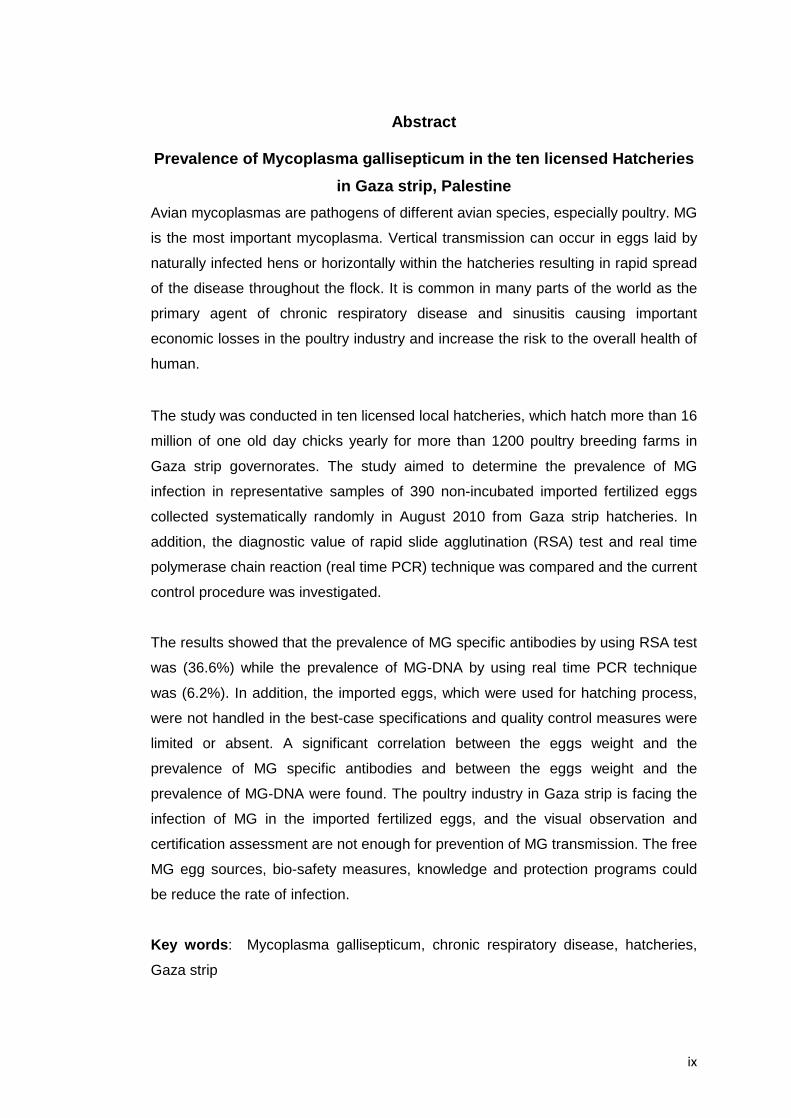