Prevalence of Malocclusion and Impacted Canine in Arab Israelian Population (Arab48)
12
International Journal of Public Health Research 2015; 3(5): 180-191 Published online June 20, 2015 (http://www.openscienceonline.com/journal/ijphr) Prevalence of Malocclusion and Impacted Canine in Arab Israelian Population (Arab48) Muhamad Abu-Hussein 1, * , Nezar Watted 2 , Abdulgani Azzaldeen 3 , Mohammad Yehia 4 , Obaida Awadi 5 , Yosef Abu-Hussein 6 1 Department of Pediatric Dentistry, University of Athens, Athens, Greece 2 Clinics and Policlinics for Dental, Oral and Maxillofacial Diseases of the Bavarian Julius-Maximilian-University, Wuerzburg, Germany 3 Department of Conservative Dentistry, Al-Quds University, Jerusalem, Palestine 4 Triangle R&D Center, Kafr Qara, Israel 5 Center for Dentistry, Research and Aesthetics, Jatt, Israel 6 Statistics and Actuarial Faculty, University of Haifa, Haifa, Israel Email address [email protected] (M. Abu-Hussein), [email protected] (N. Watted) To cite this article Muhamad Abu-Hussein, Nezar Watted, Abdulgani Azzaldeen, Mohammad Yehia, Obaida Awadi, Yosef Abu-Hussein. Prevalence of Malocclusion and Impacted Canine in Arab Israelian Population (Arab48). International Journal of Public Health Research. Vol. 3, No. 5, 2015, pp. 180-191. Abstract The maxillary canine is the second most frequently impacted tooth following third molars. The etiology of the impacted canine is obscure, but probably multifactorial. There are some evidences that patients with a few certain features of occlusion may be at higher risk to the development of impacted canine. The aim of this study was to investigate the association between occurrence of canine impaction and other aspects of occlusion. This study was conducted with patients aged 10, 2 years or older who consequently presented to Orthodontics clinic/jat, Israel. Prevalence of canine impaction in patients of an orthodontic clinic were 3,7%. Angle's Class II was more prevalent occlusion, (61,36 %) malocclusion, Angle's Class I (21,09%),whereas Angle's Class III (17,55%) malocclusion is the least prevalent among Arab population aged 10, 2-39, 5 in Israel. Canine impaction showed no significant relationship with type of malocclusion. Keywords Canine Impaction, Maxillary Canines, Malocclusion 1. Introduction Occlusion is the relationship of the teeth in the mandibular arch to those in the maxillary arch as they are brought together. The occlusion is examined and recorded as part of the oral examination. Knowledge of the occlusion of each patient can contribute significantly to complete care and instruction (1). Recognition of malocclusion assists in the referral of patients to the orthodontist, gives many valuable points of reference for patient instruction, and determines necessary adaptations in techniques (2). Epidemiological studies on malocclusion have been primarily concerned with its aetiology and distribution. Entrenched in these studies is the typological concept that suggests that all variants from a specified normal are abnormal (2). The major flaw in this concept is that it is not easy to define normality, due to the fact that there always exists degrees of natural variation among individuals of a population (3). In addition, most studies have considered the definition of malocclusion to be largely synonymous with that of the Angle’s classification (1,4). Difficulties, therefore, arise when a continuous variable such as the molar relationship is divided into a small number of ordinal categories (Class I, Class II and Class II), which are then treated as a series of independent variables (5). Angle’s classification, though clinically useful, is inadequate for epidemiological studies and as such, indices have had to evolve to focus on means of uniformly evaluating malocclusion (6). Occlusion has been defined as the interdigitation of maxillary and mandibular teeth (7). This description is misleading in that it implies that occlusion is a static contact relationship or arrangement of teeth. The practical concept of
-
Upload
abu-hussein-muhamad -
Category
Health & Medicine
-
view
47 -
download
5
Transcript of Prevalence of Malocclusion and Impacted Canine in Arab Israelian Population (Arab48)

International Journal of Public Health Research 2015; 3(5): 180-191 Published online June 20, 2015 (http://www.openscienceonline.com/journal/ijphr)
Prevalence of Malocclusion and Impacted Canine in Arab Israelian Population (Arab48)
Muhamad Abu-Hussein1, *, Nezar Watted2, Abdulgani Azzaldeen3, Mohammad Yehia4, Obaida Awadi5, Yosef Abu-Hussein6
1Department of Pediatric Dentistry, University of Athens, Athens, Greece 2Clinics and Policlinics for Dental, Oral and Maxillofacial Diseases of the Bavarian Julius-Maximilian-University, Wuerzburg, Germany 3Department of Conservative Dentistry, Al-Quds University, Jerusalem, Palestine 4Triangle R&D Center, Kafr Qara, Israel 5Center for Dentistry, Research and Aesthetics, Jatt, Israel 6Statistics and Actuarial Faculty, University of Haifa, Haifa, Israel
Email address
[email protected] (M. Abu-Hussein), [email protected] (N. Watted)
To cite this article Muhamad Abu-Hussein, Nezar Watted, Abdulgani Azzaldeen, Mohammad Yehia, Obaida Awadi, Yosef Abu-Hussein. Prevalence of
Malocclusion and Impacted Canine in Arab Israelian Population (Arab48). International Journal of Public Health Research.
Vol. 3, No. 5, 2015, pp. 180-191.
Abstract
The maxillary canine is the second most frequently impacted tooth following third molars. The etiology of the impacted canine is obscure, but probably multifactorial. There are some evidences that patients with a few certain features of occlusion may be at higher risk to the development of impacted canine. The aim of this study was to investigate the association between occurrence of canine impaction and other aspects of occlusion. This study was conducted with patients aged 10, 2 years or older who consequently presented to Orthodontics clinic/jat, Israel. Prevalence of canine impaction in patients of an orthodontic clinic were 3,7%. Angle's Class II was more prevalent occlusion, (61,36 %) malocclusion, Angle's Class I (21,09%),whereas Angle's Class III (17,55%) malocclusion is the least prevalent among Arab population aged 10, 2-39, 5 in Israel. Canine impaction showed no significant relationship with type of malocclusion.
Keywords
Canine Impaction, Maxillary Canines, Malocclusion
1. Introduction
Occlusion is the relationship of the teeth in the mandibular arch to those in the maxillary arch as they are brought together. The occlusion is examined and recorded as part of the oral examination. Knowledge of the occlusion of each patient can contribute significantly to complete care and instruction (1). Recognition of malocclusion assists in the referral of patients to the orthodontist, gives many valuable points of reference for patient instruction, and determines necessary adaptations in techniques (2).
Epidemiological studies on malocclusion have been primarily concerned with its aetiology and distribution. Entrenched in these studies is the typological concept that suggests that all variants from a specified normal are abnormal (2). The major flaw in this concept is that it is not
easy to define normality, due to the fact that there always exists degrees of natural variation among individuals of a population (3).
In addition, most studies have considered the definition of malocclusion to be largely synonymous with that of the Angle’s classification (1,4). Difficulties, therefore, arise when a continuous variable such as the molar relationship is divided into a small number of ordinal categories (Class I, Class II and Class II), which are then treated as a series of independent variables (5). Angle’s classification, though clinically useful, is inadequate for epidemiological studies and as such, indices have had to evolve to focus on means of uniformly evaluating malocclusion (6).
Occlusion has been defined as the interdigitation of maxillary and mandibular teeth (7). This description is misleading in that it implies that occlusion is a static contact relationship or arrangement of teeth. The practical concept of

International Journal of Public Health Research 2015; 3(5): 180-191 181
occlusion is to recognize the interplay between the teeth, temporomandibular joint and neuromuscular system (8).
Angle’s description of occlusion is based on the relationship of the maxillary first permanent molar to the mandibular first permanent molar. He noted that in normal occlusion, the mesio-buccal cusp of the upper first molar articulates in the buccal groove of the lower first permanent molar (9). Although his concept of occlusion places primary emphasis on the static relationship of the first permanent molars, he did say that:“…the sizes, forms, interdigitating surfaces, and positions of the teeth in the arches are such as to give one another, singly and collectively, the greatest possible support in all directions…” implying that the functional aspect of occlusion is just as important as the static relationship of teeth to each other. However, later authors still tended to emphasise the arrangement and alignment of teeth by anatomic standards without specific functional considerations for occlusion. (9,10)
The definition of occlusion in terms of alignment of teeth is not adequate to describe its functional aspect. Instead, other factors should also be considered, such as excursive movements, contacts, condylar activity and jaw relationships during function to impart the view that occlusion is a dynamic process (11,12). It was on this basis that Roth (1981) advanced his concept of “gnathology” which incorporated centric relation, cusp-fossa relationships and canine guidance. According to this concept, the occlusal anatomy is designed to guide the mandible to centric relation upon closure(8). However, its practical application in orthodontics is rarely feasible because of the difficulty of positioning the posterior teeth so that every supporting cusp occludes in a fossa (13,14). This concept is further complicated by and the controversy surrounding the definition of centric relation (15,16).
Within the distribution of occlusal forms are the conditions somewhat arbitrarily designated as ideal, normal and malocclusion, the precise differences of which are difficult to identify (2).
Angle (1907) on ideal occlusion stated that, “…each dental arch describes a graceful curve and that the teeth are so arranged as to be in greatest harmony with their fellows in the same arch, as well as with those in the opposite arch.” From a functional point of view, an ideal occlusion needs to be in complete harmony with the neuromuscular system and temporomandibular articulation (9). According to Burdi and Moyers (13) “ideal” denotes a hypothetical concept or treatment goal rarely seen in nature. (17)
In a series of publications, Garber pondered with the philosophical implications of the concepts of normal and ideal occlusion. He asked,” What is normal occlusion? Does it [normal] mean an ideal, a goal to be sought after but never found?”
In the literature, there has been a tendency to use the word “normal” to describe only ideal occlusions (14), thus all deviations from perfection are labelled malocclusions. The definition of normal occlusion is problematic.
The usual definition where normal occlusion is presumed synonymous with ideal occlusion is based on typologic concepts that require precisely defined ideal "types" which are static and unchanging. Deviations from the ideal are viewed as anomalies or degenerations. Typology is a prestatistical way of thinking that does not recognize the variation that describes natural phenomena. When typology is applied to the dentition, hardly anyone has a normal occlusion (18). The term “normal occlusion” is thus a broad and vague concept, the boundaries of which are unclear (19,20,21).
Figure 1. Angle's Classification Of Malocclusion.
Normal cannot be synonymous with ideal. A biologically valid concept of normal occlusion includes a range of occlusal traits that is compatible with health and unimpaired function (21). It allows for minor deviation from the ideal that is aesthetically satisfactory and functionally acceptable and implies variations around an average or mean value (13,21). The most rigorous definition of normal occlusion would therefore be a statistical one with stated probabilities (21).
Normal occlusion is generally accepted to be a Class I molar relationship with good alignment of all the teeth. This represents a situation that occurs in only 30-40% of the population (17). (Figure.1)
Summers in 1971 postulated that in epidemiology, normal
occlusion is confusing and thus the term occlusion is preferred, because it encompasses all variations from an ideal occlusion to a malocclusion and implies a continuous variability rather than just the invariable state. (17)
Andrews (1972) reported of six significant characteristics consistently observed in 120 casts of non-orthodontics patients with normal occlusion. He had used the centre of the clinical crowns as reference points and measured the thickness, tip and torque of each tooth (12). These constants were referred to as the “six keys to normal occlusion”. The significant features shared by all the patients were as follows:
a) Molar relationship: Corresponds with the mesiodistal relationship of upper first permanent molars of Angle with addition that the distal surface of the disto buccal cusp of the

182 Muhamad Abu-Hussein et al.: Prevalence of Malocclusion and Impacted Canine in Arab Israelian Population (Arab48)
upper first permanent molar should made contact and occluded with the mesial surface of the mesio
buccal cusp of the lower second molar. b) Correct crown angulation (mesodistal tip of the crown):
The angulation of the facial axis of every clinical crown should be positive. The extent of angulation varies according to tooth type. (positive means: the gingival part of the long axis of each crown in the upper jaw is positioned distally to the occlusal part of this axis).
c) Correct crown inclination (labiolingual or buccolingual
torque): In upper incisors, the gingival portion of the crown’s labial surface is lingual to the incisal portion. In all other crowns, including lower incisors, the gingival portion of the labial or buccal surface is labial or buccal to the incisal or occlusal portion. In upper posterior crowns (cuspids through molars), the lingual crown inclination of the buccal surfaces is slightly more pronounced in the molars than it is in cuspids and bicuspids. In lower posterior crowns (cuspids through molars), lingual inclination progressively increases.
d) Absence of rotations: Teeth should be free of undesirable rotations. If rotated, a molar or bicuspid occupies more space than it normally does. A rotated incisor may occupy less space.
e) Tight proximal contacts: In absence of abnormalities such as genuine tooth size
discrepancies, contact point should be tight. f) Flat occlusal plane: The curve of Spee should have no
more than a slight arch with the deepest curve was 1.5 mm (plane drawn from incisors to second molars). The convex curve of Spee and mandibular core line bare excessive potions of the occlusal surfaces.
Works by Roth (1981) had then added some functional keys to the previous six keys to normal occlusion by Andrew(8):
a) Centric relationship and centric occlusion should be coincident.
b) In protrusion, the incisors should disclude the posterior teeth, with the guidance provided by the lower incisal edges passing along the palatal contour of the upper incisors.
c) In lateral excursions of the mandible, th canine should guide the working side whilst all other teeth on that and the other side are discluded.
d) When the teeth are in centric occlusion, there should be even bilateral contacts in the buccal segments.
Malocclusion has been described as any deviation from the normal relation of the teeth in the same arch to each other and to the teeth in the opposite arch (11).
The World Health Organisation (1962) defined malocclusion as a dentofacial anomaly. A handicapping dentofacial anomaly is one which causes disfigurement or which impedes function and requires treatment if it is or is likely to be an obstacle to the patient’s physical or emotional well-being. Salzmann (1968) defined a handicapping malocclusion as one that adversely affects aesthetics, function or speech. However, measuring disability or handicap associated with malocclusion is challenging because the emotional impact of the malocclusion on the
individual does not always seem to be directly related to the degree of disfigurement (22,23).
The term malocclusion is an imprecise and ambiguous concept in that it can only be defined in reference to normal occlusion. The definition of malocclusion originated within the realm of corrective treatment and is thus biased (24). This approach directs attention away from the variation normally found among individuals thus suggesting that all variants from a specified normal are abnormal (1). The determination of the point at which normal variation becomes abnormal is difficult (4) and has been cited as the main contributory factor in the variation of the prevalence of malocclusion seen in epidemiological studies (2).
Proffit (1986) elaborated that malocclusion might be associated with one or more of the following (24):
a) Malalignment of individual teeth in each arch: a tooth in an arch may occupy a position deviating from the smooth curve of line by being; tipped, displaced, rotated, in infra-occlusion, in supraocclusion and transposed.
b) Malrelationship of the dental arches relative to the normal occlusion: may occur in any of the three planes of spaces: anteroposterior, vertical or transverse.
A clearer concept of malocclusion would be obtained if the occlusal variables that it comprises are brought into consideration (Lombardi, 1982). Since malocclusion is described as tooth malpositions and/or malrelationships between arches, it represents a range of deviation from the ideal (21), and therefore the developing trend has been to speak of “occlusal variation” or “occlusal traits” in order to avoid the handicapping connotation of the word malocclusion.
Today malocclusion occurs in the majority of the population. It is neither a normal or unhealthy condition (14). Malocclusion is an appreciable deviation from the ideal occlusion that may be considered aesthetically unsatisfactory (24) thus implying a condition of imbalance in the relative sizes and position of teeth, facial bones and soft tissues (lips, cheek, and tongue). It is important not to equate the possession of malocclusion with the need for a treatment instead it should be judged according to dental health, aesthetic or functional criteria namely: chewing, speech, breathing and swallowing (24,25).
The earliest published method of recording malocclusion was Angle’s classification of malocclusion . He believed that all teeth are essential, yet in function and influence, some were of greater importance than others, the most important of all being the first permanent molars, especially the upper first molars, which were called the keys to occlusion;
a) They are the biggest teeth and their anchorage is strongest.
b) Their local position in the occlusal arch supports the main masticatory duty and operation.
c) They influence the vertical distance of upper and lower jaws, the occlusal height and aesthetic proportions.
d) As the permanent molars are the first erupting teeth of permanent dentition, they have “mighty” control on the teeth erupting later behind and in front of them, as they are forced

International Journal of Public Health Research 2015; 3(5): 180-191 183
to position to the already erupted and in occlusion functioning first molars.
e) The anomalies in dental positioning are mostly due to a more prominent dislocated position of the crowns of upper permanent molars to normal, less and minor due to a dislocation of their apex.
These findings lead Angle to postulate, that “the first upper permanent molar, more than any other tooth or anatomical point gives a precise scientific basis for defining occlusal disharmony and occlusal anomalies”.
Graber (1992) described Angle’s classification as the only internationally recognized classification and widely used in epidemiological study of malocclusion. Despite such praises, the classification has been criticized by a number of authors. Graber pointed out that the Angle classification failed to distinguish between malocclusion and anteroposterior relationships(14). Rinchuse and Rinchuse (1988) proposed that the classification was not clear about the description and definition of different classes and Angle’s writing was equivocal, leading to possibility of one class overlapping into another. In addition to the above, several investigations had provided data that question the reliability of Angle’s classification.(25)
Gravely and Johnson (1974) for example, had demonstrated a poor intraexaminer and interexaminer reliability for Angle’s classification, especially in categorizing Class II division 2 malocclusion (26,27).
After the third molars, the maxillary canine is the second most frequently impacted tooth in the dental arch. The canine impaction prevalence ranging from 0.8 to 5.2 percent depending on the population examined. The prevalence of palatally impacted maxillary canines varies between 0.8% and 2.8% .(28)
Maxillary canine impaction represents 2% of orthodontic patients. In 70% to 85% of canine impactions, the canine is located palatally.
Various studies have noted several factors associated with canine impaction such as arch-length deficiency(labial canine impaction), premaxillary skeletal deficiency (labial impaction) , maxillary excess (palatal impaction), maxillary lateral incisor agenesis (unilateral PDC), bilateral small maxillary lateral incisors, absent of third molars and second premolars, and peg-shaped maxillary lateral incisor with absent of maxillary lateral incisor, deficiency in maxillary width.
The incidence of canine impaction and it’s relation with Impaction canine has not down on Arab population (Arab48) in Israel.
The aim of this study was to investigate the association between occurrence of canine impaction and type of occlusion in orthodontic seeking patients.
2. Material & Methods
All of the patients whom referred to orthodontic clinic of Center For Dentistry, Research & Aesthetics, Jatt, Almothalath, Israel, and Triangle R&D Center, Kafr Qara,
Israel. included in this study. Canine impaction were confirmed with clinical method and use of panoramic radiograph by an orthodontist. Patients with pathological reasons for canine impaction, syndromic patients with multiple impactions were excluded. An orthodontist selected type of impaction and occlusal status of patients using clinical method or panoramic radiographs and PA with Clark occlusal technique.. The data for each patient was recorded.
The records of 2200 patients attending the Center For
Dentistry, research & Aesthetics, Jatt, Israel, between,
between Jan 2006 to Dec 2013 were examined by intraoral examination, palpation, and patient records, followed by
panoramic radiographs for the study. Ethical clearance was
obtained from the Institutional Ethical Committee. A written
informed consent was obtained from all the patients. The age
of the patients ranged from 10.2 to 39.5years, with a mean of 16,2years.
The data collected for malocclusion was descriptively analysed to determine the prevailing percentage of malocclusion in the examined Arab population in Israel. The related impacted teeth interferences with malocclusion data were statistically analyzed using Pearson test and logical regression for correlation and association by SPSS. (Table.1)
3. Results
In this study 2200 patients older than 12years (846 male-38,4% & 1354 female 61.6) assessed for canine impaction.3,7% of these (82subject) had impacted canine and 96,3% were without impaction. (Table.2) (Table.3) (Fig.2)
(Fig.3) (Fig.4) The distribution of gender in patients was 36 male / 46
female (43,9% / 56,1%). malocclusion, 46,34% revealed C I 40,24 Cl II malocclusion, and 13,42 % had Cl III malocclusion and there was not prove any correlation between canine impaction and type of occlusion (p=0. 068). (Table.4) (Fig.5) (Table.5) (Fig.6)
In patients with canine impaction 57,9% had agenesis. The most prevalence of agenesis related to lateral incisor (80 case in maxilla & 87 case in mandible) and in next sequences is central incisor (1 case in maxilla & 7case in mandible), second premolar (32 case in maxilla & 69case in mandible), canine (2 case in maxilla & 1 case in mandible), first premolar (5 case in maxilla & 3 case in mandible), second molar (1 case in maxilla & 2 case in mandible). 21,7% of impaction patients revealedcrowding. 20,6% of canine impaction cases had small lateral incisor and there was significant correlation between impaction and incisor size (p=0.000). (Table.6) (Fig.7) (Table.7) (Fig.8) (Table.8) (Fig.9) (Table.9) (Fig.10)
4. Discussions
Many epidemiologic studies regarding the prevalence of malocclusion have given a vast amount of information on the different types of malocclusion; Different results have been obtained by several authors showing the wide range of

184 Muhamad Abu-Hussein et al.: Prevalence of Malocclusion and Impacted Canine in Arab Israelian Population (Arab48)
variety in malocclusion between different ethnic races. Emrich, and Blayney (1965), studied two age group from
Black and White America in USA, Group 1 consisted of 903 Black American aged 6-8-year-old; The result showed high percentage of normal occlusion (70%) and very low percentage of Angle Class II malocclusion (5%), Angle Class I malocclusion found in (23%), and Angle Class III malocclusion was only in (2%). In group 2 1,476 Black American aged 12-14 years, the result revealed less prevalence of normal occlusion (57%), Angle Class I malocclusion (32%), Angle Class II malocclusion (7%) and Angle Class III result indicated that the prevalence of malocclusion as follows: normal occlusion (69%), Angle Class I malocclusion (18%), Angle Class II malocclusion (11%) and Angle Class III malocclusion was only in (1%). whereas on 13475 white American aged 12-14years old, the result exhibited less prevalence of normal occlusion (54%) and the prevalence of Angle Class I and II malocclusion were slightly high (30%) and (15%) respectively and only (1%) of Angle Class III malocclusion was reported. (29)
Gardiner 1982 investigated 500 Libyan school children aged 10-12 years and found that the majority had Class I malocclusion (74%) while crowding was a very common finding (31%). (30)
Steigman, Kawar, and Zilberman (1983) carried study on 803 Arabian children in Israel aged 13-15-year-old and reported small percent of normal occlusion (0.3%), Angle Class I the most common malocclusion ( 85%), followed by Class II (10.2%) and Class III (1.3%) malocclusion. (31)
Farawana (1987) carried out a preliminary study on occlusion in Iraq, and found a high percentage of Angle Class II malocclusion (29.6%) combined with overjet (div I). (32)
El-Mangoury, and Mostafa (1990) studied 501 Egyptians adults aged 18-24-year-old and recorded high prevalence of normal occlusion (34.3%) followed by Class I malocclusion in (33.3%), Class II in (21%) and Class III malocclusion in (10.9 %) with higher prevalence among females. (33)
Al Emran (1990) studied 500 Saudi boys with a mean age of 14 years, and concluded that the frequency of malocclusion among Saudi children (62.4%) was either dentition, occlusal, or space anomalies. (34)
Heidi Kerosuo, (1991), conducted two studies in Tanzania and Finland. In the Finnish sample consisted of 205 Boys and 242 girls aged 12-18, and registered that the boys sample showed that (77%) had Angle Class I malocclusion, Angle Class II malocclusion in (22%) and Angle Class III malocclusion in (1%). Further, in the girls sample he noticed high percent (84%) of Angle Class I malocclusion, (15%) Angle Class II malocclusion and the percentage of Angle Class III malocclusion was the same as in boys (1%). (35)
Tipton and Rinchuse (1991) studied 101 dental school populations in Pittsburgh USA aged 18-32-year-old and reported normal occlusion in (52%), Angle Class I malocclusion in (26%), Angle Class II malocclusion in (16%) and Angle Class III malocclusion in (7%). (36)
Kerosuo, Laine, and Honkala (1991), conducted two studies in Tanzania and Finland. In Tanzania investigated 340
Tanzanian Boys and 302 Girls aged 11-18-yearold, the result showed that the boys had very high percentage of Angle Class I malocclusion (96%), and had the same percentage of Angle Class II and III malocclusion in (2%). The Girls also had reported a high prevalence of Angle Class I malocclusion (95%) and low percentage of Angle Class II and Class III malocclusion (4%) and (1%) respectively. (35)
Tang, (1994) studied 201 Chinese adults in Hong Kong aged 20 years old and observed that, (63.7%) were Angle Class I malocclusion, Angle Class II malocclusion in (16.4%) and a higher percentage of Angle Class III malocclusion (19.9%) in the sample. (37)
Alphonso Trottman (1996) examined 99 Black American in USA aged 3-5-year-old and 139 white American same age and reported a higher prevalence of Angle CI III malocclusion (17%) and a lower prevalence of Angle Class II malocclusion (7%) among the Black American sample. In the white American; Angle Class II malocclusion was found in (14%) and Class III in (8%). (38)
Proffit, Fields, and Moray (1998), in the United States; surveyed the prevalence of malocclusion on 14,000 American population; in three age groups aged: 18-50-year-old, 12-17-year-old and 8-11- year-old and observed the same percentage of normal occlusion (30%), Angle Class I malocclusion (50%) to (55%) and Angle Class III malocclusion in (1%) in the three age group. Further he found that Angle Class II malocclusion was higher in the small age group (23%). (39)
Saleh (1999) carried out study on 851 Lebanese schoolchildren aged 9-15-year-old, found that (40.3%) had normal occlusion, (35.5%) had Angle Class I malocclusion, (19%) had Angle Class II malocclusion and (5%) had Angle Class III malocclusion. (40)
Sayin, and rkkahraman (2004) carried out study in 1356 Turkish children with the mean aged 13.57 ±3.16-year-old, and noticed that the prevalence of Angle Class I malocclusion was in (64%), Angle Class II in (24.9%) and Angle Class III in (12%). (41)
Chukwudi (2004) investigated 636 Nigerian adolescents 12-17- year-old in Ibadan and found that (24%) had normal occlusion. (50%) had Angle Class I malocclusion, (14%) had Angle Class II, and (12%) had Angle Class III malocclusion. (42)
Soh, Sandham, and Chan (2005), studied 339 Asian men in Singapore aged 17-22-year-old and noticed a very high percentage of Angle Class II malocclusion (48%). Angle Class I malocclusion was found in (17%) and Angle Class III malocclusion in (18%). (43)
Behbehani, Årtun, Al-Jame, and Kerosuoc, (2005) carried out an epidemiological study of malocclusion in 1299 Kuwaitis school children aged 13-14-year-old. The result showed high percentage of Angle Class II malocclusion (31.2%), Angle class I malocclusion was in (57.8%) and (11%) had Angle Class III malocclusion.(44)
Josefsson, (2007), conducted study in 263 Swedish and 64 Eastern European immigrant adolescents aged 12 and 13years the result revealed that Angle Class I malocclusion
International Journal of Public Health Research 2015; 3(5): 180-191 185
was found in (47.0%), Angle Class II in (48.8%) and Angle Class III in (4.2%). On the other hand in the Eastern European the result was (42.2%) had Angle Class I, (46.9%) Angle Class II and (10.9%) had Angle Class III. further, the result of 118 Asian samples, showed Angle Class I in (54.6%), Angle Class II in (36.2%) and Angle Class III in (9.2 %). the result of the sample of Other Countries, showed that Angle Class I was in (47.9%), Angle Class II in (47.9%) and Angle Class III in (4.2 %). (45)
Mohammad. (2007) studied 398 Iranian children aged 13-15-yearold in Tabriz. The result showed small percentage of the children had normal occlusion (4%) and high percentage of the children had Angle Class III malocclusion (17.1). Angle Class I malocclusion was found to be reported in (57%) of the sample whereas Angle Class II malocclusion was noticed in (21.9%). (46)
Gelgöra (2007), carried out study on 2329 adolescents in Central Anatolia; aged 12-17-year-old and found the prevalence of malocclusion as follows: normal occlusion in (10.1%), Angle Class I malocclusion in (34.9%), Angle Class II in (44.7%) and Angle Class III malocclusion in (10.3%). (47)
Matilda, Åstrøm AN. (2008), performed study on 1601 Tanzanian Schoolchildren aged 12-14-year-old. The result exhibited a high prevalence of Angle Class I malocclusion (93.6%) and low prevalence of Angle Class II malocclusion (4.4%) and Angle Class III malocclusion (2.0%) (48)
Bernabe, Sheihamb, and Oliveirac (2008), conducted a research on 1675 Brazilian adolescents aged 15-16-year-old and recorded the prevalence of malocclusion as follows: normal occlusion in (12.4%), Angle Class I malocclusion in (65.8%), Angle Class II malocclusion in (16.6%) and Angle Class III malocclusion in (5.2%). (49)
AL-Hourani, (2008), conducted study on 58 untreated subjects in Syria aged 7-13-year-old.the prevalence of Angle Class I malocclusion was (51.7%), Angle Class II malocclusion was (37.9%) and Angle Class III malocclusion was (10.3%). (50)
Chu, (2009) performed study on 120 Chinese young adults in Hong Kong aged 18-27-year-old. He assessed the prevalence of malocclusion. The result showed that the most common malocclusion was manifested Angle Class I in (48%) followed by
Angle Class III in (21%), whereas normal occlusion was found in (20%) and Angle Class II in (11%). (51)
Sidlauskas, (2009), did a study on 1681 Lithuanian Schoolchildren aged 7-15-year-old the result indicated that the prevalence of malocclusion as follows: Angle Class I in (68.4%), Angle Class II in (27.7%) and Angle Class III in (2.8%). (52)
The present research was the first large population based study considering the prevalence of normal occlusion and malocclusion for Arab population in Israel (Arab48). The results of these and our study is not comparable because our study accomplished on only orthodontic patients population.
In this study, the rate of canine impaction in women was more than men (56,1% women, 43,9% men), but the
difference was not significant(p>0.05). Nagahara observed that the frequency of too the impaction is not different between men and women (53). Dachi and Howell stated that girls are likely for canine impaction twice than boys (54).
In our study regarding type of malocclusion, the rate of impaction was more in cl l, then cl II and cl III respectively. But between type of malocclusion and canine impaction was not signify cant difference (p> 0.05). AL Nimri etal found that palatal canine impaction is more probable in individuals with malocclusion cl II div2 (55). Basdra also found that the prevalence of canine impaction is more in individuals with malocclusion cl II div2 .In another study, this researcher observed that the prevalence of canine impaction was not different significantly between patients with cl III malocclusion and cl II div1 (56,57) .Leifert reported that Angle classification was not correlated to canine eruption disorder (58) .
Table 1. Means age.
Age, Impacted Min Max Avg
10.2 39.5 16.2
Table 2. Gender distribution of patients treated without/with impacted
canine.
Treated (Orth.) N=2200 %
Female 1354 61.6%
Male 846 38.4%
Impacted 82 3.7%
Non Impacted 2118 96.3%
5. Conclusion
In general prevalence of malocclusion is considered to be on increase with evolution and civilization. Although Angle’s method has been used in recording the malocclusion it does not reflect the actual orthodontic treatment needs of the society.
The findings of the present study were as follows:
-Angle's Class II was more prevalent occlusion, (61,36 %) malocclusion, Angle's Class I (21,09%),whereas Angle's Class III (17,55%) malocclusion is the least prevalent among Arab population aged 10,2-39,5 in Israel .
-Prevalence of canine impaction in patients of an orthodontic clinic were 3,7%
-Canine impaction was usually unilateral -Canine impaction was not associated with gender. -Canine impaction showed no significant relationship with
type of malocclusion, crowding, anterior cross bite and posterior cross bite -In relation to gender, females had significantly more
Angle's Class I normal occlusion than the males where as males significantly had more Angle's Class I malocclusion than the females.
-Canine impaction showed no significant relationship with type of malocclusion

186 Muhamad Abu-Hussein et al.: Prevalence of Malocclusion and Impacted Canine in Arab Israelian Population (Arab48)
Fig. 2. Gender distribution of patients treated.
Fig. 3. Proportion of patients with retention (blue) and without retention (brown).
Fig. 4. Gender distribution in retention.
International Journal of Public Health Research 2015; 3(5): 180-191 187
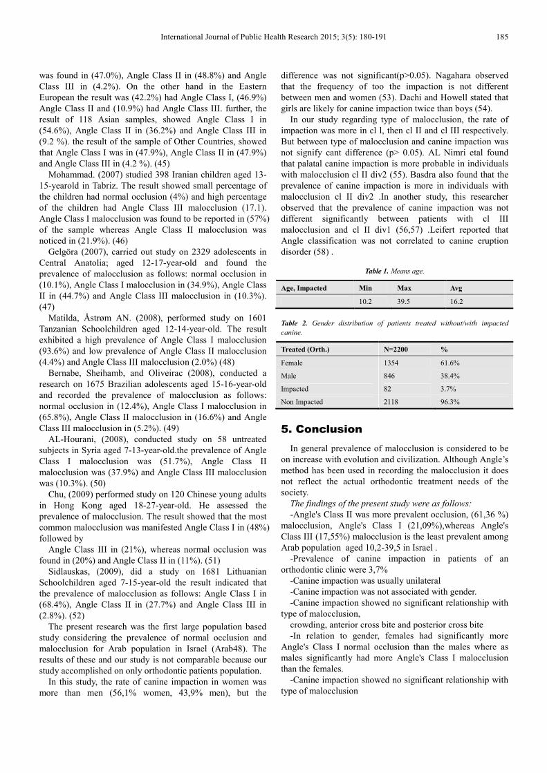
Fig. 5. Frequency distribution on the type of occlusion.
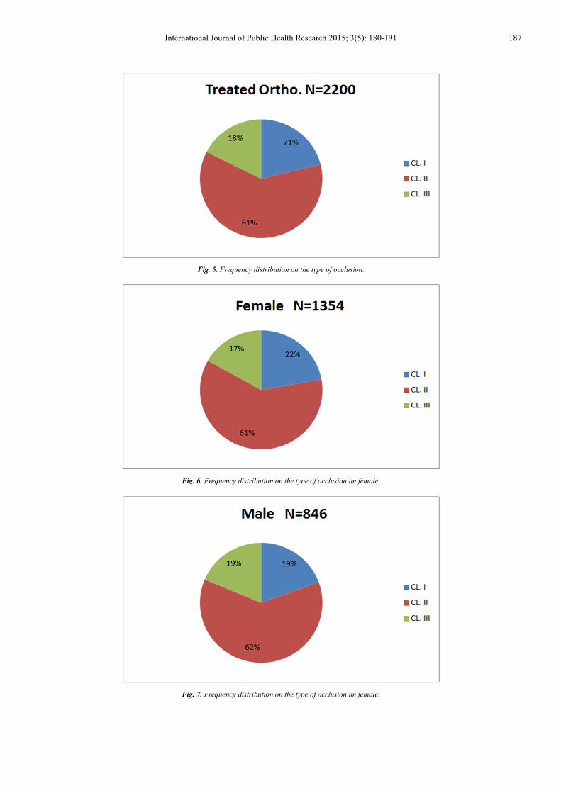
Fig. 6. Frequency distribution on the type of occlusion im female.
Fig. 7. Frequency distribution on the type of occlusion im female.

188 Muhamad Abu-Hussein et al.: Prevalence of Malocclusion and Impacted Canine in Arab Israelian Population (Arab48)
Fig. 8. Frequency distribution of canine impaction based on the type of occlusion.
Fig. 9. Frequency distribution of canine impaction based on the type of occlusion in male.
Fig. 10. Frequency distribution of canine impaction based on the type of occlusion in female.

International Journal of Public Health Research 2015; 3(5): 180-191 189
Table 3. Prevalance of impacted maxillary canine.
Impacted N=82 % %Treated
(2200)
%Investigated
Patients (4250)
Female 46 56.1% 2.1% 1.1%
Male 36 43.9% 1.6% 0.8%
Table 4. Frequency distribution on the type of occlusion.
Treated Ortho. N=2200 %
CL. I 464 21.09%
CL. II 1350 61.36%
CL. III 386 17.55%
Table 5. Frequency distribution on the type of occlusion im female.
Female N=1354 %
CL. I 300 22.16%
CL. II 826 61.00%
CL. III 228 16.84%
Table 6. Frequency distribution on the type of occlusion im male.
Male N=846 %
CL. I 164 19.38%
CL. II 524 61.94%
CL. III 158 18.68%
Table 7. Frequency distribution ofcanine impaction based on the type of
occlusion.
Impacted N=82 %
CL. I 38 46.34% CL. II 33 40.24% CL. III 11 13.42%
Table 8. Frequency distribution of canine impaction based on the type of
occlusion in male.
Impacted Male N=36 %
CL. I 14 38.89% CL. II 16 44.44% CL. III 6 16.67%
Table 9. Frequency distribution of canine impaction based on the type of
occlusion in female.
Impacted Female N=46 %
CL. I 24 52.17% CL. II 17 36.96% CL. III 5 10.87%
References
[1] Smith RJ, Bailit HL. Problems and methods in research on the genetics of dental occlusion, Angle Orthod 1977; 47: 65-77.
[2] Abu-Hussein M., Sarafianou a.,; MATHEMATICAL ANALYSIS OF DENTAL ARCH OF CHILDREN IN NORMAL OCCLUSION: A LITERATURE REVIEW, IJMD2012;2(1);33-39.
[3] Baume LJ, Maréchaux SC. Uniform methods for the epidemiologic assessment of malocclusion. The development of basic methods by the World Health Organisation and the
Fédération Dentaire Internationale. Am J Orthod 1974; 66: 121-128.
[4] Abu-Hussein Muhamad, Watted Nezar, Abdulgani Azzaldeen. The Curve of Dental Arch in Normal Occlusion. Open Science Journal of Clinical Medicine.2015, Vol. 3, No. 2, 47-54
[5] Burgersdijk R, Truin GJ, Frankenmolen F, et al. Malocclusion and orthodontic treatment need of 15-74-year-old Dutch adults. Community Dent Oral Epidemiol 1991; 19: 64-67
[6] Gravely JF, Johnson DB. Angle's classification of malocclusion: an assessment of reliability. Br J Orthod 1974; 1: 79-86.
[7] Fletcher AE: Keys and lines of occlusion. Aust Orthod J 1987; 10: 38-42.
[8] Roth RH. Functional occlusion for the orthodontist: Parts I-IV. J Clin Orthod 1981; 15: 32-51, 100-123, 174-198, 246-264.
[9] Angle EH. Malocclusion of teeth. 7th
Ed. Philadelphia. The S.S. White Dental Manufacturing Co., 1907.
[10] Dewey M, Anderson GM. Practical orthodontia. St Louis: CV Mosby; 1935.
[11] Anderson GM. Practical Orthodontics. 7th
Edition, St. Louis, Mosby 1948.
[12] Andrews LF. The six keys to occlusion. Am J Orthod 1972; 61: 297-309.
[13] Burdi AR, Moyers RE. Development of the dentition and the occlusion. Chapter 6. In Moyers RE (editor): The handbook of orthodontics. 4
th
edition. Yearbook Medical Publishers, Inc. 1988.
[14] Graber TM, Vanarsdall RL, (eds). Orthodontics. Current Principles and Techniques. 3
rd
Edition. The C.V. Mosby Company, 2000.
[15] Isaacson RJ, Christiansen RL, Evans CA, Reidel RA. Research on the variation in dental occlusion. Am J Orthod 1975; 68: 241-255.
[16] Keshvad A, Winstanley RB. An appraisal of the literature on centric relation. Part I. Jnl of Oral Rehab 2000; 27: 823-833.
[17] Moyers RE, Summers CJ. Application of the epidemiolgic method to the study of occlusal disorders. Int Dent J 1970; 20: 575-585.
[18] Saunders SR, Popovich F, Thompson GW. A family study of craniofacial dimensions in the Burlington Growth Centre sample. Am J Orthod 1980; 78: 394-403.
[19] Steigman S, Kawar M, Zilberman Y. Prevalence and severity of malocclusion in Israel Arab urban children aged 13 to 15 years of age. Am J Orthod 1983; 84: 337-343.
[20] Timm TA, Herremans EL, Ash MM. Occlusion and orthodontics. Am J Orthod 1976; 70: 138-145..
[21] Vlachos C. Occlusal principles in orthodontics. Dental Clinics of North America 1995 Apr; 39: 363-377.
[22] World Health Organisation Series. Standardisation of reporting of dental diseases and conditions: Report of an expert committee on dental health. W.H.O. Technical Report Series No. 242, 1962.

190 Muhamad Abu-Hussein et al.: Prevalence of Malocclusion and Impacted Canine in Arab Israelian Population (Arab48)
[23] World Health Organisation. Oral Health Care Systems: An International Collaborative Study. Geneva, Switzerland. Quintessence Publishing Company Ltd. London. 1985; 156-160.
[24] Proffit WR (1986). On the Aetiology of Malocclusion. BrJ Orthod, 13: 1-11.
[25] Rinchuse DJ, Rinchuse DJ. Ambiguities of Angle's classification. Angle Orthod 1988; 59: 295-298.
[26] Gravely JF, Johnson DB. Angle's classification of malocclusion: an assessment of reliability. Br J Orthod 1974; 1: 79-86.
[27] Ackerman JL, Proffit WR. The characteristics of malocclusion: A modern approach to classification and diagnosis. Am J Orthod 1969; 56: 443-454.
[28] Watted N., Abu-Hussein M.,; Prevalence of impacted canines in Arab Population in Israel, Ijphr2014, 2(6);71-7
[29] Emrich RE, Brodie AG, Blayney JR. Prevalence of Class 1, Class 2, and Class 3 malocclusions (Angle) in an urban population. An epidemiological study, Journal of dental research, 1965 Sep-Oct; 44(5):947-53.
[30] Gardiner JH. An orthodontic survey of Libyan schoolchildren British journal of orthodontics, 1982 Jan; 9(1):59-61.
[31] Steigman S, Kawar M, Zilberman Y. Prevalence and severity of malocclusion in Israeli Arab urban children 13 to 15 years of age American Journal of Orthodontics 1983;84(4):337-43.
[32] Farawana NW, Malocclusion in Iraq. Quintessence Int. 1987 Feb; 18(2):153-7.
[33] El-Mangoury NH, Mostafa YA. Epidemiologic panorama of dental occlusion, The Angle orthodontist, 1990 Fall; 60(3):207-14.
[34] Al-Emran S, Wisth PJ, Boe OE. Prevalence of malocclusion and need for orthodontic treatment in Saudi Arabia. Community dentistry and oral epidemiology, 1990 Oct; 18(5):253-5.
[35] Heidi Kerosuo TLaEH. Occlusal characteristics in groups of Tanzanian and Finnish urban schoolchildren, The Angle orthodontist, 1991(1):49-56.
[36] Tipton RT, Rinchuse DJ. The relationship between static occlusion and functional occlusion in a dental school population, Angle Orthod 1991; 61(1):57-66.
[37] Tang EL. Occlusal features of Chinese adults in Hong Kong. Australian orthodontic journal, 1994 Oct; 13(3):159-63.
[38] Alphonso Trottman aHGE, comparison of malocclusion in preschool black and white children, AmJOrthod Dentofacial Orthop 1996, 69-72
[39] Proffit WR, Fields HW, Jr., Moray LJ. Prevalence of malocclusion and orthodontic treatment need in the United States: estimates from the NHANES III survey, The International journal of adult orthodontics and orthognathicsurgery, 1998; 13(2):97-106.
[40] Saleh FK. Prevalence of malocclusion in a sample of Lebanese schoolchildren, Mediterranean Health Journal 1999:337-43.
[41] Sayin MOzr. Malocclusion and crowding in an Orthodontically Referred Turkish Population the Angle orthodontist, 2004(74):635-9.
[42] Chukwudi OO. Prevalence of malocclusion among adolescents in Ibadan, Nigeria, Am J Orthod Dentofacial Orthop 2004; 126:604-7.
[43] Soh J, Sandham A, Chan YH. Malocclusion severity in Asian men in relation to malocclusion type and orthodontic treatment need, Am J Orthod Dentofacial Orthop, 2005 Nov; 128(5):648-52.
[44] Faraja.Behbehani. Jon Årtun .a Badreia Al-Jame .b Heidi Kerosuoc, Prevalence and Severity of Malocclusion in Adolescent Kuwaitis, Med Princ Pract 2005(14):390-5.
[45] Eva Josefsson KBaRL, Malocclusion frequency in Swedish and immigrant adolescents' influence of origin on orthodontictreatment need. European journal of orthodontics, 2007;29:79-87.
[46] Mohammad. Hossein Ahangar Atashi. Prevalence of Malocclusion in 13-15 Year-old Adolescents in Tabriz Journal of Dental Research, Dental Clinics, Dental Prospects,2007; 1:1
[47] İbrahim Erhan Gelgöra AİKEE. Prevalence of Malocclusion among Adolescents in Central Anatolia,. European Journal of Dentistry 2007; 1:125-31
[48] Mtaya M BP, Åstrøm AN.,. Prevalence of malocclusion and its relationship with sociodemographic factors, dental caries and oral hygiene in 12-14 year-old Tanzanian School children, European Journal of Orthodontics; 2008.
[49] Eduardo Bernabe´ a; Aubrey Sheihamb; Cesar Messias de Oliveirac, Condition-Specific Impacts on Quality of Life Attributed to Malocclusion by Adolescents with Normal Occlusion and Class I, II and III Malocclusion, Angle Orthodontist, 2008; 78(6)
[50] AL-Hourani. Ammar, The prevalence of malocclusions in an untreated population of 7-13 year old in Hama, Syria, and the importance of early interceptive orthodontics in such cases, University of Glasgow BDS Elective Report
[51] Chu CH CB, Lo EC., Occlusion and orthodontic treatment demand among Chinese young adults in Hong Kong. Oral Health Prev Dent 2009; 7((1)):83-91.
[52] Antanas Sidlauskas KL, the prevalence of malocclusion among 7-15-year-old Lithuanian school children, Medicina (Kaunas); .45(2).
[53] Nagahara K, Yuasa S, Yamada A, Ito K, Watanabe O, Iizuka T, Sakai M, Utida H. Etiological study of relationship between impacted permanent teeth and malocclusion. Aichi Gakuin Daigaku Shigakkai Shi. 1989; 27(4): 913-24.
[54] Dachi SF, Howell FV. A survey of 3874 rou�ne full mouth radiographs. Oral Surg Oral Med Oral Pathol. 1961;14:1165–1169.
[55] Al-Nimri K, Gharaibeh T. Space conditions and dental and occlusal features in patients with palatally impacted maxillary canines: an etiological study. Eur J Orthod 2005;27:461-5.
[56] Basdra EK, Kiokpasoglou M, Stellzig A. The Class II Division 2 craniofacial type is associated with numerous congenital tooth anomalies. Eur J Orthod 2000; 22(5): 529-35.

International Journal of Public Health Research 2015; 3(5): 180-191 191
[57] Basdra EK, Kiokpasoglou MN, Komposch G. Congenital tooth anomalies and malocclusions: a genetic link? Eur J Orthod 2001;23(2):145-51.
[58] Leifert S, Jonas IE. Dental anomalies as a microsymptom of palatal canine displacement. J Orofac Orthop 2003;64(2):108-20.