Multi-Component Model Structure for Autogenous and Semi-Autogenous Mills
Upload
james-schumacherCategory
view
213download
0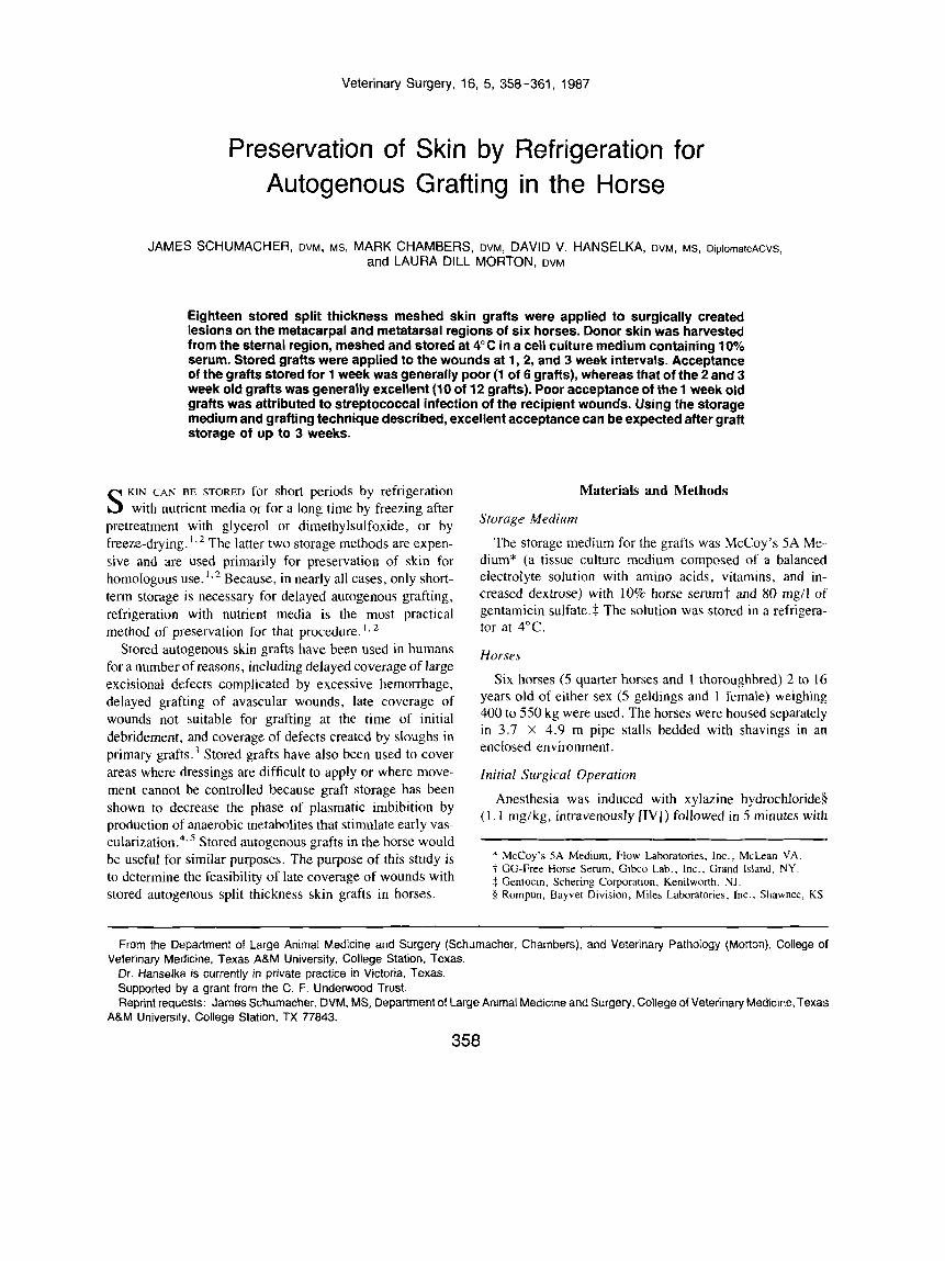
Veterinary Surgery, 16, 5, 358-361, 1987
Preservation of Skin by Refrigeration for Autogenous Grafting in the Horse
JAMES SCHUMACHER, DVM, MS, MARK CHAMBERS, DVM, DAVID V. HANSELKA, OVM, MS, DiplornateACVS, and LAURA DILL MORTON. DVM
Eighteen stored split thickness meshed skin grafts were applied to surgically created lesions on the metacarpal and metatarsal regions of six horses. Donor skin was harvested from the sternal region, meshed and stored at 4°C in a cell culture medium containing looh serum. Stored grafts were applied to the wounds at 1,2, and 3 week intervals. Acceptance of the grafts stored for 1 week was generally poor (1 of 6 grafts), whereas that of the 2 and 3 week old grafts was generally excellent (10 of 12 grafts). Poor acceptance of the 1 week old grafts was attributed to streptococcal infection of the recipient wounds. Using the storage medium and grafting technique described, excellent acceptance can be expected after graft storage of up to 3 weeks.
KIN CAN BE STORED for short periods by refrigeration S with nutrient media or for a long time by freezing after pretreatment with glycerol or dimethylsulfoxide, or by freeze-drying. ‘ , 2 The latter two storage methods are expen- sive and are used primarily for preservation of skin for homologous use.*’* Because, in nearly all cases, only short- term storage i s necessary for delayed autogenous grafting, refrigeration with nutrient media is the most practical method of preservation for that procedure.
Stored autogenous skin grafts have been used in humans for a number of reasons, including delayed coverage of large excisional defects complicated by excessive hemorrhage, delayed grafting of avascular wounds, late coverage of wounds not suitable for grafting at the time of initial debridement, and coverage of defects created by sloughs in primary graft^.^ Stored grafts have also been used to cover areas where dressings are difficult to apply or where move- ment cannot be controlled because graft storage has been shown to decrease the phase of plasmatic imbibition by production of anaerobic metabolites that stimulate early vas- cular i~at ion.~-~ Stored autogenous grafts in the horse would be useful for similar purposes. The purpose of this study is to determine the feasibility of late coverage of wounds with stored autogenous split thickness skin grafts in horses.
Materials and Methods
Storage Medium
The storage medium for the grafts was McCoy’s 5A Me- dium* (a tissue culture medium composed of a balanced electrolyte solution with amino acids, vitamins, and in- creased dextrose) with 10% horse serum? and 80 mg/l of gentamicin sulfate.$ The solution was stored in a refrigera- tor at 4°C.
Horses
Six horses (5 quarter horses and 1 thoroughbred) 2 to 16 years old of either sex ( 5 geldings and 1 female) weighing 400 to 550 kg were used. The horses were housed separately in 3.7 X 4.9 m pipe stalls bedded with shavings in an enclosed environment.
lnitial Surgical Operation
Anesthesia was induced with xylazine hydrochloride§ ( 1 . 1 mglkg, intravenously [IV]) followed in 5 minutes with
* McCoy’s 5A Medium, Flow Laboratories, Inc., McLean VA. t GG-Free Horse Serum, Gibco Lab., lnc., Grand Island, NY. $ Gentocin, Schering Corporation, Kenilwonh, NJ. 9 Rompun, Bayvet Division, Miles Laboratories, Inc., Shawnee, KS.
From the Department of Large Animal Medicine and Surgery (Schumacher, Chambers), and Veterinary Pathology (Morton), College of
Dr. Hanselka is currently in private practice in Victoria, Texas. Supported by a grant from the C. F. Underwood Trust. Reprint requests: James Schumacher, DVM, MS, Department of Large Animal Medicine and Surgery, College of Veterinary Medicine, Texas
Veterinary Medicine, Texas A&M University, College Station, Texas.
A&M University, College Station, TX 77843.
356

SCHUMACHER, CHAMBERS, HANSELKA, AND MORTON 359
ketamine hydrochloride1 (2 .2 mgikg, IV). After tracheal intubation, anesthesia was maintained with halothanell and O2 delivered through a semiclosed system. Each horse was positioned in dorsal recumbency. The metacarpal, metatar- sal, and sternal regions were clipped and a chemical depila- tory agent# was applied to the sternal area. Clipped areas were prepared for aseptic surgery. A 5 X 10 cm full thick- ness skin flap was removed from the dorsolateral aspect of each metacarpus and the right metatarsus and discarded. Split thickness skin sufficient in length to cover all three wounds was harvested from the sternal area of each horse with an electric skin graft derniatome. ** The skin was cut at widths that varied from 4 to 8 cm and at a thickness of 0.63 mm. The harvested skin was meshed with a dermatome expandertt with a three to one expansion ratio and cut into four equal sections each measuring at least 13 cni in length. Three of the graft sections were placed on separate vaseline- impregnated gauze strips$$ with the cut surface of the graft away from the gauze. The caudal edge of each graft was marked with a suture. Each of the three gauze and graft composites was rolled, with the gauze surface to the outside of the roll, and placed in a separate sterile plastic container. Enough storage solution was added to establish a ratio of I to 1.5 ml of media per square centimeter of skin (80-100 ml) and the containers were refrigerated at 4°C. Sterile petroleum-impregnated gauze was applied to each wound and secured with elastic gauze.§§ Several layers of 12 inch cotton padding77 were applied over each wound and se- cured with additional elastic gauze and elastic tape.[/// Ban- dages were removed at 3 days and the wounds were left uncovered; these wounds were cleaned and wrapped I day before stored graft application.
Graft Application
Stored grafts were applied to the wounds in the following sequence: ( I ) right hindlimb at 1 week, (2) left forelimb at 2 weeks, and (3) right forelimb at 3 weeks. The assumption was made that grafts were accepted equally on the metacar- pus and metatarsus. Horses were sedated with xylazine (200
TI Ketaset, Vererinary Products, Brisiol Laboratories, Bristo-Meyers,
1 1 Halothane, Halocarbon Laboratories, Inc., Hackensack, NJ . # Neet, Whitehall Laboratories, tnc., New York, NY. ** Electric Dennatome, Stryker Electro-Surgical Unit, Orthopedic Fram
t t Mesh Skin Graft Expander, No. P-160, Padgett Instruments. Kansas
$$ Adaptic, Johnson and Johnson Products, Inc.. Fort Dodge. IA. $8 Brown Gauze, I . R. Raynor, New Hartford, NY. YllCombine Roll, 12 inches, Chaston Medical and Surgical Products,
1111 Elastikon, Johnson and Johnson Products, Inc.. New Brunswick, NJ
Co., Syracuse. NY.
Co., Kalamazoo, MI.
City, MO.
Lake Road, Dayville, CT.
mg, IV) and butorphanol tartrate## (10 mg, IV). After surgical preparation, the granulating wound bed was scraped ( 1 week old wound) or trimmed (2 and 3 week old wounds) with a razor*** to slightly below the level of the skin surface. After hemorrhage ceased, a stored graft was applied to the wound with as little expansion as possible and secured to the wound margins with cyanoacrylate.tt? The graft was applied with the marker distad for proper hair orientation. The grafted wound was wrapped in the manner described previously.
Aftercare
All grafted wounds were unwrapped and inspected daily for graft acceptance and cleaned with dilute chlorhexi- dine$$$ (0.05%). Chloramphenicol powder$$$ (0.5 g) was applied to the wounds at grafting and at each bandage change after Streptococcus sp. infected three wounds grafted at 1 week and two grafted at 2 weeks.
Culture
A sample for bacterial culture was taken from each con- tainer of storage solution and each recipient bed after trim- ming granulation tissue before grafting. Grafted wounds were cultured for bacteria if they lost a healthy appearance or if large sloughs occurred. Antibiotic sensitivity testing was done on all bacterial isolates.
Biopsy
A biopsy sample was removed from each graft for histo- logic examination before graft application.
Assessment
Graft acceptance was estimated by visual observation as a percentage of wound area covered by the meshed graft at 14 days. Graft acceptance was categorized as either poor (0-50%), fair (50-80%), good (80-90%), or excellent (90 - 1 00%).
~ ~~ ~~ ~ ~ ~
# # Torbugesic, Veterinary Products, Bristol Laboratories, Division of
*** Wecprep. Edward Weck and Co., Inc., Research Triangle Park,
t tt Nexaband Liquid, BioNexus, lnc., Raleigh, NC. $$f Nolvasan. Fort Dodge Laboratories, Inc., Fort Dodge, IA. 1% Chloramphenicol, Rugby Laboratories, Inc.. Rockville Centre, N Y .
Bristol-Meyers Co., Syracuse, NY.
NC .

360 SKIN PRESERVATION BY REFRIGERATION FOR AUTOGENOUS GRAFTING IN THE HORSE
TABLE 1. Acceptance of Stored Grafts' after 14 Days
Horse 1 Wk 2 Wk 3 Wk No. Storage Storage Storage
~ ~ ~
1 Poor Excellent Excellent 2 Poor Excellent Excellent 3 Excellent Excellent Excellent 4 Poor Poor Excellent 5 Poor Excellent Excellent 6 Poor Excellent Good
* Excellent-90 to 100% of wound area covered by the graft: good-80 to 90% of wound area covered by the graft: fair-50 to 80% of wound area covered by the graft; poor-0 to 50% of wound area covered by the graft.
Results
Stored graft acceptance ranged from poor to excellent (Table 1). Acceptance of the grafts stored for 2 and 3 weeks was generally excellent, whereas that of the grafts stored for 1 week was generally poor.
Only one wound covered with a graft stored for 1 week had excellent acceptance (horse 3), whereas the other five had poor acceptance. One 1 week graft was lost at day 1 when horse 4 destroyed its bandage and rubbed the graft. Three grafts were partially or completely lost by infection with Beta-hemolytic Streptococcus. One graft failed to ad- here to the recipient bed. Bacterial culture of the recipient beds at the time of grafting yielded no growth except for Streptococcus organisms in horses 4 and 5. Bacterial culture of the recipient beds of horses I , 2 , and 5 taken 2 to 4 days after grafting when the graft site appeared dark red and spongy yielded Beta-hemolytic Streptococcus organisms sensitive to chloramphenicol. The limbs were swollen and painful and the graft sites were covered with exudate. Vary- ing degrees of slough had occurred in all three grafts during the 24 hours since the previous bandage change. While awaiting culture and sensitivity test results, dilute chlorhexi- dine (0.05%), ticarcillin, and penicillin were applied topi- cally on different days. These antibacterial agents were inef- fective in halting graft slough and relieving inflammation even though ticarcillin and penicillin proved to be effective in vitro. Application of chloramphenicol based on culture and sensitivity tests halted further sloughing and returned a healthy appearance to the granulation beds. Lack of adher- ence of the graft to the recipient bed of horse 6 was noted at days 1, 2, and 3 even though the graft and recipient bed appeared grossly normal. The nonadherent graft was consid- ered rejected on day 3. Bacterial culture of the graft site yielded a Staphylococcus organism. Bacterial cultures of the storage media of the grafts stored for 1 week yielded no growth.
Acceptance of grafts stored for 2 weeks was excellent in all but horse 4 (Table 1). Bacterial culture of recipient beds yielded no growth in horses 1, 2, and 5, a Beta-hemolytic Streptococcus sp. sensitive to chloramphenicol in horses 4 and 6, and a Corynebacterium sp. sensitive to chloram- phenicol in horse 3. The graft site of horse 4 became in- flamed, spongy, and covered with exudate after 3 days. Extensive destruction of the graft had occurred within the 24 hours since the previous bandage change. Application of chloramphenicol based on culture and sensitivity tests halted further sloughing and returned a healthy appearance to the granulation beds. The graft sites of horses 3 and 6 each maintained a normal healthy appearance. Bacterial culture of storage media of the grafts stored for 2 weeks yielded no growth.
Acceptance of grafts stored for 3 weeks was excellent in all but horse 6, in which the proximal edge of the graft failed to adhere to the recipient bed for an undetermined reason. Bacterial cultures of the recipient beds of horses 3 and 6 yielded a Staphylococcus sp. Bacterial culture of storage media of the grafts stored for 3 weeks yielded no growth.
Except for slight paleness, the appearance of each graft remained grossly unchanged after storage. Although all stored skin grafts appeared viable on histologic examina- tion, degenerative changes were present. These changes in- cluded vacuolar degeneration of epidermal cells, supra- basilar and intraepidermal acantholysis, and small foci of epidermal necrosis. There were no significant differences between grafts stored 1, 2, or 3 weeks.
Discussion
Estimation of the percentage of area of graft acceptance is difficult because epithelial migration and wound contracture continuously change the ratio of skin to uncovered recipient bed. Although visual observation was considered a reason- able method of assessing percentage graft acceptance, a more objective measurement can be obtained with an orthoplex digital coordinate sensor coupled to a microcomputer.6
It was assumed that there was no difference in acceptance of grafts on metacarpal and metatarsal wounds. Randomized limb grafting of many more horses than were used in this study would be necessary to determine if such a difference existed.
The difference in acceptance between grafts stored 1, 2 , and 3 weeks appeared to be due to Streptococcus infection of the 1 week old recipient wounds. The presence of bacteria other than Streptococcus and Pseudomonas sp. on the graft beds should be considered insignificant because healthy granulating wounds normally carry a resident bacterial pop-

SCHUMACHER, CHAMBERS, HANSELKA, AND MORTON 361
ulation .7 All wounds appeared healthy regardless of culture results except those colonized by Beta-hemolytic Strepto- coccus sp. Streptococcus produces enzymes that are de- structive to both the graft and the recipient bed, including fibrinolysin which interferes with the graft's fibrin attach- men^^, ' Although chloramphenicol powder appeared to control Streptococcus infection on subsequent graft sites, application of powder to wounds is usually contraindicated because the powder may act as a foreign body.9 A solution of chloramphenicol would have been a better choice for topical antibiotic treatment. Bacterial culturing of the wounds several days before graft application might have indicated the presence of Streptococcus sp. Appropriate antibiotic therapy could then have been initiated before grafting.
Human skin has been stored in refrigerated nutrient media for 6 to 8 weeks with consistently successful results." I" . I '
Storage of autogenous skin for 3 weeks should allow suffi- cient time to establish proper grafting conditions in most equine wounds. The high percentage of graft acceptance after storage periods of 3 weeks suggests that equine skin can probably be stored for longer periods if the need arises.
The skin should be stored at 4°C to reduce tissue metabo- lism to a basal level. Ice-crystal formation and hyperosmo- lar damage may occur at lower temperatures. Air must be present in the storage container to provide oxygen for cellu- lar metabolism. A balanced electrolyte solution with serum or plasma gives the most consistently successful refrigerated storage results. ' 3 The addition of vitamins, amino acids, and dextrose extends the useful storage time. ' Plasma or serum content may vary between 10 and 33%. l 4 Concen- trations above 33% have a deleterious effect because of stimulation of metabolic activity of the skin. l 4 Antigenic reactions of the serum to the graft can be avoided by use of commercial preparations of antibody-free equine serum, pooled homologous serum, or serum procured from the equine patient. l o In one study, optimum viability occurred when the media volume was approximately 1 ml for each square centimeter of skin.14 Larger ratios had an unex- plained deleterious effect on the skin. Other investigators
reported an optimum ratio of 2.4 ml of media for each square centimeter of skin." McCoy's 5A Medium contains phenol red as an indicator of catabolite production. A color change of cherry red to orange-yellow is an indication of excessive catabolite production and a need for media re- placement. Only half of the volume should be replaced because total replacement has been shown to have a deleteri- ous effect on the
Stored autogenous skin grafts would allow reduction of surgical procedures and would simplify wound management in the horse. Using the storage medium and technique de- scribed, excellent graft acceptance can be expected after graft storage of up to 3 weeks.
I .
2.
3.
4.
5.
6.
7.
8.
9.
10.
I I .
12.
13.
14.
References
Hurst LN, Brown MB, Murray KA. Prolonged life and improved quality for stored skin grafts. Plast Reconstr Surg l984;73: 105-9.
Perry VP. A review of skin preservation. Cryobiology 1966;3: 109-30.
Shephard GH. The storage of split skin grafts on their donor sites. Plast Reconstr Surg 1972;49: 115-22.
Lehman JA, Saddowi N . Delayed open skin grafting. Br J Plast Surg 1975:28:46-8.
Smahel J . Preparation phenomenon in a free skin graft. Br J Plast Surg 197 1;24: 133-9.
Pope ER, Swaini SF. Wound drainage from under full-thickness skin grafts in dogs: Part 11. Effect on cosmetic appearance. Vet Surg 1986; 15:72-8.
Grabb WC, Smith JW. Basic techniques of plastic surgery. In: Grabb WC, Smith JW, eds. Plastic surgery: a concise guide to clinical practice, 3rd ed. Boston: Little Brown and Co, 1973:31.
McGregor 1A. Fundamental techniques of plastic surgery, 6th ed. Edinburgh: Churchill Livingston, 1972:84-6.
Swaim S. Surgery of traumatized skin: management and reconstruc- tion in the dog and cat. Philadelphia: WB Saunders, 1980:150-1.
Gresham RB, Perry VP, Thompson VK. Practical methods of short- term storage of homografts. Arch Surg 1963;87:417-2 I .
Brown JB. Fryer MP, Zaydon TJ. A skin bank for postmortem homografts. Surg Gynecol Obstet 1955;101:401-12.
Hanks JH, Wallace RE. Relation of oxygen and temperature in the preservation of tissues by refrigeration. Proc Soc Exp Biol Med 1949;7 1 : 196-200.
Marrangoni AG. An experimental study on refrigerated skin grafts stored in 10% homologous serum. Plast Reconstr Surg 1950;6: 425-34.
Allgower M, Blocker TG Jr. Viability of skin in relation to various methods of storage. Texas Rep Bio Med J 1952;10:3-21.