Presence of Cells in Fresh- Frozen Allogeneic Bone … process and deep-freeze, supporting the...
6
Bone replacement materials have been widely used to reconstruct atrophic jawbones. Based on previous reports demonstrating the presence of viable cells in bone blocks even after processing by musculoskeletal tissue banks for orthopedic use, the aim of this study was to evaluate the presence of cells in bone blocks from three Brazilian tissue banks for maxillary reconstructions. All samples were processed by the respective tissue banks, according to the guidelines of the Brazilian National Sanitary Surveillance Agency. Three samples were removed from each block for subsequent histological processing and stained using hematoxylin & eosin. Further evaluation included section staining by the Feulgen method and ultrastructural analysis using scanning electron microscopy (SEM). Light microscopy images from all bone samples showed presence of osteocyte-like cells in all groups and intense Feulgen staining, demonstrating presence of DNA in bone even after tissue processing. The ultrastructural analysis showed red blood cells in lacunae within the bone tissue. In conclusion, despite bone tissue processing by the musculoskeletal tissue banks, cells may be found within the bone used for allogeneic grafts. Presence of Cells in Fresh- Frozen Allogeneic Bone Grafts from Different Tissue Banks Libério França Coutinho, Juliano Batista do Amaral, Érico Brito dos Santos, Elizabeth Ferreira Martinez, Victor Angelo M. Montalli, José Luiz Cintra Junqueira, Vera Cavalcanti de Araújo, Marcelo Henrique Napimoga São Leopoldo Mandic Institute and Research Center, Campinas, SP, Brazi Correspondence: Marcelo H. Napimoga, R. José Rocha Junqueira, 13, 13045-755 Campinas, SP, Brasil. Tel: +55 19 3211-3627. e-mail: [email protected] / [email protected] Key Words: bone banks; transplantation, homologous; dental implants. ISSN 0103-6440 Brazilian Dental Journal (2017) 28(2): 1-6 http://dx.doi.org/10.1590/0103-6440201701206 Introduction Oral rehabilitation using dental implants has become routine in dental practice, although alveolar bone resorption is still a challenge in the context of implant dentistry (1,2). In patients with significant bone loss, anchoring alternatives such as the zygomatic bone has been losing ground due to the high degree of complexity involved in such an approach, apart from the risk of morbidity associated with it. Moreover, bone grafting techniques using autologous bone from different sites such as the iliac bone, calvaria, ramus of the mandible and mentum have allowed jawbone reconstruction prior to implant placement (3,4). Autologous bone has osteoconductive, osteoinductive and osteogenic properties and, in theory, carries no risks of disease transmission or deleterious effectsfromanimmunologicalincompatibilityviewpoint. It is therefore considered the first treatment option for bone reconstruction (5). Such an approach is however riddled with morbidity, including postoperative pain at the donor site, not to mention the limited availability of bone tissue, which may hinder its applicability in most cases (6). Bone substitute methods such as allografts have been clinically used in an attempt to solve the issues of availability and morbidity, proving to be a useful option for reconstruction of atrophic jaws. Allogeneic bone blocksfeatureosteoconduction,thoughtheirosteogenic properties may be lost during tissue processing for decontamination and deep-freeze. This will ultimately cause cell death (7) and decrease immunological receptors, thus reducing immunogenicity, which could otherwise be triggered by the presence of human leukocyte antigen (HLA) on the cell membrane (8). Some studies have shown, however, that some viable cells (7,9) or even functional proteins (10) may persist despite the decontamination process and deep-freeze, supporting the literature reports on the presence of anti-HLA antibodies in patients receiving orthopedic allogeneic bone transplants (8,11-13). The objective of this study was to evaluate the presence of cells in allogeneic bone blocks from different tissuebanksinBrazilusinghistochemicaltechniques,due to the increasing use of bone transplants in Brazil for maxillofacial reconstruction and subsequent implant- supported dental rehabilitation, combined with a lack of scientific reports on the presence of cells or cell remnants in fresh bone grafts after processing and deep-freezing.
Transcript of Presence of Cells in Fresh- Frozen Allogeneic Bone … process and deep-freeze, supporting the...
Bone replacement materials have been widely used to reconstruct atrophic jawbones. Based on previous reports demonstrating the presence of viable cells in bone blocks even after processing by musculoskeletal tissue banks for orthopedic use, the aim of this study was to evaluate the presence of cells in bone blocks from three Brazilian tissue banks for maxillary reconstructions. All samples were processed by the respective tissue banks, according to the guidelines of the Brazilian National Sanitary Surveillance Agency. Three samples were removed from each block for subsequent histological processing and stained using hematoxylin & eosin. Further evaluation included section staining by the Feulgen method and ultrastructural analysis using scanning electron microscopy (SEM). Light microscopy images from all bone samples showed presence of osteocyte-like cells in all groups and intense Feulgen staining, demonstrating presence of DNA in bone even after tissue processing. The ultrastructural analysis showed red blood cells in lacunae within the bone tissue. In conclusion, despite bone tissue processing by the musculoskeletal tissue banks, cells may be found within the bone used for allogeneic grafts.
P r e s e n c e o f C e l l s i n F r e s h -Froze n A l l o g e n e i c B o n e G ra f t s f r o m D i f f e r e n t T i s s u e B a n k s
Libério França Coutinho, Juliano Batista do Amaral, Érico Brito dos Santos, Elizabeth Ferreira Martinez, Victor Angelo M. Montalli, José Luiz Cintra Junqueira, Vera Cavalcanti de Araújo, Marcelo Henrique Napimoga
São Leopoldo Mandic Institute and Research Center, Campinas, SP, Brazi
Correspondence: Marcelo H. Napimoga, R. José Rocha Junqueira, 13, 13045-755 Campinas, SP, Brasil. Tel: +55 19 3211-3627. e-mail: [email protected] / [email protected]
Key Words: bone banks; transplantation, homologous; dental implants.
ISSN 0103-6440Brazilian Dental Journal (2017) 28(2): 1-6http://dx.doi.org/10.1590/0103-6440201701206
Introduction Oral rehabilitation using dental implants has become
routine in dental practice, although alveolar bone resorption is still a challenge in the context of implant dentistry (1,2).
In patients with significant bone loss, anchoring alternatives such as the zygomatic bone has been losing ground due to the high degree of complexity involved in such an approach, apart from the risk of morbidity associated with it. Moreover, bone grafting techniques using autologous bone from different sites such as the iliac bone, calvaria, ramus of the mandible and mentum have allowed jawbone reconstruction prior to implant placement (3,4). Autologous bone has osteoconductive, osteoinductive and osteogenic properties and, in theory, carries no risks of disease transmission or deleterious effects from an immunological incompatibility viewpoint. It is therefore considered the first treatment option for bone reconstruction (5). Such an approach is however riddled with morbidity, including postoperative pain at the donor site, not to mention the limited availability of bone tissue, which may hinder its applicability in most cases (6).
Bone substitute methods such as allografts have
been clinically used in an attempt to solve the issues of availability and morbidity, proving to be a useful option for reconstruction of atrophic jaws. Allogeneic bone blocks feature osteoconduction, though their osteogenic properties may be lost during tissue processing for decontamination and deep-freeze. This will ultimately cause cell death (7) and decrease immunological receptors, thus reducing immunogenicity, which could otherwise be triggered by the presence of human leukocyte antigen (HLA) on the cell membrane (8). Some studies have shown, however, that some viable cells (7,9) or even functional proteins (10) may persist despite the decontamination process and deep-freeze, supporting the literature reports on the presence of anti-HLA antibodies in patients receiving orthopedic allogeneic bone transplants (8,11-13).
The objective of this study was to evaluate the presence of cells in allogeneic bone blocks from different tissue banks in Brazil using histochemical techniques, due to the increasing use of bone transplants in Brazil for maxillofacial reconstruction and subsequent implant-supported dental rehabilitation, combined with a lack of scientific reports on the presence of cells or cell remnants in fresh bone grafts after processing and deep-freezing.

Braz Dent J 28(2) 2017
2
L.F.
Cout
inho
et a
l.
Material and MethodsThis study was approved by the Research Ethics
Committee of the São Leopoldo Mandic Dental School (registration 1.303.728). Samples of deep-frozen allogeneic bone blocks were obtained from organ and musculoskeletal tissue banks from Passo Fundo (Hospital São Vicente de Paulo), São Paulo (Banco de Tecidos Salvador Arena - Irmandade da Santa Casa de Misericórdia) and Marilia (Unioss - University Hospital). All samples were processed by the respective tissue banks, following the guidelines of the National Sanitary Surveillance Agency (ANVISA) #220/2006 and the Brazilian National Transplant System.
Three bone blocks from different donors were obtained from each tissue bank. All the characteristic cortico-medullary bone features were present in the samples. Using a sterile 2-mm diameter trephine drill (S.I.N Implantes, São Paulo, SP, Brazil) attached to a handpiece and an electric motor (W&H Dentawerk, Burmoos GmbH Austria), as well as copious irrigation with sterile saline, sections were obtained for histological processing and subsequent hematoxylin-eosin (HE) and Feulgen stainings, as well as ultrastructural analysis under scanning electron microscopy.
For histological analysis, specimens were fixed in PBS-buffered 4% formaldehyde solution for 24 h and then decalcified in 20% formic acid solution at room temperature for 7 days. The specimens were subsequently dehydrated, cleared in xylene and embedded in paraffin. Five-micrometer-thick sections were gradually taken from the center of the specimens and stained with H&E or the Feulgen method, following the standard operating procedures of the histopathology laboratory of the São Leopoldo Mandic Dental School. A section from a fibrous hyperplastic lesion obtained from the pathology department of the São Leopoldo Mandic Dental School was used as positive control for the presence of DNA, as detected by the Feulgen staining method.
The observations and photomicrographs were made using epi-illumination under a light microscope (Zeiss Axiophot, Carl Zeiss, Gottingen, Germany) equipped with an Axiocam digital camera and using the AxioVision software (Carl Zeiss).
For the ultrastructural analysis, samples were fixed in 4% formaldehyde solution and 0.1% glutaraldehyde in 0.1 M cacodylate buffer, pH 7.4 for 1 h at room temperature. They were next rinsed in the buffer solution at 0.05 M prior to further dehydration in increasing concentrations of ethanol to critical point and HMDS. The samples were then mounted on aluminum stubs and sputter coated with gold for assessment under a Scanning Electron Microscope (JEOL Neoscope JCM-5000, Tokyo, Japan).
Microscopic analyses were descriptive and qualitative, based on the evidence of bone tissue, hematopoietic bone marrow and osteocytes.
Results Histological Analysis
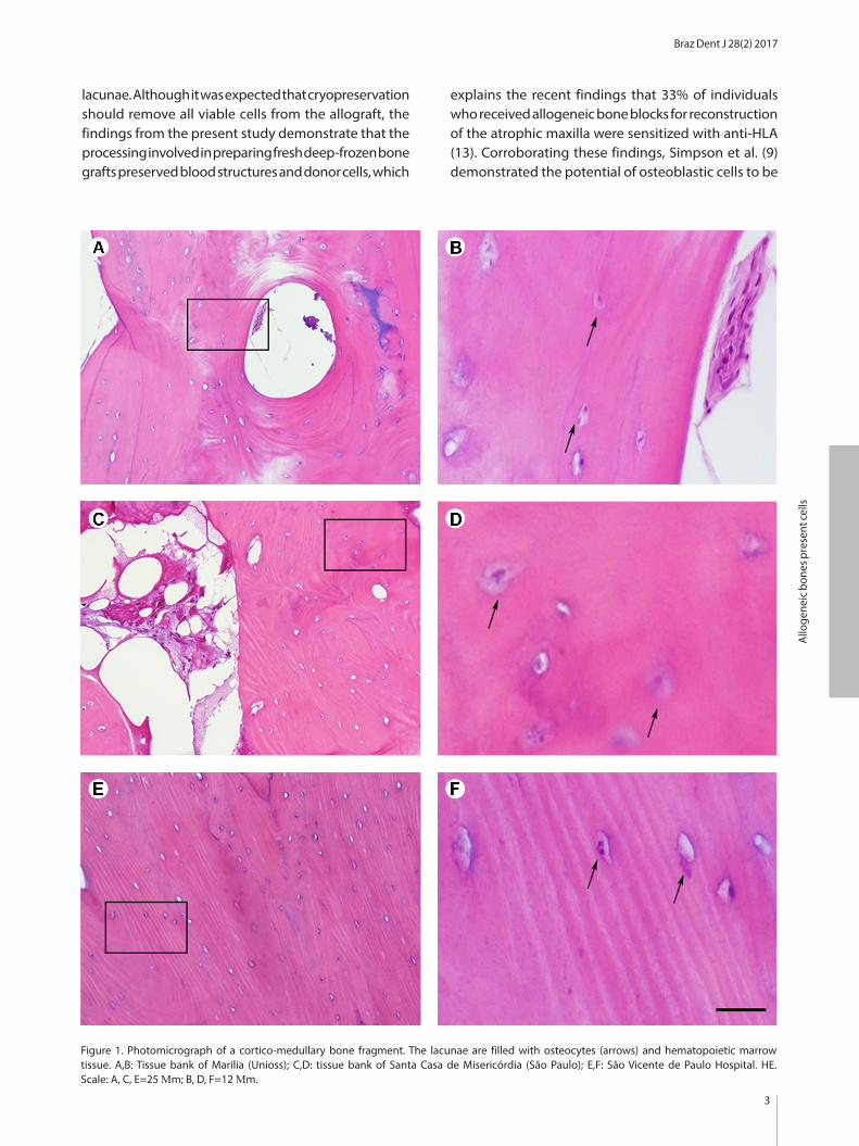
Photomicrographs from the bone samples stained with H&E and Feulgen are shown in Figures 1 and 2, respectively. All specimens presented cortico-medullary bone containing lacunae filled occasionally with osteocytes. Additionally, hematopoietic bone marrow tissue was also present (Fig. 1).
Feulgen staining confirmed the presence of DNA in all tissue samples (Fig. 2). There was presence of hematopoietic bone marrow and there were stained nuclei in all samples.
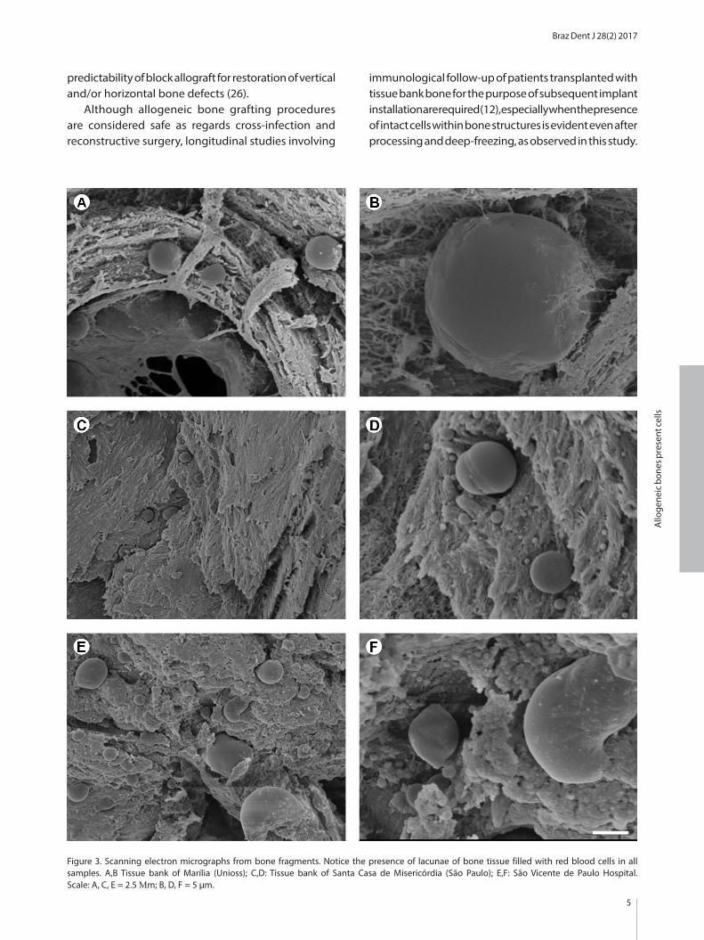
Ultrastructural AnalysisSEM photomicrographs are shown in Figure 3. The
lacunae observed within the cortico-medullary bone tissue were filled with erythrocytes.
DiscussionAutologous bone grafts are regarded as the gold
standard and despite their wide use, they are associated with considerable postoperative morbidity, with pain at the donor site as one of the most common complaints by patients, ranging in intensity from moderate to severe in 99% of patients in the first post-operative weeks (14). Allogeneic bone is considered a second line treatment option for grafting, due mainly to its osteoconductive properties (15) and high implant survival rates (16,17). The antigenicity and immune response associated with allogenic bone grafts remain poorly understood (8,18) and so, largely controversial.
There are several options available for allogeneic grafting, which include fresh frozen bone, fresh lyophilized bone and demineralized lyophilized bone (16). The antigenicity of the graft may be reduced by manipulation of the material so that it is well tolerated and the risk of cross-infection is minimized. The least immunogenic allogeneic biomaterial currently available is the lyophilized demineralized bone graft, which is particulate and therefore has limited use, especially when maxillofacial reconstruction is required.
The present study evaluated samples of fresh frozen bone grafts from three tissue banks in Brazil using optical and scanning electron microscopy. In all samples, there was organic matter, represented by medullary tissue, cellular debris, red blood cells and DNA within the bone tissue. Also within the bone matrix there was bone marrow and osteocytes occupying individually their respective
Braz Dent J 28(2) 2017
3
Allo
gene
ic b
ones
pre
sent
cells
lacunae. Although it was expected that cryopreservation should remove all viable cells from the allograft, the findings from the present study demonstrate that the processing involved in preparing fresh deep-frozen bone grafts preserved blood structures and donor cells, which
explains the recent findings that 33% of individuals who received allogeneic bone blocks for reconstruction of the atrophic maxilla were sensitized with anti-HLA (13). Corroborating these findings, Simpson et al. (9) demonstrated the potential of osteoblastic cells to be
Figure 1. Photomicrograph of a cortico-medullary bone fragment. The lacunae are filled with osteocytes (arrows) and hematopoietic marrow tissue. A,B: Tissue bank of Marilia (Unioss); C,D: tissue bank of Santa Casa de Misericórdia (São Paulo); E,F: São Vicente de Paulo Hospital. HE. Scale: A, C, E=25 Μm; B, D, F=12 Μm.

Braz Dent J 28(2) 2017
4
L.F.
Cout
inho
et a
l.
cultivated from freshly frozen allograft specimens for at least six months in a bone bank.
Despite allograft decontamination, osteocyte lacunae may remain completely intact, including hydroxyapatite crystals that are known to contribute to DNA preservation (19). In the present study, purplish-red Feulgen staining was observed in all allografts, both in osteocytes and in bone marrow cells adjacent to the bone matrix. Before this, the state of conservation of old bone samples was analyzed using histochemical techniques and it demonstrated that Feulgen staining was positive in a few cases only, suggesting that not all osteocytes preserved their DNA (20). Their well-preserved samples with a high degree of Feulgen positivity, however, demonstrated good condition of the bone tissue and consequently yielded good quality DNA, as demonstrated by amplification. In a previous study, four different allogeneic bone grafts were compared histologically, in which remnants of organic
tissue were detected in all samples, such as adipocytes, fibroblasts, osteocytes and chondrocytes, as well as presence of DNA samples, which ranged from 10.2 to 56.2 ng/mL (21). Although there is evidence that DNA may induce an immune response, cytosolic DNA may be the trigger of a strong immune response through the cGAMP synthase enzyme (22).
Previous studies have shown the development of an immune response against bone grafts, especially with respect to HLA incompatibility both in animals (23) and humans (8,11,24,25). In allograft recipients, donor cell growth can be responsible for an entire spectrum of post-implantation immune responses, which may explain the inconsistent behavior of allografts in revision arthroplasty (9,13). Notwithstanding the high success rate reported for the bone allograft survival, a systematic review demonstrated low level of scientific evidence articles with short follow-up time regarding clinical efficacy and
Figure 2. Photomicrograph of a cortico-medullary bone fragment. A,B. DNA is present in all samples, as demonstrated by stained nuclei (arrows), including in hematopoietic marrow tissue. A: fibrous hyperplasia (positive control); B: Tissue bank of Marilia (Unioss); C: Tissue bank of Santa Casa de Misericórdia (São Paulo); D: São Vicente de Paulo Hospital. Feulgen’s staining. Scale: 12 μm.
Braz Dent J 28(2) 2017
5
Allo
gene
ic b
ones
pre
sent
cells
Figure 3. Scanning electron micrographs from bone fragments. Notice the presence of lacunae of bone tissue filled with red blood cells in all samples. A,B Tissue bank of Marília (Unioss); C,D: Tissue bank of Santa Casa de Misericórdia (São Paulo); E,F: São Vicente de Paulo Hospital. Scale: A, C, E = 2.5 Μm; B, D, F = 5 μm.
predictability of block allograft for restoration of vertical and/or horizontal bone defects (26).
Although allogeneic bone grafting procedures are considered safe as regards cross-infection and reconstructive surgery, longitudinal studies involving
immunological follow-up of patients transplanted with tissue bank bone for the purpose of subsequent implant installation are required (12), especially when the presence of intact cells within bone structures is evident even after processing and deep-freezing, as observed in this study.

Braz Dent J 28(2) 2017
6
L.F.
Cout
inho
et a
l.
In summary, frozen bone grafts from three tissue banks in Brazil harbored whole cells even after tissue processing and deep-freezing.
ResumoSubstitutos ósseos têm sido amplamente utilizados para reconstrução de ossos maxilares atróficos. Uma vez que relatos anteriores demonstram a existência de células viáveis em blocos ósseos após processamento pelos bancos de tecidos músculo-esqueléticos para uso ortopédico, o objetivo deste trabalho foi avaliar a presença de células provenientes de três bancos de tecidos músculo-esqueléticos do Brasil. Todas as amostras foram processadas pelos respectivos bancos seguindo as normas da Agência Nacional de Vigilância Sanitária brasileira. Foi realizada a retirada de 3 amostras de cada bloco, que foram destinadas para processamento histológico e subsequente coloração por hematoxilina e eosina e Feugen e análise ultra-estrutural através de microscopia eletrônica de varredura (MEV). As imagens de microscopia de luz de todos os fragmentos de enxertos ósseos mostraram presença de células do tipo osteócito em todos os grupos avaliados, bem como intensa coloração por Feulgen, demonstrando a presença de DNA nos ossos mesmo após o processamento realizado. As análises ultraestruturais evidenciaram a presença de hemácias nas lacunas do tecido ósseo. Conclui-se que mesmo após os processamentos realizados pelos bancos de tecidos músculo-esqueléticos é possível encontrar células nos ossos utilizados para enxertia alógena.
References 1. Esposito M, Grusovin MG, Chew YS, Coulthard P, Worthington
HV. Interventions for replacing missing teeth: 1- versus 2-stage implant placement. Cochrane Database Syst Rev 2009;8:CD006698.
2. Rogers GF, Greene AK. Autogenous bone graft: basic science and clinical implications. J Craniofac Surg 2012;23:323-327.
3. Crespi R, Vinci R, Capparè P, Gherlone E, Romanos GE. Calvarial versus iliac crest for autologous bone graft material for a sinus lift procedure: a histomorphometric study. Int J Oral Maxillofac Implants 2007;22:527-532.
4. Sakka S, Krenkel C. Simultaneous maxillary sinus lifting and implant placement with autogenous parietal bone graft: outcome of 17 cases. J Craniomaxillofac Surg 2011;39:187-191.
5. Misch CM. Maxillary autogenous bone grafting. Dent Clin North Am 2011;55:697-713.
6. Pelegrine AA, Sorgi da Costa CE, Sendyk WR, Gromatzky A. The comparative analysis of homologous fresh frozen bone and autogenous bone graft, associated or not with autogenous bone marrow, in rabbit calvaria: a clinical and histomorphometric study. Cell Tissue Bank 2011;12:171-184.
7. Heyligers IC, Klein-Nulend J. Detection of living cells in non-processed but deep-frozen bone allografts. Cell Tissue Bank 2005;6:25–31.
8. Ward WG, Gautreaux MD, Lippert DC, Boles C. HLA sensitization and allograft bone graft incorporation. Clin Orthop Relat Res 2008;466:1837-1848.
9. Simpson D, Kakarala G, Hampson K, Steele N, Ashton B. Viable cells survive in fresh frozen human bone allografts. Acta Orthop 2007;78:26-30.
10. Sousa APS, Martinez EF, Napimoga MH. Osteoinductive potential of allogeneic human bone blocks in vitro. Rev Assoc Paul Cir Dent
2014;69:75-78.11. Graham SM, Leonidou A, Aslam-Pervez N, Hamza A, Panteliadis
P, Heliotis M, et al.. Biological therapy of bone defects: the immunology of bone allo-transplantation. Expert Opin Biol Ther 2010;10:885-901
12. Napimoga MH, Lacerda PE, Succi GM, Pelegrine AA. The use of allogeneic bone for reconstruction of the jaw can induce anti-HLA antibodies. Implant News 2015;12:357-363.
13. de Lacerda PE, Pelegrine AA, Teixeira ML, Montalli VA, Rodrigues H, Napimoga MH. Homologous transplantation with fresh frozen bone for dental implant placement can induce HLA sensitization: a preliminary study. Cell Tissue Bank 2016; May 5 DOI. 10.1007/s10561-016-9562-9.
14. Barone A, Varanini P, Orlando B, Tonelli P, Covani U. Deep-frozen allogeneic onlay bone grafts for reconstruction of atrophic maxillary alveolar ridges: a preliminary study. J Oral Maxillofac Surg 2009;67:1300-1306.
15. Nazirkar G, Singh S, Dole V, Nikam A. Effortless effort in bone regeneration: a review. J Int Oral Health 2014;6:120-124.
16. Acocella A, Bertolai R, Ellis E 3rd, Nissan J, Sacco R. Maxillary alveolar ridge reconstruction with monocortical fresh-frozen bone blocks: a clinical, histological and histomorphometric study. J Craniomaxillofac Surg 2012;40:525-533.
17. Pimentel AC, Napimoga, MH; Manzi, MR; Sendyk, WR. Reconstruction of the edentulous mandible with fresh frozen bone grafts and implants: a 4-year report of a prospective clinical study. Cell Tissue Bank 2014;15:1-6.
18. Spin-Neto R, Stavropoulos A, de Freitas RM, Pereira LA, Carlos IZ, Marcantonio E Jr. Immunological aspects of fresh-frozen allogeneic bone grafting for lateral ridge augmentation. Clin Oral Implants Res 2013;24:963-968.
19. Lindahl T. Instability and decay of the primary structure of DNA. Nature 1993;362:709-715.
20. Guarino FM, Angelini F, Odierna G, Bianco MR, Di Bernardo G, Forte A, et al.. Detection of DNA in ancient bones using histochemical methods. Biotech Histochem 2000;75:110-117.
21. Fretwurst T, Spanou A, Nelson K, Wein M, Steinberg T, Stricker A. Comparison of four different allogeneic bone grafts for alveolar ridge reconstruction: a preliminary histologic and biochemical analysis. Oral Surg Oral Med Oral Pathol Oral Radiol 2014;118:424-431.
22. Civril F, Deimling T, de Oliveira Mann CC, Ablasser A, Moldt M, Witte G, et al.. Structural mechanism of cytosolic DNA sensing by cGAS. Nature 2013;498:332-337.
23. Stevenson S, Li XQ, Martin B. The fate of cancellous and cortical bone after transplantation of fresh and frozen tissue-antigen-matched and mismatched osteochondral allografts in dogs. J Bone Joint Surg Am 1991;73:1143-1156.
24. Lee MY, Finn HA, Lazda, VA, Thistlethwaite JR Jr, Simon MA. Bone allografts are immunogenic and may preclude subsequent organ transplants. Clin Orthop Relat Res 1997;340:215-219.
25. Piaia M, Bub CB, Succi GM, Torres M, Costa TH, Pinheiro FC, et al.. HLA-typing analysis following allogeneic bone grafting for sinus lifting. Cell Tissue Bank 2017;18:75-81.
26. Araújo PP, Oliveira KP, Montenegro SC, Carreiro AF, Silva JS, Germano AR. Block allograft for reconstruction of alveolar bone ridge in implantology: a systematic review. Implant Dent 2013;22:304-308.
Received July 26, 2016Accepted January 4, 2017