PreparationofReversibleColorimetricTemperatureNanosensorsa...
4
Published: March 04, 2011 r2011 American Chemical Society 2434 dx.doi.org/10.1021/ac200196y | Anal. Chem. 2011, 83, 2434–2437 LETTER pubs.acs.org/ac Preparation of Reversible Colorimetric Temperature Nanosensors and Their Application in Quantitative Two-Dimensional Thermo-Imaging Xu-dong Wang, † Xin-hong Song, † Chun-yan He, ‡ Chaoyong James Yang, † Guonan Chen, ‡ and Xi Chen* ,† † Department of Chemistry and Key Laboratory of Analytical Sciences of Xiamen University, College of Chemistry and Chemical Engineering and State Key Laboratory of Marine Environmental Science, Xiamen University, Xiamen, 361005, China ‡ Ministry of Education Key Laboratory of Analysis and Detection Technology for Food Safety, Department of Chemistry, Fuzhou University, Fuzhou, 350002, China b S Supporting Information T he temperature sensor, as one of the earliest developed and most abundant devices (shared 75-80% of the world sensor market 1 ), has countless applications in daily usage: meteorology, aerodynamics, scientific research, industry, military technology, medical care, etc. As a result of safety and environmental conta- mination considerations, the uses of traditional liquid mercury- or kerosene-filled temperature sensors have been gradually reduced. Thermocouples and infrared thermometers have, thus, become the main products and dominate the mainstream of the temperature sensor market. However, these sensors are hard to miniaturize and unsuitable for temperature sensing on the micro/nanoscale. 2 Conversely, since they are easily miniaturized, noninvasive, accurate, and enable remote micro/nanoscale ima- ging, optical thermo-responsive sensors 3-7 have recently at- tracted much attention. Several attempts have been made to prepare thermo-responsive micro/nanoparticles employing quan- tum dots, 8,9 upconverting nanoparticles, 10 rhodamine, 11 ruthenium complex, 7,12 lanthanide complex, 2,13-16 nanogel, 17 and conju- gated polymer. 18 However, these sensors all suffer from cytoto- xicity, 19 irreversibility, 18 cross-sensitivity to oxygen, or their large size. 12 More importantly, these sensors lack the ability for real- time quantitative imaging, which is, however, essential for monitoring in microfluidic research, microreactor reaction, and hyperthermal tumor treatment 20 and studying heat-driven hy- drodynamics. Thus, achieving real-time quantitative imaging is a great challenge. Herein, we fabricated reversible colorimetric temperature nanosensors (CTNSs) by encapsulating two color luminescent dyes in gas-impermeable polymer nanoparticles. These CTNSs present obvious and full reversible temperature response and enable both rapid colorimetric temperature estima- tion using the eyes and quantitative two-dimensional (2-D) thermo-imaging. Heat-exchange induced fluid motion was, for the first time, rapidly, precisely, and quantitatively imaged using a digital camera, which provided good temporal and spatial resolution. ’ EXPERIMENTAL SECTION Materials. 4,4 0 -Bis(2-benzoxazolyl)stilbene, tris(benzoylacetonato) mono(phenanthroline)europium(III), and poly (vinylidene chloride- co-acrylonitrile) [P(VDC- co-AN), M w ∼150 000] were purchased from Sigma-Aldrich (www.sigmaaldrich.com). All reagents were analytical grade and used without further purification. Preparation of CTNSs. The CTNSs were prepared following the precipitation method. 21 Typically, 40 mg of P(VDC-co-AN), 0.5 mL of 4,4 0 -bis(2-benzoxazolyl)stilbene saturated acetone solution, and 4.0 mg of tris(benzoylacetonato)mono(phenan- throline)europium(III) were dissolved in 20 mL of acetone. With rigorous stirring, 60 mL of double distilled water was poured into the solution, and the acetone was removed at reduced pressure. The prepared CTNSs were stored at 4 °C in the dark. Received: January 24, 2011 Accepted: February 28, 2011 ABSTRACT: Reversible colorimetric temperature nanosensors were prepared using a very simple precipitation method to encapsulate two color luminescent dyes. These nanosensors presented obvious reversible temperature response and enabled both rapid colorimetric temperature estimation using the eyes and quantitative two-dimensional thermo-imaging. Heat-ex- change induced fluid motion was, for the first time, rapidly, precisely, and quantitatively imaged by just taking color pic- tures, and this presented good temporal and spatial resolution for studying heat-driven hydrodynamics. These nanosensors should have great application in micro/nanoscale research and also fabrication into films for macroscopic study.
Transcript of PreparationofReversibleColorimetricTemperatureNanosensorsa...

Published: March 04, 2011
r 2011 American Chemical Society 2434 dx.doi.org/10.1021/ac200196y |Anal. Chem. 2011, 83, 2434–2437
LETTER
pubs.acs.org/ac
Preparation of Reversible Colorimetric Temperature Nanosensors andTheir Application in Quantitative Two-Dimensional Thermo-ImagingXu-dong Wang,† Xin-hong Song,† Chun-yan He,‡ Chaoyong James Yang,† Guonan Chen,‡ and Xi Chen*,†
†Department of Chemistry and Key Laboratory of Analytical Sciences of Xiamen University,College of Chemistry and Chemical Engineering and State Key Laboratory of Marine Environmental Science, Xiamen University,Xiamen, 361005, China‡Ministry of Education Key Laboratory of Analysis and Detection Technology for Food Safety, Department of Chemistry,Fuzhou University, Fuzhou, 350002, China
bS Supporting Information
The temperature sensor, as one of the earliest developed andmost abundant devices (shared 75-80% of the world sensor
market1), has countless applications in daily usage: meteorology,aerodynamics, scientific research, industry, military technology,medical care, etc. As a result of safety and environmental conta-mination considerations, the uses of traditional liquid mercury-or kerosene-filled temperature sensors have been graduallyreduced. Thermocouples and infrared thermometers have, thus,become the main products and dominate the mainstream of thetemperature sensor market. However, these sensors are hard tominiaturize and unsuitable for temperature sensing on themicro/nanoscale.2 Conversely, since they are easily miniaturized,noninvasive, accurate, and enable remote micro/nanoscale ima-ging, optical thermo-responsive sensors3-7 have recently at-tracted much attention. Several attempts have been made toprepare thermo-responsive micro/nanoparticles employing quan-tum dots,8,9 upconverting nanoparticles,10 rhodamine,11 rutheniumcomplex,7,12 lanthanide complex,2,13-16 nanogel,17 and conju-gated polymer.18 However, these sensors all suffer from cytoto-xicity,19 irreversibility,18 cross-sensitivity to oxygen, or their largesize.12 More importantly, these sensors lack the ability for real-time quantitative imaging, which is, however, essential formonitoring in microfluidic research, microreactor reaction, andhyperthermal tumor treatment20 and studying heat-driven hy-drodynamics. Thus, achieving real-time quantitative imaging is agreat challenge. Herein, we fabricated reversible colorimetrictemperature nanosensors (CTNSs) by encapsulating two colorluminescent dyes in gas-impermeable polymer nanoparticles.
These CTNSs present obvious and full reversible temperatureresponse and enable both rapid colorimetric temperature estima-tion using the eyes and quantitative two-dimensional (2-D)thermo-imaging. Heat-exchange induced fluid motion was, forthe first time, rapidly, precisely, and quantitatively imaged using adigital camera, which provided good temporal and spatialresolution.
’EXPERIMENTAL SECTION
Materials.4,40-Bis(2-benzoxazolyl)stilbene, tris(benzoylacetonato)mono(phenanthroline)europium(III), and poly (vinylidene chloride-co-acrylonitrile) [P(VDC-co-AN), Mw ∼150000] were purchasedfrom Sigma-Aldrich (www.sigmaaldrich.com). All reagents wereanalytical grade and used without further purification.Preparation of CTNSs. The CTNSs were prepared following
the precipitation method.21 Typically, 40 mg of P(VDC-co-AN),0.5 mL of 4,40-bis(2-benzoxazolyl)stilbene saturated acetonesolution, and 4.0 mg of tris(benzoylacetonato)mono(phenan-throline)europium(III) were dissolved in 20 mL of acetone.With rigorous stirring, 60 mL of double distilled water waspoured into the solution, and the acetone was removed atreduced pressure. The prepared CTNSs were stored at 4 �C inthe dark.
Received: January 24, 2011Accepted: February 28, 2011
ABSTRACT:Reversible colorimetric temperature nanosensorswere prepared using a very simple precipitation method toencapsulate two color luminescent dyes. These nanosensorspresented obvious reversible temperature response and enabledboth rapid colorimetric temperature estimation using the eyesand quantitative two-dimensional thermo-imaging. Heat-ex-change induced fluid motion was, for the first time, rapidly,precisely, and quantitatively imaged by just taking color pic-tures, and this presented good temporal and spatial resolutionfor studying heat-driven hydrodynamics. These nanosensors should have great application in micro/nanoscale research and alsofabrication into films for macroscopic study.
2435 dx.doi.org/10.1021/ac200196y |Anal. Chem. 2011, 83, 2434–2437
Analytical Chemistry LETTER
Characterization of the Prepared CTNSs. Fluorescenceprofiles were obtained from a Hitachi F-4500 fluorometer(Hitachi Co. Ltd., Japan). A 365 nm UV lamp was used to excitethe CTNSs. A Hitachi S-4800 scanning electron microscope(SEM, Hitachi, Japan,) was used to characterize the particlemorphology. A Nikon D300 CMOS digital camera (Nikon Co.Ltd., Japan) was used for recording color images. Temperaturewas controlled using a Julabo F12-ED (Julabo Inc., Germany)refrigerated/heating circulator. Precise solution temperatureswere calibrated using a thermocouple based temperature meter(Yudian AI-5600. http://www.yudian.com/).
’RESULTS AND DISCUSSION
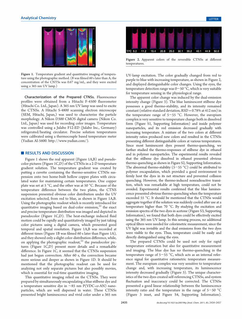
Figure 1 shows the real apparent (Figure 1A,B) and pseudo-color pictures (Figure 1C,D) of the CTNSs in a 2-D temperaturegradient solution. The temperature gradient was created byputting a cuvette containing the thermo-sensitive CTNSs sus-pension onto two home-built hollow copper plates with circu-lated water for maintaining certain temperatures. One copperplate was set at 5 �C, and the other was at 50 �C. Because of thetemperature difference between the two plates, the CTNSsuspension presented obvious color distribution under the UVexcitation selected, from red to blue, as shown in Figure 1A,B.Using the photographic readout which is recently introduced forquantitative imaging based on dual-color emission,22 the exactand precise temperature distribution was imaged and depicted inpseudocolor (Figure 1C,D). The heat-exchange induced fluidmotion could be rapidly and quantitatively imaged by just takingcolor pictures using a digital camera, which presented goodtemporal and spatial resolution. Figure 1A,B was recorded atdifferent times (Figure 1B was filmed 60 s later than Figure 1A),and they showed only a slight color-distribution difference, while,on applying the photographic readout,22 the pseudocolor pic-tures (Figure 1C,D) present more details and a remarkabledifference. In Figure 1C, it seemed that the CTNSs suspensionhad just begun convection. After 60 s, the convection becamemore serious and deeper as shown in Figure 1D. It should bementioned that the photographic readout can be used foranalyzing not only separate pictures but also possibly movies,which is essential for real-time quantitative imaging.
This quantitative imaging relied on the CTNSs. They wereprepared by simultaneously encapsulating a blue stilbene dye anda temperature sensitive dye in ∼85 nm P(VDC-co-AN) nano-particles, which are well dispersed in water. These CTNSspresented bright luminescence and vivid color under a 365 nm
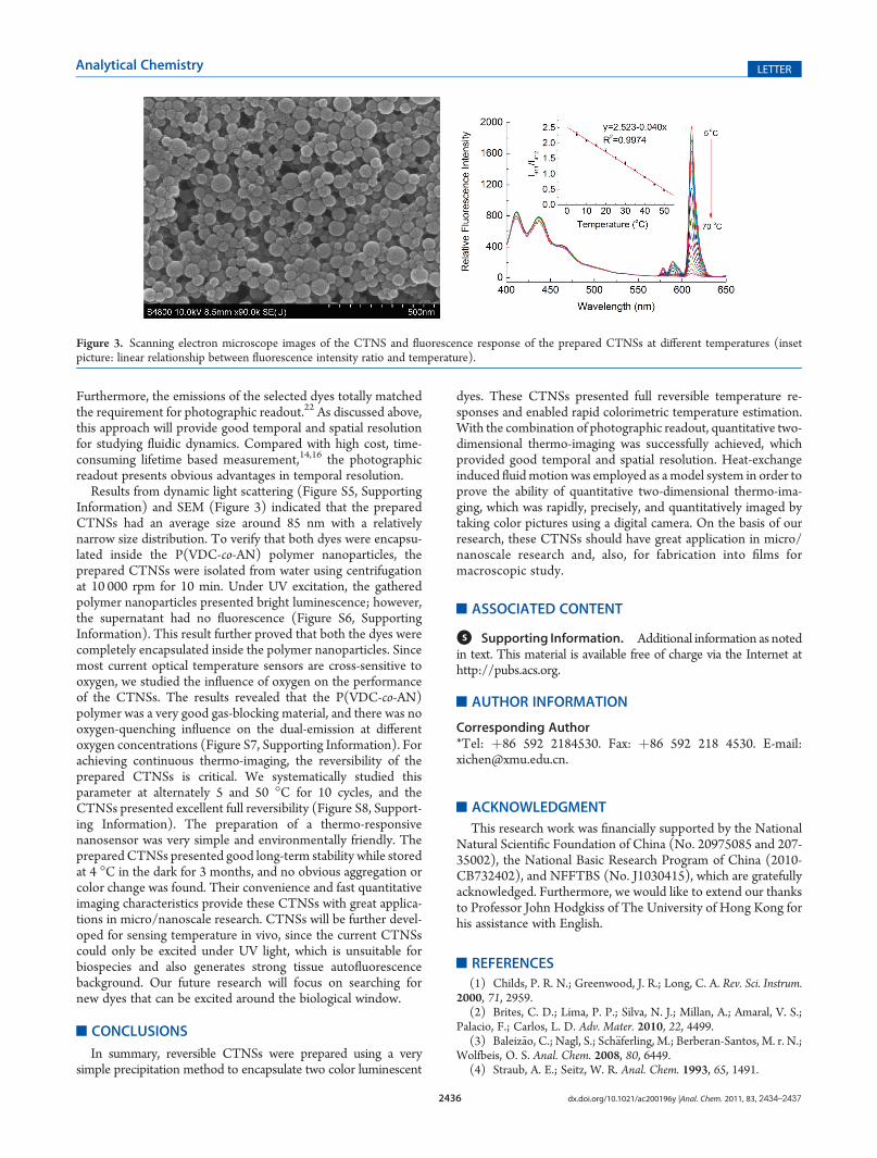
UV-lamp excitation. The color gradually changed from red topurple to blue with increasing temperature, as shown in Figure 2,and displayed distinguishable color changes. Using the eyes, thetemperature detection range was 0-50 �C, which is very suitablefor temperature sensing in the physiological range.
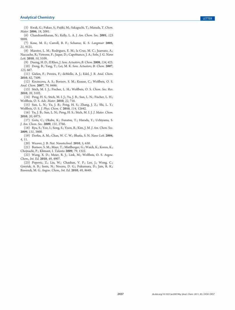
The apparent color change was induced by the dual-emissionintensity change (Figure 3). The blue luminescent stilbene dyepossesses a good thermo-stability, and its intensity remainedconstant (relative standard deviation, RSD= 0.78% at 412 nm) inthe temperature range of 5-55 �C. However, the europiumcomplex is very sensitive to temperature change both in dissolvedform (Figure S1, Supporting Information) and inside polymernanoparticles, and its red emission decreased gradually withincreasing temperature. A mixture of the two colors at differentintensity ratios produced new colors and resulted in the CTNSspresenting different distinguishable colors at various temperatures.Since most luminescent dyes present thermo-quenching, wefurther studied the thermo-responses of stilbene dye in ethanoland in polymer nanoparticles. The experimental results revealedthat the stilbene dye dissolved in ethanol presented obviousthermo-quenching as shown in Figure S2, Supporting Information.The abnormal thermo-stability (5-55 �C) could be attributed topolymer encapsulation, which provided a good environment tofirmly host the dyes in its net structure and prevented collisionquenching. However, the thermo-induced nonradioactive transi-tion, which was remarkable at high temperature, could not beavoided. Experimental results confirmed that the blue lumines-cence presented obvious thermo-quenching when the temperatureexceeded 55 �C. It should be mentioned that the CTNSs wouldaggregate together if the solution was suddenly cooled after use at atemperature higher than 70 �C. By studying the excitation andemission spectra of the two dyes in acetone (Figure S3, SupportingInformation), we found that both dyes could be effectively excitedusing the 365 nm UV lamp. In this sensing process, no additionaloptical filters were needed for colorimetric measurement, since theUV light was invisible and the dual emissions from the two dyeswere visible to the eyes. Thus, temperature could be easily anddirectly distinguished using the eyes.
The prepared CTNSs could be used not only for rapidtemperature estimation but also for quantitative measurementand imaging. The blue dye has no thermo-quenching in thetemperature range of 5-55 �C, which acts as an internal refer-ence signal for quantitative ratiometric temperature measure-ment. The europium complex was very sensitive to temperaturechange and, with increasing temperature, its luminescenceintensity decreased gradually (Figure 3). The unique character-istics of the two dyes created self-referencing CTNSs, and systemfluctuation and inaccuracy could be corrected. The CTNSspresented a good linear relationship between the luminescenceintensity ratio and the temperature in the range of 5-50 �C(Figure 3 inset, and Figure S4, Supporting Information).
Figure 1. Temperature gradient and quantitative imaging of tempera-ture using the photographic method. (B was filmed 60 s later than A; theconcentration of the CNTSs was 0.67 mg/mL, and they were excitedusing a 365 nm UV lamp.)
Figure 2. Apparent colors of the reversible CTNSs at differenttemperatures.
2436 dx.doi.org/10.1021/ac200196y |Anal. Chem. 2011, 83, 2434–2437
Analytical Chemistry LETTER
Furthermore, the emissions of the selected dyes totally matchedthe requirement for photographic readout.22 As discussed above,this approach will provide good temporal and spatial resolutionfor studying fluidic dynamics. Compared with high cost, time-consuming lifetime based measurement,14,16 the photographicreadout presents obvious advantages in temporal resolution.
Results from dynamic light scattering (Figure S5, SupportingInformation) and SEM (Figure 3) indicated that the preparedCTNSs had an average size around 85 nm with a relativelynarrow size distribution. To verify that both dyes were encapsu-lated inside the P(VDC-co-AN) polymer nanoparticles, theprepared CTNSs were isolated from water using centrifugationat 10 000 rpm for 10 min. Under UV excitation, the gatheredpolymer nanoparticles presented bright luminescence; however,the supernatant had no fluorescence (Figure S6, SupportingInformation). This result further proved that both the dyes werecompletely encapsulated inside the polymer nanoparticles. Sincemost current optical temperature sensors are cross-sensitive tooxygen, we studied the influence of oxygen on the performanceof the CTNSs. The results revealed that the P(VDC-co-AN)polymer was a very good gas-blocking material, and there was nooxygen-quenching influence on the dual-emission at differentoxygen concentrations (Figure S7, Supporting Information). Forachieving continuous thermo-imaging, the reversibility of theprepared CTNSs is critical. We systematically studied thisparameter at alternately 5 and 50 �C for 10 cycles, and theCTNSs presented excellent full reversibility (Figure S8, Support-ing Information). The preparation of a thermo-responsivenanosensor was very simple and environmentally friendly. Theprepared CTNSs presented good long-term stability while storedat 4 �C in the dark for 3 months, and no obvious aggregation orcolor change was found. Their convenience and fast quantitativeimaging characteristics provide these CTNSs with great applica-tions in micro/nanoscale research. CTNSs will be further devel-oped for sensing temperature in vivo, since the current CTNSscould only be excited under UV light, which is unsuitable forbiospecies and also generates strong tissue autofluorescencebackground. Our future research will focus on searching fornew dyes that can be excited around the biological window.
’CONCLUSIONS
In summary, reversible CTNSs were prepared using a verysimple precipitation method to encapsulate two color luminescent
dyes. These CTNSs presented full reversible temperature re-sponses and enabled rapid colorimetric temperature estimation.With the combination of photographic readout, quantitative two-dimensional thermo-imaging was successfully achieved, whichprovided good temporal and spatial resolution. Heat-exchangeinduced fluidmotion was employed as amodel system in order toprove the ability of quantitative two-dimensional thermo-ima-ging, which was rapidly, precisely, and quantitatively imaged bytaking color pictures using a digital camera. On the basis of ourresearch, these CTNSs should have great application in micro/nanoscale research and, also, for fabrication into films formacroscopic study.
’ASSOCIATED CONTENT
bS Supporting Information. Additional information as notedin text. This material is available free of charge via the Internet athttp://pubs.acs.org.
’AUTHOR INFORMATION
Corresponding Author*Tel: þ86 592 2184530. Fax: þ86 592 218 4530. E-mail:[email protected].
’ACKNOWLEDGMENT
This research work was financially supported by the NationalNatural Scientific Foundation of China (No. 20975085 and 207-35002), the National Basic Research Program of China (2010-CB732402), and NFFTBS (No. J1030415), which are gratefullyacknowledged. Furthermore, we would like to extend our thanksto Professor John Hodgkiss of The University of Hong Kong forhis assistance with English.
’REFERENCES
(1) Childs, P. R. N.; Greenwood, J. R.; Long, C. A. Rev. Sci. Instrum.2000, 71, 2959.
(2) Brites, C. D.; Lima, P. P.; Silva, N. J.; Millan, A.; Amaral, V. S.;Palacio, F.; Carlos, L. D. Adv. Mater. 2010, 22, 4499.
(3) Baleiz~ao, C.; Nagl, S.; Sch€aferling, M.; Berberan-Santos, M. r. N.;Wolfbeis, O. S. Anal. Chem. 2008, 80, 6449.
(4) Straub, A. E.; Seitz, W. R. Anal. Chem. 1993, 65, 1491.
Figure 3. Scanning electron microscope images of the CTNS and fluorescence response of the prepared CTNSs at different temperatures (insetpicture: linear relationship between fluorescence intensity ratio and temperature).
2437 dx.doi.org/10.1021/ac200196y |Anal. Chem. 2011, 83, 2434–2437
Analytical Chemistry LETTER
(5) Kwak, G.; Fukao, S.; Fujiki, M.; Sakaguchi, T.; Masuda, T. Chem.Mater. 2006, 18, 2081.(6) Chandrasekharan, N.; Kelly, L. A. J. Am. Chem. Soc. 2001, 123
9898.(7) Kose, M. E.; Carroll, B. F.; Schanze, K. S. Langmuir 2005,
21, 9121.(8) Maestro, L. M.; Rodriguez, E. M.; la Cruz, M. C.; Juarranz, A.;
Naccache, R.; Vetrone, F.; Jaque, D.; Capobianco, J. A.; Sole, J. G. NanoLett. 2010, 10, 5109.(9) Duong, H. D.; Il Rhee, J. Sens. Actuators, B: Chem. 2008, 134, 423.(10) Dong, B.; Yang, T.; Lei, M. K. Sens. Actuators, B: Chem. 2007,
123, 667.(11) Gielen, F.; Pereira, F.; deMello, A. J.; Edel, J. B. Anal. Chem.
2010, 82, 7509.(12) Kocincova, A. S.; Borisov, S. M.; Krause, C.; Wolfbeis, O. S.
Anal. Chem. 2007, 79, 8486.(13) Stich, M. I. J.; Fischer, L. H.; Wolfbeis, O. S. Chem. Soc. Rev.
2010, 39, 3102.(14) Peng, H. S.; Stich, M. I. J.; Yu, J. B.; Sun, L. N.; Fischer, L. H.;
Wolfbeis, O. S. Adv. Mater. 2010, 22, 716.(15) Sun, L. N.; Yu, J. B.; Peng, H. S.; Zhang, J. Z.; Shi, L. Y.;
Wolfbeis, O. S. J. Phys. Chem. C 2010, 114, 12642.(16) Yu, J. B.; Sun, L. N.; Peng, H. S.; Stich, M. I. J. J. Mater. Chem.
2010, 20, 6975.(17) Gota, C.; Okabe, K.; Funatsu, T.; Harada, Y.; Uchiyama, S.
J. Am. Chem. Soc. 2009, 131, 2766.(18) Ryu, S.; Yoo, I.; Song, S.; Yoon, B.; Kim, J. M. J. Am. Chem. Soc.
2009, 131, 3800.(19) Derfus, A. M.; Chan, W. C. W.; Bhatia, S. N. Nano Lett. 2004,
4, 11.(20) Weaver, J. B. Nat. Nanotechnol. 2010, 5, 630.(21) Borisov, S. M.; Mayr, T.; Mistlberger, G.; Waich, K.; Koren, K.;
Chojnacki, P.; Klimant, I. Talanta 2009, 79, 1322.(22) Wang, X. D.; Meier, R. J.; Link, M.; Wolfbeis, O. S. Angew.
Chem., Int. Ed. 2010, 49, 4907.(23) Popovic, Z.; Liu, W.; Chauhan, V. P.; Lee, J.; Wong, C.;
Greytak, A. B.; Insin, N.; Nocera, D. G.; Fukumura, D.; Jain, R. K.;Bawendi, M. G. Angew. Chem., Int. Ed. 2010, 49, 8649.