Preparation and Functionalization of Gold...
18
J. Surface Sci. Technol., Vol 29, No. 3-4, pp. 1-18, 2013 © 2013 Indian Society for Surface Science and Technology, India. Preparation and Functionalization of Gold Nanoparticles IGNÁC CAPEK Slovak Academy of Sciences, Polymer Institute and Institute of Measurement Science, Dúbravská cesta, Bratislava, Slovakia; Faculty of Industrial Technology, TnUni, Púchov, Slovakia Abstract — The abilities to control the size of gold nanoparticles and to manipulate them on a nanometer scale are priority subjects in the field of nanotechnology. To synthesize gold nanomaterials in controlled sizes and dimensions, various approaches have been developed. These systems can be made up of several different microenvironments: a continuous medium formed by the alkane or water, a disperse phase formed by microdroplets, micelles or surface active compounds and a tensioactive film, which separates the hydrophilic phase from the alkane and allows the solubilization to occur. Micelle or microemulsion approach allows for a unique encapsulated volume of controllable size through which reactions and subsequent development of metal and metallic compounds can be produced. The precipitation of prime nanoparticles is based on the supersaturation of solution by reactants and stabilization of formed nanoparticles by surfactants. The existence of these microenvironments gives these systems a particular ability to modulate the chemical reactivity due to the compartmentalization of the reactants in different microenvironments. These nanoparticles have become the focus of intensive research due to their unique applications in mesoscopic physics and in the fabrication of nanoscale devices. Keywords : Gold nanoparticles, preparation, passivation, functionalization and Ostwald ripening. INTRODUCTION While the most ancient use of colloidal gold is believed to have been in Egypt by alchemists, the brilliant colors of nanosized colloidal particles of silver, gold and copper were used in staining glasses as back as the 17th century. The use of gold colloid in biological applications began in 1971, when the immunogold staining procedure was invented. *Author for correspondence. E-mail : [email protected], [email protected] 1 2 3 4 5 6 7 8 9 10 11 12 13 14 15 16 17 18 19 20 21 22 23 24 25 26 27 28 29 30 31 32 33 34 35 36 37 38 39 Art-2
Transcript of Preparation and Functionalization of Gold...
J. Surface Sci. Technol., Vol 29, No. 3-4, pp. 1-18, 2013© 2013 Indian Society for Surface Science and Technology, India.
Preparation and Functionalization of GoldNanoparticles
IGNÁC CAPEKSlovak Academy of Sciences, Polymer Institute and Institute of Measurement Science,Dúbravská cesta, Bratislava, Slovakia; Faculty of Industrial Technology, TnUni, Púchov, Slovakia
Abstract — The abilities to control the size of gold nanoparticles and to manipulate them ona nanometer scale are priority subjects in the field of nanotechnology. To synthesize goldnanomaterials in controlled sizes and dimensions, various approaches have been developed. Thesesystems can be made up of several different microenvironments: a continuous medium formedby the alkane or water, a disperse phase formed by microdroplets, micelles or surface activecompounds and a tensioactive film, which separates the hydrophilic phase from the alkane andallows the solubilization to occur. Micelle or microemulsion approach allows for a uniqueencapsulated volume of controllable size through which reactions and subsequent developmentof metal and metallic compounds can be produced. The precipitation of prime nanoparticles isbased on the supersaturation of solution by reactants and stabilization of formed nanoparticlesby surfactants. The existence of these microenvironments gives these systems a particular abilityto modulate the chemical reactivity due to the compartmentalization of the reactants in differentmicroenvironments. These nanoparticles have become the focus of intensive research due to theirunique applications in mesoscopic physics and in the fabrication of nanoscale devices.
Keywords : Gold nanoparticles, preparation, passivation, functionalization and Ostwald ripening.
INTRODUCTION
While the most ancient use of colloidal gold is believed to have been in Egypt byalchemists, the brilliant colors of nanosized colloidal particles of silver, gold andcopper were used in staining glasses as back as the 17th century. The use of goldcolloid in biological applications began in 1971, when the immunogold stainingprocedure was invented.
*Author for correspondence. E-mail : [email protected], [email protected]
123456789101112131415161718192021222324252627282930313233343536373839
Art-2
2 Capek
123456789101112131415161718192021222324252627282930313233343536373839
The preparation of noble metal nanoparticles has received considerable attentionin recent decades because nanoparticles possess unconventional physical and chemicalproperties [1] Nanoparticles exhibit novel material properties which largely differ fromthe bulk materials due to these small sizes, including quantum size effect onphotochemistry, nonlinear optical properties of semiconductor or the emergence ofmetallic properties with the size of the particles [2]. As nanomaterials of noble metals,gold nanoparticles have extensive applications, such as antibacterial materials [3],antistatic materials, cryogenic superconducting materials [4], biosensor materials andso on.
Many nanogold-based environmental technologies (e.g., sensors, sorbents,reactants…) are under very active research and development, and are expected toemerge as the next generation environmental technologies to improve or replacevarious conventional environmental technologies in the near future [5]. Some of themost promising near term realizations of nanotechnology are at the interface ofphysical, chemical and biological systems. Because many biomolecules have specificbinding properties in self-assembly processes, they are attractive materials fornanotechnology.
Most existing approaches explore the strong affinity of thiols to gold [6] andthe use of disulfides [7] and thioethers [8] as capping agents as well. Polymersfunctionalized with molecular recognition groups [9], thioacetate groups [10] andtetradentate thioether ligands [11] have recently been used to mediate the formationof spherical or related assemblies of gold nanoparticles. These approaches have shownremarkable capabilities in assembling nanoparticles into functional nanostructures, theability to control their size and shape.
SYNTHETIC APPROACHES
A number of techniques have been used for producing metal nanoparticles, includingvapor phase techniques [12], sol-gel methods [13], sputtering [14], precipitation [15],soft- and hard-templates, chemical and bioreduction,synthesis in micellar solutions [16,17], etc. Two main methods can be employed for the preparation of goldnanoparticles: coprecipitation and chemical reduction. In both cases, the presence ofsurfactant is required to govern the growth process. Typically, the coprecipitationreactions involve the thermal decomposition of organometallic precursors [18]. Thechemical reduction occurring in colloidal assemblies is another approach for theformation of size- and shape-controlled nanoparticles [19]. A major benefit of chemicalmethods is their relatively inexpensive investment of capital equipment.
Preparation and Functionalization of Gold Nanoparticles 3
123456789101112131415161718192021222324252627282930313233343536373839
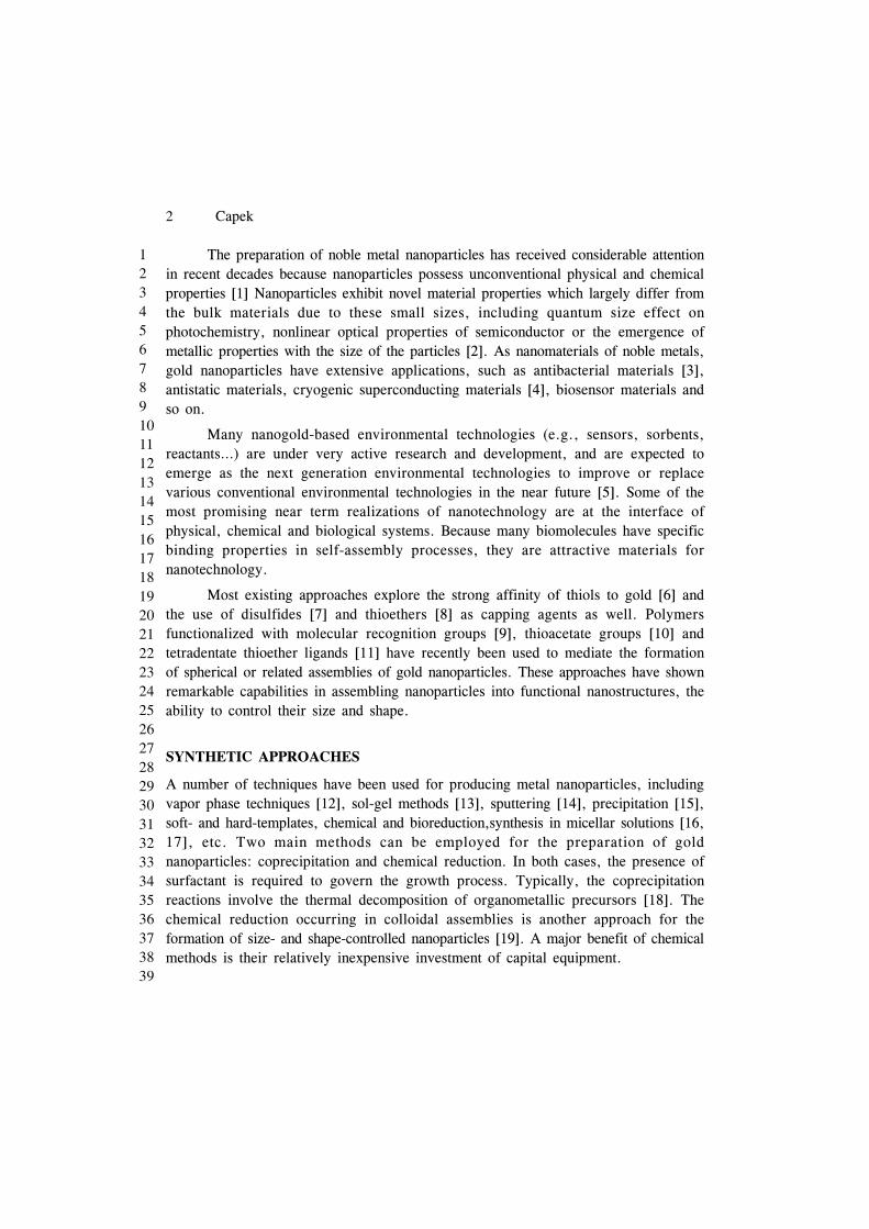
The successful utilization of gold nanoparticles (AuNPs) in biological assaysrelies on the availability of synthetic methods generating nanoparticles with the desiredcharacteristics, namely high solubility in water, and adequate morphology, sizedispersion, and surface functionalities. Of the chemical processes, reverse micelle(microemulsion) synthesis has been recently demonstrated to be a viable method forproducing a wide array of noble metal nanoparticles over a relatively narrow particlesize distribution [20]. Reverse micelle synthesis utilizes the natural phenomenoninvolving the formation of spheroidal aggregates in a solution when a surfactant isintroduced to an organic solvent, formed either in the presence or in the absence ofwater [21]. Micelle formation allows for a unique encapsulated volume of controllablesize through which reactions and subsequent development of metal and metalliccompounds can be produced. Aggregates containing (= [water]/[surfactant], seeabove) of less than 15 can be called as reverse micelles and have hydrodynamicdiameters in the range of 4-10 nm [22], whereas ù greater than 15 constitutemicroemulsions, which have a hydrodynamic diameter range between 5 and 50 nm.Once the right microemulsions are obtained, the method of particle preparation consistsin mixing of two microemulsions carrying the appropriate reactants in order to obtainthe nanoparticles [23,24] (Fig. 1).
Fig. 1. Proposed mechanism for the formation of gold nanoparticles by the microemulsion ap-proach.
4 Capek
123456789101112131415161718192021222324252627282930313233343536373839
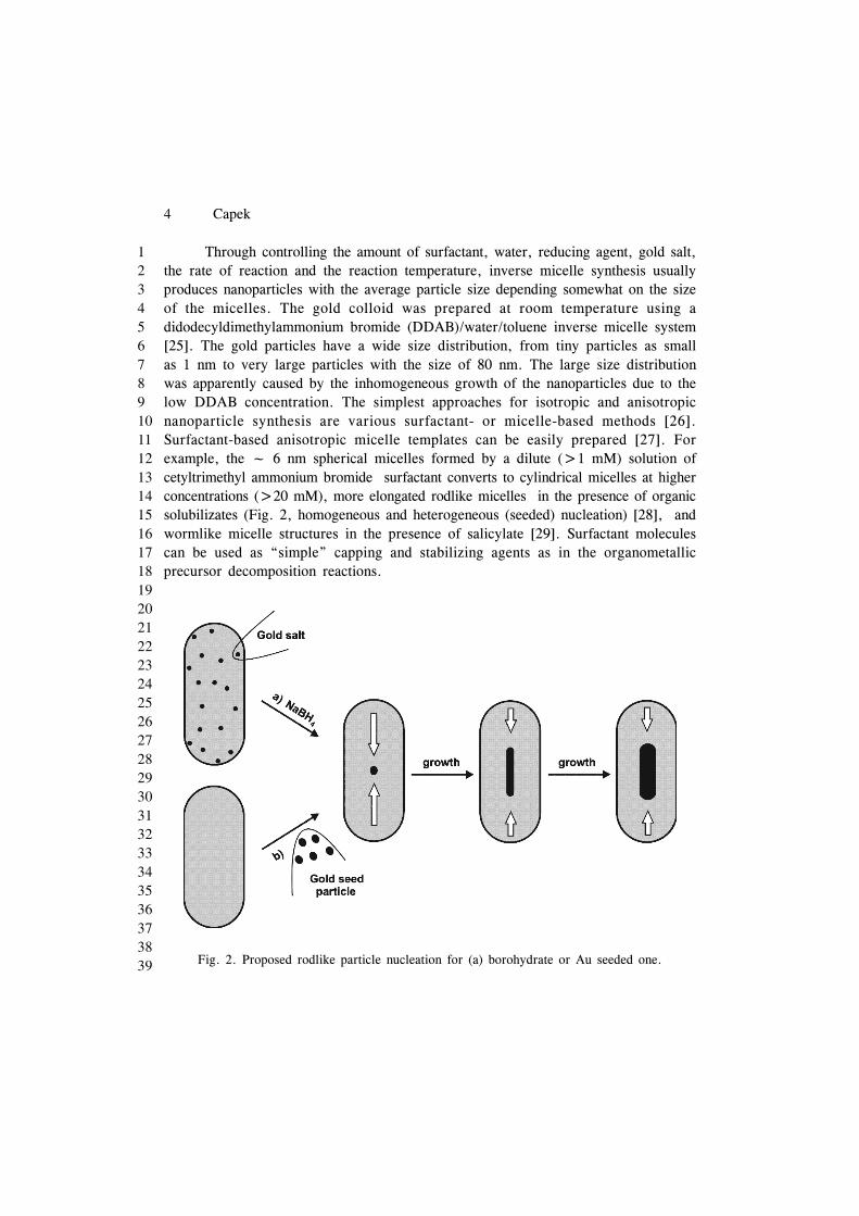
Through controlling the amount of surfactant, water, reducing agent, gold salt,the rate of reaction and the reaction temperature, inverse micelle synthesis usuallyproduces nanoparticles with the average particle size depending somewhat on the sizeof the micelles. The gold colloid was prepared at room temperature using adidodecyldimethylammonium bromide (DDAB)/water/toluene inverse micelle system[25]. The gold particles have a wide size distribution, from tiny particles as smallas 1 nm to very large particles with the size of 80 nm. The large size distributionwas apparently caused by the inhomogeneous growth of the nanoparticles due to thelow DDAB concentration. The simplest approaches for isotropic and anisotropicnanoparticle synthesis are various surfactant- or micelle-based methods [26].Surfactant-based anisotropic micelle templates can be easily prepared [27]. Forexample, the ~ 6 nm spherical micelles formed by a dilute (>1 mM) solution ofcetyltrimethyl ammonium bromide surfactant converts to cylindrical micelles at higherconcentrations (>20 mM), more elongated rodlike micelles in the presence of organicsolubilizates (Fig. 2, homogeneous and heterogeneous (seeded) nucleation) [28], andwormlike micelle structures in the presence of salicylate [29]. Surfactant moleculescan be used as “simple” capping and stabilizing agents as in the organometallicprecursor decomposition reactions.
Fig. 2. Proposed rodlike particle nucleation for (a) borohydrate or Au seeded one.
Preparation and Functionalization of Gold Nanoparticles 5
123456789101112131415161718192021222324252627282930313233343536373839
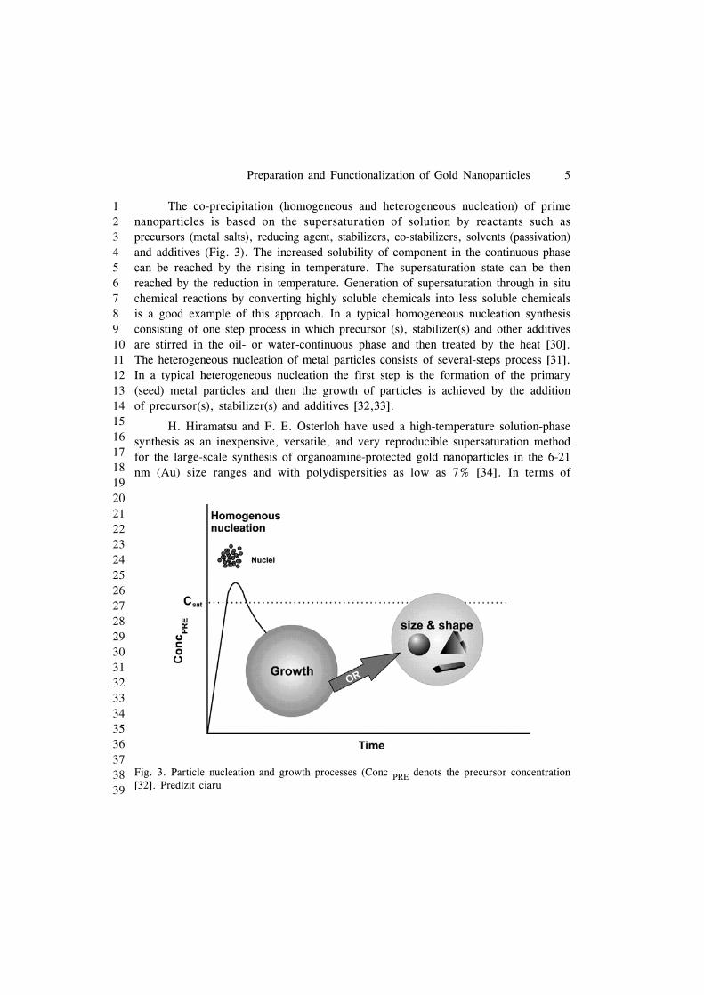
The co-precipitation (homogeneous and heterogeneous nucleation) of primenanoparticles is based on the supersaturation of solution by reactants such asprecursors (metal salts), reducing agent, stabilizers, co-stabilizers, solvents (passivation)and additives (Fig. 3). The increased solubility of component in the continuous phasecan be reached by the rising in temperature. The supersaturation state can be thenreached by the reduction in temperature. Generation of supersaturation through in situchemical reactions by converting highly soluble chemicals into less soluble chemicalsis a good example of this approach. In a typical homogeneous nucleation synthesisconsisting of one step process in which precursor (s), stabilizer(s) and other additivesare stirred in the oil- or water-continuous phase and then treated by the heat [30].The heterogeneous nucleation of metal particles consists of several-steps process [31].In a typical heterogeneous nucleation the first step is the formation of the primary(seed) metal particles and then the growth of particles is achieved by the additionof precursor(s), stabilizer(s) and additives [32,33].
H. Hiramatsu and F. E. Osterloh have used a high-temperature solution-phasesynthesis as an inexpensive, versatile, and very reproducible supersaturation methodfor the large-scale synthesis of organoamine-protected gold nanoparticles in the 6-21nm (Au) size ranges and with polydispersities as low as 7% [34]. In terms of
Fig. 3. Particle nucleation and growth processes (Conc PRE denots the precursor concentration[32]. Predlzit ciaru
6 Capek
123456789101112131415161718192021222324252627282930313233343536373839
achievable particles sizes, polydispersites, and simplicity (only three reagents,tetrachloroauric acid, oleylamine, and a solvent are required) the method is superiorto that of Jana et al. [35]. The syntheses are fast, very reproducible, and simple.The particles are stable in dried form and they can be easily modified withhydrophobic and hydrophilic thiols to afford nanoparticles that are soluble in organicsolvents or in water.
Most commonly, gold nanoparticles are synthesized by chemical orelectrochemical reduction of a gold(III) precursor compound in the presence of acapping agent, i.e. a compound able to bind to the nanoparticle surface blocking itsgrowth beyond the nanometer range and stabilizing the colloid in the particular solventused. Control over the shape and size of the AuNPs is usually achieved through thecareful selection of the experimental conditions, namely reducing agent, solvent,reaction time, temperature, and capping agent. Controlled nucleation and separationof nucleation from growth are the keys to synthesizing near-monodisperse goldnanoparticles in the 1–15 nm size range [36]. This can be achieved either by providinga controlled number of preformed gold nanoparticles as nucleation centers in a growthmedium where no secondary nucleation can occur—the seeding growth method [36]- or by varying the ratio of strong and weak reducing agents [35]. Key goals in thesynthesis of nanoparticles are that the synthesis gives nanostructures of a specific sizeand size distribution and that the synthesis is reproducible [37]. A common approachis to use capping agents with strong affinity for gold, e.g. thiol capping agents. Thisallows the synthesis of AuNPs but usually only soluble in organic solvents [6]. Anadditional step is required for the extraction of the particles into water. Exchangeof strongly binding capping agents is, however, usually cumbersome, which makesthis type of AuNP less versatile for various modifications and biological applications.The reverse is true for the capping agents with weak affinity for gold. The citratereduction method is the most commonly used method for preparation of sphericalAuNPs for biological assays due to its simplicity and high yield [38]. The use ofcitrate as a capping agent is very convenient due to its easy post-synthesis treatment,since it can be easily replaced by other capping agents, e.g. thiol capping agents,bearing an appropriate functionality for binding of the biological analyte of interest.
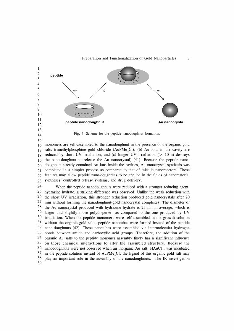
Djalali et al. have reported a novel method to produce gold nanoparticles insoft templates (doughnut-shaped nanoreactors), peptide nanodoughnuts [39]. The nano-doughnuts were self-assembled from peptides and organic gold salts. Various shapesof peptide/protein assemblies have been produced in biomaterials [40]. MonodisperseAu nanocrystals grew inside the cavities of peptide nanodoughnuts by the reductionof Au ions trapped in the cavities and the resulting Au nanocrystals were extractedby destroying the nano-doughnuts via long UV irradiation (Fig. 4. (a) peptide
Preparation and Functionalization of Gold Nanoparticles 7
123456789101112131415161718192021222324252627282930313233343536373839
monomers are self-assembled to the nanodoughnut in the presence of the organic goldsalts trimethylphosphine gold chloride (AuPMe3Cl), (b) Au ions in the cavity arereduced by short UV irradiation, and (c) longer UV irradiation (> 10 h) destroysthe nano-doughnut to release the Au nanocrystal) [41]. Because the peptide nano-doughnuts already contained Au ions inside the cavities, Au nanocrystal synthesis wascompleted in a simpler process as compared to that of micelle nanoreactors. Thosefeatures may allow peptide nano-doughnuts to be applied in the fields of nanomaterialsyntheses, controlled release systems, and drug delivery.
When the peptide nanodoughnuts were reduced with a stronger reducing agent,hydrazine hydrate, a striking difference was observed. Unlike the weak reduction withthe short UV irradiation, this stronger reduction produced gold nanocrystals after 20min without forming the nanodoughnut-gold nanocrystal complexes. The diameter ofthe Au nanocrystal produced with hydrazine hydrate is 23 nm in average, which islarger and slightly more polydisperse as compared to the one produced by UVirradiation. When the peptide monomers were self-assembled in the growth solutionwithout the organic gold salts, peptide nanotubes were formed instead of the peptidenano-doughnuts [42]. Those nanotubes were assembled via intermolecular hydrogenbonds between amide and carboxylic acid groups. Therefore, the addition of theorganic Au salts to the peptide monomer assembly likely has a significant influenceon those chemical interactions to alter the assembled structure. Because thenanodoughnuts were not observed when an inorganic Au salt, HAuCl4, was incubatedin the peptide solution instead of AuPMe3Cl, the ligand of this organic gold salt mayplay an important role in the assembly of the nanodoughnuts. The IR investigation
Fig. 4. Scheme for the peptide nanodoughnut formation.

8 Capek
123456789101112131415161718192021222324252627282930313233343536373839
suggests that the organic Au salts are incorporated in the peptide self-assemblies andcontribute to the nanodoughnut formation. The amide peak shifts were observed inamide-containing selfassembled monolayers, after gold salts were bound to their amidegroups [43]. When the peptide nano-doughnuts were weakly reduced by UV irradiationin 20 min, gold nanocrystals were observed inside the doughnut cavities. The particlesin the center of the doughnut cavities are identified as gold nanocrystals from theSFM phase images, the TEM images, and the electron diffractions. In fact, theincorporation of the gold nanocrystal increases the mechanical strength of the peptidenanodoughnut. After those samples were dried on mica surfaces, the peptidenanodoughnuts without gold nanocrystals collapsed and displayed a deformed ringshape, whereas the peptide nanodoughnuts with gold nanocrystals inside the cavitiesshowed a monodisperse and isotropic ring shape.
A solid-phase place exchange reaction can be also used to synthesize goldnanoparticles with monofunctional group attached to the surface [44]. This approachis based on a “catch and release” mechanism. Bifunctional thiol ligands with acarboxylic end group were first immobilized on a solid support such as a polymerresin with a controlled density. The density was low enough that neighboring thiolligands were far apart from each other. When the modified polymer support wasincubated in a butanethiol-protected gold nanoparticle solution, a one-to-one placeexchange reaction took place between the polymer-bound thiol ligands and thenanoparticles. After cleaving off from the solid support, nanoparticles with a singlecarboxylic group were obtained as the major product. Jacobson et al. published analmost identical approach toward the synthesis of gold nanoparticles with a singleamino acid moiety [45]. These nanoparticles with a single functional group attachedcan be treated as giant “molecules” and linked together into very sophisticatedstructures through traditional chemical reactions, just like the total synthesis ofcomplicated natural product from small molecular units.
STABILIZATION
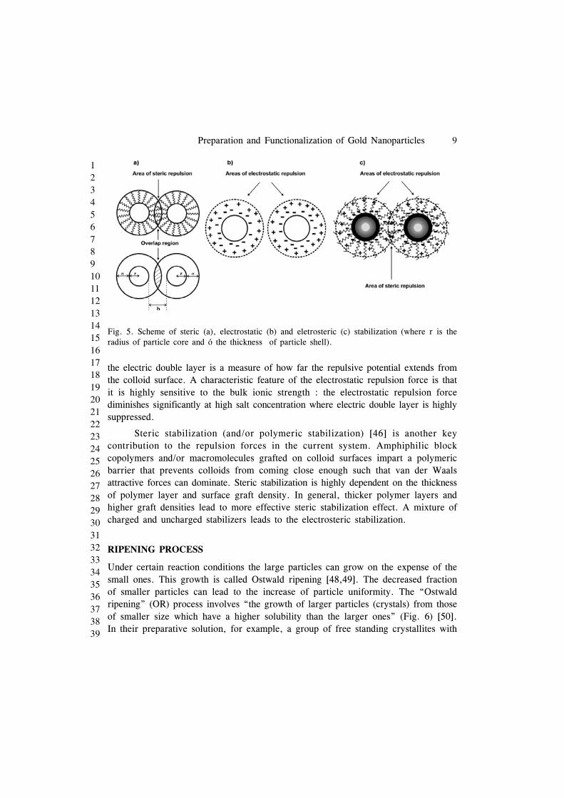
Whether these gold colloids are stabilized or undergo aggregation depends on the netpotential of interparticle attraction and repulsion forces. The interparticle attractionforce is van der Waals force, which is responsible for the gold nanoparticle (AuNP)aggregation. The two major repulsion forces that contribute to AuNP stabilization areelectrostatic and steric repulsion forces (Fig. 5) [46]. Electrostatic repulsion resultsfrom the negatively or positively charged ionic groups at ionic stabilizers. Thecharges, together with the counterions in the medium, form a repulsive electric doublelayer that stabilizes colloids against van der Waals attraction [47]. The thickness of
Preparation and Functionalization of Gold Nanoparticles 9
123456789101112131415161718192021222324252627282930313233343536373839
the electric double layer is a measure of how far the repulsive potential extends fromthe colloid surface. A characteristic feature of the electrostatic repulsion force is thatit is highly sensitive to the bulk ionic strength : the electrostatic repulsion forcediminishes significantly at high salt concentration where electric double layer is highlysuppressed.
Steric stabilization (and/or polymeric stabilization) [46] is another keycontribution to the repulsion forces in the current system. Amphiphilic blockcopolymers and/or macromolecules grafted on colloid surfaces impart a polymericbarrier that prevents colloids from coming close enough such that van der Waalsattractive forces can dominate. Steric stabilization is highly dependent on the thicknessof polymer layer and surface graft density. In general, thicker polymer layers andhigher graft densities lead to more effective steric stabilization effect. A mixture ofcharged and uncharged stabilizers leads to the electrosteric stabilization.
RIPENING PROCESS
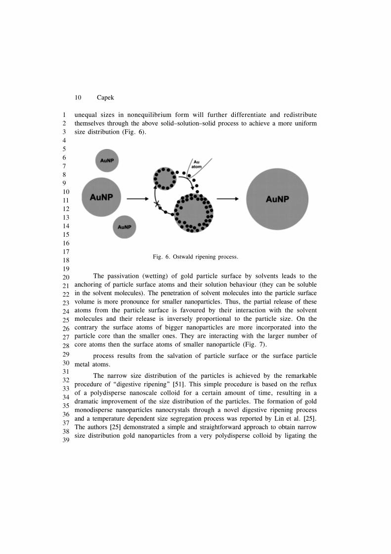
Under certain reaction conditions the large particles can grow on the expense of thesmall ones. This growth is called Ostwald ripening [48,49]. The decreased fractionof smaller particles can lead to the increase of particle uniformity. The “Ostwaldripening” (OR) process involves “the growth of larger particles (crystals) from thoseof smaller size which have a higher solubility than the larger ones” (Fig. 6) [50].In their preparative solution, for example, a group of free standing crystallites with
Fig. 5. Scheme of steric (a), electrostatic (b) and eletrosteric (c) stabilization (where r is theradius of particle core and ó the thickness of particle shell).
10 Capek
123456789101112131415161718192021222324252627282930313233343536373839
unequal sizes in nonequilibrium form will further differentiate and redistributethemselves through the above solid–solution–solid process to achieve a more uniformsize distribution (Fig. 6).
Fig. 6. Ostwald ripening process.
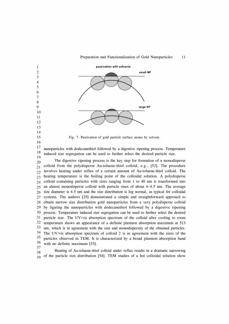
The passivation (wetting) of gold particle surface by solvents leads to theanchoring of particle surface atoms and their solution behaviour (they can be solublein the solvent molecules). The penetration of solvent molecules into the particle surfacevolume is more pronounce for smaller nanoparticles. Thus, the partial release of theseatoms from the particle surface is favoured by their interaction with the solventmolecules and their release is inversely proportional to the particle size. On thecontrary the surface atoms of bigger nanoparticles are more incorporated into theparticle core than the smaller ones. They are interacting with the larger number ofcore atoms then the surface atoms of smaller nanoparticle (Fig. 7).
process results from the salvation of particle surface or the surface particlemetal atoms.
The narrow size distribution of the particles is achieved by the remarkableprocedure of “digestive ripening” [51]. This simple procedure is based on the refluxof a polydisperse nanoscale colloid for a certain amount of time, resulting in adramatic improvement of the size distribution of the particles. The formation of goldmonodisperse nanoparticles nanocrystals through a novel digestive ripening processand a temperature dependent size segregation process was reported by Lin et al. [25].The authors [25] demonstrated a simple and straightforward approach to obtain narrowsize distribution gold nanoparticles from a very polydisperse colloid by ligating the
Preparation and Functionalization of Gold Nanoparticles 11
123456789101112131415161718192021222324252627282930313233343536373839
nanoparticles with dodecanethiol followed by a digestive ripening process. Temperatureinduced size segregation can be used to further select the desired particle size.
The digestive ripening process is the key step for formation of a monodispersecolloid from the polydisperse Au-toluene-thiol colloid, e.g., [52]. The procedureinvolves heating under reflux of a certain amount of Au-toluene-thiol colloid. Theheating temperature is the boiling point of the colloidal solution. A polydispersecolloid containing particles with sizes ranging from 1 to 40 nm is transformed intoan almost monodisperse colloid with particle sizes of about 4–4.5 nm. The averagesize diameter is 4.5 nm and the size distribution is log normal, as typical for colloidalsystems. The authors [25] demonstrated a simple and straightforward approach toobtain narrow size distribution gold nanoparticles from a very polydisperse colloidby ligating the nanoparticles with dodecanethiol followed by a digestive ripeningprocess. Temperature induced size segregation can be used to further select the desiredparticle size. The UV/vis absorption spectrum of the colloid after cooling to roomtemperature shows an appearance of a definite plasmon absorption maximum at 513nm, which is in agreement with the size and monodispersity of the obtained particles.The UV/vis absorption spectrum of colloid 2 is in agreement with the sizes of theparticles observed in TEM. It is characterized by a broad plasmon absorption bandwith no definite maximum [53].
Heating of Au-toluene-thiol colloid under reflux results in a dramatic narrowingof the particle size distribution [54]. TEM studies of a hot colloidal solution show
Fig. 7. Passivation of gold particle surface atoms by solvent.
12 Capek
123456789101112131415161718192021222324252627282930313233343536373839
formation of spherically shaped particles with sizes of about 4 nm. They have atendency to organize into 2D layers. Some of the particles from the hot colloidorganize in nice 3D structures. The remarkable effect of the digestive ripeningprocedure is the great improvement of the size distribution. The amazing result isthat the particles predominantly organize on the TEM grid in large 3D structures inonly about 15 min after the digestive ripening process is finished. A small numberof areas of 2D arrangement are also observed. Even larger 3D structures (>3 m)are observed. The results suggest that the activation energy for 2D organization islower compared to that of 3D organization. Of course, one of the most interestingfeatures of the synthetic sequence reported herein is the digestive ripening step, andthe mechanism for this remarkable process is not entirely clear. Only a few usefulfacts are known. First of all, nanoparticles are the necessary starting material; thatis, normal gold powder is not susceptible to digestive ripening, showing again thatnanosized particles are intrinsically more chemically reactive than bulk samples [55].
FUNCTIONALIZATION
The ligand exchange reaction is an extremely versatile tool for the preparation offunctionalized metal nanoparticles [56]. This method is fast and simple to use; it allowsone to introduce functional groups that are incompatible with other methods fornanoparticle synthesis. A report by the R. W. Murray group suggests a new role ofreaction conditions in mediating the exchange reaction [57]. It was demonstrated thattriphenylphosphine (TPP)-stabilized gold nanoparticles [58] undergo ligand exchangereactions with a few -functionalized thiols to produce functionalized nanoparticlesthat preserve the core dimensions of the precursor particles but exhibit highlyincreased stability against heat, aggregation, and decomposition [59]. The stronginteraction between n-alkylthiols and the gold surface provides the most popularmethod for the attachment of molecular groups [60]. Such bonding is convenient toengineer, but is reversible at moderate temperatures and kinetically unstable withrespect to movement of thiols on the surface [60]. Thus, groups that are depositedonto a metallic surface cannot be precisely fixed with respect to one another, althoughaverage spacing can be arranged by diluting the monolayer of functionalized alkylthiolswith analogues lacking the functional group [61]. Precise, angstrom-level control ofreactivity in nanotechnology requires nanoscale building blocks of known structureat atomic resolution. 60 vynechana
The passivation of gold particles with acetone leads to the gold-acetone colloid(1) which has a brown color, particles well-dispersed in solvent and particles ranging
Preparation and Functionalization of Gold Nanoparticles 13
123456789101112131415161718192021222324252627282930313233343536373839
from 10 to 50 nm in pure acetone solvent [54]. The addition of toluene changes thecolor of the Au-acetone-toluene-thiol colloid to a dark brown color (colloid 2). TEMstudies of this colloid show particles ranging from 5 to 40 nm with no definitegeometrical shapes [52]. Both stabilization (steric and electrostatic) processes take placeduring the warm up step, which has to be carried out slowly in order to ensure goodstabilization. Au-toluene-thiol colloid (colloid 2) was obtained by vacuum evaporationof all the acetone from colloid 1. Drastic change of the size and shape of the particlesis characteristic at this stage. Nearly spherical particles with sizes in the range of1-6 nm are dominant. There are also a small number of larger particles (10-40 nm)like those in the initial acetone-containing colloid (colloid 1). One possible explanationfor the change of size and shape of the gold particles induced by the removal ofacetone is due to the change in interaction particle-solvent. In colloid 1 the amountof acetone is in great excess. In great excess of acetone the gold particles are stronglysolvated by acetone and the attachment of dodecanethiol (RSH) molecules on theparticles’ surface is suppressed. Acetone, with its nonbonding electron pairs, can serveas a reasonably good ligand for gold but can only compete with RSH at high acetoneconcentrations. Therefore, as acetone is removed, the thiol competes better and better.This effect would be enhanced by the fact that the long-chained thiol is less solublein acetone than in toluene. Acetone acts as a preliminary stabilizing agent, which issubstituted by dodecanethiol molecules when acetone is evaporated. This ensures gooddispersity of the thiol-ligated gold particles in the toluene medium. In addition, tolueneis anticipated to achieve much better wetting of the thiol molecules on the goldparticles’ surface compared to the more polar solvent acetone. In favor of this areresults obtained for the wetting of undecanethiol self-assembled monolayers on goldsurface by water and hexadecane [62]. It was found that hexadecane as a nonpolarsolvent wetted the thiol molecules on the gold surface much better compared to water[62]. It is reasonable to expect a similar wetting trend for acetone, hexane, toluene,etc. due to their different polarities.
Although gold nanoparticles can be prepared from various materials by severalmethods [63], the coupling and functionalization with biological components has onlybeen carried out with a limited number of chemical methods. To apply gold colloidsin newly developed biomodifying medical assay systems, a simple and facile meansof anchoring different ligand biomolecules onto particle surfaces are strongly requiredas well as the stability in the physiological condition should be improved. Particularly,color changes induced by the association of nanometer-sized gold particles providea basis of a simple yet highly selective method for detecting specific biological

14 Capek
123456789101112131415161718192021222324252627282930313233343536373839
reactions between anchored ligand molecules and receptor molecules in the milieu.Colloidal gold nanoparticles, in particular, have found application in a variety of assayformats in which analyte binding is coupled to particle adsorption. With decreasinggold colloidal particle size, however, colloidal stability decreases significantly due toincreased particle surface energy. Such gold nanoparticles aggregate in high ionicstrength milieu as well as adsorb biomolecules such as proteins and DNAnonspecifically, resulting in reduced sensitivity and selectivity when used as colloidalsensor systems in biological fluids.
Functionalization of gold nanoparticles involves the use of functional ligandsin which a moiety is used for anchorage to the particle while the other is directedto the outer-surface for specific interaction with biomolecules. For example, thiol-modified oligonucleotides have been used to functionalize AuNPs for specific detectionof nucleic acid sequences in biological samples. Functionalization of Au-NPs withbiomolecules other than nucleic acids has also been used in order to developmethodologies suitable for clinical diagnostics. These include : 1) antibodies for signalenhancement in immunoassays [64]; 2) carbohydrate functionalization to study specificmolecular interactions [65]; and 3) surface functionalization with ligands that can betailored for specific protein binding [66] or direct binding of peptides and proteinsto the Au-nanoparticle surface [67].
CONCLUSION
Gold nanomaterials have been synthesized using a variety of methods. Two mainapproaches for the preparation of gold nanoparticles precipitation and chemicalreduction were discussed. In both cases, the presence of surfactant is required toinitiate the particle nucleation and govern the growth process. The precipitation ofprime nanoparticles is based on the supersaturation of solution by reactants andadditives. Generation of supersaturation through in situ chemical reactions byconverting highly soluble chemicals into less soluble chemicals is a good exampleof this approach. The reverse microemulsion synthesis has been recently demonstratedto be a viable method for producing a wide array of gold nanoparticles over arelatively narrow particle size distribution. A major benefit of chemical methods istheir relatively inexpensive investment of capital equipment.
There are several reasons for the synthesis and use of AuNPs in nanotechnologyas well as in nanomedicine. (i) First of all, gold compounds have long been used inmedicine throughout the history of civilization. (ii) It is easy to synthesize AuNPs
Preparation and Functionalization of Gold Nanoparticles 15
123456789101112131415161718192021222324252627282930313233343536373839
by several simple, economically cheap, safe and reliable (above mentioned) methods;(iii) it can be synthesized from sizes of 2–500 nm by changing the reactionparameters; (iv) it can be easily synthesized with different shapes (spheres, rods, tubes,wires, ribbons, plate, cubic, hexagonal, triangular) usingsoft andhard templates andchanging reaction conditions; (v) due to the presence of a negative charge on thesurface of AuNPs, they are highly reactive, which helps to modify the surface ofAuNPs using several biomolecules. Due to the strong interaction between the goldsurface and thiol/amine containing molecules (organic molecules, DNA, protein,enzyme etc.) the surface of AuNPs can be easily modified or functionalized and (vii)finally, it is well established that AuNPs are biocompatible and non-toxic.
ACKNOWLEDGEMENT
This research was supported by the APVV-0125-11 project.
LITERATURE
1. R. F. Service, Science, 271, 920 (1996).
2. C. Feldmann, Adv. Funct. Mater., 13, 101 (2003).
3. H. Q. Jiang, S. Manolache, A. C. L. Wong and F. S. Denes, J. Appl. Polym. Sci.,93, 1411 (2004).
4. S. Hirano, Y. Wakasa, A. Saka, S. Yoshizawa, Y. Oya-Seimiya, Y. Hishinuma, A.Nishimura, A. Matsumoto and H. Kumakura, Phys., C, 392, 458 (2003).
5. A. Modi, N. Koratkar, E. Lass, B. Q. Wei and P. M. Ajayan, Nature, 424, 171(2003).
6. M. Brust, M. Walker, D. Bethell, D. J. Schiffrin and R. Whyman, J. Chem. Soc.,Chem. Commun., 801 (1994).
7. Y. S. Shon, C. Mazzitelli and R. W. Murray, Langmuir, 17, 7735 (2001).
8. X. M. Li, M. R. de Jong, K. Inoue, S. Shinkai, J. Huskens and D. N. Reinhoudt,J. Mater. Chem., 11, 1919 (2001).
9. A. K. Boal, F. Ilhan, J. E. DeRouchey, T. Thurn-Albrecht, T. P. Russell and V.M. Rotello, Nature, 404, 746 (2000).
10. L. C. Brousseau, J. P. Novak, S. M. Marinakos and D. L. Feldheim, AdV. Mater.,11, 447 (1999).
11. M. M. Maye, S. C. Chun, L. Han, D. Rabinovich and C. J. Zhong, J. Am. Chem.Soc., 124, 4958 (2002).
12. R. W. Siegel and S. Ramasamy, J. Mater. Rev., 3, 1367 (1998).
16 Capek
123456789101112131415161718192021222324252627282930313233343536373839
13. B. J. Fegley, P. White and H. K. Bowen, Am. Ceram Soc. Bull., 64, 1115 (1985).
14. P. Fayet, L. Woste, Zeitschrift für Physik D, Atoms, Molecules and Clusters, 3,177 (1986).
15. Z. X. Tang, C. M. Sorensen, K. J. Klabunde and G. C. Hadjipanayis, J. ColloidInterface Sci., 146, 38 (1991).
16. N. R. Jana, L. Gearheart and C. J. Murphy, Journal of Physical Chemistry B, 105,4065 (2001).
17. M. Green, Chem. Commun., 3002 (2005).
18. F. Dumestre, B. Chaudret, C. Amiens, M. C. Fromen, M. J. Casanove, P. Renaudand P. Zurcher, Angew. Chem., 114, 4462 (2002).
19. M. P. Pileni, Nat. Mater., 2, 145 (2003).
20. I. Capek, Nanocomposite structures and dispersions, Eds. D. Mobius and R. Muller,Elsevier, London (2006).
21. R. D. K. Misra, S. Gubbala, A. Kale and W. F. Egelhoff, J. Mater. Sci. Eng. B,111, 164 (2004).
22. M. C. McLeod, R. S. McHenry, E. J. Beckman and C. B. Roberts, J. Phys. Chem.B, 107, 2693 (2003).
23. T. K. Jain, G. Cassin, J. P. Badiali and M. P. Pileni, Langmuir, 12, 2408 (1996).
24. I. Capek, Adv. Colloid Interface Sci., 110, 49 (2004).
25. X. M. Lin, C. M. Sorensen and K. J. Klabunde, J. Nanoparticle Res., 2, 157(2000).
26. N. R. Jana, Angew. Chem., 116, 1562 (2004).
27. N. R. Jana and T. Pal, J. Surf. Sci. Technol., 17, 191 (2001).
28. M. Toernblom and U. Henriksson, J. Phys. Chem. B, 101, 6028 (1997).
29. Z. Lin, J. J. Cai, L. E. Scriven and H. T. Davis, J. Phys. Chem., 98, 5984 (1994).
30. T. Hyeon, S. S. Lee, J. Park, Y. Chung and H. Bin Na, J. Am. Chem. Soc., 123,2798 (2001).
31. S. Sun, C. B. Murray, D. Weller, L. Folks and A. Moser, Science, 287, 1989(2000).
32. Y. Jun, J. H. Lee, J. Choi and J. Cheon, J. Phys. Chem. B, 109, 14795 (2005).
33. V. K. La Mer and R. H. Dinegar, J. Am. Chem. Soc., 72, 4847 (1950).
34. H. Hiramatsu and F. E. Osterloh, Chem. Mater., 16, 13, 2509 (2004).
35. N. R. Jana and X. G. Peng, J. Am. Chem. Soc., 125, 14280 (2003).
36. C. J. Murphy and N. R. Jana, Adv. Mater., 14, 80 (2002).

37. A. Roucoux, J. Schultz and H. Patin, Chem. Rev., 102, 3757 (2002).
38. J. Turkevich, P. C. Stevenson and J. Hillier, Discuss Faraday Soc., 11, 55 (1951).
39. R. Djalali, Y. F. Chen and H. Matsui, J. Am. Chem. Soc., 125, 5873 (2003).
40. K. Keren, R. S. Berman, E. Buschstab, U. Sivan and E. Braun, Science, 302, 1380(2003).
41. R. Djalali, J. Samson and H. Matsui, J. Am. Chem. Soc., 126, 7935 (2004).
42. H. Matsui and B. Gologan, J. Phys. Chem. B, 104, 3383 (2000).
43. A. Manna, T. Imae, K. Aoi, M. Okada and T. Yogo, Chem. Mater., 13, 1674(2001).
44. J. G. Worden, A. W. Shaffer and Q. Huo, Chem. Commun., 518 (2004).
45. K. M. Soon, D. W. Mosley, B. R. Peelle, S. Zhang and J. M. Jacobson, J. Am.Chem. Soc., 126, 5064 (2004).
46. W. R. J. Glomm, Dispersion Sci. Technol., 26, 389 (2005).
47. R. J. Hunter, Foundations of Colloid Science; Oxford University Press : New York,(2004).
48. W. Oswald, Phys. Chem., 37, 385 (1901).
49. Y. De Smet, L. Deriemaeker and R. Finsy, Langmuir, 13, 6884 (1997).
50. W. Oswald, Z. Phys. Chem., 34, 495 (1900).
51. X. M. Lin, G. M. Wang, C. M. Sorensen and K. J. Klabunde, J. Phys. Chem. B,03, 26, 5488 (1999).
52. S. Stoeva, K. J. Klabunde, C. M. Sorensen and I. Dragieva, J. Am. Chem. Soc.,124, 2305 (2002).
53. T. L. Ferrell, T. A. Callcott and R. Warmark, J. Am. Sci., 73, 344 (1985).
54. S. Lin, M. T. Franklin and K. J. Klabunde, Langmuir, 2, 259 (1986).
55. K. J. Klabunde, R. S. Mulukutla, Nanoscale Materials in Chemistry; K. J. Klabunde,Ed. Wiley Interscience : New York Chapt., 7, 223 (2001).
56. M. J. Hostetler, A. C. Templeton and R. W. Murray, Langmuir, 15, 3782 (1999).
57. Y. Song, T. Huang and R. W. Murray, J. Am. Chem. Soc., 125, 11694 (2003).
58. G. Schmid, R. Pfeil, R. Boese, F. Brandermann, S. Meyer, G. H. M. Calis andJ. W. A. Van der Velden, Chem. Ber., 114, 3634 (1981).
59. M. G. Warner, S. M. Reed and J. E. Hutchison, Chem. Mater., 12, 3316 (2000).
60. A. C. Templeton, W. P. Wuelfing and R. W. Murray, Acc. Chem. Res., 33, 27(2000).
61. B. T. Houseman and M. Mrksich, Angew. Chem. Int. Ed., 38, 782 (1999).

18 Capek
123456789101112131415161718192021222324252627282930313233343536373839
62. C. D. Bain, J. Evall and G. M. Whitesides, J. Am. Chem. Soc., 111, 7155 (1989).
63. D. L. Feldheim and C. D. Keating, Chem. Soc. Rev., 27, 1 (1998).
64. B. Y. Hsieh, Y. F. Chang, M. Y. Ng, W. C. Liu, C. H. Lin, H. T. Wu and C.Chou, Anal. Chem., 79, 3487 (2007).
65. R. Ojeda, J. L. de Paz, A. G. Barrientos, M. Martin-Lomas and S. Penades,Carbohydr. Res., 342, 448 (2007).
66. C. C. You, A. Verma and V. M. Rotello, Soft Matter, 2, 190 (2006).
67. R. Levy, Chembiochem., 7, 1141 (2006).