PRELIMINARY STUDY OF VON WILLEBRAND FACTOR …eprints.usm.my/39371/1/PRELIMINARY_STUDY_OF_VON... ·...
39
PRELIMINARY STUDY OF VON WILLEBRAND FACTOR PROFILES OF THE DIFFERENT ABO BLOOD GROUP AMONG MALAY POPULATION BY DR. ROHAIDA BINTI ABDUL RAHMAN DISSERTATION SUBMITTED IN PARTIAL FULFILMENT OF THE REQUIREMENT FOR THE DEGREE OF MASTER OF MEDICINE (TRANSFUSION MEDICINE) ADVANCED MEDICAL AND DENTAL INSTITUTE (AMDI) UNIVERSITI SAINS MALAYSIA 2016
Transcript of PRELIMINARY STUDY OF VON WILLEBRAND FACTOR …eprints.usm.my/39371/1/PRELIMINARY_STUDY_OF_VON... ·...

PRELIMINARY STUDY OF VON WILLEBRAND
FACTOR PROFILES OF THE DIFFERENT ABO BLOOD
GROUP AMONG MALAY POPULATION
BY
DR. ROHAIDA BINTI ABDUL RAHMAN
DISSERTATION SUBMITTED IN PARTIAL FULFILMENT OF
THE REQUIREMENT FOR THE DEGREE OF MASTER OF
MEDICINE (TRANSFUSION MEDICINE)
ADVANCED MEDICAL AND DENTAL INSTITUTE (AMDI)
UNIVERSITI SAINS MALAYSIA
2016
ii
DECLARATION
I hereby declare that this research has been sent to Universiti Sains Malaysia (USM) for
the degree of Master of Medicine (Transfusion Medicine). It has not been sent to any
other universities. With that, this research can be used for consultation and can be
photocopied as a reference.
DR. ROHAIDA BINTI ABDUL RAHMAN
IPM0003/13

iii
ACKNOWLEDGEMENT
Bismillahirrahmanirrahim Alhamdulillahirabbila’lamin.
The highest gratitude of all goes to Allah the Almighty for His love and blessing, for
giving me the opportunity to live in good health, giving me the guidance in making
decisions, helping me to go through all the obstacles and difficulties during my study and
finally completing my dissertation. Peace and blessing be upon Prophet Muhammad
(pbuh) who had performed his duty to deliver the messages to all mankind. His patience
had inspired me to face all the problems with confidence and looking forward to the future
ahead with positive outlook.
I would like to take this opportunity to express my deepest gratitude to my supervisors,
Dr.Rafeezul Bin Mohamed from Advanced Medical and Dental Institute and Dr.Tun
Maizura Binti Mohd Fathullah from National Blood Centre, Kuala Lumpur for their
guidance, supervision and comments. Without all that, it will be impossible for me to
complete this dissertation. Special thanks to Dr.Rohayu Binti Hami, Dr. Noor Suzana
Binti Mohd Shariff and Mr. Nizuwan Bin Azman from Advanced Medical and Dental
Institute, for their contributions in the statistical analysis of this study.
Appreciation to the Director of National Blood Centre, Kuala Lumpur, Dr.Noryati Binti
Abu Amin for giving me permission to perform this study at National Blood Centre. I am
indebted to all the staffs at the haemostasis laboratory especially Cik Faridah Binti
Afandi, Puan Mariana Binti Mohamed and Puan Sufiza Binti Jamaluddin as they had
taught me a lot in the aspect of laboratory testing.
I would like to dedicate this thesis to my father, Haji Abdul Rahman Bin Yiacob who
had always been the motivating factor behind me, giving me the best could he provided
me with educations since my childhood and put his trust in me to succeed despite all odds.
iv
To my beloved mom, Hajjah Rakhayah Binti Mohd Amin, no words can express my
gratitude towards her, for her prayer and her encouragements. To my dear husband
Khairul Saleh Bin Abdullah, thank you for your love and understanding that had kept me
strong throughout the years. To my three lovely children, Farah Nur Alia, Muhammad
Aniq Rayyan and Muhammad Aqeef Iman, I really hope that your patience of waiting for
me to complete the journey of my study will be well worth. I wish to extend my sincere
gratitude to all my friends, batch 2013 Transfusion Medicine Master Students. Thank you
for all your help, support and advice towards the completion of the thesis.
v
TABLE OF CONTENTS
Declaration……………………………...……………………………………………….ii
Acknowledgement............................................................................................................iii
Table of Contents .............................................................................................................v
List of Tables .................................................................................................................xii
List of Figures ...............................................................................................................xiv
List of Abbreviations .....................................................................................................xv
Abstrak ..........................................................................................................................xvi
Abstract........................................................................................................................xviii
CHAPTER 1- INTRODUCTION
1.1 Overview ....................................................................................................................1
1.1.1 Von Willebrand Factor………….................................................................1
1.1.2 Von Willebrand Disease…………...............................................................2
1.1.3 Von Willebrand Disease: epidemiology.......................................................7
1.1.4 Malaysia: Multiracial country………….…….……....................................8
1.1.5 Ethnic variation in von Willebrand Factor...................................................9
1.1.6 vWF level influence by ABO blood group……………….…………........10
1.2 List of definition........................................................................................................11
1.3 Research Justification and Benefits .........................................................................12
vi
1.4 Research Objectives..................................................................................................13
1.4.1 General Objective ………………………………………………………..13
1.4.2 Specific Objectives.....................................................................................13
1.5 Research Hypothesis ................................................................................................13
1.6 Conceptual Framework …………………………………………………………….14
CHAPTER 2- LITERATURE REVIEW
2.1 The Malays…………………...……………………………………………….……15
2.2 Malays and thromboembolic and bleeding event.…………………...……………..16
2.3 Malays and Von Willebrand Disease.……………………………………...………16
2.4 Diagnosis of Von Willebrand Disease.…..................................................................17
2.5 VWF levels varies among population...…………...………..…………………...…21
2.6 ABO blood group and plasma VWF ………………...…….....................................23
2.7 Von Willebrand versus smoking habits..……………………...................................24
2.8 Von Willebrand versus gender.……… ……............................................................25
2.9 Von Willebrand versus age group.……………..………………….….….…...……25
2.10 Von Willebrand versus body mass index……………………..……...….….…….26
CHAPTER 3- MATERIALS AND METHODS
3.1 Study Design..............................................................................................................27
vii
3.2 Study location......................…..................................................................................27
3.3 Study Variables..........................................................................................................28
3.3.1 Dependent Variables ..................................................................................27
3.3.2 Independent Variables................................................................................27
3.4 Subjects……………..................................................................................................28
3.4.1 Inclusion Criteria……….…………….…………….…………………….29
3.4.2 Exclusion Criteria.......................................................................................29
3.5 Sample Size...............................................................................................................30
3.6 Sampling Method......................................................................................................34
3.7 Duration of study………………………….………………………..........................34
3.8 Research tools and materials….……........................................................................34
3.8.1 Porforma………………………………………………………………….34
3.8.2 Laboratory Apparatus and Equipment……………...…………………….35
3.8.3 Chemicals and Reagents………………………………………………….36
3.8.4 Kits and Consumable………………………………….………………….36
3.9 Flow chart of study……………..…….……………………………...……………..37
3.10 Statistical Analysis .................................................................................................38
3.11 Operational Definition………………………………………………………….…38
3.12 Ethical Issues ..........................................................................................................40
3.13 Blood Samples Preparation and Test Procedur.......................................................41
viii
3.13.1 ELISA …………………………………………………………………..41
3.13.2 ACL Top 500 ……………………………………………….…………..43
i) Factor VIII.……………………………………...……………………43
ii) Von Willebrand Antigen.……………………………………………44
iii) RiCof.…………………………………………………….…………45
CHAPTER 4- RESULTS
4.0 Introduction...............................................................................................................47
4.1 Descriptive Analysis..…………....………………………...……………….47
4.1.1 Distribution of demographic characteristics and smoking
habit…………………………………………………………...47
4.1.2 Distribution of blood group.……………………………………48
4.1.3 Distribution of Von Willebrand profiles...…...…………….…..48
4.2 Statistical Analysis (Univariate Analysis).….…………...…………………51
4.2.1 Difference of distribution of vWF profiles between blood
group……………………………………………………………51
4.2.2 Association of demographic characteristics and smoking habit
among donors with vWF profiles…………………..…………..52
i) Association of smoking habits with vWF profiles……........………...52
ii) Association of gender with vWF profiles…………………...………53
ix
iii) Association of age group with vWF profiles…….……...………….53
iv) Association of BMI with vWF profiles………….………………….54
4.3 Statistical Analysis (Multivariate Analysis)…..…………..……………55
CHAPTER 5- DISCUSSSION
5.0 Overview...................................................................................................................57
5.1 Bleeding history ……………………………………………………………………57
5.2 von Willebrand profiles in Malays.…..……..………………...................................58
5.2.1 Genetics variants among different ethnic group ………………...……….59
5.2.2 Method of testing ……………………………...…………………………60
5.2.3 Pre-analytical variables …………………………………………………..60
5.3 Prevalance of low von Willebrand profiles among Malays.…………...…………..61
5.4 Ratio of vWF activity: vWF antigen.…………………...……………….……...….62
5.5 ABO blood group and Von Willebrand Profiles.......................................................63
5.5.1 ABO and vWF antigen....……………………………………...…………63
5.5.2 ABO and Factor VIII.……………………………………………...…..…64
5.5.3 ABO and vWF RiCof……………………………………..…...…………64
5.5.4 ABO and vWF CBA……………...………………………………………65
5.6 Association of smoking habits with vWF profiles....................................................68
5.7 Association of age group with vWF profiles.............................................................69
x
5.8 Association of gender with vWF profiles..................................................................70
5.9 Association of BMI with vWF profiles.....................................................................70
5.10 Conclusion……………………………………………….………………………..71
CHAPTER 6- CONCLUSION, RECOMMENDATIONS AND LIMITATIONS
6.1 Conclusion.................................................................................................................72
6.2 Limitations of the current study.................................................................................72
6.3 Recommendation for Future Research......................................................................72
REFERENCE.................................................................................................................74
APPENDICES…………………………………………………………………………86
Appendix 1 Approval from Medical and Ethics Committee, Ministry of
Health……………………………………………………….......87
Appendix 2 Annual Ethical Renewal for 2016……………….……………..89
Appendix 3 Approval from Human Research Ethics Committee (HREC).....90
Appendix 4 Participants Information Sheet (English)....................................92
Appendix 5 Participants Information Sheet (Malay)......................................97
Appendix 6 Subject Consent Form (English)................................................102
Appendix 7 Subject’s Material Publication Consent (English).....................103
Appendix 8 Participant Consent Form (Bahasa Melayu)..............................104
xi
Appendix 9 Subject’s Material Publication Consent (Bahasa Melayu).......105
Appendix 10 Bleeding Tendency Questionnaire from NHLBI.......................106
Appendix 11 Blood Donor Registration Form (Bahasa Melayu)...................107
Appendix 12 Research PORFORMA…………..............................................111
Appendix 13 Preventive Maintenance of ACL Top 500…………….....……112
Appendix 14 Validation of ACL Top 500………...........……………………114
Appendix 15 Preventive Maintenance of Centrifuge Kubota………........….115
Appendix 16 Validation of Centrifuge Kubota……........………...…………116
Appendix 17 Preventive Maintenance of ELISA Reader…………....……...117

xii
LIST OF TABLES
Page
Table 1.1 Classification of von Willebrand disease. 3
Table 1.2 Guideline to diagnose vWD
(National Heart, lung and Blood Institute-2008).
5
Table 3.1 vWF Antigen Levels and Function in Healthy Thais. 30
Table 3.2 Sample size to determine vWF profiles of different ABO
blood group among Malays donors.
31
Table 3.3 Sample size to compare the vWF profiles of different ABO
blood group among Malays blood donors.
32
Table 3.4 List of laboratory apparatus and equipment 35
Table 3.5 List of chemicals and reagents. 36
Table 4.1 Demographic characteristics and smoking habits among the
Malay donors.
47
Table 4.2 Distribution of blood group between the regular Malay
donors.
48
Table 4.3 Distribution of vWF profiles in Malays. 48
Table 4.4 Distribution of vWF profiles and blood group correlation. 49
Table 4.5 The prevalence of low vWF profiles level (less than 50 IU/dl)
and blood group correlation.
49
Table 4.6 Ratio of vWF activity: vWF antigen in Malays 50
Table 4.7 Ratio of vWF activity:vWF antigen according to blood group
in Malays
50

xiii
Table 4.8 Difference of distribution of vWF profiles between blood
group
51
Table 4.9 Association of smoking habits with vWF profiles. 52
Table 4.10 Association of gender with vWF profiles. 53
Table 4.11 Association of age group with vWF profiles. 53
Table 4.12 Association of BMI with vWF profiles. 54
Table 4.13 Multivariate analysis between blood group and vWF profiles. 55
xiv
LIST OF FIGURES
Figure 1.1 Conceptual Framework. 14
Figure 3.1 Study flow chart. 37
Figure 3.2 ELISA Reader Infinite F50. 46
Figure 3.3 Automated Coagulation Analyser ACL TOP 500. 46
Figure 5.1 ABO blood group and risk of thrombosis and bleeding. 67
xv
LIST OF ABBREVIATIONS
vWF von Willebrand Factor.
ADAMTS13 A Disintegrinlike And Metalloprotease domain (reprolysin
type) with thromboSpondin type 1 motif, member 13].
vWD von Willebrand Disease.
FVIII Factor VIII.
APTT Activated Partial Thromboplastin Time test.
PAI-1 Plasmin Activator Inhibitor-1.
vWF:Ag von Willebrand antigen.
RiCof Ristocetin Cofactor Assay.
CBA Collagen Binding Assay.
ACL Automated Coagulometer.
BMI Body Mass Index.
AMDI Advance Medical and Dental Institute.
USM Universiti Sains Malaysia.
NBCKL National Blood Centre Kuala Lumpur.
ANOVA Analysis of Variance.
MANOVA Multivariate Analysis of Variance.
MOH Ministry of Health.
WHO World Health Organization.
NHLBI National Heart, Lung, and Blood Institute.
xvi
ABSTRAK
Latar Belakang: Di kalangan 28 juta penduduk Malaysia pada tahun 2010, sebanyak
0.002% pesakit von Willebrand dilaporkan dengan 63% daripadanya berbangsa Melayu.
Disebabkan kepelbagaian genetik pada gene vWF dan faktor-faktor lain, paras vWF di
dalam darah dan presentasi penyakit berbeza antara individu. Objektif kajian ini adalah
untuk mendapatkan data profil vWF di kalangan Melayu yang berkumpulan darah ABO
yang berbeza dan untuk melihat hubungkait demografi data dan status merokok, dengan
paras profil vWF.
Kaedah: Seramai 140 penderma darah Melayu yang mempunyai kumpulan darah ABO
yang berbeza, terlibat dalam kajian keratin rentas yang dijalankan di Pusat Darah Negara,
Kuala Lumpur. Paras FVIII diukur dengan kaedah ‘coagulometric clot detection’, vWF
antigen dan RiCof, dengan kaedah ‘latex particles agglutination’ dan CBA dengan
kaedah ‘ELISA’.
Keputusan: Majoriti penderma (59.3%) berumur 30-49 tahun, lelaki (81.43%), bukan
perokok (74.3%) dan, mempunyai berat badan berlebihan atau obesiti (40.7%). We found
the vWF profiles were higher in B blood group, followed by A and O. Didapati paras
profil vWF tertinggi adalah pada kumpulan darah B diikuti oleh A dan O. Paras (IU/dL)
Faktor VIII, antigen vWF, RiCof dan CBA di kalangan kumpulan darah A ialah 138.77
+ 37.74, 143.30 + 45.20, 100.97 + 24.47, 95.80 + 32.55, kumpulan darah B ialah 144.43
+ 31.69, 151.37 + 40.79, 107.93+ 25.95 dan 103.78 + 31.74, manakala kumpulan O ialah
115.10 + 29.65, 104.96 + 40.11, 83.26 + 21.63 dan 87.86 + 24.31. Prevalens vWF antigen
dan CBA yang rendah (<50 IU/dL) adalah jarang pada Melayu iaitu 1.4% dan 0.7%.
Kesemua mereka berkumpulan darah O dan A. Tiada subjek yang mempunyai vWF
antigen <30 IU/dL. Seorang subjek mempunyai RiCof <30 IU/dL tetapi parameter ujian
xvii
lainnya adalah normal. Dalam kes ini, perlu diulangi ujian RiCof dan ujian lanjut
mungkin juga perlu bagi memastikan beliau tiada penyakit von Willebrand Jenis 2.
Nisbah purata RiCof: antigen di kalangan kumpulan A, B dan O adalah 0.70 + 0.56, 0.71
+ 0.51 dan 0.79 + 0.64, manakala nisbah CBA: antigen adalah 0.67 + 0.66, 0.68 + 0.58
dan 0.84 + 0.66. Didapati juga paras CBA mempunyai hubungkait yang signifikan dengan
kumpulan umur. Kajian juga mendapati paras vWF antigen di kalangan Melayu sedikit
tinggi tetapi nisbah vWf aktiviti: antigen rendah berbanding populasi kulit putih, India,
Cina dan Thailand.
Kesimpulan dan cadangan: Seperti kajian sebelumnya, subjek berkumpulan O
mempunyai paras profil vWF yang rendah berbanding bukan O. Adalah dicadangkan
untuk mengambil nisbah aktiviti vWF: antigen <0.6 sebagai aras untuk meningkatkan
pengesanan vWD varian di kalangan Melayu. Bagi kajian yang akan datang, dicadangkan
untuk membuat ujian molekular di kalangan pesakit von Willebrand berbangsa Melayu
bagi mengenalpasti kewujudan varian pada gene vWF yang mungkin menyebabkan paras
dan aktiviti vWF protein yang berbeza di kalangan bangsa tersebut berbanding dengan
populasi lain. Dicadangkan juga untuk membuat kajian yang melibatkan populasi yang
lebih besar dengan penyertaan pelbagai kaum di Malaysia dan subjek berkumpulan darah
AB kerana saiz sampel yang kecil menjadikan limitasi pada kajian kali ini.
Kata Kunci: Profile Faktor von Willebrand, Kumpulan darah ABO, Melayu
xviii
ABSTRACT
Background: In the year 2010, 0.002% von Willebrand disease patients were reported
among 28 million of Malaysia population, with 63% of them from the Malay ethnicity.
Due to multiple genetic makeup on vWF gene and other factors, the vWF level in the
bloodstream and disease presentation vary among different individuals. The objective of
this research is to obtain the data of vWF profiles of the different ABO blood type among
Malays and to observe the association of demographic characteristic and smoking habit
with the profiles.
Methodology: One hundred and forthy (140) of the different ABO blood group Malay
donors were involved in the cross sectional study administered in the NBCKL. FVIII,
vWF antigen and RiCof, and CBA levels were measured by coagulometric clot detection,
latex particles agglutination and ELISA methods respectively.
Results: Majority of the donor (59.3%) were aged between 30-49 years, male (81.43%),
non-smoker (74.3%) and, overweight and obese (71.4%). We found that the vWF profiles
were higher in B blood group, followed by A and O. The levels (IU/dL) of FVIII, vWF
antigen, RiCof and CBA in A blood group were 138.77 + 37.74, 143.30 + 45.20, 100.97
+ 24.47, 95.80 + 32.55 respectively, in B blood group were 144.43 + 31.69, 151.37 +
40.79, 107.93 + 25.95 and 103.78 + 31.74 respectively and, in O blood group were 115.10
+ 29.65, 104.96 + 40.11, 83.26 + 21.63 and 87.86 + 24.31 respectively. The prevalence
of low (<50 IU/dl) vWF antigen and CBA were rare among the Malays which were 1.4%
(n=2) and 0.7% (n=1) respectively. All of them were in the O and A blood groups. None
of the subject had vWF antigen <30 IU/dL. One subject had RiCof <30 IU/dL but the
other test parameters were normal. In this case, RiCof test should be repeated and further
investigation may require to exclude vWD Type 2. The average ratio of RiCof: antigen

xix
among A, B and O groups were 0.70 + 0.56, 0.71 + 0.51 and 0.79 + 0.64 whereas the
ratio of CBA: antigen were 0.67 + 0.66, 0.68 + 0.58 and 0.84 + 0.66. It was also observed
that the level of CBA was significantly-interrelated with the age group. The vWF antigen
in Malays were slightly higher but the average ratio of vWF activity: antigen was slightly
lower compared to Caucasians, Indian, Chinese and Thais.
Conclusion and recommendation: Similarly with the previous studies, subjects in O
group had lower vWF profiles compared to non-O. We recommended to set up cut-off
0.6 for the ratio of vWF activity: antigen to improve detection of vWD variance in
Malays. Molecular study among Malay vWD patients is suggested to find out the
existence of variant on vWF genes, which may result in a different vWF levels and
activities in Malays. It is also suggested that a bigger scale population-based study should
be administered with the participation of multiple ethnics in Malaysia and for those with
AB blood type as smaller sample size has been a limitation in this study.
Keywords: von Willebrand Factor profiles, Malays, ABO blood group
1
CHAPTER 1
INTRODUCTION
1.1 OVERVIEW
1.1.1 von Willebrand Factor (vWF)
vWF is a plasma protein that mediates the initial adhesion of platelets at the sites of
vascular injury. It also acts as a carrier for blood clotting Factor VIII (FVIII) and stabilizes
the factor, hindering it from proteolysis in the circulation (Sadler, 1998). Therefore, any
defect in vWF can cause bleeding due to impaired platelet adhesion capability or by
reducing the concentration of FVIII.
vWF is synthesized by the endothelial cells (EC) throughout the body and stored in
weibel-palade bodies. The small amount of the factors is synthesized by megakaryocytes
and stored in the alpha granule of the platelets (Sadler, 1998). vWF is a large multimer
glycoprotein, ranging in size from 500,000 to more than 20 million daltons with more
than 2 micrometres in length (Mohanty and Shetty, 2014). It is coded by a gene located
at a short arm of chromosome 12 (Ginsburg et al., 1985). ADAMTS13 is a plasma
metalloprotease that cuts the vWF protein at the peptide bond Tyr1605-Met1606
(Doldan‐Silvero et al., 2008). It will rapidly reduce the size of the vWF multimer at the
time it is secreted into the plasma thus determining the vWF’s specific activity. The
interaction of vWF with the platelet and collagen depends on the size of its multimers.
The most effective size to assist wound healing are 5000–10 000 kilo daltons, under
conditions of high shear stress (Stockschlaeder et al., 2014).
2
As an acute phase reactant protein, the levels of vWF in circulation varies. It is increased
during acute infectious illnesses and dropped after the recovery period (Pottinger et al.,
1989). The level also varies among normal individuals or when measured repeatedly
among the same individuals (Werner et al., 1995). Orstavik (1985) reported that most
(60%) of the reasons for the variance among individuals were due to genetic factors in
which 30% of that, was due to the effect of ABO blood type (Orstavik et al., 1985).
The fate of the vWF in circulation depends on the size of the protein multimer, the
interaction with the platelet or other cells, the exposure to ADAMTS13 for proteolysis
and the rate of clearance in the liver or spleen (Sadler, 2009).
1.1.2 von Willebrand Disease (vWD)
vWD is the commonest hereditary bleeding disorder worldwide. The disease is a highly
heterogeneous disorder with bleeding event, ranging from asymptomatic or very minimal
bleeding symptom to very severe life threatening haemorrhage (Mohanty and Shetty,
2014). It is caused by the deceased amount or abnormal function of the vWF protein.
There are 3 types of vWD that have been reported. vWD Type 1 and 3 are due to
quantitative defect of vWF protein, whereas Type 2 is caused by qualitative defects of the
protein. In vWD Type 1, there is a minimal deficiency of plasma vWF whereas in vWD
Type 3, the level of the factor in circulation is very low or almost nil.
The mild quantities deficiency of vWF in vWD Type 1 leads to mild bleeding tendency
while in Type 3 leads to life threatening bleeding events. Table 1.1 showed the
classification of vWD as adapted from (Gill, 2004).
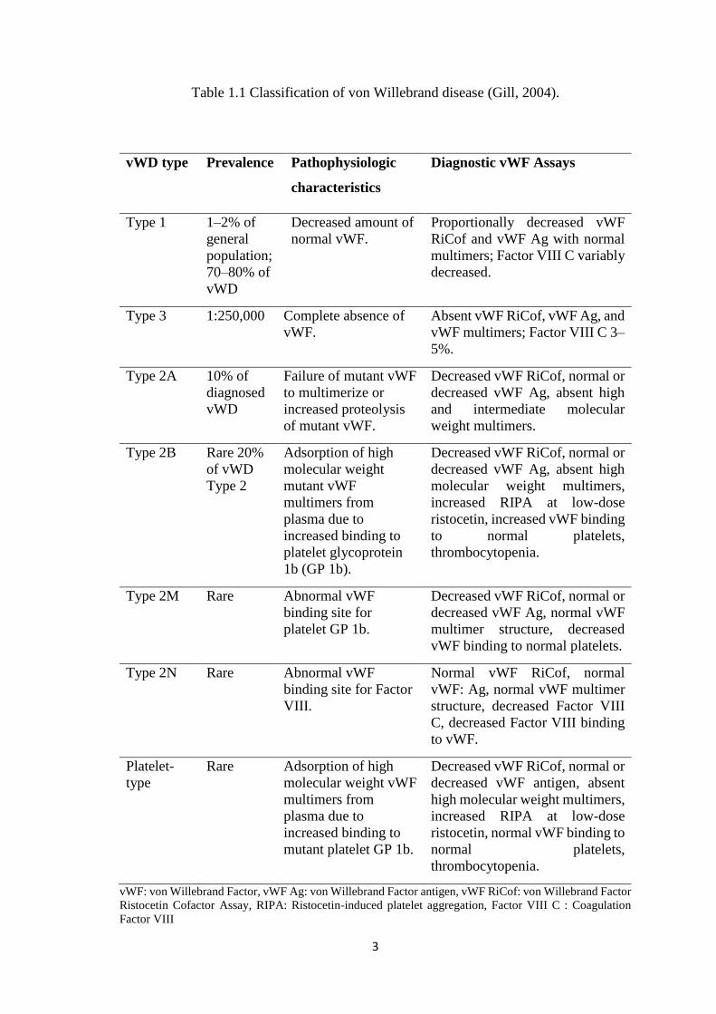
3
Table 1.1 Classification of von Willebrand disease (Gill, 2004).
vWD type Prevalence Pathophysiologic
characteristics
Diagnostic vWF Assays
Type 1 1–2% of
general
population;
70–80% of
vWD
Decreased amount of
normal vWF.
Proportionally decreased vWF
RiCof and vWF Ag with normal
multimers; Factor VIII C variably
decreased.
Type 3 1:250,000 Complete absence of
vWF.
Absent vWF RiCof, vWF Ag, and
vWF multimers; Factor VIII C 3–
5%.
Type 2A 10% of
diagnosed
vWD
Failure of mutant vWF
to multimerize or
increased proteolysis
of mutant vWF.
Decreased vWF RiCof, normal or
decreased vWF Ag, absent high
and intermediate molecular
weight multimers.
Type 2B Rare 20%
of vWD
Type 2
Adsorption of high
molecular weight
mutant vWF
multimers from
plasma due to
increased binding to
platelet glycoprotein
1b (GP 1b).
Decreased vWF RiCof, normal or
decreased vWF Ag, absent high
molecular weight multimers,
increased RIPA at low-dose
ristocetin, increased vWF binding
to normal platelets,
thrombocytopenia.
Type 2M Rare Abnormal vWF
binding site for
platelet GP 1b.
Decreased vWF RiCof, normal or
decreased vWF Ag, normal vWF
multimer structure, decreased
vWF binding to normal platelets.
Type 2N Rare Abnormal vWF
binding site for Factor
VIII.
Normal vWF RiCof, normal
vWF: Ag, normal vWF multimer
structure, decreased Factor VIII
C, decreased Factor VIII binding
to vWF.
Platelet-
type
Rare Adsorption of high
molecular weight vWF
multimers from
plasma due to
increased binding to
mutant platelet GP 1b.
Decreased vWF RiCof, normal or
decreased vWF antigen, absent
high molecular weight multimers,
increased RIPA at low-dose
ristocetin, normal vWF binding to
normal platelets,
thrombocytopenia.
vWF: von Willebrand Factor, vWF Ag: von Willebrand Factor antigen, vWF RiCof: von Willebrand Factor
Ristocetin Cofactor Assay, RIPA: Ristocetin-induced platelet aggregation, Factor VIII C : Coagulation
Factor VIII
4
The heterogeneous of the disease presentations and the laboratory findings which may
overlap with normal subjects, make it challenging for the clinicians to establish the
diagnosis of vWD. The molecular study may correspond to the specific variants, but has
a wide range of genetic mechanisms (Fernández and de Alarcón, 2014).
Currently, diagnosis of vWD is based on an array of laboratory tests (vWF antigen, Factor
VIII, vWF Ristocetin Cofactor Assay and VwF Collagen Binding Assay) together with
the history of increased bleeding tendency, which usually is also present in the family
members. The blood investigation should be carried out in a dedicated laboratory that is
qualified to perform all the tests correctly and providing the patients with a balanced view
of their bleeding risks. In Malaysia, the National Blood Centre in Kuala Lumpur is one
of the accredited laboratories to perform all of these tests.
Family and patient’s bleeding history are very important in determining the diagnosis of
vWD and serve better prediction of future bleeding than depending only on plasma vWF
level. Further testing on subjects with the vWF level of 30 to 50 IU/dL will not identify
the clinically significant patients whereas testing on the bleeding patients will identify
only the relatively small number of patients with clinical severity of vWD Type 1, 2 and
3, and larger numbers with a moderately low vWF level. (Sadler, 2009). However, the
high prevalence of mild bleeding symptoms even in a normal individual requires the
usage of a standardized questionnaire and bleeding score for the identification of patients
who require further laboratory evaluation for vWD (Rodeghiero et al., 2005). In the
current study, we used the standardized questionnaire extracted from Guidelines from the
National Heart, Lungs, And Blood Institute (NHLBI) 2008 (Nichols et al., 2008) as a
screening tool to exclude any bleeding tendencies in donors and their family members.
5
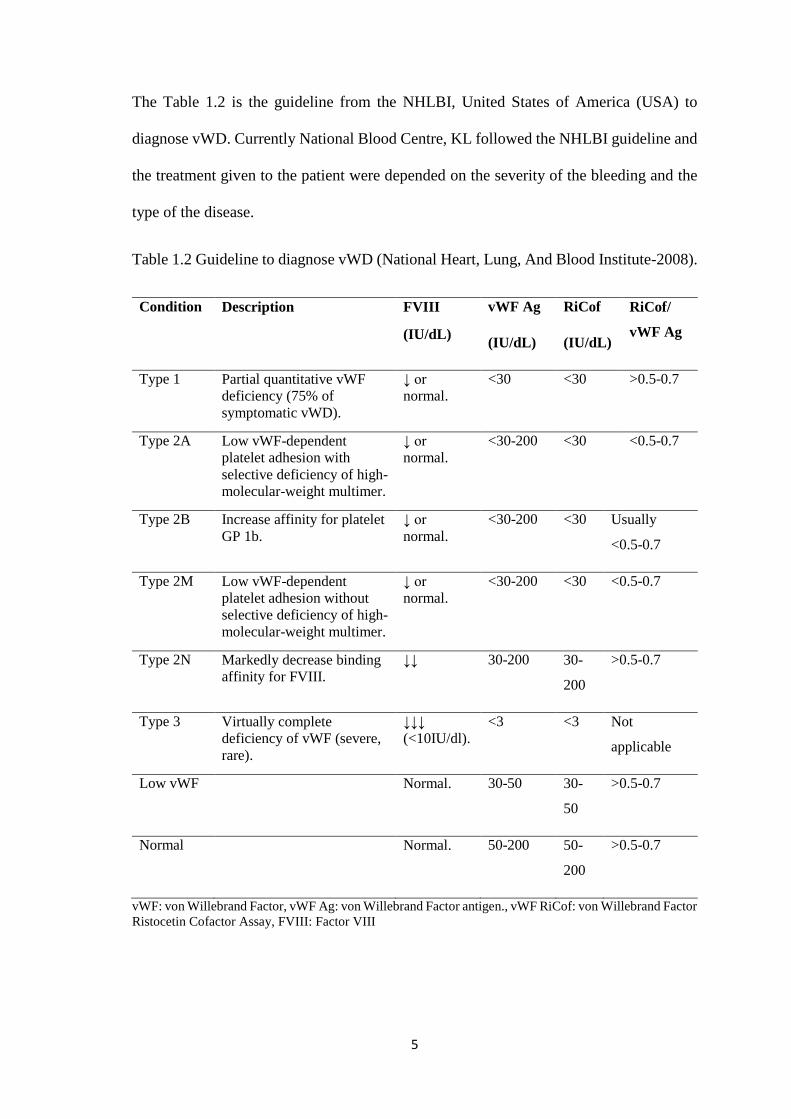
The Table 1.2 is the guideline from the NHLBI, United States of America (USA) to
diagnose vWD. Currently National Blood Centre, KL followed the NHLBI guideline and
the treatment given to the patient were depended on the severity of the bleeding and the
type of the disease.
Table 1.2 Guideline to diagnose vWD (National Heart, Lung, And Blood Institute-2008).
Condition Description FVIII
(IU/dL)
vWF Ag
(IU/dL)
RiCof
(IU/dL)
RiCof/
vWF Ag
Type 1 Partial quantitative vWF
deficiency (75% of
symptomatic vWD).
↓ or
normal.
<30 <30 >0.5-0.7
Type 2A Low vWF-dependent
platelet adhesion with
selective deficiency of high-
molecular-weight multimer.
↓ or
normal.
<30-200 <30 <0.5-0.7
Type 2B Increase affinity for platelet
GP 1b.
↓ or
normal.
<30-200 <30 Usually
<0.5-0.7
Type 2M Low vWF-dependent
platelet adhesion without
selective deficiency of high-
molecular-weight multimer.
↓ or
normal.
<30-200 <30 <0.5-0.7
Type 2N Markedly decrease binding
affinity for FVIII.
↓↓ 30-200 30-
200
>0.5-0.7
Type 3 Virtually complete
deficiency of vWF (severe,
rare).
↓↓↓
(<10IU/dl).
<3 <3 Not
applicable
Low vWF Normal. 30-50 30-
50
>0.5-0.7
Normal Normal. 50-200 50-
200
>0.5-0.7
vWF: von Willebrand Factor, vWF Ag: von Willebrand Factor antigen., vWF RiCof: von Willebrand Factor
Ristocetin Cofactor Assay, FVIII: Factor VIII
6
Currently, the National Blood Centre, KL followed the NHLBI guidelines and the
treatment given to the patients are dependent on the severity of the bleeding and the types
of the disease.
The NHLBI chose the vWF antigen and RiCof of less than 30 IU/dL as the level for a
definitive diagnosis of vWD because of the high prevalence of the O blood group in the
United States of America (USA) which is associated with low vWF antigen levels, the
absence of genetic abnormality in patients with mild to moderate level of RiCof and the
presence of a significant number of individuals with a bleeding history but no underlying
disease detected.
The absent of vWF (vWD Type 3) and many qualitative defects (vWD Type 2) are
straightforward diagnosis but not in vWD Type 1 due to a broader distribution of normal
vWF level, the high prevalence of mild bleeding symptoms even in a normal person and
the weak relationship between vWF level and bleeding event. Therefore, none of the vWF
level can isolate patients into a group with clearly different clinical features (Sadler,
2003). Consequently, many vWD Type 1 patients do not have the specific bleeding
disease at all which limits the value of the diagnosis. False positive diagnosis of vWD
Type 1 also causes many patients to be subjected to risky, expensive and ineffective
treatments, while the actual causes of symptoms are overlooked and untreated. Many
patients had to change their lifestyles due to fear of bleeding and some have even been
denied of insurance coverage.
Recent studies of vWD Type 1 patients have defined several pathophysiologic
mechanisms that determine the vWF plasma concentration but the relationship between
the vWF levels and the probability of bleeding remains inconclusive and the difference
between ‘normal’ and ‘low’ is undistinguishable. These problems might be resolved by
7
an epidemiologic approach to vWF and other risk factors for the bleeding event (Sadler,
2009).
The treatment of vWD is dependent on the severity of the bleeding symptoms, the type
of the disease and the type of surgical procedure that the patient plans to proceed with.
The treatments include drugs to induce the releasing of vWF and FVIII into the blood
circulation to prevent lysis of the blood clots, to control heavy menstrual bleeding in
women and to replace the vWF.
While inherited vWD is the commonest inherited bleeding disorder, acquired vWD is a
rare bleeding disorder. It is associated with other diseases such as lymphoproliferative or
myeloproliferative disorders, malignancies, immunologic diseases, various congenital
cardiac defects or other drug related ailements. The production and function of vWF in
this type of diseases are normal, but the factor is rapidly eliminated from circulation by
autoantibodies, absorption of the vWF onto the cancer cell clones or loss of high-
molecular-weight vWF multimers under the high shear stress (Federici, 2006).
1.1.3 von Willebrand disease: Epidemiology
Previously, vWD is classified as a rare disease (Office of Rare Diseases of the National
Institutes of Health, US). However, Rodeghiero (1987) revealed that the prevalence of
the Italian population with vWD was between 0.57%-1.15% (Rodeghiero et al., 1987).
Meanwhile, Werner (1993) showed that the prevalence among the USA population was
at 1.3% (Werner et al., 1993). In 2010, Bowman reported that there were relatively low
prevalence of medically significant bleeding patients attending primary health clinics and
a low prevalence of symptomatic vWD in the primary care setting which was at least 1 in
1000. They suggested that further investigations needed to be carried out to investigate
the discrepancies of these prevalent findings (Bowman et al., 2010).
8
The disease shows no geographical nor ethnic preferences. As an autosomal disorder,
both genders inherit the mutant vWF alleles equally, but women show frequent bleeding
symptoms by almost 2:1 probably because of the excessive per vaginal bleeding during
their reproductive age (Lillicrap, 2013).
Data on the vWD epidemiology in developing countries are very limited. Although there
is no accurate data on the estimation of vWD prevalence in those countries, the available
data suggests that the diagnosed cases were less than the actual number, accounting for
only 6% to 13% of patients with hereditary bleeding disorders. The number with severe
diseases tend to be much higher, particularly in certain parts of the world where
consanguineous marriage is common (Srivastava and Rodeghiero, 2005). In Malaysia,
only 0.002% (n=464) of the diagnosed cases were reported among the 28 million of the
population (World Federation of Hemophilia, 2010). Most of the patients were diagnosed
to have vWD Type 1 (77.2%) (Periayah et al., 2016). This corresponds with the estimated
prevalence in some countries where the symptomatic patients that was seen at
haemostasis clinics ranging from 23 to 110 per million population (0.0023–0.01%)
(Nichols et al., 2008). In another study conducted by Hassan (2012), it is found that vWD
were common among menorrhagia patients who attended the gynaecology clinic in
Hospital Universiti Sains Malaysia, Kelantan, which accounted up to 13.3%. They
suggested that vWD testing should be provided to complete the diagnostic work-up for
menorrhagia (Hassan et al., 2012). It was similar with other studies which reported that
the frequency of vWD in menorrhagia ranges from 5% to 20% (Kujovich, 2005).
1.1.4 Malaysia is a multiracial country
An interesting fact about the Malaysian society is the diversity of its ethnic composition.
It is the result of large population movements in the nineteenth and early twentieth
9
centuries (Schafgans, 1998). Currently, Malaysia has a population of 31.2 million,
consisting of 15.3 million of women and 16.4 million of men where 68.6% of them are
Malays, 23.4% are Chinese, and 7% are Indians and 1% of other ethnic groups
(https://www.statistics.gov.my/). Therefore, we have decided to perform the study among
the Malays because it represents the majority of the country’s population (68.6%) and
the majority (63.0%) of the vWD patients diagnosed in Malaysia (Periayah et al., 2016).
In the National Blood Centre, KL the majority (49.5%) of donors were also Malays
(Blood Bank Information System [BBIS] National Blood Centre Kuala Lumpur 2015).
The distribution of the ABO blood group among our blood donors, have been reported by
Musa in 2012. They revealed about 34.5 % of Malays were in the O group, 27.5% were
in group B, 30.5% were in group A and 7.5% were in group AB (Musa et al., 2012). In
addition, Manoharan (2013) also reported that 39% of Malays were in group O, 32% were
in group B, 23% were in group A and 13% were in group AB as the subjects of the study
were students at Asia Metropolitan University of Malaysia. In the current study, we
included the blood group O, A and B of Malay donors, but excluded group AB as it is
less prevalent among our population (Manoharan et al., 2013; Musa et al., 2012)
1.1.5 Ethnic variation in von Willebrand Factor
In previous population studies, they found that vWF were higher among African
Americans than Caucasians. These racial differences in vWF further complicated the
issues surrounding a diagnosis of vWD (Miller et al., 2001). Another study performed in
South Africa with a distinct ethnic mixture of Africans, Caucasians and Indians reported
that the African Americans had significantly higher vWF antigen and Factor VIII levels
when compared to others. However, they found that the Indians had comparable levels of
vWF with Caucasians. They suggested that the influence of ethnicity on the vWF levels
10
should also be considered in the clinical and laboratory evaluation of vWD (Sukhu et al.,
2003). This is contradictory to the earlier study conducted by Werner (1993), who found
that there were no significant differences in vWF activity via ethnicity but they only
carried out the study focusing on the age group of paediatric subjects (Werner et al.,
1993). Later, Johnsen (2013) in the NHLBI exome sequencing project found that some
vWF missensed variants, which were commonly or uncommonly present among certain
ethnicity had contributed to the phenotypic variation of the vWF and Factor VIII (Johnsen
et al., 2013).
Even when they had different ethnicity than Caucasians, Rojnuckarin (2005) reported that
the vWF profiles among Thais were comparable with the earlier reports in the USA
population (Rojnuckarin et al., 2005).
1.1.6 von Willebrand Factor level influence by ABO group
ABO blood groups greatly influence the plasma level of vWF as the O blood group
subjects have a lower vWF levels compared to non O blood group (Franchini et al., 2007).
Rojnuckarin (2005) found that AB blood group subjects had the highest level of vWF
followed by B, A and O (Rojnuckarin et al., 2005). The ABO blood group appeared to
strongly influence the clearance of vWF, but not its protein synthesis or its release from
EC (Gallinaro et al., 2008). In a study conducted by Orstavik (1985) revealed that Factor
VIII was dependent on vWF antigen levels and 30% of the genetic variance of vWF
antigen were due to the effect of ABO blood group (Orstavik et al., 1985). They also
found that the concentration of plasma vWF and Factor VIII were the lowest in O, higher
in A2, and highest in A1 and B group subjects (Orstavik et al., 1985). For the diagnosis
of vWD among the different ABO blood group, the National Heart, Lung, and Blood
Institute chose the less than 30 IU/dL plasma vWF level for a definitive diagnosis of vWD
11
because there is a high prevalence of O blood group in the USA which is associated with
‘low’ vWF levels. They did not use different reference ranges for diagnosing vWD among
group O individuals. In this current study, we focus on blood group A, B and O as AB
group is less prevalence among our population, our blood donors and Malay donors at
NBCKL.
1.2 List of definitions
1.2.1 Body mass index: Is a measure of body fat based on height and weight that
applies to adult men and women. BMI categories:
Underweight = <18.5
Normal weight = 18.5–24.9
Overweight = 25–29.9
Obesity = 30 or greater
(http://www.nhlbi.nih.gov/)
1.2.2 vWF antigen: Test to measures the quantity of a vWF in the circulation.
1.2.3 ADAMTS13: A Disintegrin like And Metalloprotease domain (reprolysin
type) with ThromboSpondin type 1 motif, member 13]. A
plasma protein that cleaves multimeric vWF.
1.2.3 RiCof: Ristocetin Cofactor Activity; test to evaluate the capability
of vWF to bind platelet glycoprotein 1b (GP 1b) and
promote platelet plug formation.
1.2.4 CBA: Collagen Binding Assay; test to evaluate the capability of
vWF to bind to collagen, mimicking the interaction with
the subendothelial matrix at the site of vascular injury.
(http://practical-haemostssis.com/)

12
1.2.5 Rare disease: Disease that affects less than 200,000 people in the US
population.
(https://www.genome.gov/)
1.3 Research Justification and Benefits
vWD is the commonest hereditary bleeding tendency disorder encountered in almost 1%
of the worldwide population (Kouides and Kreuz, 2009). However, the diagnosis of vWD
Type 1 is very challenging due to the broad distribution of normal vWF levels, the high
prevalence of mild bleeding symptoms even in the normal population and the weak
relationship between vWF levels and the bleeding events.
In the National Blood Centre, Kuala Lumpur (NBCKL), the interpretation of laboratory
results for vWF profiles follows the guidelines from the NHLBI. However, the normal
range that is being used by the NHLBI was established according to the Caucasians
population. There are several studies found that the levels of vWF and Factor VIII were
significantly higher in blacks compared to the white population (Fleming, 2003;
Gomperts et al., 1976; Kadir et al., 1999; Miller et al., 2001) proposed that the reference
ranges of laboratory haemostasis investigations, cannot be used as a reference across the
world as the range is largely based on the results obtained from the Caucasians.
Up to the best of our knowledge, the normal reference range of the vWF profiles in the
Malaysian population is not established yet. This study will serve as preliminary data of
the vWF profiles among the Malay population in our country. If the data obtained from
this study is largely deviated from the NHLBI 2008 data, we then have to conduct a larger
scale population-based study in the future to obtain our own normal vWF profiles
reference range. It may include the 3 major races in Malaysia (Malays, Chinese and
Indians), including all ABO blood groups.
13
There are limited studies on vWF levels among the Asian compared to the western
population. It is hope that this study will serve as a preliminary data on vWF antigen and
activity levels in the healthy Malay population in Malaysia.
1.4 Research Objectives
1.4.1 General Objectives
To study von Willebrand Factor profiles of the different ABO blood group among Malay
donors at NBCKL in 2015.
1.4.2 Specific Objectives
1) To determine the von Willebrand Factor profiles (Factor VIII, von Willebrand
antigen, RiCof and Collagen Binding Assay) of the different ABO blood group among
Malay donors at NBCKL in 2015.
2) To compare the von Willebrand Factor profiles (Factor VIII, von Willebrand
antigen, RiCof and Collagen Binding Assay) with the different ABO blood group among
Malay donors at NBCKL in 2015.
3) To assess the influence of demographic data (gender, age group and BMI) and smoking
habit with the von Willebrand Factor profiles among Malay donors at NBCKL in 2015.
1.5 Research Hypothesis
1) There are differences in the von Willebrand Factor profiles in different ABO
blood groups among Malay donors at NBCKL.
2) There are association between demographic data (gender, age group and BMI)
and smoking habit, and the von Willebrand Factor profiles among Malay donors
at NBCKL.
14
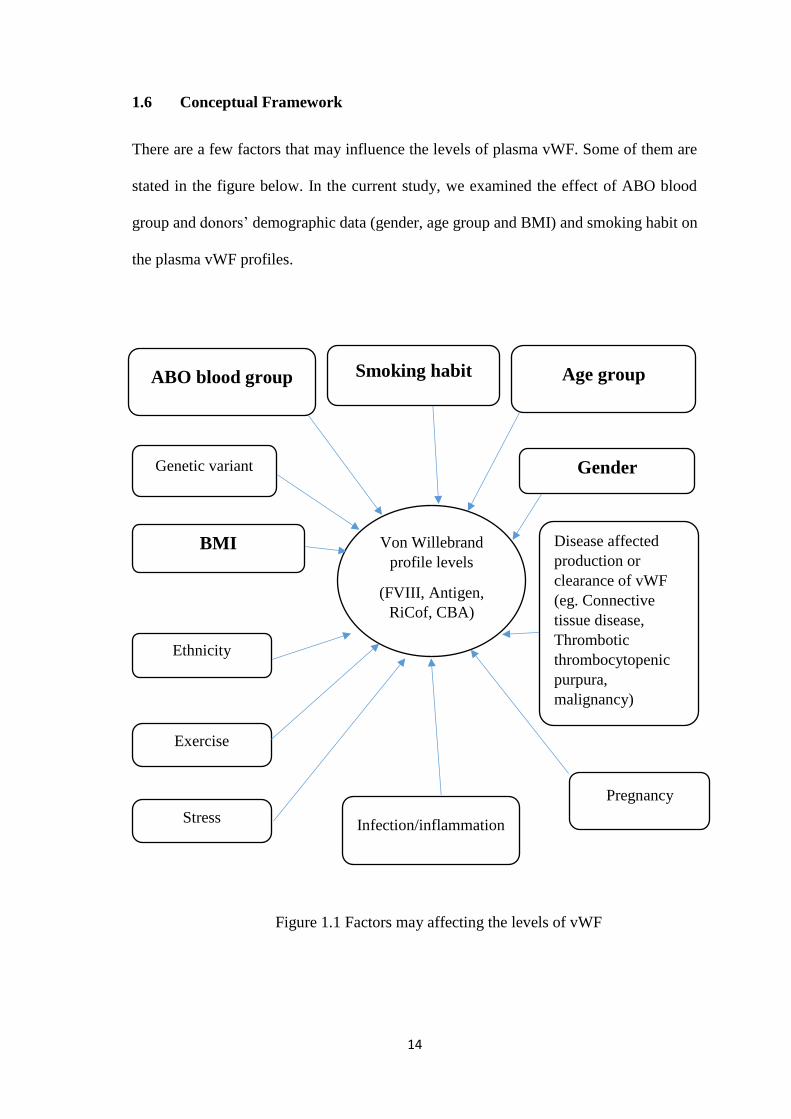
1.6 Conceptual Framework
There are a few factors that may influence the levels of plasma vWF. Some of them are
stated in the figure below. In the current study, we examined the effect of ABO blood
group and donors’ demographic data (gender, age group and BMI) and smoking habit on
the plasma vWF profiles.
Figure 1.1 Factors may affecting the levels of vWF
Von Willebrand
profile levels
(FVIII, Antigen,
RiCof, CBA)
Smoking habit ABO blood group
BMI
Gender
Age group
Genetic variant
Disease affected
production or
clearance of vWF
(eg. Connective
tissue disease,
Thrombotic
thrombocytopenic
purpura,
malignancy)
Infection/inflammation
Ethnicity
Exercise
Pregnancy
Stress
15
CHAPTER 2
LITERATURE REVIEW
2.1 The Malays
The Malays is the race of people who live mainly in Peninsular Malaysia and a portion
of the adjacent island of Southeast Asia, including the coast of Borneo, the east coast of
Sumatra and small islands that lie between these areas (http://sabrizain.org/malaya/).
Based on their migrations a few centuries ago, there are various sub-ethnic groups in the
Malay population present currently, which are believed to have different ancestral origins.
The major sub-ethnics are Melayu Jawa, Melayu Minang, Melayu Bugis, Melayu
Kelantan and Melayu Kedah (Hatin et al., 2011). Some of the present day Malays are
mixed with modern Chinese, Indian, Arab and Thai blood (http://sabrizain.org/malaya/).
Most of the Malays in Malaysia are practising Islam as their religion, speak Malay
language and practice Malay customs (adat) and cultures.
The Malays is one of the unique ethnicity in the world with different genetic variances
compared to others. In the fields of population genetics and forensics, the human X
chromosome has been focused on by many researchers in recent years. Samejima (2012)
has carried out a genetic study on the X-chromosomal short tandem repeats (X- STRs)
and found 12 X-STRs in the Malay population that differed from East Asian, European,
or African populations (Samejima et al., 2012). In the genetic study for drug metabolism,
Teh (2001) found that the genetic polymorphism of CYP2D6 in Malays was different
from the Chinese and Far Eastern races. These variances result in ethnic differences in
the metabolism of CYP2D6 drugs (Teh et al., 2001). The genetics differences are also
present among sub-ethnic group as previously reported by Hatin (2014) and that there is
a genetic mixture among sub-ethnic Malays in Peninsular Malaysia (Hatin et al., 2011).

16
These genetic variants may increase risk among the Malays to inherit certain diseases or
resistance to certain drugs.
2.2 Malays and thromboembolic and bleeding event
Up to the best of our knowledge, there are very limited published studies on
thromboembolic or bleeding event among the Malays. Nawawi (2002) reported that the
prevalence of the coronary risk factors among rural Malays in Malaysia was high
according to the Global Risk Assessment. Apart from genetic predisposition, the high
prevalence was probably due to the rapid socioeconomic development at the rural areas
(Nawawi et al., 2002). Loo (2012) had described a few studies on the prevalence of stroke
(bleeding or ischaemic) in different states in Malaysia where they found about 86·1% of
the patients were Malays and 13·9% were Chinese in Kelantan whereby 55.7% were
Chinese, 28.9% were Malays and 14.2% were Indians in Pulau Pinang (Loo and Gan,
2012). This may reflect upon the local population as many Malays reside in Kelantan
compared to the majority of Chinese located in Pulau Pinang especially on the island.
However, these also may reflect the differences of genetic among the ethnicity which
results in different risks of thromboembolic or bleeding incident. In 2004, Kandasami had
conducted a study to look into the prevalence of bleeding events (peptic ulcer disease)
among different ethnicity in Malaysia and found over presented symptoms in the Chinese
but similar to the ethnic distribution in the Malays and Indians (Kandasami et al., 2004).
2.3 Malays and von Willebrand disease
Apart from collecting, processing and distributing of blood and its components to
hospitals, the National Blood Centre, Kuala Lumpur also provides expert medical
services and groundwork for research in the field of haematology. Based on the recent
update from the National Blood Centre, the effect of vWD was quite low among
17
Malaysians due to under reporting even after many conferences, campaigns, awareness
and colloquia that have been organized. Only 545 cases were reported from the year 1979
to 2013 (Periayah et al., 2016). The prevalence was highest in the Malays (63%) followed
by Indians (15.2%) and lowest in Chinese (5.5%) Most of the patients were diagnosed to
have vWD Type 1 and about 40% of them were males and 60% were females (Periayah
et al., 2016).
2.4 Diagnosis of von Willebrand disease
vWD is a heterogeneous disorder and a very complex disease (Sadler and Gralnick, 1994).
It is very difficult to establish the diagnosis of vWD especially in Type 1 disease due to
the wide distribution of the normal vWF level, high prevalence of mild bleeding
symptoms even in a normal population and the weak relationship between vWF levels
and bleeding events. In Type 1, the vWF level is not obviously low, but usually near to
the lower end of the normal vWF range. The wide distribution of vWF levels in a normal
population, makes the situation more complicated. Almost 95% of the plasma vWF levels
lie between 50 to 200 IU/dL among the 300 million of the USA population and around
7.5 million people who had the vWF levels less than 50 IU/dL would be at risk for the
diagnosis of vWD Type 1 (Nichols et al., 2008). The cut-off level of 50 IU/dL was chosen
as normal because less than that showed increased bleeding risk with a relative risk of
2.0-3.9 (Sadler, 2003).
Nitu-Whalley (2000) had conducted a retrospective study to investigate the difficulties in
making a diagnosis of vWD Type 1 in one of the Hemophilia Centre in United Kingdom
(Nitu-Whalley et al., 2000). They found that among previously diagnosed vWD Type 1,
41% of them were in group O and had between 30-50 IU/dL of vWF levels, with or
without a history of increase in bleeding tendencies. Those groups of patients might
18
require reclassification as ‘not vWD’ and searching for alternative diagnosis for the
bleeding symptoms might be needed.
In view of the bleeding history, the standardized questionnaire is extremely important to
screen bleeding risks among the population. Rodeghiero (2005) proposed the use of a
standardized questionnaire and bleeding score to identify the patients who need
laboratory evaluation for the vWD (Rodeghiero et al., 2005). Friberg (2006) observed
that about 23% of Swedish girls reported 3 or more haemorrhagic symptoms when using
a self-reported questionnaire (Friberg et al., 2006), whereas Rodeghiero (2005) revealed
that by using a standardized questionnaire, they found less than 1% of normal control had
3 or more of the symptoms (Rodeghiero et al., 2005).
Apart from the bleeding history, the diagnosis of vWD is also based on an array of
laboratory tests to investigate the amount and function of the protein. The tests looked at
the amount of vWF (vWF antigen) and determining the activity of the protein; to carry
and stabilize the Factor VIII (Factor VIII Assay), to bind to the glycoprotein 1b on the
platelet surface (vWF RiCof) and to bind to the collagen (vWF CBA). The primary site
of platelet binding is in the A1 domain of vWF, whereas collagen binding is in the A3
domain. The interaction of platelets with vWF is forced in vivo by shear stress, which
causes a conformational change of the protein and allows its binding to the platelet
glycoprotein 1b (Sadler, 1998). In vitro, the interaction is aggravated by the antibiotic
ristocetin, in the lack of shear stress condition (Scott et al., 1991).
Similar to the vWF RiCof, vWF CBA relies on the size of the vWF multimeric in which
the larger size will bind more avidly than the smaller forms. However, Dean (2000)
conducted a study to compare the vWF CBA and vWF RiCof and found that the vWF
CBA was most helpful in the classification of vWD Type 2 variants, using a low vWF
19
activity: vWF antigen ratio (Dean et al., 2000). Similarly, Casonato (2001) observed that
in disease Type 2A and 2B, even when both vWF CBA and vWF RiCof were decreased,
the vWF CBA was more constant and they suggested to include vWF CBA to the test
panel for diagnosis of vWD (Casonato et al., 2001). A ratio of vWF CBA: vWF antigen
of less than 0.5 is consistent with types 2A and 2B disease, whereas a normal ratio (0.5
or more) are associated with types 2M and 2N disease (Popov et al., 2006). Flood (2013)
found that vWF CBA provides a sensitive method to capture the variant of vWD
especially by using the lowest (0.6) cut-off of vWF CBA: vWF antigen ratio (Flood et
al., 2013).
In early 2000, Favaloro reported that the ability to discriminate the subtype of vWD was
dependant on the type of collagen used in CBA in which Type III or a mixture of Type
I/III showed highest sensitivity (Favaloro et al., 2000). In the current study, we used
collagen Type III to accurately discriminate the subtype if found. Riddell (2002) reported
that even if vWF CBA is a sensitive method to detect functional variants related to the
loss of high molecular weight multimers, it’s incapable to detect defective platelet-
binding vWD variants in the presence of normal high molecular weight multimers but
vWF RiCof does. They concluded that the vWF CBA should be used in association with
vWF RiCof rather than as a replacement for it (Riddell et al., 2002). A guideline from the
UK Hemophilia Centre Doctors’ Organization, 2014 also recommended to use both vWF
RiCof and CBA to increase the ability to detect Type 2 variants and clear definition of
vWD Type 1 (Laffan, M.A et al., 2014).
An alternative to vWF RiCof, some laboratories offer vWF-Activity (vWF-Ac) to assess
the function of vWF to bind to the platelet receptors without the presence of ristocetin.
Geisen (2014) in a comparison study between vWF activity and vWF RiCof on
20
aggregometer showed a good correlation between the two methods (Geisen et al., 2014).
Other laboratory tests such as Ristocetin Induced Platelet Aggregation (RIPA) and
multimeric analysis are also essential in order to differentiate the different subtypes of
vWD.
Apart from that, molecular study is also one of the methods to distinguish vWD and its
variants. vWD is highly heterogeneous due to the molecular mechanisms that produce
various clinical presentation and laboratory findings. However, the requirement for the
study in vWD is variable because the usefulness of genetic testing varies for different
vWD subtypes. In case of vWD Type 1, the picture of the disease is not really clear as the
causative molecular defect is unidentified in a significant number of cases, and even in
those cases in which the causative mutation is known. The association of molecular
pathology is not necessarily understood (Keeney et al., 2008). Although genetic analysis
is not mandatory to diagnose vWD or to define a classification type, it may be useful in
isolated situations (Favaloro et al., 2010).
Nowadays, various tests are available to diagnose vWD, however NHBLI in their
guidelines suggested carrying out three initial tests namely vWF antigen, vWF RiCof and
Factor VIII (Nichols et al., 2008). If one or more results is noted to be abnormal, further
test such as vWF CBA, RIPA, Factor VIII binding, platelet vWF studies, multimer
distribution, ratio of vWF RiCof: vWF antigen and molecular study are recommended to
distinguish the type of vWD and its variants. In NBCKL, vWD patients are normally
classified based on the 3 essential laboratory investigations namely Factor VIII, vWF
antigen and vWF CBA (Periayah et al., 2016). However, in the current study, we
performed a full investigation panel of vWF profiles including vWF RiCof.