
Experimental evaluation of protection and immunogenicity ...

Research ArticlePreliminary Evaluation of the Safety and Immunogenicity of anAntimalarial Vaccine Candidate Modified Peptide (IMPIPS)Mixture in a Murine Model
Jennifer Lambraño,1,2Hernando Curtidor,1 Catalina Avendaño ,3Diana Díaz-Arévalo ,1,4
Leonardo Roa ,3Magnolia Vanegas,1Manuel E. Patarroyo,1,5 andManuel A. Patarroyo 1,4
1Fundación Instituto de Inmunología de Colombia (FIDIC), Bogotá, Colombia2Master’s Programme in Biochemistry, Medical School, Universidad Nacional de Colombia, Bogotá, Colombia3Faculty of Animal Science, Universidad de Ciencias Aplicadas y Ambientales (U.D.C.A), Bogotá, Colombia4School of Medicine and Health Sciences, Universidad del Rosario, Bogotá, Colombia5Pathology Department, Medical School, Universidad Nacional de Colombia, Bogotá, Colombia
Correspondence should be addressed to Manuel A. Patarroyo; [email protected]
Received 3 October 2019; Accepted 3 December 2019; Published 30 December 2019
Academic Editor: Pedro A. Reche
Copyright © 2019 Jennifer Lambraño et al. This is an open access article distributed under the Creative Commons AttributionLicense, which permits unrestricted use, distribution, and reproduction in anymedium, provided the original work is properly cited.
Malaria continues being a high-impact disease regarding public health worldwide; the WHO report for malaria in 2018 estimatedthat ~219 million cases occurred in 2017, mostly caused by the parasite Plasmodium falciparum. The disease cost the lives of morethan 400,000 people, mainly in Africa. In spite of great efforts aimed at developing better prevention (i.e., a highly effective vaccine),diagnosis, and treatment methods for malaria, no efficient solution to this disease has been advanced to date. The FundaciónInstituto de Inmunología de Colombia (FIDIC) has been developing studies aimed at furthering the search for vaccinecandidates for controlling P. falciparum malaria. However, vaccine development involves safety and immunogenicity studiesregarding their formulation in animal models before proceeding to clinical studies. The present work has thus been aimed atevaluating the safety and immunogenicity of a mixture of 23 chemically synthesised, modified peptides (immune protection-inducing protein structure (IMPIPS)) derived from different P. falciparum proteins. Single and repeat dose assays were thusused with male and female BALB/c mice which were immunised with the IMPIPS mixture. It was found that single and repeatdose immunisation with the IMPIPS mixture was safe, both locally and systemically. It was observed that the antibodies sostimulated recognised the parasite’s native proteins and inhibited merozoite invasion of red blood cells in vitro when evaluatingthe humoral immune response induced by the IMPIPS mixture. Such results suggested that the IMPIPS peptide mixture couldbe a safe candidate to be tested during the next stage involved in developing an antimalarial vaccine, evaluating local safety,immunogenicity, and protection in a nonhuman primate model.
1. Introduction
Malaria represents one of the greatest public health problemsworldwide. According to the World Health Organization(WHO), ~219 million new malaria-related cases occurredin 2017 accompanied by ~435,000 deaths. The African conti-nent was the most affected region in the world (92% of casesand 93% of deaths) [1]. The Global Technical Strategy forMalaria 2016-2030 (WHO) has suggested reducing malarial
incidence and mortality by at least 90% and eliminating itin at least 35 countries by 2030 through prevention, diagno-sis, and treatment strategies [2].
No significant progress has been observed to date regard-ing the reduction of cases of malaria worldwide despite thediffering strategies used for combating this disease (usinginsecticide-impregnated mosquito nets for controlling thevector, chemoprophylaxis, and case management) [1, 2].The most recurrent problem is concerned with the increase
HindawiJournal of Immunology ResearchVolume 2019, Article ID 3832513, 12 pageshttps://doi.org/10.1155/2019/3832513

in strains which are resistant to antimalarial drugs andinsecticide-resistant mosquitoes; this has necessitated thedevelopment and combined use of new control and preven-tion methods, especially a vaccine having high protectioncapability as time elapses [1, 3].
The Fundación Instituto de Inmunología de Colombia(FIDIC) has thoroughly demonstrated the feasibility of achemically synthesised, multistage, multiantigen, minimumsubunit-based (~20 amino acid-long peptide) vaccine by fol-lowing a completely functional approach [4, 5]. This has ledto ascertaining that peptides derived from the main proteinsparticipating in merozoite (Mrz) invasion of RBCs [6] specif-ically bind to human RBCs and that sporozoites (Spz) invad-ing hepatic cells [7, 8] bind to the HepG2 hepatocellularcarcinoma cell line [9–12].
Immunogenicity and protection assays in Aotusmonkeyshave shown that high activity binding peptides (HABPs) [12]having a conserved sequence (cHABP) have not induced animmune response, suggesting that despite the importance oftheir biological role, they are immunologically silent [4, 5].By contrast, HABPs having a variable sequence (vHABPs)have induced a nonprotective immune response (or only ashort-term one) [4, 13], an immune evasion mechanism forthese sequences (smokescreens distracting the immuneresponse) [14–16]. However, when some cHABP residues[17, 18] have been replaced by amino acids (aa) having simi-lar mass and volume, but different polarity, modified ana-logues (mHABPs) have been seen to induce a protectiveimmune response in Aotus monkeys against experimentalchallenge [5, 13, 19–22].
Nuclear magnetic resonance (NMR) and in silico struc-tural binding studies have shown that mHABPs havingpolyproline II (PPIIL) helix structures [23, 24] can bindto HLA-DRβ1∗ molecules covering most MHC-II allelevariants [21, 25, 26] and have greater interaction withthe T cell receptor (TCR). This would suggest the stableformation of the MHC-II-mHABP-TCR trimer complexand thus the capability for inducing a protective immuneresponse [27–31]. Protection-inducing mHABPs have thusbeen called Pf immune protection-inducing protein struc-tures (IMPIPS) in view of their close structure-protectionrelationship [32, 33].
This study thus used a murine model for evaluating theimmunogenicity, local toxicity, and systemic toxicity [34–38]of a mixture of 23 IMPIPS. These were derived from the mainP. falciparum Spz (circumsporozoite protein 1 (CSP-1),thrombospondin-related anonymous protein (TRAP), spo-rozoite threonine and asparagine-rich protein (STARP),sporozoite microneme proteins essential for cell traversal(SPECT-1 and SPECT-2), cell-traversal protein for ooki-netes and sporozoites (CelTOS), and sporozoite invasion-associated protein 1 and 2 (SIAP-1 and SIAP-2)) [9, 11,39, 40], as well as Mrz proteins (apical membrane antigen-1(AMA-1), erythrocyte-binding protein 175 (EBA-175),erythrocyte-binding protein 140 (EBA-140), serine repeatantigen (SERA-5), merozoite surface protein-1 (MSP-1),and histidine-rich protein II (HRP-II)) [10, 39, 40]. Previousstudies testing these peptides individually have shown thatthe antibodies induced were able to recognise the original
template protein when expressed as a recombinant (Supple-mentary Table 1).
2. Materials and Methods
2.1. Peptide Synthesis and Purification. Twenty-three poly-mer peptides (Table 1) were modified following previouslyreported principles [32, 33, 40, 41] to render them immuno-genic and then synthesised using solid-phase multiple pep-tide synthesis following the tert-butyloxycarbonyl (t-Boc)synthesis strategy described by Merrifield [42] and modifiedby Houghten [43]. All peptides were derived from fully con-served and functionally relevant regions of the correspondingproteins. Such a Merck-Hitachi L-6200 A chromatograph(Merck) fitted with a UV-VIS L-4250 210nm wavelengthdetector was used for determining synthesised peptide purityby high-performance reversed-phase liquid chromatography.A Microflex mass spectrometer (Bruker Daltonics) was thenused for characterising them by matrix-assisted laser deso-rption/ionisation-time of flight (MALDI-TOF).
2.2. Animals. Forty BALB/c mice (20 males and 20 females)were used for evaluating the formulation’s safety and immu-nogenicity. The mice were aged 5 to 6 weeks when they wereimmunised, according to WHO recommendations [37]. Themice were acquired from the Universidad Nacional deColombia’s Faculty of Animal Science’s Biotherium.
2.2.1. Single Dose Local Tolerance. This assay enabled deter-mining possible inflammatory reactions at the different treat-ments’ inoculation sites [37]; this involved using 18 BALB/cmice (9 males and 9 females), following the protocols estab-lished by the regulatory authorities [34–37]. The animalswere randomly assigned to 3 groups (Table 2), each consist-ing of 3 males and 3 females, in line with the principles ofreduction and refinement [44].
The animals were immunised by subcutaneous (SC)route at the base of the tail with 100μL of the formulation.The animals were observed twice a day after they had beenimmunised for detecting changes in their behaviour, signsof disease, or toxicity. The injection site and the tissue aroundit were examined 1, 3, 24, 48, and 72 hours after immunisa-tion to ascertain the presence of erythema, oedema, eschar,and necrosis; the parameters described by Cox were usedfor evaluating their degree [45]. The animals were anesthetisedwith ketamine (80-120mg/kg) and xylazine (5-16mg/kg)by intraperitoneal (IP) route on the third day and sacri-ficed by cervical dislocation.
2.2.2. Repeat Doses. Twenty-two BALB/c 5- to 6-week-oldmice (11 males and 11 females) were randomly distributedinto three groups for evaluating possible toxic reactions pro-duced by repeat inoculations (SC) of the Pf-IMPIPS peptidemixture (30μg in total). Toxicity due to repeat doses canoccur as a result of repeat administration of a product overa specific period [35–37] (Table 3).
The animals were immunised 4 times with a 14-day inter-val as the amount of doses in an animal model must be equalto or greater than the amount of doses for clinical assays [37],
2 Journal of Immunology Research

and it would be expected that the amount of doses adminis-tered would not exceed two in clinical assays (Figure 1).
Each group was immunised with 100μL of the formula-tion on days 0, 14, 28, and 42; the formulation was adminis-tered by SC route at the base the tail. The immunisation siteswere examined 1, 3, and 24 h after each injection looking forsigns of erythema, oedema, eschar, and necrosis.
The animals were observed twice per day for evidence ofany adverse reaction to the injection or the presence of dis-ease, and a weekly physical examination was made for mon-itoring every animal’s overall state of health. Their weightand food consumption were also monitored before beginningthe immunisation protocol and after immunisation on days0, 3, and 7 and every week thereafter until day 70.
Mouse body temperature was measured with an infra-red thermometer (Benetech GM320) before and after eachimmunisation (0, 4, and 24 h) at five different sites on theirabdomens. The average of five readings was recorded [46].
Blood samples were taken from the facial vein beforeimmunisation and on days 1, 3, 40, 43, and 70 followingthe first immunisation to rule out acute and chronic alter-ations and in case of any abnormal findings. These sampleswere used for evaluating blood urea nitrogen (BUN), creati-nine (CRE), haematocrit (HCT), red blood cell (RBC) count,white blood cell (WBC) count, and total plasma protein
Table 3: Repeat doses: distribution of the groups of mice accordingto treatment.
Treatment Size
Group 1 Physiological saline solution (PSS) (control) 3M+3F
Group 2 IMPIPS mixture+PSS (1 : 1) 4M+4F
Group 3 IMPIPS mixture+Freund’s adjuvant∗ (1 : 1) 4M+4F∗The first immunisation was made with complete Freund’s adjuvant andthose thereafter with Freund’s incomplete adjuvant.
Table 1: List of peptides (IMPIPS) included in the mixture.
mHABP Sequence Protein Theoretical mass (kDa) Mass (m/z)32958 CGGNGNGQGLNMNNPPNFNVDENAGC CSP 2,436.8 2,436.9
25608 CGKNSFSLGENPNANPGC CSP 1,809.3 1,807.1
24312 CGDLGHVNGRDTMNNIVDENKYGC TRAP 2,715.4 2,713.3
24242 CGVWDEWSPVSTAVGMGTRSRKGC TRAP 2,568.8 2,567.3
24250 CGKSLDIERKMADPQAQDNNGC TRAP 2,393.8 2,392.2
24254 CGGAATPYSGEPSPFDEVLGEEGC TRAP 2,372.9 2,373.2
24320 CGVIKHMRFHADYQAPFLGGGYGC STARP 2,628.8 2,626.0
38150 CGTDLILKALGKLQNTNKGC SPECT 2,090.9 2,089.3
38890 CGSDYTKALAAEAKVSYWGIGC SPECT-2 2,435.1 2,436.2
38128 CGKLTPISDSFDSDDTKESYDKGC SPECT-2 2,612.3 2,611.7
38976 CGVDTTIWSGVNNLSHVALDGGC SPECT-2 2,316.0 2,316.3
38880 CGETAVGALQADEIWNYNTGC CELTOS 2,212.9 2,213.9
38162 CGKTQGHSYHLRRKNGVKHPVYGC SIAP-1 2,726.6 2,729.2
38884 CGGLHYSTDSQPNLDISFGELGC SIAP-2 2,411.0 2,411.3
13486 CGMIKASFDPTGAFKSPRYKSHGC AMA-1 2,589.6 2,588.8
37206 CGNDKLYFDEYWKVIKKDGC EBA-175 2,425.2 2,405.3
24292 CGLTNQNINIDQEFNLMKHGFHGC EBA-175 2,734.4 2,732.0
22690 CGNNIPSRYNLYDKMLDLDGC EBA-175 2,405.2 2,402.2
36620 CGLKNKETTKDYDMFQKIDSFLGC EBA-140 2,785.6 2,781.7
22796 CGDNILVKMFKVIENNDKSELIGC SERA 2,683.6 2,681.7
23426 CGKKVQNLTGDDTADLATNIVGGC SERA 2,394.0 2,395.4
10014 CGEVLYHVPLAGVYRSLKKQLEGC MSP-1 2,663.5 2,662.9
24230 CGSAFDDNLTAANAMGLILNKRGC HRP-2 2,456.4 2,453.4
m/z: mass-to-charge ratio.
Table 2: Single dose: distribution of the groups of mice according to treatment.
Treatment Size
Group 1 Physiological saline solution (PSS) (control) 3M+3F
Group 2 IMPIPS mixture (30 μg in total)+PSS (1 : 1) 3M+3F
Group 3 IMPIPS mixture (30 μg in total)+Freund’s adjuvant∗ (1 : 1) 3M+3F∗The immunisation was made with complete Freund’s adjuvant.
3Journal of Immunology Research

(TPP) levels. The animals were anesthetised with ketamine(80-120mg/kg) and xylazine (5-16mg/kg) on day 70 by IProute and sacrificed by cervical dislocation [47].
The mice were necropsied, and kidney, heart, duode-num, spleen, and liver samples were taken for histologicalstudy to evaluate possible damage. The samples were kept in10% formaldehyde and processed and analysed at the Uni-versidad de Ciencias Aplicadas y Ambientales (U.D.C.A)pathology laboratory.
2.3. Immunogenicity
2.3.1. Indirect Immunofluorescence Assay (IFA). The indirectimmunofluorescence assay (IFA) was used for evaluating anantibody’s ability to recognise the parasite’s native proteins,according to a previously reported methodology [48]. Briefly,RBC infected with mature schizont-stage parasites weretaken from a P. falciparum FCB2 strain continuous culturesynchronised with 5% sorbitol, at 5% to 7% parasitaemia.The RBCs were washed three times with phosphate-bufferedsaline (PBS) (7.2 to 7.4 pH) and spun at 1,200 g for 5min.The pellet was suspended in filtered PBS until reaching 1%final dilution, 20μL/well of this suspension was seeded on 8-well slides and left to settle for 20minutes, and the supernatantwas collected. The slides were left to dry at room temperature(RT); they were then blocked with 30μL/well PBS-1%skimmed milk for 10min at (RT), washed once with PBS(7.2 to 7.4 pH) for 5min, and left to dry.
Then, in duplicate, 2.5μL/well serum from the finalbleeding was seeded on 8-well slides at 1 : 20 dilution inPBS. The slides were incubated in a moist chamber for30min, washed with PBS (7.2 to 7.4 pH) six times for 5mineach wash, and left to dry. This was followed by placing10μL/well fluorescein isothiocyanate- (FITC-) labelled anti-mouse IgG (Vector Laboratories, Inc.) in each well at 1 : 20dilution in PBS as well as 1 : 80 4μL/well Evans blue to reducebackground and increase contrast in the imaging/reading.The slides were incubated in a moist chamber in the darkfor 30min, washed, and left to dry. The slides were observed
with a fluorescence microscope (Olympus B51) at 1,000xmagnification. The assay was made by pooling all the serafrom each group due to the low serum volume available.Serum from a P. berghei-infected mouse was used as the pos-itive control. The immunofluorescence signal was analysedsemiquantitatively in the photographs already taken as fol-lows: (+++) corresponds to the maximum fluorescence(positive control), (0+) to the negative control, (++) toIMPIPS plus adjuvant, and (+) to IMPIPS plus PSS.
2.3.2. Invasion Inhibition Assay. The P. falciparum FCB-2strain (parasite ring stage) culture, previously synchronisedwith 5% sorbitol, 1-8 h postinvasion, was used for determin-ing the ability of serum from the mice immunised with theIMPIPS mixture to inhibit Mrz invasion of erythrocytes[49]. A 384-well plate [50] seeded with 1.7μL/well parasiteculture (2% haematocrit and 0.1% parasitaemia) was incu-bated with 10μL/well of serum from the final bleedingwhich had been inactivated (preimmune and postimmune)at 20%, 10%, 5%, and 2.5% concentrations (%v/v). Eachwell’s final volume was 50μL, which was completed withRPMI 1640 media (Gibco); each sample was analysed induplicate. The plate was incubated at 37°C for 48 h in a5% O2, 5% CO2, and 90% N2 atmosphere. Parasitised RBCs(pRBCs) and sera from control group mice (day 0) wereused as the negative control, whilst human RBCs with chlo-roquine (150 nM) were used as the positive control for theinvasion inhibition assay. Parasite culture supplementedwith healthy human plasma was used as the culture control.The plate was spun at 1,800 rpm after 48 h incubation, andthe culture supernatants were removed; the cells were thenlabelled with 1X 50μL SYBR Green (1 : 10,000) (Invitrogen)for 30min in the dark. Cell suspensions were washed threetimes with PBS and analysed by flow cytometry (FACS-Canto II, Becton Dickinson). FlowJo 7.5 (Tree Star, Inc.)was used for analysing the data [51]. The assay was madeby pooling all the sera from each group due to the lowserum volume available.
0 14 28 42Day 701 3 4340
Immunisation
Final bleedingEuthanasia
Bleed
Figure 1: Immunisation scheme for evaluating local tolerance and systemic toxicity due to repeat doses in mice.
4 Journal of Immunology Research

2.4. Statistical Analysis. GraphPad Prism 7 software wasused for statistically analysing the data for each group ofanimals (minimum and maximum values, the mean andstandard deviation (SD)). The Shapiro-Wilk test of normalitywas used for comparing the groups of animals according totreatment; ANOVA was then used for analysing normallydistributed data, whilst nonnormally distributed data wasanalysed by the Tukey or Kruskal-Wallis multiple compari-son test and Dunn’s multiple comparison test. Differenceswere considered statistically significant at p < 0:05. Statasoftware’s linear regression model was used for statisticallyanalysing histopathological results, using Pearson’s Χ2 test,for determining whether the IMPIPS mixture was toxic forthe organs and tissues analysed here.
2.5. Ethical Statements. The mice were maintained accordingto the bioethical regulations laid down in Colombian Law84/1989 [52], Colombian Ministry of Health resolution8430/1993 [53], and the Guide for the Care and Use of Labo-ratory Animals of the National Institutes of Health [54]. Reg-ulations stipulated by the American Veterinary MedicalAssociation’s (AVMA) Panel on Euthanasia (2013) were alsoconsidered [47]. All the animals were fed on Rodent Diet5010 (LabDiet) and provided with water ad libitum. The Uni-versidad de Ciencias Aplicadas y Ambientales (U.D.C.A)ethics committee, regulated by Agreement 285/2008, ChapterVII, endorsed this research.
3. Results
3.1. Local Tolerance: Single Dose in Mice. No deaths occurredin any of the groups being studied for this assay. None of theanimals (females or males) in the different groups treatedand evaluated 1, 3, 24, 48, and 72 h postimmunisation hadlesions at the inoculation site, compared to the control group(not immunised).
3.2. Local and Systemic Tolerance: Repeat Doses in Mice.There were no deaths in any of the groups of animals whenevaluating local toxicity due to repeat doses of the IMPIPSmixture. No local adverse reactions were observed (postim-munisation), such as erythema, oedema, eschar, or necrosis,at the inoculation site in any of the groups.
3.2.1. Physiological Parameters. The Shapiro-Wilk test ofnormality confirmed a normal distribution for male andfemale weight values. Average weight gain according to themeans for each experimental group and the SD (mean ± SD)obtained for each treatment for male mice immunised withPSS, IMPIPS+PSS, and MPIPS+adjuvant was 25:32 ± 2:56 g,25:04 ± 3:06 g, and 23:34 ± 3:38 g, respectively, whilst forthe females immunised with just PSS, IMPIPS+PSS, andIMPIPS+adjuvant, this was 19:70 ± 1:99 g, 19:23 ± 2:39 g,and 19:38 ± 3:01, respectively (Supplementary Figure 1).Statistical analysis revealed no significant differences betweenthe groups (p > 0:05).
Mean weekly consumption of food by the males immu-nised with PSS, IMPIPS+PSS, and MPIPS+adjuvant was30:69 ± 1:43 g, 30:2 ± 1:25 g, and 30:97 ± 2:99 g, respectively,compared to the females immunised with PSS, IMPIPS+
PSS, and MPIPS+adjuvant (24:05 ± 1:25 g, 24:88 ± 1:20 g,and 25:27 ± 1:72 g, respectively) (Supplementary Figure 1).Statistical analysis did not reveal any significant differencesbetween the groups (p > 0:05).
The temperature of male mice immunised with PSS,IMPIPS+adjuvant, and IMPIPS+PSS ranged from 30.72°Cto 32.27°C, 29.56°C to 32.63°C, and 30.57°C to 32.23°C,respectively, during the study, whilst for females immunisedwith PSS, IMPIPS+adjuvant, and IMPIPS+PSS, it ranged from31.05°C to 37.57°C, 29.62°C to 33.91°C, and 31.2°C to 33.35°C,respectively (Supplementary Figure 1). Overall, temperaturesremained between the minimum and maximum ranges ofthe control group.
3.2.2. Haematological Parameters. The haematic picture eval-uated parameters related to erythrocytes (erythrocyte count(RBC), haematocrit (HCT), haemoglobin (Hb), and totalplasma proteins (TPP)) and leukocytes (leukocyte (WBC)count) for determining possible alterations caused by theformulation. The reference values for analysing each bio-chemical parameter were determined by mean control ± SD;no significant differences (p > 0:05) were observed regardingeither erythrocytes or leukocytes. Complete blood cell count(CBC) values came within the stated parameters, except forday 3 when control group females had a slight reduction inhaematocrit and haemoglobin, possibly due to previous bleed-ings. By contrast, an increase in leukocytes was observed incontrol group males; this increase could have been caused bystress due to the bleeding.
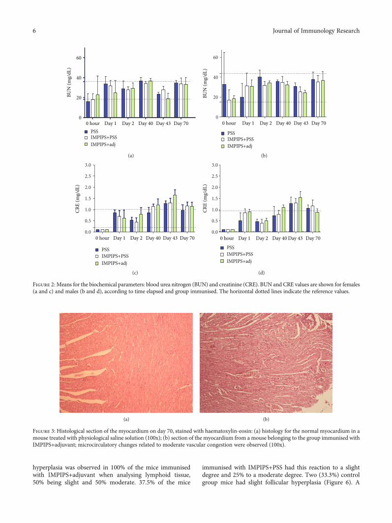
Renal function was evaluated by measuring BUN andCRE. ANOVA analysis identified no statistically significantdifferences between the different treatments when comparingvalues between the groups or when comparing control groupvalues (saline solution) to those for the other study groups(p > 0:05) (Figure 2). The reference values for analysing eachbiochemical parameter were determined from the means forthe controls ± SD (Figure 2).
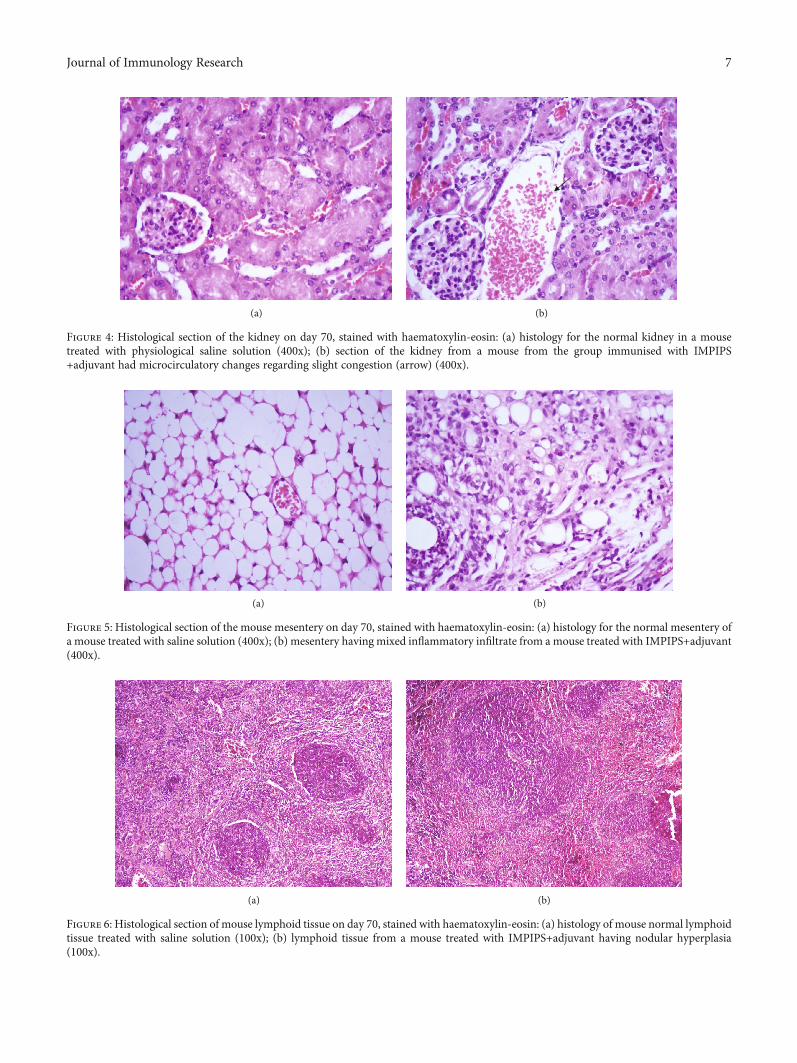
3.2.3. Histopathology. Microcirculatory changes related toslight and moderate vascular congestion were observed inthe myocardium in 2/4 females immunised with theIMPIPS+adjuvant formulation; there was no evidence ofcongestion in the other animals from the same group or fromthe other groups (p > 0:05). No microcirculatory changeswere seen in any of the immunised animals, such as oedemaand/or haemorrhage, inflammatory infiltrate, structuralchanges, or binucleation (Figure 3).
Slight congestion was observed in the kidneys of at leastone animal from every group. These changes occurred morein females than in males, since a lesion was found in just onemale compared to 6 females (2 from each group) in whichcongestion was observed. No microcirculatory changes suchas oedema and haemorrhage, inflammatory infiltrate, struc-tural changes, or binucleations were observed in any of thestudy groups (Figure 4).
No macroscopic or microscopic alterations were observedwhen analysing the duodenum, though 50% of the miceimmunised with IMPIPS+adjuvant had mixed inflammatoryinfiltrate in the mesentery (Figure 5). Likewise, follicular
5Journal of Immunology Research
hyperplasia was observed in 100% of the mice immunisedwith IMPIPS+adjuvant when analysing lymphoid tissue,50% being slight and 50% moderate. 37.5% of the mice
immunised with IMPIPS+PSS had this reaction to a slightdegree and 25% to a moderate degree. Two (33.3%) controlgroup mice had slight follicular hyperplasia (Figure 6). A
(a) (b)
Figure 3: Histological section of the myocardium on day 70, stained with haematoxylin-eosin: (a) histology for the normal myocardium in amouse treated with physiological saline solution (100x); (b) section of the myocardium from a mouse belonging to the group immunised withIMPIPS+adjuvant; microcirculatory changes related to moderate vascular congestion were observed (100x).
PSSIMPIPS+PSSIMPIPS+adj
0
20
40
60
BUN
(mg/
dL)
0 hour Day 1 Day 2 Day 40 Day 43 Day 70
(a)
PSSIMPIPS+PSSIMPIPS+adj
0
20
40
60
BUN
(mg/
dL)
0 hour Day 1 Day 2 Day 40 Day 43 Day 70
(b)
PSSIMPIPS+PSSIMPIPS+adj
0.0
0.5
1.0
1.5
2.0
2.5
3.0
CRE
(mg/
dL)
0 hour Day 1 Day 2 Day 40 Day 43 Day 70
(c)
PSSIMPIPS+PSSIMPIPS+adj
0.0
0.5
1.0
1.5
2.0
2.5
3.0
CRE
(mg/
dL)
0 hour Day 1 Day 2 Day 40 Day 43 Day 70
(d)
Figure 2: Means for the biochemical parameters: blood urea nitrogen (BUN) and creatinine (CRE). BUN and CRE values are shown for females(a and c) and males (b and d), according to time elapsed and group immunised. The horizontal dotted lines indicate the reference values.
6 Journal of Immunology Research
(a) (b)
Figure 6: Histological section of mouse lymphoid tissue on day 70, stained with haematoxylin-eosin: (a) histology of mouse normal lymphoidtissue treated with saline solution (100x); (b) lymphoid tissue from a mouse treated with IMPIPS+adjuvant having nodular hyperplasia(100x).
(a) (b)
Figure 5: Histological section of the mouse mesentery on day 70, stained with haematoxylin-eosin: (a) histology for the normal mesentery ofa mouse treated with saline solution (400x); (b) mesentery having mixed inflammatory infiltrate from a mouse treated with IMPIPS+adjuvant(400x).
(a) (b)
Figure 4: Histological section of the kidney on day 70, stained with haematoxylin-eosin: (a) histology for the normal kidney in a mousetreated with physiological saline solution (400x); (b) section of the kidney from a mouse from the group immunised with IMPIPS+adjuvant had microcirculatory changes regarding slight congestion (arrow) (400x).
7Journal of Immunology Research

scale was generated to semiquantify follicular hyperplasiaaccording to the number of nodules using a magnificationof 400x (Supplementary Table 2).
3.3. Immunogenicity
3.3.1. Determining Anti-IMPIPS Serum Ability to RecogniseP. falciparum-Infected RBC by IFA. An indirect immunofluo-rescence assay was used for determining anti-IMPIPS serumability to recognise pRBC. The serum from mice immunisedwith IMPIPS+adjuvant as well as that from those immu-nised with IMPIPS+PSS was able to recognise pRBC(Figure 7). Considering that some P. berghei proteins sharehigh identity with their P. falciparum counterparts, such asenolase [55], the higher fluorescence intensity observed inthe positive control might be due to the higher number ofproteins being recognised versus just the six blood-stageproteins being recognised from animals immunised withthe peptide mixture.
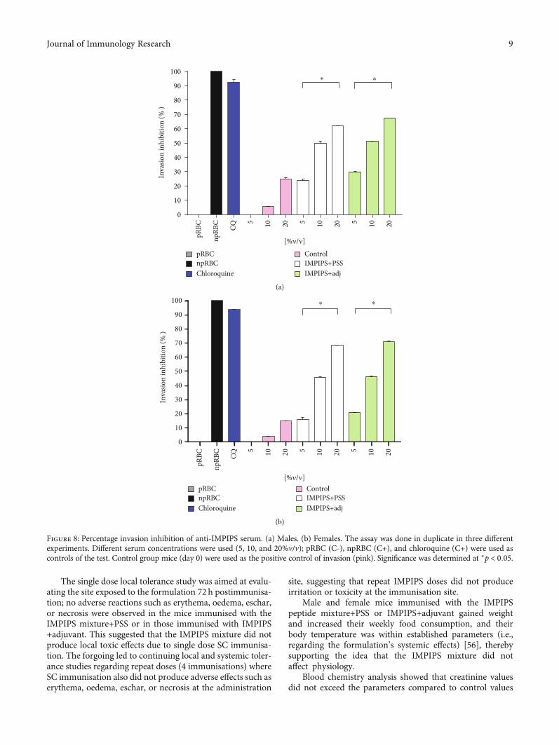
3.3.2. Determining Anti-IMPIPS Antibodies’ MerozoiteInvasion Inhibition Capability. The functional role of anti-bodies stimulated by immunisation with IMPIPS was deter-mined by an in vitro invasion inhibition assay. The serumfrom animals immunised with the IMPIPS peptide mix-ture+PSS was able to inhibit invasion, maximum valuesbeing 61.84% for males and 68.34% for females. Likewise,
the serum from the animals immunised with the IMPIPSpeptide mixture+adjuvant had 67.62% maximum invasioninhibition values for males and 70.82% for females. Itwas found that inhibition was concentration dependent(p < 0:05). No statistical difference was observed betweeninhibition percentages for the females compared to thosefor the males (p > 0:05) (Figure 8). Sera surpassing 70% wereconsidered strong inhibitors, whilst those ranging from 50%to 69% were considered medium-high inhibitors. Those hav-ing 30% to 49% were considered medium-low inhibitors,those from 10%-29% are low inhibitors, and those < 9% wereconsidered negative.
4. Discussion
Toxicological studies of the formulation to be used in clinicalstudies are of the utmost importance when developing vac-cines as they provide information about possible adverseeffects which might arise due to the formulation, either atthe inoculation site or in the different organs and tissues ofsubjects being vaccinated [37, 38]. This study thus evaluatedthe safety and immunogenicity of a mixture of 23 modifiedpeptides derived from 8 Spz proteins (CSP-1, TRAP, STARP,SPECT-1 and SPECT-2, CelTOS, and SIAP-1 and SIAP-2)and 6 Mrz proteins (AMA-1, EBA-175, EBA-140, SERA-5,MSP-1, and HRP-II) [9–11] in a murine model as a syntheticantimalarial vaccine candidate.
(a) (b)
(c) (d)
Figure 7: Immunofluorescence assay with mouse anti-IMPIPS antibodies in Plasmodium falciparum-infected erythrocytes (FCB2 strain).Results were analysed semiquantitatively according to fluorescence intensity from null (0+) to maximum fluorescence (+++). (a) PBS(negative control) (0+). (b) Serum from a P. berghei-infected mouse (positive control) (+++). (c) Serum from the group of miceimmunised with IMPIPS plus adjuvant (++). (d) Serum from the group immunised with IMPIPS plus PSS (+). Each study was done induplicate. Mouse serum and the FITC-labelled anti-mouse IgG (green fluorescence) were used at 1 : 20 dilution. The samples wereanalysed with a fluorescence microscope (Olympus B51) with an immersion objective (1,000x).
8 Journal of Immunology Research
The single dose local tolerance study was aimed at evalu-ating the site exposed to the formulation 72 h postimmunisa-tion; no adverse reactions such as erythema, oedema, eschar,or necrosis were observed in the mice immunised with theIMPIPS mixture+PSS or in those immunised with IMPIPS+adjuvant. This suggested that the IMPIPS mixture did notproduce local toxic effects due to single dose SC immunisa-tion. The forgoing led to continuing local and systemic toler-ance studies regarding repeat doses (4 immunisations) whereSC immunisation also did not produce adverse effects such aserythema, oedema, eschar, or necrosis at the administration
site, suggesting that repeat IMPIPS doses did not produceirritation or toxicity at the immunisation site.
Male and female mice immunised with the IMPIPSpeptide mixture+PSS or IMPIPS+adjuvant gained weightand increased their weekly food consumption, and theirbody temperature was within established parameters (i.e.,regarding the formulation’s systemic effects) [56], therebysupporting the idea that the IMPIPS mixture did notaffect physiology.
Blood chemistry analysis showed that creatinine valuesdid not exceed the parameters compared to control values
0
10
20
30
40
50
60
70
80
90
100
Inva
sion
inhi
bitio
n (%
)
⁎⁎
pRBC
npRB
C
CQ
5 10 20 5 10 20 5 10 20
[%𝜈/𝜈]
npRBCChloroquine
Control IMPIPS+PSSIMPIPS+adj
pRBC
(a)
Inva
sion
inhi
bitio
n (%
)
0
10
20
30
40
50
60
70
80
90
100
pRBC
npRB
C
CQ
5 10 20 5 10 20 5 10 20
npRBCChloroquine
Control IMPIPS+PSSIMPIPS+adj
pRBC
⁎⁎
[%𝜈/𝜈]
(b)
Figure 8: Percentage invasion inhibition of anti-IMPIPS serum. (a) Males. (b) Females. The assay was done in duplicate in three differentexperiments. Different serum concentrations were used (5, 10, and 20%v/v); pRBC (C-), npRBC (C+), and chloroquine (C+) were used ascontrols of the test. Control group mice (day 0) were used as the positive control of invasion (pink). Significance was determined at ∗p < 0:05.
9Journal of Immunology Research

during the first bleeding. Creatinine values on day 40exceeded the parameters; however, they became reduced byday 70, coming within normal parameters for the males. Suchtransitory increase could have been due to stress or dehydra-tion since, unlike other mammals, mice excrete creatinine intheir urine. Once the situation had become resolved, creati-nine returned to its normal values [56]. BUN values camewithin normal parameters. Since no other damage wasobserved on day 70, the histological study of the kidneys ver-ified that an increase in creatinine was due to prerenal causesand not to renal damage.
Regarding histological analysis, the microcirculatorychanges in the myocardium compatible with congestion didnot arise from administering the IMPIPS peptide mixture,since control group animals also had this pathology. Suchchanges could mainly have been due to the hypovolemicshock caused by the final bleeding; this would have occurredbecause haemorrhagic shock affects tissue perfusion [57].
Mixed inflammatory infiltrate was observed in the histo-logical study of the mesentery; this description refers to local-ised accumulations of mononuclear and polymorphonuclearcells, indicating the presence of a foreign body in acute phase,i.e., causing the antigenic stimulus to continue. This findingin the animals immunised with IMPIPS+adjuvant and notin those immunised with IMPIPS+PPS or in the controlgroup indicated that the antigen continued being active. Thiscould have been caused by the formation of a deposit at theinjection site due to the adjuvant’s mechanism of action(doses 2, 3, and 4 of the formulation were administered byIP route) [58].
The nodular hyperplasia observed in animals’ lymphoidtissue is mainly due to normal lymphoid nodule inflamma-tion in response to an antigen. Such response in this casewas triggered by the immunisation; such reaction is alsoknown as reactive lymphoid hyperplasia [59] which, asexpected, was much stronger in the animals immunised withthe formulation containing IMPIPS+adjuvant.
FIDIC’s previous studies have shown that individualimmunisation of IMPIPS in Aotus monkeys has stimulatedthe production of antibodies which have been able to recog-nise parasite proteins in their native form and induce a pro-tective immune response, determined by the total absenceof parasites in the blood following experimental challenge[30, 32, 33]. The present study highlighted the fact that serumfrom male and female mice immunised with the IMPIPSmixture+adjuvant or IMPIPS+PSS recognised the parasitein the Mrz stage. This indicated that although the peptideshad been modified for their presentation by human MHC-II [27, 29, 31], the mixture was capable of inducing animmune response against the native proteins from whichthey were derived, even in a murine model, thereby reinforc-ing the idea of using IMPIPS in an antimalarial vaccine [60].Such response was seen in the immunofluorescence andinvasion inhibition assays.
5. Conclusions
Local tolerance and systemic safety tests regarding single andrepeat doses in this study showed no toxicity induced by the
IMPIPS mixture in a murine model 70 days after the firstimmunisation, reaffirming that peptide-based vaccines canrepresent a safe option. The IMPIPS mixture was immuno-genic in a murine model, even when the peptides weredesigned for human MHC-II. Such results suggested thatthe IMPIPS mixture is safe and thus further immunogenic-ity and protection assays in a nonhuman primate modelsuch as the Aotus spp. monkey but delivered with adjuvantsauthorised for human use are recommended.
Data Availability
The data used to support the findings of this study areincluded within the article.
Conflicts of Interest
The authors declare that there is no conflict of interestregarding the publication of this paper.
Acknowledgments
We would like to thank Jason Garry for translating thismanuscript. This work was financed by COLCIENCIAS callfor projects 725/2015, the Fundación Instituto de Inmunolo-gía de Colombia (FIDIC), and the Universidad de CienciasAplicadas y Ambientales (U.D.C.A).
Supplementary Materials
Supplementary Figure 1: physiological parameters for maleand female mice: weight, weekly consumption of food, andbody temperature values. Dotted lines show upper and lowernormal values. Supplementary Table 1: publications referen-cing peptides included in the present study, in which the anti-bodies raised recognise the corresponding protein expressedas a recombinant. Supplementary Table 2: semiquantitativescale of follicular hyperplasia using a 400x magnification.(Supplementary Materials)
References
[1] World Health Organization (WHO), World Malaria Report2018, Geneva, 2018.
[2] World Health Organization (WHO),Global Technical Strategyfor Malaria 2016-2030, 2015.
[3] D. Menard and A. Dondorp, “Antimalarial drug resistance: athreat to malaria elimination,” Cold Spring Harbor Perspectivesin Medicine, vol. 7, no. 7, article a025619, 2017.
[4] M. E. Patarroyo and M. A. Patarroyo, “Emerging rules forsubunit-based multiantigenic, multistage chemically synthe-sized vaccines,” Accounts of Chemical Research, vol. 41, no. 3,pp. 377–386, 2008.
[5] M. E. Patarroyo, A. Bermudez, and M. A. Patarroyo, “Struc-tural and immunological principles leading to chemically syn-thesized, multiantigenic, multistage, minimal subunit-basedvaccine development,” Chemical Reviews, vol. 111, no. 5,pp. 3459–3507, 2011.
[6] Z. Bozdech, M. Llinás, B. L. Pulliam, E. D. Wong, J. Zhu, andJ. L. DeRisi, “The transcriptome of the intraerythrocytic
10 Journal of Immunology Research
developmental cycle of Plasmodium falciparum,” PLoS Biol-ogy, vol. 1, no. 1, p. E5, 2003.
[7] K. Kaiser, K. Matuschewski, N. Camargo, J. Ross, and S. H.Kappe, “Differential transcriptome profiling identifies Plasmo-dium genes encoding pre-erythrocytic stage-specific proteins,”Molecular Microbiology, vol. 51, no. 5, pp. 1221–1232, 2004.
[8] E. Lasonder, C. J. Janse, G. J. van Gemert et al., “Proteomicprofiling of Plasmodium sporozoite maturation identifiesnew proteins essential for parasite development and infec-tivity,” PLoS Pathogens, vol. 4, no. 10, article e1000195,2008.
[9] J. E. Garcia, A. Puentes, and M. E. Patarroyo, “Developmentalbiology of sporozoite-host interactions in Plasmodium falci-parum malaria: implications for vaccine design,” ClinicalMicrobiology Reviews, vol. 19, no. 4, pp. 686–707, 2006.
[10] L. E. Rodriguez, H. Curtidor, M. Urquiza, G. Cifuentes,C. Reyes, and M. E. Patarroyo, “Intimate molecular interac-tions of P. falciparum merozoite proteins involved in invasionof red blood cells and their implications for vaccine design,”Chemical Reviews, vol. 108, no. 9, pp. 3656–3705, 2008.
[11] H. Curtidor, M. Vanegas, M. P. Alba, and M. E. Patarroyo,“Functional, immunological and three-dimensional analysisof chemically synthesised sporozoite peptides as componentsof a fully-effective antimalarial vaccine,” Current MedicinalChemistry, vol. 18, no. 29, pp. 4470–4502, 2011.
[12] M. Urquiza, L. E. Rodriguez, J. E. Suarez et al., “Identificationof Plasmodium falciparum MSP-1 peptides able to bind tohuman red blood cells,” Parasite Immunology, vol. 18, no. 10,pp. 515–526, 1996.
[13] F. Espejo, M. Cubillos, L. M. Salazar et al., “Structure, immu-nogenicity, and protectivity relationship for the 1585 malarialpeptide and its substitution analogues,” Angewandte Chemie,International Edition, vol. 40, no. 24, pp. 4654–4657, 2001.
[14] S. Casares and T. L. Richie, “Immune evasion by malaria par-asites: a challenge for vaccine development,” Current Opinionin Immunology, vol. 21, no. 3, pp. 321–330, 2009.
[15] H. Hisaeda, K. Yasutomo, and K. Himeno, “Malaria: immuneevasion by parasites,” The International Journal of Biochemis-try & Cell Biology, vol. 37, no. 4, pp. 700–706, 2005.
[16] M. Hommel, “Antigenic variation in malaria parasites,”Immunology Today, vol. 6, no. 1, pp. 28–33, 1985.
[17] H. Curtidor, M. Urquiza, J. E. Suarez et al., “Plasmodiumfalciparum acid basic repeat antigen (ABRA) peptides: eryth-rocyte binding and biological activity,” Vaccine, vol. 19,no. 31, pp. 4496–4504, 2001.
[18] M. Ocampo, M. Urquiza, F. Guzmán et al., “Two MSA 2peptides that bind to human red blood cells are relevantto Plasmodium falciparum merozoite invasion,” The Journalof Peptide Research, vol. 55, no. 3, pp. 216–223, 2000.
[19] G. Cifuentes, A. Bermudez, R. Rodriguez, M. Patarroyo, andM. Patarroyo, “Shifting the polarity of some critical residuesin malarial peptides binding to host cells is a key factor inbreaking conserved antigens code of silence,”Medicinal Chem-istry, vol. 4, no. 3, pp. 278–292, 2008.
[20] F. Guzman, K. Jaramillo, L. M. Salazar, A. Torres, A. Rivera,and M. E. Patarroyo, “1H-NMR structures of the Plasmodiumfalciparum 1758 erythrocyte binding peptide analogues andprotection against malaria,” Life Sciences, vol. 71, no. 23,pp. 2773–2785, 2002.
[21] L. M. Salazar, M. P. Alba, H. Curtidor et al., “Changing ABRAprotein peptide to fit into the HLA-DRβ1∗0301 molecule ren-
ders it protection-inducing,” Biochemical and BiophysicalResearch Communications, vol. 322, no. 1, pp. 119–125, 2004.
[22] M. E. Patarroyo, G. Cifuentes, A. Bermúdez, and M. A.Patarroyo, “Strategies for developing multi-epitope, subunit-based, chemically synthesized anti-malarial vaccines,” Journalof Cellular and Molecular Medicine, vol. 12, no. 5B, pp. 1915–1935, 2008.
[23] A. Bermudez, M. P. Alba, M. Vanegas, M. A. Patarroyo,and M. E. Patarroyo, “Specific β-turns precede PPIIL structuresbinding to allele-specific HLA-DRβ1∗ PBRs in fully-protectivemalaria vaccine components,” Frontiers in Chemistry, vol. 6,p. 106, 2018.
[24] C. Reyes, A. Moreno-Vranich, and M. E. Patarroyo, “The roleof pi-interactions and hydrogen bonds in fully protective syn-thetic malaria vaccine development,” Biochemical and Bio-physical Research Communications, vol. 484, no. 3, pp. 501–507, 2017.
[25] A. Bermúdez, M. P. Alba, M. Vanegas, and M. E. Patarroyo,“3D structure determination of STARP peptides implicatedin P. falciparum invasion of hepatic cells,” Vaccine, vol. 28,no. 31, pp. 4989–4996, 2010.
[26] M. P. Alba, L. M. Salazar, L. E. Vargas, M. Trujillo, Y. Lopez,and M. E. Patarroyo, “Modifying RESA protein peptide 6671to fit into HLA-DRβ1∗ pockets induces protection againstmalaria,” Biochemical and Biophysical Research Communica-tions, vol. 315, no. 4, pp. 1154–1164, 2004.
[27] M. E. Patarroyo, G. Cifuentes, C. Piraján, A. Moreno-Vranich,and M. Vanegas, “Atomic evidence that modification of H-bonds established with amino acids critical for host-cell bind-ing induces sterile immunity against malaria,” Biochemicaland Biophysical Research Communications, vol. 394, no. 3,pp. 529–535, 2010.
[28] M. E. Patarroyo, A. Bermudez, and M. P. Alba, “The highimmunogenicity induced by modified sporozoites’ malarialpeptides depends on their phi (ϕ) and psi (ψ) angles,” Bio-chemical and Biophysical Research Communications, vol. 429,no. 1-2, pp. 81–86, 2012.
[29] M. E. Patarroyo, H. Almonacid, and A. Moreno-Vranich, “Therole of amino acid electron-donor/acceptor atoms in host-cellbinding peptides is associated with their 3D structure andHLA-binding capacity in sterile malarial immunity induc-tion,” Biochemical and Biophysical Research Communications,vol. 417, no. 3, pp. 938–944, 2012.
[30] A. Bermúdez, D. Calderon, A. Moreno-Vranich et al.,“Gauche+ side-chain orientation as a key factor in thesearch for an immunogenic peptide mixture leading to acomplete fully protective vaccine,” Vaccine, vol. 32, no. 18,pp. 2117–2126, 2014.
[31] M. E. Patarroyo, A. Moreno-Vranich, and A. Bermúdez, “Phi(Φ) and psi (Ψ) angles involved in malarial peptide bondsdetermine sterile protective immunity,” Biochemical and Bio-physical Research Communications, vol. 429, no. 1-2, pp. 75–80, 2012.
[32] M. E. Patarroyo, A. Bermúdez, M. P. Alba et al., “IMPIPS: theimmune protection-inducing protein structure concept in thesearch for steric-electron and topochemical principles forcomplete fully-protective chemically synthesised vaccinedevelopment,” PLoS One, vol. 10, no. 4, article e0123249, 2015.
[33] M. E. Patarroyo, M. A. Patarroyo, L. Pabón, H. Curtidor, andL. A. Poloche, “Immune protection-inducing protein struc-tures (IMPIPS) against malaria: the weapons needed for beat-ing Odysseus,” Vaccine, vol. 33, no. 52, pp. 7525–7537, 2015.
11Journal of Immunology Research
[34] International Conference on Harmonisation (ICH), “ICH S6(R1) Preclinical safety evaluation of biotechnology-derivedpharmaceuticals,” vol. 6, 2011.
[35] European Medicines Agency (EMA), “Guideline on repeateddose toxicity,” vol. 99, 2010.
[36] European Agency for the Evaluation of Medicinal Products(EMEA), Note for guidance on non-clinical local tolerance test-ing of medicinal products, London, 2001.
[37] World Health Organization (WHO),Annex 1WHO guidelineson nonclinical evaluation of vaccines, 2005.
[38] World Health Organization (WHO), Handbook: Good Labo-ratory Practice (GLP): Qquality Practices for Regulated Non-clinical Research and Development, 2nd edition2nd edition, ,2009.
[39] M. Patarroyo, M. P. Alba, R. Rojas-Luna, A. Bermudez, andJ. Aza-Conde, “Functionally relevant proteins in Plasmodiumfalciparum host cell invasion,” Immunotherapy, vol. 9, no. 2,pp. 131–155, 2017.
[40] M. E. Patarroyo, G. Arévalo-Pinzón, C. Reyes, A. Moreno-Vranich, and M. A. Patarroyo, “Malaria parasite survivaldepends on conserved binding peptides’ critical biological func-tions,” Current Issues in Molecular Biology, vol. 18, pp. 57–78,2016.
[41] H. Curtidor, C. Reyes, A. Bermúdez, M. Vanegas, Y. Varela,and M. Patarroyo, “Conserved binding regions provide theclue for peptide-based vaccine development: a chemical per-spective,” Molecules, vol. 22, no. 12, article 2199, 2017.
[42] R. B. Merrifield, “Solid phase peptide synthesis. I. The synthe-sis of a tetrapeptide,” Journal of the American Chemical Soci-ety, vol. 85, no. 14, pp. 2149–2154, 1963.
[43] R. A. Houghten, “General method for the rapid solid-phasesynthesis of large numbers of peptides: specificity of antigen-antibody interaction at the level of individual amino acids,”Proceedings of the National Academy of Sciences, vol. 82,no. 15, pp. 5131–5135, 1985.
[44] European Medicines Agency, Guideline on the principles ofregulatory acceptance of 3Rs (replacement, reduction, refine-ment) testing approaches, 2016.
[45] S. Cox, “Irritation and local tissue tolerance studies in pharma-cetical safety assessment,” in Preclinical Development Hand-book: Toxicology, S. Cox, Ed., pp. 233–268, John Wiley &Sons, 2008.
[46] T. W. Adamson, D. Diaz-Arevalo, T. M. Gonzalez, X. Liu, andM. Kalkum, “Hypothermic endpoint for an intranasal invasivepulmonary aspergillosis mouse model,” Comparative Medi-cine, vol. 63, no. 6, pp. 477–481, 2013.
[47] American Veterinary Medical Association (AVMA), AVMAguidelines for the euthanasia of animals, 2013.
[48] L. E. Rodriguez, R. Vera, J. Valbuena et al., “Characterisationof Plasmodium falciparum RESA-like protein peptides thatbind specifically to erythrocytes and inhibit invasion,” Biolog-ical Chemistry, vol. 388, no. 1, pp. 15–24, 2007.
[49] C. Lambros and J. P. Vanderberg, “Synch stages in culture ofPlasmodium falciparum,” The Journal of Parasitology, vol. 65,pp. 418–420, 2012.
[50] E. Duncan and E. Bergmenn-Litner, “Miniaturized growthinhibition assay to assess the anti-blood stage activity of anti-bodies,” in Malaria Vaccines Methods, A. Vaughan, Ed.,pp. 153–166, Springer, 2015.
[51] A. Bei and M. Duraisingh, “Measuring Plasmodium falciparumerythrocyte invasion phenotypes using flow cytometry,” in
Malaria Vaccines Methods, A. Vaughan, Ed., pp. 167–186,Springer, 2015.
[52] Congreso Nacional de Colombia, Ley 84 de 1989, Congreso deColombia, Colombia, 1989.
[53] Ministerio de Salud de Colombia, Resolución Número 8430 de1993, Colombia, 1993.
[54] National Research Council of the National Academies, Guidefor the Care and Use of Laboratory Animals, Eighth editio-nEighth edition, , 2011.
[55] S. Dutta, A. Tewari, C. Balaji et al., “Strain-transcendingneutralization of malaria parasite by antibodies against Plasmo-dium falciparum enolase,”Malaria Journal, vol. 17, no. 1, p. 304,2018.
[56] M. T. Whary, N. Baumgarth, J. G. Fox, and S. W. Barthold,“Biology and diseases of mice,” in Laboratory animal medicine2, J. G. Fox, L. C. Anderson, G. M. Otto, K. R. Pritchett-Corn-ing, and M. T. Whary, Eds., pp. 43–149, Elsevier, 2015.
[57] N. T. Veith, T. Histing, M. D. Menger, T. Pohlemann, andT. Tschernig, “Helping prometheus: liver protection in acutehemorrhagic shock,” Annals of Translational Medicine,vol. 5, no. 10, p. 206, 2017.
[58] S. Awate, L. A. Babiuk, and G. Mutwiri, “Mechanisms of actionof adjuvants,” Frontiers in Immunology, vol. 4, p. 114, 2013.
[59] P. J. Haley, “The lymphoid system: a review of species differ-ences,” Journal of Toxicologic Pathology, vol. 30, no. 2,pp. 111–123, 2017.
[60] M. E. Patarroyo, M. P. Alba, C. Reyes, R. Rojas-Luna, andM. A. Patarroyo, “The Malaria Parasite's Achilles' Heel:Functionally-relevant Invasion Structures,” Current Issues inMolecular Biology, vol. 18, pp. 11–20, 2016.
12 Journal of Immunology Research

Stem Cells International
Hindawiwww.hindawi.com Volume 2018
Hindawiwww.hindawi.com Volume 2018
MEDIATORSINFLAMMATION
of
EndocrinologyInternational Journal of
Hindawiwww.hindawi.com Volume 2018
Hindawiwww.hindawi.com Volume 2018
Disease Markers
Hindawiwww.hindawi.com Volume 2018
BioMed Research International
OncologyJournal of
Hindawiwww.hindawi.com Volume 2013
Hindawiwww.hindawi.com Volume 2018
Oxidative Medicine and Cellular Longevity
Hindawiwww.hindawi.com Volume 2018
PPAR Research
Hindawi Publishing Corporation http://www.hindawi.com Volume 2013Hindawiwww.hindawi.com
The Scientific World Journal
Volume 2018
Immunology ResearchHindawiwww.hindawi.com Volume 2018
Journal of
ObesityJournal of
Hindawiwww.hindawi.com Volume 2018
Hindawiwww.hindawi.com Volume 2018
Computational and Mathematical Methods in Medicine
Hindawiwww.hindawi.com Volume 2018
Behavioural Neurology
OphthalmologyJournal of
Hindawiwww.hindawi.com Volume 2018
Diabetes ResearchJournal of
Hindawiwww.hindawi.com Volume 2018
Hindawiwww.hindawi.com Volume 2018
Research and TreatmentAIDS
Hindawiwww.hindawi.com Volume 2018
Gastroenterology Research and Practice
Hindawiwww.hindawi.com Volume 2018
Parkinson’s Disease
Evidence-Based Complementary andAlternative Medicine
Volume 2018Hindawiwww.hindawi.com
Submit your manuscripts atwww.hindawi.com