Pregnancy and Congenital Heart Disease...1 Pregnancy and Congenital Heart Disease What the Nurse...
17
1 Pregnancy and Congenital Heart Disease What the Nurse Caring for a Patient with Congenital Heart Disease Needs to Know Annette Haynes, MS, RN, CNS, CCRN Cardiology Clinical Nurse Specialist Stanford Hospital and Clinics Palo Alto, California Patricia L. Woods, MSN, RN, CNP - Adolescent/Adult Adult Congenital Heart Disease Nurse Practitioner Cardiology, Oregon Health & Science University Portland, Oregon Introduction: Most women with congenital heart disease (CHD) will reach child-bearing age. Many women with complex CHD have a moderate to high risk for both the mother and her fetus during pregnancy. For women wishing to have children, planning for pregnancy for a patient with CHD starts prior to conception and continues after delivery. All practitioners involved in the care of a patient need to be aware of the most up-to-date guidelines and information about CHD and pregnancy. Ideally, appropriate counseling should begin during pediatric care. The following document provides guidelines for the care of pregnant women with CHD. This link will provide more comprehensive, disease specific information. http://www.heartdiseaseandpregnancy.com/ Key Components: Preconception Counseling (Wald, 2009) o Recommended for any patient with CHD o Risks of pregnancy Evaluate risks related to both mother and baby Increased risk to mother, pregnancy discouraged Severe pulmonary hypertension Severe left heart obstructive lesions Marfan syndrome with increased aortic root diameter Cardiomyopathies with ventricular dysfunction Increased risk to fetus Poor maternal functional class Maternal cyanosis Maternal CHD with left heart obstruction Prenatal History (Canobbio, 2017) o Evaluation of maternal risks is most reliable when using a risk algorithm. o Risk scores/stratification systems are helpful to identify at risk patients to predict/plan for adverse maternal cardiac events.
Transcript of Pregnancy and Congenital Heart Disease...1 Pregnancy and Congenital Heart Disease What the Nurse...

1
Pregnancy and Congenital Heart Disease What the Nurse Caring for a Patient with Congenital Heart Disease Needs to Know
Annette Haynes, MS, RN, CNS, CCRN
Cardiology Clinical Nurse Specialist
Stanford Hospital and Clinics
Palo Alto, California
Patricia L. Woods, MSN, RN, CNP - Adolescent/Adult
Adult Congenital Heart Disease Nurse Practitioner
Cardiology, Oregon Health & Science University
Portland, Oregon
Introduction:
Most women with congenital heart disease (CHD) will reach child-bearing age. Many women
with complex CHD have a moderate to high risk for both the mother and her fetus during
pregnancy. For women wishing to have children, planning for pregnancy for a patient with CHD
starts prior to conception and continues after delivery. All practitioners involved in the care of a
patient need to be aware of the most up-to-date guidelines and information about CHD and
pregnancy. Ideally, appropriate counseling should begin during pediatric care. The following
document provides guidelines for the care of pregnant women with CHD. This link will provide
more comprehensive, disease specific information. http://www.heartdiseaseandpregnancy.com/
Key Components:
Preconception Counseling (Wald, 2009)
o Recommended for any patient with CHD
o Risks of pregnancy
Evaluate risks related to both mother and baby
Increased risk to mother, pregnancy discouraged
Severe pulmonary hypertension
Severe left heart obstructive lesions
Marfan syndrome with increased aortic root diameter
Cardiomyopathies with ventricular dysfunction
Increased risk to fetus
Poor maternal functional class
Maternal cyanosis
Maternal CHD with left heart obstruction
Prenatal History (Canobbio, 2017)
o Evaluation of maternal risks is most reliable when using a risk algorithm.
o Risk scores/stratification systems are helpful to identify at risk patients to
predict/plan for adverse maternal cardiac events.

2
o Recommendations are supported by the World Health Organization (WHO) and
the American Heart Association (AHA).
o The Modified WHO Classification of Pregnancy Risk:
Class I conditions:
Associated with no detectable increased risk of maternal mortality
and no/mild increase in morbidity
Conditions include uncomplicated, small patent ductus arteriosus,
mild pulmonic stenosis, or mitral valve prolapse; successfully
repaired simple lesions (atrial or ventricular septal defect, patent
ductus arteriosus, or anomalous pulmonary venous drainage); and
isolated atrial or ventricular ectopic beats
Class II conditions:
Associated with small increased risk of maternal mortality or
moderate increase in morbidity
Conditions include unrepaired atrial or ventricular septal defect,
repaired tetralogy of Fallot, most arrhythmias
Class II to III conditions:
Depends on individual
Conditions include mild left ventricular impairment, hypertrophic
cardiomyopathy, native or bioprosthetic valvular heart disease not
considered WHO I or IV, repaired coarctation, Marfan syndrome
with aortic dimension <40mm without aortic dissection, and
bicuspid aortic valve with ascending aorta diameter <45mm
Class III conditions:
Associated with significantly increased risk of maternal mortality
or severe morbidity
Conditions include a mechanical valve, systemic right ventricle,
Fontan circulation, cyanotic heart disease (unrepaired), other
complex congenital heart disease, bicuspid aortic valve with
ascending aortic diameter of 45 to 50 mm, and Marfan syndrome
with aortic diameter of 40 to 45 mm
Class IV conditions:
Associated with extremely high risk of maternal mortality or
severe morbidity
Pregnancy is contraindicated
o Risk Stratification (See Table 1 below)
Table 1. Risk Stratification of Pregnancy in Women with Congenital Heart Disease
Low Moderate Very High
Atrial septal defect Repaired heart disease Pulmonary hypertension
Mild/moderate valvular
regurgitation
Mechanical valve
replacement
Shunt Lesions with
Eisenmenger syndrome
Mild/moderate pulmonary
stenosis
Uncomplicated repaired
coarctation of the aorta
Severe heart muscle disease
(cardiomyopathy)
VSD
PDA

3
Mild atrial and ventricular
arrhythmias
Hypertrophic
cardiomyopathy
Uncorrected cyanotic congenital
heart disease
Successful repaired lesions:
Secundum ASD
VSD
PDA
TAPV
Marfan syndrome or coarctation of
the aorta with aortic aneurysm
Ischemic Cardiomyopathy
Planning for conception and pregnancy
o Individualized
For a low risk pregnancy
Evaluate around 20 weeks by their cardiologist
Deliver in the community
Fetal echo at 20 weeks.
For moderate to severe risk pregnancy
Provide care at a center with expertise in CHD
Involves individualized plan based upon:
o Maternal risk factors
o Complexity of CHD
o Functional capacity
o Other existing or potential clinical issues
o Social situation
o Insurance coverage
o Location of high-risk obstetric and adult CHD providers
o Involves interdisciplinary team
Patient, significant other, other family members as desired
High-risk perinatal/neonatologists
Adult Congenital Heart Disease Cardiologists
Anesthesiologists
Nurse practitioners
Additional specialists as indicated (electrophysiology, hematology,
pulmonology, etc.)
o Interdisciplinary Care Meetings (See Attachment A for suggested content of
documentation for team meetings)
Initiated by preconception plan/birth plan
Frequency and team members will vary based upon:
Needs of both mother and baby
Services provided at delivering hospital
Needs to clearly communicate all facets of care
Needs to include both mother and father
Held at least bi-monthly

4
Topics include:
Vary depending on events during pregnancy
Cardiac diagnosis (diagrams are very helpful)
Potential risks of pregnancy
Identification of all members of the care team with contact
information
Due Date
Planned mode of delivery
Planned location of delivery and post-partum care
Cardiac Monitoring during and/ after deliver
IV lines: CVP? Art line?
Specialized nursing plan
For very high risk deliveries consider the presence of a critical care
RN in the delivery room
For delivery in a critical care unit consider the presence of an
obstetric nurse during the delivery and post-partum periods
Discussion regarding medication regime
Desire/ability of patient to breast feed
Presence of family during delivery and post-partum period
Update of family members during delivery
Cardiac surveillance required during pregnancy.
o Maternal surveillance
Follow plan created prior to conception (See components of plan outlined
above and documentation in Appendix A)
Determined by risk factors (See Table 1 on risk stratification above)
Determined by tolerance to normal physiological changes during
pregnancy (See Table 2 below)
Fluctuations in circulating blood volume
Increase in BNP
Decrease in plasma albumin
Increase in heart rate with lower threshold for arrhythmias
Remodeling of arterial vasculature to accommodate increased
blood volume
Volume Cardiac Output Heart Rate Blood Pressure
1st Trimester
2nd Trimester
3rd Trimester

5
Table 2. Normal Physiological Changes during Pregnancy
Assessment includes:
o Physical examination (See Table 3 below for normal and abnormal
cardiovascular findings)
o Diagnostic and genetic testing
o Symptoms (See Table 4 below)
o Hemodynamic changes (See Table 2 above)
o Complications (See Table 5)
Table 3. Normal and Abnormal Physical Exam during Pregnancy
Exam Abnormal
Observed color:
No color changes
Observed color findings:
Cyanosis
Clubbing
Dependent edema
Rales
Distended neck veins
EKG Changes:
Nonspecific ST and T-wave changes
Shift in electrical axis, leftward to
rightward with physical displacement of
the heart.
PMI: laterally displaced by displacement
of heart
EKG Findings:
Arrhythmias
Labor/Delivery
Postpartum

6
Auscultatory Changes:
First heart sound increases in loudness,
increased splitting of S1, attributed to
early closure of mitral valve.
Second heart sound: at 30 weeks, splitting
of the second sound may occur;
PMI laterally displaced by displacement of
heart
Third heart sound is heard in up to 90% of
women
Internal mammary murmur, Mammary
Souffle. Heard in 15% of postpartum patients
Auscultatory Findings:
S4
Harsh murmurs
Diagnostic Testing
12-lead electrocardiogram
Cardiopulmonary exercise testing
Cardiac imaging
o Echocardiogram
o MRI/CT imaging as needed
Genetic testing and counseling (Genetic basis for congenital heart defects,
Circulation 2007)
All patients with CHD
o Specifically test for 22q11 deletion in patients with
conotruncal defects
Tetralogy of Fallot
VSD with aortic arch anomaly
Truncus arteriosus
Interrupted aortic arch
Discontinuous branch pulmonary arteries (PA)
Evaluate risk of fetus to have CHD and/or other genetic
disease/syndrome
Other consultations as indicated
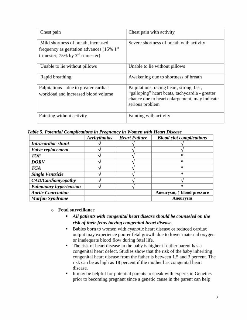
Table 4. Normal and Abnormal Symptoms during Pregnancy
Normal Abnormal
Necessary to report and evaluate symptoms
listed below.
Fatigue Symptoms at rest
7
Chest pain Chest pain with activity
Mild shortness of breath, increased
frequency as gestation advances (15% 1st
trimester; 75% by 3rd trimester)
Severe shortness of breath with activity
Unable to lie without pillows Unable to lie without pillows
Rapid breathing Awakening due to shortness of breath
Palpitations – due to greater cardiac
workload and increased blood volume
Palpitations, racing heart, strong, fast,
“galloping” heart beats, tachycardia - greater
chance due to heart enlargement, may indicate
serious problem
Fainting without activity Fainting with activity
Table 5. Potential Complications in Pregnancy in Women with Heart Disease
Arrhythmias Heart Failure Blood clot complications
Intracardiac shunt
Valve replacement
TOF *
DORV *
TGA *
Single Ventricle *
CAD/Cardiomyopathy
Pulmonary hypertension *
Aortic Coarctation Aneurysm, ↑ blood pressure
Marfan Syndrome Aneurysm
o Fetal surveillance
All patients with congenital heart disease should be counseled on the
risk of their fetus having congenital heart disease.
Babies born to women with cyanotic heart disease or reduced cardiac
output may experience poorer fetal growth due to lower maternal oxygen
or inadequate blood flow during fetal life.
The risk of heart disease in the baby is higher if either parent has a
congenital heart defect. Studies show that the risk of the baby inheriting
congenital heart disease from the father is between 1.5 and 3 percent. The
risk can be as high as 18 percent if the mother has congenital heart
disease.
It may be helpful for potential parents to speak with experts in Genetics
prior to becoming pregnant since a genetic cause in the parent can help

8
answer questions about the risk of transmitting the genetic condition to the
baby. The cause of congenital heart disease is unknown in most cases.
Risk factors associated with an increased rate of congenital heart disease
are shown in Table 6 below. Because of the increased risk of transmitting
congenital heart disease, fetal ultrasound is recommended.
The fetal ultrasound is performed by specially trained sonographers and
physicians between 20 and 24 weeks of gestation to check the baby’s heart
for congenital defects.
22q11 deletion testing is recommended for all pregnant patients with:
Tetralogy of Fallot
VSD with aortic arch anomaly
Truncus arteriosus
Interrupted aortic arch
Discontinuous branch PA’s
Follow up as indicated by identified risk factors or fetal diagnosis of CHD
Table 6. Fetal Risk Factors Associated with Congenital Heart Disease
Medication management during pregnancy
o Identify any medications that would need to be discontinued prior to pregnancy
Maternal alcohol or drug abuse during pregnancy
Exposure to certain environmental agents (pesticides, lead)
Maternal CHD – risk of fetal CHD increased by 18%
Paternal CHD – risk of fetal CHD increased by 1.5-3%
Maternal heart disease with cyanosis or reduced cardiac output –
increased risk of intrauterine growth retardation (IUGR)
Maternal viral infection, such as German measles
Maternal fever early in pregnancy or around conception
Maternal diabetes (not gestational diabetes)
Maternal obesity
Poor maternal nutrition
Chromosomal or genetic abnormalities (Down syndrome) in the fetus
Certain medications taken during pregnancy (ACE inhibitors,
Coumadin)

9
o Remember that almost all cardiovascular medications cross the placenta
o Anticoagulation:
All forms increase the risk of:
Spontaneous abortion
Retroplacental bleeding
Stillbirth
Fetal death
Increased risk of clot formation in pregnancy
Hypercoagulable state
Platelet adhesion with decreased fibrinolysis
Increased risk of valve thrombosis or embolic events
Specifically in pts with mechanical valve in MV position
Medications
Coumadin
o Safest for mother
o Teratogenic – see midline deformities
9% risk, less if daily dose < 5 mg
Avoid 1st trimester
Acceptable during 2nd trimester to middle of 3rd
trimester
o Concern during 3rd trimester related to immature fetal liver
Heparin
o Often used in 1st trimester until 13-14 weeks
May then resume warfarin
Increased risk of clot
Prosthetic valve in mitral position
Disc type of prosthetic valve
TPA/streptokinase does not cross placenta.
o Dose – prolong aPTT by 2X control 6 hours after
administration initiated
Low molecular weight Heparin (LMWH)
o Increased risk of thrombosis in prosthetic valves
o More predictable bioavailability.
o Anti-Xa monitoring, 4 hours post dose, weekly with goal of
1.0-1.2 unit/ml
o Aspirin may be used adjunctively.
ASA
o Acceptable during pregnancy
o Dose - 81 mg dose safe without premature fetal duct
closure
o Fetotoxic medications
Angiotensin converting enzyme (ACE)/angiotensin receptor blocker
(ARB)/aldosterone antagonists
Strictly contraindicated
Increased risk of fetal renal malformation, IUGR

10
Alternative afterload-reducing agents
Aldosterone
Oral isosorbide dinitrate
Systemic afterload
Hydralazine
Nitrates
o Antiarrhythmic medications
Clinically significant arrhythmias common
Increased with history of arrhythmias
Lower threshold for ventricular arrhythmias, reentrant SVT
Often related to:
o Extra volume load
o Enhanced adrenergic receptor excitability
o Presence of surgical scar
Place patients with documented arrhythmias during pregnancy on
continuous cardiac monitoring (direct or tele monitoring) throughout
labor, delivery, and the postpartum period
Medications
Beta blockers
o Extensively used
o Generally safe, except for atenolol
o Atenolol
Associated with pre term labor
Crosses placenta
Associated with fetal growth retardation
Newborn bradycardia
Hypoglycemia.
Calcium channel blockers
o Most experience with verapamil
o Less with diltiazem and nifedipine
o Generally safe
o Use with caution with magnesium
o Less desirable near delivery/breastfeeding.
Digoxin
o Extensive experience
o Crosses placenta but not associated with teratogenicity
Diuretics
o Generally safe
o Aggressive use may decrease placental blood flow
o Limited data on safety of aldactone
LESION Specific Concerns During Pregnancy
Complications in women with CHD (See Table 5 above for summary of potential
complications in women with CHD)
o Associated with specific lesions
Aortic valve (AV) stenosis

11
Mild/Asymptomatic-> well tolerated
Moderate->usually well tolerated, provided normal LV function
Severe (AVA<1cm2, mean grad >50 mmHg)
o Decreased PVR
Will exaggerate gradient
May provoke symptoms
Management
Bedrest
Beta blockers to increase diastolic filling
time
May consider balloon valvuloplasty – shield
gravid uterus
Acute afterload reduction following delivery is
Particularly dangerous
May require invasive monitoring for 24 hours
during and post delivery
Pulmonary Stenosis
Usually well tolerated unless RV hypertension present
See accelerated degeneration of a bioprosthetic valve in any
position
o May occur during or shortly after delivery.
Mitral Stenosis
Common problem in areas where rheumatic heart disease is
endemic.
Increased HR and volume lead to:
o Increased atrial stretch
o Pulmonary congestion
Mild to moderate – management
o Beta blockers
Slow heart rate
Increase diastolic filling time
o Aspirin
Decreases the risk of embolic events
o Diuretics
Use with caution
Improve volume status
Severe – may consider balloon valvuloplasty
Simple Congenital Lesions
Atrial septal defect (ASD)
o Well tolerated, even large defects
o Exceptions
Pulmonary hypertension (PH)
Atrial fibrillation
o Risk of paradoxical embolism increased
Ventricular septal defect (VSD)
o Usually well tolerated

12
o Unless PH present
Patent ductus arteriosus (PDA)
o Small->well tolerated
o Large->increased volume load
o High risk if PH is present
Aortic abnormalities
Coarctation
o Generally well tolerated
o May assist stage II with a C-section
o Deliver by C-section for any concern for aortic instability
Aortic Risks in connective tissue disease
o Greatest risk of complications including aortic dissection –
last trimester and early post-partum period
o Risk stratification:
Growth rate
Normal = 1.3 mm/year
Increased = 1.9 mm/year with bicuspid AV
Rate of progression
Aortic diameter
Size > 45mm – should replace prior to
pregnancy or deliver with C-section
Age
BSA
Family history of dissection
Connective tissue disease (Syndromes: Marfan,
Turners, Ehlers-Danlos, Loeys-Dietz)
o Management of conditions with connective tissue disease
Marfan Syndrome
Increased risk for dissection for aorta >40-
45mm
Continue beta blockade
Surveillance echo imaging every 6-8 weeks
Invasive arterial pressure monitoring and
assisted 2nd stage with C-section
Turner Syndrome
Associated with left-sided cardiac
abnormalities – BAV (20%), CoA (12%)
Increased risk of aortic dilation/dissection
(50%), even without risk factors
Have increased risk of spontaneous
dissection
Associated with vascular abnormalities
Hypertension (50%)
Pregnancy occurs with infertility
interventions

13
o Frequent surveillance echo once
aorta>2cm/m2 due to increased risk
of dissection
C-section indicated for aorta > 27 mm/m2
Ehlers-Danlos Syndrome
Recommended to deliver all patients by C-
section.
Tetralogy of Fallot
Following surgical correction
o Risk dependent on RV function, pulmonary insufficiency
(PI), and tricuspid regurgitation (TR)
o Requires assessment of hemodynamics and arrhythmia
status prior to pregnancy
Ebstein
Risk dependent on degree of TR
Higher risk in the setting of atrial communication and/or atrial
arrhythmias
Assess for bypass tracts/SVT risk prior to pregnancy
Cyanotic Heart Disease
High Risk for mother and fetus
Decrease in PVR may worsen cyanosis by increase Qs and
decrease Qp
Increased chance of paradoxical embolism
Assess RV function prior to pregnancy
Pulmonary hypertension (HTN)
Higher risk if pulmonary pressures >60% systemic
Eisenmenger syndrome, mortality approaches 50%
Termination should be considered given risks
ICU delivery with invasive pressure monitoring
Left lateral decubitus positioning may be helpful.
Implement DVT Bundle to prevent PE
Highest risk period is 24 hours following delivery due to:
o Sudden shifts in volume status and anemia
o Acute afterload reduction
o Profound vagal changes
Heart-related conditions in otherwise healthy women who become pregnant (gestational)
o Peripartum cardiomyopathy
Rare form of heart muscle weakness
Occurs most often in women over 30 years of age
Most common during the last trimester of pregnancy or within six months
after delivery
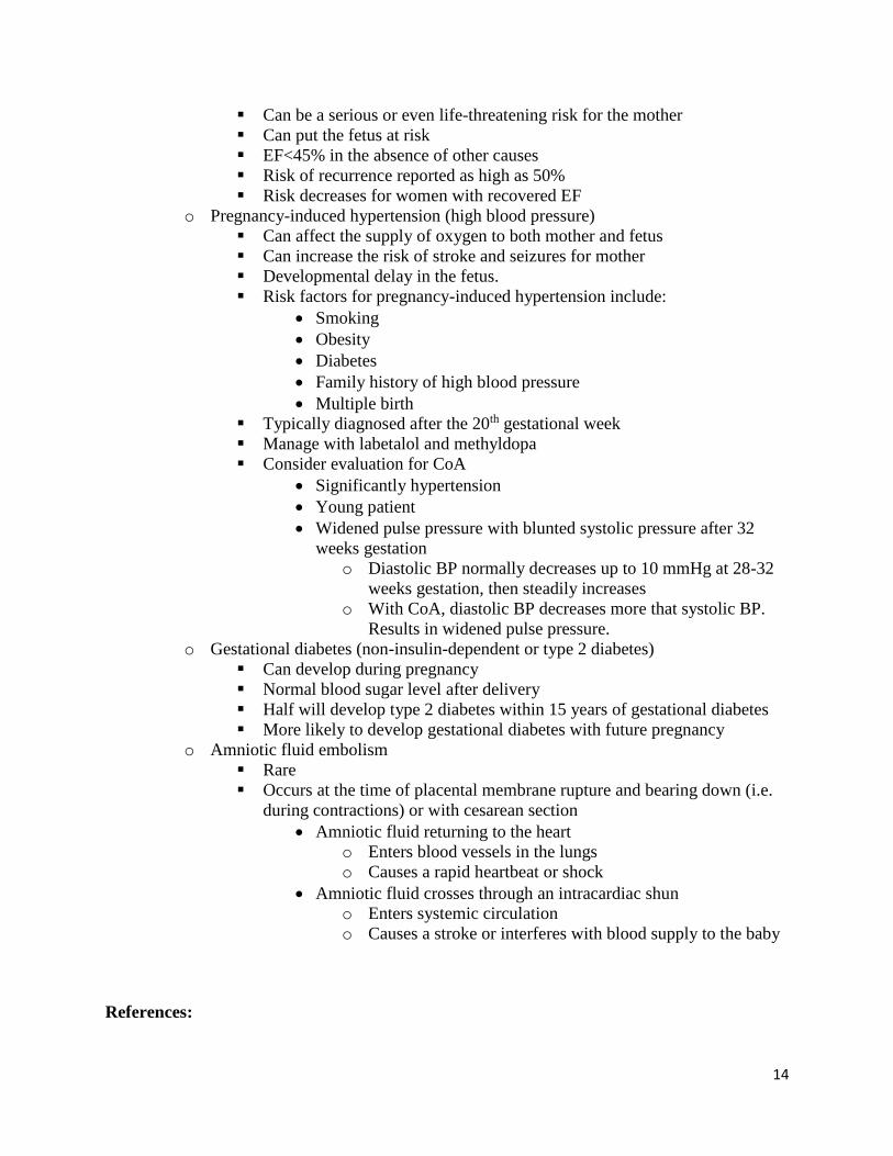
14
Can be a serious or even life-threatening risk for the mother
Can put the fetus at risk
EF<45% in the absence of other causes
Risk of recurrence reported as high as 50%
Risk decreases for women with recovered EF
o Pregnancy-induced hypertension (high blood pressure)
Can affect the supply of oxygen to both mother and fetus
Can increase the risk of stroke and seizures for mother
Developmental delay in the fetus.
Risk factors for pregnancy-induced hypertension include:
Smoking
Obesity
Diabetes
Family history of high blood pressure
Multiple birth
Typically diagnosed after the 20th gestational week
Manage with labetalol and methyldopa
Consider evaluation for CoA
Significantly hypertension
Young patient
Widened pulse pressure with blunted systolic pressure after 32
weeks gestation
o Diastolic BP normally decreases up to 10 mmHg at 28-32
weeks gestation, then steadily increases
o With CoA, diastolic BP decreases more that systolic BP.
Results in widened pulse pressure.
o Gestational diabetes (non-insulin-dependent or type 2 diabetes)
Can develop during pregnancy
Normal blood sugar level after delivery
Half will develop type 2 diabetes within 15 years of gestational diabetes
More likely to develop gestational diabetes with future pregnancy
o Amniotic fluid embolism
Rare
Occurs at the time of placental membrane rupture and bearing down (i.e.
during contractions) or with cesarean section
Amniotic fluid returning to the heart
o Enters blood vessels in the lungs
o Causes a rapid heartbeat or shock
Amniotic fluid crosses through an intracardiac shun
o Enters systemic circulation
o Causes a stroke or interferes with blood supply to the baby
References:

15
Balci,A., Sollie-Szarynska, K. M,, van der Bijl, A. G., et al. (2014). Prospective validation and
assessment of cardiovascular and offspring risk models for pregnant women with congenital
heart disease. HEART, 100, 1373-1381.
Brickner, M. E. (2014) Cardiovascular management in pregnancy: Congenital heart disease.
Circulation, 130, 273-282.
Canobbio, M. M., Warnes, C. A., Aboulhosn, J., et al. on behalf of the American Heart
Association on Cardiovascular and Stroke Nursing; Council on Clinical Cardiology; Council on
Cardiovascular Disease in the Young; Council on Functional Genomics and Translational
Biology; and Council on Quality of Care and Outcomes Research. (2017). Management of
pregnancy in patients with complex congenital heart disease. A scientific statement for
healthcare professionals from the American Heart Association [Published online January 12,
2017]. Circulation, 135;XXX-XXX. doi:10.1161/CIR.00000000000000458
Pierpont, M. E., Basson, C. T., Benson, D. W. Jr., et al. (2007). Genetic basis for congenital heart
defects: current knowledge: a scientific statement from the American Heart Association
Congenital Cardiac Defects Committee, Council on Cardiovascular Disease in the Young:
endorsed by the American Academy of Pediatrics. Circulation, 115(23), 3015-38.
Wald, R. M., Colman, J. M. (2009). Pregnancy and contraception. In Warnes CA, editor: Adult
Congenital Heart Disease, Oxford, Wiley-Blackwell.
16
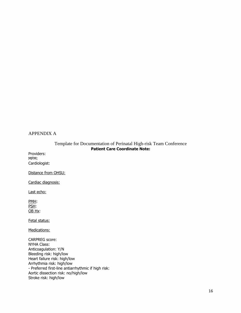
APPENDIX A
Template for Documentation of Perinatal High-risk Team Conference Patient Care Coordinate Note:
Providers: MFM:
Cardiologist:
Distance from OHSU:
Cardiac diagnosis:
Last echo:
PMH: PSH:
OB Hx:
Fetal status:
Medications:
CARPREG score:
NYHA Class:
Anticoagulation: Y/N Bleeding risk: high/low
Heart failure risk: high/low Arrhythmia risk: high/low
- Preferred first-line antiarrhythmic if high risk:
Aortic dissection risk: no/high/low Stroke risk: high/low
17
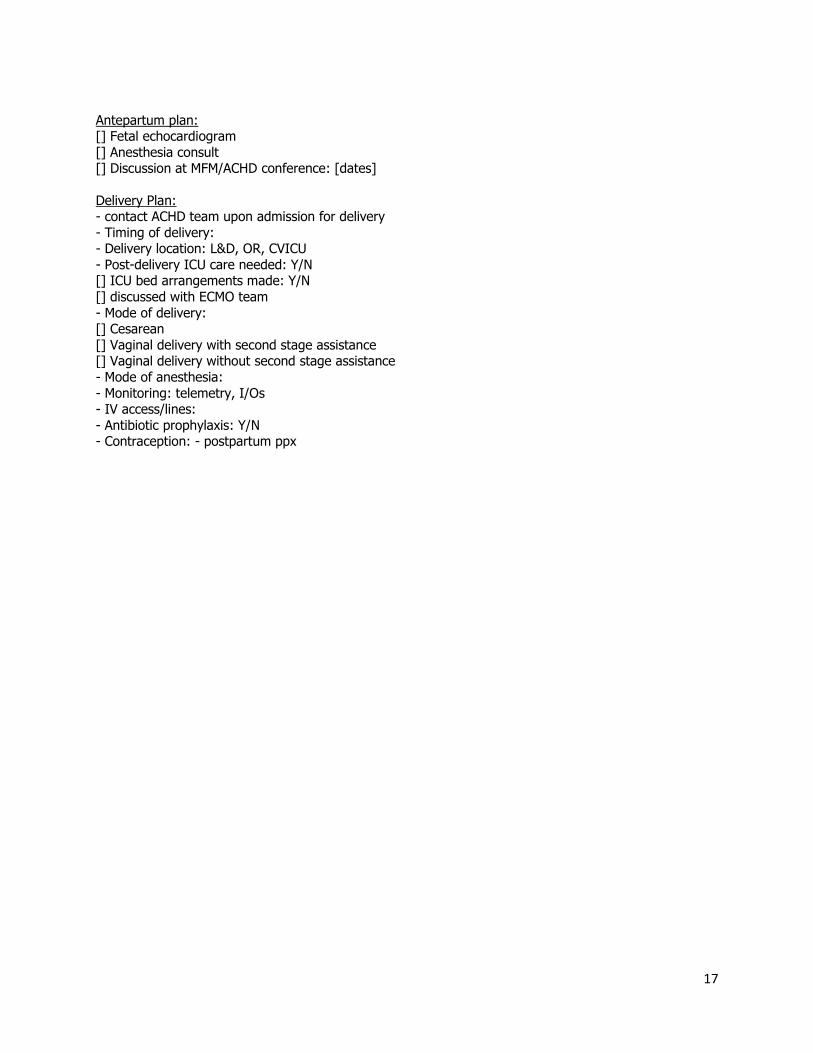
Antepartum plan: [] Fetal echocardiogram
[] Anesthesia consult [] Discussion at MFM/ACHD conference: [dates]
Delivery Plan: - contact ACHD team upon admission for delivery
- Timing of delivery: - Delivery location: L&D, OR, CVICU
- Post-delivery ICU care needed: Y/N [] ICU bed arrangements made: Y/N
[] discussed with ECMO team
- Mode of delivery: [] Cesarean
[] Vaginal delivery with second stage assistance [] Vaginal delivery without second stage assistance
- Mode of anesthesia:
- Monitoring: telemetry, I/Os - IV access/lines:
- Antibiotic prophylaxis: Y/N - Contraception: - postpartum ppx