PREFERRED PRACTICES
120
PREFERRED PRACTICES IN PEDIATRIC OPHTHALMOLOGY FOR INDIA
Transcript of PREFERRED PRACTICES

PREFERRED PRACTICESIN PEDIATRIC OPHTHALMOLOGY FOR INDIA


CONTENTS1. History and Physical Examination in Children 1
2. Refraction in Children 2
3. Binocular Vision Assessment in Children 2
4. Contact Lens Referral and Prescribing Guidelines in Children 12
5. Amblyopia 13
6. Sedation Protocol 20
7. Child with Developmental Delay 20
8. Nystagmus 23
9. Epiphora 25
10. Strabismus – Comitant 28
11. Incomitant Strabismus 37
12. Botulinum Toxin Injection 39
13. Allergic Conjunctivitis 40
14. Pediatric Corneal Opacities 42
15. Pediatric Cataract 44
16. Subluxated Lens 50
17. Congenital Disc Anomalies 52
18. Retinopathy of Prematurity 56
19. Retinal Dystrophies 61
20. Pediatric Ocular Trauma 66
21. Pediatric Low Vision Care 69

This preferred practices in pediatric ophthalmology is intended to serve as a guide to the clinician seeing child patients with eye problems. The manual tries to be comprehensive and not exhaustive.
Clinicians with predominantly or large pediatric ophthalmic practice are advised to read specialized texts for in depth and more upto date management of their patients.
It gives details of examination techniques in children and selectively elaborates on treatment. This handbook also includes sections describing strabismus in adults as most strabismus regardless of age are managed by pediatric ophthalmologists.
GUIDELINE OBJECTIVESThis is a clinical guideline for the general ophthalmologists and also practicing pediatric ophthalmologists for their basic and routine Outpatient. In depth study of the listed topics is essential for full time pediatric ophthalmologists and this is not a substitute for that.
To develop an appropriate timetable for eye and vision examinations for pediatric patients To select appropriate examination procedures for all pediatric patients To minimize or avoid the adverse effects of eye and vision problems in children through early identification, education, treatment, and prevention.

Foreword
India is the second most populous country in the world and home to over 20% of the world’s blind population. Unfortunately, India is also home to the largest number of blind children in any one country.
To address this, Orbis launched the India Childhood Blindness Initiative (ICBI) and pioneered the introduction of comprehensive pediatric ophthalmology services in several states.
In our journey towards eliminating needless blindness, Orbis has worked with numerous partners including the Centers of Excellence in eye care - Aravind Eye Care System, L V Prasad Eye Institute, and Sankara Nethralaya. We established Pediatric Ophthalmology Learning and Training Centers (POLTCs) with a structured curriculum to train cadres of professionals to specifically address the eye care needs of children. These initiatives among others contributed to the development of pediatric ophthalmology as a distinct sub-specialty in the Indian ophthalmology landscape. Also, under the aegis of ICBI, Orbis has supported the development of 31 Children’s Eye Centers (CEC) across 17 states of the country which remains the world’s largest network of Children’s Eye Centers in any one country; and the network continues to grow.
The logical next step was to consolidate the knowledge and practices of our partners. Discussions with our partners and practicing pediatric ophthalmologists reinforced the need for a Pediatric Ophthalmology Preferred Practices specific to the Indian context. We consulted our POLTCs and requested Sankara Nethralaya, Chennai to lead the development of this India-specific Preferred Practices in Pediatric Ophthalmology Feedback from POLTC Faculty and Orbis Volunteer Faculty in India was received and incorporated giving us this document which we are happy to share with you.
I thank Dr. T S Surendran and the team at Sankara Nethralaya for taking up this task and sincerely appreciate their outstanding efforts. I would also like to thank Dr. Vijayalakshmi, Dr. Ramesha Kekunnaya, Dr. Mihir Kothari, Dr. Pariskhit Gogate, and Dr. Suma Ganesh for their valuable suggestions and comments which greatly helped in making this Preferred Practices in Pediatric Ophthalmology what it is.
We hope pediatric ophthalmologists across the country will find this a useful resource in our constant endeavour to provide quality eye care to every child in need.
Best wishes,Dr. Rahul AliCountry Director – IndiaOrbis International

Foreword
I congratulate Orbis for bringing out this book and all others who contributed to it.
Pediatric ophthalmology has evolved in India for almost three decades and I have an umbilical cord attachment to the subject which started with just around a dozen ophthalmologists and has spread to over a hundred ophthalmologists at present in our country. Seeds were sown both in government and non-governmental organizations to spread the message.
A lot of interest has been shown by the young post graduates with Pediatric ophthalmology as their specialization. The speciality gets support from optometrists, anaesthetists, nursing and para medical staff. It is heartening to see many centers taking up this speciality. After all, taking care of children’s eyes is a big challenge.
Pediatric ophthalmology has a wide spectrum starting from refractive errors, congenital cataract, congenital glaucoma, ptosis and other oculoplasty disorders to retinopathy of prematurity, retinoblastoma and more.
Once again, I thank Orbis and all other contributors for bringing out this book.
Wishing the book and its readers the very best.
Dr. T S SurendranVice Chairman and Director of Pediatric Ophthalmology DepartmentSankara Nethralaya
“The Preferred Practices in Pediatric Ophthalmology formulated by Orbis in pediatric ophthalmology comes from the collective wisdom of its 30+ partner organizations, some of the most active/proactive and best pediatric eye care centers in the world. The Preferred Practices in Pediatric Ophthalmology shall go a long way in raising the standard of children’s vision care delivered in the Indian sub-continent and beyond. But tit will be a true success when the Preferred Practices in Pediatric Ophthalmology is widely circulated and used, by all eye care practitioners (ophthalmologists, optometrists and orthoptists) in India and the neighbouring countries”.
-Dr. Parikshit Gogate, Pediatric Ophthalmologist, Dr. Gogate’s Eye Clinic
“The Preferred Practices in Pediatric Ophthalmology for India compiled by the pediatric ophthalmology team at Sankara Nethralaya is an excellent resource for all the pediatric ophthalmologists and general ophthalmologists practicing pediatric ophthalmology across India. The academic content is excellent and will surely benefit all”.
-Dr. Suma Ganesh, Head of Department, Pediatric Ophthalmology and Strabismus, Head of Medical Education Department, Dr. Shroff’s Charity Eye Hospital
“Pediatric ophthalmology in countries like India is rapidly gaining due attention with eye care units extending their services and addressing the specific child eye care needs.. This module, on preferred practices has been designed to suit the need of pediatric ophthalmology personnel at various levels. The Preferred Practices in Pediatric Ophthalmology’s relevance stands in its modern day practice information such as the recommendations on prescribing low vision devices and the inclusion of separate guidelines to rehabilitate visually impaired infants and children with and without other developmental disorders”.
I congratulate all the authors who have put in their time and energy; and greatly appreciate the tremendous efforts of the Orbis team to bring out this publication.
-Dr. P. Vijayalakshmi, Chief, Pediatric Ophthalmology & Strabismus Department, Aravind Eye Hospital
“This concise document on clinical pediatric ophthalmology can serve as a mini-textbook. It can be used by the residents in ophthalmology and clinicians as a preferred practices for various common pediatric eye diseases. The indexing of the topics is simple and finding the relevant text is very easy. The information is the latest and accurate . General ophthalmologists will find it useful as a ready reckoner and pediatric ophthalmologists can use it as a check list while working in the clinic”.
-Dr Mihir Kothari, Director, Jyotirmay Eye Clinic, Ocular Motility Lab and Pediatric Low Vision Center
“Preferred Practices in Pediatric Ophthalmology’ by Orbis is really a value addition to professionals involved in children’s eye care in India and other developing countries. It fulfils a much needed guideline for eye care including strabismus and eye movement disorders in children. These are standard protocols followed or adopted by most pediatric ophthalmologists which will benefit many”.
-Ramesh Kekunnaya, MD,FRCS, Head & Consultant, Pediatric Ophthalmology, Strabismus & Neuro-Ophthalmology Jasti V Ramanamma Children’s Eye Care Center L V Prasad Eye Institute (LVPEI)

About the AuthorsThe consultants of pediatric ophthalmology department, Sankara Nethralaya are the main
contributors. They are as follows:
Dr Surendran T S Director
Dr Sumita Agarkar Deputy Director
Dr Meenakshi Swaminathan Senior Consultant
Dr Kavitha Kalaivani Senior Consultant
Dr Srikanth Ramasubramanian Associate Consultant
Dr Akila Ramkumar Associate Consultant
The other contributors from Sankara Nethralaya include:
Dr G Suganeshwari Consultant- Vitreoretinal Services
Dr Bipasha Mukherjee Director - Orbit, Oculoplasty, Reconstructive
and Aesthetics Services
Dr Rajeswari M Head, Contact Lens Department
Ms Jameel Rizwana Hussaindeen Head, Binocular Vision Clinic
Ms Kalpa N Senior Optometrist, Pediatric Optometry
Ms Sarika G Senior Optometrist, Low Vision Care Services
Ms Sailaja MVS Senior Optometrist, Low Vision Care Services
The other contributors were the pediatric ophthalmology fellows:
Dr Jayesh Patil. Dr Soumya Nambiar and Dr Shruti Nishanth
The Preferred Practices in Pediatric Ophthalmlogy would not have been possible without the valuable contribution of Sankara Nethraya. We extend our immense gratitude to them.
Sankara Nethralaya (Main Campus)
No. 41 (old 18), College Road, Nungambakkam,
Chennai - 600 006, Tamil Nadu , India.
T: 91-044-42271500 | F: +91-44-28254180
www.sankaranethralaya.org
Email: [email protected], [email protected]

1
HISTORY TAKING AND PHYSICAL EXAMINATION IN CHILDREN
PATIENT HISTORY
PRESENTING COMPLAINT
1) Blurred Vision
• Monocular/ Binocular
• Distance/ Near(Difficulty with viewing blackboard/ television/ holding books closer)
• Squeezing of eyes
• Frequent blinking
• Day/ Night difference
2) Photophobia
3) Squint
• Onset, duration and progression
• Variability
• Intermittent or constant
• Preferred eye
• Eye movement limitation
4) Abnormal head posture/Head oscillation
• Face turns/ Head tilt/ Chin up or down
• Intermittent/ Constant
5) Wobbly/ Shaking eyes
• Timeof on set and duration
• Any precipitating event like trauma, surgery, fever
6) Asthenopia
• Related to a particular activity
• Related to certain time of day
• Any associated diplopia or blurring of vision
• Symptoms relieved when initiating activity is discontinued or when patient reads with one eye closed
7) Watering / Epiphora
8) Itching and redness of eyes
9) White Reflex
10) Drooping of the eyelid
11) Diplopia
• Monocular or binocular
• If Monocular: consider corneal or pupillary abnormality, lenticular opacification or retinal pathology
• Binocular: strongly suggests strabismic origin
• Horizontal or vertical separation of images.
• Torsional can be observed by asking patient to look at the edge of a door and observe tilt
• Diurnal variation
• Worse in which gaze
• Worse for distance or near
• Precipitating factor
PAST HISTORY
• History of patching
• History of spectacle wear
• History of trauma to the eye/head
• Past ocular or strabismus surgery or
general surgery
BIRTH HISTORY
• Full term or premature
• Age of gestation
• Birth weight
• Mode of delivery (normal/ caesarean/ forceps)
• H/o birth asphyxia
• H/o fever with rashes in mother during pregnancy
• H/o neonatal convulsions/ jaundice
• H/o CRS and NICU admission
• H/o oxygenation therapy
DEVELOPMENT AL MILESTONES
• Global motor and speech development
• Any regression of milestones
• Hyperactive behaviour (autism)
• Learning disability
• Related investigations and rehabilitation done like CT or MRI

2
FAMILY HISTORY
• H/o consanguinity in parents
• Presence of hereditary forms of strabismus
• F/h of other inherited eye diseases in the family members
• Pedigree chart in selected cases
REVIEW OF SYSTEMS
• Neurologic symptoms
• Headache, seizures, ataxia, muscle weakness, fatigue, ptosis, bowel and bladder incontinence, facial asymmetry
• Cardiac
• Congenital heart disease, mitral valve prolapse
• Respiratory
• Wheezing, allergic rhinitis
• Tachypnoea
• Other systemic abnormalities and syndromes
EXAMINATION
VISUAL
1) 0-6 Months
• Menace reflex
• Visual dampening of induced nystagmus on rotation
2) 6 Months -4 Years
• Fixation preference tests
• Preferential looking visual acuity test
• Teller acuity cards / Lea Gratings
• Cardiff Acuity Cards
An infant undergoing visual assessment with Lea paddles
3) 4-6 Years
• Lea Symbols chart
• Broken Wheel acuity cards
• HOTV test
4) 6 Years and Above
• Snellen letter / ETDRS logMAR vision chart)
• KAY pictures
REFRACTION
• Cycloplegic retinoscopy
• Near retinoscopy/ Dynamic retinoscopy)
• Static retinoscopy
• Subjective refraction
BINOCULAR VISION, ACCOMMODATION AND OCULAR MOTILITY IN CHILDREN
• Cover tests (cover uncover/ alternate prism cover test)
• Hirschberg test
• Krimsky test
• Brückner test
• Versions and ductions
• Fusion and stereopsis
• Bielschowsky three step test
If Required:
• Near point of convergence (NPC)
• Positive and negative fusional vergences (prism bar/step vergence testing)
• Accommodative amplitude and facility

3
OCULAR ASSESSMENT
• Evaluation of the anterior segment and adnexa
• Evaluation of the posterior segment
• Assessment of pupil lary responses
• Visual field screening (confrontation)
• Color vision testing
– Pediatric color vision testing (matching chart by Helveston)
– Ishihara screening plates• Measurement of intraocular pressure (IOP)
The following section will give specific guidelines for examination of children of various age groups
EXAMINATION OF INFANTS AND TODDLERS (BIRTH TO 3 YEARS)
GENERAL CONSIDERATIONS
Children in this age group generally perform best
if the examination takes place when they are
alert. Examination early in the morning or after an
infant’s nap is usually most effective. Infants tend
to be more cooperative and alert when feeding.
Hence, it is also helpful to suggest that the parent
bring a bottle for the child. Bottle feeding is
strongly discouraged by the pediatrics all over
the world for kids < 6 months age. Exclusive
breast feeding is the norm. Hence, let the mother
breast feed the baby in the feeding room and then
examine the baby.
Modifications include relying more on objective
examination procedures and performing tests
considerably more rapidly than with older
children.
1) Visual Acuity
Assessment of visual acuity for infants and toddlers may include these procedures:
• Menace reflex – in the dark, suddenly shine the bright light viz. full illumination of indirect ophthalmoscope. It is the first response that is
important and only the positive test confirms development of the visual pathways.
• Visual dampening (within first 10 seconds) after rotation for 30 seconds on the parent’s shoulder or by the ophthalmologist
• Fixation - central, steady, maintained
• 10-PD test
• Fixation preference tests /Resistance to occlusion
• Preferential looking visual acuity test
• Lea Symbol chart
Forced-choice preferential looking with the Teller acuity cards or electrodiagnostic testing should be considered to obtain a more precise measure of baseline visual acuity.
2) Refraction
Traditional subjective procedures for the assessment of refractive error may be ineffective with infants or toddlers because of short attention span and poor fixation. As a result, the examiner will need to rely on objective measures of refraction. The three most commonly used procedures are:
• Cycloplegic retinoscopy
• Near retinoscopy
• Dynamic retinoscopy
It is important for the examiner performing cycloplegic retinoscopy in an infant or toddler to take several precautions:
• Select the cycloplegic agent carefully (e.g., fair-skinned children with blue eyes may exhibit an increased response to drugs and darkly pigmented children may require more frequent or stronger dosages)
• Avoid overdosage. For instance, children with down syndrome, cerebral palsy, trisomy 13 and 18, and other central nervous system disorders in whom there may be an increased reaction to cycloplegic agents, two % homatropine eyedrops may be used
• Beware of biological variations in children (e.g., low weight infants < 5kg require 50% dilution)
Cyclopentolate hydrochloride is the cycloplegic

4
agent of choice but it is contraindicated in children with neurological disease or history of convulsions. One drop should be instilled twice, five minutes apart, in each eye, using strength of 0.5% for children from birth to one year and one % for older children. The regimen that works well in children with dark irides is one drop of cyclopentolate one % followed five minutes later by one drop of tropicamide one % and followed 5 minutes later by one more drop of one % cyclopentolate. Retinoscopy may be performed 45 minutes after instillation. The use of loose lenses or a lens rack is recommended for retinoscopy. Atropine refraction may be considered if we suspect:
1. Pseudomyopia or accommodative spasm
2. Accommodative esotropia with small residual squint over glasses
3. Vergence anomalies
4. Varying retinoscopy values
Near retinoscopy: without an accommodative target in dark, the illumination of the streak works as the non accommodative target is another objective method of estimating refractive error in infants and toddlers. However, it has not been found reliable for quantification of the refractive error.Near retinoscopy may have some clinical value in the following situations:
• When frequent follow-up is necessary
• When the child is extremely anxious about instillation of cycloplegic agents
• When the child has had or is at risk of an adverse reaction to cyclopentolate or tropicamide
The average refractive error in children from birth to one year of age is about two diopters (D) of hyperopia (standard deviation 2 D). Astigmatism up to 2 D is common in children under three years of age. Studies show that 30-50% of infants less than 12 months of age have significant astigmatism, which declines over the first few years of life, becoming stable by approximately 2½ to five years of age. Low amounts of anisometropia are common and variable in infants. The clinician may choose to monitor these levels of refractive error rather than prescribe a lens correction.
Dynamic retinoscopy is not same as near retinoscopy. While in near retinoscopy, child fixates at the light filament of the retinoscope and the accommodation is still assumed to be at rest, in dynamic retinoscopy, the child is accommodating on an appropriate object at near (33cm). It is necessary to diagnose accommodation failure especially in patients with down syndrome, cerebral vision impairment, foveal hypoplasia and internal ophthalmoplegia.
BINOCULAR VISION AND OCULAR MOTILITY
The following procedures are useful for assessing binocular function:
• Cover test
• Hirschberg test
• Krimsky test
• Bruckner test
• Versions
Pupillary evaluation, anterior segment and posterior segment examination to be performed as mentioned earlier.
EXAMINATION OF 3-5 YEAR OLD CHILDREN
GENERAL CONSIDERATIONS
Although the vast majority of children in this age group can communicate verbally, it is preferable in most cases for the parent/caregiver to accompany the child into the examination room. It is important to ensure that the child feels relaxed and at ease, which is often best accomplished by beginning the examination with procedures that appear less threatening.
Modifications include reliance on objective examination techniques, limited use of subjective techniques requiring verbal interaction, and performing testing considerably more rapidly than is typically used for older children.
EXAMINATION SEQUENCE
1) Visual Acuity
• Lea Symbols chart
• Broken Wheel acuity cards

5
• HOTV test
2) Refraction
• Static retinoscopy
• Cycloplegic retinoscopy
3) Binocular Vision, Accommodation, and Ocular Motility
• Cover test
• Positive and negative fusional vergences (prism bar/step vergence testing)
• Near point of convergence (NPC)
• Stereopsis
• Versions
Pupillary evaluation, anterior segment and posterior segment examination to be performed as mentioned earlier.
EXAMINATION OF OLDER CHILDREN (>5 YEARS OF AGE)
GENERAL CONSIDERATIONS
Some of the issues relating to younger children also apply to this population, particularly children younger than eight years old. Age-appropriate examination and management strategies should be used. Although most of the examination procedures used with this age group are identical to those recommended for adults, age-appropriate modifications of instructions and targets often may be required.
EXAMINATION SEQUENCE
1) Visual Acuity
Visual acuity may be assessed with the Snellen acuity chart (modified for children six to eight years of age). A recommended modification is the isolation of one line, or even one-half line of letters, rather than projection of a full chart.
2) Refraction
• Static (distance) retinoscopy
• Cycloplegic retinoscopy
• Subjective refraction
3) Binocular Vision, Accommodation, and Ocular Motility
• Cover test
• Near point of convergence (NPC)
• Positive and negative fusional vergences
• Accommodative amplitude
• Stereopsis
• Versions
Pupillary evaluation, anterior segment and posterior segment examination to be performed as mentioned earlier.
PATIENT EDUCATIONEducating parents or caregivers about any eye or vision disorders and vision care.
The importance of adhering to an eye and vision examination schedule should be emphasized educating parents/caregivers and children about eye safety, particularly regarding sports-related eye safety.
Importance of early, preventive eye care, including examinations at the age of six months, at age three, before entering first grade, and periodically during the school years.
RECOMMENDED EYE EXAMINATION FREQUENCY FOR THE PEDIATRIC PATIENT
AT BIRTH: RED REFLEX TEST
Detailed examination for ‘at risk infants’
BIRTH TO 24 MONTHS
Asymptomatic/risk-free: At six months of ageAt risk: At six months of age or as recommended
2 TO 5 YEARSAsymptomatic/risk-free: At three years of ageAt risk: At three years of age or as recommended
6 TO 18 YEARSAsymptomatic/risk-free: Before first standard and every two years thereafterAt risk: Annually or as recommended
Coordination, Frequency, and Extent of CareThe child’s first eye and vision examination should be scheduled at six months of age (or sooner if signs or symptoms warrant). When no

6
abnormalities are detected at this age, the next examination should be scheduled at age three.
The child considered at risk for the development of eye and vision problems may need additional testing or more frequent re-evaluation. Factors placing an infant, toddler, or child at significant risk for visual impairment include:
• Prematurity, low birth weight, oxygen at birth, grade III or IV intraventricular hemorrhage
• Family history of retinoblastoma, congenital cataracts, or metabolic or genetic disease
• Infection of mother during pregnancy (e.g., rubella, toxoplasmosis, venereal disease, herpes, cytomegalovirus, or human immunodeficiency virus). Difficult or assisted labour, which may be associated with fetal distress or low APGAR scores
• High refractive error
• Strabismus
• Anisometropia
• Known or suspected central nervous system dysfunction evidenced by developmental delay, cerebral palsy, dysmorphic features, seizures, or hydrocephalus
REFRACTIONDo subjective refraction if possible
Do cycloplegic refraction in all patients reporting to the pediatric ophthalmology clinic.
INDICATIONS FOR CYCLOPLEGIC REFRACTION
1) Refractive errors
• To assess the refractive power of the eye (Myopia, Hyperopia and Astigmatism)
• Poor cooperation/ fixation during refraction
• Fluctuations in the refractive error while performing dry retinoscopy
• Vision not correlating with the dry refraction
• To rule out latent hyperopia
• Refractive surgery – to assess the refractive error accurately and rule out latent component
2) Accommodative anomalies (accommodative spasm)
3) Amblyopia (penalization)
4) Strabismus (accommodative and partially accommodative esotropia)
Common cycloplegic agents
Agent Strength (percentage)
Mydriasis Cycloplegia Side effects
Maximum (min)
Recovery time
Maximum (min)
Recovery time
Phenylephrine 2.5 20 2-3hours None Tachycardia, hypertension
Tropicamide 0.5,1 20-40 2-6 hours 30 2-6h
Cyclopentolate 0.5,1,2 30-60 6-24 hours 25-75 6-24h Psychosis, seizure, gastro-intestinal disturbance
Homatropine 2,5 40-90 1-3 days 30-60 1-3days Ataxia
Atropine 0.5,1 30-60 7-14 days 60-180 3-12 days Dryness of mouth, Flushing, tachycardia, fever, delirium, irritability

7
CHOICE OF CYCLOPLEGIC AGENT
REGIMEN WHEN USING CYCLOPENTOLATE
One drop of 1% cyclopentolate is instilled at five minutes interval, followed by one drop of 0.5% tropicamide, again followed by instillation of 1% cyclopentolate once after five minutes interval.
Perform a cycloplegic refraction between 45 minutes and 75 minutes after the last drop instillation. If the cycloplegic refraction cannot be performed between 45 and 75 minutes, instill another drop of cyclopentolate one% in each eye and wait a minimum of thirty minutes more.
Regimen when not using cyclopentolate (if contraindicated especially when the child has seizures or is on antiseizure medication).
One drop of Homatropine 2% is applied followed by one drop of 0.5% tropicamide at five minutes interval. Cycloplegic refraction can be carried out appropriately in all new patients below 12 years of age, and in all children with refractive errors detected for the first time.
Atropine (1%) Ointment
• Dose: three times a day x three days
• Recommended in:
• Accommodative esotropia
• Accommodative spasm
• Varying retinoscopy values
Drops for premature babies and infants <six months of age: Recommended eye drops are tropicamide 0.5%-1% with phenylephrine 2.5%. two to three instillations of each of these drops, five minutes apart, Mydriasis in fifteen to twenty minutes and effect lasts thirty to forty five minutes.
Do wipe the drops spilled over cheeks as it can be absorbed from skin and cause increased heart rate
Avoid 10% phenylephrine in premature babies as it causes severe tachycardia, hyperthermia and dehydration
ROP drops can be prepared by mixing 3 ml of 1% tropicamide with 1 ml of 10% phenylephrine. The solution can be stored at room temperature for three days. This preparation is available commercially as well.
NOTE:
Adverse effects to be explained to parents
Children with light Colored eyes and infants may need modification of dose.
Avoid atropine in down’s syndrome, cerebral palsy, trisomy 13, 18 and other CNS disorders.
Cyclopentolate is contraindicated in children with seizures, mental retardation, cerebral palsy and other neurological abnormalities
Method• Do retinoscopy after maximum cycloplegia is
achieved
• Darken the room to avoid distraction
• Child can fixate on retinoscope light
• Use loose lenses to neutralize the reflex
• Post- mydriatic test – To be done in older
children
Infants Tropicamide(1%) 1 + 1=20min
Premature infants ROP drops (3:1)(Tropicamide + Phenylephrine)
Delayed milestones, seizures and CNS disorders Homatropine (0.5% -1%) + Tropicamide (1%) 1+1+1=30 min
Down’s syndrome Cyclopentolate(0.5% - 1%) + Tropicamide(1%) 1+1+1=30min
Normal children(1 – 16years) Cyclopentolate (0.5% - 1%) + Tropicamide (1%) 1+1+1 = 30min
8
TESTS PERFORMED SPECIFIC TO CONDITIONS
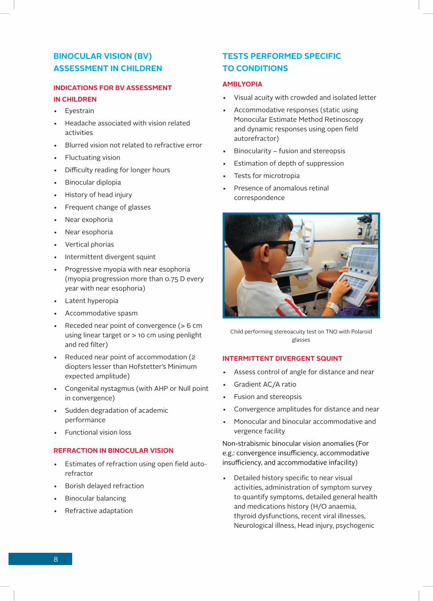
AMBLYOPIA
• Visual acuity with crowded and isolated letter
• Accommodative responses (static using Monocular Estimate Method Retinoscopy and dynamic responses using open field autorefractor)
• Binocularity – fusion and stereopsis
• Estimation of depth of suppression
• Tests for microtropia
• Presence of anomalous retinal correspondence
Child performing stereoacuity test on TNO with Polaroid glasses
INTERMITTENT DIVERGENT SQUINT
• Assess control of angle for distance and near
• Gradient AC/A ratio
• Fusion and stereopsis
• Convergence amplitudes for distance and near
• Monocular and binocular accommodative and vergence facility
Non-strabismic binocular vision anomalies (For e.g.: convergence insufficiency, accommodative insufficiency, and accommodative infacility)
• Detailed history specific to near visual activities, administration of symptom survey to quantify symptoms, detailed general health and medications history (H/O anaemia, thyroid dysfunctions, recent viral illnesses, Neurological illness, Head injury, psychogenic
BINOCULAR VISION (BV) ASSESSMENT IN CHILDREN
INDICATIONS FOR BV ASSESSMENT IN CHILDREN• Eyestrain
• Headache associated with vision related activities
• Blurred vision not related to refractive error
• Fluctuating vision
• Difficulty reading for longer hours
• Binocular diplopia
• History of head injury
• Frequent change of glasses
• Near exophoria
• Near esophoria
• Vertical phorias
• Intermittent divergent squint
• Progressive myopia with near esophoria (myopia progression more than 0.75 D every year with near esophoria)
• Latent hyperopia
• Accommodative spasm
• Receded near point of convergence (> 6 cm using linear target or > 10 cm using penlight and red filter)
• Reduced near point of accommodation (2 diopters lesser than Hofstetter’s Minimum expected amplitude)
• Congenital nystagmus (with AHP or Null point in convergence)
• Sudden degradation of academic performance
• Functional vision loss
REFRACTION IN BINOCULAR VISION
• Estimates of refraction using open field auto-refractor
• Borish delayed refraction
• Binocular balancing
• Refractive adaptation

9
illness, medications for seizures, psychogenic illness)
• Refraction
• Sensory and motor evaluation
• Horizontal and vertical phoria for distance and near
• Near point of convergence
• Fusional vergence for distance and near
• Vergence facility
• Near point of accommodation
• Accommodative responses using Monocular Estimate method Negative and Positive relative accommodation
• Accommodative facility
ACCOMMODATIVE ESOTROPIA
• Accommodative amplitudes
• Gradient AC/A ratio
• Response CA/C and AC/A ratio
• Near angles with and without addition
• MEM retinoscopy
ACCOMMODATIVE SPASM
• Refraction with and without cycloplegia
• Phoria/ tropia with and without cycloplegia
• AC/A ratio
• Cycloplegic and non-cycloplegic automated refraction measurements
• Biometry (To document axial length)
• Accommodation responses (to check lead of accommodation)
• Subjective acceptance with and without cycloplegic correction
• Progressive myopia
• Phoria for distance and near
• Gradient AC/A ratio
• Accommodative responses (to see if lag of accommodation is > +0.75 D)
• Near esophoria
NYSTAGMUS
• Assess the null point and AHP
• Adaptation with Yoke prisms
• Try Base out prisms for Nystagmus that dampens with convergence
• Binocular visual acuity with and without prisms
• AHP with and without prisms
• Photographic and video-graphic documentation
DIPLOPIA
• Magnitude of phoria/ tropia in all gazes for distance and near
• Hess/ Diplopia charting for Palsy and Paresis
• Vertical fusional reserves in congenital/ longstanding superior oblique palsy
• Compensatory fusional vergence reserves in intermittent squint
• Contact lenses for Anisometropia and reassess fusion
• Prisms for fusion
• Vision therapy to improve fusional vergence reserves in intermittent squint
• In Fresnel prisms, visual acuity with and without Fresnel prisms
HEAD INJURY (WITH NORMAL/ NEAR NORMAL VISUAL ACUITY)• Accommodation and vergence
• Saccades and pursuits
• Reading parameters and eye movements assessment using Developmental eye movement test and Readalyzer Infrared eye movement tracking system
• Visual information processing assessment (Tests for visual perceptual skills)
• Prisms as field expanders in required cases
• Yoke prisms in reading difficulty/ hemianopia
• Learning related vision problems
• Review with recommendation from educational/school psychologist
• Visual information processing assessment (tests for visual perceptual skills)
• Accommodation and vergence testing
• Oculomotor testing (Saccades and Pursuits)
10
VISION THERAPY
• Home therapy/ In-office vision therapy indicated for
• Non-strabismic binocular vision anomalies
• Amblyopia
• Intermittent squint
• Learning related vision problems
GOALS OF VISION THERAPY
• Anti-suppression
• Monocular accommodation, fixation and ocular motility training
• Gross vergence and training to appreciate physiological diplopia
• Smooth and jump vergence training
• Binocular accommodation training
• Visual information processing training
• Integration of procedures
PRESCRIPTION OF GLASSES IN CHILDREN
DECIDING FACTORS WHILE PRESCRIBING GLASSES• Age of the child
• Visual needs
• Symptoms
• Strabismus
• Amblyopia
• Apkhakia /pesudophakia
MYOPIA• Give full correction including cylinder
• Anisomyopia of more than 3D is amblyogenic
• Contact lenses, in high myopes, to avoid image minification may be prescribed in older children
• Avoid overcorrection of myopia in orthophoric children
• Do not prescribe myopic correction without a cycloplegic refraction
• Look for ocular or systemic causes in case of high myopia
HYPEROPIA• Insignificant hyperopia i.e., upto +3.0, in the
absence of esotropia or reduced vision, can be left uncorrected
• Hyperopia can be amblyogenic if:
– At 0-1 years it is > 4D – At 1-2 years more than 3D – At 2-6 years more than 2D• If there is an esodeviation full cycloplegic
correction is prescribed
• Bifocals can be prescribed in case of high AC/A ratio
In school-going children, less than full cycloplegic correction (reduce 1-2-D) could be prescribed, to avoid distance blur, even if there is esotropia.Hyperopia may be corrected, even if insignificant, if there is a strong family history of
11
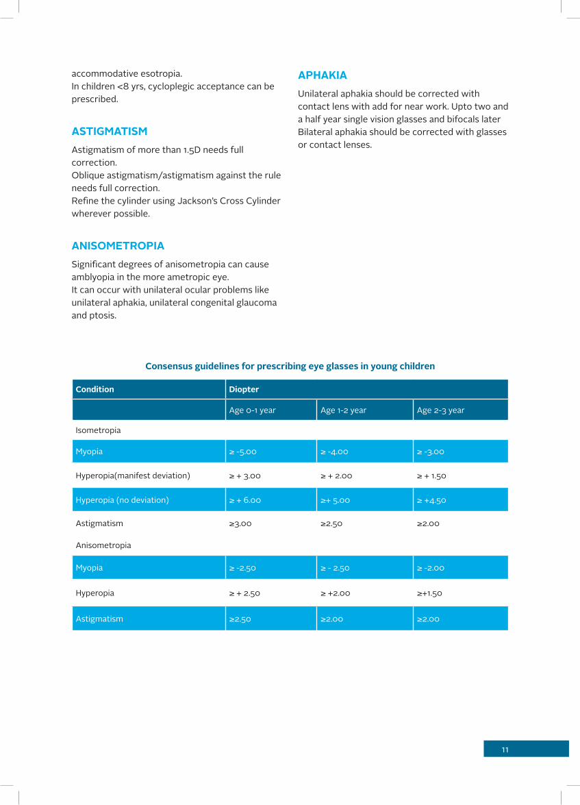
Condition Diopter
Age 0-1 year Age 1-2 year Age 2-3 year
Isometropia
Myopia ≥ -5.00 ≥ -4.00 ≥ -3.00
Hyperopia(manifest deviation) ≥ + 3.00 ≥ + 2.00 ≥ + 1.50
Hyperopia (no deviation) ≥ + 6.00 ≥+ 5.00 ≥ +4.50
Astigmatism ≥3.00 ≥2.50 ≥2.00
Anisometropia
Myopia ≥ -2.50 ≥ - 2.50 ≥ -2.00
Hyperopia ≥ + 2.50 ≥ +2.00 ≥+1.50
Astigmatism ≥2.50 ≥2.00 ≥2.00
Consensus guidelines for prescribing eye glasses in young children
accommodative esotropia.In children <8 yrs, cycloplegic acceptance can be prescribed.
ASTIGMATISMAstigmatism of more than 1.5D needs full correction.Oblique astigmatism/astigmatism against the rule needs full correction.Refine the cylinder using Jackson’s Cross Cylinder wherever possible.
ANISOMETROPIASignificant degrees of anisometropia can cause amblyopia in the more ametropic eye.It can occur with unilateral ocular problems like unilateral aphakia, unilateral congenital glaucoma and ptosis.
APHAKIAUnilateral aphakia should be corrected with contact lens with add for near work. Upto two and a half year single vision glasses and bifocals later Bilateral aphakia should be corrected with glasses or contact lenses.

12
• Patient’s parents are taught procedures how to insert, remove and clean the CL
• To insert CL when the baby is awake and remove just after sleeping, though it is an extended wear material
• Lens power is calculated from the cycloplegic retinoscopy value, 12 mm vertex conversion and intermediate distance. No add prescribed in case of aphakia
• Base curve is determined after keratometry and considering lens thickness
• Lens diameter 2mm less than HVID
• Review every three months
• Spare lenses dispensed to use in case of lens loss
PRESCRIBING FOR > 3 YEARS OLD• Lens made for distance power
• Near add prescribed whenever necessary
• Six months follow-up advised
• On parent’s request or if clinically recommended, soft or soft toric can be tried
General Note:
• Patients are referred with contact lens for squint evaluation and diplopia evaluation whenever indicated
• Prescriptions for age group less than two year old babies valid for three to six months only
• For age group 2 - 18 prescriptions valid for 6-12 months only
• The choice of lens is usually advised as higher oxygen permeable and disposability in children. In case of RGP three to six months disposal recommended with proper care regimen
CONTACT – LENS (CL) REFERRAL AND PRESCRIBING GUIDELINES IN CHILDREN
INDICATIONS• Refractive errors: Improves effective power,
clarity of vision:
– Myopia – Hyperopia – Astigmatism – Aphakia• Corneal scar – vision improvement
• Corneal dystrophies and degenerations – vision improvement
• Anisometropia – vision improvement, diplopia management and avoiding amblyopia, given along with patching therapy
• Amblyopia - vision improvement, diplopia management and avoiding amblyopia, given along with patching therapy
PROCEDURES FOLLOWED IN THE CONTACT LENS PRESCRIBING• Counseling parents about need for contact
lens care and maintenance
• Cycloplegic refraction
• Evaluation of anterior segment - evaluate for corneal staining, suture if any
• If loose sutures or staining present- remove the loose sutures and treat corneal abrasions before CL trial
• Horizontal Visible Iris Diameter (HVID)
• Lid tone
• Keratometry –under sedation or under GA for infants and uncooperative children
• Choice of lens decided as per the above preliminary evaluation and the available contact lens
PRESCRIBING FOR BABIES LESS THAN TWO YEARS:• First choice of lens - RGP extended wear 60
DK

13
AMBLYOPIA
DISEASE DEFINITIONAmblyopia is a unilateral or, less commonly, bilateral reduction of best-corrected visual acuity that cannot only and directly be attributed to the effect of a structural abnormality of the eye or the visual pathways. Amblyopia is caused by abnormal visual experience early in life resulting from one of the following:
• Strabismus
• Anisometropia or high bilateral refractive errors (ametropia)
• Visual deprivation
PATIENT POPULATION• Children younger than six years are at highest
risk for development of amblyopia as this is the age of plasticity. However, age up to 12 has been associated with development of amblyopia
• Goals
• The goals of the patient care process are as follows:
PREDICT DISEASE• Identify children at risk
DIAGNOSE • Examine and diagnose the child with
amblyopia or risk factors for amblyopia at the earliest possible stage
• Identify etiology of amblyopia and formulate an appropriate treatment plan
INFORM • Inform the family/caregiver of the diagnosis,
treatment options, and care plan
TREAT • Treat infants and children with amblyopia
in order to improve visual acuity, facilitate the treatment of strabismus, and reduce the likelihood of vision-related disability
MONITOR• Re-evaluate the patient and adjust the
treatment plan as necessary
DIAGNOSIS
HISTORY
Although a thorough history generally includes the following items, the exact composition varies with the patient’s particular problems and needs:• Demographic data, including identification of
parent/caregiver, and patient’s gender and date of birth
• Documentation of identity and relationship of the person from whom history is sought
• The identity of other pertinent health care providers
• The chief complaint and reason for the eye evaluation
• Current eye problems
• Ocular history, including prior eye problems, diseases, diagnoses, and treatments
• Systemic history; birth weight; prenatal and perinatal history that may be pertinent (e.g., alcohol, tobacco, and drug use during pregnancy); past hospitalizations and operations; general health and development
• Current medications and allergies
• Family history of eye conditions and relevant systemic diseases
• Review of systems
EXAMINATION
• The eye examination consists of an assessment of the physiological function and the anatomic status of the eye and visual system. Documentation of the child’s level of cooperation with the examination can be useful in interpreting the results and in making comparisons among the examinations over time. In general, the examination may include the following elements:
• Assessment of visual acuity and fixation pattern
• Ocular alignment and motility
• Red reflex or binocular red reflex (Brückner) test
14
• Pupil examination
• External examination
• Anterior segment examination
• Cycloplegic retinoscopy/refraction
• Funduscopic examination
• Binocularity/stereoacuity testing
• Assessment of Visual Acuity and Fixation Pattern
The method of evaluating visual acuity varies according to the child’s age and level of cooperation. Preverbal children should be checked for objection to cover and the presence of a fixation preference.
When possible, monocular distance visual acuity should be determined utilizing a recognized optotype, such as the tumbling-E, Lea figures, or Snellen letters.
Anomalous head posture is suggestive of peeking around the occluder. An occlusive patch over the non-tested eye can distinguish between peeking and possible eccentric fixation.
Monocular visual acuity testing for patients with nystagmus may require blurring of the contralateral eye with a high plus lens (+4.00 D to +5.00 D). Binocular and monocular testing also should be performed for patients with nystagmus.
Testing visual acuity with isolated targets (figures or letters) is the quickest way to assess the vision in preverbal children. But it does lead to falsely elevated visual acuities. Isolated acuities should be compared with visual acuities taken with linear targets or crowding bars. The difference between linear and isolated acuities should be noted at each visit, if possible. This difference is a way of quantifying the depth of amblyopia from visit to visit.
Under ideal circumstances, visual acuity testing conditions should be standardized in each examination room and at each visit, so that the same viewing distance and lighting conditions are used. Some children are more amenable to testing at shorter distances.
The testing distance, type of optotype, and whether the optotype is presented as a line at a time or isolated, should be documented. Patients should be encouraged to learn optotype-equivalent tests at the earliest possible age.
OCULAR ALIGNMENT AND MOTILITYOcular alignment is assessed by using the corneal light reflection, the binocular red reflex (Brückner) test, or the cover test.[A:III] Ocular versions and ductions should be tested even in the young infant. In the inattentive or uncooperative patient, eye movements may be tested using the oculocephalic rotations maneuver (doll’s head) or assessed by spontaneous eye movements.
RED REFLEX / BINOCULAR RED REFLEX (BRÜCKNER) TEST
The red reflex and/or binocular red reflex test should be performed to identify opacities of the ocular media.
The red reflex of each eye is assessed by looking at each eye with a direct ophthalmoscope from a distance of about 18 inches. The examiner should answer three questions:
1. Is there a red reflex from each eye?
2. Are the red reflexes from each eye symmetrical?
3. Is the quality of the red reflex normal for the individual child (taking into account skin tone and race or ethnicity)?
The binocular red reflex (Brückner) test allows an assessment of the clarity of the visual axis and an indirect assessment of ocular alignment as well as large and/or asymmetric refractive errors. The binocular red reflex test is performed in a dimly lit room with the examiner at a distance of about 30 inches (0.75 meter) from the child.
The examiner overlaps both pupils simultaneously, creating a binocular red reflex with the largest circular light of a direct ophthalmoscope set to focus on the ocular surface, usually at zero. The examiner then assesses the quality of the redness seen within the child’s pupils. Normally, the red reflex from each eye should be of the same color

15
and brightness. Abnormalities include asymmetric reflexes when one reflex is duller or a different color, a white reflex, a partially or totally obscured reflex, or crescents present in the reflex.
PUPIL EXAMINATION
Even in small infants, the pupils should be assessed for direct and consensual response to light and for the presence of a relative afferent defect.
This can be done with a penlight, a direct ophthalmoscope, or a transilluminator. Pupil evaluation in infants and children may be difficult due to active hippus or shift in the patient’s fixation and accommodative status. In general, amblyopia is not associated with a detectable afferent pupillary defect.
EXTERNAL EXAMINATION
External examination involves assessment of the eyelids, eyelashes, lacrimal apparatus, and orbit. The anatomy of the face (including the lids, interocular distance, and presence or absence of epicanthal folds), orbital rim, and presence of oculofacial anomalies should be noted.
The position of the head and face (including head tilt or turn) should be noted. Children with prominent epicanthal folds and normal ocular alignment may appear to have esotropia (pseudo-esotropia). Distinctive features unusual for the family may suggest the presence of a congenital anomaly and merit an assessment of other physical abnormalities (e.g., ears, hands).
ANTERIOR SEGMENT EXAMINATION
To evaluate further opacities of the ocular media, the cornea, anterior chamber, iris, and lens should be evaluated with slit-lamp biomicroscopy, if possible. Slit-lamp biomicroscopic evaluation is indicated for older children or for younger children who are cooperative. In infants and young children, a hand-held slit-lamp biomicroscope may be helpful. Some children may need to be restrained, sedated, or undergo an eye examination under general anesthesia when apparent abnormalities warrant a detailed examination.
CYCLOPLEGIC RETINOSCOPY/REFRACTION
Determination of refractive errors is important in the diagnosis and treatment of amblyopia or strabismus. Patients should receive an accurate cycloplegic refraction either by retinoscopy or by subjective refraction. Prior to cycloplegia, dynamic retinoscopy provides a rapid assessment of accommodative function and may be helpful in evaluating a child with high hyperopia or possible accommodative insufficiency.
FUNDUSCOPIC EXAMINATION
Posterior segment structures should be examined, preferably with an indirect ophthalmoscope. The optic disc, macula, retina, vessels, and the choroid of the posterior regions should be examined. Examination of the peripheral retinal and scleral indentation, if indicated, may require sedation or general anesthesia (e.g., evaluation for retinoblastoma).
BINOCULARITY / STEREOACUITY TESTING
Testing for binocular fusion (e.g., Worth 4-dot test) or the presence of stereopsis (e.g., Random- Dot E test or Stereo Fly test) may be useful in detecting ocular misalignment or amblyopia. Fusion and stereoacuity testing at distance (20 feet or 6 meters) as well as near (13 inches or 0.33 meter) may also be helpful.
CRITERIA FOR DIAGNOSISAmblyopia in the absence of strabismus, unequal refractive error, or media opacity is rare. A careful search for an alternate diagnosis with associated visual loss should be carried out if an obvious cause is not present. Amblyopia is diagnosed when the criteria in Table 1 are met and the cause is identified (Table 2).
MANAGEMENTIn managing amblyopia, the ophthalmologist strives to improve visual acuity by using two basic strategies. The first is to present a clear retinal image to the amblyopic eye by eliminating causes of visual deprivation and correcting visually important refractive errors.
The second strategy is to make the child use the amblyopic eye. While not always achievable,

16
the treatment goal is to achieve equalization/normalization of fixation patterns or visual acuity.
Child wearing an indigenously prepared patch
The recommended treatment should be based on the patient’s age; visual acuity; compliance with previous treatment; and physical, social, and psychological status.
CHOICE OF THERAPY
The following therapies are used alone or in combination as required to achieve the therapeutic goal:
• Optical correction
• Occlusion
• Penalization
• Surgery to treat the cause of the amblyopia
• In the Amblyopia Treatment Study Group trials, mild to moderate amblyopia is defined as visual acuity in the amblyopic eye of 20/80 or better. Severe amblyopia is defined as visual acuity in the amblyopic eye of 20/100 to 20/400
OPTICAL CORRECTION
Refractive correction alone improves visual acuity in at least one-third of children three to seven years old with untreated anisometropic amblyopia.
In some cases where amblyogenic risk factors are present (e.g., unilateral keratopathy, a small monocular cataract, or ocular conditions that can cause anisometropia such as unilateral ptosis and hemangioma), it may be useful to institute preventive therapy using eyeglasses to correct refractive error and/or occlusion therapy.
In general, eyeglasses are well tolerated by children, especially when there is visual improvement. Accurate fitting and maintaining proper adjustment facilitate acceptance. Straps may be useful in babies; cable temples and spring hinges are helpful in keeping eyeglasses on active young children. Polycarbonate lenses have greater safety and are preferable for children, especially if they are amblyopic.
Some children require optical correction in conjunction with occlusion or penalization for effective amblyopia therapy. Refractive surgery is performed in some instances of anisomyopia associated with poor visual acuity.
OCCLUSION
The physiologic benefit of occlusion is that it produces the greatest decrease in neural signals from the dominant eye, as demonstrated by recordings from the visual cortex in experimental animals.
TYPES OF OCCLUDERS
1. Adhesive skin patch(self-made with micropore tape and tissue paper)
2. Commercially available opticlude3. Doyen’s occluder4. Contact lens occluder
OCCLUSION SCHEDULE
Some practitioners believe that full-time occlusion of the nonamblyopic eye may improve visual acuity more rapidly than part-time patching.

17
However, recent Amblyopia Treatment trials have shown that 6 waking hours of prescribed daily patching produces an improvement in the visual acuity that is similar in magnitude to full-time occlusion therapy when treating severe amblyopia (worse than 20/80) in children under seven years of age. In children with moderate amblyopia (20/40-20/80), two waking hours of prescribed daily patching produces an improvement in visual acuity that is similar in magnitude to the improvement produced by six hours of prescribed daily patching.
ADJUSTMENT OF OCCLUSION
Children treated with full-time or near full-time occlusion may develop strabismus or occlusion amblyopia in the previously better-seeing eye. It is called reverse amblyopia. On the other hand, in some children, occlusion therapy may improve strabismus. Frequency of follow-up visits: as in Table 3.
WHAT TO LOOK FOR DURING FOLLOW-UP?
1. Visual acuity of both eyes on the same chart after giving 5-10 minutes for acclimatization
2. Fixation pattern
3. Presence of occlusion amblyopia
WHEN SHOULD OCCLUSION BE STOPPED?
1. When vision becomes equal in both eyes
2. When there is true alternation of fixation
3. No visual improvement after three to six months of occlusion despite good compliance.
FOLLOW-UP AFTER COMPLETION OF TREATMENT
1. Follow-up till twelve years of age.
2. Infants should be followed up every six weeks.
3. Children between three to six years should be followed every three months.
4. Children above six years should be followed up every six months.
Side effects of treatment are well known and usually mild and transient. Skin irritation can be minimized with commercial preparations, such as unscented skin cream, which can be applied to
irritated areas when the child is not wearing the patch. The parent/caregiver should be advised that children wearing a patch should be monitored carefully to avoid accidents.
PENALIZATION
Penalization refers to a therapeutic technique performed by optically defocusing the eye with better vision by using cycloplegia or by altering the eyeglass lens to cause decreased vision in the nonamblyopic eye. This technique may be considered only for children with:
• Occlusion nystagmus
• Occlusion failures
• For children non compliant to occlusion therapy
• For children who require maintenance treatment
Penalization can be performed full time or part time. Penalization is not effective for children with severe amblyopia, who require occlusion.
In hyperopic patients and patients with mild myopia (i.e., less than or equal to –1.0 D), defocusing of the fixing eye may be achieved by using topically applied atropine, homatropine, or cyclopentolate. Atropine should be used with caution during the first year of life because of systemic side effects and the possibility of blur-induced amblyopia. Applying direct digital pressure over the lacrimal sac for 20 to 30 seconds may reduce systemic toxicity when using atropine and other cycloplegic agents.
In children who show no improvement with atropine or who do not tolerate patching or eye drops, optical penalization can be used. Optical penalization can be done by changing the refractive correction of the dominant eye to induce blur or by using translucent filters (such as frosted tape or Bangerter semi-translucent filters). The success of these techniques has been variable, and they have not been tested with randomized clinical trials.
SURGERY
Surgical procedures are recommended when the cause of the amblyopia can be attributed to a reparable opacification of the media, such as

18
cataract, non-clearing vitreous opacities, corneal opacities, or blepharoptosis, and are severe enough to prevent successful therapy without surgical correction. Although strabismus surgery may facilitate amblyopia management in selected cases, it usually does not eliminate the need for amblyopia treatment.
The role of refractive surgery in treating anisometropic amblyopia is controversial. Recent studies have shown that photorefractive keratectomy in non-compliant children with anisometropic amblyopia and LASIK in adults with amblyopia can be safely performed. visual acuity and stereopsis have improved in most eyes, even in older children.
Opacification of the vitreous cavity from hemorrhage or inflammatory debris also can produce deprivation amblyopia and may necessitate vitrectomy. If subluxation of a clear lens causes significant optical defocus that is not correctable with eyeglasses or contact lenses, a lensectomy may be necessary.
• Drugs like Levodopa have not come into completely accepted treatment regimens
MAINTENANCE THERAPY
If maintenance therapy is advised, the child may participate in choosing the method. Maintenance methods include:
• Part-time patching
• Full or part-time optical penalization
• Full or part time cycloplegic penalization
When maximum visual acuity improvement has been obtained, or there is no improvement in vision despite fully compliant patching for three months, the treatment may be tapered and eventually stopped.
Patients who are functionally monocular should wear proper protective eyewear full time, even if they do not require corrective lenses. A frame approved by the American National Standards Institute Standard No. Z87.1 with polycarbonate lenses should be worn for daily wear and low- eye-risk sports. For most ball and contact sports, polycarbonate sports goggles should be worn,
and head and face protection should be added for higher risk activities. Functionally monocular individuals should use approved protective eyewear when participating in contact sports or other potentially harmful activities, such as those that involve pellet guns, paintballs, and personal use of fireworks. Special goggles, industrial safety glasses, side shields, and full-face shields should be used in these cases. Functionally monocular patients should be aware of the need to have regular eye examinations throughout their lives.
PREVENTION AND EARLY DETECTIONAll children should undergo eye and vision screening, because screening is most effective when performed periodically throughout childhood.
In addition, children with risk factors for amblyopia should have a comprehensive ophthalmic evaluation. Some risk factors include:
• Family history of amblyopia or strabismus
• Childhood cataract or glaucoma
• Premature birth of less than 30-week gestation
1,500 grams, and delayed visual or neurologic maturation of unclear etiology.
Reduction or prevention of risk factors such as premature birth and detrimental prenatal environmental influences such as substance abuse and smoking can result in a decrease in the incidence of amblyopia.
COUNSELING AND REFERRALAmblyopia is a long-term problem that requires commitment from the parent/caregiver and ophthalmologist to achieve the best possible outcome.The ophthalmologist should discuss the findings of the evaluation with the patient, when appropriate, as well as the parent/caregiver. The ophthalmologist should explain the disorder and recruit the family in a collaborative approach to therapy. Parents/caregivers of pediatric patients who understand the diagnosis and rationale for treatment are more likely to adhere to treatment recommendations.
19
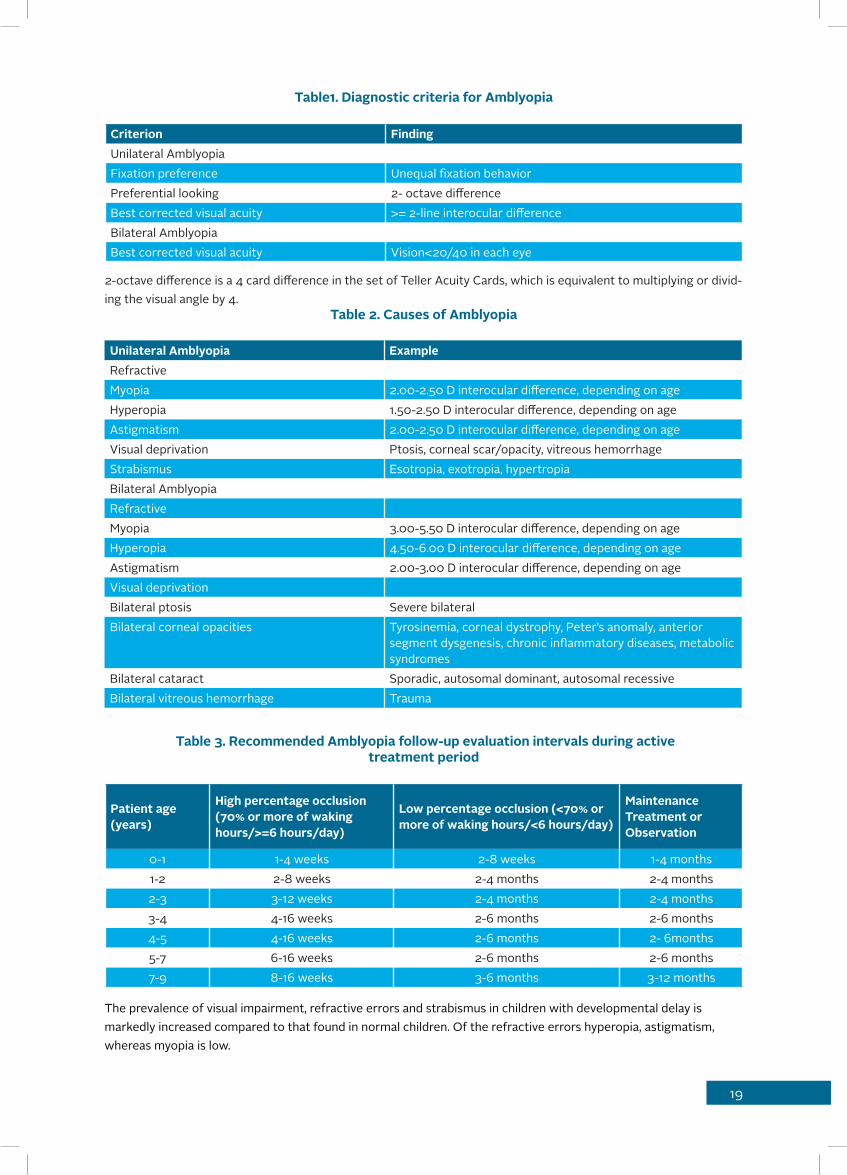
Table1. Diagnostic criteria for Amblyopia
Criterion FindingUnilateral AmblyopiaFixation preference Unequal fixation behaviorPreferential looking 2- octave differenceBest corrected visual acuity >= 2-line interocular differenceBilateral AmblyopiaBest corrected visual acuity Vision<20/40 in each eye
2-octave difference is a 4 card difference in the set of Teller Acuity Cards, which is equivalent to multiplying or divid-ing the visual angle by 4.
Table 2. Causes of Amblyopia
Unilateral Amblyopia ExampleRefractiveMyopia 2.00-2.50 D interocular difference, depending on ageHyperopia 1.50-2.50 D interocular difference, depending on ageAstigmatism 2.00-2.50 D interocular difference, depending on ageVisual deprivation Ptosis, corneal scar/opacity, vitreous hemorrhageStrabismus Esotropia, exotropia, hypertropiaBilateral AmblyopiaRefractiveMyopia 3.00-5.50 D interocular difference, depending on ageHyperopia 4.50-6.00 D interocular difference, depending on ageAstigmatism 2.00-3.00 D interocular difference, depending on ageVisual deprivationBilateral ptosis Severe bilateralBilateral corneal opacities Tyrosinemia, corneal dystrophy, Peter’s anomaly, anterior
segment dysgenesis, chronic inflammatory diseases, metabolic syndromes
Bilateral cataract Sporadic, autosomal dominant, autosomal recessiveBilateral vitreous hemorrhage Trauma
Table 3. Recommended Amblyopia follow-up evaluation intervals during activetreatment period
Patient age(years)
High percentage occlusion (70% or more of waking hours/>=6 hours/day)
Low percentage occlusion (<70% or more of waking hours/<6 hours/day)
Maintenance Treatment or Observation
0-1 1-4 weeks 2-8 weeks 1-4 months1-2 2-8 weeks 2-4 months 2-4 months2-3 3-12 weeks 2-4 months 2-4 months3-4 4-16 weeks 2-6 months 2-6 months4-5 4-16 weeks 2-6 months 2- 6months5-7 6-16 weeks 2-6 months 2-6 months7-9 8-16 weeks 3-6 months 3-12 months
The prevalence of visual impairment, refractive errors and strabismus in children with developmental delay is markedly increased compared to that found in normal children. Of the refractive errors hyperopia, astigmatism, whereas myopia is low.
20
SEDATION PROTOCOL IN PEDIATRIC AGE GROUP
INDICATIONS• Suture removal• Change of eye-pad dressings• Measurement of IOP• Removal of IV access
ASSESSMENT • Detailed history and physical examination• Previous history of any sedation administered• History of allergic to any medications
FASTING• Two hrs NPO for clear fluids/ solids etc• Preparation of the child• Consent for oral sedation• Measure body weight• Assess airway• Emergency trolley with necessary drugs• Suction apparatus with attachement• Baseline measurement of HR/SaO2,
respiratory rate and sedation score (see attached)
• Connect to pulse oximeter• Agent used • Syr. Pedicloryl 50mg/kg body weight
(Triclofos Oral solution 500mg/5ml)• Post-procedure • Monitor and record vital signs every 15
minutes• Child should be “left-lateral position” and nil
per oral until the sedation score returns to baseline level
UMMS- THE UNIVERSITY MICHIGAN SEDATION SCORE• Awake and alert• Minimally sedated; may appear tired/sleepy,
responds to verbal conversation and or sound• Moderately sedated; somnolent/sleeping;
easily roused with light tactile stimulation or simple verbal command - also known as “conscious sedation”
• Deep sedation; deep sleep, rousable only with deep or significant physical stimulation
• Unrousable
CHILD WITH DEVELOMENTAL DELAY
AIM OF EXAMINING A CHILD WITH DELAYED DEVELOPMENT• Early detection to optimize their learning
capacity and independence
• To find out the treatable causes of visual handicap in these children
• To study type of developmental delay whether
• Isolated or global delay and their appropriate referral
• For accurate health surveillance and educational planning for the child
• Identification of possible risk of having affected children in the future
HISTORY• Detail history with regards to
• Birth history
PRENATAL HISTORY
• Potential teratogens including alcohol, medications, vitamins, maternal infection (rubella, cytomegalovirus, toxoplasmosis, varicella), maternaldiabetes, hyperthermia, maternal phenylketonuria
• Prenatal tests (eg, amniocentesis, ultrasound)
PERINATAL HISTORY
• Gestation, mode of delivery, Apgar scores, resuscitation
• Birthweight, length, head circumference
• Feeding, muscle tone, other problems
POSTNATAL HISTORY
Milestones, school performanceEvidence of regression (this may be a clue to inborn error of metabolism or neurodegenerative process)• Unusual behaviour, personality
• Coordination, seizures, unusual movements
• Increased or decreased tone
21
• Growth: height, weight, head circumference
• Previous illnesses
• Vision, hearing
• Immunization
FAMILY HISTORY
• Three generations, maternal and paternal
• Consanguinity
• Previous pregnancy outcomes: miscarriages, stillbirths, neonatal or childhood deaths, infertility
• Family history of birth defects, childhood deaths, mental retardation, speech delay, learning disabilities, autism and known genetic conditions
TREATMENT AND REHABILITATION HISTORY
Special School
• Physiotherapy or occupational therapy
• Visual stimulation exercise
• Medical or surgical treatment taken
• Physical examination
To classify the child into syndromic versus non syndromic developmental delay
– To look in detail for minor anomalies, particu larly of the face, head and hands.
– Try to first determine whether the child looks like either of the parents or siblings.
– Compare photographs of the siblings or par ents at a similar age maybe useful as a com parison.
– To describe the features (eg, coarse, – myopathic), or a more specific description of
each feature is required (eg, hypertelorism,) – Mongoloid and anti mongoloid slant, cranio
synostosis etc – A careful neurological examination is
important – Noting abnormalities of muscle tone and
strength – Carefully examining the skin for hyper- or
hypopigmentation Café-au-lait spots may signal neurofibromatosis
– Vascular tumours or hemangiomas may sug gest certain genetic disorders or syndromes
– Other unusual findings such as anomalies of the genitalia, connective tissue and/or joint
abnormalities and internal anomalies (espe cially cardiac and renal) should be noted.
– A reference to pediatric for other system ic evaluation is mandatory
VISUAL ACUITY TESTING In < 1 Year
• CSM
• Fixing and following light
• Indirect methods: red reflex,resisting to occlusion
Flash VEP
1-2 Years
Candy bead testPreferentiallooking testsFlash VEP
2-3 Years
Sheridan gardnerAssessment of functional visual acuity like
reaching out to objects or toys, social smile, holding at toys, side gazes etc)
Ophthalmological conditions to look for Refractive errors
• Most of these children are hypermetropics and hypo accomodators
Squint
• Exotropia is most commonly seen
• A careful surgical planning is required in these cases
• Complications of general anesthesia also to be kept in mind
Optic Atrophy
• Look for other neurological abnormalities
Delayed visual maturation
• Vision therapy exercises may help in these cases
Cortical Visual Impairment
Cortical visual impairment is a neurologic impairment defined as bilateral loss of central vision (visual acuity) caused by damage to the

22
central nervous system.• Retinopathy of prematurity (ROP)
• Papilloedema
• Nystagmus
• Cataract
INVESTIGATIONS
GENETICS
• Karyotyping to assess for chromosomal abnormalities when necessary
• Endocrinology when necessary
• TSH, free T4
• Referral to endocrinology should be considered
METABOLIC
Metabolic screening – glucose, electrolytes, serum lactate, ammonia, liver function tests, pyruvate, albumin, triglycerides, uric acid, serum quantitative amino acids, urine organic acids, acylcarnitines, creatine phosphokinase (if suspecting myopathy)
• Referral to physician should be considered
• Neurology:
• EEG
• CT/MRI
• Referral to nurology if any of these tests are considered
INTERVENTION
EARLY INTERVENTION
• Physiotherapy
• Occupation therapy
• Speech therapy
• Other service
• Psychologist
• Early childhood educator
• Behavioral therapist
• Pediatric
PHYSIOTHERAPY
• Achievement of physical milestones such as
sitting, crawling and standing
• Improved independence in activities of daily living
• Improved posture, muscle strength, balance and coordination
• Verbal and non-verbal communication skill development
• Language understanding
• Social communication use (e.g., greeting people and playing with peers)
• Speech clarity (ability to produce and combine speech sounds)
• Fluency
OCCUPATIONAL THERAPY
• Improve trunk control
• Fine motor skills (hand function)
• Activities of daily living (e.g., dressing, feeding and swallowing)
VISUAL DEVELOPMENT THERAPY
The capacity of visual system can be improved through the presentation of specific material activities
It should go in systemic procedure to get desired visual behaviorMany children with neurological visual impairment develop higher level of vision after receiving visual development therapy
Level I
To be performed in a dark room
Phase 1- Light and white paper
Phase 2-Light and colored paper
Phase 3- Light with translucent plastic ball /toys
Phase 4- Move lighted objects across the line of sight
Level II
Perform the above phases in a room with normal lighting.
Level III
Perform the above phases with a bright shiny

23
object such as Christmas decorations or glittering papers in a room with low level lighting.
Level IV
Start using brightly colored large toys instead of lights or objects.
Level V
Show bright high contrast black and white patterns; followed by facial expression flash cards.
VISION DEVELOPMENT
Awareness> Attention> UnderstandingLight> Object> PeopleFixation> TrackingLarge> Small
REHABILITATION & EDUCATION
Parents should be guided to select the suitable medium and mode of education
NYSTAGMUS
Repetitive, regular, rhythmic, involuntary, to-and-fro, oscillatory movements of the eyes
TYPES• CEMAS classification
– Idiopathic Nystagmus Syndrome (INS ) – Fusional Maldevelopment Nystagmus Syn
drome (FMNS)Names like INS not congenital nystagmus)• Other classifications:
– Pendular – Jerk
ETIOLOGICAL CLASSIFICATION• Physiological
– Optokinetic nystagmus – End point nystagmus – Physiological vestibular nystagmus
CONGENITAL• Infantile nystagmus
• Latent nystagmus
• Spasmus nutans
• Nystagmus blockage syndrome
ACQUIRED• Acquired (Ocular) pendular nystagmus
• Peripheral vestibular nystagmus
• Central vestibular nystagmus
– Upbeat – Downbeat – Periodic alternating nystagmus • Gaze paretic nystagmus
• See saw nystagmus
PRESENTATION • Visual acuity ranges from profound visual loss
to minimal dysfunction
• Nystagmus
• Strabismus
• Photophobia

24
HISTORY• What is the duration of diminution of vision?
• Is the diminution of vision slowly progressive?
• Is there any associated decrease in side vision?
• Is there any associated decrease in night vision?
• Is there any photophobia?
• Is there any strabismus?
• Is there any history of consanguinity amongst parents?
• Is there any family member affected?
• Is there any mental retardation/ developmental delay?
• Are there any other systemic abnormalities?
• Is there any history of taking medications?
EXAMINATION• Abnormal head posture
• Null point
• Abnormal head movements
• Head bobbing
• Systemic anomalies
• Neurological anomalies
• Visual acuity and refraction
• Binocular function testing+ sensory evaluation
• Color vision
• Extraocular movements and associated strabismus
• Evaluation of nystagmus
– Type (Jerk/Pendular) – Unilateral/Bilateral and
symmetrical/asymmetrical – Direction (horizontal, vertical, torsional) – Whether uniplanar or multiplanar? – Frequency – Amplitude – Null point – Saccades and pursuits• Pupil evaluation
• Slit lamp examination
– Cornea- opacities – Iris –transillumination defects, aniridia – Lens – congenital cataract
• IOP
• Fundus
• Indirect ophthalmoscopy with 20 D and 78D
Disc: Optic disc anomaliesArteries: Attenuation, abnormal orientation (Morning glory syndrome)Macula: Foveal hypoplasia, albinismPeriphery: Depigmented, retinochoroidal abnormalities Cone Rod dystrophies ROP
INVESTIGATIONS • If you suspect ocular anomaly as the cause for
nystagmus
• Investigations mainly to confirm the diagnosis
– OCT – ERG – VEP• Acquired nystagmus associated with
oscillopsia/ vertical torsional / multiplanar
• Suspect CNS abnormality
• Neuroimaging
– MRI – CT
TREATMENT• Glass prescription
• Optical management
– Overminus lens – Prisms- yoked prisms or base out prisms – Contact lens• Concomitant amblyopia treatment
• Low vision aids
• Medications
– Baclofen – Gabapentine – Botox• Surgical correction for associated strabismus
• Surgical management of nystagmus
• Kestenbaum-Anderson procedure
– Artificial divergence surgery – Retroequatorial recession of horizontal
recti – Anterior tenotomy and reattachment – Neurosurgery • Systemic examinations
25
• Genetic counseling
• Advise ocular examination of other family members
• Do not give a very poor prognosis to the patients, emphasize on the positive side- the patient is very unlikely to be completely blind
• Routine monitoring every year or as and when required to treat the complications associated with the disease
• Rehabilitation
• Information about new scientific developments
EPIPHORA
COMMON CAUSES• Congenital Naso-lacrimal Duct Obstruction
(Most common)
• Vernal/allergic conjunctivitis
• Ophthalmia neonatorum
• Congenital Glaucoma
DIFFERENTIAL DIAGNOSIS• Ophthalmia neonatorum
• In first month of life
• Conjunctival hyperaemia, copious discharge
• Needs immediate attention
CONGENITAL NASO-LACRIMAL DUCT OBSTRUCTION• Epiphora usually after one month of birth as
there is no lacrimal secretion earlier than that.
• Clear serous, mucoid or mucopurulent discharge specially if superadded infection
• There is regurgitation of discharge from punctum on digital pressure over lacrimal sac area (ROPLAS- Regurgitation on pressure from lacrimal sac)
• May have associated episodes of dacrocystitis
• Dacryocoele may also be present which decreases after pressure over cyst, with discharge through punctum
LACRIMAL PUNCTUAL ATRESIA
• Overflow of clear tears
• No regurgitation on pressure over lacrimal sac area
CONGENITAL GLAUCOMAEpiphora is usually accompanied with following:
• Photophobia
• Blepharospasm
• Megalocornea

26
• Corneal haze
• Blue sclera
• Haab’s striae
VERNAL/ALLERGIC CONJUNCTIVITIS• Seasonal waxing and waning of episodes
• Severe itching and rubbing
• Ropy discharge
• Conjunctival pigmentation
• Cornea may have SPKS, shield ulcers in severe cases
• Often have associated atopy
CLINICAL EXAMINATION
HISTORY
• Onset
• Associated symptoms
• History of prematurity
• Family history
OPHTHALMIC EXAMINATION• Fixation preference for any eye
• Always rule out refractive error by cycloplegic refraction
• CNLDO is commonly associated with refractive errors and amblyogenic factors like strabismus and anisometropia
CLINICAL EXAMINATION • Look for punctal atresia or agenesis,note
punctal apposition also
• Look for lacrimal sinus or fistula
• Anterior segment examination with slit lamp or hand held slit lamp.
• Can use Indirect ophthalmoscope with 20 D lens for magnification if handheld slit lamp not available.
• Look out for foreign body , caterpillar hair if epiphora is unilateral by everting the lid .Double eversion may require short GA
• Ophthalmia neonatorum, if suspected, should be dealt on emergency basis
• Glaucoma also needs early intervention and treatment so attempt should made to check IOP either under sedation or general anesthesia
• Corneal surface abnormalities
• Digital pressure on sac area to test if any regurgitation
• Fundus evaluation is mandatory
INVESTIGATION• It is useful to plan evaluation under anesthesia
if cause of watering is not clear on clinical examination
• DCG, Dacrocystography, to find out exact site of obstruction.
• Nasal endoscopy if bony obstruction suspected or to rule out antecedent nasal pathology
MANAGEMENT
OPHTHALMIA NEONATORUM
• Prevention: Prophylactic application of Erythromycin eye ointment or Povidone iodine eye drops in both eyes immediately after birth in both eyes
• Antibiotic treatment needs to be started immediately
• In case of gonoccocal infection, require oral antibiotics
GLAUCOMA
• Discussed in detail in glaucoma section
ALLERGIC CONJUNCTIVITISPREVENTION
• Protective glasses
• Avoid pets, dust, travelling in a window seat
• Hand hygiene
• Mast cell stabilizers (Cromolyn sodium four%) can be started one to two weeks if a definite waxing period is noted over the years
TREATMENT
Mast cell stabilizers: They act by preventing the degranulation of the mast cells.

27
• Olopatadine one% twice a day or two% once a day
• Cromolyn sodium one%
• Ketotifen fumarate (0.025%)
Steroids: It depends upon severity of the symptoms Presence of corneal involvement is an indication for steroids. If steroids are indicated , a short tapering course of low dose steroids can be prescribed .Take few minutes to explain potential adverse effects like glaucoma to parents . This will prevent self medication
• Flurometholone one%
• Prednisolone acetate
Immunomodulators • Tacrolimus ointment over the lid
Lubricants
• They help in maintaining ocular surface integrity. Patients with chronic allergic condition often develop secondary dry eye
Follow up
• These children need regular follow up to detect any glaucomatous changes, limbal stem cell deficiency or cataractous changes in lens
CONGENITAL NASO-LACRIMAL
Duct Obstruction
• Conservative
• Criggler’s massage:
• Appropriate technique is of utmost importance.
• Press with little finger above medial canthus area and massage downwards, backwards and medially with same force throughout.
• 20 strokes three times a day at least for six weeks . To be continued till the duct becomes patent or child is one year old whichever is earlier
• To be continued for two months even if ROPLAS becomes negative .
• Lid hygiene
• Antibiotic eye drops can be prescribed if there is a superimposed infection leading to profuse mucoid or mucopurulent discharge
PROBING AND SYRINGING
After one year of age
• Indication for early probing:
• If child needs intraocular surgical intervention earlier. For e.g. glaucoma, cataract
• Acute dacrocystitis causing fistula
Procedure
• We prefer general anesthesia, can be done as in office procedure under topical anesthesia but not recommended in older children
• Dilatation of puncti with punctum dilator
• Lacrimal sac irrigation by syringing through lower puncta
• Surgeon should proceed with probing even if syringing is patent
• Bowmans probe 0 or 1 first advanced vertically in punctum
• Traction over lateral canthus with finger
• Probe will become horizontal and then passed towards sac area. Look for any obstruction, resistance
• If resistance felt, smaller size probe inserted to rule out stenosis Vs obstruction
• If resistance felt gradually increasing force can be applied to clear obstruction or stenosis
• When tip encounters nasal bony wall, retract a probe a little and pivot to direct it downwards, medially. Remove lid traction at this oint
• Sudden decrease in resistance is felt if there is distal membranous obstruction which got overcome
• Put one more probe in nasal cavity below inferior turbinate and look for any movement in primary probe
• Can be confirmed by repeating syringing.
• If firm or hard resistance felt, then its either because of hypertrophic inferior turbinateor bony ostium
• Can try infracture of inferior turbinate in such cases
Post op care• Antibiotic and steroid drops for a week,
• To continue Criggler’s massage for 2 weeks

28
• Complications:
• Minor nasal bleeding
• Creation of false passage
• Failure
• Recurrence commonly within 2 months
If probing fails or recurrence occurs
• Repeat probing can be done after 2 months with inferior turbinate infracture in other cases
• Balloon dilatation or intubation can be used
• In cases of bony ostium no role of repeat probing
• If repeat probing fails or in cases of bony ostium:
• Can wait and watch till 2 years as bone development is complete
• Periodic follow ups
• Dacrocystorhinostomy is indicated
• Decision of timing depends upon severity of signs and symptoms
Punctal stenosis/atresia
• Punctal perforation with 26G needle with punctal dilatation is sufficient in most cases
• If not successful for maldeveloped puncta may need incisional punctoplasty with/ without intubation
• Conjunctivo-dacro-cysto-rhinostomy if no punctum
STRABISMUS - COMITANT
INTRODUCTIONStrabismus is a common condition in childhood affecting 2.1% of the population, with an increased prevalence associated with assisted delivery (forceps or caesareansection), low birth weight (including premature infants), neuro-developmental disorders. Neuro-developmental strabismus (associated with a neuro-developmentalproblem) is independently associated with maternal smoking later in pregnancy, maternal illnesses in pregnancy and low birth weight for gestational age.
Approximately 60% have eso-deviations and 20% exo-deviations.
Strabismus may lead to a failure to develop binocular vision, and amblyopia, either of which may prevent an individual pursuing certain occupations. The appearance of ocular misalignment may interfere with social and psychological development with potentially serious effects for all patients with strabismus.
Timely diagnosis and appropriate treatment of children with strabismus can reduce the prevalence of amblyopia and ocular misalignment in later childhood and adult life. Correction of strabismus has been reported to improve motor coordination.
Strabismus may be the presenting symptom in children with a serious eye or brain condition (e.g. retinoblastoma, hydrocephalus or brain tumour). All professionals involved with the management of strabismus need to be able to recognize this, and either initiate onward referral or arrange for appropriate investigation and management.
AIMS OF MANAGEMENT OF STRABISMUS
• To maintain or restore optimal visual acuity in each eye
• To maintain or restore normal (or subnormal) binocular single vision
• To restore appropriate ocular alignment
• To eliminate double vision or other induced symptoms (e.g. asthenopia)
• To correct significant abnormal

29
(compensatory) head posture
• To improve binocular visual field (in the case of esotropia correction)
• To detect/exclude serious underlying ocular or neurological disease
CONCOMITANT SQUINT
HISTORY
• Age of onset of squint
• Precipitating factors like trauma, febrile illness.
• Associated abnormal eye movements or abnormal head position
• Symptoms of asthenopia
• Double vision
• Eye closure in bright light
• Previous surgery for strabismus
• Occlusion therapy and compliance
• Previous spectacle wear
• Application of drops like phospholine Iodide
• Any other surgery like R.D, cataract, glaucoma implant, sinus surgery or neuro surgery
BIRTH AND MEDICAL HISTORY
• Significant antenatal history
• Maternal infection, any drugs taken during pregnancy
• Any untoward event during delivery
• Birth trauma, forceps, birth asphyxia
• Gestational age
• Birth weight of the child
• History of being kept in incubator
• History of oxygenation
• Developmental milestones, both physical and mental
• Any associated systemic problem
• Other associated neurological problems like cerebral palsy, epilepsy, mental retardation and craniofacial anomalies
• History of any other handicap
FAMILY HISTORY
• Consanguinity
• Similar problems in other siblings
• History of any inherited eye or systemic condition
• Response of other members of family to surgery
• High ametropia
EXTERNAL EXAMINATION
• Shape of head
• Facial asymmetry
• Dysmorphic facial features like sparse hair, frontal bossing
• Ear abnormalities
• Any other deformities like extra digits
• General appearance including obesity
EXAMINATION
• Sensory evaluation has to be done before motor evaluation
• Binocular vision assessment
• Check binocular sensory status
Fusion – Worth 4 dot test for distance and near
Stereopsis – use Titmus fly/Lang’s chart/TNO
Retinal correspondence – use Bagolini’s glasses/Worth four dot test/Synaptophore
Suppression – Worth four dot test/ Bagolini’s glasses
• Motor fusion can be assessed using the 20 base out test in preverbal children, and more formally in older children in a step wise manner using a prism bar/loose prisms and red filter
• Note any abnormal head posture like head tilt, face turn etc
• Assess lid position – Note any ptosis, lid retraction
OCULAR ALIGNMENT/ MOTOR EVALUATION
QUALITATIVE TESTS
• Do a cover test, cover uncover and alternate cover test for distance and near targets

30
(accommodative target must be used to increase accommodative stimulus)
• It is important to note if there is any fixation preferenceand the degree of control (if present)
QUANTITATIVE TEST
• Measure angle of deviation near and distance, with and without glasses using alternate prism cover test (APCT) and simultaneous prism cover test (SPCT)
• Measure angle of deviation in all gazes with correction
• Hirschberg’s test and modified Krimsky in case of uncooperative patients and those with poorfixation/vision
• Note A or V or X pattern and DVD if any
• The presence of any torsion can be assessed using a double Maddox rod, single Maddox rod and fundus examination
• Note any nystagmus – in primary position and other gazes
• Mention type of nystagmus, jerk or pendular, mention amplitude and frequency, notenull point if any or if it dampens on convergence
• In children with nystagmus particular attention should be given to the visualacuity with both eyes open, near visual acuity (with both eyes open), fusion and anycompensatory head posture to aid surgical planning
Diagramatic representation of ocular motility
OCULAR MOTILITY
Check ductions and versions
Note any limitation, overaction and underaction of musclesThe use of a diagram to document extra ocular movements is considered best practice. In small children it may not be possible to bring
the eyes into all areas adequately. Occlusion of an eye for a short period (up to an hour) or use of the vestibulo-ocular system may help in demonstrating full abduction.Convergence is tested using an accommodative target.
VISUAL ACUITY ASSESSMENTRECORD VISUAL ACUITY FOR NEAR AND DISTANCE
Fixation pattern may be used as a gross comparison between the two eyes where formal testing is not possible. The two eyes can be separated using a vertically acting 10 or 12 dioptre prism (usually held base down). This is only necessary if there is no strabismus or the angle of strabismus is very small. A fogging lens (for example a plus lens) can be used instead of occlusion in the assessment of children with manifest or latent nystagmus. It is preferable to use a crowded test as early as possible for accurate detection of amblyopia. Near chart letter matching may be a more appropriate measure of near acuity thanreading text, if children are still learning to read.
DO RETINOSCOPY AND SUBJECTIVE REFRACTION
Retinoscopy is carried out in a semi-darkened room using hand-held lenses, or trial frame, to neutralise the retinoscopic reflections along the visual axis. It is important to maintain the child’s attention and fixation should be on the retinoscope lightIt is rarely necessary to perform an examination under anesthesia in order to carryout refraction and fundus examination and its routine use should be discouraged.
A repeat refraction every 12 months is advised. If visual acuity fails to improve or deteriorates, it may be necessary to repeat the refraction, (along with a repeat fundus examination) or consider alternative diagnoses
DO CYCLOPLEGIC REFRACTION
• A post cycloplegic subjective refraction is a useful exercise in selected cases.
31
• Adequate cycloplegia for retinoscopy may be obtained 30 minutes following the instillation of cyclopentolate one% eye drops.
• Below the age of six months mydriatics are used in lower concentration to reduce the risk of toxicity
• Cycloplentolate 0.5%
DECISIONS BASED ON VISUAL ACUITY
• Treatment with glasses
• Amblyopia therapy (occlusion, penalization)
• Frequency of follow up visits
• Consideration for further investigations
• Timing of strabismus surgery
ADDITIONAL TESTSIf indicated, measurement of AC/A ratio, 4ΔB O test, simultaneous prism cover test, fusional amplitudes, forced duction test, ERG.Use of the fixation circle in an ophthalmoscope can identify foveal fixation, or lack of it, which is particularly helpful in microtropia.
ANTERIOR SEGMENT EXAMINATION BY SLIT LAMP/TORCH LIGHT
• Note any congenital anomalies like aniridia, coloboma, heterochromia, cataract
• Check pupillary reaction
FUNDUS EXAMINATION
Includes examining optic nerve and quality of the nerve fibre layer, macula and retinal periphery.The use of the indirect ophthalmoscope is recommended as standard in small children (less than five years).
In older children, the direct or 90 dioptre/slit lamp examination can add information.
If restraint is required, this is permitted to allow fundus examination to exclude life threatening examination findings such as papilloedema or optic atrophy. Parents should be requested to consent to have their child restrained.
If the examination is inadequate for any
reason, the difficulty in the examination should be documented and a date set for a repeat examination.
EXAMINATION
Order of Management
1. Refractive error correction2. Amblyopia treatment3. Strabismus management
CORRECT REFRACTIVE ERROR BY APPROPRIATE GLASSES
• Children’s spectacles should always be provided with plastic lenses to reduce the risk of injury. Advise careful fitting, using adequate support for the nasal bridge and sufficient size of lens to avoid children looking over the top of the glasses
• In all forms of esotropia, full correction of hypermetropia is the treatment of choice (having corrected the retinoscopy for working distance only). This is often known as the ‘full correction’. There is no requirement to subtract any lens power for cycloplegia
• In exotropia, uncorrected hypermetropia might be preferable to aid exotropia control, assuming visual acuity is good
• The main aims of further review are to further diagnose and classify the strabismus, monitor visual development (visual acuity and binocular function), treat amblyopia and manage the strabismus to obtain, maintain or restore binocular single vision where potential is present (e.g. prisms, alteration of / or addition of lenses, exercises)
• A period of refractive adaptation is recommended after glasses have been prescribed, until the vision is stable, as the visual acuity can improve with glasses alone, even in strabismic amblyopia. This may take up to 18 weeks
• The frequency of follow up visits will depend on many factors, such as age and change in treatment and treatment effect
• An annual refraction is required to monitor changes in the glasses prescription. An earlier repeat is indicated if the vision fails to improve
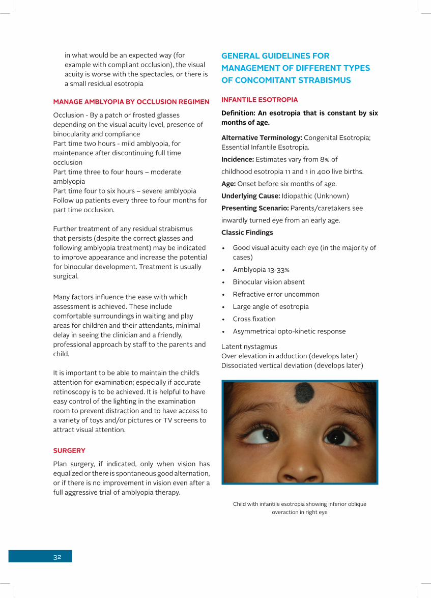
32
in what would be an expected way (for example with compliant occlusion), the visual acuity is worse with the spectacles, or there is a small residual esotropia
MANAGE AMBLYOPIA BY OCCLUSION REGIMEN
Occlusion - By a patch or frosted glasses depending on the visual acuity level, presence of binocularity and compliancePart time two hours - mild amblyopia, for maintenance after discontinuing full time occlusionPart time three to four hours – moderate amblyopiaPart time four to six hours – severe amblyopiaFollow up patients every three to four months for part time occlusion.
Further treatment of any residual strabismus that persists (despite the correct glasses and following amblyopia treatment) may be indicated to improve appearance and increase the potential for binocular development. Treatment is usually surgical.
Many factors influence the ease with which assessment is achieved. These include comfortable surroundings in waiting and play areas for children and their attendants, minimal delay in seeing the clinician and a friendly, professional approach by staff to the parents and child.
It is important to be able to maintain the child’s attention for examination; especially if accurate retinoscopy is to be achieved. It is helpful to have easy control of the lighting in the examination room to prevent distraction and to have access to a variety of toys and/or pictures or TV screens to attract visual attention.
SURGERY
Plan surgery, if indicated, only when vision has equalized or there is spontaneous good alternation, or if there is no improvement in vision even after a full aggressive trial of amblyopia therapy.
GENERAL GUIDELINES FOR MANAGEMENT OF DIFFERENT TYPES OF CONCOMITANT STRABISMUS
INFANTILE ESOTROPIA
Definition: An esotropia that is constant by six months of age.
Alternative Terminology: Congenital Esotropia; Essential Infantile Esotropia.
Incidence: Estimates vary from 8% of
childhood esotropia 11 and 1 in 400 live births.
Age: Onset before six months of age.
Underlying Cause: Idiopathic (Unknown)
Presenting Scenario: Parents/caretakers see
inwardly turned eye from an early age.
Classic Findings
• Good visual acuity each eye (in the majority of cases)
• Amblyopia 13-33%
• Binocular vision absent
• Refractive error uncommon
• Large angle of esotropia
• Cross fixation
• Asymmetrical opto-kinetic response
Latent nystagmusOver elevation in adduction (develops later)Dissociated vertical deviation (develops later)
Child with infantile esotropia showing inferior oblique overaction in right eye
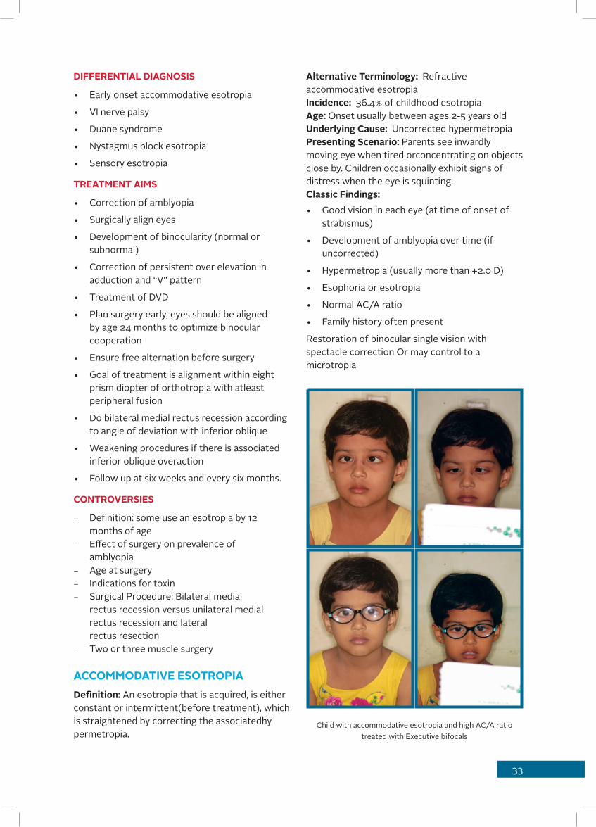
33
DIFFERENTIAL DIAGNOSIS
• Early onset accommodative esotropia
• VI nerve palsy
• Duane syndrome
• Nystagmus block esotropia
• Sensory esotropia
TREATMENT AIMS
• Correction of amblyopia
• Surgically align eyes
• Development of binocularity (normal or subnormal)
• Correction of persistent over elevation in adduction and “V” pattern
• Treatment of DVD
• Plan surgery early, eyes should be aligned by age 24 months to optimize binocular cooperation
• Ensure free alternation before surgery
• Goal of treatment is alignment within eight prism diopter of orthotropia with atleast peripheral fusion
• Do bilateral medial rectus recession according to angle of deviation with inferior oblique
• Weakening procedures if there is associated inferior oblique overaction
• Follow up at six weeks and every six months.
CONTROVERSIES
– Definition: some use an esotropia by 12 months of age
– Effect of surgery on prevalence of amblyopia
– Age at surgery – Indications for toxin – Surgical Procedure: Bilateral medial
rectus recession versus unilateral medial rectus recession and lateral rectus resection
– Two or three muscle surgery
ACCOMMODATIVE ESOTROPIADefinition: An esotropia that is acquired, is either constant or intermittent(before treatment), which is straightened by correcting the associatedhy permetropia.
Alternative Terminology: Refractive accommodative esotropiaIncidence: 36.4% of childhood esotropiaAge: Onset usually between ages 2-5 years oldUnderlying Cause: Uncorrected hypermetropiaPresenting Scenario: Parents see inwardly moving eye when tired orconcentrating on objects close by. Children occasionally exhibit signs of distress when the eye is squinting.Classic Findings:• Good vision in each eye (at time of onset of
strabismus)
• Development of amblyopia over time (if uncorrected)
• Hypermetropia (usually more than +2.0 D)
• Esophoria or esotropia
• Normal AC/A ratio
• Family history often present
Restoration of binocular single vision with spectacle correction Or may control to a microtropia
Child with accommodative esotropia and high AC/A ratio treated with Executive bifocals

34
DIFFERENTIAL DIAGNOSIS• Non accommodative esotropia
• Constant esotropia (binocularity not
• re-established with spectacle correction)• Also known as partially accommodative
esotropia or constant esotropia with an accommodative element
• VI nerve palsy• Congenital esotropia
• Cyclical esotropia
• Convergence excess esotropia• Near esotropia
TREATMENT
• Full cycloplegic hypermetropic correction
• Orthoptic treatment to expand base in fusion range
• Delay in initiation of treatment increases the likelihood of not responding to
• anti-accommodative therapy
• Treat amblyopia
• Gradually decrease hyperopic correction if good control of ensuing esophoria
• Can try Phospholine Iodide drops in cases of poor compliance with glasses. Instruct parents about possible side effects
• Follow up frequently if on Phospholine Iodide
CONTROVERSIES
• Role of surgery
• Indication for miotic drops
• Role of refractive surgery
PARTIALLY ACCOMODATIVE ESOTROPIADefinition: A group of esotropias that are helped, but not cured, with glasses for hypermetropiaAlternative Terminology: Partial Accommodative esotropia Constant Esotropia with an accommodative elementIncidence: 10% of Childhood Esotropia11Age: two to five years oldUnderlying Cause:• Hypermetropia
• Poor fusional reserves
• Esophoria
• Not fully understood
PRESENTING SCENARIO • Esotropia seen when tired or concentrating on
close objects.
• Classic Findings:
• Amblyopia
• Absent binocular function
• Suppression of squinting eye
• If small angle, development of abnormal retinal correspondence (ARC)
• Hypermetropia
• Constant esotropia even with “full correction”
• Family history
DIFFERENTIAL DIAGNOSIS
• Fully accommodative esotropia
• VI nerve palsy
• Congenital esotropia
• Cyclical esotropia
• Convergence excess esotropia
TREATMENT
• Full cycloplegic hypermetropic correction
• Amblyopia treatment (occlusion, atropine, other penalization techniques)
• Prism adaption prior to surgery
• Be conservative about planning surgery
• Rule out latent uncorrected hyperopia before proceeding with surgery
• Do a bilateral medial rectus recession for residual angle of deviation overfull cycloplegic correction
• To plan under-correction
• Explain parents that child needs to wear glasses even after surgery
• Restoration of binocular function rare
ESOTROPIA WITH HIGH AC/A RATIODefinition: An intermittent esotropia with binocular single vision present at distance fixation

35
but esotropia on accommodation for near fixation.Terminology: The term convergence excess is sometimes used to include patients with a constant esotropia and no binocular vision.A near esotropia is a condition with an increased angle for near viewing but not associated with a high AC/A ratio. The term non-accommodative convergence excess is sometimes used for this condition.Near distance disparity is considered relevant if over 8-10 prism dioptres.Incidence: 27% of esotropia have near distance disparity, but the prevalence of convergence excess is less.Age: Onset is usually between one to four years old, but can be up to aged 10 years.Underlying Cause: High AC/A ratio: not fully understood what underlies this.
PRESENTING SCENARIO
Parents see inwardly moving eye when tired or concentrating on near objects. Children occasionally exhibit signs of distress when the eye is squinting. Older children may report double vision at near.
CLASSIC FINDINGS
• Good vision each eye
• Amblyopia rare
• Binocular in the distance, reduced at near unless corrected with near add
• May control to a fully accommodative esotropia at distance (with hypermetropic glasses)
Variable control at near• Esotropia at near, which may be phoric to a
non accommodative target
• High AC/A – leading to deterioration of control
• Poor near controlled binocular vision (CBA)
DIFFERENTIAL DIAGNOSIS
• Non accommodative esotropia
• Constant esotropia
• Near esotropia
• Hypo accommodative esotropia
• Congenital esotropia
TREATMENT
• Full cycloplegic hypermetropic correction when present
• Treat amblyopia if any
• Prescribe executive bifocals bisecting the pupil if distance deviation is less that 15Δ
• Acceptable response is fusion at distance with less than 10 prism diopter of residual esotropia through bifocal at near fixation
• Orthoptic exercises
• Miotics - phopholine Iodide
• Surgery
INDICATIONS FOR SURGERY
• Reducing binocularity at near
• Reducing control with other forms of treatment (e.g. bifocals)
• Parent/Doctor preference over other treatments (e.g. bifocals)
• Orthoptic exercises not progressing
• Consider prism adaptation to the near angle
TYPE OF SURGERY
• Large bilateral medial rectus muscle recession - if distance deviation is more than 15Δ even with glasses
• Pulley surgery
• Slanted recession
• Posterior fixation
• Medial rectus recession with resection
CONTROVERSIES
• The terminology
• Use of bifocal glasses
• Indications for surgery
• Surgical Procedure
NON-ACCOMMODATIVE ESOTROPIADefinition: An esodeviation occurs after six months of age that is not improved with hypermetropia correction.Alternative Terminology: Acquired non-accommodative esotropiaIncidence: 16% of childhood esotropia11, 17.7/100,000 live births

36
Age: two to five years oldUnderlying Cause: Not fully understoodPresenting Scenario: Esotropia seen when tired or concentrating on close objects.Classic Findings:Onset maybe acuteFamily history of strabismus 34%Reduced binocular functionSuppression of squinting eyeAmblyopia 41%Low hypermetropia or emmetropia (Mean of +1.42)Esophoria breaking down to esotropiaFamily history
DIFFERENTIAL DIAGNOSIS
• Accommodative esotropia
• VI nerve palsy
• Cyclical esotropia
• Other neurological disease
TREATMENT
• Trial of full hypermetropic correction
• Amblyopia treatment (occlusion, atropine, other penalization techniques)
• Prism adaption prior to surgery
• Surgery to restore ocular alignment (73%)
INTERMITTENT DIVERGENT SQUINTDefinition: An intermittent exotropia, with a larger angle at distance.
Terminology: Intermittent exotropia, distance exotropia, exotropia ofdivergence excess type.Incidence: Up to one% of all children, or 32 per 100,000 of children under 19.Age: Onset usually between two to four years oldUnderlying Cause: Unknown
PRESENTING SCENARIO
Parents see outwardly moving (or turned) eyes when tired, day-dreaming or in bright sunlight.Uniocular eye closing in bright sunlight.- diplophotophobiaDouble vision rare. Usually few if any child reported symptoms.
CLASSIC FINDINGS
• Good visual acuity
• Refractive error – same as population
• Amblyopia in 16% (VA = 6/9 or worse)12
• Control variable – worse for distance fixation
• Good sensory single vision for near
• Assess stereopsis
• Measure squint angle for both near and distance
• Rule out pseudo divergence excess if angle is more for distance than near by controlling fusion and accommodation
• Observe if measurement of deviation is unreliable or if child is able to control deviation well
• Hemi-field suppression (not universal – some have double vision)
• Poor motor fusional reserves
• Exophoria/exotropia on cover testing
• Convergence normal
• AC/A normal, true high or false high (tenacious proximal fusion)
A patient of intermittent exotropia with poor control for distance
DIFFERENTIAL DIAGNOSIS
• Infantile exotropia (usually constant, onset before six months of age)
• Sensory exotropia (poor unilateral vision)
• Convergence weakness (bigger angle for near)
• Convergence insufficiency (poor convergence)

37
TREATMENT
• Prescribe corrective lenses for refractive errors
• Myopia and astigmatism should be fully corrected
• Ignore hyperopia of up to +2.00D
• High hyperopia > 4 D needs to be corrected as optical blur may itself increase exotropia
• Over minus lens effective as temporary measure to delay surgery in pre-school age children
• Maintain/treat visual acuity
• Monitor control
• Unilateral or alternating occlusion1
• Minus lens therapy
• Exercises
• Surgical correction depending on the angle of the deviation
INDICATIONS FOR SURGERY
• Increase in the angle of deviation with deteriorating control
• Decrease in stereoacuity
• Coexisting convergence insufficiency refractory to orthoptictreatment
• Asthenopia
• Development of amblyopia/suppression
• Surgeon/parent/patient preference
• To improve or maintain binocular single vision
TYPE OF SURGERY Unilateral recess resect, unilateral or bilateral lateral rectus recession
CONTROVERSIES The value (if any) of orthoptic exercises/vision therapyIndications for surgeryIdeal age for surgeryNatural historyLong term prognosis
COMPLICATIONS Post operative over corrections.Managed by alternate patching, temporary prisms, botulinum toxin or re-operation
INCOMITANT SQUINT
A squint is termed incomitant when the deviation varies in size with the direction of gaze or with the eye used for fixating
HISTORY• Age of onset of squint
• Precipitating factors like trauma, febrile illness
• Diplopia – duration, mode of onset, diurnal variation
• Abnormal head posture
• Systemic conditions like DM, IHD, and Thyroid disorder
• Abnormal eye movements Oscillopsia, if nystagmus is present
• Associated neurological symptoms like loss of consciousness, convulsion, motor weakness, slurring of speech, giddiness etc
• Previous surgery for strabismus
• Occlusion therapy and compliance
• Previous spectacle wear
• Any other surgery like R.D, cataract, glaucoma implant, sinus surgery or neuro surgery
BIRTH HISTORY AND MEDICAL HISTORY
• Gestational age
• Significant antenatal history
• Maternal infection, any drugs taken during pregnancy
• History of any other handicap
• Any untoward event during delivery
• Birth trauma, forceps, birth asphyxia
• Birth weight of the child
• History of being kept in incubator and history of oxygenation
• Is the child thriving well?
• Developmental milestones, both physical and mental
• Any associated systemic problem
• Other associated neurological problems like cerebral palsy, epilepsy, mental retardation and craniofacial anomalies
38
FAMILY HISTORY
• Consanguinity
• Similar problems in other siblings
• History of any inherited eye or systemic condition
EXAMINATION • Record visual acuity for distance and near
• Do dynamic retinoscopy
• Note any abnormal head posture
• Do cover tests
• Krimsky test or Hirschberg’s test in case of uncooperative patients and those with poor fixation/vision
• Note difference between primary and secondary deviation
• Check versions and ductions
• Note limitation, over action and under action of muscles
• Note any nystagmus – mention type of nystagmus, jerk or pendular, note null point if any
ANTERIOR SEGMENT EXAMINATION BY SLIT- LAMP / TORCH LIGHT • Note any congenital anomalies like aniridia,
coloboma, heterochromia, and cataract.
• Check pupillary reaction
At Squint Clinic
• Review history
• Note any anomalous head posture, position of lids
• Perform cover tests
• Measure angle of deviation, both primary and secondary; in all cardinal positions of gaze
• In adults assess if single binocular vision is achieved in primary position and reading position with prisms
• Recheck ductions and versions
• Check torsion by Maddox rod/double Maddox rod
• Do diplopia charting/Hess charting
• Do Parks three step test in case of vertical strabismus
• Perform forced duction test if indicated
• Detailed fundus evaluation
• Neuroimaging in cases of recent onset paralytic strabismus in the absence of diabetes, hypertension, in cases of trauma, or when multiple cranial neuropathies or suspected space -occupying lesion
• Order thyroid function tests and rule out myasthenia, when aetiology is unclear
• Order cross consultation with neuro-ophthalmologist if indicated
MANAGEMENT• Correct refractive error by appropriate glasses
• Manage amblyopia if any, by occlusion regimen
• Observation if paralysis is of acute onset
• Botulinum toxin injection into the antagonist muscle in selected cases
• Patch to relieve diplopia
• Prisms to achieve binocular vision in case of small angle deviation
• Do surgery in patients with large angle deviation, after waiting for at least six months after the onset, for recovery and stability
• Surgical plan is based on Hess charting, measured angle of deviation
• Adjustable suture technique is better, as results are unpredictable

39
Blepharospasm
• Very effective in the treatment of essential Blepharospasm
• Repeated injections are required every two to three months
• Hemifacial spasm
• Nystagmus
• Induce ptosis in patients with corneal disease
INJECTION PROCEDURE
• Commercial preparations are Oculinum, Dysport, and Botox
• Reconstitution 2.5 units per 0.1 ml
• Take consent
• Explain procedure
• Injection is made directly into the selected extra ocular muscle or the orbicularis
• In adults, injection is performed using a portable electromyography device; children require GA
• It can be injected subcutaneously at sites depending on muscles that need to be paralyzed
• Rule out secondary causes of blepharospasm such as dry eyes and foreign body
• Essential blepharospasm needs no neuroimaging prior to injection, whereas
• Hemifacial spasm needs neuroimaging prior to injection
COMPLICATIONS
Common
• Ptosis, incomplete eyelid closure, dry eye, and induced vertical strabismus
Rare
• Scleral perforation, retrobulbar hemorrhage, pupillary dilatation, and permanent diplopia
BOTULINUM TOXIN INJECTION
It is frozen lyophilized protein drug produced by the bacterium clostridium botulinum. Out of six toxins produced by clostridium botulinum, type A is used clinically.
MECHANISM OF ACTION Acts by blocking the release of acetylcholine at the neuro muscular junctionProduces temporary, reversible, dose-related paralysis, of the muscle It is bound and internalized in 24 to 48 hours within local motor nerve terminals, where it remains for many weeks.The injected extra ocular muscle lengthens and its antagonist contracts
ONSET OF ACTION
• Tow to four days
DURATION OF ACTION
• Extra ocular muscles – five to eight weeks
• Orbicularis muscle – 12 to 15 weeks
INDICATIONS AND CLINICAL USES
Strabismus
• Best results are achieved when fusion is present to stabilize the alignment
• Small to moderate angle esotropia or exotropia (<40 PD)
• Postoperative residual strabismus (2-8 weeks after surgery)
• To eliminate diplopia in acute paralytic strabismus (commonly sixth nerve)
• Cyclic esotropia
• Active thyroid eye disease (when surgery is inappropriate)
• Supplement to MR recession in large angle infantile esotropia
• Botulinum injection is ineffective in large deviations, restrictive strabismus (trauma, chronic TED), A and V patterns, DVD, oblique muscle disorders, and chronic paralytic strabismus
40
COURSE OF DISEASE Seasonal
• Minimal sequelae
Vernal
• Eyelid thicknening
• Corneal ulcer
• Corneal vascularization
• Keratoconus
• Visual loss
GPC
• Ptosis
SYMPTOMS AND SIGNS OF CONJUCTIVITIS Seasonal
• Symptom
• Watery discharge
Signs
• Bilateral
• Chemosis
• Conjuctival injection
Vernal
• Symptom
• Severe itching
• Mucoid discharge
Signs
• Bilateral
• Papillary hypertrophy of superior tarsal conjuctiva
• Limbal trantas spots
• Limbal papillae
• Corneal epithelial erosions
• Corneal neovascularization and scarring
• Corneal vernal plaque
• Shield ulcer
ALLERGIC CONJUCTIVITIES
Types
• Seasonal allergic conjunctivities
• Vernal conjunctivitis
• Atopic conjunctivitis
• Giant papillary conjunctivitis (GPC)
ASSOCIATED / PREDISPOSING FACTORS
Seasonal : Environmental allergens
Vernal : Hot dry environments
Atopy : Genetic predisposition
GPC : Soft contact lens
Prolonged wear of contact lens
Protruding sutures
Prosthesis
NATURAL HISTORY Seasonal
• Recurrent
Vernal
• Childhood onset
• Chronic course
• Exacerbates in spring
• Reduces in activity as the child grows older
Atopic
• Childhood onset
• Chronic course
• Acute exacerbation
GPC
• Gradual increase in symptoms with contact lens wear
• Exposed corneal / scleral sutures

41
Atopic
• Symptom
• Itching
• Mucoid discharge
Signs
• Bilateral
• Papillary hypertrophy of superior and inferior tarsal conjunctiva
• Eczemoid blepharities
• Eyelid scaring
• Periorbital darkening
• Loss of eye lashes
• Puctate epithelial erosions
• Corneal neovascularization and scarring
• Keratoconus
• Subcapsular cataract
• Pseudogerontoxon
Slit lamp photos of VKC with shield ulcer, giant papillae, perilimbal pigmentation and pseudogerontoxon
GPC
• Symptom
• Mucoid discharge
Signs
• Papillary hypertrophy of superior tarsal conjuctiva
• Severe cases ptosis
TREATMENT
General Treatment
• Do not rub the eyes because rubbing can cause mechanical mast cell degranulation and worsening of symptoms
• Cool compresses can help reduce eyelid and periorbital edema
• Frequent use of refrigerated artificial tears
• Avoidance or reduction of contact with known allergens
Seasonal Allergic Conjunctivitis
Mild
• Antihistamines/ Vasoconstrictors/topical histamine receptor antagonist
• Recurrent/Persistent : Mast cell stabilizer/combination (mast cell stabilizer+ antihistamines)
• Additional measures
• Artificial tears
• Cool compresses
• Oral antihistamines
• Allergen avoidance
Vernal Conjunctivitis
• General treatment
• Cool compresses
• Ocular lubricants
• Topical and oral antihistamines
• Topical mast cell stabilizers
• Eg: Cromolyn sodium (not useful in acute symptoms)
• Topical antihistamines and mast-cell stabilizers (preferred)
• Eg: olopatadine
• Topical corticosteroids
• Used only in refractory cases /severe +active cases
• Used as pulse therapy for two weeks
• Baseline and periodic measurement of IOP and pupillary dilation should be performed to evaluate for cataract and glaucoma

42
• Soft steroids like loteprednol and fluromethalone can be used two to four times/ day/ 2 weeks
• Topical cyclosporine two%
• Useful in refractory cases and in whom topical steroids can’t be used / complication to steroid
Atopic Kera to conjuctivitis
In addition to the above treatment modalities:
• Tacrolimus ointment 0.03% is used for children 2 years to 15 years old; either 0.03% or 0.1% is used for patients 16 years and older
• Systemic antihistamines
• Dermatologist reference
Giant Papillary Conjunctivitis
• Removal of the causative entity like protruding knots
• If contact lens induced mild GPC replacing lenses more frequently, decreasing contact lens wearing time, increasing the frequency of enzyme treatment, using preservative-free lens care systems, change over to daily disposable
• Severe GPC - avoid contact lens for two weeks, topical steroids
PEDIATRIC CORNEAL OPACITIES
CAUSES
COMMON
• Post traumatic scar
• Corneal ulcer scarring
• Peter’s anomaly (anterior segment dysgenesis)
• Descemet’s tear (Birth trauma)
RARE
• Xerosis (Vitamin A deficiency)
• Sclerocornea
• Limbal dermoids
• Corneal dystrophy
• Metabolic disorders
WORK UP
HISTORY
• Establish the etiology
• Associated redness, purulent discharge
• Malnutrition, diarrhoea, measles
• Systemic anomalies
• Consanguinity
• Birth history
• Birth trauma
• Family history of ocular ailment
• Previous surgical or medical treatment
VISUAL SYMPTOMS• Uniocular vs binocular
• Strabismus
• Nystagmus
EXAMINATION
VISUAL ACUITY
• Dilated and undilated with Snellens if possible or with Lea or Cardiff or Lea paddles
• Fixation preference, if any with CSM grading
43
• Retinoscopy undilated and cycloplegic
• Look for glow in undilated Vs dilated examination
• Any abnormal corneal reflex/scissoring
• Keratometry, baseline for further evaluation
• Vision with trial Contact lens
• Strabismus or Nystagmus
ANTERIOR SEGMENT EXAMINATION
• Microcornea
• Size of opacity
• Location - pupillary, paraxial, limbal
• Grading - Nebular, macular, leucomatous
• Stromal extent
• Endothelial status
• Limbal stem cell assessment
• Associated corneal edema
• Anterior chamber - activity, dysgenesis
• Iris - Polycoria, abnormal pattern
• Pupil - Reaction to light, corectopia
• Lens - Cataractous
• IOP
• Gonioscopy if possible
• Pachymetry
FUNDUS EXAMINATION
• Media clarity
• Any posterior segment pathology
• EUA if required for complete ophthalmic evaluation and CL trial can be planned.
REFERRAL
• To pediatric if suspecting inborn errors of metabolism or Vitamin A deficiency
INVESTIGATIONS
• Corneal topography
• Anterior segment OCT/UBM
• B - scan if no fundus view
MANAGEMENT
• Opacities are irreversible
• Treatment of systemic/ocular disease if any
VISUAL REHABILITATION
Causes of Decrease in Vision:• Refractive error - Regular or irregular
astigmatisms
• Visual axis obscuration
• Associated eye conditions, e.g. Glaucoma,
• Amblyopia - refractive or stimulus deprivation
REFRACTIVE CORRECTION
• Glasses or Contact - Lenses
• Contact-Lenses indicated in irregular anterior corneal surface
TREATMENT OF ASSOCIATED EYE CONDITIONS
• Glaucoma can be managed medically or surgically
• Limbal stem cell deficiency if suspected, may improve with Amniotic membrane grafting or Limbal stem cell transplant
VISUAL AXIS OBSCURATION
• Small central corneal opacity
• Trial of dilating drops
• Tropicamide 1%, 3 times a day in waking hours at 5 hours interval with last application 5 hours before sleeping
• Needs close follow up of child and good compliance from parents and teacher
LARGE CENTRAL OPACITY
• Broad optical PI in the quadrant over clear cornea
KERATOPLASTY
• Lamellar keratoplasty may help in epithelial dystrophy
• Penetrating keratoplasty is usually required.
LIMITATION
• Age matched donors
• Corneal curvature changes in infants give rise to refractive surprises

44
• Amblyopia restricts visual restoration
• Long term complications like glaucoma, cataract and retinal detachment
AMBLYOPIA THERAPY
• Appropriate correction of refractive error
• Visual axis clearance
• Part time occlusion of preferred eye
PEDIATRIC CATARACT
OVERVIEWDiagnosis, evaluation, surgical procedure and post - operative care in pediatric cataract is significantly different compared to that of a typical adult cataract patient
Cataract removal is only the first step in a long process. Importance of refraction, amblyopia therapy and need for follow up cannot be underplayed.
A close follow up is required following surgery to monitor visual axis to prevent amblyopia, to give appropriate refractive correction and to detect asymptomatic complications like glaucomaIt is estimated that one in 250 children have some type of cataract
CLASSIFICATION
According to age of onset
• Congenital (at birth)
• Infantile (first year of life)
• Developmental (two to 10 years)
According to morphology
Types of congenital cataracts: Posterior polar, Blue dot, Zonular cataract and Sutural cataract
• Blue dot cataract
• Anterior polar cataract
• Anterior subcapsular cataract
• Lamellar cataract
45
• Zonular / Nuclear
• Posterior subcapsular
• Posterior lenticonus
• Total cataract
• Persistent fetal vasculature
AETIOLOGY
• Idiopathic – Most common (60%), Mostly bilateral
• Hereditary - (30%) Mostly autosomal dominant, bilateral
• Usually no ocular or systemic association
Antenatal Infections
• TORCH
• Rubella most common in India
• First trimester of pregnancy
Metabolic
• Galactosemia
• Homocystinuria (mostly ectopia lentis)
Trauma
• Traumatic cataracts are common in pediatric age group with both penetrating as well as blunt injury
• Steroid induced cataracts are also common especially after injudicious use of topical steroids for allergic conjunctivitis
ASSOCIATIONS
Ophthalmic
• Microcornea (Most common association)
• Microphthalmos
• Persistent fetal vasculature
– Persistent pupillary membrane – Stretching of the ciliary processes – Iridohyaloid blood vessels – Persistence of the posterior fibrovascular
sheath of the lens – Persistent hyaloid artery – Tent-shaped retinal detachment• Aniridia
• Peter’s anomaly
SYSTEMIC ASSOCIATIONS
• Lowe syndrome (oculo-cerebral-renal syndrome)
– X-linked recessive – Renal tubular dysfunction – Hyperparathyroidism – Hypophosphatemia• Down syndrome (Trisomy 21)
– Blue dot cataract most common• Galactosemia:
– Liver dysfunction – Renal tubular dysfunction• Homocystinuria
– Osteoporosis – Mental retardation in 50% – Thromboembolism• Marfan syndrome
– Cardio-vascular disorder – Aortic root dilation- most common• Congenital rubella syndrome
– Patent ductus arteriosus – Sensory-neural hearing deficit
HISTORY• When was white reflex noted?
• Unilateral/Bilateral
• Abnormal eye movements
• Squint
• Visual behaviour
FAMILY HISTORY
• H/o childhood cataract in parents/ siblings
• H/o metabolic or systemic disorder in siblings/parents
• H/o consanguinity
ANTE-NATAL HISTORY
• H/o any infection
• H/o rash/fever especially in first trimester
BIRTH HISTORY
• Gestational age
• Birth weight
• Type of delivery

46
• Perinatal history
DEVELOPMENTAL HISTORY
• Trauma
• Redness, pain
• H/o chronic steroid use
• Topical/Systemic medication
• H/o vaccination
EXAMINATION
GENERAL EXAMINATION
• General condition
• Weight
• Motor milestones
• Notable systemic features
VISUAL FUNCTION• Fixation with light/objects . Look for
CSM
• Fixation preference
• Resistance to occlusion
• Visual acuity function with age matched cards can be tried in preverbal/older children like Teller acuity charts, Cardiff charts, Lea symbols, Snellen’s
SQUINT EVALUATION• For distance and near
• Look for the preferred eye
• Presence of strabismus with fixation preference indicates unequal vision as well as longstanding nature of the visual deprivation
• Look for Nystagmus which is indicative that cataract is visually significant and requires surgical intervention. Subtle nystagmus is often best seen on slit lamp
OCULAR ADNEXA• Rule out naso-lacrimal duct obstruction
by pressing over sac area and noting regurgitation of discharge if any
• Any septic focus on lids in form of stye, chalazion and forehead
REFRACTION• Dry retinoscopy
• Look for glow on distant direct ophthalmoscopy, helpful in cases of asymmetrical cataracts as well as small opacities
• Cycloplegic refraction
SLIT LAMP EXAMINATION (UNDILATED AND DILATED)• Co-operation can be an issue in younger
children
• Be ready with a plan, whatever is most important should be examined first
• Try examining the child as far as possible without touching
• Conjunctiva: Congestion, ciliary injection if any
• Cornea: Size, Opacity
• AC depth
• Pupillary reaction
• Lens: Size of cataract, is it obscuring visual axis, is it dense enough to cause vision deprivation, whether it is symmetrical in both eyes
• Morphology of cataract: Zonular or posterior capsular or lenticonus as this gives a clue to the visual prognosis
• Phacodonesis or subluxation if any
• Anterior capsular status, presence of lens matter / blood in AC in case of trauma
• Posterior capsular dehiscence, often coexists with lenticonus
• Coexisting anterior segment anomalies like iris coloboma, lens coloboma, aniridia
• Look for evidence of previous episodes of inflammation in form of KPs or posterior synechiae
• Intraocular pressure if co-operative
• Gonioscopy
• Fundus examination if possible. Surprisingly, in

47
several cases despite dense central cataracts periphery is relatively clear allowing a good fundus view
• Examination of parents and siblings can also give important clues to etiology
ANCILLARY TEST• Ultrasonography if there is no fundus view
Look for attached retina, any PFV. In cases of trauma, usg is invaluable to rule out IOFB, vitritis or Vitreous hemmorrhage etc
• UBM- Few indications, may be helpful in certain cases to assess zonular status
FACTORS INFLUENCING TIMING OF SURGERY Laterality: Unilateral or bilateral asymmetrical cataracts will need early intervention The eye with denser cataract needs to be operated first. Bilateral symmetrical cataracts are less amblyogenic than unilateral cataracts .
Morphology: Posterior cataracts are more vision threatening compared to nuclear or anterior cataract and need early intervention.
Size of Cataract: Small cataracts not obscuring visual axis can be left alone, for instance, blue dot cataract.
If lenticular opacity is less than three mm of size and in visual axis, a trial of dilating drops (tropicamide) can be given. However these children need a close watch.We prescribe tropicamide one% at five hourly intervals in waking hours.Cataract more than three mm in size involving visual axis needs surgical intervention.Presence of strabismus or nystagmusare indications for early intervention.
ANTERIOR CAPSULAR RUPTURE/ INTUMESCENT LENS• Early surgery is required to prevent
inflammation or pressure spike
• In symmetrical bilateral cataracts we plan other eye surgery after one week
• Time of surgery
Congenital Cataract: four to six weeks of age depending on fitness from point of view of general anesthesia
Counseling the Parents: Parents are your best partners in care and they should be equally involved in the decision making . Parental counseling should stress on:
• Most probable aetiology (Why it happened?)
• Need for investigations
• Pros and cons of surgical plan specially involving IOL
• Rehablitation after surgery, stressing the need for spectacles, amblyopia therapy, and the need for lifelong follow up
• Long term complications like glaucoma and retinal detachment
• Last but not the least, genetic counseling and screening of siblings
MANAGEMENT
SURGICAL MANAGEMENT
Pre-operative
Lab investigations
1) To establish the etiology
– TORCH titre – Urine to screen for inborn errors of
metabolism
• Unilateral cataracts are most often secondary to an isolated ocular abnormality or idiopathic and thus do not require extensive work up
2) Review by a pediatric to rule out systemic associations and for fitness for anesthesia
ANESTHESIA
• General anesthesia
IOL POWER CALCULATION• Pre-operatively if co-operative can be done
under GA if required
• Formula used: SRK II formula for most children
– Hoffer Q if AL less than 20mm – SRK/T if AL > 24 mm
48
SELECTION OF IOL POWER FOR CHILDREN
• One way to undercorrect is to follow the “Rule Of Seven” i.e Seven minus age of the pt in years is the amount of undercorrection required for that age. For instance a three year old is kept 4 D undercorrected (7-3 =4)
• Undercorrection to leave the children with hyperopia is the preferred strategy
• Undercorrection is based on age
• Under one year of age +5 D to +6 D hyperopia
• One to three years +3D to+ 4D of hyperopia
• Four to six years +2D to +3 D of hyperopia
• Seven to 10 years +1.5D to +2D of hyperopia
• 10 -12 years +1 D of hyperopia
• 13 years and above +0 .5D or can choose emetropic power
• Please remember that these are general guidelines and you may have to customize
• The common factors like age associated axial length, corneal curvature (deviation from normalcy), laterality, mental status commonly influence the decision on IOL power to be implanted
• In unilateral cataracts , it may be wise to match it with the refractive error of the other eye so as to not induce anisometropia inadvertantly
• For traumatic cataracts or in cases where lens is sulcus fixated we undercorrect the power further by +1.0 D
• In general, as for as our experience goes, it is very difficult to predict emmetropisation in a given child. Hence, we prefer a little less undercorrection and the actual IOL power implantation after the age of 10 which definitely reduces the amount of post operative refractive error and increases the visual outcome
SURGERY PROTOCOL• Examination under anesthesia: Preferable to
do to complete baseline evaluation
• Biometry if not done preoperatively
• When a mobile biometry not possible in the
operating room; Axial length alone can be measured preoperatively which almost all infants\toddlers cooperate, followed by a keratometry(which invariably not possible in this age group) on the table after anesthesia and calculate the power with computer assistance
• IOP with Perkin’s tonometer during early induction phase before starting IV anaesthetic
• Mention time of IOP check
• Pachymetry
• Corneal diameter
• Gonioscopy with Koeppe’s lens
• Anterior segment examination with hand held slit lamp
SURGICAL MANAGEMENT
PLAN OF SURGERY
• <6 months of age or corneal diameter < 9.5 mm or axial length < 18 mm
• Lens aspiration with posterior capsulotomy with anterior vitrectomy
• 6 months to one year of age with corneal diameter >10.5 with no anterior segment dysgenesis with axial length of at least 18 mm
• Lens aspiration with posterior capsulorrhexis and anterior vitrectomy with an intraocular lens can be planned. Only one rider Intraocular lens must be placed in the bag and not in the sulcus
• One year - eight years of age – Lens aspiration with posterior capsulotomy with anterior vitrectomy with IOL unless IOL is contraindicated
• Eight years and above – lens aspiration with IOL with / without posterior capsulotomy
• Corneal diameter < 6 mm -lensectomy using pars plana approach
POST OPERATIVE MANAGEMENT• Intense topical steroids to be started on
hourly basis initially with gradual tapering over six weeks
• Topical antibiotics (moxifloxacin) for two weeks
49
• Cycloplegic drops three times a day for two to three weeks
• Manage inflammation with aggressive topical steroid and cycloplegic; rarely systemic steroids may be required as a short course
VISUAL REHABILITATION
APHAKIA
• Bilateral – With glasses or contact lenses corrected for near vision Unilateral – With Contact lens
PSEUDOPHAKIA
• <2years monofocal for near vision
• >2 years bifocals
• Contact Lenses:
• Silsoft lenses – Silicon hydrogel lenses
• Available at 1D difference till +20D then every 3D difference till 32D
• Customized RGP lens
• Amblyopia therapy based on age should be initiated along with correction in early postoperative period
POST-OP FOLLOW UP
• At six weeks
• Check vision, fixation preference, refraction, IOP
• Fundus evaluation
• Reinforce need for patching and glasses
FURTHER FOLLOW UP
• At three to four months till two years of age, then six monthly till six years of age
• At each visit look for change in refraction, visual axis clarity, IOP measurement and detailed fundus evaluation including special attention on disc
EUA
• If child is uncooperative for exam, EUA should be plannedatleast once a year. EUA should include detailed examination, pachymetry, axial length measurement, gonioscopy and fundus evaluation
TIME OF SECONDARY IOL IMPLANTATION
• If corneal diameter is at least 10.5 mm and axial length atleast 18 mm and adequate capsular support for PC IOL is present, secondary IOL can be planned at around two years of age, ideally before starting school
• If capsular support is inadequate,iris fixated lens or a scleral fixated lens (SFIOL) can be planned after 6 years of age
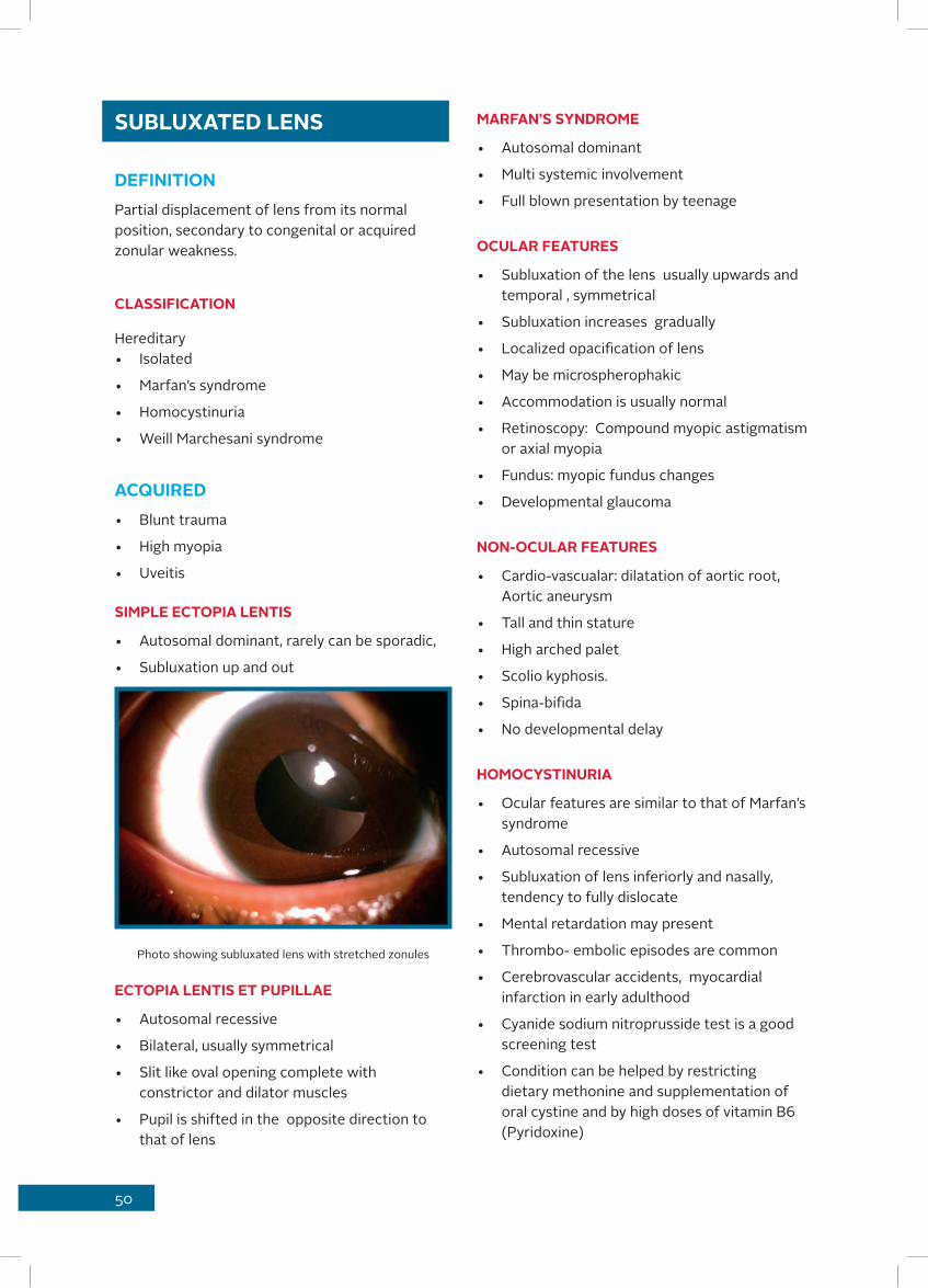
50
MARFAN’S SYNDROME
• Autosomal dominant
• Multi systemic involvement
• Full blown presentation by teenage
OCULAR FEATURES
• Subluxation of the lens usually upwards and temporal , symmetrical
• Subluxation increases gradually
• Localized opacification of lens
• May be microspherophakic
• Accommodation is usually normal
• Retinoscopy: Compound myopic astigmatism or axial myopia
• Fundus: myopic fundus changes
• Developmental glaucoma
NON-OCULAR FEATURES
• Cardio-vascualar: dilatation of aortic root, Aortic aneurysm
• Tall and thin stature
• High arched palet
• Scolio kyphosis.
• Spina-bifida
• No developmental delay
HOMOCYSTINURIA
• Ocular features are similar to that of Marfan’s syndrome
• Autosomal recessive
• Subluxation of lens inferiorly and nasally, tendency to fully dislocate
• Mental retardation may present
• Thrombo- embolic episodes are common
• Cerebrovascular accidents, myocardial infarction in early adulthood
• Cyanide sodium nitroprusside test is a good screening test
• Condition can be helped by restricting dietary methonine and supplementation of oral cystine and by high doses of vitamin B6 (Pyridoxine)
SUBLUXATED LENS
DEFINITIONPartial displacement of lens from its normal position, secondary to congenital or acquired zonular weakness.
CLASSIFICATION
Hereditary• Isolated
• Marfan’s syndrome
• Homocystinuria
• Weill Marchesani syndrome
ACQUIRED• Blunt trauma
• High myopia
• Uveitis
SIMPLE ECTOPIA LENTIS
• Autosomal dominant, rarely can be sporadic,
• Subluxation up and out
Photo showing subluxated lens with stretched zonules
ECTOPIA LENTIS ET PUPILLAE
• Autosomal recessive
• Bilateral, usually symmetrical
• Slit like oval opening complete with constrictor and dilator muscles
• Pupil is shifted in the opposite direction to that of lens

51
WEILL MARCHESANI SYNDROME
• Autosomal dominant or recessive
Components
• Microspherophakia
• Anterior dislocation most common
• Lenticular myopia
• Pupillary block glaucoma is most troublesome complication
• The patients have a short stubby stature
• Brachycephly
• Cardiac anomalies
WORK UP OF A CASE OF SUBLUXATED LENS
HISTORY
• Vision
• Squint
• Family history
• Trauma,(Unilateral)
• Developmental history
• Medical history
• Associated systemic features
CLINICAL EXAMINATION
• Refraction
• Through phakic and aphakic part
• Strabismus, preference for any particular eye
SLIT LAMP EXAMINATION
• Both dilated and undilated to assess whether pupillary area is mostly phakic or aphakic
• Direction of subluxation
• Clarity of lens
• Irregularly depth of AC
• Phacodonesis, iridodonesis
• Zonules are visible , stretched or broken
• Other associated ocular anomalies
• Look for signs or sequalae of trauma
FUNDUS EXAMINATION
• Myopic macular degeneration
• Peripheral retinal degeneration, breaks, lattices
• Physician/pediatric review to rule out systemic manifestations and appropriate treatment for same
INVESTIGATIONS
• Screening for inborn errors of metabolism
• Urine HPLC test for homocystinuria
• Echocardiogram to evaluate cardiac status
• Ultrasound biomicroscopy to evaluate zonular status to help in surgical plan
CAUSE OF DECREASE OF VISION
• Refractive error
• Amblyopia
• Glaucoma
• Retinal detachment
MANAGEMENT
• Genetic counseling
• Frequent follow ups as condition is progressive in most instances.
• Sibling examinations
OPTICAL CORRECTION
• Best possible refractive correction phakic or aphakic by glasses/contact lens
• Bifocals if prescribing glasses for aphakic portion.
• Treatment of amblyopia by patching
• It is necessary to recheck vision with new glasses for both distance and near
SURGICAL MANAGEMENT
INDICATION
• Anterior dislocation is an emergency as there is risk of acute rise in IOP and corneal decompensation
• Posteriorly dislocated lens may need removal
• Cataractous lens in visual axis causing a drop

52
in vision
• Pupil bisecting subluxation leading to difficulty in using refractive prescription
• Myopic astigmatism refractory to both glasses and contact lenses
• Asymmetric optical correction
• Occasionally along with posterior segment surgery for treatment of RD. etc
SURGICAL OPTIONS
• Lensectomy with or without scleral fixated intraocular lens in cases where there is significant zonular loss . Scleral fixation of intraocular lens is deferred in children below six years of age
• Phacoemulsification with intraocular lens implantation with capsular tension rings in case of localized zonular damage, mostly in acquired cases
• Peripheral iridotomy in cases prone for pupillar block
• Pars plana vitrectomy and lensectomy if dislocated lens
OPTIONS OF INTRAOCULAR LENS
• SFIOL with thorough core vitrectomy is preferable
• Iris claw lens
• Posteriorly fixated iris claw lens
CONGENITAL DISC ANOMALIES
PRESENTATION • Visual acuity ranges from total blindness to
minimal dysfunction
– Strabismus – Nystagmus
HISTORY
• What is the duration of diminution of vision?
• Is the diminution of vision slowly progressive?
• Is there any nystagmus or strabismus?
• Is there any history of consanguinity amongst parents?
• Is there any family member affected?
• Is there any mental retardation/ developmental delay?
• Are there any other systemic abnormalities?
EXAMINATION
• Best corrected visual acuity
• Slit lamp examination
• Lens changes
• IOP
• Fundus
• Indirect ophthalmoscopy with 20 D and 78D
Disc: Size (Hypoplasia, megalopapilla)
Shape: (Tilted disc, pit, coloboma)Margins: (Drusen)Color: Pallor (Autosomal dominant optic
atrophy)
Arteries: Attenuation, abnormal orientation (Morning glory syndrome)Macula: Dragged, distance from disc to macula, sub retinal fluid.
Fundus photo showing optic disc coloboma

53
INVESTIGATIONS
• Color vision
• Visual fields
• Fundus photo
• Neuro imaging
• ERG
• VEP
TREATMENT
• Glass prescription
• Low vision aids
• Systemic examinations
• Genetic counseling
• Advice ocular examination of other family members
• Do not give a very poor prognosis to the patients, emphasize on the positive side- the patient is very unlikely to be completely blind
• Routine monitoring every year or as and when required to treat the complications associated with the disease
• Rehabilitation
• Information about new scientific developments
OPTIC NERVE HEAD DRUSEN
• Rare, mostly bilateral
• Calcium deposits within the optic nerve head not evident at birth but more visible around first decade
• Can be superficial or deeply buried
INVESTIGATIONS
• A and B scan ultrasonography: disc elevation with high reflective spike
• CT scan
• Visual fields – enlarged blind spot
• Autofluorescence
• Ocular complications:
• Asymptomatic visual loss or transient vision loss
• Ischemic optic neuropathy
• Peripapillary serous chorioretinopathy
• Venous occlusions
HYPOPLASTIC DISC
• Most common optic disc anomalies
• Subnormal number of optic nerve axons with normal mesodermal elements and gial tissue
• Unilateral or bilateral
CLINICAL PRESENTATION
• VA - Variable according to severity (6/6 to NPL)
• Small disc surrounded by halo (double ring sign)
• Disc to macula ratio 2.62 as opposed to 2.94 in normal
• Vessel normal caliber but may be tortuous
• Occasional Association: Disorders involving mid line structures of brain
• De Morsier syndrome (septo-optic dysplasia)
• Absence of septum pellucidum and corpus callosum
• Triad of short stature, nystagmus and
Disc Anomalies Systemic Associations
Optic disc hypoplasia De Morsier syndrome (septo-optic dysplasia)
Optic disc dysplasia Transphenoidal form of basal encephalocele
Morning glory disc Basal encephalocele
Disc coloboma Papillorenal (renal-coloboma) syndrome Patau syndrome (trisomy 13) and cat-eye syndrome (trisomy 22) ‘CHARGE’
Tilted disc Situs inversus
54
hypoplastic disc
• Maternal diabetes
Other ocular associations: defective Color vision, relative afferent pupillary defect, foveal hypoplasia, aniridia, microphthalomos
DYSPLASTIC DISC
• Strikingly deformed disc with anomalous retinal vasculature
• Associated with morning glory disc anomaly with transphenoidal form of basal encephalocele
• Associated with infrapapillary tongue shaped retinochoroidal depigmentation
• Investigations: Neuroimaging to look for transphenoidal encephalocele
MORNING GLORY DISC:
• Very rare, usually unilateral, more common in females
CLINICAL FEATURES
• Visual acuity – Vary from NPL to 6/6
• Large disc with funnel-shaped excavation
• Glial tissue within base
• Spoke-like emerging vessels, more in number
• Surrounding chorioretinal pigmentary disturbance
• Complications: Serous retinal detachment in about 30%
• Occasional Association: Basal encephalocele which is frequently associated with mid-facial anomalies
MYELINATED NERVE FIBRES
• Myeliation begins at fifth month of gestation and progresses to reach lamina cribrosa by birth
• If it extends intraocularly anteriorly over the surface of the disc and retina, is called as myelinated nerve fibres
• Usually unilateral
• Visual acuity – 6/6 to 6/60 depends on the macular involvement
TYPES
• Isolated
– Peripapillary – Extensive
Investigations: Visual fields; enlarge blind spot
DISC COLOBOMA
• Rare, unilateral or bilateral
• Usually sporadic - occasionally dominant
• Incomplete or abnormal coaptation of proximal end of embryonic fissure
• Visual Acuity – depends on involvement of papillomacular bundle
Amblyopia treatment helps to improve visual acuity Ocular Associations: May be associated with other colobomas (choroid, retina, iris, ciliary), microphthalmia, orbital cyst, retinal detachment
• Occasional Systemic Associations of Optic Disc Coloboma:
1. CNS Malformation - Basal encephalocele and cysts
2. Chromosomal Anomalies - Patau syndrome (trisomy 13) and cat-eye syndrome (trisomy 22)
3. ‘CHARGE’ - Coloboma, heart defects, choanal atresia, aetarded development, genital and ear anomalies
4. Other Syndromes - Meckel-Gruber, Goltz, Lenz microphthalmos, Walker-Warburg and Goldenhar
INVESTIGATIONS
B scan optic disc Coloboma: Cup shaped defect in the optic disc Low reflectivity echo
OPTIC DISC PIT
• It is a congenital excavation of the optic nerve head that may be associated with other abnormalities of the optic nerve and peripapillary retina
• Incidence: One in 10,000 people, with no gender predilection, and are usually sporadic

55
CLINICAL FINDINGS
• Asymptomatic
• Visual acuity: 6/6 to 6/60
• Disc pit may lead to serous detachments of the retina, with associated full-thickness or laminar retinal holes, retinal pigment epithelium mottling and general cystic changes.
INVESTIGATIONS
• OCT- shows a schi sis like separation between the inner and outer retina and a larger retinal detachment
• Visual fields: Arcuate scotoma, enlarged blind spot
• FFA: No dye accumulation in the area of the serous detachment
MANAGEMENT
• Laser photocoagulation: Burns between optic Disc and serous retinal detachment
• Macular buckling
• Vitrectomy
Morning glory syndrome Disc coloboma
Females No gender predisposition
Unilateral Bilateral
Disc lies within the excavation Excavation lies within the optic disc
Central glial tuft No glial tuft
Abnormal retinal vasculature Normal retina vasculature
Basal encephalocele is common Uncommon
Rarely associated with genetic disorders Associated with multisystem genetic disorder
TILTED DISC
• Tilted disc syndrome, or Fuchs Coloboma, the superior pole of the optic disc appear elevated with posterior displacement of the inferior nasal disc
• It has an obliquely oriented long axis
ASSOCIATED FEATURES
• Myopia
• Scleral crescent located inferiorly or inferonasally
• Situs inversus (a nasal detour of the temporal retinal vessels as they emerge from the disc before turning back temporally)
• Posterior ectasia of the inferior nasal fundus
INVESTIGATIONS
Visual fields: Bitemporal hemianopia, which is typically incomplete and preferentially involves the superior quadrants and does not respect the vertical midlineTilted discs, myopic astigmatism, bilateral decreased vision, and visual difficulty at night should suggest the possibility of X-linked congenital stationary night blindness
MISCELLANEOUS OPTIC DISC ANOMALIES
• Peripapillary staphyloma
• Megalopapilla
• Optic nerve aplasia
56
Use ROP data sheet ( Refer Annexure 1)
Examine the cornea and anterior chamber
Mention extent of pupillary dilation, presence of iris vascular engorgement, persistent pupillary membrane, vitreous haze or hemorrhage
Record location of ROP-zone 1, zone 2 or zone 3
Enter number of clock hours involved.Note severity of the disease by stages 1 through 5.
Look for signs of Posterior Plus disease (sufficient vascular dilatation and tortuosity present in at least 2 quadrants of the eye ), A + symbol is added to the ROP stage number to designate the presence of plus disease1. For example - stage 2 ROP combined with posterior vascular dilatation and tortuosity should be written “stage 2 +ROP.”
Look for signs of Pre-Plus disease2 and can be noted beside the stage, for example – “stage 2 with pre-plus disease”.
Look for signs of Aggressive posterior retinopathy (AP-ROP)3
Notice previous treatment marks– cryo or laser scars, if any.
Watch for Apnoeic spells (crying is a good sign)Dragging of optic disc as a sequalae of retinopathy of prematurity
TREATMENT
If normal retinal vascularization is seen 360 degrees up to ora serrata, only follow-up examination is needed perhaps at six months. 1Plus disease is defined as a degree of dilation and tortuosity of the posterior retinal blood vessels as defined by a standard photograph.
2Pre-Plus disease is defined as abnormal dilatation and tortuosity of the posterior pole vessels that are insufficient for the diagnosis of plus disease but that demonstrate more arterial tortuosity and more venous dilatation than normal.
3AP-ROP Posterior pole is defined as vascular dilation and tortuosity of all 4 quadrants that is out of proportion to the peripheral retinopathy most commonly in zone I causing inability to differentiate arterioles and venules.
RETINOPATHY OF PREMATURITY
MINIMAL RELEVANT HISTORY TO BE RECORDED
GESTATION AGE (WEEKS AT DELIVERY)
• Chronological age (weeks after birth)
• Birth weight
• Reason for NICU admission
• Whether any supplemental oxygen therapy given (ventilator support)
• Type, duration of oxygen therapy
• H/o seizure disorder)
• H/o blood transfusions
• H/o neonatal septicemia
• H/o concurrent illnesses
• H/o multiple births
• Previous treatment if any
EXAMINATION Dilated fundus examination with Indirect ophthalmoscopy with scleral depression three to four weeks after birth or before discharge from the neonatal unit whichever is earlier.
Dilate with combination of phenylephrine 2.5% and tropicamide 1% instilled twice at 10 minutes interval with simultaneous punctual occlusion.
In lieu of ideal combination of phenylephrine 10% and tropicamide 1% as mentioned above , one can mix 1cc of 10% phenylephrine with 3cc of 1% tropicamide ( both commonly available). This gives a combination of phenylephrine 2.5% and tropicamide 0.75%.
• Instill a drop of paracaine (topical anaesthetic)
• Use Alfonso or other infant speculum for exposure
• Scleral indentation is done with a Wire vectis
A condensing lens of 20 D is used for comparison with standard photograph. 28D/ 30 D lens may also be used.
Data Recording

57
It is assumed that 3600 examination could be performed adequately and normal vascularization was noted up to ora serrata. In case of any doubt a review after one week is advised.
If retinal vasculature is immature and extends into zone II but no retinopathy is present, follow-up examination should be planned at two weeks. Either normalization takes place or ROP develops.
If retinal vasculature is immature and extends into zone I but no retinopathy is present and there is no plus disease, weekly examination is mandatory. Sometimes it can develop into Aggressive posterior ROP (AP-ROP) which does not necessarily follow classical 1-3 staging.
Stage 1 or 2 ROP in zone II/III with no Plus disease, follow-up examination should be planned at 1 to 2 weeks.
Stage 1 or 2 ROP in zone I or Stage 3 ROP in zone II, with no Plus disease, follow-up examination should be planned at <1 week .
Refer flow chart 1 & 2 for complete planning.
Type 2 ROP4 needs to be watched and treatment should be considered only if they progress to type 1 ROP5 or threshold ROP.
5Type 1 ROP is defined as Zone 1, any stage with plus disease, or zone 1, stage 3 without plus disease or Zone 2, stage 2 or 3 with plus disease (type 1 includes eyes with threshold ROP).
4Type 2 ROP is defined as Zone 1, stage 1 or 2 without plus disease or Zone 2, stage 3 without plus disease.
INDICATIONS FOR LASER TREATMENT
Type 1 ROP will need laser treatment as described below within 72 hours of detection.
Aggressive posterior retinopathy (AP-ROP) will need laser treatment as described below within 48 hours of detection.
INDICATIONS FOR SURGICAL REPAIR
Stages 4 and 5 will require surgical repair of retinal detachment with scleral buckling, lens sparing vitrectomy with or without Preoperative Intravitreal AntiVEGF or combined lensectomy + vitrectomy.
Treatment with Indirect Laser Photocoagulation:
Indirect laser photocoagulation is done using topical anesthesia in the operation theatre or NICU with constant cardiac and respiratory monitoring by the anaesthetist or peadiatrician.
Laser treatment anterior to the ridge to the entire avascular retina up to the ora. Treatment to be instituted within 72 hours of detection.
If zone 1 or posterior zone 2 disease, then split the treatment into two sessions. In session one cover three to four rows immediately anterior to the ridge. In session two cover the remaining avascular retina.
Topical steroid with or without antibiotic is prescribed in q.i.d dosage for five days.
SURGICAL MANAGEMENT
Scleral Buckling in ROP:
• Surgical repair of retinal detachment with scleral buckling done for Stage 4a /4b/ rare cases of stage 5
• Exoplant used is 240 band
• Scleral tunnels are made
• Placement of the band at site of highest ridge
• Indentation facilitated by paracentesis
• Removal of buckle or band cutting - approximately at one year age to prevent erosion into developing eye
VITRECTOMY IN ROP
Lens sparing vitrectomy with or without

58
Preoperative Intravitreal AntiVEGF or combined lensectomy + vitrectomy.
Intravitreal AntiVEGF is given in half the adult dose (0.625mg 0.75mm to 1mm from pars plana) if required after explaining the possible risk in O.T following sterile precautions under topical anesthesia
*Currently AntiVEGF is used for failed laser therapy or prior to surgery in very florid disease rather than as primary treatment.
FOLLOW UP
If initial fundus exam was normal and in cases with spontaneous regression of ROP as well as laser induced regression of ROP, repeat exam after six months to see indications of myopia, strabismus, amblyopia.
Watch for delayed complications like macular dragging, cataract, glaucoma or retinal detachment later on.
If significant refractive error is present as mentioned below - correction of the same with glasses is mandatory:
Myopia of more than >– 4.00 D
1. Hyperopia with no manifest deviation of >+6.00D
2. Hyperopia with esotropia >+2.00 D
3. Astigmatism of > 3.00 Dcyl
4. Any anisometropia of > +2.5 Dsp / > - 2.5 Dsp / > 2.5Dcyl

Annexure: I
59

60
Normal up to Ora
Follow up 3-6 months
Avascular RetinaNo ROP
Follow up2 weeks
Progressto ROP
Weeklyfollow up
RegressingROP
ROP needingtreatment
Initial Visit 3- 4 weeks
Flow Chart: 1
Regressed ROP Persistant Plus
AntiVEGF
Progressto ROP Scleral
Buckle
Lens sparingVitrectomy
ExtramacularTraction
PosteriorTraction Stage V
Progressed TRD
Lensectomy+ Vitrectomy
ROP on treatmentwith Laser
Flow Chart: 2

61
CONDITIONS CAUSING ROD – CONE
Dysfunction
• Leber’s Congenital Amaurosis (LCA)
• Typical retinitis pigmentosa (RP)
• Congenital stationary night blindness (CSNB)
• Melanoma-associated retinopathy
• Cancer-associated retinopathy
• Gyrate atrophy and choroideremia
CONDITIONS AFFECTING CONE – ROD
Dysfunction
• Stargardt’s disease
• Fundus flavimaculatus
• Atypical’ RP
• Bests’ disease
• Adult vitelliform
• Pattern dystrophies
• Dominant drusen
• Rod monochromacy and S-cone monochromacy
TESTS DONE FOR ROD – CONE DYSFUNCTION
• Best-corrected VA
• Contrast sensitivity (e.g.Pelli-Robson, low contrast VA)
• Color vision (e.g. Standard D15, Desaturated D15)
• Peripheral visual field assessment (full-field static screening)
• Dilated fundus examination
Child undergoing ERG test
RETINAL DYSTROPHIES
Inherited retinal dystrophies are many and varied, encompassing RPE (retinal pigment epithelium) and photoreceptor-based diseases.
They principally affect, or initially affect either rod (known as ‘rod-cone’) or cone (known as ‘cone-rod’) function.
Depending on the type of dystrophy, central or peripheral vision may be affected first, or scotopic versus photopic visual function.
Advanced stage retinal dystrophies are usually associated with low vision or legal blindness.
Some inherited retinal dystrophies are described as ‘stationary’ but are slowly progressive.
Nystagmus is the most definitive presenting sign of a hereditary retinal disorder. Poor visual funct ion may be the presenting abnormality in a
young child, and school-aged children with retinal d iseaseIllay present after a failed vi sion screening.
PRESENTING SYMPTOMS OF ROD – CONEDysfunction
• Decreasing night vision (night blindness)
• Peripheral visual field loss
• Poor light/ dark adaptation
• Defective Color vision
PRESENTING SYMPTOMS FOR CONE – ROD Dysfunction
• Decreasing visual acuity• Decreasing contrast sensitivity• Defective Color vision• Central visual field loss
• Poor light/ dark adaptation
FAMILY HISTORY
• To establish an inheritance pattern
• A retinal dystrophy should be suspected based on family history, fundus appearance and/or specific patient symptoms, particularly if bilateral

62
• Optical coherence tomography
• Fundus autofluorescence
• Dark adaptation
• Electroretinography (full-field flash/pattern/multifocal) and/or electro-oculography
TESTS FOR CONE – ROD DYSFUNCTION
• Best-corrected VA
• Contrast sensitivity (e.g. Pelli-Robson)
• Color vision (e.g. Standard D15, Desaturated D15)
• Central visual field assessment (e.g. Humphrey 24-2, 10-2, Macula)
• Macula photostress test
• Dilated fundus examination
• Optical coherence tomography
• Fundus autofluorescence
• Dark adaptation
Electroretinography (full-field flash/pattern/multifocal) and/or electro-oculographyManagement of these patients revolves around early diagnosis and specialized genetic counseling in order to decrease morbidity through optimal preventative care. Treatment options for these conditions are limited and tend to be focused around optometric visual rehabilitation where possible (eg use of low vision aids, orientation and mobility training).
PATIENT EDUCATION
Once diagnosed with a retinal dystrophy, affected individuals should be educated about the condition and the likely prognosis.Individuals should be made aware of possible treatment options
REVIEW
Regular review is to be done to consider medical or surgical interventions that may be beneficial during the disease course, and to exclude ocular comorbidity. Testing should include a specific focus on associations with the individual’s disease Review is beneficial with regard to maximizing visual function and testing should always involve at least:
• Careful refraction
• Assessment of the need for low vision
• Appropriate tests
Fundus photo of a child suspected with leber’s congenital amaurosis
REVIEW SCHEDULE
• Developed on an individual basis
• Most tests should be performed every 1 to 2 years
ELECTROPHYSIOLOGICAL TESTING
• To be done under GA for < one year
• Unreliable in children < one year
• To be repeated every two to three years to assess the progressive course of the disease
EMERGING MANAGEMENT CONSIDERATIONS
New management options for inherited retinal dystrophies ranging from gene therapy and oral or topical interventions designed to slow photoreceptor degeneration, through to stem cell transplants and bionic eyes under trial should be informed to parents and if approved in future will be contacted for same.
GENETIC TESTING
Genetic testing to be done for selected inherited retinal dystrophies which can help determine the likelihood of an asymptomatic family member developing the retinal dystrophy. It can also be useful for family planning decisions.

63
VISION REHABILITATION
The procurement of and training in the use of appropriate low vision aids. It should focus on reading, activities of daily living
ORIENTATION AND MOBILITY
Orientation and mobility instruction, where appropriate, should address the risk of falls and safety of the individual by assessing and modifying environmental and/or behavioral factors.
VOCATIONAL GUIDANCE
Vocational planning assistance is given to individuals experiencing severe vision loss, especially during school age
COUNSELING
Emotional and psychological well-being should be addressed using a multidisciplinary approach. Appropriate certification to be provided with a view to granting entitlements (pensions, benefits and travel concessions) to those with any visual impairment and their carer/s.
STARGARDT’S DISEASE
Description: Also known as juvenile macular dystrophy, this is the most common form of inherited macular degeneration and accounts for 7% of all retinal dystrophies.
Inheritance: Mainly AR but there is a rare AD variant.
Presentation
• childhood (about six years old) to early adulthood
• bilateral (usually) decreased central vision. This is often out of proportion to the clinical picture and these children may initially be labelled as malingerers
• There is also progressive Color blindness
Signs
• A relatively normal looking fundus except for a heavily pigmented RPE
• Atrophic macular degeneration - bull’s eye appearance as a result of atrophy of RPE
around a normal central core of RPE, a ‘beaten metal’ appearance, pigment clumping or marked geographical atrophy
• Normal visual fields in most cases
• ERG – normal in early stages, becomes abnormal in later
• EOG – subnormal
Differential diagnosis
• Fundus albipunctatus
• RP albescens
• Drusen
• Cone dystrophy
• Batten’s disease
• Chloroquine
• hydroxychloroquinemaculopathy
Work – Up
• History – age at onset
• Fundus examination
• FFA – silent choroid due to lipofuschin in RPE cells
• ERG and EOG
• Visual fields
Treatment
• Low vision aids
• Genetic counseling
Prognosis
generally is poor. Once vision drops below 6/12, progression is rapid and the visual prognosis is poor. However, active steps in visual rehabilitation can achieve and maintain some degree of independence.
JUVENILE BEST’S DISEASE
Description: This rare condition is also known as vitelliform (macular dystrophy. Thereare distinct, identifiable stages in this disease.
Inheritance • AD with variable penetrance
• Carriers may have normal fundus but abnormal EOG
64
Presentation
• Decreased vision or asymptomatic
• Characterized by a yellow, round subretinalmacular lesion which grows over years, eventually to give rise to a characteristic round egg-yolk appearance (lipofuschin) and which may be later associated with a pseudo-hypopyon
• Bilateral lesions located at fovea of 1 – 2 disc diameters in size
• Subnormal changes showing severe loss of light response occur in EOG readings in children before they are symptomatic or anything is seen clinically
• Lesions degegenerate in 20% and may develop CNV, hemorrhage and scarring
Work – Up
• Family history - examine family members
• Fundus examination – macula with 90 D lens
• EOG – highly specific, detects carrier state of the disease
• ERG - Normal
• FFA – to detect CNV
Treatment
• Amsler’s grid given and instructed on its use and return immediately if change noted
Prognosis
• Declining visual acuity may be a reflection of macular scarring.
LEBER’S CONGENITAL AMAUROSIS
Description: Group of hereditary (usually autosomal recessive) retinal diseases that affect the rods and cones.
Inheritance: AR.
Presentation
• Severe vision loss noted in infancy. Vision ranges from 20/200 to bare light perception in most patients
• Nystagmus
• Poorly reactive pupils
• Exhibit the characteristic ‘oculodigital sign’:
Constant rubbing of the eyes results in orbital fat resorption and subsequent enophthalmos (eyes sunken into sockets)
• Ocular features: Blindness, hypermetropia, keratoconus, keratoglobus, early cataracts
• Funduscopic appearance - a normal appearance, particularly in infancy; to pigment clumping in the retinal pigment epithelium (RPE)
ERG – Extinguished
• Nervous system - learning disabilities, deafness, epilepsy
• Other systems - renal and musculoskeletal abnormalities, endocrine dysfunction
• Associated systemic diseases - Alstrom syndrome, Battendisease (neuronal ceroid-lipofuscinosis), Joubert syndrome, peroxisomal diseases (Zell wager disease, neonatal adrenoleukodystrophy, and infantile Refsum disease), and SeniorLoken syndrome
Genetic Testing: To predict prognosis and counseling
Prognosis
• Very poor
CONGENITAL STATIONARY NIGHT BLINDNESS
Description
• Group of non progressive retinal disorders characterized predominantly by abnormal function of the rod system
Inheritance
• Depending on the subtype, may be AD, AR or XL(most common)
Presentation
• Nyctalopia
• Presents in infancy, with myopia, nystagmus and strabismus with normal fundus in XL and AR
• Dark adaptation – Abnormal
• ERG pattern - “Negative” Dark-adapted ERG: a large a-wave and a reduced (negative) b-wave

65
• Oguchi disease and fundus albipunctatus are forms of CSNB with abnormal fundi
Prognosis
• There is no progression
CONGENITAL MONOCHROMATISM
Description
• This is used to describe various degrees of rod monochromatism or cone monochromatism
Inheritance• Depending on subtype, AR or XL
Presentation
• Poor color vision, poor central vision, nystagmus, hemeralopia and photophobia in rod monochromatism
• Color vision – Abnormal
• Fundus – Normal, with absent foveal reflex
• ERG – Extinguished cone responses but normal rod response
• There may be some Color perception in incomplete rod monochromatism.
• Cone monochromatism is associated with better visual acuity (6/6 to 6/9) than rod monochromatism
• Treatment
• Dark glasses/red glasses
• Low vision aids
• Prognosis - There is no progression.
• Retinitis Pigmentosa
Presentation
• Decreased night vision
• Loss of peripheral vision
• Decrease in central vision in late stage
• Triad – Arteriolar attenuation, bony spicules, waxy pallor disc
• CME, PSC, epiretinal membrane
• Visual fields – Ring scotoma and small central field (tubular vision) later stage
Inheritance
• AR – Most common
• AD – Least severe
• X R – Rare, most severe
Work – up
• Family history diagnosing and counseling purposes
• Visual fields
• ERG – In mothers of male patient (women carriers)
• Serum phytanic acid to r/o refsum’s disease if neurologic abnormalities is present
• Lipid profile, serom protein and peripheral blood smears for acanhocytosis – abetalipoproteinemia
• Kearns – Sayre syndrome – ECG for heart blocks
Treatment
• Genetic counseling
• Tinted glasses
• Low vision aids and vocational rehabilitation
Prognosis
• Disease is progressive. Guarded visual prognosis.Apart from diagnosing and counseling on inheritance, Almost all the children of retinal dystrophies should have consultation with the vision rehabilitationist especially on the mode of their education

66
Under general anesthesia with a good assistant and nursing staff and a good microscope surgery should be performedWound involving only the cornea and or limbus interrupted 10’o nylon sutures are used.• Any exposed iris / uveal tissue should be
abscised
• Iris tissue must be cleared away from the wound
• All the sutures buried
• Should avoid intraocular manipulations
• Should ensure formed globe
WOUND EXTENDING TO THE SCLERA
• Need a good exposure to visualize the total extent
• The wound should be first approximated at the limbus and then sutured on either sides
SCLERA TEARS
• The clue to identify the location of the tear through an intact conjunctiva is to look for localized or eccymosis
• In case of inability to localize the probable site, its useful to follow and order by making peritomies and exploring the globe with minimal dissections and hooking of the recti where necessary
• Very rarely a wound may be impossible to localise which may mean a very posterior dehiscence which may not be amenable to repairing by the anterior approach.
• Once the extent is indentified 6’o vicrly interrupted sutures are used to repair the sclera
• When the wound extends under an extra-ocular muscle it gives better exposure to first put placement sutures with 6’o vicrly on the muscle, disinsert it then do the sclera suturing and finally make sure to suture back the muscle to its insertion
PEDIATRIC TRAUMATIC CATARACT MANAGEMENT TIMING
During Primary Repair• Best avoided
• Only if capsule ruptured with lens matter in
PEDIATRIC OCULAR TRAUMA
CHILDREN BY THEIR NATURE ARE TRAUMA PRONEOcular trauma in children poses unique challenges starting from assessment, timing of treatment and the early occurrence of amblyopia. So, standard guidelines are needed to streamline management. Various groups have applied the Birmingham ocular trauma score for pediatric ocular trauma and found it to be useful as a predictor of visual outcome.
BIRMINGHAM OCULAR TRAUMA SCORE
EXAMINATION
Good history and patience usually pays in being able to make a good clinical assessment of children presenting with acute trauma. But there may be quite a few occasions when we may need a short sedation or anesthesia to have a thorough evaluation. In such situations we should ensure complete examination including a team of doctors from various specialities if indicated and decisions made. It may always be a useful practice to inform the parents that examination findings may warrant immediate surgery so that repeat anesthesias could be avoided.
Only minimum necessary investigations are required before the preliminary management like a CT scan to r/o fractures or foreign bodies. More detailed investigations can be reserved after the management of the acute trauma.
TIMING OF TREATMENT
Indications for immediate surgical intervention in the order of priority:• Any open globe
• Closed globe with ruptured lens
• Blow out fractures with muscle entrapment
• Trauma to the naso-lacriaml system
• Lid tears
OPEN GLOBE INJURIES
Aim is to restore globe integrity and prevent introducing infection intraocularly

67
the anterior chamber
• Very small corneal wound with traumatic cataract and normal posterior segment at presentation in a very young child – this can help early rehabilitation and avoid a second anesthesia when anyway an intraocular lens implantation is not an option
• All other occasions it is best to plan cataract management after primary repair
IMMEDIATELY AFTER PRIMARY REPAIR (FIVE DAYS TO TWO WEEKS)
• When the globe is intact and in a more elective manner cataract management is preferred
• Without implanting intraocular lens(IOL)
• Younger than two years of age
• Corneal sutures interfere with IOL power calculations but cataract has to be removed due to inflammation or intraocular pressure issues
• Corneal sutures interfere with IOL power calculations but cataract has to be removed due to impending risk of irreversible amblyopia
• When the capsular support is inadequate
• In less than six year old posterior capsule management is also necessary leaving a rim of capsule for future IOl implantation
• With IOL implantation
• Preferred whenever possible
• Peripheral corneal wound which doesn’t interfere with IOL power calculations
ELECTIVE CATARACT MANAGEMENT (LATER THAN SIX WEEKS)
• Any child above six years of age• After suture removal and when able to get a
reliable IOL power
PROCEDURE
• When IOL implantation is not planned
• A bimanual limbal aspiration is done with irrigation aspiration probe
• Capsule can usually be managed with the help
of instruments like curved intraocular scissors, capsular forceps, vitrectomy cutter, etc.
• This ensures minimal entry wounds and a closed chamber
• Posterior capsule is also opened at least 5 to 6 mm with whichever instrument possible and a limited anterior vitrectomy performed
• When planning to implant IOL the lens aspiration is best done bimanually and once capsular integrity is ensured and IOL placement confirmed the main entry can be made
• Wound should be sutured with 10”o vicryl or nylon sutures whenever indicated
• Corneal wound integrity is checked before closing
• Additional procedures like pupilloplasty, iridodialysis repair, etc, must be managed concurrently
• An indirect ophthalmoscopy to evaluate the posterior segment in all trauma patients
IOL POWER CALCULATIONS
The challenge is mainly in obtaining a reliable keratometry valueClear cornea – routine method using auto/manual keratometry and standard formulas like SRK 2• If corneal sutures present – to wait for teo
weeks after suture removal
• Manual K/topography or SIM K
• Routine formulas
• Parents should be primed about the probability of requiring contact lenses for the residual astigmatism
CHOICE OF IOL
• If in the bag standard single piece hydrophilic acrylic lenses
• If in the sulcus three piece acrylic lenses or PMMA lenses depending on the adequacy of the capsular support
• Sclera fixated lenses in children older than eight in the absence of capsular support
VISUAL REHABILITATION
• Contact lenses whenever IOL has not been

68
implanted once the corneal sutures have been removed
• Glasses even earlier if age is highly amblyogenic
• Secondary IOL implantation as early as reliable IOL power calculation can be made wherever possible
• Along with refractive correction, parallelly patching regime according to the age and severity of amblyopia has to be started
• Regular follow ups
PEDIATRIC LOW VISION CARE
INTRODUCTION
The major causes of blindness in children vary widely from region to region, being largely determined by socio-economic development, and the availability of primary health care and low vision care services1. The prevalence of blindness in children ranges from approximately 0.3/1000 children in affluent regions to 1.5/1000 in the poorest communities. Reliable population-based data on the causes of blindness in children are difficult to obtain in developing countries2. There is an increasing awareness about the needs of students with low vision, particularly in developing countries where programs of integrated education are being developed. However, the appro priate low vision services are usually mandatory in order to improve their residual vision3.
DEFINITION OF LOW VISION
A child with low vision is one who has impairment of visual functioning even after treatment and/
or standard refractive correction, and has a visual acuity of less than 6/18 to light perception, or a visual field of less than 10 degrees from the point of fixation in the better eye. But who uses, or is potentially able to use, vision for the planning and/or execution of a task for which vision is essential (WHO, 1992).
IMPACT OF LOW VISION
Functionally, low vision is described as irreversible visual loss and a reduced ability to perform many daily activities, such as recognizing people in the street, reading blackboards, writing at the same speed as peers, and playing with friends.
CAUSES OF LOW VISION
The major causes of visual impairment among children includes Stargardt’s disease, Myopic degeneration, Oculocutaneous Albinism, Retinitis Pigmentosa, Maculopathy, Optic Atrophy, Corneal opacities, Rod cone dystrophy and other heredomacular degenerations3.
CLASSIFICATION OF CONDITIONS CAUSING LOW VISION
There are various ocular conditions that cause low vision. These conditions may be congenital or acquired. The number of conditions is too large to list here. However, they can be broadly classified in terms of the impact on what the person with low vision sees.
These categories are: Overall blurred vision Central field loss Peripheral field lossMultiple field loss
Category Corrected VA-Better Eye WHO Definition Working Indian Definition
0 6/6-6/18 Normal Normal Normal
1 <6/18- 6/60 Visual Impairment Low Vision Low Vision
2 <6/60- 3/60 Severe Visual Low Vision Blind
3 <3/60- 1/60 Blind Low Vision Blind
4 <1/60- PL Blind Low Vision Blind
5 NPL Blind Total Blindness
Categories of Visual Impairment

69
PEDIATRIC LOW VISION CARE
The first thing the clinician needs to understand is that the final management in the low vision care clinic can have no impact on the ocular condition of the child. A child is referred to the low vision clinic after being diagnosed with a condition that cannot be improved further with treatment. The management aims to improve the way that the existing vision is already being used by the child. Therefore, the low vision examination has two primary goals – a) to identify and understand how well the child uses residual vision (or) functional vision assessment b) to explore conditions or modifications for maximizing or enhancing visual performance.
LOW VISION EXAMINATIONSteps in examination of child with low vision:Observation – Observe how the child utilizes vision for moving or to locate an object or for communication from the time of entry into the clinic.Interview [History taking] – Understand the difficulties the child has in utilizing vision for visual tasks and identify visual tasks that are important for the child to execute.Assessment/Functional vision- Clinical assessment of visual functions – Visual acuity, contrast sensitivity, color vision, visual fields.End-point of the above low vision examination: Goal setting.
EXTERNAL OBSERVATIONThe foremost step towards assessing a child with low vision begins even before the child enters the room. The clinician must carefully observe the mannerisms, visual habits and body language of the child. The cues that an experienced practitioner can pick up from this initial assessment go a long way in successful counseling and management of the child.
Details to observe for before beginning the low vision evaluation:
Level I: In the waiting room/ waiting area
How actively does the child use vision to gather
Figure: Sample picture of a scenery seen by an individual with normal vision
Figure: Sample of scenery as seen by a child with overall blurred vision
Figure: Sample of scenery as seen by a child with central field loss
Figure: Sample of scenery as seen by a child with peripheral field loss
70
input from the surrounding?• Does the child try to actively observe what is
going on?
• Does child make visual contact while interacting with family members/attender?
Level II: On entering the examination room
How does the child navigate the environment?• With the help of the care giver?
• With the help of a cane?
• Tries to use tactile input by trailing fingers along walls or furniture
• Is independent but visually (with or without additional head movement) scans the environment for obstacles
• Has poor navigation skills: Bumps into objects
• Walks independently, confidently without any aid
Level III: Mannerisms/Posture
• Head down: [may be due to glare or reduced inferior field of view]
• Squeezing eyes: Uncorrected refractive error/ glare
• Chin up/ Chin down posture
• Head nodding
• Eccentric viewing / Fixation
• Eye-poking/Rubbing
• Presence of nystagmus with/without compensatory head postures
Level IV: Physical Appearance
• Shoddily dressed
– Mismatched colors – Improperly buttoned – Poorly tied shoelaces• Untidy clothes
– Unkempt appearance - Matted hair – Spilled food stains on clothes of geriatric
population• Frailty
– Any leg/arm weakness [Favours a particular leg while walking or a particular hand for hold ing reading material]
– Tremors – The last two are particularly important for
deciding in the use of handheld devices. • Children diagnosed to have retinitis
pigmentosa
– Obesity – Polydactyly – Clubbed fingers [syndactyly] – Albinism: – Sunburn – Nystagmus – Any unhealed lesions or deep abrasions on
skin [may have poor platelet aggregation]
Level V: While interacting with the child
• Does the child expect the care giver to be in close proximity? Is the child dependent on caregiver for the visual task?
• Does the child interact directly with the examiner?
• Mode of exploring surroundings – Tactile/Auditory/Visual: Children with profound visual impairment may begin to depend on other senses to explore their surroundings, or continue to use vision – understanding their method for sensory input could help in suitable counseling on the mode of education
• Voice modulation: Does the child talk with halts/in a monotone ?
• Does child make eye contact while talking or at least attempt to fixate eccentrically
• Child is enthusiastic about the outcome of the test/does not interact at all/is not interested in the test
• Attempt to gauge the expectations of the child: high/reasonable/low
Level VI: While interacting with the care giver
• Is the care giver/family aware about the ocular condition of the child
• Are they over protective/deny care
HISTORY TAKING
Child Details
• Age
• Occupation
71
• Living situation and main care giver [number of family members, care given, source of family income gives a general idea about the child’s psychological outlook, need for independence and how much support will be available from the family]
• General health (Diabetes, Hypertension, following trauma or surgery, are important determinants of success of low vision management and also influence prognosis of the ocular condition) – in case of acquired conditions or worsening conditions, it would be a good practice to know when it was detected, specific milestones in disease history (worsening, surgeries, systemic/visual disability onset)
• Relate ocular conditions to systemic syndromes, especially those that can be potentially life threatening. For example: metabolic disorders, Wilms tumour in aniridia, syndromes in children that can have impact on developmental milestones such as Bardett Biedl Syndrome in Retinitis Pigmentosa, etc
• Document family history of consanguinity or ocular disease to understand the mode of inheritance and the prognosis of the condition.
• Other limitations such as physical handicap, hearing impairment, mental retardation
Previous Low Vision Care
• Has the child tried any device/devices before
– Type of device – Details (Magnification/Color of tint) – For what purpose – How useful was it/ Success with device – Frequency of use – Duration of use• Does patient have/know about disability
certificate
– Used associated concessions/rights?This gives an idea of what the child has tried before and how much success or failure has been faced on previous exposure to low vision care. Also, it would be a guideline to understand the child’s demands and visual tasks. Details about disability concessions would inform the practitioner about how well educated or well informed the subject is about the ocular condition or the government’s facilities for the same.
Difficulty with Visual Tasks
The most important thing to discern is about the onset of difficulty with visual task and whether the problem is progressively worsening, stable or if the vision is improving. Visual tasks are divided into three categories:• Distance
• Intermediate
• Near
Distance visual tasks Include tasks such as
• Face recognition
• Seeing blackboard [For students]
• Seeing PowerPoint displays
• Seeing signboards/bus numbers/ shop names/ street names
• Seeing curbs or steps
• General sight seeing
• Near visual tasks may be broadly classified as:
• Seeing textbooks/ notebooks [ School children]
• Reading office print/ handwritten material at the office
• Reading newspapers/ novels/ magazines
• Reading bills/ price tags/ medicine labels
• Using mobile phone
The examiner should take care to distinguish between difficulty in reading a text and difficulty in prolonged reading or reading in bright/dim light.
Writing tasks such as
• Signing, filling accounts
• Generally children with field loss have will problems writing along a straight line or complain of overlapping of letters while writing
INTERMEDIATE VISUAL TASKS
• Using computers/mobile phones
• Problems with computers may arise from:
• Difficulty with contrast [due to impaired contrast sensitivity]
• Difficulty finding options on screen [due to peripheral field loss]

72
• Skipping lines or mixing up lines [due to central field loss]
• Reading text on screen [reduced near vision]
LIGHT SENSITIVITY
Various ocular conditions causing low vision cause problems such as:
– Photophobia – Difficulty seeing in dim light – Delayed dark adaptation – Delayed light adaptation – Glare – Veiling / Disability glare
Knowing the child’s light sensitivity, one can help in modifying the daily environment of the child, providing non optical devices such as filters or peaked caps to cope with dim light, etc.
MOBILITY TASKS
Differentiate between difficulty with navigating in familiar and unfamiliar environments to difficulty only with moving around in dim light and brightly lit surroundingsAlso question what compensating techniques the child uses such as:
– Having an assistant – Help of the care giver – Use of walking stick / cane – Does the child just sit at home due to mobility
problems?This question guides the practitioner in deciding the best mode of intervention for difficulty with mobility and determining the need for orientation and mobility training.
ACTIVITIES OF DAILY LIVING
Activities of daily living [ADL] are those activities that are a part of daily life and are an absolute necessity:
– Personal grooming – Personal hygiene – Finding food on plate and eating – Currency identification – Inserting keys into doors – Matching colors of clothes, etc
Various techniques can be taught by rehabilitation therapists when non optical devices do not help patients with low vision / visual impairment to perform activities of daily living independently. The parameters that functional vision assessment
looks at areVisual acuityContrast sensitivityColor visionVisual field
IDEAL VISUAL ACUITY CHART IN LOW VISION CLINIC
• Standardized testing at different distances (LogMAR)
• Equal number of letters on each line (more than 3)
• Equal distance (same as letter height) between letters on a row
• Space between two rows equals letter height of smaller row
• Contrast of letters to background 95% or more
RECOMMENDATIONS FOR VISUAL ACUITY TESTING
• Chart lighting: 85 Cd/m2-300 Cd/m2
– Use variable lighting to assess optimum light ing for best visual acuity• Start at close working distance
• Test better eye first, then worse eye and then binocularly. Document whether monocular (which eye) or binocular performance is better
• Allow abnormal head postures and eccentric viewing
• Do not point to letters or provide cues – allow to skip letters on a line (gives an idea of letters being skipped on a line due to scotoma)
• Allow time to scan and coax where required
• Positively encourage further reading. Pointing to a letter, expecting a better performance on the test should be at the end and both visual acuities should be documented.
• Use of pinholes is not recommended when visual acuity is assessed at less than four meters.(less than four meters cannot be considered optical infinity and therefore may not yield correct results for paraxial approximation. However, pinholes of larger aperture sizes are still used in some low

73
vision clinics to detect uncorrected refractive error) In the case of patients who have sufficient vision to respond at four meters or larger distances may not always perceive an improvement especially in cases of field loss (especially central) or impaired contrast sensitivity (due to luminance decrease with pinhole).
– Large aperture pinholes are available for special use in the low vision clinic where indicated.
–Note: The need for visual acuity testing in the low vision clinic is to get an idea of functional vision or how the patient uses his vision in daily life and to also identify the smallest size of letters that can be read in ideal conditions such as a close working distance or given adequate time.Commonly used visual acuity chart in the low vision clinicBailey Lovie high contrast visual acuity chart
Figure: High contrast Bailey Lovie chart
• One of the earliest charts used to implement logarithmic progression for letter size
– Letters have equal legibility. The letters are combinations from a 10-letter set.
– Uses five Sloan sans-serif letters per line – Equal spacing between characters (same as
letter height), uniform spacing between rows
(same as letter height of next row) – Logarithmic progression of text – letters dou
ble in size every three lines – If the chart is moved closer by 0.1 log steps,
the subject will read an additional row – Each letter can be scored to calculate final
visual acuity score. (0.02 per letter, 0.1 per row) Berkeley Rudimentary Vision test kit(Bailey, 2012).• Three pairs of cards (25 cm2 area)
• First pair: Single tumbling E (STE) – 100M, 65M, 40M. 25M
• Second pair: Grating acuity-200M, 125M, 80M, 50M
• Third pair: White field projection, black-white discrimination
SCREENING METHOD
• Step 1: Show 25M single tumbling E(STE) at 100 cm → Pass →Use LogMAR
• Step 2: Fail 25M at 100 cm→ Show 100M at 100cm → Pass → Assess rest of visual acuity with Ste (Visual acuity will be between 100 and 25M)
• Step 3: Fail 100M at 100 cm→Check with 100M at 25cm → Pass → Assess rest of visual acuity with STE at 25 cm
• Step 4: Fail STE at 25 cm → Use grating acuity 200M at 25cm→ Pass → Assess visual acuity with grating acuity cards at 25 cm
• Step 5: Fail Grating acuity at 25 cm → Show Quad or Hemifield white projection in four quadrants of visual field → Pass → Document light projection or
• Step 6: Fail white field projection at 25 cm → Use penlight to assess light perception
Parameters to be documented when assessing visual acuity in a child with low vision - 1)
Testing distance
M Notation
LogMAR acuity
Snellen (Feet)
Snellen (Meters)
100 cm 100M 2 20/2000 6/600 63M 1.8 20/1250 6/378 43M 1.6 20/800 6/258 20M 1.4 20/500 6/12025 cm 100M 2.6 20/8000 6/2400 63M 2.4 20/5000 6/1500 43M 2.2 20/3200 6/960 20M 2 20/2000 6/600

74
Letter size 2) Testing distance 3) Qualitative components - missing letters, detects on pointing, speed of reading, etc.
NEAR VISUAL ACUITY
Notations used in near visual acuity testingN Notation (Point Notation)• Non standardized
• Varies based on type of font
• 1 point – 1/72 of an inch (slug into which the text fits)
• Actual size of lower case text: 1 [point – 1/144 inch)
M NOTATION
• Sloan’s M notation can be converted based on the distance at which it is read.
• 1M subtends 5 minutes of an arc at 1 meter
• 8 points (N8) = 1M
SNELLEN EQUIVALENT
Near vision charts are calibrated for a certain working distance (usually 40 cm) and the corresponding Snellen acuity is given based on letter size.
TYPES OF NEAR VISION CARDS
Figure: MN read chart
• Single word
– May be word, number or symbol – Example: Lighthouse “game” or number cards• Continuous text
– Word reading• Sets of unrelated words along a single line
• Example: Bailey Lovie word reading chart
– Continuous reading tests• A complete sentence of about 10 words –
usually simple and makes sense
• Example: MN Read Card (Minnesota Near Vision reading test)
recommendations for visual acuity testing for near• Chart lighting: 85 Cd/m2-300 Cd/m2
– use variable lighting to assess optimum light ing for best visual acuity• Start at close working distance/allow patient
to adjust for optimal comfort
• Test better eye first, then worse eye and then binocularly. Document whether monocular (which eye) or binocular performance is better
• Allow abnormal head postures and eccentric viewing
• Allow changing orientation of text – vertical orientation is easier to read for patients with macular degeneration
• Do not point to letters or provide cues – allow to skip letters on a line(gives an idea of letters being skipped on a line due to scotoma)
• Allow time to scan and coax where required
• Positively encourage further reading. If you feel that by pointing to a missed letter/word the patient may perform better on the test, do it at the end and document both visual acuities
• Parameters to document:
– Letter size – Working distance – Reading speed: Number of words read per
minute (document for smallest text size that can be read)
– Critical print size: Minimum print size for maximum reading speed. (Up to a certain acuity level, the patient may read fast and the reading speed may then drop off slowly. It is necessary
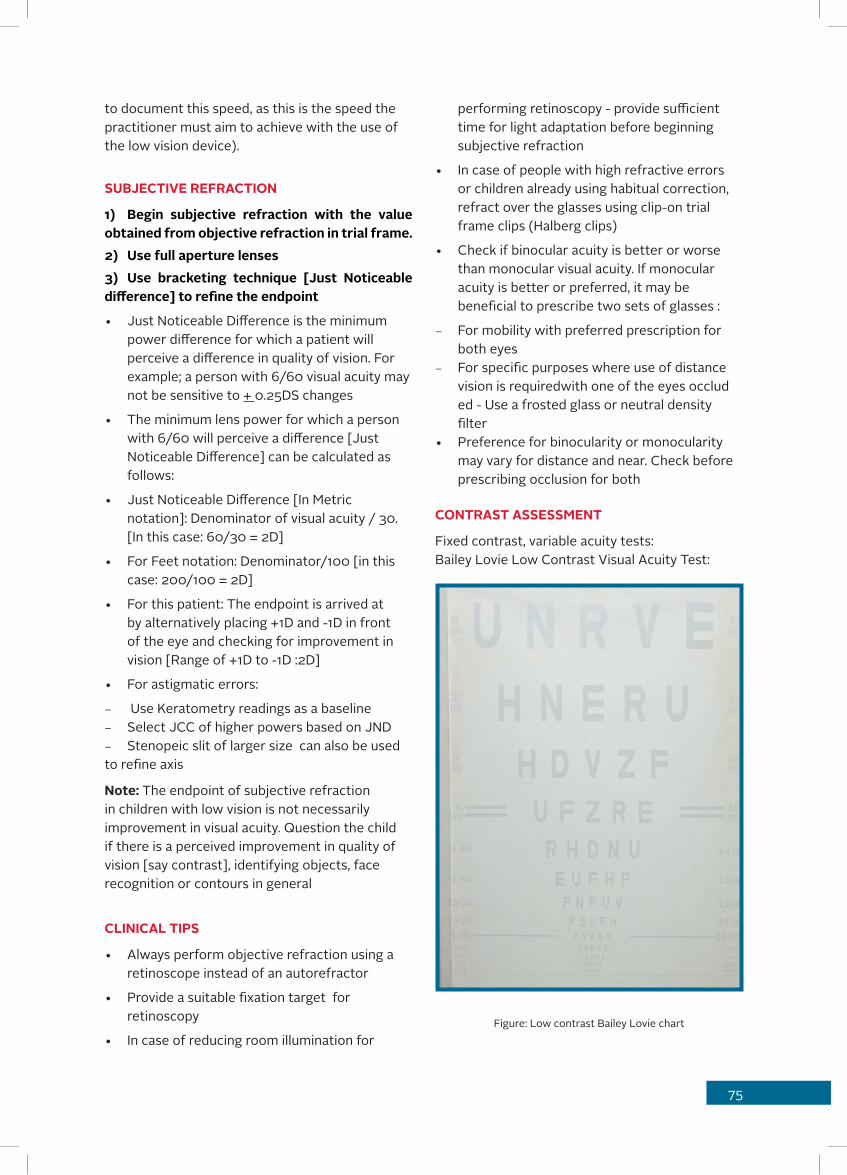
75
to document this speed, as this is the speed the practitioner must aim to achieve with the use of the low vision device).
SUBJECTIVE REFRACTION
1) Begin subjective refraction with the value obtained from objective refraction in trial frame.2) Use full aperture lenses3) Use bracketing technique [Just Noticeable difference] to refine the endpoint
• Just Noticeable Difference is the minimum power difference for which a patient will perceive a difference in quality of vision. For example; a person with 6/60 visual acuity may not be sensitive to + 0.25DS changes
• The minimum lens power for which a person with 6/60 will perceive a difference [Just Noticeable Difference] can be calculated as follows:
• Just Noticeable Difference [In Metric notation]: Denominator of visual acuity / 30. [In this case: 60/30 = 2D]
• For Feet notation: Denominator/100 [in this case: 200/100 = 2D]
• For this patient: The endpoint is arrived at by alternatively placing +1D and -1D in front of the eye and checking for improvement in vision [Range of +1D to -1D :2D]
• For astigmatic errors:
– Use Keratometry readings as a baseline – Select JCC of higher powers based on JND – Stenopeic slit of larger size can also be used
to refine axis
Note: The endpoint of subjective refraction in children with low vision is not necessarily improvement in visual acuity. Question the child if there is a perceived improvement in quality of vision [say contrast], identifying objects, face recognition or contours in general
CLINICAL TIPS
• Always perform objective refraction using a retinoscope instead of an autorefractor
• Provide a suitable fixation target for retinoscopy
• In case of reducing room illumination for
performing retinoscopy - provide sufficient time for light adaptation before beginning subjective refraction
• In case of people with high refractive errors or children already using habitual correction, refract over the glasses using clip-on trial frame clips (Halberg clips)
• Check if binocular acuity is better or worse than monocular visual acuity. If monocular acuity is better or preferred, it may be beneficial to prescribe two sets of glasses :
– For mobility with preferred prescription for both eyes
– For specific purposes where use of distance vision is requiredwith one of the eyes occlud ed - Use a frosted glass or neutral density filter • Preference for binocularity or monocularity
may vary for distance and near. Check before prescribing occlusion for both
CONTRAST ASSESSMENT
Fixed contrast, variable acuity tests:Bailey Lovie Low Contrast Visual Acuity Test:
Figure: Low contrast Bailey Lovie chart
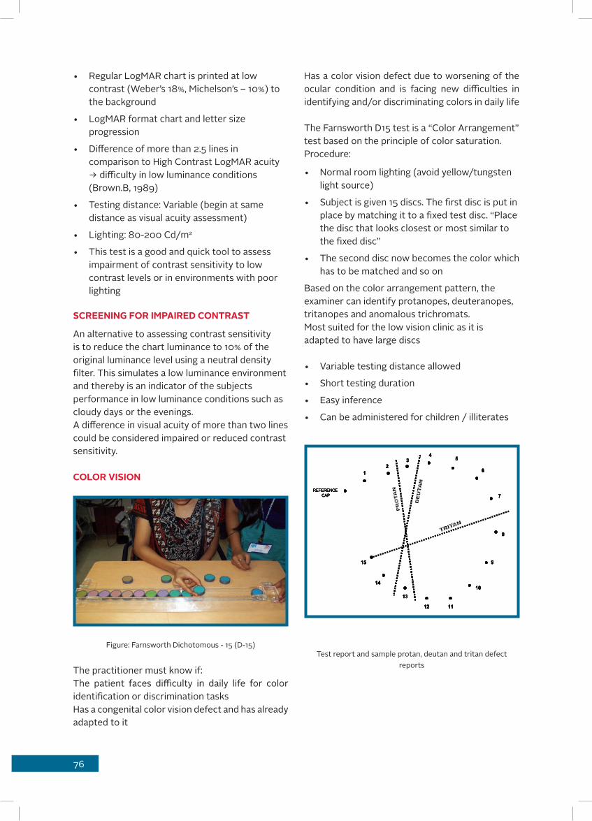
REFERENCE CAP
12
34
5
6
7
8
9
10
1112
13
14
15
REFERENCE CAP
12
34
5
6
7
8
9
10
1112
13
14
15
REFERENCE CAP
12
34
5
6
7
8
9
10
1112
13
14
15
REFERENCE CAP
12
34
5
6
7
8
9
10
1112
13
14
15
76
• Regular LogMAR chart is printed at low contrast (Weber’s 18%, Michelson’s – 10%) to the background
• LogMAR format chart and letter size progression
• Difference of more than 2.5 lines in comparison to High Contrast LogMAR acuity → difficulty in low luminance conditions (Brown.B, 1989)
• Testing distance: Variable (begin at same distance as visual acuity assessment)
• Lighting: 80-200 Cd/m2
• This test is a good and quick tool to assess impairment of contrast sensitivity to low contrast levels or in environments with poor lighting
SCREENING FOR IMPAIRED CONTRAST
An alternative to assessing contrast sensitivity is to reduce the chart luminance to 10% of the original luminance level using a neutral density filter. This simulates a low luminance environment and thereby is an indicator of the subjects performance in low luminance conditions such as cloudy days or the evenings.A difference in visual acuity of more than two lines could be considered impaired or reduced contrast sensitivity.
COLOR VISION
Figure: Farnsworth Dichotomous - 15 (D-15)
The practitioner must know if:The patient faces difficulty in daily life for color identification or discrimination tasksHas a congenital color vision defect and has already adapted to it
Has a color vision defect due to worsening of the ocular condition and is facing new difficulties in identifying and/or discriminating colors in daily life
The Farnsworth D15 test is a “Color Arrangement” test based on the principle of color saturation.Procedure:
• Normal room lighting (avoid yellow/tungsten light source)
• Subject is given 15 discs. The first disc is put in place by matching it to a fixed test disc. “Place the disc that looks closest or most similar to the fixed disc”
• The second disc now becomes the color which has to be matched and so on
Based on the color arrangement pattern, the examiner can identify protanopes, deuteranopes, tritanopes and anomalous trichromats.Most suited for the low vision clinic as it is adapted to have large discs
• Variable testing distance allowed
• Short testing duration
• Easy inference
• Can be administered for children / illiterates
Test report and sample protan, deutan and tritan defect reports

77
MISCELLANEOUS ASSESSMENTOCULOMOTOR ASSESSMENT
Check for extraocular motility as limitations would interfere with eye scanning to compensate for field loss
SACCADES AND PURSUITS
Saccades: Ability to shift fixation or refixate from one object to another
Test • Hold two fingers a few inches apart
• Ask patient to shift fixation from one finger to the other and back a few times
• Look for smooth eye movements, quick refixation
• Pursuit: Ability to track an object
• Test:
• Ask patient to look at a target such as a pencil, your finger, etc
• Move a pencil or finger horizontally or vertically across
• Check for ability to follow without losing track
Impaired saccades or pursuits could result in difficulty in using magnifiers comfortably for scanning text or tracking a line completely.
Impairment could also indicate damage to higher order centers such as neural pathways or the cortex. This is most usually seen following cerebrovascular accidents (strokes), diffuse cerebral atrophy and causing hemianopias, altitudinal field defects etc.
Part IIILow Vision Assessment for pre-verbal children
GOALS FOR LOW VISION EXAMINATION
1) Learning to Communicate
An infant with low vision may not be able to make eye contact or imitate the mother smiles/coos. This in turn will give poor feedback to the parents. It can also result in depression for the parents who are unable to bond with the baby and are already depressed with the idea of bringing up a baby with visual impairment. It can have a life-long impact on the parent-child relationship.A child with low vision who does not make eye contact could be affected in peer relationships, may be misunderstood by parents to be stubborn and face many problems in peer-groups, school as well as the home.
2) Exploring Environment
An infant with low vision is exploring the environment and learning about colors, visual textures and recognizing items visually. The infant learns to name objects by looking and pointing at them. When infants have difficulty saying, parents are at a loss about teaching infants about the environment. This impacts the development of the infant. They may also fear stimuli that are scary or unfamiliar to them. Seeing clearly or contrast enhancement is essential to manage this aspect. Further, this is the period when the child begins to learn mobility skills. Necessary training during this time can have a major impact in visual developmental process.For the child with low vision, various aspects in school and the surrounding are being explored. Mobility, learning to ride small vehicles like two wheelers are a part of this experience. Experiments in science and other components of study enhance understanding. Poor participation in these experiences can lead to insecurity and a sense of loss.Aspects of vision that need to be evaluated have their application in:• Communication
• Orientation and mobility
• Activities of daily living
• Sustained near vision tasks
The examiner must explore what type of

78
techniques will best help the infant/child to carry out the above tasks:• Blind: Use sensory substitution techniques for
the tasks
• Low vision techniques: Needs optical enhancement/magnification
• Sighted techniques: Can manage or needs environmental/contrast enhancement
Based on which of these techniques, the infant/child will be using most of the time, the examiner may have a better guideline to decide the mode of managementTherefore, the goal of functional examination in pediatric population is to determine if their visual functions would be suitable for sensory substitution/low vision/sighted technique for a specific task.Dr. Lea Hyvarinen recommends the following scoring for the techniques:
Technique ScoreBlind 1Low Vision 2Sighted 3
For infants, the activities of daily living and sustained near vision tasks could be combined. The total score could then be between 3-9. An infant with a score of 9 would be sighted while score of 3 would be using sensory substitution techniques most often.
Points Interpretation3 Functional Blindness4 Profound Low Vision5-6 Severe Low Vision7 Moderate Low Vision8 Mild Low Vision
9 Sighted
Based on this interpretation, the examiner must exercise judgement to determine appropriate management, mode of schooling, etc.For school children, the scoring would be 4-12, as activities of daily living would be separate from near vision tasks.Tools in low vision examinationVisual Acuity• Lea’s tests
– Symbols: House, heart, apple, circle – Numbers – Grating acuity tests/paddles• HOTV
• Teller acuity cards
• Opto Kinetic Nystagmus Drum
REFRACTION
Perform manual retinoscopy (use a single large and interesting lighted target in the room instead of an optotype)
– The reader is requested to refer to other liter-ature dedicated to pediatric visual examination for further information.• Evaluate the quality of reflex without
cycloplegia to understand
– Type of error – Presence of opacities – Position of pupil and how dynamic accommo
dation is – Type of fixation
Use halberg clips if there is a habitual correctionPerform cycloplegic refraction to determine actual error (exercise care in use of cycloplegic agents and correlate with underlying systemic conditions)
CLINICAL TIP
Perform dynamic retinoscopy for all children to determine difficulty with accommodation. Most children are prescribed “approach magnification” or to use close working distance to read print at near. However, prolonged reading at close working distance can cause significant strain especially when there is difficulty with accommodation. The only way to pick this up is to perform dynamic retinoscopy.
Method• Attach a target that is two lines above the
near visual acuity of the patient to the retinoscope
• Ask the patient to look at a distant target. Observe the retinoscopic reflex
• Ask the patient to look at the target attached to the retinoscopy ahead. Observe time taken for accommodation to occur/change in retinoscopic reflex
• Perform dynamic retinoscopy at working
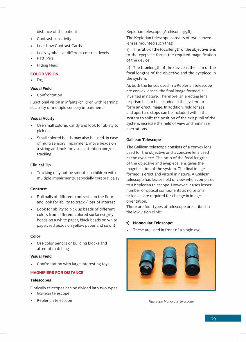
79
distance of the patient
• Contrast sensitivity
• Leas Low Contrast Cards
– Lea’s symbols at different contrast levels• Patti Pics
• Hiding Heidi
COLOR VISION• D15
Visual Field• Confrontation
Functional vision in infants/children with learning disability or multiple sensory impairment:
Visual Acuity
• Use small colored candy and look for ability to pick up.
• Small colored beads may also be used. In case of multi sensory impairment, move beads on a string and look for visual attention and/or tracking
Clinical Tip
• Tracking may not be smooth in children with multiple impairments, especially cerebral palsy
Contrast
• Roll balls of different contrasts on the floor and look for ability to track / loss of interest
• Look for ability to pick up beads of different colors from different colored surfaces(grey beads on a white paper, black beads on white paper, red beads on yellow paper and so on)
Color
• Use color pencils or building blocks and attempt matching
Visual Field
• Confrontation with large interesting toys.
MAGNIFIERS FOR DISTANCE
Telescopes
Optically telecopes can be divided into two types:• Galilean telescope
• Keplerian telescope
Keplerian telescope [Atchison, 1996]. The Keplerian telescope consists of two convex lenses mounted such that:1) The ratio of the focal length of the objective lens to the eyepiece forms the required magnification of the device
2) The tubelength of the device is the sum of the focal lengths of the objective and the eyepiece in the system.
As both the lenses used in a Keplerian telescope are convex lenses, the final image formed is inverted in nature. Therefore, an erecting lens or prism has to be included in the system to form an erect image. In addition, field lenses and aperture stops can be included within the system to shift the position of the exit pupil of the system, increase the field of view and minimize aberrations.
Galilean Telescope
The Galilean telescope consists of a convex lens used for the objective and a concave lens used as the eyepiece. The ratio of the focal lengths of the objective and eyepiece lens gives the magnification of the system. The final image formed is erect and virtual in nature. A Galilean telescope has lesser field of view when compared to a Keplerian telescope. However, it uses lesser number of optical components as no prisms or lenses are required for change in image orientation. There are four types of telescope prescribed in the low vision clinic:
1) Monocular Telescope:
• These are used in front of a single eye
Figure 4.11 Monocular telescope

80
• Are portable
• Used for spotting tasks
• Can be used for copying from the blackboard in classrooms for students
• Can also be used for tourism and spotting
• Can be combined with a reading cap for near tasks
• Cannot be used for mobility
2) Binocular telescope
• Similar to monocular telescopes except for larger field of view due to binocularity
• Heavier than monocular telescopes
• Less compact
• Useful for spotting only
• Cannot be used for long duration due to weight and bulk
• Very useful for individuals who cannot tolerate occlusion due to nystagmus or reduced field loss
Figure 4.12 Binocular telescope
3) Ocutech telescope
This is a bioptic monocular telescopeThe individual can use the optics of the spectacle for general tasks and look through the telescope only when there is an object of interest.Especially prescribed for driving – following rigorous training program and special driving test in countries where it is approved.(Not approved in India)
Figure 4.13 Bioptic monocular telescope
MAGNIFIERS FOR NEAR
Spectacle magnifiers (+5D to +20.0D – higher plus can be prescribed subject to availability)Hand magnifiers (1.2x – 14x)Stand magnifiers (4x – 14x)Dome magnifiers (1.8x – 2.2x)Bar magnifiers (1.5x - 2.2x)Fresnel sheet magnifiers (up to 3x)The above mentioned magnifications refer to the equivalent power of the system and not manufacture mentioned magnifications.Closed Circuit Television Systems (CCTV)Spectacle magnifiers (Relative Distance Magnification): Prescribing high ADD that can be incorporated in the spectacle up to +5.0D Prescribing separate reading glasses with ADD higher than +5.00D. If the spectacles are intended for binocular use, either decentration of the lenses or incorporation of base-in prism is necessary. This is because the interpupillary distance (IPD) will differ from that of near PD measured for 40 centimeters.
Figure.4.1 Half eyes

81
Clinical pearlHigh powered lenses will have aberrations on moving away from the optical center of the lens. Therefore, decentration or incorporation of prisms is essential for best vision with the glasses. In case the patient has eccentric fixation or is not viewing the center of the lens, appropriate changes have to be made.1 mm of base In decentration for every diopter of ADD, for each eye may satisfy the base in prism requirement of many cases for binocular devices.
Example Distance IPD 64 mm. ADD 8D. 8mm decentration = 8 Δ Base In for each eye’s dist PD.Distance IPD 68 mm. ADD 10D. 10mm decentration = 10 Δ Base in for each eye.The higher the addition, more the meter angle of convergence and therefore, the requirement for more base in prisms. If near P.D is considered, the base in prisms should be calculated for ADDs over +3.0 D only, i.e., for a 10D add deduct 3D and calculate for 7D only. A rule of thumb is to incorporate two prisms more than the power in each eye. For example For +6.0D ADD, 8 prism BI in each eye would equal 16 prism BI.
POWER BASE-IN PRISM+6.0 D 8 prisms for each lens+8.0 D 10 prisms for each lens+10.0 D 12 prisms for each lens
Half eye frames can be used binocularly for reading powers of 6D – 10D. Half-eye glasses give the wearer freedom to look over the frame for distance visual tasks.Beyond +10.0D binocularity may not be possible in spite of equal or near-equal visual acuity, primarily due to the close working distance.
Hand Magnifiers
A plus (Convex) lens is held at a distance from the object such that the object is at the focal length of the lens. This makes the object appear closer and larger than the actual size of the object: 1. The eye-to-lens distance and the lens-to-
object distance can be varied making this a very flexible magnifier
2. The larger the eye-to-lens distance, the lesser the field of view and vice versa.
3. The larger the lens-to-object distance, lesser the magnification.
4. Can be prescribed in combination with a spectacle magnifier. The resulting magnification can be calculated using the formula for equivalent power: FE = F1+F2-dF1F2 and the magnification =FE/4 where F1 and F2 are the powers of the plus lens and the hand held magnifier and d is the distance between the lens and the magnifier.
Figure.4.2.a Non-illuminated Hand Magnifier
Figure.4.2.b Illuminated Hand Magnifier

82
POCKET MAGNIFIERS
• As the name indicates, they are constructed to be carried around in the pocket
• These are hand-held magnifiers with slide-in mechanism into a case to protect the lens from scratching. The case itself functions as the handle of the “hand-held”
• They are intended for spot or quick reading.
• These are generally small aperture lenses
Figure: 4.3 Pocket Magnifiers (Clockwise)
STAND MAGNIFIERS
• Option of choice for prolonged reading when higher levels of magnification are required
Figure 4.3a: 6X Cutaway Stand Magnifier
• Magnifiers with internal illumination are available
• Generally placed in contact with reading material. The height of the stand is the same as the focal length of the lens or lesser than the focal length of the lens
• If the height of the stand is less than the focal length of the lens, the patient will have to accommodate or require ADD to see clearly through the stand magnifiers
Figure 4.3b: 10X Adjustable Stand Magnifier
DOME MAGNIFIERS
These are also called Visolett or bright field or paper weight magnifiers.These are magnifiers with a plano-convex construction and have better light gathering properties than other optical low vision devices. They are only possible with low magnification up to 4.0X
Figure 4.4 Dome Magnifier 4.0X (above) and 2.25X (below)
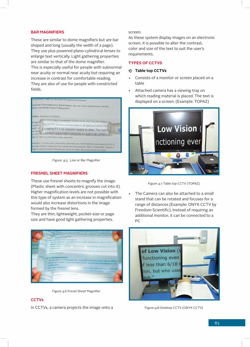
83
screen. As these system display images on an electronic screen, it is possible to alter the contrast, color and size of the text to suit the user’s requirements.
TYPES OF CCTVS
1) Table top CCTVs
• Consists of a monitor or screen placed on a table
• Attached camera has a viewing tray on which reading material is placed. The text is displayed on a screen. (Example: TOPAZ)
Figure 4.7 Table top CCTV (TOPAZ)
• The Camera can also be attached to a small stand that can be rotated and focuses for a range of distances (Example: ONYX CCTV by Freedom Scientific). Instead of requiring an additional monitor, it can be connected to a PC
Figure.4.8 Desktop CCTV (ONYX CCTV)
BAR MAGNIFIERS
These are similar to dome magnifiers but are bar shaped and long (usually the width of a page). They use plus powered plano-cylindrical lenses to enlarge text vertically. Light gathering properties are similar to that of the dome magnifier.This is especially useful for people with subnormal near acuity or normal near acuity but requiring an increase in contrast for comfortable reading. They are also of use for people with constricted fields.
Figure: 4.5. Line or Bar Magnifier
FRESNEL SHEET MAGNIFIERS
These use fresnel sheets to magnify the image. (Plastic sheet with concentric grooves cut into it). Higher magnification levels are not possible with this type of system as an increase in magnification would also increase distortions in the image formed by the fresnel lens. They are thin, lightweight, pocket-size or page size and have good light gathering properties.
Figure 4.6 Frenel Sheet Magnifier
CCTVs
In CCTVs, a camera projects the image onto a

84
2) Mouse model
• The CCTV camera is hand held like a mouse and is attached to a television or PC screen
• The text is placed under the mouse and displayed on the screen
Figure 4.9 Mouse-Model CCTV (Bonita)
3) Head Mounted
• Similar to the mouse model - handheld camera
• Screen is a binocular system that can be worn on the head in front of the eyes
4) Portable
• These are small video magnifiers with screen sizes that are between two to five inches high.
• The camera is inbuilt under the screen and the device can be moved directly over the material like a stand magnifier
Figure 4.10.a. Ruby CCTV (Freedom Scientific), being used for spotting
Figure 4.10.b. Ruby CCTV (Freedom Scientific) reading, continuous reading
Figure 4.10.c. Ruby CCTV (Freedom Scientific) being used for writing tasks

85
Magnifier Reading task Spotting task Writing task Hands free
Spectacle magnifier
Hand magnifier
Stand Magnifier
Dome Magnifier
Bar Magnifier
Fresnel Magnifier
CCTV
Table discussing type of magnifier, type of magnification, available magnification levels and type of illumination possible with the given magnifier
Table showing most suitable tasks with specific type of magnifier
Magnifier Type of Magnification Magnification level Illumination
Spectacle magnifier Approach 1.5x – 6x External
Hand magnifier Approach 1.5x – 14x Internal possible
Stand Magnifier Approach + Angular 4x - 14x Internal possible
Dome Magnifier Approach + Angular 2x - 4x Light gathering
Bar Magnifier Approach 1x - 5x Light gathering
Fresnel Magnifier Approach 1x - 3x Light gathering + External possible
CCTV Real Image Magnification upto 100X Internal
PRESCRIBING MAGNIFYING DEVICES
Prescribing for Distance Visual Tasks
Formula to calculate magnification:
Distance
Magnification = Best corrected distance visual acuity
NEEDED DISTANCE VISUAL ACUITY
For example, the best corrected distance VA of a 10 year old boy is 6/36 and needed visual acuity to view black board is 6/12. Required magnification is 36/12 = 3 X
Near
Although there are several formulae to calculate the tentative magnification for near, the most acceptable formula which is in use nowadays is

86
• Medical management and life skill device
• Assistive technology- Computer and mobile modifications, computer and mobile softwares
RELATIVE SIZE MAGNIFYING DEVICE
These devices assist patients with low vision by mechanically increasing font size. For example, large print magazines, telephones, with large numbers, large size playing cards, jumbo clocks, large insulin syringes.
Glare Contrast and Lighting Control Device
• Filters• Peaked cap for glare
• Use of tablelamps to increase illuminance• Typoscopes to improve contrast and cut off
specular reflection• Hand held torch in dimly lit environments• Multiple pinhole spectacles: Useful for
multiple corneal opacities for glare reduction
TINTS AND FILTERS
Types of FiltersContrast Enhancing Filters
• Yellow or yellow-orange filters
• High absorption filters
• Red/Dark amber
• Green
• Grey
PHOTOCHROMICS:
Prescribe with caution in children with delayed light adaptation or dark adaptation.
SENSORY SUBSTITUTION DEVICES
The visually impaired can make use of other senses such as touch [tactile], hearing [auditory] and smell [olfactory] to substitute for the lack of vision. Any device that makes use of these other senses and devices the visually impaired is called a sensory substitution device. For example, Braille [tactile], audio books, etc.
MEDICAL MANAGEMENT AND LIFE SKILL
Devices
Certain tasks are absolutely essential and necessary for daily living such as, currency
the equivalent viewing power [EVP]
The calculated magnification would be
EVP = [BCVA / TVA] * [100 / WD] = [12 / 4] * [100 / 30] = 3*3.33 = 9.99D = ~10D → ~2.5XEVP = [BCVA / TVA] * [100 / WD] BCVA - Best corrected visual acuityTVA - Target visual acuityWD - Working distance
The options for prescription would be
A spectacle magnifier of +10.00DA stand magnifier of 2.5 X magnificationA hand magnifier of 2.5 X magnificationA near telescope of 2.5 X magnificationIn this case, the option of choice would be a spectacle magnifier or a stand magnifier as they are apt for continuous reading – such as for the newspaper.
Prescription of telescopes for near visual tasks requires a motivated patient who is very particular about working distance and is not worried about cosmesis or field of view.
CLINICAL TIP
The minimum field of view through the near magnifier should be 4-6 characters to ensure comfortable reading.
NON OPTICAL MANAGEMENT
A non-optical device is a device that increases the ease of performance of a task just by increasing size, increasing contrast, reducing glare or simply due to sensory substitution. Non optical devices may be classified as follows:• Relative size device
• Glare, contrast and lighting control
• Postural devices
• Hand writing and written communication device
• Orientation and mobility techniques and devices
• Sensory substitution device
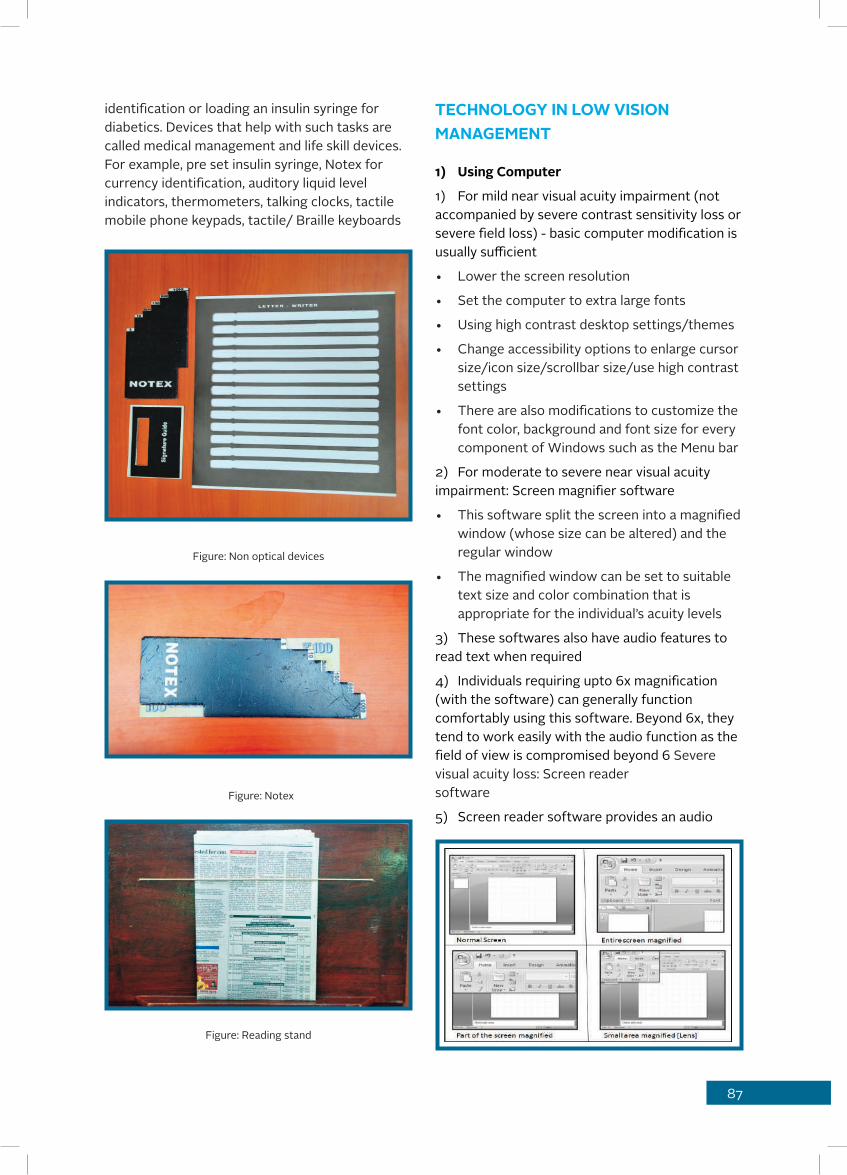
87
identification or loading an insulin syringe for diabetics. Devices that help with such tasks are called medical management and life skill devices.For example, pre set insulin syringe, Notex for currency identification, auditory liquid level indicators, thermometers, talking clocks, tactile mobile phone keypads, tactile/ Braille keyboards
Figure: Non optical devices
Figure: Notex
Figure: Reading stand
TECHNOLOGY IN LOW VISION MANAGEMENT
1) Using Computer
1) For mild near visual acuity impairment (not accompanied by severe contrast sensitivity loss or severe field loss) - basic computer modification is usually sufficient
• Lower the screen resolution
• Set the computer to extra large fonts
• Using high contrast desktop settings/themes
• Change accessibility options to enlarge cursor size/icon size/scrollbar size/use high contrast settings
• There are also modifications to customize the font color, background and font size for every component of Windows such as the Menu bar
2) For moderate to severe near visual acuity impairment: Screen magnifier software
• This software split the screen into a magnified window (whose size can be altered) and the regular window
• The magnified window can be set to suitable text size and color combination that is appropriate for the individual’s acuity levels
3) These softwares also have audio features to read text when required
4) Individuals requiring upto 6x magnification (with the software) can generally function comfortably using this software. Beyond 6x, they tend to work easily with the audio function as the field of view is compromised beyond 6 Severe visual acuity loss: Screen reader software
5) Screen reader software provides an audio

88
output everytime a key is pressed.
6) They have a wide variety of shortcut keys to enable easy use.
7) JAWS (Job Access With Speech Software by Freedom Scientific Inc) is the most commonly available software in India. It is also available with a voice having an Indian accent.
READING SOFTWARE AND DEVICES
• Various software with OCR (Optical Character Recognition) are available
• These software use OCR technology to convert text on a scanned document to audio.(Example: Kurzweil, OpenBook)
• Stand alone devices that use OCR technology are also available. These devices have a camera that captures an image of text and reads text aloud.(Example: SARA Stand-Alone Reading Appliance, Freedom Scientific Inc, Intel Reader by Intel)
Figure: SARA(Stand Alone Appliance , Freedom Scientific Inc, Intel reader ny Intel)
SENSORY SUBSTITUTION DEVICES
Refreshable Braille Displays: These can be connected to a computer. The display then shows tactile dots on the screen that can be felt by the user. Some displays also have a braille keyboard attachment that can be used to input text. Bluetooth displays are also available that can be connected to mobile phones.
Digital Book Reader Hardware/Software: Similar to music players, these devices convert electronic books into a DAISY format(Digital Accesible Information System). The books can be read in audio format and the user can toggle between chapters, pages, paragraphs or lines of text using buttons available on the player. (Example: BookSENSE, BuddyPlayer)
Figure: Buddy Player
Digital talking books are audio/visual books that can be magnified and read, or listened to depending on the user’s comfort.GPS devices: These devices use ultrasonic technology to detect objects/obstacles in the
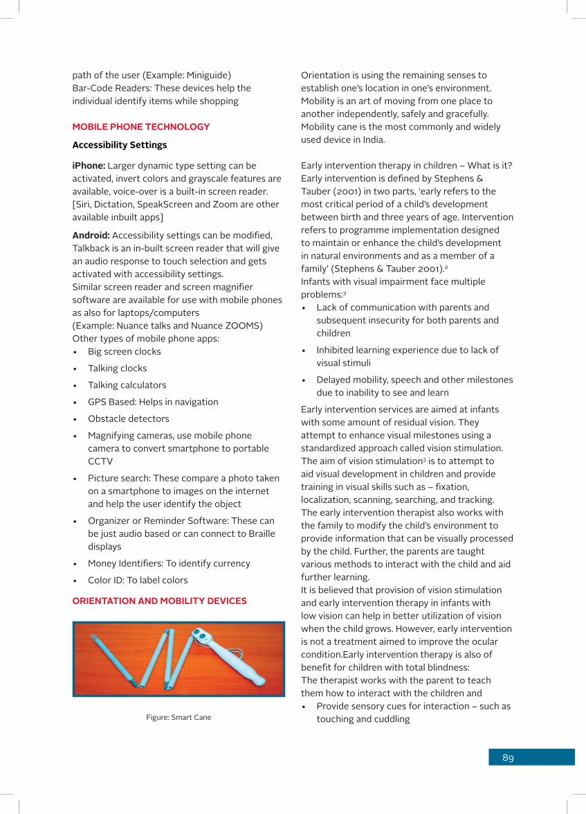
89
path of the user (Example: Miniguide)Bar-Code Readers: These devices help the individual identify items while shopping
MOBILE PHONE TECHNOLOGY
Accessibility Settings
iPhone: Larger dynamic type setting can be activated, invert colors and grayscale features are available, voice-over is a built-in screen reader. [Siri, Dictation, SpeakScreen and Zoom are other available inbuilt apps]
Android: Accessibility settings can be modified, Talkback is an in-built screen reader that will give an audio response to touch selection and gets activated with accessibility settings.Similar screen reader and screen magnifier software are available for use with mobile phones as also for laptops/computers(Example: Nuance talks and Nuance ZOOMS)Other types of mobile phone apps:• Big screen clocks
• Talking clocks
• Talking calculators
• GPS Based: Helps in navigation
• Obstacle detectors
• Magnifying cameras, use mobile phone camera to convert smartphone to portable CCTV
• Picture search: These compare a photo taken on a smartphone to images on the internet and help the user identify the object
• Organizer or Reminder Software: These can be just audio based or can connect to Braille displays
• Money Identifiers: To identify currency
• Color ID: To label colors
ORIENTATION AND MOBILITY DEVICES
Figure: Smart Cane
Orientation is using the remaining senses to establish one’s location in one’s environment.Mobility is an art of moving from one place to another independently, safely and gracefully. Mobility cane is the most commonly and widely used device in India.
Early intervention therapy in children – What is it?Early intervention is defined by Stephens & Tauber (2001) in two parts, ‘early refers to the most critical period of a child’s development between birth and three years of age. Intervention refers to programme implementation designed to maintain or enhance the child’s development in natural environments and as a member of a family’ (Stephens & Tauber 2001).2Infants with visual impairment face multiple problems:3• Lack of communication with parents and
subsequent insecurity for both parents and children
• Inhibited learning experience due to lack of visual stimuli
• Delayed mobility, speech and other milestones due to inability to see and learn
Early intervention services are aimed at infants with some amount of residual vision. They attempt to enhance visual milestones using a standardized approach called vision stimulation. The aim of vision stimulation3 is to attempt to aid visual development in children and provide training in visual skills such as – fixation, localization, scanning, searching, and tracking.The early intervention therapist also works with the family to modify the child’s environment to provide information that can be visually processed by the child. Further, the parents are taught various methods to interact with the child and aid further learning. It is believed that provision of vision stimulation and early intervention therapy in infants with low vision can help in better utilization of vision when the child grows. However, early intervention is not a treatment aimed to improve the ocular condition.Early intervention therapy is also of benefit for children with total blindness:The therapist works with the parent to teach them how to interact with the children and• Provide sensory cues for interaction – such as
touching and cuddling

90
• Provide sensory cues for environmental interaction
• Enhance other senses
• Learn pre-cane skills – Pre cane skills can also be taught to preschoolers and children below the age of five. This helps them to learn to navigate the environment at a young age and prepares them to use the cane easily in the future
MANAGING FIELD LOSS
In general people with field loss have difficulty staying on a line or reading continuously due to skipping or mixing lines.
This can be managed by following compensating strategies:
Single finger guide: Thumb of left hand is placed at beginning of sentence and moved for each sentence.
Double finger guide: Thumb of left hand is placed at beginning of sentence while index finger of right hand is used to track the sentence. Left hand serves as guide to move back when the sentence is completed.
Ruler: Ruler is placed under the sentence being read (as if to underline)
Typoscope: Typoscope is used instead of ruler to track the line
CENTRAL FIELD LOSS
1) Steady eye strategy
2) Eccentric viewing training
1) Subjectively
i) Using Amsler grid ii) Using clock face
2) Objectively:
i) Scanning Laser Ophthalmoscopeii) Microperimeter
MANAGING PERIPHERAL FIELD LOSS
A visual field of more than 10 degrees is sufficient for mobility in well illuminated and non-cluttered unfamiliar surroundings (Pelli DG, 1987).
A visual field of less than 10 degrees requires the use of additional orientation and mobility techniques for mobility (Ferraro, J. and Jose, R. T. (1983).)

91
Management - A Capsule
Visual Task Management Options
Options for Impaired Contrast
Distance
Seeing Blackboard
1. Go closer to board (Approach magnification)
1. Use black board (Avoid green board, white board)
2. Use telescope2. For white board- Use black marker only
3. Use distance CCTV
3. Avoid glare on board, use overhead light on board
Recognizing faces 1. Use contrast cues
1. Use voice/external mannerism as cue
2. Approach magnification
Watching television
1. Approach magnification
2. Relative size magnifi-cation (Large screen)
3. Open field Galilean Binoculars (SEETV)
4. Bioptic (OR) Specta-cle mounted monocular/Binocular telescope
Shop names/Door numbers
Approach magnification
Monocular telescope
Sighted guide assistance (SS)
Traffic Lights Bioptic telescope (if eligible to drive)
Sighted guide assistance (SS)
Crossing Roads
telescope not recom-mended as it limits field of view
Sighted guide assistance (SS)
DrivingBioptic telescope (if vision with telescope eligible to drive)
Seeing Curbs or Steps
Use additional lighting
Contrast edge of steps
Use cane/sighted guide (SS)
SS- Sensory Substitution
Visual Task Management Options
Options for Impaired Contrast
Near
Continuous reading Large print books
Photocopy colored text to black and white
Reading ADD/Spectacle magnifier
Yellow overlay
Stand magnifier Illuminated magnifiers
CCTV CCTV
Screen reading software(SS)
Use e-book readers where possible (flexible for glare control/color/ reversed contrast/ magnification)
Reading stand Use additional lighting
Cheque books/Form filling/Writing
spectacle magnifier/ADD Bold pen/marker
Bold tipped pen/marker
Use additional lighting
Cutaway stand magnifier Portable CCTV
Large field hand magnifier
Portable CCTV
Reading/Writing stand
Price tags/Bills/Medicine labels/Expiry date
Spectacle magnifier/ADD
Use additional lighting
Hand held magnifier Portable CCTV
Portable CCTV
Mobile Phone Accessibility options/App
Talks/Mobile magnifier software
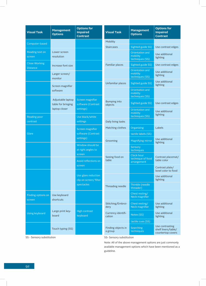
92
Visual Task Management Options
Options for Impaired Contrast
Computer-based
Reading text on
screen
Lower screen
resolution
Close Working
DistanceIncrease font size
Larger screen/
monitor
Screen magnifier
software
Adjustable laptop
table for bringing
laptop closer
Screen magnifier
software (Contrast
settings)
Reading poor
contrast
Use black/white
settings
Glare
Screen magnifier
software (Contrast
settings)
Window should be
at right angles to
screen
Avoid reflections on
screen
Use glare reduction
clip on screen/ filter
spectacles
Finding options on
screen
Use keyboard
shortcuts
Using keyboardLarge print key-
board
High contrast
keyboard
Touch typing (SS)
SS - Sensory substitution
Visual Task Management Options
Options for Impaired Contrast
Mobility
Staircases Sighted guide SS) Use contrast edges
Orientation and mobility techniques (SS)
Use additional lighting
Familiar places Sighted guide SS) Use contrast edges
Orientation and mobility techniques (SS)
Use additional lighting
Unfamiliar places Sighted guide SS) Use additional lighting
Orientation and mobility techniques (SS)
Bumping into objects Sighted guide SS) Use contrast edges
Orientation and mobility techniques (SS)
Use additional lighting
Daily living tasks
Matching clothes Organizing Labels
tactile labels (SS)
Grooming Magnifying mirror Use additional lighting
Sensory techniques
Seeing food on table
Clock hour technique of food arrangement
Contrast placemat/table color
Contrast plate/bowl color to food
Use additional lighting
Threading needle Thimble (needle threader)
Chest resting/Neck magnifier
Stitching/Embroi-dery
Chest resting/Neck magnifier
Use additional lighting
Currency identifi-cation Notex (SS) Use additional
lighting
tactile cues (SS)
Finding objects in a group
Searching techniques
Use contrasting shelf liners/table/countertop covers
SS- Sensory substitution
Note: All of the above management options are just commonly available management options which have been mentioned as a guideline.

93
MULTIDISCIPLINARY MODEL OF MANAGEMENTHolistic management of the patient with low vision does not end with the Low Vision Care practitioner. Rehabilitation is the road to compre-hensive management of the patient. The following is the basic skeleton of the multiple disciplines involved in management of the patient with low vision:
WHOM TO REFER AND WHERE
AT THE INFANT LEVEL
Special educators and rehabilitation therapists are important to provide vision stimulation and vision training so that the child may learn to use residual vision and develop various visual skills to maxi-mum use of functional vision.
AT THE SCHOOL LEVELIt is the special educator and the rehabilitation therapist who play the major role. Once the device has been prescribed by the optometrist and various guidelines have been suggested, the special educator and rehabilitation therapist are necessary for implementation of the suggestions and for helping the child cope with their sighted peers. Modes of schooling available for children with low vision:1. Regular education: Ordinary schooling with no specific modification except use of low vision devices if required.
2. Integrated Education: Ordinary school with additional special educator who gives child special classes or attention when required. No changes are made in daily classroom and class teacher has no special training to handle special needs.
3. Inclusive Education: Children of different types of disability in the same school or class. The class teacher is trained to handle children with special needs. Special concessions are made by the teacher to incorporate needs such as- slow speed, use of special devices, using sensory substitution when child cannot use visual means.
4. Special education: Special school where all children have some form of visual impairment and sensory substitution is the primary method of teaching.
AT THE ADOLESCENT LEVEL
At this stage, the adolescent needs help from Career counselors, special educators, vocational trainers in order to decide the best vocation that will suit them for the future and acquire necessary skills for future career.
AT THE ADULT LEVEL
The most important role would be played by:1) Rehabilitation therapist: For training in use of devices
2) Orientation and Mobility therapist: For independent mobility
3) Social worker: To help the individual and
family cope with the onset of low vision
CASE REPORTS
Case 1
General History: A 14-year-old female diagnosed with high myopia and associated retinal finding of dry macula (Myopic Retinal Degeneration) was referred to the Low Vision Care Clinic as she had discontinued school three months back. She was accompanied by her parents. There was no family history of parental consanguinity. Her younger sister had myopia [around -3.00D and no visual impairment]. There was no other relevant history.Previous Low Vision Care: Nil
Present ComplaintsDistance visual tasks: The patient had difficulty seeing the blackboard and recognizing the fac-es of people who were more than three metres away. She watched television from a distance of one metre.Near visual tasks: The patient was able to man-age at a close working distance of around 15 centimeters.ADL/Mobility: These tasks were independent.Light Sensitivity: There were no complaints regarding light sensitivity.Additional History: Classroom environmentSchool: Private school (English medium), State Board of Education syllabus Medium of Teaching: English

94
Class Strength: 30Academic performance of patient: Average (50-60%)Reason for discontinuing school: Unable to see blackboard from front bench at a distance of three meters. Parents say teacher wrote letters of size about one finger length (~2.5 inches or 6-7 cms)Color of board: BlackLetters: White chalk used, Colored chalk: rareClass lighting: Sufficient (no glare and no com-plaints of dim/poor lighting, including cloudy days)Internal lighting present in class: three fluores-cent lightsExternal Lighting: three large windowsSeating position: Third row, center(No flexibility in seating, not allowed to copy or reference friends notebook)Visual requirements: Seeing blackboardExamination:Previous Glass Prescription:OD: -13.50 DS / -2.00C x 50OS: -12.00 DS/ -1.25 DC X 45Distance visual acuity with glassesOD: 3/30+2 , 6/60+2
OS: 3/19, 6/38B/O: 3/19, 6/38Near visual acuity with glasses: OD/OS/B/O: N8 @ 15 cmReading Speed: 40 wpmUnaided near visual acuity: B/O: N5 @ 10 cmReading speed: 60 wpmRefractionRetinoscopyOD: -11.50DS/-1.00 DC X 60OS:-11.25 DS/-1.25 DC X 120Dynamic Retinoscopy: (MEM)Accommodative Lag: +1.00 DS to +1.25DS Subjective Refraction OD: -11.50DS/-1.00 DC X 60 [3/9.5 , 6/19]OS:-11.25 DS/-1.25 DC X 120 [3/9.5 , 6/19]ADD: OU: Does not prefer ADDUnaided near visual acuity: N5 with ease @ 10 cms
Low Contrast Visual Acuity: 3/19 [6/38] → Bor-derline impairmentD15: NormalAmsler: No scotoma detectedConfrontation: Normal peripheral fieldsLVD Trial for distance Required visual acuity: 6/6Magnification required: = 3XTrial 1: 3x Monocular Handheld Telescope: 6/6 with easeTelescope training was given – Able to localize, hold fixation, scan, track and copy from text on sample blackboard.Copies text at speed of 30-40 wpm.LVD Trial for near: Reads up to N8 with correc-tionReads up to N4 unaidedTrial 1: Add +1.00DS to +5.00DS tried in 1.00D steps. Patient does not report significant differ-ence/improvementTrial 2: With 4x Dome Magnifier: N4; Reads with ease @ 40 cms Reading speed: 80 wpmDiscussion: High myopia is associated with var-ious complications such as retinal holes, retinal detachment open angle glaucoma and macular degeneration1. Myopic macular degeneration does not result in blindness with prognosis being loss of visual acuity up to 3/60.2
Another problem for subjects with high myopia irrespective of other complications is close work-ing distance for near. The close working distance devices in seeing finer details3 and in improving retinal image quality.4 Low vision correction is appropriate in high myopia5 when an increase in working distance is desired. In this case, magnification was prescribed keeping in mind that accommodative demand had to be reduced. This is because it has been shown that increased accommodative lag and near induced transient myopia can further cause progression of myopia.6
Further, the use of magnification would improve retinal image quality thereby reducing strain, es-pecially where it would be necessary to see finer details.

95
Final Rx1. New Rx for distance2. 3x Monocular handheld telescope for use with
blackboard3. To use dome magnifier for prolonged reading
tasks, to reduce eyestrain, reference atlas, maps, dictionaries or fine print
4. To use reading stand for prolonged reading tasks to aid posture and maintain working distance
Other Advice: The condition and prognosis were clearly explained to the parents. The parents were reassured about closer working distance and the need to use a magnifier for comfortable reading at close working distance was explained. The child was advised to continue her schooling with the help of low vision care. They were also counseled about available options for computer modification and magnification.
References
1) Pierro et al. Peripheral retinal changes and axial myopia. Retina 1992; 12(1): 12-17
2) Saw S.-M. Et al. Myopia and associated pathological complications. Ophth Phys Opt. 2005; 25: 381-391
3) Fonda G. Low vision correction for high myopia. Surv Ophth. 1992; 36(4): 313-317
4) Charman WN. Near vision, lags of accommodation and myopia. 1999; 19(2): 126-133.
5) Collins et al. Retinal image quality, reading and myopia. Vis Res. 2006; 46: 196-215.
6) Ciuffreda KJ, Vasudevan B. Nearwork-induced transient myopia (NITM) and permanent myopia-is there a link? Ophthalmol Phys Optics. Mar 2008; 28(2):102-114.
Case 2
General History: An eight- year-old female diag-nosed with congenital stationary night blindness was accompanied by her parents to the low vision care clinic. Her difficulty with dimly lit environ-ment was noticed by the parents at around one
year of age. She was staying with her parents and two elder brothers. At the time she was studying in Std II. Her parents did not have a consanguin-eous marriage but her mother’s age was around 34 when the child was born. Her father who was working in an automobile assembly company was the source of family income.
Previous History: She had no history of using any low vision devices.External observation showed she had good fix-ation but difficulty in searching for a pen among various items placed on the table.
Present Complaints:
Distance Visual Tasks: Difficulty with seeing the blackboard in the classroom (three Meters), see-ing objects at distance while sightseeing. She was able to manage all near acuity tasks at a closer working distance.
Mobility and ADL: She was able to move around independently but had difficulty in dim light and took long time to adjust to dimly lit surroundings. She had independent activities of daily living with-in the limitations of her age.
Additional History: Classroom environment:
• Seated at three meters from blackboard
• Use of white chalk. Colored chalk used occasionally
• Allowed to come close to the board
• No note-taking until class IV in the school
• Artificial room lighting – five fluorescent lights
• Class strength: 40 students
• Medium of instruction: English
• Type of school: Private
• Syllabus: State board
Patient requirement: Seeing blackboard.Examination
Visual Acuity
Distance Visual Acuity with Bailey Lovie LogMAR chart:
OD/OS/B/o: 3/12 [6/24]
Near visual acuity: N10 @25 centimeters with the

96
MN Read chart at 10 wpm good reading skills There was no difference between monocular and binocular visual acuities. There was no significant refractive error on objective refraction.
Dynamic Retinoscopy: Accommodative lag: +1.00DS
Cycloplegic refraction: No refractive error requir-ing correction detected.Low contrast visual acuity with Bailey Lovie Low Contrast LogMAR chart was 3/15[6/30] (Within normal limits).
Color Vision: Able to match a few colors on the D15 panel but could not comprehend the entire test.
Confrontation: Mild peripheral field deficit be-yond 50 degree visual field.
Trial of Low Vision Devices1. 4x monocular handheld telescope: 6/6 [no spe-cific preference for right or left eye]
Near
ADD: OD/OS: +2.00DS: N4 @ 20cm
The child seemed to be able to read better and reading speed improved to 25 wpm for finer print (>N10 size)
Discussion
Congenital stationary night blindness is consid-ered to have an X-linked pattern of inheritance. The prevalence of the condition is not known. It is characterized by non-progressive defective dark adaptation, myopia and associated nystagmus or strabismus in some cases. The visual acuity ranges from 6/9 to 6/60 and myopia from mod-erate[-0.25D to -4.75D] to high[>-10.00DS].1
Binoculars are generally prescribed for children who have difficulty with focusing or have motor problems as the weight and field of view of the binoculars makes it easier to handle for shorter periods of time.2 For this patient, binoculars were suggested in order to habituate the patient to the use of binoculars for comfortable distance view-ing and to accustom the patient to the use of a
low vision device for distance vision tasks. Pre-scription of Bifocal Rx with higher ADD need to be considered as the first choice for children with low vision, if they are too young to handle any low vision device.
Final Rx
New Bifocal Rx with +2.00DS ADD.The parents were advised to purchase over the counter binoculars of 3x or more magnification. A letter was given to the school to provide a sepa-rate seating arrangment closer to the blackboard at less than two meters when required. (Currently, students were not required to copy any notes from the blackboard).
Advice
The child was advised to be seated in a separate chair (ahead of the front row) closer to board at ~2m. Letter was given to school management recommending closer seating arrangement, in-creased lighting level and compensatory time for completing her examinations. The parents were advised to increase the illumination level at home especially during night time. The child needs to be instructed on caution regarding mobility tasks. Option of low vision devices were explained to parents which will be beneficial in the future. The patient was advised to come for an annual low vision care visit with her school books and class notes.
References
Boycott KM et al. X-linked Congenital Stationary Night Blindness. GeneReviews [Internet]. Pagon RA, Bird TD, Dolan CR, et al., editors.Seattle (WA): University of Washington, Seattle; 2003. [http://www.ncbi.nlm.nih.gov/books/NBK1245/]Jose RT. Understanding Low Vision. American Foundation for the blind 1983. Pgs 280-284.

97
Appendix - I
LOW-VISION ASSESSMENT WORKSHEET
START TIME: AGE/SEX:
DATE: PATIENT ACCOMPANIED BY:
OPTOMETRIST:
CONTACT DETAILS:
DIAGNOSIS:
ONSET OF DIFFICULTY WITH VISUAL TASKS
[CONDITION: IMPROVING/FLUCTUATING/WORSENING/STABLE]FAMILY HISTORY: PARENTAL CONSANGUINITY: YES / NOOCULAR DISEASES : YES / NO IF YES DETAILS:OTHERS:GENERAL HEALTH:OTHER LIMITATIONS/DISABILITIES: YES/NOWALKING/TREMORS/HEARING LOSS/MULTIPLE DISABILITIES/DELAYED MILESTONES/OTHERS EXTERNAL OBSERVATION:EDUCATIONAL QUALIFICATION: OCCUPATION:LIVING SITUATION:SOURCE OF FAMILY INCOME:PREVIOUS LOW VISION CARE: YES / NO IF YES, WHERE:IF YES:a) SELF PURCHASEDb) PRESCRIBEDC) PRESCRIBED BUT NOT BOUGHTa) OPTICALDISTANCE: TELESCOPE : MONOCULAR / BINOCULARS / SPECTACLE MOUNTED/ SEE TV/OTHERSMAGNIFICATION:NEAR: HAND-HELD /POCKET/ STAND /SPECTACLE / DOME MAGNIFIER/ OTHERS MAGNIFICATION:b) NON- OPTICAL: NOTEX / TYPOSCOPE / READING LAMP / READING STAND/ NEEDLE THREADER / SIGNATURE GUIDE/ TORCH/ CAP/ SUNGLASSES/OTHERSc) ASSISTIVE DEVICES / SOFTWARE: USING PREVIOUS LVD’S SINCE HOW LONG:

98
USING PREVIOUS LVD’S FOR WHAT PURPOSE:AVERAGE USING TIME PER DAY:ANY DIFFICULTY WITH DEVICES: YES / NOIF YES DIFFICULTY IN: PRESENT COMPLAINTSDIFFICULTY IN DISTANCE VISION: YES / NO IF YES, SEEING BLACK BOARD / RECOGNIZING FACES / WATCHING T V/ BUS NUMBERS / SHOP NUMBERS / SEEING TRAFFIC LIGHTS SEEING STREET SIGNS / CROSSING ROADS / DRIVING / GETTING AROUND PEOPLE OR OBJECTS / SEEING CURBS OR STEPS
OTHERS:DIFFICULTY IN READING FOR NEAR: YES / NO / N.ACOLLEGE BOOKS / SCHOOL BOOKS / OFFICE BOOKS / REGULAR PRINT BOOKS / NEWS PAPER/ COMPUTER PRINT OUTS/ OFFICE RELATED MATERIALS /CHEQUE BOOKS/ CHALLAN BOOKS/ RELIGIOUS BOOKS / PRICE TAGS / BILLS / MEDICINE LABELS / EXPIRY DATE / TEXT IN MOBILE/ HAND WRITTEN MATERIAL/ MANAGING ACCOUNTS
OTHERS:ARE YOU MORE COMFORTABLE /DOES DIFFICULTY REDUCE ONTAKING READING MATERIAL CLOSER? YES/NOREADING IN INCREASED LIGHT? YES/NOTO WHAT EXTENT READING IS A PRIORITYACADEMIC / EDUCATIONAL / OCCUPATIONAL / PLEASURE / INFORMATION/
OTHERS:DIFFICULTY IN WRITING: YES / NO/ N.ASEEING TEXT WRITTEN BY SELF/ WRITING ALONG A STRAIGHT LINE/ SIGNING CHEQUE BOOKS / SIGNING OFFICE PAPERS/ OTHERSDIFFICULTY WORKING WITH COMPUTERS: YES / NO / N.A.IF YES: SEEING KEYBOARD / FINDING OPTIONS ON SCREEN /READING TEXT ON SCREEN/ CLOSE WORKING DISTANCE/CONTRAST OF LETTERS/ GLARE – SCREEN IS TOO BRIGHT/OTHERS:

99
DIFFICULTY IN MOBILITY TASKS: YES/ NO
STAIRCASES / FAMILIAR PLACES / UNFAMILIAR PLACES/GETTING AROUND PEOPLE/BUMPING INTO OBJECTS/ STEPS ON OBJECTS ON THE FLOOR/OTHERSMANAGING BY: USING CANE / HAVE AN ASSISTANT OUTDOORS /RESTRICTED TO HOME /NIL
DIFFICULTY WITH: SUNLIGHT/BRIGHT LIGHT: YES / NO DIM LIGHT: YES / NODELAYED ADAPTATION: YES / NO IF YES :LIGHT / DARKDISABILITY/DISCOMFORT GLARE YES / NO
DIFFICULTY WITH DAILY LIVING ACTIVITIES: YES / NO IF YES DIFFICULTY IN:GETTING DRESSED/ BUTTONING CLOTHES/ TYING SHOELACES/ PERSONAL HYGIENE / SEEING FOOD ON TABLE/THREADING NEEDLE / STITCHING/ EMBROIDERY/ DOING HOUSE WORK / COOK-ING/CURRENCY IDENTIFICATION/ COLOR PERCEPTION / IDENTIFYING OBJECTS IN A GROUP/GROPING FOR OBJECTS ON FLOOR OTHERS:AWARE ABOUT DISABILITY CERTIFICATE OR CONCESSION: YES / NO / N.A.ALREADY AVAILING CONCESSIONS FOR DISABILITY: YES / NO / N.A. IF YES WHAT? :ADDITIONAL HISTORY:PATIENT’S VISUAL NEEDS / EXPECTATIONS: PREVIOUS GLASS PRESCRIPTION : Duration: OD) ADD: OD: OS) OS:A. DISTANCE B. NEAR C. BIFOCALS D. ANY TINTS E. ANY ADDITIONAL RX:
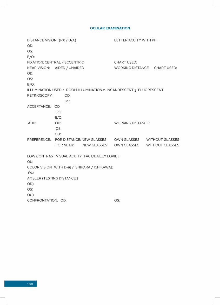
100
OCULAR EXAMINATION
DISTANCE VISION: (RX / U/A) LETTER ACUITY WITH PH : OD: OS: B/O: FIXATION: CENTRAL / ECCENTRIC CHART USED: NEAR VISION: AIDED / UNAIDED WORKING DISTANCE CHART USED:OD: OS:B/O:ILLUMINATION USED: 1. ROOM ILLUMINATION 2. INCANDESCENT 3. FLUORESCENT RETINOSCOPY: OD: OS:ACCEPTANCE: OD: OS: B/O: ADD: OD: WORKING DISTANCE: OS: OU: PREFERENCE: FOR DISTANCE: NEW GLASSES OWN GLASSES WITHOUT GLASSES FOR NEAR: NEW GLASSES OWN GLASSES WITHOUT GLASSES
LOW CONTRAST VISUAL ACUITY [FACT/BAILEY LOVIE]: OU:COLOR VISION [WITH D-15 / ISHIHARA / ICHIKAWA]: OU:AMSLER (TESTING DISTANCE:)OD) OS)OU)CONFRONTATION: OD: OS:

101
LOW VISION AID TRIAL
PREFERRED EYE (SUBJECTIVE) OD / OS / OUTRIAL FOR DISTANCE: (OPTICAL): OD / OS / OU: CURRENT VISUAL ACUITY: REQUIRED VISUAL ACUITY: REQUIRED MAGNIFICATION: PREFERENCE:
TRIAL FOR NEAR: OD / OS / OU:CURRENT NEAR ACUITY: CURRENT WORKING DISTANCE: REQUIRED NEAR ACUITY: REQUIRED MAGNIFICATION:PREFERENCE: READING SPEED:
ILLUMINATION PREFERENCE: 1. ROOM ILLUMINATION 2. INCANDESCENT 3. FLUORESCENT
TEXT USED:TRIAL OF NON-OPTICAL DEVICES: DONE / NOT REQUIRED:
NOTEX / TYPOSCOPE / READING STAND / NEEDLE THREADER /SIGNATURE GUIDE/LETTER WRIT-ER OTHERS:TINTS: (TRIAL PERFORMED OUTDOORS / INDOORS / BOTH) DONE/NOT REQUIRED:TRIAL:
PREFERRED TINT:VISION:
COMPUTER SOFTWARE: DEMO GIVEN/ RECOMMENDED /NOT REQUIRED: □ MAGIC □ JAWS □ OPEN BOOK □ KURZWEIL □ BIG-SHOT □ ZOOMTEXT □ KINDLE □ TEXT ALOUD □ CHANGE COMPUTER SETTINGS (RESOLUTION /CONTRAST) □ MAGNIFIER SETTINGS (START MENU) □ OTHERS:
MOBILE SOFTWARE: RECOMMENDED/NOT REQUIRED: □ MOBILE SPEAK □ MOBILE MAGNIFIER □ TALKS □ ZOOM □ OTHERS
OTHER REMARKS: / HANDWRITING SAMPLE:

102
CASE SUMMARY – Low Vision Care Clinic (LVC)
(Relevant Options Are Ticked)
BEST CORRECTED VISUAL ACUITYDISTANCE: OD: NEAR: OD: ( ) OS: ( ) OS:COLOR VISION: □ Normal □ Protan defect □ Deutan defect □ Tritan defect □ Non specific defect □ Deferred / Not tested CONTRAST SENSITIVITY: □ Normal □ Impaired contrast sensitivity □ Deferred / Not testedVISUAL FIELDS: □ Normal □ Central scotomas (AMSLER / CONFRONTATION) □ Peripherally constricted □ Distortions □ Tubular fields / Temporal island of vision □ Deferred / Not tested
FINAL Rx
ADVICE:Explained □ condition □ possible prognosis
CONTRAST ENHANCING MEASURES EXPLAINED □ Increase task illumination – Fluorescent / Incandescent □ Use black pens /8B pencils /10B pencils Felt tip pens □ Use large black dial watches with white letters □ Mark borders of staircases / doors / cupboards / switchboards with contrast colors □ Paint kitchen counter with two contrasting colors. □ Use contrasting cutlery – plates / tea cups. □ Use contrasting colored equipment in bathrooms.
MEASURES FOR COLOR ENHANCEMENT EXPLAINED: □ NOT REQUIRED □ Label pens / pencils □ Use patterns □ Mark clothes on inside with buttons/textures □ Others
MODIFICATIONS FOR ACTIVITIES OF DAILY LIVING EXPLAINED □ NOT REQUIRED □ Chime clock □ Furniture arrangement □ Food arrangement □ Kitchen arrangement □ Others
FOR MOBILITY TASKS □ NOT REQUIRED □ Head scanning explained □ Eccentric viewing explained □ Need for Orientation and Mobility Training explained □ Others
REFERRED FOR: □ NOT REQUIRED □ Orientation and Mobility train-ing □ Daily Living Skills □ Vocational guidance / Independent Living skills/ Com-puter software training □ Disability certificate □ Others:
MOBILE SOFTWARE:COMPUTER SOFTWARE:OTHER ADVICE:OTHER SERVICES REQUIRED:RTC TO LVC CLINIC: SIGNATURE
END TIME:

103
Appendix – I a
CLASS ROOM ENVIRONMENT
Standard: Informant: Mode of education: a) Special school b) Regular school c) Private tutorSchool name/ College name: Class teacher / Principal name:Address of the school/ college:1. Class strength: 2. Seating position: Center / Corner3. Approx distance from board: 4. Which bench / row: 5. Color of writing material on board: 6. Color of the board: 7. Number of windows in class: 8. Sitting beside window:9. Amount of light in class: Dimly illuminated / Optimally illuminated / Glaring (too bright)
Academic performance: Good peer-group interaction: Yes / No Good parent-teacher interaction: Yes / NoIs there any difficulty in copying from black board: Yes / NoIf yes: managing by: Copying from friends / Going close to the black board / Taking friends note-books to home / Teacher’s dictation.Others: Using extra hours for writing exams: Yes / NoUse of special assistance: Yes / No If yes: Scribe / Braille / Talking books/ Audio cassettes Others:Since how long:Any difficulty with special assistance:Is the teacher co-operative and ready to give individual attention: Yes / No / NA:Will the school management be ready to make changes in the setup: Yes / No / NALetter to the class teacher required: Yes / No / NA

104
Appendix – I b
D-15 RECORDING SHEETTEST PERFORMED: OD/ OS/ OU
Array:Impression: Normal Color Vision / Protan / Deutan / Tritan / Non Specific color defect
APPENDIX – I C
FACT recording sheet
REFERENCE CAP
12
34
5
6
7
8
9
10
1112
13
14
15
REFERENCE CAP
12
34
5
6
7
8
9
10
1112
13
14
15
REFERENCE CAP
12
34
5
6
7
8
9
10
1112
13
14
15
REFERENCE CAP
12
34
5
6
7
8
9
10
1112
13
14
15
105
APPENDIX – II a
LETTER TO SCHOOL MANAGEMENT
LOW VISION CARE CLINIC Date:
TO WHOMSOEVER IT MAY CONCERN
This is to state that Master/Miss.______________________________ who is studying________STD in your school (Reference MRD No.: _____________) was evaluated in the Low Vision Care Clinic. He was noted to have _____________ Visual Impairment in both the eyes due to retinal problem.
This vision will not be sufficient to view the black board comfortably. He needs to be seated closer to the black board (i.e, one meter distance) or in the front row (without rotation). Most of his learning will be through listening to the classes, so dictating as much as possible will help him in a large way.
Kindly allow him to copy notes from his friend’s notebook in case he is not able to copy from the black board directly. In addition, he can be given compensatory time concession of minimum 30 minutes for all examinations (school level and for board exams)
[Source: F.No.16-110/2003-DD.III, Government of India, Ministry of Social Justice and Empowerment, New Delhi, India]
Please extend your co-operation so that his ocular condition doesn’t interfere with his academic performance.
Thank you for your kind co-operation.
Yours sincerely
(__________)
Optometrist
Low Vision Care Clinic
Sankara Nethralaya
Chennai- Tamil Nadu
Phone: 044-42271519

106
APPENDIX – II b
LETTER FOR DISABILITY CERTIFICATE
LOW VISION CARE CLINIC Date:
TO WHOMSOEVER IT MAY CONCERN
This is to state that Mr/Ms. ____________________-_________ (Reference number:___________) was evaluated in the Low vision care clinic and was noted to have best corrected visual acuity of __________ in the right eye and ___________ in the left eye due to _____________.
He can be considered as ________% visually handicapped [Source: No. 16-18/96-NI.I, Government of India, Ministry of Social Justice & Empowerment, New Delhi, India Category _______] and he is eligible for possible concessions and benefits according to this category.
Yours sincerely
(______________)
Optometrist
Low Vision Care Clinic
Sankara Nethralaya
Chennai- Tamil Nadu
Phone: 044-42271519
Appendix – III
Rehabilitation Center
Sample - Rehabilitation wise contacts• Readers facility
• Blind School
• Early Intervention center
• For children with learning difficulty
• Special school
– Integrated schools and Special educators – Inclusive school – Braille Training (Pre-school)• Vocational Training
• Disability certificate - SRTC
– Computer software training – Mobile software – Job placement

107
1. Karishma Enterprises132, Market Tower B, Cuffe Parade, Mumbai 400 005. IndiaPhone: 022-22181853; Fax: 022-22153291Email: [email protected]: www.brailleworldindia.com
2. Barrier Break Technologies101, Highway Commercial ComplexIB Patel Road, Goregoan (E), Mumbai- 400 063, IndiaPhone: +91 (22) 2685 9730/ 2686 0485/6cell: +91 99202 29081E-mail: [email protected]: http://www.barrierbreak.com
3. Om Tao Scientific Apparatus3-2-320/2, Chappal Bazar KachigudaHyderabad - 500 027Tel: 9885054759Email: [email protected]
4. JUTRON VisionA-6, Nand Complex, Near Aims Oxygen Juna Padra RoadBaroda-390020Phone: +91 -99987-02020Fax: 91-265-2321323Email: [email protected]
5. ABISee, INC141 Parker St, Suite 201, Maynard MA 01754, USAPhone: 800-681-5909Website: www.abisee.com
6. Telesensory520 Almanor AvenueSunnyvale, CA 94085, USAWebsite: www.telesensory.comEmail: [email protected]
7. Dolphin Computer Access Ltd.Technology House,Blackpole Estate WestWorcester, WR3 8TJUnited KingdomTel: +44 (0) 1905 754 577UK Local Rate (from a BT landline): 0845 130 5353Fax: +44 (0) 1905 754 559Email: [email protected]
8. Freedom Scientific11800 31st Court NorthSt. Petersburg, FL 33716-1805, USAPhone: 1-727-803-8000 (worldwide)Fax: 1-727-803-80Web-site: www.freedomscientific.com
Appendix - IV
SOURCES OF LOW VISION & ASSISTIVE DEVICES

108
Appendix - V
Useful web links related to Low vision
1) For children with low vision http://www.lowvisionkids.com
2) Visual perception and tracking skills - http://www.eyecanlearn.com/#Peripheral
3) Visual stimulation exercises - http://www.whiteboardroom.org.uk/LearningSystem/PortalHome/ preDefault.asp?Resource=AEE03C0E-AE07-497D-9FA7-989D8654B96F&ResourceID=7041
4) Punarbhava
(National Interactive Portal that provides information for persons with disability)
5) National Initiative for the Blind (NIB) - Centre for computer training
http://acharya.iitm.ac.in/projects/trgschools.php
6) International Council for Education of People with Visual Impairment
(ICEVI) - Education for all children and youth with visual impairment
http://www.icevi.org/
7) Information / links useful for patients - awareness
8) Cell phone accessibility for visually impaired
http://www.nfb.org/images/nfb/Publications/fr/fr28/3/fr280314.htm
9) Computer softwares & Internet accessibility for the disabledscreen magnifiers
http://www.lowvision.org/access_to_the_internet.htm
http://www.daisyindia.org/02HelloDAISY.html
http://www.daisyindia.org/index.htm
10) National Association for Visually Handicapped (NAVH)
Low vision services to assistive devices to support groups
http://www.navh.org/about/accessibility.htm
11) Vocational training center
http://www.dget.gov.in/mes/
12) Reading materials
13) Engineering books – Audio format
www.samritatrust.org
14) Special schools for MR children
• Statewise contacts
http://www.udaan.org/parivaar/india.html
• Countrywise contacts
http://www.Pediatric.com/fordoctor/schools/view_schools.aspx?id=3&cat_id=Special%20 Schools%20for%20mentally%20Handicapped%20&%20Spastic%20children
Ministry of social Justice: http://socialjustice.nic.in/policiesacts3.php
To know about concessions given for person with multiple disabilities:
http://www.fourstepsindia.org/index.html
National Trust schemes:
http://www.thenationaltrust.co.in/nt/index.php?option=com_

109
content&task=view&id=193&Itemid=265
15) Link to National trust for special children - statewise contacts
http://thenationaltrust.in/NewWeb/snaplist.mht
16) Eligibility of driving in India
http://www.delhitrafficpolice.nic.in/old-age-and-driving.htm#q14
17) Braille press and other provisions for persons with multiple disabilities
http://socialjustice.nic.in/brailpress.php
18) Job placement
www.enable-india.org Enable India
http://www.eyeway.org/include/cl-jobs.php Eyeway.org
http://www.disabilityindia.org/forum/viewtopics.cfm?Forum=13 Disabilityindia.org
http://www.ncpedp.org/employ/placements/viewopenings.php NCPEDP
19) Information regarding available sources for all visually impaired (partially sighted, blind, etc)
20) http://www.sightlosssolutions.org/
21) ROPARD is The Association for Retinopathy of Prematurity and Related
Diseases – awareness link for parents
22) http://www.ropard.org/information-for-parents?start=1
23) Link provides information especially for blinds
24) http://www.hadley.edu/7_c_links.asp
25) Blind resource center for visually impaired and Blind
26) http://www.nyise.org/speech/homepage.htm
27) Personal Web Sites of the Blind and Visually Impaired
28) http://www.lowvision.org/personal_web_sites_of_the_blind_.htmL
110
Reference:
1) Gilbert, Clare, and Allen Foster. “Childhood blindness in the context of VISION 2020: the right to sight.” Bulletin of the World Health Organization 79.3 (2001): 227-232.
2) Sharma, M. K., et al. “A profile of low vision among the blind school students in Lumbini Zone of Nepal.” Nepalese Journal of Ophthalmology 2.2 (2010): 127-131.
3) Shah, Mufarriq, et al. “Causes of visual impairment in children with low vision.” J Coll Physicians Surg Pak 21.2 (2011): 88-92.
4) Colenbrander, A. (2010). Assessment of functional vision and its rehabilitation.Acta ophthalmologica, 88(2), 163-173.
5) Bailey, I. L., Jackson, A. J., Minto, H., Greer, R. B., & Chu, M. A. (2012). The Berkeley rudimentary vision test. Optometry & Vision Science, 89(9), 1257-1264.
6) Brown, B., & Lovie-Kitchin, J. E. (1989). High and low contrast acuity and clinical contrast sensitivity tested in a normal population. Optometry & Vision Science, 66(7), 467-473.
7) Hyvärinen, L. (2000). HOW TO CLASSIFY Pediatric LOW VISION?.Extraídoel, 25.
8) [http://www.lea-est.fi/en/assessme/ Pediatriclow_vis.html]
9) Hassan et al. What is the minimum field of view required for efficient navigation. Vision Res 2007; 47: 2115-2123
10) http://www.visionaware.org/info/everyday- living/essential-skills/an-introduction-to- orientation-and-mobility-skills/123
11) Stephens L.C, Tauber S.K (2001) cited in Case-Smith J (2001) Occupational Therapy for Children, 4th edition, Mosby Inc, Elsevier Science, United States of America.
12) Hyvärinen, L. (2000). HOW TO CLASSIFY Pediatric LOW VISION?.Extraídoel, 25.
13) Verezen CA, Muelendijks CF et al. Long-term evaluation of eccentric viewing spectacles in patients with bilateral central scotomas. Optom Vis Sci. 2006; 83(2): 88-95
14) Romayananda, N, Wong, SW, Elzeneiny, IH, Chan, GH. Prismatic scanning method for improving visual acuity in patients with low
vision. Ophthalmology. 1982;89:937–945
15) Smith, HJ, Dickinson, CM, Cacho, I, Reeves, BC, Harper, RA. A randomized controlled trial to determine the effectiveness of prism spectacles for patients with age-related macular degeneration. Arch Ophthalmol.2005;123:1042–1050.
16) Pelli, D. G. (1987). The visual requirements of mobility. In Low Vision (pp. 134-146). Springer New York.
17) Ferraro, J. and Jose, R. T. (1983). Training programs for individuals with restricted fields. In R. T. Jose (Ed.), Understanding Low Vision, American Foundation for the Blind, New York. Vol. 14, 363-376
18) Szlyk, J. P., Seiple, W., Laderman, D. J., Kelsch, R., Ho, K. and McMahon, T. (1998). Use of bioptic amorphic lenses to expand the visual field in patients with peripheral loss. Optometry and Vision Science 75, 518- 524.
19) Onufryk M. Ophthalmic prismatic image relocating eye glasses for persons having retinitis pigmentosa and hemianopia and method for making same. USA Patent #5,323,190 1994.
20) Pambakian, A., Currie, J., & Kennard, C. (2005). Rehabilitation strategies for patients with homonymous visual field defects. Journal of Neuro-Ophthalmology,25(2), 136-142.
21) Apfelbaum, H. L., Ross, N. C., Bowers, A. R., & Peli, E. (2013). Considering apical scotomas, confusion, and diplopia when prescribing prisms for homonymous hemianopia. Translational vision science & technology, 2(4).


ORBIS INTERNATIONALIndia Country Office, 6th Floor, Fortune Tower II, Plot No. 406, Udyog Vihar Phase III, Gurgaon - 122016, India T: 91 124 4284601 | F: 91 124 4284602 | www.orbis.org