Preemie Primer 4.19 - bchsfoutreach.ucsf.edu · ‒suctioning, PIV insertion, CT insertion, loud...
60
4/17/2018 1 Preemie Primer… the ABCs of Preemie Problems Tanya Kamka, MSN, RNC-NIC Neonatal Outreach Educator Objectives ▪ Describe the nursing interventions to reduce the incidence of intraventricular hemorrhage in the preterm infant ▪ Use evidence based practice to influence care of the infant at risk for intraventricular hemorrhage ▪ Identify developmental and physiological differences between the term and premature infant Upon completion of this course, the learner will be able to: 2 ▪ Brain development is activity-dependent • experiences and stimuli "shape" the way the connections are made. • different beginnings foster different end points • Interventions can increase cell maturation, such as environmental enrichment Neurodevelopment
Transcript of Preemie Primer 4.19 - bchsfoutreach.ucsf.edu · ‒suctioning, PIV insertion, CT insertion, loud...

4/17/2018
1
Preemie Primer… the ABCs of Preemie Problems
Tanya Kamka, MSN, RNC-NICNeonatal Outreach Educator
Objectives
▪ Describe the nursing interventions to reduce the incidence of intraventricular hemorrhage in the preterm infant
▪ Use evidence based practice to influence care of the infant at risk for intraventricular hemorrhage
▪ Identify developmental and physiological differences between the term and premature infant
Upon completion of this course, the learner will be able to:
2
▪Brain development is activity-dependent
• experiences and stimuli "shape" the way the connections are made.
• different beginnings foster different end points
• Interventions can increase cell maturation, such as environmental enrichment
Neurodevelopment

4/17/2018
2
Anatomy!!▪ Ventricles
▪ Intraventricular Foramen
▪ Cerebral aqueduct
▪ Choroid plexus
▪ Germinal matrix
Introductory sentence Arial – 21pt font
4
Anatomy!!
Introductory sentence Arial – 21pt font
5
Germinal Matrix▪ Highly vascularized and poorly supported
• Involutes over time
‒ 23-24 weeks 2.5 mm width
‒ 32 weeks 1.4 mm width
‒ 36 weeks involute
6

4/17/2018
3
Periventricular-Intraventricular Hemorrhage (IVH)▪ Occurs once germinal matrix hemorrhage extends into the lateral
ventricle
▪ Risk factors: prematurity (less than 34 weeks), respiratory failure, increasing arterial blood pressure, perinatal asphyxia
▪ Incidence:
• 10 to 15% of infants with hemorrhages
• 30 to 40% of preterm infants <30 weeks or <1500 grams
• Higher risk if <28 weeks
• 2 to 3% of term infants
7
8
So why are
preemies at high risk?!
9
This is what we
are working with…

4/17/2018
4
Pathogenesis
Thought to be caused by capillary bleeding.
▪ Major factors:
• Intra-vascular factors
‒ Loss of cerebral autoregulation
‒ Abrupt alterations in cerebral blood flow and pressure
• Vascular factors
‒ Germinal matrix-vulnerable to hypoxia
‒ Reperfusion injury
• Extravascular factors
‒ Poor vascular support in cerebral tissue
10
IVH Risk FactorsAbrupt changes in cerebral circulation
▪ Rapid changes in PaCO2
▪ Rapid changes in aortic pressure
• Rapid infusion of volume expander
• Excessive increase in vasopressor infusion
• Noxious procedures
‒ suctioning, PIV insertion, CT insertion, loud noises, aggressive handling
▪ Large PDA with left-to-right shunt
▪ Elevated venous pressure from tension pneumothorax or excessive ventilator pressures
11
IVH Timing and Progression
▪ May begin in utero, but usually begins after birth
▪ Hemorrhages may be small at first, then progress to larger hemorrhages later
▪ Most large or progressive IVH’s begin in the first week of life
• Why is this important to the bedside nurse?
12
4/17/2018
5
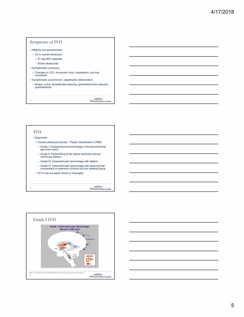
Symptoms of IVH
▪ Majority are asymptomatic
• Dx is cranial ultrasound
‒ 4th day 90% detected
‒ Serial ultrasounds
▪ Symptomatic (common)
• Changes in LOC, movement, tone, respirations, and eye movement
▪ Symptomatic (uncommon) catastrophic deterioration
• Stupor, coma, decerebrate posturing, generalized tonic seizures, quadraparesis
13
IVH▪ Diagnostic:
• Cranial ultrasound (serial) – Papile Classification (1988):
‒ Grade I: Subependymal hemorrhage in the periventricular germinal matrix.
‒ Grade II: Partial filling of the lateral ventricles without ventricular dilation.
‒ Grade III: Intraventricular hemorrhage with dilation
‒ Grade IV: Intraventricular hemorrhage with parenchymal involvement or extension of blood into the cerebral tissue
• LP to rule out septic shock or meningitis
14
Grade I IVH
15
http://www.slideshare.net/PediatricHomeService/brain-injury-in-preterm-infants
4/17/2018
6
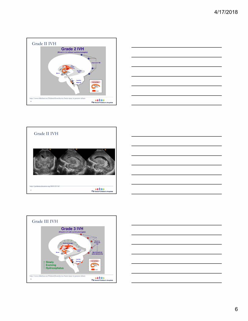
Grade II IVH
16
http://www.slideshare.net/PediatricHomeService/brain-injury-in-preterm-infants
Grade II IVH
17
http://pediatriceducation.org/2005/03/14/
Grade III IVH
18
http://www.slideshare.net/PediatricHomeService/brain-injury-in-preterm-infants

4/17/2018
7
Grade III IVH
19
http://pediatriceducation.org/2005/03/14/
Grade IV IVH
20
http://www.slideshare.net/PediatricHomeService/brain-injury-in-preterm-infantshttp://www.nrdaddy.com/lectures/ivh_pvl/ivhgrad_4a.htm
Grade IV IVH
21
http://www.slideshare.net/PediatricHomeService/brain-injury-in-preterm-infantshttp://www.nrdaddy.com/lectures/ivh_pvl/ivhgrad_4a.htm

4/17/2018
8
22
http://www.slideshare.net/PediatricHomeService/brain-injury-in-preterm-infantshttp://www.nrdaddy.com/lectures/ivh_pvl/ivhgrad_4a.htm
IVH Outcomes
▪ Small (Grade I)
• Neurodevelopmental disability similar to premature infants without IVH
▪ Moderate (Grade II-III)
• Neurodevelopmental disability in 40%
• Mortality 10%
• Progressive hydrocephalus in 20%
▪ Severe (Grade PVHI)
• Major neurodevelopmental disability in 80%
• Mortality rate 50-60%
• Hydrocephalus common in survivors
Introductory sentence Arial – 21pt font
23
Periventricular Leukomalacia (PVL)
▪ Ischemic, necrotic periventricular white matter lesions of arterial origin
▪Risk factors: systemic hypotension, recurrent apnea with bradycardia
▪Pathophysiology
▪ Incidence
24

4/17/2018
9
PVL
▪Clinical presentation:
• Acute phase: hypotension and lethargy
• 6 to 10 weeks later:
‒ Irritable
‒Hypertonic
‒ Increased arm flexion and leg extension
‒ Frequent tremors
‒Abnormal Moro reflex
25
PVL
▪Diagnostic:
• Cranial ultrasound
• CT
• MRI
• Initial presentation: PV echodensities
• Later: PV cystic changes
26
Periventricular Leukomalacia
27
http://www.armobgyn.com/en/Neurosonography.htm

4/17/2018
10
Periventricular Leukomalacia
28
http://www.armobgyn.com/en/Neurosonography.htm
Periventricular Leukomalacia
29
http://www.armobgyn.com/en/Neurosonography.htm
PVL
▪ Outcome:
• Based on location and extent of the injury
• Major motor deficits
• Significant upper arm involvement is associated with intellectual deficits
• Visual impairment
• Lower limb weakness
30

4/17/2018
11
PVL Outcome
31
http://www.nrdaddy.com/lectures/ivh_pvl/prog4.htmhttp://www.perinatal.nhs.uk/reviews/cp/cp_causes.htm
Post Hemorrhagic Hydrocephalus
▪ Frequent complication of GM-IVH
• Clot obstructing CSF flow at the level of the aquaduct of Sylvius
32
Hydrocephalus
33
http://www.spinabifida.net/hydrocephalus-in-children-adults-facts-treatment-symptoms.html

4/17/2018
12
Hydrocephalus
▪ Clinical presentation:
• Rapid increase in head size
• Episodic apnea and bradycardia
• Lethargy
• Increased ICP
• Tense, bulging anterior fontanel
• Separated cranial sutures
• Ocular movement abnormalities
34
Post Hemorrhage Ventricle DeviceExternal Ventricular Drain/Reservoir/Shunt/
▪EVD
▪Ommaya reservoir
▪Ventriculoperitoneal (VP) shunt
35
Willows Vision Appeal,. (2015). Willow's Story. Retrieved 10 November 2015, from http://www.willowsvisionappeal.com/willows-story.htmlMskcc.org,. (2015). About Your Ommaya Reservoir Placement Surgery for Pediatric Patients | Memorial Sloan Kettering Cancer Center. Retrieved 10 November 2015, from https://www.mskcc.org/cancer-care/patient-education/about-your-ommaya-reservoir-placement-surgerySeattlechildrens.org,. (2015). Hydrocephalus Treatment | Seattle Children’s Hospital. Retrieved 10 November 2015, from http://www.seattlechildrens.org/medical-conditions/brain-nervous-system-mental-conditions/hydrocephalus-treatment/
Patient Care and Management
▪ Prevent preterm birth
▪ Promote in utero transport
▪ Promote a no stressful intrapartum course
▪ Provide efficient resuscitation with expedient intubation
▪ Cluster care activities and promote appropriate handling
▪ Minimize noxious stimuli
▪ Avoid events associated with wide swings in arterial and venous pressures (i.e.: seizures, apnea, etc.).
▪ Prevent blood pressure swings – slow volume replacement
36

4/17/2018
13
Patient Care and Management
▪ Avoid over ventilation leading to pneumothorax
▪ Use inline suctioning devices
▪ Use noninvasive monitoring devices
▪ Monitor and maintain normal pH
▪ Correct abnormal clotting
▪ Be alert to signs of hemorrhage (changes in LOC, etc.)
37
38
Educate and
support the
parents
Neuroprotection in the NICU
▪ IVH Bundles
▪ Antenatal steroids and Magnesium Sulfate
▪ Delayed Cord Clamping
▪ The “Golden” Hour / CPQCC Delivery Room Toolkit
▪ Neutral head positioning
▪ NIDCAP
▪ Developmental care
39

4/17/2018
14
IVH Bundles
40
Potentially Better Practices to Prevent Brain Injury
McLendon et al. Pediatrics, 2003; Carteaux, et al, Pediatrics, 200341
1. Antenatal steroids & magnesium
2. Optimize management and delivery at center with a NICU
3. Early management by a Neonatologist/NNP
4. Minimize pain and stress
1. Avoid pain and stress
2. Developmental Care
5. Optimal positioning (midline)
6. Treat hypotension
7. Judicious indomethacin use
8. Optimize respiratory management
9. Limit sodium bicarbonate use
10. Use post-natal dexamethasone judiciously
The “Golden” Hour (Delivery Room Toolkit)▪ Based on principles from cardiovascular and emergency medicine
▪ First hour of life is a time of critical transition and adaptation
▪ Management has been show to impact long term outcomes
▪ Structured focus on thermoregulation, minimizing energy consumption, and respiratory support
▪ Measurable data points include: time to admission, admission temperature, admission glucose, initiation of IV fluids with glucose and amino acids
▪ What does your “Golden” hour look like?
42Castrodale, V. and Rinehart, S. (2014). The Golden Hour: improving the stabilization of the very low birth-weight infant. Advances in Neonatal Care, 14(1):9-14.

4/17/2018
15
The Patent Ductus Arteriosus (PDA) and the Preterm Baby
Tanya Kamka, RNC-NIC, MSNNeonatal Outreach Educator
Normal Cardiovascular Function: Review
44Uptodate.com,. (2015). Identifying newborns with critical congenital heart disease. Retrieved 22 October 2015, from http://www.uptodate.com/contents/identifying-newborns-with-critical-congenital-heart-disease?source=search_result&search=congenital+heart+disease&selectedTitle=1~150
Fetal Circulation▪ Gas exchange is liquid to liquid
▪ Organ of respiration is placenta
▪ High flow, low resistance
▪ Fetal lungs
• Low flow, high resistance
▪ PA’s constricted
▪ High right heart and lung pressures
▪ Low left heart pressures
▪ Open fetal shunts
45

4/17/2018
16
Review of
FETAL SHUNTS
The PDA and the Fetus
The ductus arteriosus
serves to divert blood
away from the fluid-filled
lungs toward the
descending aorta
and placenta
In the Preterm Neonate…
A PDA is normal on DOL 3 and may remain
open through the first week of life in 50% of
preterm infants
48

4/17/2018
17
In the Preterm Neonate…
▪The presence of a hemodynamically
significant PDA with a large left-to-right shunt
is a common cause of morbidity in the
extremely premature neonate
49
Incidence of PDA by Postnatal Age
Incidence (%)0-24 hr 24-48 hr 48-72 hr 72-96 hr
Gest(wk)
Healthy RDS Healthy RDS Healthy RDS Healthy RDS
>40 55 0 0 0
38-40 85 50 5 0
34-37 96 42 12 4
30-33 87 87 31 56 13 25 0 11
<29 80 88 40 84 20 77 7 65
A Hemodynamically Significant PDA May Increase the Risk of…
▪ Intraventricular hemorrhage
▪ Pulmonary edema/hemorrhage
▪ Necrotizing enterocolitis
▪ BPD/ventilator dependence
▪ Retinopathy of prematurity
▪ Surgical intervention
▪ Death
51

4/17/2018
18
Pathophysiology of a PDA…
▪Term infants-
• Left ventricular distension may produce a higher ventricular end-diastolic pressure at smaller ventricular volumes
▪Preterm infants
• Ventricles are less distensible than term and generate less force per gram of myocardium
• The relative lack of ventricular distensibility is more a function of the tissue rather than poor muscle function
52
Redistribution of Systemic Blood Flow
Even with a small PDA blood is shunted away from the:
• Skin
• Bone
• Muscle
• GI tract
• Kidneys
53
Shunting May Cause…
▪Decreased perfusion
• Due to a drop in diastolic pressures
▪Decreased blood flow to organs
▪Localized vasoconstriction… why?
▪Organs may experience significant
hypoperfusion before there are any signs of left
ventricular compromise
54

4/17/2018
19
How Does a PDA Present?
55
Actual Monitor of Preemiewith Significant PDA
PDA Presentation
▪Usually asymptomatic when ductus is small
▪Bounding pulses
▪Palmar pulses
▪Active precordium
▪Wide swings in oxygen saturation
▪Murmur
▪Widening pulse pressure (>20mm Hg)
▪ Low diastolic pressure
56
PDA Presentation
▪ ↑ Vascular markings on CXR
▪ ↑ Heart size is a late sign
▪ Apnea or worsening respiratory status
▪ Prolonged capillary fill time from ↓ systemic output
57

4/17/2018
20
Diagnostics for Diagnosis of PDA
▪Chest x-ray
▪Echocardiogram
▪BNP
58
Laboratory Findings with PDA
▪B-Type Natriuretic Peptide (BNP) is released from the heart in response to increased wall tension.
▪Can be useful to help evaluate the left to right shunting through the ductus.
▪Normal Value
• Normal <25
• >100 indicates significant left to right shunt
PDA: Treatment Modalities
▪Conservative measures are employed initially:
▪ Fluid Restriction
▪ Diuretics (lasix)
▪ Positive end-expiratory pressure: useful in reducing left-to-right shunt via PDA

4/17/2018
21
Fluid restriction▪Restricted fluid administration reduces the risk of PDA and NEC and demonstrates trends towards
reducing the risk of BPD, IVH, and death(Cochrane Database, 2010)
▪Total fluids on admission will be 80mL/kg/day
• Slow increases
▪Be cautious of flushes
▪Document, document, document
PDA Treatment Modalities
▪Pharmacologic
• Indomethacin (Indocin)
• Neoprofin (Ibuprofen)
• Acetaminophen (Paracetamol)
62
Indomethacin IV Administration
Special Considerations:
Rapid infusions of intravenous indomethacin have been associated with significant reductions in
cerebral blood flow.

4/17/2018
22
UCSF Indomethacin Therapy: Intravenous Administration
▪Administer by syringe infusion pump over 30 minutes
▪Flush what is left in the tubing with 1 ml NS over 30 minutes
▪Administer into dedicated peripheral IV
Indomethacin Therapy
▪Notify provider for…
• Creatinine >2mg/dL
• UO <1mL/kg/hr
• Abdominal distension
• Platelets <100k
• Bilious gastric residual
• Hemoccult positive stool
Contraindications to Indomethacin
▪ Suspected CHD
▪ Known GI or renal anomaly
▪ Poor renal function
▪ Bleeding disorders or thrombocytopenia
▪ Necrotizing enterocolitis
▪ Sepsis

4/17/2018
23
Ibuprofen Side Effects
▪ Inhibits platelet aggregation, so signs of bleeding should be monitored
▪ Ibuprofen solution may be irritating to the tissue; therefore, it should be administered carefully to avoid IV extravasation
▪ Ibuprofen is known to displace bilirubin from albumin binding sites. and should be used with caution in patients with an elevated total bilirubin
Ibuprofen Contraindications
▪Proven or suspected infection that is untreated
▪Congenital heart disease
▪Active bleeding, especially ICH or GI
▪Thrombocytopenia, coagulation defects
▪Suspected NEC
▪Significant impairment of renal function
Ibuprofen – Adverse events
▪Bleeding
▪Skin lesion/irritation
▪Hypoglycemia
▪Hypocalcemia
▪Adrenal insufficiency
▪Respiratory failure
▪ IVH and renal insufficiency have also been reported

4/17/2018
24
Acetaminophen Therapy
▪NSAIDs come with many risks
▪Acetaminophen is an alternative for hemodynamically significant PDA
▪ IV or Oral
▪No need to stop feeds
Early Treatment Versus Delayed Conservative Treatment of the Patent Ductus Arteriosus (PDA:TOLERATE)
▪Goal of the trial is to compare 2 treatment approaches
• Early treatment
• Conservative approach
▪Hypothesis is the treatment of a moderate PDA will decrease need for respiratory support, diuretics, gavage feeding, ligation, and further intervention
https://clinicaltrials.gov/ct2/show/NCT01958320

4/17/2018
25
Alphabet Soup of Preemie Problems: RDS & BPD
73
NICU Respiratory History
▪1940’s and 50’s Oxygen and more oxygen
▪1963 Patrick Bouvier Kennedy died of RDS at 34 weeks/2100 gms
▪1971 George Gregory uses CPAP for RDS
▪1980’s Jet ventilators used for neonates
▪1990 FDA approval for surfactant use
▪1990’s -2000’s the more pressure the better
▪Today back to CPAP and gentle ventilation
74
Old School…
▪Intubation based on size, gestational age
▪High pressures, high O2
▪Chest physiotherapy ATC
▪Suction whether you need it or not
▪“Babies do not feel pain.”

4/17/2018
26
Changes in Respiratory Management of the Preterm Neonate
▪Antenatal steroids▪Surfactant replacement therapy▪Permissive hypercapnia▪Gentler ventilatory modes (HFOV, PC, PS)▪Inhaled nitric oxide▪Non-invasive ventilation: NCPAP/SiPAP, HFNC, RAM▪Judicious use of oxygen▪Improved survival of preterm babies
Lung Development
77
Respiratory Vulnerabilities
▪Newborn susceptible to respiratory distress due to limits on respiratory system:
•Ribcage not well ossified
•Respiratory muscles have low endurance and strength
•Floppy chest wall offers little resistance to collapse
•Obligate nose breathers
•Tongue large, trachea and glottis small

4/17/2018
27
Respiratory Vulnerabilities
▪Close proximity of trachea to bronchi -> rapid transmission of infection
▪Immature mucosal lining -> less protection against infection
▪Large dead space -> need to breathe twice as fast as adult to oxygenate
Respiratory Vulnerabilities
▪Higher metabolic rate at rest: increased O2 consumption compared to adult
▪Operate close to maximum capacity normally
▪Little compensatory reserve for increased demands
Respiratory Distress Syndrome
▪Most common cause of respiratory distress in premature infants
•Incidence: inversely proportional to GA
•Most babes <29 wks have RDS
▪Small proportion of term infants develop RDS
•Damage to Type II cells and/or enzyme pathways
•IDM’s, C-sections, asphyxia

4/17/2018
28
Risk Factors for Developing Respiratory Distress Syndrome
Increased risk
▪Premature Birth
▪Sex
▪Infant of a Diabetic Mother
▪Asphyxia
▪C-section without labor
▪Caucasian race
▪Chorioamnioitis
▪Hydrops
Decreased risk
▪Chronic intrauterine stress
▪PPROM
▪Maternal HTN
▪IUGR/SGA
▪Antenatal steroids
▪Mat. drug use
▪Tocolytic agents
RDS Pathophysiology
▪Insufficient volume of surfactant
•Unstable alveoli collapse
•Normal Functional Residual Capacity not established
•Each breath requires increased energy output
RDS Prevention ▪Antenatal steroids (Betamethasone 48 hrs PTD)• Accelerate lung maturation• Single course for women between 24 and 34 wks
gestation at risk for preterm delivery• Reduces incidence of RDS by 50% in infants < 31
wks• Decreases neonatal mortality by 30%• Decreases incidence of IVH and NEC• No adverse consequences identified at this dose• Ongoing trials evaluating use of repeated doses in
undelivered moms at continued high risk

4/17/2018
29
Unique Needs of the ELBW Baby: It’s All About Protection
▪What we do during those first minutes, to hours can affect outcomes
▪All systems immature and vulnerable
6/27/201785
The Golden Hour
▪Definition
•Taken from shock trauma medicine indicating the critical first hour as the most important for improving survival
•For the extremely preterm neonate, initial steps undertaken to optimize coordination and execution of care with the goal of minimizing injury and improving outcomes
GOAL: resuscitation without injury!
6/27/201786
RDS Treatment Strategies▪Initial DR stabilization -California Perinatal Quality Care Collaborative (CPQCC) guidelines
▪Surfactant replacement
▪Support ventilation (lung protection is goal)
‒Non-invasive ventilation
‒Gentler mechanical ventilation
▪Fluids, electrolytes, glucose, calories, normothermia
▪Prevent complications
•Infection
•BPD/CLD
▪Inhaled nitric oxide

4/17/2018
30
Oxygen therapy
▪Most commonly used drug in neonatal care▪No concentration is “safe”▪Use blender, analyzers▪Continuous monitoring (HR, sats)▪Humidify, warm▪Maintain stable delivery, concentration▪Wean slowly with continuous monitoring▪O2 saturation: target ranges
•Under 1000gms: 85%-92% (PaO2 45-60 torr)***
•Beyond 36wks: 90-95%
Desaturation Episodes: Critical Points▪Fewer desaturations with proper positioning• Abdomen• Developmental positioning, nesting▪Minimal handling during desaturation episode• Assess need for sedation
▪Be patient, most will recover with no intervention▪O2 titrations used as last resort for desaturation episodes▪Set tight sat alarms on monitor▪Document episodes, interventions, infant’s response
Bronchopulmonary Dysplasia (BPD)

4/17/2018
31
BPD
BPD Incidence
▪Inversely proportional to gestational age
▪Of survivors corrected to 36 weeks
•85% of babies born at 22 weeks had an O2 requirement
•22% of babies born at 28 weeks had an O2 requirement
(Stoll et al., 2010)
BPD
▪Chronic lung disease
▪Inversely proportional to gestational age
▪Develops after acute pulmonary disease
▪Chronic, constant recurrent lung injury⦿Lung injury and repair occur simultaneously
with organ growth and development
▪Prolongs need for O2 , mechanical ventilation

4/17/2018
32
Factors that Contribute to BPD
BPD
Hyperoxic Injury
Inflammation
Nutritional Problems Genetics
Sex-specific Differences
Alterations in Microbiome
Increased Pulmonary Arterial Muscularization
Barotrauma
Fetal Programing
Other
BPD: Multifactoral
•Lung immaturity: prematurity•Lung injury/inflammation‒ Barotrauma/volutrauma▪O2 toxicity: high or low levels, short or long term▪Occurs in ventilated infants without O2
•Infection‒Chorioamnionitis ▪Inflammatory cytokines present in high levels in amniotic fluid of babes who develop BPD
BPD Treatment▪Lung protection
•Extubate to nasal CPAP or SiPAP with low FiO2 to minimize toxicity to lung
•No air leak (PIE or pneumo)•Best ventilation strategy yet to be determined•Sats of 88% to 92% will provide adequate tissue oxygenation, help avoid complications of BPD
•Better weight gain •Prevent development of pulmonary hypertension, right-sided heart failure
•Avoid hyperoxia: ROP

4/17/2018
33
Retinopathy Of Prematurity
Road to ROP
•When babies are born premature, the blood vessels on the retina are not fully developed
•After birth, the vessels may begin to grow very quickly
•This rapid growth can damage the retina
Definition to ROP
•Improper growth of the blood vessels on the retina and the damage caused by the growth
•One of leading causes of childhood blindness

4/17/2018
34
Incidence
•ROP is rare in infants > 32 weeks GA and heavier than 1500 grams
•Approximately 50% of infants with a birth weight between 1000 and 1250 grams will have some degree of ROP
•90% of infants < 750 grams and/or born < 28 weeks GA develop ROP
The Road to ROP
•The preterm infant is in a hypoxic environment in utero (Pa02 25 to 35 mmHg)
•The physiologic hypoxia stimulates the expression of vascular endothelial growth factor (VEGF)
The Road to ROP
▪In infants born prematurely, the relative increase in oxygen extra utero (hyperoxia) inhibits VEGF, causing vasoconstriction of fragile, immature retinal vessels which progress to vaso-obliteration
▪The retina responds by creating LOTS of vessels (vaso-proliferation) .

4/17/2018
35
The Road to ROP
▪If the new vessels proceed to develop abnormally the capillaries may extend into the vitreous body and/or over the surface of the retina.
▪Leakage of fluid or hemorrhage from the abnormal vessels may occur.
The Road to ROP
•Traction may pull the macula out of normal position affecting visual acuity.
•Retinal detachment may occur, resulting in blindness

4/17/2018
36
The Road to ROP
▪Zone refers to the location (I,II, III) - how far developing retinal vessels have progressed
The Road to ROP
Stages of ROP
•Stage 1: Mildly abnormal growth of retinal vessels. Usually gets better without any treatment and has no long-term effects

4/17/2018
37
Stages of ROP
•Stage 2: Growth of retinal vessels is moderately abnormal. Usually gets better without any treatment and has no long-term effects
Stages of ROP•Stage 3: Ridge with Fibrovascular proliferation. Growth of retinal vessels is severely abnormal. Infants with Stage 3 may require treatment and have a higher risk for long-term problems
Stages of ROP
•Stage 4: Partial retinal detachment. Usually requires treatment and may lead to long-term vision problems or blindness
•Stage 5: Complete retinal detachment. Requires treatment and may lead to long-term vision problems and blindness

4/17/2018
38
Stages of ROP
•Plus Disease – Sign of ROP advancing quickly.
Usually requires treatment
•Plus disease frequently leads to vessel contraction and scar formation, which in turn, leads to macular displacement.
•Rush disease: Aggressive form of ROP. Develops between 3-5 weeks after delivery, may progress rapidly.
Prevention
•Treat Oxygen like a drug
•Monitor Pa02 on ABGs – keep 50 to 70 mmHg
•Even if the infant requires only room air, the Pa02 can rise to 60 to 100mmHg
Prevention
•Set high alarm limits on pulse oximeter
•If the infant is on supplemental oxygen, keep saturations 85 to 92%
•Sp02 values > 93% are associated with Pa02 values of > 80 mmHg

4/17/2018
39
Prevention
▪Protect the eyes
•Premature infants eyelids are paper thin and do not sufficiently block light.
Cover eyes whenever possible, especially when under exam lights
The Road to ROP
▪Surveillance
▪Cryosurgery (rarely done)
▪Laser Surgery
▪Intravitreal administration of Anti-vascular endothelial growth factor agents (Avastin)
▪Retinal Buckling for complete detachment
Follow up Exams for ROP
▪Immature Retinal Vasculature
•Every 2-3 weeks
▪If ROP present:
•Exam every 1-2 weeks
•More often depending on severity of disease

4/17/2018
40
The Road to ROP
•Eye exams and mydriatic eyedrops can produce:
– hypertension
–reflex bradycardia
–apnea
Complications of ROP
•Varying degrees of visual impairment may require corrective lenses or surgery.
▪Strabismus ( Crossed-eyed)
▪Nystagmus (Rapid involuntary motion of the eyeball)
▪Glaucoma (Abnormal high fluid pressure in the eye)
▪Cataracts (Opacity of the lens)
▪Amblyopia (“Lazy eye” Dimness of sight – Visual transmission not recognized properly)
▪Macular ectopia (displacement of ocular muscles)
▪Myopia (nearsightedness)
Alphabet soup of preemie problems…MBM & NEC
Tanya Kamka, MSN, RNC-NICNeonatal Outreach Educator
Samantha Wynn, RN, BSN, Cynthia Jensen, MS

4/17/2018
41
How long are the small intestines?
121
McElroy, S. (2017). Innate Immunity and NEC. Presentation, UC Davis.
122
Small Intestines Tight Junctions
123

4/17/2018
42
Gastrointestinal Development
▪ Exposure to enteral nutrition leads to rapid differentiation and development
▪ Development of GIT mucosal immunity due to dietary antigens (or lack of)
▪ Mucosal immune system begins to distinguish between safe nutrients and foreign pathogens
▪ What are we putting into our babies??
Neonatal phase
124
125
When Good Guts Go Bad…
Necrotizing Enterocolitis
126

4/17/2018
43
What is NEC?▪Definition: an acquired disease that affects the GI system, particularly that of premature infants. It is characterized by inflammation of the bowel wall followed by areas of necrosis, most commonly in the terminal ileum and proximal colon, but may affect any or all of the small and large intestines.
127
Verklan, M., & Walden, M. (2015). Core curriculum for neonatal intensive care nursing (5th ed.).
Incidence▪BW 501 to 750 g – 12 percent risk, 42 percent mortality with NEC
▪BW 751 to 1000 g – 9 percent risk, 29 percent mortality with NEC
▪BW 1001 to 1250 g – 6 percent risk, 21 percent mortality with NEC
▪BW 1251 to 1500 g – 3 percent risk, 16 percent mortality with NEC
128
Verklan, M., & Walden, M. (2015). Core curriculum for neonatal intensive care nursing (5th ed.).
What is or isn’t NEC?
129
Gordon, P. (2017). We have to redefine NEC. Presented at, UC Davis

4/17/2018
44
Early vs Late onset▪ Early onset
▪ Usually term
▪ First 7 days
▪ Critically ill
▪ Never fed
▪ Vascular cause of intestinal injury
▪ Late onset
▪ > 7 days
▪ Growing preemie
▪ Has been fed
▪ Luminal cause of intestinal injury
NEC Pathogenesis▪Prematurity
•Propensity towards gut inflammation
•Impaired intestinal barrier function
•Decreased intestinal motility
•Deficient mucosal enzymes, hormones, pepsin, and gastric acid
•Immature autoregulation of microcirculation
•Lack of amniotic fluid
•Immature mucin layer
131
NEC Pathogenesis▪Enteral Feeds
•Formula feeding
•Fortification
▪Abnormal intestinal microbiota
•Decreased commensal flora
•Increased pathogenic bacteria
•Prolonged antibiotic therapy
•Acid suppression medications
132

4/17/2018
45
NEC Pathogenesis
▪Gut Ischemia
•Abnormal gut vascular regulation
•More prone to hypoxic events
▪Inflammatory response
•Genetic factors?
•TLR4?
133
Etiology
Common final pathway – the endogenous production of inflammatory mediators that
precipitate intestinal injury
Model of Pathogenesis
▪ Subclinical event (hypoxia, ischemia)
▪ Intestines become colonized
▪ Bacteria bind to injured mucosa
▪ Inflammatory response
▪ Increased permeability of mucosa
▪ Translocation of bacteria
▪ Inflammatory response accelerates
▪ Inflammatory mediators, reactive oxygen species further injure mucosa
▪ Maladaptive vasoconstrictive response leads to ongoing ischemia/reperfusion injury
▪ End result – cycle of feedback mechanisms ultimately causing necrosis and perforation

4/17/2018
46
Gastrointestinal Development
136
Medline Plus. (2017). Fetal development: MedlinePlus Medical Encyclopedia.Medlineplus.gov. Retrieved 15 April 2017, from https://medlineplus.gov/ency/article/002398.htm
Bacteria = Sharks
Epithelium = The Wall
Mucin = The Moat
Rest of the body = town
TLR-4, Immune Cells, etc = sentries
Commensal Biofilm = The Barges
Fats = Seagulls(complacent,
harmless, unhelpful)
Hypoxemia/ Hypovolemia = Natural Disaster
If persists can destroy the wall and overfill the bay allowing unrestricted access to the town!
In medical terms, it can lead to damage inflammation and shock.
In the adult intestine…
Gastrointestinal Development
137
Medline Plus. (2017). Fetal development: MedlinePlus Medical Encyclopedia.Medlineplus.gov. Retrieved 15 April 2017, from https://medlineplus.gov/ency/article/002398.htm
Breast MilkIn the premature intestine…• Walls aren’t as strong• Moat isn’t as wide• Lots more sentries• Not as many barges
Gastrointestinal Development
138
Medline Plus. (2017). Fetal development: MedlinePlus Medical Encyclopedia.Medlineplus.gov. Retrieved 15 April 2017, from https://medlineplus.gov/ency/article/002398.htm
Breast MilkBreast Milk• Has growth factors to increase
mucin production• Has HMOs that distract pathogens
and feed commensals
Formula• Less protection against pathogen invasions• Over expressed TLR-4 (sentries) sends out
alarm causing excessive inflammatory response (town panic & wall damage)
Both are still at risk for “natural disasters” but hopefully the breast fed
infant can weather the storm a little better

4/17/2018
47
Diagnosing NEC
Bell’s Staging for NEC
▪ Most commonly used in practice
▪ Staging based on severity of systemic, intestinal, and
radiographic findings
▪ Treatment is directed at the clinical signs rather than the
particular stage of NEC
▪ Each advancing stage includes the characteristics of the
previous stage plus additional findings due to increasing
severity of the disease
Stage 1-Suspected NEC
▪Gastric residuals
▪Abdominal distension
▪Heme positive stools
▪X-ray normal to mild distension
▪ Temp instability
▪A’s and B’s

4/17/2018
48
Stage 2- Proven NEC
▪ Absent bowel sounds
▪ Abdominal tenderness
▪ Pneumatosis intestinalis
▪ Portal venous gas
▪ Mild metabolic acidosis
▪ ↓ Platelets
▪ Radiograph findings include intestinal dilation, ileus, pneumatosis intestinalis, and ascites
▪ Stage IIA “mildly ill”, stage IIB “moderately ill”
Stage 3-Advanced NEC▪Severely ill
▪Marked distension
▪Signs of peritonitis
▪Hypotension
▪Bradycardia
▪Severe apnea
▪Metabolic and respiratory acidosis
▪Disseminated intravascular coagulation (DIC)
▪Stage IIIA intact bowel, stage IIIB perforated bowel visualized as a pneumoperitoneum
Diagnosing NEC▪Based on the presence of the characteristic clinical features of:
•Abdominal distention
•Rectal bleeding
•Abdominal radiographic finding of pneumatosis intestinalis.
▪Assessment of infants with suspected NEC includes:
•abdominal imaging, blood studies, stool analysis, and sepsis evaluation
▪Future Biomarkers? iFABP? PAF?

4/17/2018
49
Presentation – clinical findings
▪Abdominal distension
▪ Feeding residuals, often bilious
▪Gross or occult blood in stool
▪Abdominal tenderness
▪Erythema or bluish discoloration of abdominal wall
▪Absent bowel sounds
▪Non-specific signs (temp. instability, glucose instability, lethargy, apnea/bradycardia, hypotension)
Visual Exam

4/17/2018
50
Abdominal Distension/ Increased AG▪What is the distension from?
▪What has been the baseline abdominal exam
▪ Last stool
▪ Is he on CPAP or been recently bag/mask ventilated? Does he have an OGT in place and vented?
▪Are there any other concerning signs? – glucose instability, lethargy, temp. instability, apnea, etc.
▪ Is the infant hungry and eager to nipple feed?

4/17/2018
51
Residuals▪ Is the residual bilious?
▪ Is the baby stooling at least q12h, moderate amounts?
▪Has the infant been recently bag/mask ventilated?
▪What position has the baby been in?
▪Has the last few hours been stressful for the infant? (blood draws, lots of handling, procedures)
▪ Is the physical exam benign?
▪Could there be an ileus? – morphine, infection, post-op
▪Refeed residual if non-bilious, partially digested milk.
Heme Positive Stools
▪ Is the stool meconium?
▪ Is the infant receiving glycerin suppositories?
▪Does the infant have a fissure?
▪Has the infant had abdominal surgery?
Presentation – Laboratory Findings
▪Glucose instability
▪Abnormally high or low WBC
▪ Left shift of WBC
▪ Thrombocytopenia
▪Metabolic acidosis
▪DIC
▪CRP

4/17/2018
52
Radiologic Findings
Normal Abdominal Xray
▪ Uniform size and shape of loops
▪ Diameter approximates vertebra
▪ Gas throughout entire bowel tract
▪ Gas pattern should vary on subsequent x rays
▪ No free air in abdomen and liver is opaque
Presentation – Radiographic Findings▪Non-specific bowel dilatation
▪ Thickening of bowel wall
▪ Fixed, dilated loop unchanged on >1 radiograph
▪Pneumatosis intestinalis
▪Portal venous gas
▪Pneumoperitoneum
▪ Falciform ligament may be outlined
▪ Football sign

4/17/2018
53

4/17/2018
54
160
Ultrasound▪Used to look for bowel wall thickening
▪ Free air
▪Portal Venous gas
Management- Suspected NEC▪ NPO
▪ Fluid/electrolyte and parenteral nutrition management
▪ Gastric decompression
▪ Labs
▪ Antibiotics (Ampicillin and Gentamicin, Vancomycin, Cefotaxime, Flagyl?)
▪ Correct metabolic acidosis
▪ Serial xrays
▪ Respiratory & Circulatory support
▪ Rule out other causes of distension
▪ Transfusions?
▪ Close monitoring

4/17/2018
55
Management – Definite NEC▪Obtain consult with surgical team
▪Serial KUB’s
▪NPO 7-10 days
▪Careful I/O’s – maintain bowel perfusion, may require fluid resuscitation
▪Circulatory support
▪Respiratory support
▪PT, PTT, fibrinogen, platelets
▪ Frequent abg’s, electrolytes
▪Antibiotics 7-14 days
Surgical TreatmentMedical management is appropriate in most cases
▪Operative intervention indications:
• Perforation, evidence of necrotic bowel (fixed loop, metabolic acidosis, DIC, shock), or progressively worsening clinical condition despite intensive medical management
▪Surgical Options:
▪Peritoneal drain
▪Exploratory laparotomy for resection of necrotic bowel
Surgical Considerations▪Peritoneal drain +/- irrigation
• Allows time for baby to stabilize
• Better able to delineate viable gut when do operate
• Borderline bowel segments may recover
• Intestinal perforation may be definitively treated
• Laparotomy indicated if not improving in 24-48 hrs

4/17/2018
56
Surgical Considerations
▪Exploratory laparotomy for resection of necrotic bowel
▪Primary Anastomoses
▪Ostomy & Mucous Fistula
▪Clip & Drop

4/17/2018
57
Prevention▪Human milk
▪ pasteurized MBM is not as protective▪ Intestinal priming (gut stimulation feedings)
▪ Promotes structural and functional maturation
▪ Promotes acquisition of normal flora
▪ Stimulate release of gastric hormones
▪ Slow, but not too slow, feed advance
▪Minimize prolonged antibiotic use
▪ Antenatal glucocorticoids for lung maturation also accelerate intestinal maturation
▪ Prebiotics, probiotics, postbiotics
How can Human Milk Prevent NEC?
170
http://www.human-milk.com/infographic/leaflet.html
Oligosaccharides & the Gut
171
▪Human Milk Oligosaccharides
▪3rd most abundant biomolecule in human milk
▪Un-digestible by humans
“It’s All About the Breast Milk Sugars,
Baby”
German, B. (2017). The Evolution of Lactation as a Guide to Understanding Nourishment and Prevention in the 21st Century. Presentation, UC Davis.

4/17/2018
58
Colostrum Care
172
▪ Oropharyngeal administration
▪ Use syringe to place directly onto the
▪ oral mucosa in the buccal cavity for
▪ absorption via the mucosa
▪ Allows for systemic absorption of the cytokines and pancreatic secretory trypsin inhibitor (PSTI)
▪ Rich source of Oligosaccharides
▪ May reduce time to full feeds
Trophic feeds
173
▪ Improved feeding tolerance
▪ More rapid advancement to full feeds
▪ Improved weight gain
▪ Shortened duration of TPN
▪ Less phototherapy
▪ Reduction in direct bilirubin
▪ Decreased length of stay
▪ Does not increase risk of NEC!
▪ May be considered for an infant who is hemodynamically stable on a low dose of dopamine and/or with an Umbilical Arterial Line in place
Feeding Protocol for Preterm Infants▪Goal
▪ Preterm: 150-160 ml/kg/day of 24cal/oz; fortify when feed volume is 60ml/kg/day
▪ Term: 150-165 ml/kg/day of 20cal/oz
▪Consider increasing concentration of feeds if poor weight gain, volume intolerance or fluid restriction, or increased energy requirements.
▪ Preterm infants should receive 24cal/oz for at least one week before increasing to 27cal/oz
▪ Consider hind milk

4/17/2018
59
Prevention - Disadvantages of NPO▪ “The wisdom of stopping all enteral intake is counter-intuitive to the ontologic processes that started in utero” Edmund Gamma, Lyle Browne, State Univ. NV
▪Has not uniformly provided protection, may simply delay the onset of NEC
▪Predisposes to injury when finally fed
• Gut atrophy
• Decreased mucin production and enzyme activity
• Decreased secretion of IgA
• Increased transmural penetration of bacteria, and antigens
• Increased susceptibility to infection
• Autodigestion
Enteral Nutrition
176
▪ Breast milk is best
▪ Donor breast when MBM not available
▪ Use of standardized feeding guideline
▪ Reduced risk of NEC
▪ Less variability
▪ Achieve full feeds earlier
Neonatal nurses go beyond the numbers to take in the entire baby. Their nursing is particular, contextual, and holistic. It is an art. Xrays and lab values pale in comparison with the vigilance and commitment of an expert nurse.

4/17/2018
60
QUESTIONS??