
PredictorsofNonsentinelNodalInvolvementto ...downloads.hindawi.com/archive/2011/539503.pdf ·...
8
International Scholarly Research Network ISRN Oncology Volume 2011, Article ID 539503, 7 pages doi:10.5402/2011/539503 Research Article Predictors of Nonsentinel Nodal Involvement to Aid Intraoperative Decision Making in Breast Cancer Patients with Positive Sentinel Lymph Nodes Ern Yu Tan, 1 Bernard Ho, 2 Juliana J. C. Chen, 1 Pey Woei Ho, 1 Christine Teo, 1 Arul Earnest, 3 and Patrick M. Y. Chan 1 1 Department of General Surgery, Tan Tock Seng Hospital, 11 Jalan Tan Tock Seng, Singapore 308433 2 Department of Pathology, Tan Tock Seng Hospital, 11 Jalan Tan Tock Seng, Singapore 308433 3 Centre for Quantitative Medicine, Duke-NUS Graduate Medical School Singapore, 8 College Road, Singapore 169857 Correspondence should be addressed to Ern Yu Tan, [email protected] Received 27 April 2011; Accepted 27 June 2011 Academic Editors: A. E. Pinto, H. Rizos, E. Sueoka, and A. H. Wolfson Copyright © 2011 Ern Yu Tan et al. This is an open access article distributed under the Creative Commons Attribution License, which permits unrestricted use, distribution, and reproduction in any medium, provided the original work is properly cited. Background. Up to 60% of patients with a positive sentinel lymph node (SLN) have no additional nodal involvement and do not benefit from completion axillary lymph node dissection (ALND). We aim to identify factors predicting for non-SLN involvement and to validate the MSKCC nomogram and Tenon score in our population. Methods. Retrospective review was performed of 110 consecutive patients with positive SLNs who underwent ALND over an 8-year period. Results. Fifty patients (45%) had non- SLN involvement. Non-SLN involvement correlated positively with the number of positive SLNs (P = 0.04), macrometastasis (P = 0.01), and inversely with the total number of SLNs harvested (P = 0.03). The MSKCC nomogram and Tenon score both failed to perform as previously reported. Conclusions. The MSKCC nomogram and Tenon score have limited value in our practice. Instead, we identified three independent predictors, which are more relevant in guiding the intraoperative decision for ALND. 1. Introduction Sentinel lymph node (SLN) biopsy is the current standard of care for T1 T2 breast cancers with no clinically palpable axillary lymph nodes. Based on the principle of sequential directional lymphatic drainage from the breast, the SLN is hypothetically the first axillary node to receive lymphatic drainage from the breast. It, therefore, follows that if the SLN is free of metastatic tumour deposits, the rest of the ax- illary nodes are not expected to be involved either. This oblit- erates the need for full axillary lymph node dissection (ALND) when the SLN is negative for metastases. ALND, in- volving the removal of the level I and II axillary nodes, is now reserved for instances where the SLN is positive for metastases or where SLN biopsy is contraindicated. This has resulted in more than 50% of patients with T1 and T2 tum- ours being spared the arm morbidity of ALND because of the less extensive dissection [1, 2]. However, it is known that up to 60% of patients with a positive SLN do not have additional nodal involvement, suggesting that these patients are overtreated by the current practice of ALND whenever the SLN is positive [3, 4]. The removal of uninvolved nodes serves neither to aid prognosti- cation nor to guide adjuvant therapy, yet exposes the patient to the risk of lymphedema and its associated complications. This has led to some questioning the need for ALND in all instances of SLN positivity [5]. The American College of Sur- geons Oncology Group Z0011 trial is the latest to show that women with T1 and T2 tumours who undergo lumpectomy derive little additional benefit from ALND since any residual disease in the level I and II nodes appear to be effectively eradicated by postoperative irradiation and chemotherapy [6, 7]. It remains to be seen whether the evidence so far will change current guidelines. Most centres continue to advocate ALND when the SLN is positive. Given the reluctance of most surgeons to leave behind residual disease in the axilla,
Transcript of PredictorsofNonsentinelNodalInvolvementto ...downloads.hindawi.com/archive/2011/539503.pdf ·...

International Scholarly Research NetworkISRN OncologyVolume 2011, Article ID 539503, 7 pagesdoi:10.5402/2011/539503
Research Article
Predictors of Nonsentinel Nodal Involvement toAid Intraoperative Decision Making in Breast CancerPatients with Positive Sentinel Lymph Nodes
Ern Yu Tan,1 Bernard Ho,2 Juliana J. C. Chen,1 Pey Woei Ho,1 Christine Teo,1
Arul Earnest,3 and Patrick M. Y. Chan1
1 Department of General Surgery, Tan Tock Seng Hospital, 11 Jalan Tan Tock Seng, Singapore 3084332 Department of Pathology, Tan Tock Seng Hospital, 11 Jalan Tan Tock Seng, Singapore 3084333 Centre for Quantitative Medicine, Duke-NUS Graduate Medical School Singapore, 8 College Road, Singapore 169857
Correspondence should be addressed to Ern Yu Tan, [email protected]
Received 27 April 2011; Accepted 27 June 2011
Academic Editors: A. E. Pinto, H. Rizos, E. Sueoka, and A. H. Wolfson
Copyright © 2011 Ern Yu Tan et al. This is an open access article distributed under the Creative Commons Attribution License,which permits unrestricted use, distribution, and reproduction in any medium, provided the original work is properly cited.
Background. Up to 60% of patients with a positive sentinel lymph node (SLN) have no additional nodal involvement and do notbenefit from completion axillary lymph node dissection (ALND). We aim to identify factors predicting for non-SLN involvementand to validate the MSKCC nomogram and Tenon score in our population. Methods. Retrospective review was performed of 110consecutive patients with positive SLNs who underwent ALND over an 8-year period. Results. Fifty patients (45%) had non-SLN involvement. Non-SLN involvement correlated positively with the number of positive SLNs (P = 0.04), macrometastasis(P = 0.01), and inversely with the total number of SLNs harvested (P = 0.03). The MSKCC nomogram and Tenon score bothfailed to perform as previously reported. Conclusions. The MSKCC nomogram and Tenon score have limited value in our practice.Instead, we identified three independent predictors, which are more relevant in guiding the intraoperative decision for ALND.
1. Introduction
Sentinel lymph node (SLN) biopsy is the current standardof care for T1 T2 breast cancers with no clinically palpableaxillary lymph nodes. Based on the principle of sequentialdirectional lymphatic drainage from the breast, the SLN ishypothetically the first axillary node to receive lymphaticdrainage from the breast. It, therefore, follows that if theSLN is free of metastatic tumour deposits, the rest of the ax-illary nodes are not expected to be involved either. This oblit-erates the need for full axillary lymph node dissection(ALND) when the SLN is negative for metastases. ALND, in-volving the removal of the level I and II axillary nodes, isnow reserved for instances where the SLN is positive formetastases or where SLN biopsy is contraindicated. This hasresulted in more than 50% of patients with T1 and T2 tum-ours being spared the arm morbidity of ALND because of theless extensive dissection [1, 2].
However, it is known that up to 60% of patients witha positive SLN do not have additional nodal involvement,suggesting that these patients are overtreated by the currentpractice of ALND whenever the SLN is positive [3, 4]. Theremoval of uninvolved nodes serves neither to aid prognosti-cation nor to guide adjuvant therapy, yet exposes the patientto the risk of lymphedema and its associated complications.This has led to some questioning the need for ALND in allinstances of SLN positivity [5]. The American College of Sur-geons Oncology Group Z0011 trial is the latest to show thatwomen with T1 and T2 tumours who undergo lumpectomyderive little additional benefit from ALND since any residualdisease in the level I and II nodes appear to be effectivelyeradicated by postoperative irradiation and chemotherapy[6, 7]. It remains to be seen whether the evidence so far willchange current guidelines. Most centres continue to advocateALND when the SLN is positive. Given the reluctance ofmost surgeons to leave behind residual disease in the axilla,

2 ISRN Oncology
a reliable means of predicting the likelihood of non-SLN in-volvement will be a step towards refining the indication forALND. To date, there are 4 nomograms (Memorial Sloan-Kettering Cancer Centre [MSKCC] nomogram [8], Mayonomogram [9], Cambridge nomogram [10] and the Stanfordnomogram [11]), 3 scoring systems (the Tenon score [12],the MD Anderson Cancer Centre score [13] and the scoredeveloped by Saidi et al. [14]) and 2 recursive partitioningtools developed by Kohrt et al. [11]. In a direct comparisonof these models, the MSKCC nomogram and the Tenonscore performed best and were the only 2 models with anarea under the curve (AUC) of more than 0.75 [15].
In our practice, the decision for ALND is made intraop-eratively based on frozen section analysis of the SLN. Apartfrom guiding the decision to proceed with ALND duringsurgery, a reliable means of predicting the status of the non-SLN nodes will also reduce patient recall rates should theinitial frozen section analysis be false negative. We, therefore,aim to determine the incidence of non-SLN involvement inour patients, and to identify factors that may predict for this.In addition, we aim to validate the MSKCC nomogram andthe Tenon score in our local population.
2. Methods
A retrospective review was performed of 110 consecutive pa-tients with a positive SLN who underwent ALND from 1stJanuary 2001 to 31st December 2008 in our department. Thisstudy has ethics committee approval (DSRB D/10/029). Inour department, the majority of SLN biopsy is performedwith blue dye alone. Two mL of undiluted patent V blue dyeis injected into the subareolar plexus after the patient is putunder general anaesthesia, followed by 5 minutes of manualmassage. The SLN is identified as a blue-coloured node witha blue lymphatic channel leading up to it. Beginning fromthe year 2006, all SLNs were routinely submitted for intra-operative frozen section analysis (a total of 64 patients).This involves the documentation of the number and size ofSLNs submitted for analysis, followed by histological exam-ination of serial sections stained with haematoxylin and eosin(H&E). Each SLN is serially sliced at 2 to 3 mm gross in-tervals, and all slices (the entire node) are snap frozen in liq-uid nitrogen and then placed in frozen section embeddingmedium on a cryostat object disk. Each slice is then furthersectioned at intervals of 40 µm to obtain at least 3 sections,which are stained in H&E and examined using routine lightmicroscopy. The presence or absence of metastatic depositsis noted and communicated to the surgeon immediately. Ifthe SLN is positive for macro- or micrometastasis, ALND isimmediately proceeded with; if negative, no further axillarydissection is performed. The entire SLN is then formalinfixed and paraffin embedded to obtain permanent sectionsfor final analysis, where an additional 1 to 6 levels of eachslice of the SLN are examined. It is not routine in our practiceto perform immunohistochemistry or quantitative real-timepolymerase chain reactions on H&E-negative sections. Forthe 46 patients who underwent surgery prior to 2006, onlyserial H&E analysis of the permanent sections of the SLN wasperformed (without frozen section analysis).
The presence of metastatic deposits in the SLN and non-SLNs was correlated with standard clinicopathological pa-rameters including histology of the primary tumour, tumoursize, tumour grade, presence of lymphovascular invasion,and hormone and human epidermal growth factor receptor2 (HER2) receptor status. Note was also made of whetherthe metastatic deposit was classified as a macrometastasis ormicrometastasis. Macrometastases are defined as cell clustersthat are more than 2 mm in diameter; micrometastatic de-posits are defined as cell clusters that are between 0.2 mmand 2 mm in diameter (denoted as N1mic according to the 6thAJCC classification). Correlation analyses were performedusing either the chi square test or Fisher’s exact test whereappropriate. Correlation with tumour grade was evaluatedusing the Chi square test for trend. All univariate analyseswere performed with GraphPadPrism version 4 (GraphPadsoftware Inc., San Diego CA). The Cox proportional hazardregression model was used to identify independent riskfactors for non-SLN involvement. This was carried outusing the Stata package release 8.1 (Stata Corporation, 4905Lakeway Drive, College Station, Texas 77845, USA). A fullmodel was first created to include all potentially importantexplanatory variables. At each step, the variable with thesmallest contribution to the model was removed, until a finalbackward stepwise model was obtained. A 2-tailed P valuetest was used in all analyses and a P value of less than 0.05was considered statistically significant.
The probability of non-SLN involvement was calculat-ed based on the MSKCC nomogram and Tenon score.The MSKCC nomogram is based on tumour histology andgrade, pathological tumour size, multifocality, lymphovas-cular invasion, oestrogen receptor (ER) status, the numberof positive and negative SLNs, and the mode of detection (inour study, SLN biopsy cases prior to 2006 were performedwith serial H&E examination of the permanent sections, andthose from 2006 onwards were assessed with intra.opera-tive frozen section analysis). The combined score of eachvariable is translated into a predicted probability of non-SLN involvement. The nomogram is available as an elec-tronic calculator on the MSKCC website (http://www.mskcc.org/mskcc/html/15938.cfm). The Tenon score is calculatedbased on three variables: the ratio of positive SLN to the totalnumber of SLN removed, the presence of micrometastasisin the SLN, and the primary tumour size, combined to givea score from 0 to 7 [12]. Receiver operating characteristic(ROC) curves based on both models were generated and thearea under ROC curve (AUC) calculated using Stata packagerelease 8.1. AUC ranged from 0 to 1, where 1 indicates per-fect concordance, 0.5 indicates no better concordance thanchance, and 0 indicates perfect disconcordance.
3. Results
From 1st January 2001 to 31st December 2008, a total of551 patients underwent SLNB in our centre; of these, 110patients underwent completion ALND after a positive SLNwas found. Median patient age was 53 years (30 to 80 years).All patients were diagnosed with invasive carcinoma; 88%(97 of 110) were classified as invasive ductal carcinoma, 10%
ISRN Oncology 3
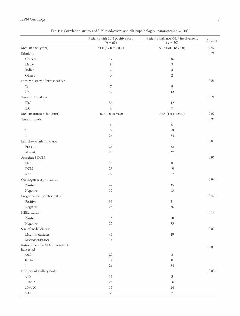
Table 1: Correlation analyses of SLN involvement and clinicopathological parameters (n = 110).
Patients with SLN positive only(n = 60)
Patients with non-SLN involvement(n = 50)
P value
Median age (years) 54.0 (37.0 to 80.0) 51.5 (30.0 to 77.0) 0.42
Ethnicity 0.70
Chinese 47 36
Malay 8 8
Indian 2 4
Others 3 2
Family history of breast cancer 0.53
Yes 7 8
No 52 42
Tumour histology 0.20
IDC 56 42
ILC 4 7
Median tumour size (mm) 20.0 (4.0 to 80.0) 24.5 (1.0 t o 55.0) 0.03
Tumour grade 0.99
1 5 6
2 28 19
3 26 23
Lymphovascular invasion 0.81
Present 26 22
Absent 29 27
Associated DCIS 0.97
EIC 10 8
DCIS 25 19
None 22 17
Oestrogen receptor status 0.84
Positive 42 35
Negative 17 13
Progesterone receptor status 0.42
Positive 31 21
Negative 28 26
HER2 status 0.16
Positive 16 10
Negative 27 33
Size of nodal disease 0.01
Macrometastases 46 49
Micrometastases 14 1
Ratio of positive SLN to total SLNharvested
0.01
<0.5 20 8
0.5 to 1 14 8
1 26 34
Number of axillary nodes 0.03
<10 11 3
10 to 20 25 16
20 to 30 17 24
>30 7 7

4 ISRN Oncology
Table 1: Continued.
Patients with SLN positive only(n = 60)
Patients with non-SLN involvement(n = 50)
P value
Distant recurrence 0.34
Yes 5 7
No 55 43
Median Tenon score 5.0 (1.5 to 7.0) 5.75 (2 to 7) <0.001
Median MSKCC probability 19.5 (3.0 to 74.0) 41.0 (6.0 to 89.0) <0.001
0
0
0.25
0.25
0.5
0.5
0.75
0.75
1
1
1-specificity
Sen
siti
vity
Area under ROC curve = 0.6938
Figure 1: Receiver operating characteristics (ROCs) curve assessingthe discriminatory ability of the MSKCC nomogram.
as invasive lobular carcinoma. One patient had a medullarycarcinoma and another a mucinous carcinoma. Medianpathological tumour size was 21 mm (4 mm to 80 mm), andmedian tumour grade was 2. Seventy percent of patientshad oestrogen receptor (ER) positive tumours, and 47% hadprogesterone receptor (PR) positive tumours. The mediannumber of SLN harvested was 2 (1 to 9). All except 9SLN biopsies were performed using blue dye alone, 6 wereperformed using dual blue dye and radiocolloid, and 3were performed with radiocolloid alone. Median numberof axillary lymph nodes harvested (including SLNs) was 22(ranging from 7 to 58).
The SLN was the only positive axillary node in 60 of110 patients (55%). The likelihood of non-SLN involvementcorrelated positively with pathological tumour size (P =0.03); median tumour size was 24.5 mm in cases of non-SLNinvolvement compared to 20 mm in those where only theSLN was involved. Non-SLN involvement was also inverselycorrelated with the ratio of positive SLNs to the total numberof SLNs harvested (P = 0.01) (Table 1). Those with a ratioof 0.5 or more were 3 times more likely to have non-SLNinvolvement (P = 0.04, OR 2.63, 95% CI 1.04 to 6.64),implying that additional non-SLN involvement became lesslikely when more harvested SLNs were negative. The size ofthe metastatic deposits also correlated with the likelihoodof non-SLN involvement, with macrometastasis (rather thanmicrometastasis) being associated with involvement of thenon-SLNs (P = 0.01, OR = 14.9, 95% CI 0.01 to 0.53)
0
0
0.25
0.25
0.5
0.5
0.75
0.75
1
1
1-specificity
Sen
siti
vity
Area under ROC curve = 0.7085
Figure 2: Receiver operating characteristics (ROC) curve assessingthe discriminatory ability of the Tenon score.
(Table 1). Non-SLN involvement did not correlate withtumour histology, tumour grade, lymphovascular invasion,or hormone receptor status (P > 0.05) (Table 1). Non-SLN involvement also did not increase the likelihood ofdistant recurrence. On multivariate analysis, the number ofpositive SLNs, total number of SLNs harvested, presenceof micrometastasis, and the total number of axillary nodesharvested during ALND independently predicted for non-SLN involvement (P < 0.05) (Table 2). Interestingly, tumoursize did not remain significant (P = 0.14) (Table 2).
The MSKCC nomogram and the Tenon score were vali-dated in our study population. Based on the MSKCC nom-ogram, the median calculated probability in the group withSLN involvement alone was 19.5%, significantly lower thanthe calculated probability of 41.0% in the group with addi-tional non-SLN involvement (P < 0.001) (Table 1). Thediscriminatory ability, as calculated from the area under thereceiver operating curve (ROC) (AUC), was 0.69 (Figure 1).Similarly, the Tenon score also differentiated the group withSLN involvement alone from the group with additional non-SLN involvement (median score of 5.0 and 5.75 resp., P <0.001), with an AUC of 0.71 (Figure 2).
The subgroup of 15 patients (13.6%) with micrometas-tasis was further evaluated. Median tumour size was 20 mm(12 mm to 40 mm), median tumour grade was 2, andlymphovascular invasion was present in 10 patients (66.7%).Thirteen tumours were classified as invasive ductal carci-noma, 1 as invasive lobular carcinoma, and 1 as mucinous

ISRN Oncology 5
Table 2: Multivariate analysis Cox regression model for non-SLN involvement for standard clinicopathological parameters (n = 110).
Odds ratio P value 95% confidence interval
Number of positive SLNs 2.05 0.04 1.04–4.01
Total number of SLNs harvested 0.73 0.03 0.55–0.96
Micrometastasis 0.06 0.01 0.01–0.54
Tumour size 1.03 0.14 0.99–1.07
Total number of axillary LN harvested 1.06 0.03 1.01–1.12
carcinoma. Calculated median Tenon score was 3.5 (1.5 to5.0), and median predicted probability based on the MSKCCnomogram was 18% (7 to 51%). Only 1 patient (with a25 mm invasive lobular carcinoma) had additional non-SLNinvolvement; calculated Tenon score was 5.0, and MSKCCpredicted probability of non-SLN involvement was 18%.
4. Discussion
Involvement of the SLN raises the possibility of tumourspread to the rest of the axillary nodes. Current guidelinestherefore recommend ALND whenever the SLN is involvedby tumour, including by micrometastasis. The rationale forthis is implied from previous experience with breast conserv-ing surgery where it was shown that optimal postoperativeirradiation did not reduce the risk of local recurrence if thesurgical margins were inadequate, implying that residual dis-ease may not be completely eradicated by adjuvant treat-ment. The Z0011 trial, however, reported results to the con-trary. This study found no increase in recurrence nor anysurvival disadvantage in women with a positive SLN who didnot undergo completion ALND, suggesting that any residualdisease in the non-SLNs nodes may be effectively eradicatedby adjuvant radiation and chemotherapy [6, 7]. It should,however, be noted that subjects included in the Z0011 studywere a highly selected group, having only limited nodaldisease, postoperative chemotherapy and chest wall irradia-tion which included the level I and II nodal basins. It is,therefore, too premature to conclude that it is safe to omitALND in all patients with a positive SLN.
ALND is theoretically unnecessary when there is no addi-tional involvement of the non-SLN nodes. In our study,55% of patients with a positive SLN had no involvement ofadditional non-SLNs, in agreement with reports in the litera-ture [16]. Although several models have been developed topredict the likelihood of non-SLN involvement, none havegained acceptance into routine practice. We have chosen tovalidate the MSKCC nomogram and the Tenon score as theywere found to outperform other available models [15]. Bothmodels are easy to use, the Tenon score being based on thesum of 3 variables to give a total score of 7, and an onlinecalculator being available for the MSKCC nomogram. Boththe MSKCC nomogram and Tenon score performed morepoorly in our study population as compared to previous re-ports in Western populations, with an AUC of 0.69 and 0.71respectively [8, 9, 17–19]. Although patients with SLN in-volvement alone had a significantly lower Tenon score ascompared to those with additional non-SLN involvement,
the median score of 5.0 was higher than the proposed thresh-old. Barranger and colleagues proposed a threshold of 3.5since scores of 3.5 or less were associated with a 97.3%likelihood of having no additional non-SLN metastases [12].If this threshold had been applied, only 24 patients in ourstudy population would have avoided ALND. The medianMSKCC probability among those with SLN involvementalone was 19.5%. Similarly, if the proposed threshold of10% was taken, only 15 patients would have avoided ALND[8]. These findings suggest that a higher threshold maybe necessary for Asian patients and may explain why bothmodels performed more poorly in our study population. Onthe other hand, only 1 of the 11 patients who underwenta second surgery for ALND after metastatic deposits werefound on the examination of the permanent sections had ad-ditional non-SLN involvement. This patient had a Tenonscore of 5.0 and a predicted probability of 18% on theMSKCC nomogram. Although higher than the respectiveproposed thresholds for both models, both scores still fallwithin the median scores of those with SLN involvementalone in our study. Further studies in a larger study popu-lation will be needed to define an appropriate threshold forour population.
In our current practice, the decision ALND is made in-traoperatively based on results of frozen section analysis ofthe SLN. This limits the types of variables that can be usedto predict non-SLN involvement. Currently available mod-els, including the MSKCC nomogram and Tenon Score, in-clude variables which require analysis of the permanent for-malin-fixed paraffin-embedded sections and can only becalculated postoperatively. The ratio of positive SLNs in re-lation to the total number of SLNs harvested has emergedas an independent predictor of non-SLN involvement in ourstudy. This ratio was also found to be significant in both theMSKCC and Tenon models. In our study, 57% of patientswith a positive SLN to total SLN ratio of 1 were found tohave additional non-SLN metastases, as compared to 28%of those with a ratio of less than 0.5. The reliability of thisratio depends largely on the accuracy of SLN identificationand intraoperative frozen section analysis. In our practice,the majority of SLN biopsy is performed with blue dye alone.Our results (SLN nonidentification rate of 2.6% and a falsenegative rate of 4.5%) are comparable to accepted standards(unpublished manuscript) [2, 4]. A possible criticism is thatfewer SLNs are identified using blue dye alone. However, themedian number of SLNs harvested per patient in our studywas similar to that harvested in the study population fromwhich the Tenon score was derived, where both blue dye and

6 ISRN Oncology
radiocolloid were used in combination [12]. Although wefound the total number of SLNs harvested to be inverselycorrelated with the likelihood of non-SLN involvement, wefailed to define an optimal number of SLNs that should beharvested. The median number of SLNs harvested in our pa-tients was 2, occurring in 68 patients (62%). Although non-SLN involvement appeared more likely in patients when lessthan 2 SLNs were harvested, this did not reach statisticalsignificance. Some studies have suggested that 3 is the opti-mal number of SLNs that should be harvested; fewer SLNscarry the risk of understaging, while the examination ofmore than 3 nodes does not increase sensitivity [20, 21]. Oursample size is likely too small for a significant correlationwith the total number of SLNs harvested to be observed. Weroutinely perform intraoperative frozen section SLN anal-ysis, with a false negative rate of 16%, often resulting frommicrometastasis being found in deeper layers of the perma-nent sections (unpublished manuscript).
The group of 15 patients with micrometastasis in the SLNis of particular interest. Current guidelines recommendALND when the SLN is involved by micrometastasis since thepossibility of non-SLN involvement cannot be ignored [22].On the other hand, we have observed that micrometastasisalone predict for a low likelihood of additional non-SLNinvolvement; only 1 of the 15 patients was found to have non-SLN involvement. Several authors have proposed that a sub-group of patients with micrometastasis have such a negligiblerisk of non-SLN involvement that completion ALND maynot be necessary; this group includes patients with tumoursize of less than 10 mm, or less than 20 mm if of tubular,colloid, or medullary histology and micrometastatic depositsless than 1 mm in size [3, 22–25]. Of note is that more than70% (11 of 15) of the patients with micrometastasis in ourstudy had a Tenon score of 3.5 or less, suggesting that theymight have avoided ALND if the threshold of 3.5 had beenapplied. Only 3 patients had a predicted MSKCC probabilityof less than 10%. It would seem that the Tenon score per-forms better, although both models have been reported toperform equally well in patients with micrometastasis [15,26]. However, our study population is too small to allow usto draw any firm conclusions.
5. Conclusion
Although both the MSKCC nomogram and Tenon scoredifferentiated between patients with SLN involvement onlyand those with non-SLN involvement, they performed lesswell than previously reported. It is possible that the proposedthreshold for both models may need to be adjusted for Asianpopulations. The number of positive SLNs, the total numberof SLNs harvested, and the size of the tumour deposits withinthe SLN were found to be independent predictors of non-SLN involvement. These variables are particularly relevant toour current practice where the decision for ALND is based onintraoperative frozen section SLN analysis. Further studies toevaluate the predictive potential of these factors will no doubtbe useful in reducing the rate of unnecessary ALND in thosewith a negligible likelihood of non-SLN involvement.
Conflict of Interests
None of the authors have a conflict of interests either finan-cial or of a personal nature.
References
[1] U. Veronesi, G. Paganelli, G. Viale et al., “A randomizedcomparison of sentinel-node biopsy with routine axillarydissection in breast cancer,” The New England Journal ofMedicine, vol. 349, no. 6, pp. 546–553, 2003.
[2] R. E. Mansel, L. Fallowfield, M. Kissin et al., “Randomizedmulticenter trial of sentinel node biopsy versus standardaxillary treatment in operable breast cancer: the ALMANACtrial,” Journal of the National Cancer Institute, vol. 98, no. 9,pp. 599–609, 2006.
[3] C. Reynolds, R. Mick, J. H. Donohue et al., “Sentinel lymphnode biopsy with metastasis: can axillary dissection be avoidedin some patients with breast cancer?” Journal of ClinicalOncology, vol. 17, no. 6, pp. 1720–1726, 1999.
[4] D. N. Krag, T. B. Julian, S. P. Harlow et al., “NSABP-32:phase III, randomized trial comparing axillary resection withsentinal lymph node dissection: a description of the trial,”Annals of Surgical Oncology, vol. 11, no. 3, supplement, pp.208S–210S, 2004.
[5] B. Cady, “Case against axillary lymphadenectomy for mostpatients with infiltrating breast cancer,” Journal of SurgicalOncology, vol. 66, no. 1, pp. 7–10, 1997.
[6] A. E. Giuliano, L. McCall, P. Beitsch et al., “Locoregionalrecurrence after sentinel lymph node dissection with orwithout axillary dissection in patients with sentinel lymphnode metastases: the American college of surgeons oncologygroup z0011 randomized trial,” Annals of Surgery, vol. 252, no.3, pp. 426–432, 2010.
[7] A. E. Giuliano, K. K. Hunt, K. V. Ballman et al., “Axillarydissection vs no axillary dissection in women with invasivebreast cancer and sentinel node metastasis: a randomizedclinical trial,” JAMA, vol. 305, no. 6, pp. 569–575, 2011.
[8] K. J. Van Zee, D. M. E. Manasseh, J. L. B. Bevilacqua et al., “Anomogram for predicting the likelihood of additional nodalmetastases in breast cancer patients with a positive sentinelnode biopsy,” Annals of Surgical Oncology, vol. 10, no. 10, pp.1140–1151, 2003.
[9] A. C. Degnim, C. Reynolds, G. Pantvaidya et al., “Nonsentinelnode metastasis in breast cancer patients: assessment of anexisting and a new predictive nomogram,” American Journalof Surgery, vol. 190, no. 4, pp. 543–550, 2005.
[10] A. Pal, E. Provenzano, S. W. Duffy, S. E. Pinder, and A. D.Pumshotham, “A model for predicting non-sentinel lymphnode metastatic disease when the sentinel lymph node ispositive,” British Journal of Surgery, vol. 95, no. 3, pp. 302–309,2008.
[11] H. E. Kohrt, R. A. Olshen, H. R. Bermas et al., “New modelsand online calculator for predicting non-sentinel lymph nodestatus in sentinel lymph node positive breast cancer patients,”BMC Cancer, vol. 8, article 66, 2008.
[12] E. Barranger, C. Coutant, A. Flahault, Y. Delpech, E. Darai,and S. Uzan, “An axilla scoring system to predict non-sentinel lymph node status in breast cancer patients withsentinel lymph node involvement,” Breast Cancer Research andTreatment, vol. 91, no. 2, pp. 113–119, 2005.
[13] R. F. Hwang, S. Krishnamurthy, K. K. Hunt et al., “Clin-icopathologic factors predicting involvement of nonsentinelaxillary nodes in women with breast cancer,” Annals of SurgicalOncology, vol. 10, no. 3, pp. 248–254, 2003.

ISRN Oncology 7
[14] R. F. Saidi, P. S. Dudrick, S. G. Remine, and V. K. Mittal, “Non-sentinel lymph node status after positive sentinel lymph nodebiopsy in early breast cancer,” American Surgeon, vol. 70, no. 2,pp. 101–105, 2004.
[15] C. Coutant, C. Olivier, E. Lambaudie et al., “Comparison ofmodels to predict nonsentinel lymph node status in breastcancer patients with metastatic sentinel lymph nodes: a pro-spective multicenter study,” Journal of Clinical Oncology, vol.27, no. 17, pp. 2800–2808, 2009.
[16] T. Kim, A. E. Giuliano, and G. H. Lyman, “Lymphatic mapp-ing and sentinel lymph node biopsy in early-stage breast carci-noma: a metaanalysis,” Cancer, vol. 106, no. 1, pp. 4–16, 2006.
[17] M. L. Smidt, D. M. Kuster, G. J. van der Wilt, F. B. Thunnissen,K. J. Van Zee, and L. J. A. Strobbe, “Can the Memorial Sloan-Kettering Cancer Center nomogram predict the likelihood ofnonsentinel lymph node metastases in breast cancer patientsin The Netherlands?” Annals of Surgical Oncology, vol. 12, no.12, pp. 1066–1072, 2005.
[18] C. Coutant, R. Rouzier, E. Fondrinier et al., “Validation of theTenon breast cancer score for predicting non-sentinel lymphnode status in breast cancer patients with sentinel lymph nodemetastasis: a prospective multicenter study,” Breast CancerResearch and Treatment, vol. 113, no. 3, pp. 537–543, 2009.
[19] C. E. Dauphine, J. S. Haukoos, M. P. Vargas, N. M. Isaac,I. Khalkhali, and H. I. Vargas, “Evaluation of three scoringsystems predicting non sentinel node metastasis in breast can-cer patients with a positive sentinel node biopsy,” Annals ofSurgical Oncology, vol. 14, no. 3, pp. 1014–1019, 2007.
[20] M. A. Lynch, J. Jackson, J. A. Kim, and R. A. Leeming,“Optimal number of radioactive sentinel lymph nodes toremove for accurate axillary staging of breast cancer,” Surgery,vol. 144, no. 4, pp. 525–532, 2008.
[21] D. J. Dabbs and R. Johnson, “The optimal number of sentinellymph nodes for focused pathologic examination,” BreastJournal, vol. 10, no. 3, pp. 186–189, 2004.
[22] G. Houvenaeghel, C. Nos, H. Mignotte et al., “Micrometas-tases in sentinel lymph node in a multicentric study: predictivefactors of nonsentinel lymph node involvement—groupe desChirurgiens de la Federation des Centres de Lutte Contre LeCancer,” Journal of Clinical Oncology, vol. 24, no. 12, pp. 1814–1822, 2006.
[23] G. Viale, E. Maiorano, G. Mazzarol et al., “Histologic detectionand clinical implications of micrometastases in axillary sen-tinel lymph nodes for patients with breast carcinoma,” Cancer,vol. 92, no. 6, pp. 1378–1384, 2001.
[24] V. J. Kamath, R. Giuliano, E. L. Dauway et al., “Characteristicsof the sentinel lymph node in breast cancer predict furtherinvolvement of higher-echelon nodes in the axilla: a study toevaluate the need for complete axillary lymph node dissec-tion,” Archives of Surgery, vol. 136, no. 6, pp. 688–692, 2001.
[25] P. Freneaux, C. Nos, A. Vincent-Salomon et al., “Histologicaldetection of minimal metastatic involvement in axillarysentinel nodes: a rational basis for a sensitive methodologyusable in daily practice,” Modern Pathology, vol. 15, no. 6, pp.641–646, 2002.
[26] S. Alran, Y. De Rycke, V. Fourchotte et al., “Validation andlimitations of use of a breast cancer nomogram predictingthe likelihood of non-sentinel node involvement after positivesentinel node biopsy,” Annals of Surgical Oncology, vol. 14, no.8, pp. 2195–2201, 2007.

Submit your manuscripts athttp://www.hindawi.com
Stem CellsInternational
Hindawi Publishing Corporationhttp://www.hindawi.com Volume 2014
Hindawi Publishing Corporationhttp://www.hindawi.com Volume 2014
MEDIATORSINFLAMMATION
of
Hindawi Publishing Corporationhttp://www.hindawi.com Volume 2014
Behavioural Neurology
EndocrinologyInternational Journal of
Hindawi Publishing Corporationhttp://www.hindawi.com Volume 2014
Hindawi Publishing Corporationhttp://www.hindawi.com Volume 2014
Disease Markers
Hindawi Publishing Corporationhttp://www.hindawi.com Volume 2014
BioMed Research International
OncologyJournal of
Hindawi Publishing Corporationhttp://www.hindawi.com Volume 2014
Hindawi Publishing Corporationhttp://www.hindawi.com Volume 2014
Oxidative Medicine and Cellular Longevity
Hindawi Publishing Corporationhttp://www.hindawi.com Volume 2014
PPAR Research
The Scientific World JournalHindawi Publishing Corporation http://www.hindawi.com Volume 2014
Immunology ResearchHindawi Publishing Corporationhttp://www.hindawi.com Volume 2014
Journal of
ObesityJournal of
Hindawi Publishing Corporationhttp://www.hindawi.com Volume 2014
Hindawi Publishing Corporationhttp://www.hindawi.com Volume 2014
Computational and Mathematical Methods in Medicine
OphthalmologyJournal of
Hindawi Publishing Corporationhttp://www.hindawi.com Volume 2014
Diabetes ResearchJournal of
Hindawi Publishing Corporationhttp://www.hindawi.com Volume 2014
Hindawi Publishing Corporationhttp://www.hindawi.com Volume 2014
Research and TreatmentAIDS
Hindawi Publishing Corporationhttp://www.hindawi.com Volume 2014
Gastroenterology Research and Practice
Hindawi Publishing Corporationhttp://www.hindawi.com Volume 2014
Parkinson’s Disease
Evidence-Based Complementary and Alternative Medicine
Volume 2014Hindawi Publishing Corporationhttp://www.hindawi.com
![Study on Indoor Navigation System solution for Tan Tock ...€¦ · TAN TOCK SENG 1] Introduced to the Virtual NUS (VNUS) team to understand and learn how they build the new NUS Map](https://static.fdocuments.us/doc/165x107/5f6c0c2e2241d62139793d49/study-on-indoor-navigation-system-solution-for-tan-tock-tan-tock-seng-1-introduced.jpg)


![[Oon seng tan]-problem-based_learning_innovation_(book_fi)](https://static.fdocuments.us/doc/165x107/58e4f2631a28ab87378b6623/oon-seng-tan-problem-basedlearninginnovationbookfi.jpg)


