Predictors of seizure occurrence in children undergoing pre-surgical monitoring
7
Predictors of seizure occurrence in children undergoing pre-surgical monitoring Chellamani Harini a,d, *, Kanwaljit Singh a,b,d , Masanori Takeoka a , Isha Parulkar a,c , Ann Marie Bergin a , Tobias Loddenkemper a , Sanjeev V. Kothare a a Division of Clinical Neurophysiology, Department of Neurology, Boston Children’s Hospital, Harvard Medical School, Boston, MA, United States b Lurie Center, Massachusetts General Hospital for Children, Harvard Medical School, Boston, MA, United States c Alpert Medical School, Brown University, Providence, RI, United States 1. Introduction Up to 40% of epilepsy patients newly treated with multiple Anti-Epileptic Drugs (AEDs) may remain pharmacologically intractable. 1 Surgical resection can be an effective therapy for patients with resectable lesions or for non-lesional patients with a focal epileptogenic zone. Children with refractory epilepsy can benefit from early surgical intervention if they qualify for epilepsy surgery. Long-term monitoring (LTM) with video-EEG is an important diagnostic tool to evaluate children with intractable epilepsy to determine the extent and location of their epileptogenic zone, the removal of which is essential for freedom from seizures. 2 The decision to admit children to the LTM unit for pre-surgical workup is influenced by such factors as the presence of focal epilepsy, seizure frequency and severity, intractability, AED intolerance/side effects, family atti- tudes, age, and the presence of lesions, among others. Seizure 22 (2013) 640–646 A R T I C L E I N F O Article history: Received 17 January 2013 Received in revised form 21 April 2013 Accepted 22 April 2013 Keywords: Seizure frequency Predictors of seizure occurrence Pre-surgical monitoring A B S T R A C T Purpose: Long-Term-Monitoring (LTM) is a valuable tool for seizure localization/lateralization among children with refractory-epilepsy undergoing pre-surgical-monitoring. The aim of this study was to examine the factors predicting occurrence of single/multiple seizures in children undergoing pre- surgical monitoring in the LTM unit. Methods: Chart review was done on 95 consecutive admissions on 92 children (40 females) admitted to the LTM-unit for pre-surgical workup. Relationship between occurrence of multiple (3) seizures and factors such as home seizure-frequency, demographics, MRI-lesions/seizure-type and localization/AED usage/neurological-exam/epilepsy-duration was evaluated by logistic-regression and survival-analysis. Home seizure-frequency was further categorized into low (up-to 1/month), medium (up-to 1/week) and high (>1/week) and relationship of these categories to the occurrence of multiple seizures was evaluated. Mean length of stay was 5.24 days in all 3 groups. Results: Home seizure frequency was the only factor predicting the occurrence of single/multiple seizures in children undergoing presurgical workup. Other factors (age/sex/MRI-lesions/seizure-type and localization/AED-usage/neurological-exam/epilepsy-duration) did not affect occurrence of single/ multiple seizures or time-to-occurrence of first/second seizure. Analysis of the home-seizure frequency categories revealed that 98% admissions in high-frequency, 94% in the medium, and 77% in low-frequency group had at-least 1 seizure recorded during the monitoring. Odds of first-seizure increased in high vs. low-frequency group (p = 0.01). Eighty-nine percent admissions in high-frequency, 78% in medium frequency, versus 50% in low-frequency group had 3 seizures. The odds of having 3 seizures increased in high-frequency (p = 0.0005) and in medium- frequency (p = 0.007), compared to low-frequency group. Mean time-to-first-seizure was 2.7 days in low- frequency, 2.1 days in medium, and 2 days in high-frequency group. Time-to-first-seizure in high and medium-frequency was less than in low-frequency group (p < 0.0014 and p = 0.038). Conclusion: Majority of the admissions (92%) admitted to the LTM-unit for pre-surgical workup had at- least one seizure during a mean length of stay of 5.24 days. Home seizure-frequency was the only predictor influencing occurrence of single/multiple seizures in the LTM unit. Patients with low seizure-frequency are at risk for completing the monitoring with less than the optimum number (<3) of seizures captured. ß 2013 British Epilepsy Association. Published by Elsevier Ltd. All rights reserved. * Corresponding author at: Instructor in Neurology, Harvard Medical School, Division of Epilepsy & Clinical Neurophysiology, Boston Children’s Hospital, Fegan 9, 300 Longwood Avenue, Boston, MA 02115, United States. Tel.: +1 617 355 5606; fax: +1 617 730 0463. E-mail address: [email protected] (C. Harini). d These authors contributed equally to the manuscript. Contents lists available at SciVerse ScienceDirect Seizure jou r nal h o mep age: w ww.els evier .co m/lo c ate/ys eiz 1059-1311/$ – see front matter ß 2013 British Epilepsy Association. Published by Elsevier Ltd. All rights reserved. http://dx.doi.org/10.1016/j.seizure.2013.04.019
Transcript of Predictors of seizure occurrence in children undergoing pre-surgical monitoring

Seizure 22 (2013) 640–646
Predictors of seizure occurrence in children undergoing pre-surgical monitoring
Chellamani Harini a,d,*, Kanwaljit Singh a,b,d, Masanori Takeoka a, Isha Parulkar a,c,Ann Marie Bergin a, Tobias Loddenkemper a, Sanjeev V. Kothare a
a Division of Clinical Neurophysiology, Department of Neurology, Boston Children’s Hospital, Harvard Medical School, Boston, MA, United Statesb Lurie Center, Massachusetts General Hospital for Children, Harvard Medical School, Boston, MA, United Statesc Alpert Medical School, Brown University, Providence, RI, United States
A R T I C L E I N F O
Article history:
Received 17 January 2013
Received in revised form 21 April 2013
Accepted 22 April 2013
Keywords:
Seizure frequency
Predictors of seizure occurrence
Pre-surgical monitoring
A B S T R A C T
Purpose: Long-Term-Monitoring (LTM) is a valuable tool for seizure localization/lateralization among
children with refractory-epilepsy undergoing pre-surgical-monitoring. The aim of this study was to
examine the factors predicting occurrence of single/multiple seizures in children undergoing pre-
surgical monitoring in the LTM unit.
Methods: Chart review was done on 95 consecutive admissions on 92 children (40 females) admitted to
the LTM-unit for pre-surgical workup. Relationship between occurrence of multiple (�3) seizures and
factors such as home seizure-frequency, demographics, MRI-lesions/seizure-type and localization/AED
usage/neurological-exam/epilepsy-duration was evaluated by logistic-regression and survival-analysis.
Home seizure-frequency was further categorized into low (up-to 1/month), medium (up-to 1/week) and
high (>1/week) and relationship of these categories to the occurrence of multiple seizures was
evaluated. Mean length of stay was 5.24 days in all 3 groups.
Results: Home seizure frequency was the only factor predicting the occurrence of single/multiple
seizures in children undergoing presurgical workup. Other factors (age/sex/MRI-lesions/seizure-type
and localization/AED-usage/neurological-exam/epilepsy-duration) did not affect occurrence of single/
multiple seizures or time-to-occurrence of first/second seizure.
Analysis of the home-seizure frequency categories revealed that 98% admissions in high-frequency,
94% in the medium, and 77% in low-frequency group had at-least 1 seizure recorded during the
monitoring. Odds of first-seizure increased in high vs. low-frequency group (p = 0.01). Eighty-nine percent
admissions in high-frequency, 78% in medium frequency, versus 50% in low-frequency group had �3
seizures. The odds of having �3 seizures increased in high-frequency (p = 0.0005) and in medium-
frequency (p = 0.007), compared to low-frequency group. Mean time-to-first-seizure was 2.7 days in low-
frequency, 2.1 days in medium, and 2 days in high-frequency group. Time-to-first-seizure in high and
medium-frequency was less than in low-frequency group (p < 0.0014 and p = 0.038).
Conclusion: Majority of the admissions (92%) admitted to the LTM-unit for pre-surgical workup had at-
least one seizure during a mean length of stay of 5.24 days. Home seizure-frequency was the only predictor
influencing occurrence of single/multiple seizures in the LTM unit. Patients with low seizure-frequency are
at risk for completing the monitoring with less than the optimum number (<3) of seizures captured.
� 2013 British Epilepsy Association. Published by Elsevier Ltd. All rights reserved.
Contents lists available at SciVerse ScienceDirect
Seizure
jou r nal h o mep age: w ww.els evier . co m/lo c ate /ys eiz
1. Introduction
Up to 40% of epilepsy patients newly treated with multipleAnti-Epileptic Drugs (AEDs) may remain pharmacologicallyintractable.1 Surgical resection can be an effective therapy for
* Corresponding author at: Instructor in Neurology, Harvard Medical School,
Division of Epilepsy & Clinical Neurophysiology, Boston Children’s Hospital, Fegan
9, 300 Longwood Avenue, Boston, MA 02115, United States. Tel.: +1 617 355 5606;
fax: +1 617 730 0463.
E-mail address: [email protected] (C. Harini).d These authors contributed equally to the manuscript.
1059-1311/$ – see front matter � 2013 British Epilepsy Association. Published by Else
http://dx.doi.org/10.1016/j.seizure.2013.04.019
patients with resectable lesions or for non-lesional patients with afocal epileptogenic zone.
Children with refractory epilepsy can benefit from early surgicalintervention if they qualify for epilepsy surgery. Long-termmonitoring (LTM) with video-EEG is an important diagnostic toolto evaluate children with intractable epilepsy to determine theextent and location of their epileptogenic zone, the removal of whichis essential for freedom from seizures.2 The decision to admitchildren to the LTM unit for pre-surgical workup is influenced bysuch factors as the presence of focal epilepsy, seizure frequency andseverity, intractability, AED intolerance/side effects, family atti-tudes, age, and the presence of lesions, among others.
vier Ltd. All rights reserved.

C. Harini et al. / Seizure 22 (2013) 640–646 641
The goal of a successful pre-surgical evaluation is to capturemultiple habitual seizures and, if indicated, also to obtainfunctional studies (SPECT, PET scans) that best define theepileptogenic zone. However, the optimal duration of monitoringnecessary to achieve this goal in children is unclear. It is suggestedthat for the purposes of diagnosis and classification, monitoringlonger than 3 days should be considered among adolescents withparoxysmal events occurring on less than a daily basis.3 Only smallnumber of articles address the occurrence of seizures in the LTMunit among patients undergoing pre-surgical workup.4–7
Pre-surgical monitoring can be expensive and may place thepatient at risk for prolonged seizures and seizure clusters. To usethis expensive resource (LTM) efficiently, our goal was to assessfactors influencing seizure occurrence during a pre-surgicalworkup. The aim of this study was to examine the factorspredicting the occurrence of single/multiple seizures in childrenundergoing pre-surgical monitoring in the LTM unit.
2. Methods
2.1. IRB approval
This study was approved by the Institutional Review Board atBoston Children’s Hospital.
2.2. Inclusion criteria
We retrospectively reviewed the medical records of allconsecutive pre-surgical admissions to the LTM Unit from October2009 to May 2011. Only admissions in patients undergoing anoninvasive pre-surgical evaluation were included. Patientsadmitted for the characterization and classification of events
Excluded admis sions w ith
events with out EEG
correlates
Retrospe ctive med ica l record review of all con 2009 – Ma
995
Patients eli gib le fo analys
95 admiss ions o (3 patients wer e m
Fig. 1. Flow diagram showing the selection process for patie
were excluded. Only seizure events with an EEG correlate wereincluded. Patients with early discharge (�3 days) were excluded, asthese patients were monitored for an insufficient length of time tocapture seizures.
Fig. 1 depicts the selection process of the patients whose datawas used for the final analysis in this study.
2.3. Clinical information
Information on the occurrence of single and multiple (�3)seizures, as well as on the total number of seizures captured uponeach admission, was abstracted from the medical records. Aseizure was defined as an event with an EEG correlate.
Information, including demographic data on patients, neuro-logical examination, seizure localization/type, presence of lesionson MRI, and number of antiepileptic medications (AED) used, werecollected. Lesions included malformations of the cortical develop-ment, focal encephalomalacia or gliosis, mesial temporal lobesclerosis, tumors, and vascular malformations (including SturgeWeber syndrome). Seizure localization included temporal andextra-temporal, multi-regional/non-localized, and generalizedseizure onsets.
2.4. Seizure frequency assessment at baseline
The baseline seizure frequency over a period of at-least 3months as reported by the patient or caregiver at the time ofadmission or last clinic visit was obtained. Because the baselineseizure frequency varied among patients, it was further catego-rized into 3 groups as follows: Low-frequency: �1 seizure/month;medium-frequency: >1 seizure/month to 1 seizure/week; andhigh-frequency: >1 seizure/week over a period of at least 3
95
96
101
995 Excluded admis sions on
patients undergoing
invasive pre -su rgica l
evaluation or those admitted
for the char acterization and
classificati on of events
894
Excluded admis sions w ith
early dis charge
01
secut ive e pilepsy ad mission s from Octobe r
y 2011
r stu dy and da ta
is :
n 92 patients onito red t wice)
nts whose data were reviewed for analysis in the study.
C. Harini et al. / Seizure 22 (2013) 640–646642
months.8 If the caregiver reported minimal and maximal seizuresper month, then the 2 numbers were averaged to calculate theseizure frequency.
The time from admission to seizure onset was calculated indays, with noon of the day of admission to noon of the next daybeing counted as day 1.
The occurrence of prolonged seizures (>5–30 min) and statusepilepticus (>30 min),9 as well as the total length of stay, werecollected.
One hundred one admissions of 98 patients undergoing pre-surgical monitoring were initially identified. Five patients did nothave an EEG correlate to their events, and one had an earlydischarge due to discordant data; these patients were subsequent-ly excluded from the analysis. The final analysis included 95admissions in 92 patients and included three children who wereadmitted twice; these admissions were counted as independentobservations. A second analysis (not reported here) after excludingone observation each from these three patients was notsignificantly different from the reported analysis.
2.5. Anti-epileptic medication (AED) reduction
AED reduction is standard for patients undergoing LTM for pre-surgical/other monitoring.5,10,11 As a standard protocol in our LTMunit, patients had a �50% reduction in dosage of their AEDs on day1 after the PET scan, and AEDs were stopped completely on day 2.
2.6. Video-EEG
All patients admitted underwent continuous digital video-EEGmonitoring throughout their hospitalization. Twenty-eight elec-trodes were placed according to the 10–20 international system toinclude FT9 and FT10 electrodes. Appropriate re-application,including the replacement (�10%) of electrodes based on a dailyimpedance check, was performed. The 24 h recording wasscreened by a trained technologist, and relevant samples werereviewed by a board-certified epileptologist.
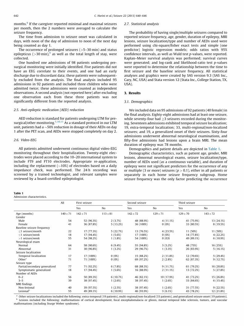
Table 1Admission characteristics.
All First seizure
Yes No
Age (months) 140 � 71 142 � 71 113 � 81
Gender
Male 54 52 (96.3%) 2 (3.7%)
Female 41 36 (87.8%) 5 (13.2%)
Baseline seizure frequency
�1 seizure/month 22 17 (77.3%) 5 (22.7%)
�1 seizure/week 18 17 (94.4%) 1 (5.6%)
>1 seizure/week 55 54 (98.2%) 1 (1.8%)
Neurological exam
Normal 64 58 (90.6%) 6 (9.4%)
Abnormal 31 30 (96.8%) 1 (3.2%)
Seizure localization
Temporal localization 17 17 (100%) 0 (0%)
Othera 71 71 (100%) 0 (0%)
Seizure type
Partial/secondary generalized 77 71 (92.2%) 6 (7.8%)
Symptomatic generalized 18 17 (94.4%) 1 (5.6%)
Number of AEDs
0–2 56 50 (89.3%) 6 (10.7%)
3–5 39 38 (97.4%) 1 (2.6%)
MRI findings
Non-lesional 40 39 (97.5%) 1 (2.5%)
Lesionalb 55 49 (89.1%) 6 (10.9%)
a Other seizure localizations included the following: extra-temporal (19 patients), mub Lesions included the following: malformations of cortical development, focal en
malformations (including Sturge Weber syndrome).
2.7. Statistical analysis
The probability of having single/multiple seizures compared toreported seizure frequency, age, gender, duration of epilepsy, MRIlesions, seizure localization/type and number of AEDs used wereperformed using chi-square/fisher exact tests and simple (onepredictor) logistic regression models; odds ratios with 95%confidence intervals, as well as Wald test p-values, were reported.Kaplan–Meier survival analysis was performed; survival curveswere generated; and log-rank and likelihood-ratio test p-valueswere reported to determine the relationship between the time tofirst seizure and the baseline seizure frequency. All statisticalanalyses and graphics were created by SAS version 9.3 (SAS Inc.,Cary, NC, USA) and Stata version 12 (Stata Inc., College Station, TX,USA).
3. Results
3.1. Demographics
We included data on 95 admissions of 92 patients (40 female) inthe final analysis. Eighty-eight admissions had at least one seizure,while seventy-four had �3 seizures recorded during the monitor-ing. Seventeen admissions exhibited temporal seizure localization;19, extra-temporal localization; 33, multi-regional/non-localizedseizures; and 19, a generalized onset of their seizures. Sixty-fouradmissions underwent abnormal neurological examinations, andfifty-five admissions had lesions upon a brain MRI. The meanduration of epilepsy was 78 months.
Demographics and patient details are depicted in Table 1.Demographic characteristics, such as patient age, gender, MRI
lesions, abnormal neurological exams, seizure localization/type,number of AEDs used (as a continuous variable), and duration ofepilepsy were not significant predictors for the occurrence of anyor multiple (3 or more) seizures (p > 0.1), either in all patients orseparately in each home seizure frequency subgroup. Homeseizure frequency was the only factor predicting the occurrence
Second seizure Third seizure
Yes No Yes No
142 � 72 129 � 71 129 � 70 143 � 72
48 (88.9%) 4 (11.1%) 41 (75.9%) 13 (24.1%)
36 (100%) 0 (0%) 33 (80.5%) 8 (19.5%)
13 (76.5%) 4 (23.5%) 11 (50%) 11 (50%)
17 (100%) 0 (0%) 14 (77.8%) 4 (22.2%)
54 (100%) 0 (0)% 49 (89.1%) 6 (10.9%)
55 (94.8%) 3 (5.2%) 48 (75%) 16 (25%)
29 (96.7%) 1 (3.35) 26 (83.9%) 5 (16.1%)
15 (88.2%) 2 (11.8%) 12 (70.6%) 5 (29.4%)
69 (97.2%) 2 (2.8%) 62 (87.3%) 9 (12.7%)
68 (88.3%) 9 (11.7%) 61 (79.2%) 16 (20.8%)
16 (88.9%) 2 (11.1%) 13 (72.2%) 5 (27.8%)
46 (82.1%) 10 (17.9%) 41 (73.2%) 15 (26.8%)
38 (97.4%) 1 (2.6%) 33 (84.6%) 6 (15.4%)
38 (97.4%) 1 (2.6%) 31 (77.5%) 9 (22.5%)
46 (93.9%) 3 (6.1%) 43 (78.2%) 12 (21.8%)
lti-regional/non-localized (33 patients), and generalized seizure onset (19 patients).
cephalomalacia or gliosis, mesial temporal lobe sclerosis, tumors, and vascular

C. Harini et al. / Seizure 22 (2013) 640–646 643
of any or multiple seizures (Fisher-exact test p = 0.01 and 0.0007,respectively).
3.2. Seizure frequency sub-group analysis
There were twenty-two admissions in the low-frequency,eighteen in the medium-frequency and fifty-five in the high-frequency group. Seventeen admissions in the low-frequency,seventeen in the medium-frequency, and fifty-four patients in thehigh-frequency group had at least one epileptic seizure during themonitoring period. All admissions in the medium and high-frequency group had a second seizure, while only 13 out of 22admissions in the low-frequency group had a second seizure.Eleven out of twenty-two admissions in the low-frequency grouphad three or more seizures during the monitoring period comparedto 14 (out of 18) in the medium-frequency group and 49 (out of 55)in the high-frequency group.
The odds of having a seizure increased by 15.88 (OR = 15.88,p = 0.01) in the high-frequency compared to low-frequency group.However, the odds of having a seizure did not significantly differbetween the high-frequency and medium-frequency groups(OR = 3.17, p = 0.42) or between the medium-frequency and low-frequency groups (OR = 5.0, p = 0.16).
The odds of having a second seizure increased in the high-frequency (OR = 37, p = 0.001) and medium-frequency groups(OR = 12, p = 0.03), compared to the low-frequency group. Theodds of having a second seizure did not significantly differ betweenthe medium- and high-frequency groups (OR = 3.18, p = 0.42).
The odds of having 3 or more seizures increased in the high(OR = 8.2, p = 0.0005) and medium-frequency groups (OR = 3.5,p = 0.007), compared to the low-frequency group. The odds ofhaving 3 or more seizures did not significantly differ between thehigh and medium-frequency groups (OR = 0 2.3, p = 0.23).
The mean time to the first seizure onset among all admissionswas 2.2 days (median 2), representing 2.7 days in the low-frequency group (median 3), 2.1 days in the medium-frequencygroup (median 2), and two days in the high-frequency group(median 1.5).
The median time to second seizure was less than twenty-fourhours from the first seizure in all three groups. In thirteenadmissions, the time interval between the first and second seizurewas longer than twenty-four hours (Table 2).
3.3. Seizure frequency and time to first seizure
Kaplan–Meier survival analysis showed that the time to onsetof the first seizure in the high-frequency group was significantlyless than that in the low-frequency group (log-rank and likelihood-ratio, p = 0.0004 and 0.0014, respectively). Similarly, the time toonset of the first seizure in the medium-frequency group wassignificantly less than that in the low-frequency group (log-rankand likelihood-ratio, p = 0.038 and 0.038, respectively). The high-and medium-frequency groups did not significantly differ in thetime to onset for the first seizure (both log-rank and likelihood-ratio, p > 0.1).
Kaplan–Meier survival analysis did not show any significantdifferences in the time to the occurrence of the second seizure from
Table 2Time to onset (days) of first and second seizures.
Baseline seizure frequency First seizure Second seizure
Mean � SD Median Mean � SD Median
�1 seizure/month (low) 2.8 � 0.83 3 1.23 � 0.60 1
�1 seizure/week (medium) 2.1 � 1.25 2 1.41 � 0.62 1
>1 seizure/week (high) 2.0 � 1.53 1.5 1.12 � 0.43 1
the first seizure in any 3 baseline seizure frequency categories(p > 0.1).
Fig. 2 displays a composite of the Kaplan–Meier curves showingthe survival probability of the 3 groups as a function of time to firstseizure onset.
Seven out of the 95 admissions did not have a seizure during themonitoring period.
The mean length of stay for all admissions was 5 days in thehigh-frequency group, 6 days in the medium-frequency group and5.5 days in the low-frequency group. Both the mean and median
Fig. 2. Kaplan–Meier survival curves demonstrating the relationship between time
to first seizure and baseline seizure frequency.

C. Harini et al. / Seizure 22 (2013) 640–646644
durations of stay in all admissions that had seizures during themonitoring period was five days. The mean length of stay inadmissions for patients not experiencing a seizure was seven days(median 6).
The total number of seizures captured during the monitoringperiod ranged from 0–18 seizures (median 2.5 seizures) in the low-frequency group, 0–22 seizures (median 5.5) in the medium-frequency group, and the highly variable range of 0–384 seizures inthe high-frequency group (median 10) (Fig. 3).
In our series, two admissions had status-epilepticus, and twelveadmissions had prolonged seizures lasting between 5 and 30 min.None of the patients with prolonged seizures or status epilepticusrequired ICU care. Seven patients were in the high-frequencygroup, three were in the medium-frequency group, and two werein the low-frequency group. The two patients (in the low-frequency group) received low-dose lorazepam (0.05 mg/kg) to
Fig. 3. Bar graphs showing the occurrence of first, second and third seizures
according to the 3 baseline seizure frequencies.
stop their prolonged seizures but continued to have seizures thenext day. Five patients received no treatment, and their prolongedseizures resolved spontaneously. The rest of the patients (n = 7)received benzodiazepine (n = 5), valproate (n = 1) and/or phenyto-in (n = 3). Patients with status-epilepticus had electroclinicalcomplex-partial seizures without overt tonic-clonic activity orany cardiopulmonary compromise.
4. Discussion
4.1. Summary
The majority of the LTM admissions for pre-surgical workup(92%) had at least one epileptic seizure during a stay of meanlength 5.24 days. Home seizure frequency was the only factorinfluencing the occurrence of single or multiple seizures duringmonitoring. None of the other factors examined, such as age,gender, presence or absence of MRI lesions, seizure types andlocalization, number of AEDs used, abnormal neurological exam orduration of epilepsy modified the occurrence of single/multipleseizures in any of the patients or the individual sub-groups.Admissions with high or medium home seizure frequencies (>1/month) were more likely to have multiple (3 or more) seizures inthe LTM than those in the low-frequency group for a similar meanlength of stay (�5 days). The time to the first epileptic seizure wasshorter in the high- and medium-frequency groups (mean 2.1days) than in the low-frequency group (mean 2.8 days).
4.2. Group differences
Home seizure frequency best predicted the occurrence ofseizures. Although most of our admissions had at least 1 seizureduring monitoring, the sub-group analysis on seizure frequencycategories revealed that the low-seizure frequency group is at riskfor completing the monitoring with a low number of seizurescaptured (<3).
We found that 98% of the high-frequency group versus 77% ofthe low-frequency group had a seizure during the monitoring.Todorov et al.4 found that a 5-day stay was required to record atleast 1 seizure in 90% of the patients evaluated for non-invasiveand invasive pre-surgical monitoring. The authors did not relatetheir findings to home seizure frequency and assumed that theseizure frequency must be relatively high due to the selection ofpatients with refractory epilepsy. The occurrence of at least 1seizure in most patients is most likely a reflection of the severity ofdisease observed in patients referred for a pre-surgical workup.This finding could also reflect the referral practices of theneurologists, who are biased towards referring children with veryfrequent seizures, as shown by the more than twice as large anumber of patients being monitored in the high-frequency groupthan in the medium- and low-frequency groups in our series.
Home seizure frequency also predicted the occurrence ofmultiple (�3) seizures during monitoring. The pre-surgical workupis considered successful if a consensus can be reached regardingthe decision of whether to proceed with surgery. The ictal dataobtained during multiple seizures provides information critical tosuccessfully planning epilepsy surgery in a majority of cases. Theoptimal number of seizures needed to complete the pre-surgicalworkup varies depending upon several factors, including thequality of the video-EEG recording, the number of seizure types,and concordant data from other tests. It has been estimated that incases of temporo-limbic epilepsy, a minimum of five seizuresshould be captured for the identification of the primary epilepto-genic focus in adults.12 Others have suggested that 3 or 4 identicalevents from a single seizure type may be sufficient to planfor epilepsy surgery in adults.2 Nevertheless, clinically useful

C. Harini et al. / Seizure 22 (2013) 640–646 645
information may be gained even after 1 or 2 seizures. Amongpatients undergoing pre-surgical monitoring, various studies havereported that decisions regarding epilepsy surgery could be madein 55–69% of the cases.5–7 The duration of monitoring in thesestudies ranged between 3.5–5 days. Although we did not examinewhether the information obtained during LTM was sufficient tocome to conclusions regarding epilepsy surgery in the groupsstudied, it is likely that the number of seizures captured duringmonitoring will impact the decision made regarding surgery. In ourstudy, nearly half of all children in the low-frequency group did nothave multiple seizures. Thus, we believe that the low-frequencygroup is possibly at risk for not successfully completing the pre-surgical monitoring.
Previous adult studies have reported that the self-reporting ofseizures is unreliable; unrecognized seizures are frequent,13 andadult patients failed to document 55% of all recorded seizures inthe LTM unit.14 In contrast, few pediatric studies have suggestedthat the frequency of the paroxysmal events was the only factordetermining the successful recording of that event during video-EEG monitoring,5,6 which is in agreement with our findings.Underreporting may be less of a problem in the pediatricpopulation compared to the adult population, as the parent/caregiver tends to be more observant and can provide morereliable information about this vulnerable population. However, ifthe seizures are subtle, underreporting can still occur.
4.3. Time to first seizure
Our results in children regarding time-to-first-seizure onsetwere remarkably similar to those reported in the adult popula-tion.10 Although we found that the difference in time-to-seizureoccurrence was statistically significant between the low-frequencygroup and the other groups, we consider this difference (of lessthan one day) not to be clinically significant. A second seizureoccurred within 24 h of the first seizure in all 3 groups, and this wasnot statistically significant among the groups. Hence, although thehome seizure frequency influenced the occurrence of single/multiple seizures during the period of monitoring, it did not appearto affect the time of occurrence of the seizures (either the first orthe second seizure) in a clinically meaningful way.
4.4. Length of stay
The duration of monitoring can influence the number ofseizures captured during LTM. The length of stay in our study wasfairly consistent with that recorded in previous studies.4,5,15
Another study 16 reported that 3 or more days were required tocharacterize paroxysmal episodes in 35% of their patients, as wellas that it was not rare to require more than a week to observe thefirst event. Although it is possible that more patients in the low-frequency group would have had a seizure if they stayed longer,children do not tolerate a longer duration of monitoring well; thisoften plays a major role in deciding the duration of monitoring. Allof the patients in our series had a similar length of stay in theattempt to capture multiple seizures, regardless of their homeseizure frequency.
Our data could help to modify clinical practice if confirmedprospectively. To capture multiple seizures, a longer duration ofmonitoring, i.e., longer than 5–6 days (current practice), may benecessary for patients with a home seizure frequency of �1 seizureper month.
Five patients in the low-frequency group, and one in each ofthe medium- and high-frequency groups, did not have anyseizures. The patient without a seizure in the high-frequencygroup turned out to have a nonepileptic event upon subsequentmonitoring. If we exclude this patient from analysis, then all of
patients in the high-frequency group had a seizure during themonitoring period.
4.5. Limitations
The main limitation of our study is the relatively small samplesize in the low-/medium-frequency groups. This small sample sizeposes a risk for false-negative or false positive-results. Anotherlimitation is the retrospective nature of the study. However, weincluded all consecutive patients admitted for a pre-surgicalworkup such that there was no inclusion bias. We believe that thestudy population provides a representative sample of patientadmissions for such a work-up. The accuracy of parent-reportedseizure-frequencies is a concern. We arbitrarily categorized theseizure frequency into 3 groups. Parents are better at reportingmonthly, weekly or more than weekly seizures, and hence wechose this categorization. Although we find that certain highlymotivated parents keep accurate seizure logs, most either do notmaintain a seizure log or fail to produce it during a clinic visit. Thedata on management decisions based on pre-surgical video-EEGmonitoring and post-surgical outcomes were not collected in ourstudy. Because this is a retrospective observational analysis, thefindings from this study must be verified prospectively.
5. Conclusions
Multiple seizures are required to demonstrate the consistencyof ictal onset for the localization of epilepsy during pre-surgicalmonitoring. Home seizure frequency correlated with the occur-rence of single/multiple seizures during LTM. None of the otherfactors (age/sex/MRI-lesions/neurological-exam/epilepsy-dura-tion/seizure type and localization/AED usage) influenced theoccurrence of single/multiple seizures during monitoring. Nearlyhalf of all admissions in the low-frequency group (or 36% of thosewho had a seizure) did not have multiple (3 or more) seizurescaptured. This result may necessitate repeated monitoring or alengthy inpatient stay during pre-surgical monitoring for lowseizure frequency patients. Multiple ways of inducing seizuresduring LTM may also be helpful for at-risk patients (low seizurefrequency group); these methods may include hyperventila-tion,17,18 sleep deprivation, a slow medication taper a few daysbefore admission, photic stimulation or other triggers, such ascircadian misalignment. In addition, cost has to be factored into theanalysis when monitoring patients for pre-surgical evaluationbecause patients with low seizure frequencies may need a longerduration or repeat monitoring.
Author contributions
Drs. Harini, Loddenkemper, Takeoka, Bergin and Kotharedesigned and conceptualized the study. Dr. Harini and Ms.Parulkar assisted in data collection. Dr. Singh performed statisticalanalysis on the data. Drs. Harini, Singh and Kothare drafted themanuscript. All authors participated in revising and editing themanuscript.
Disclosures of conflicts of interest
Dr. Kothare interprets video-EEGs and routine EEG in theDivision of Clinical Neurophysiology at Children’s Hospital Bostonand has received research support from Eisai Inc. and the NIH. Dr.Loddenkemper receives funding from the Epilepsy Foundation ofAmerica, and serves on the Laboratory Accreditation Board for LongTerm (Epilepsy and ICU) Monitoring (ABRET). Ms. Parulkar, Drs.Harini, Singh, Takeoka and Bergin have no conflicts of interest toreport. We confirm that we have read the Journal’s position on the

C. Harini et al. / Seizure 22 (2013) 640–646646
issues involved in ethical publication and affirm that this report isconsistent with those guidelines.
References
1. Kwan P, Brodie MJ. Early identification of refractory epilepsy. New EnglandJournal of Medicine 2000;342:314–9.
2. Rosenow F, Luders H. Presurgical evaluation of epilepsy. Brain 2001;124:1683–700.
3. Asano E, Pawlak C, Shah A, Shah J, Luat AF, Ahn-Ewing J, Chugani HT. Thediagnostic value of initial video-EEG monitoring in children – review of 1000cases. Epilepsy Research 2005;66:129–35.
4. Todorov AB, Lesser RP, Uematsu SS, Yankov YA, Todorov Jr AA. Distributionin time of seizures during presurgical EEG monitoring. Neurology 1994;44:1060–4.
5. Riquet A, Lamblin MD, Bastos M, Bulteau C, Derambure P, Vallee L, Auvin S.Usefulness of video-EEG monitoring in children. Seizure 2011;20:18–22.
6. Chen LS, Mitchell WG, Horton EJ, Snead 3rd OC. Clinical utility of video-EEGmonitoring. Pediatric Neurology 1995;12:220–4.
7. Alving J, Beniczky S. Diagnostic usefulness and duration of the inpatient long-term video-EEG monitoring: findings in patients extensively investigated be-fore the monitoring. Seizure 2009;18:470–3.
8. Loddenkemper T, Kellinghaus C, Wyllie E, Najm IM, Gupta A, Rosenow F, LudersHO. A proposal for a five-dimensional patient-oriented epilepsy classification.Epileptic Disorders 2005;7:308–16.
9. Sillanpaa M, Shinnar S. Status epilepticus in a population-based cohort withchildhood-onset epilepsy in Finland. Annals of Neurology 2002;52:303–10.
10. Eisenman LN, Attarian H, Fessler AJ, Vahle VJ, Gilliam F. Self-reported seizurefrequency and time to first event in the seizure monitoring unit. Epilepsia2005;46:664–8.
11. Yen DJ, Chen C, Shih YH, Guo YC, Liu LT, Yu HY, Kwan SY, Yiu CH. Antiepilepticdrug withdrawal in patients with temporal lobe epilepsy undergoing presur-gical video-EEG monitoring. Epilepsia 2001;42:251–5.
12. Blum D. Prevalence of bilateral partial seizure foci and implications for electro-encephalographic telemetry monitoring and epilepsy surgery. Electroencepha-lography and Clinical Neurophysiology 1994;91:329–36.
13. Blum DE, Eskola J, Bortz JJ, Fisher RS. Patient awareness of seizures. Neurology1996;47:260–4.
14. Hoppe C, Poepel A, Elger CE. Epilepsy: accuracy of patient seizure counts.Archives of Neurology 2007;64:1595–9.
15. Haut SR, Swick C, Freeman K, Spencer S. Seizure clustering during epilepsymonitoring. Epilepsia 2002;43:711–5.
16. Friedman DE, Hirsch LJ. How long does it take to make an accurate diagnosisin an epilepsy monitoring unit? Journal of Clinical Neurophysiology 2009;26:213–7.
17. Abubakr A, Ifeayni I, Wambacq I. The efficacy of routine hyperventilation forseizure activation during prolonged video-electroencephalography monitor-ing. Journal of Clinical Neuroscience 2010;17:1503–5.
18. Arain AM, Arbogast PG, Abou-Khalil BW. Utility of daily supervised hyperven-tilation during long-term video-EEG monitoring. Journal of Clinical Neurophysi-ology 2009;26:17–20.