
Predictors and outcomes of stent thrombosis - European Heart Journal
9
European Heart Journal (2002) 23, 124–132 doi:10.1053/euhj.2001.2707, available online at http://www.idealibrary.com on Predictors and outcomes of stent thrombosis An intravascular ultrasound registry N. G. Uren, S. P. Schwarzacher, J.A. Metz, D. P. Lee, Y. Honda, A. C. Yeung, P. J. Fitzgerald and P. G. Yock, on behalf of the POST Registry investigators Division of Cardiovascular Medicine, Stanford University School of Medicine, Stanford, California, U.S.A. Aims To investigate whether intravascular ultrasound provides additional information regarding the prediction of stent thrombosis, a retrospective multicentre registry was designed to enrol patients with stent thrombosis following stent deployment under ultrasound guidance. Methods and Results A total of 53 patients were enrolled (mean age 61 9 years) with stable angina (43%), unstable angina (36%), and post-infarct angina (21%) who under- went intracoronary stenting. The majority had balloon angioplasty alone prior to stenting (94%) with 6% also undergoing rotational atherectomy. The indication for stenting was elective (53%), suboptimal result (32%) and bailout (15%). There were 1·6 0·8 stents/artery with 87% undergoing high-pressure dilatation (14 atmospheres). The minimum stent area was 7·7 2·8 mm 2 with a mean stent expansion of 81·5 21·9%. Overall, 94% of cases demonstrated one abnormal ultrasound finding (stent under-expansion, malapposition, inflow/outflow disease, dissection, or thrombus). Angiography demonstrated an abnormality in only 32% of cases (chi-square=30·0, P<0·001). Stent thrombosis occurred at 132 125 h after deployment. Myocardial infarction occurred in 67% and there was an overall mortality of 15%. Conclusion On comparison with angiography, the vast majority of stents associated with subsequent throm- bosis have at least one abnormal feature by intravascular ultrasound at the time of stent deployment. (Eur Heart J 2002; 23: 124–132, doi:10.1053/euhj.2001.2707) 2001 The European Society of Cardiology Key Words: Intravascular ultrasound, intracoronary stents, thrombosis. See page 97, doi:10.1053/euhj.2001.2810 for the Editorial comment on this article Introduction Although the incidence of stent thrombosis has diminished with improvement in stent design, deploy- ment, and adjunctive pharmacology, it continues to occur in up to 1·9% of cases with a high associated morbidity and mortality [1–5] . At the current world-wide level of intracoronary stenting with this incidence, stent thrombosis may occur in up to 12 000 patients per year. There are several clinical, lesion-related, stent-related and technique-dependent predictors of stent thrombosis which have come from retrospective analysis of single centre stent experience in recent years. However, there are no current multicentre trial data available to allow confident prediction of this event in any individual patient. The initial use of intravascular ultrasound to deploy the Palmaz–Schatz stent and newer generation stents has contributed to improved stent deployment through appropriate balloon sizing and the use of high- pressure dilatation to achieve full stent expansion, com- plete apposition to the vessel wall, and full lesion coverage [1] . However, it is not certain whether the quali- tative information derived from ultrasound analysis of a deployed stent will provide additional features predictive of subsequent stent thrombosis. Thus, this retrospective registry was designed to collect clinical, angiographic and ultrasound data on patients undergoing stent deployment who went on to sustain a stent thrombosis, with additional data on the specific treatment and outcomes of this event. Revision submitted 27 March 2001, accepted 28 March 2001, and published online 2 October 2001. Correspondence: Neal Uren MD MRCP FESC, Depart- ment of Cardiology, Royal Infirmary, 1 Lauriston Place, Edinburgh EH3 9YW, U.K. 0195-668X/02/020124+09 $35.00/0 2001 The European Society of Cardiology
Transcript of Predictors and outcomes of stent thrombosis - European Heart Journal

European Heart Journal (2002) 23, 124–132doi:10.1053/euhj.2001.2707, available online at http://www.idealibrary.com on
Predictors and outcomes of stent thrombosis
An intravascular ultrasound registry
N. G. Uren, S. P. Schwarzacher, J. A. Metz, D. P. Lee, Y. Honda,A. C. Yeung, P. J. Fitzgerald and P. G. Yock, on behalf of the
POST Registry investigators
Division of Cardiovascular Medicine, Stanford University School of Medicine, Stanford,California, U.S.A.
Introduction
Although the incidence of stent thrombosis hasdiminished with improvement in stent design, deploy-ment, and adjunctive pharmacology, it continues tooccur in up to 1·9% of cases with a high associatedmorbidity and mortality[1–5]. At the current world-widelevel of intracoronary stenting with this incidence, stentthrombosis may occur in up to 12 000 patients per year.There are several clinical, lesion-related, stent-relatedand technique-dependent predictors of stent thrombosiswhich have come from retrospective analysis of single
0195-668X/02/020124+09 $35.00/0
centre stent experience in recent years. However, thereare no current multicentre trial data available to allowconfident prediction of this event in any individualpatient. The initial use of intravascular ultrasound todeploy the Palmaz–Schatz stent and newer generationstents has contributed to improved stent deploymentthrough appropriate balloon sizing and the use of high-pressure dilatation to achieve full stent expansion, com-plete apposition to the vessel wall, and full lesioncoverage[1]. However, it is not certain whether the quali-tative information derived from ultrasound analysis of adeployed stent will provide additional features predictiveof subsequent stent thrombosis. Thus, this retrospectiveregistry was designed to collect clinical, angiographicand ultrasound data on patients undergoing stentdeployment who went on to sustain a stent thrombosis,with additional data on the specific treatment andoutcomes of this event.
Revision submitted 27 March 2001, accepted 28 March 2001, andpublished online 2 October 2001.
Correspondence: Neal Uren MD MRCP FESC, Depart-ment of Cardiology, Royal Infirmary, 1 Lauriston Place,Edinburgh EH3 9YW, U.K.
Aims To investigate whether intravascular ultrasoundprovides additional information regarding the prediction ofstent thrombosis, a retrospective multicentre registry wasdesigned to enrol patients with stent thrombosis followingstent deployment under ultrasound guidance.
Methods and Results A total of 53 patients were enrolled(mean age 61�9 years) with stable angina (43%), unstableangina (36%), and post-infarct angina (21%) who under-went intracoronary stenting. The majority had balloonangioplasty alone prior to stenting (94%) with 6% alsoundergoing rotational atherectomy. The indication forstenting was elective (53%), suboptimal result (32%) andbailout (15%). There were 1·6�0·8 stents/artery with 87%undergoing high-pressure dilatation (�14 atmospheres).The minimum stent area was 7·7�2·8 mm2 with a meanstent expansion of 81·5�21·9%. Overall, 94% of casesdemonstrated one abnormal ultrasound finding (stentunder-expansion, malapposition, inflow/outflow disease,
dissection, or thrombus). Angiography demonstrated anabnormality in only 32% of cases (chi-square=30·0,P<0·001). Stent thrombosis occurred at 132�125 h afterdeployment. Myocardial infarction occurred in 67% andthere was an overall mortality of 15%.
Conclusion On comparison with angiography, thevast majority of stents associated with subsequent throm-bosis have at least one abnormal feature by intravascularultrasound at the time of stent deployment.(Eur Heart J 2002; 23: 124–132, doi:10.1053/euhj.2001.2707)� 2001 The European Society of Cardiology
Key Words: Intravascular ultrasound, intracoronarystents, thrombosis.
See page 97, doi:10.1053/euhj.2001.2810 for the Editorialcomment on this article
� 2001 The European Society of Cardiology

Stent thrombosis 125
Methods
Study design
The Predictors and Outcomes of Stent Thrombosis(POST) Registry was designed to identify the intra-vascular ultrasound predictors of acute (<24 h) andsubacute (24 h to 4 weeks) stent thrombosis. Thismulticentre, retrospective registry enrolled cases from1991 to 1996 from North America, Europe andJapan (see Appendix). After enrolment, cases of stentthrombosis were identified where intravascular ultra-sound was used during deployment and analysis wasbased on a clinical registry compiled from case reportforms, intravascular ultrasound imaging tapes andangiograms at deployment.
Inclusion criteria
Inclusion criteria for the registry were intentionallybroad to maximize enrolment. All clinical presentationsincluding elective or emergency stent deployment wereincluded. Single and multiple stents of all stent typeswere enrolled. Pre-stent treatment with any transcath-eter therapy was allowed. The major criterion was thatan intravascular ultrasound examination was performedpost-deployment and that images were of sufficientquality to be interpreted by the core laboratory. A totalof nine participating centres from North America, sevenfrom Europe and two from Japan contributed cases tothe study. A total of 53 cases of stent thrombosis fulfilledthe inclusion criteria and were enrolled in the registry.
Case report forms were designed to include threesections: (i) clinical details, (ii) procedure details, and(iii) outcome and treatment of stent thrombosis. Inaddition, details of the daily course of antiplatelet andantithrombotic therapy were documented on a separateclinical report form. From these data, the clinical detailsof the initial interventional case was determined andanalysed.
determined by planimetry at the distal and proximalstent edges as well as at the minimum stent cross-sectional area. Stent under-expansion was defined asminimum stent area <80% of mean proximal and distalreference lumen areas. Stent deployment was alsoassessed according to the MUSIC criteria, namely,complete apposition and expansion (minimum stent area[MSA] >90% average of proximal/distal segmentsor MSA >100% of lowest reference segment or MSA>90% proximal segment, revised to 80%, 90%, 80%respectively if MSA >9 mm2)[6].
The definition chosen for significant inflow diseasewas that the proximal reference lumen area was lessthan the distal reference lumen area in the presence of�40% plaque area in either segment. Outflow diseasewas defined as the presence of plaque �40% vesselarea. The presence of calcium and the plaque mor-phology (fibrous, fibro-fatty or fibro-calcific) in thereference segments was also documented. Residual dis-section or edge tears were defined as a visible flapoutside stent edges with blood speckle behind theintimal flap or at mid-stent articulation for thePalmaz–Schatz design when present. Malappositionwas identified by recognition of a relatively echo-freespace between the struts and intima with evidence ofblood speckle. Plaque protrusion was defined as visibletissue on the luminal side of the stent struts, butlacking any of the identifying characteristics of throm-bus. Thrombus was defined as the presence of tissuewithin but adherent to the stent with an ultrasoundintensity less than half of the adventitial signal. Whenpresent, a scintillating appearance, a lobular irregularedge, microchannels and/or movement in an undulat-ing manner separate from the artery confirmed thepresence of thrombus.
In order to compare the data from the POST registrywith comparable control data, qualitative and quanti-tative ultrasound variables from one registry ofintravascular ultrasound-guided stenting and two pro-spective studies of intravascular ultrasound-guidedagainst angiography-guided stenting were used. Fourvariables — percent stent expansion, malapposition,edge tears/dissection, and thrombus — from POST werecompared with the equivalent ultrasound parametersfrom three other studies. These were (1) STRUT (StentTreatment Region assessed by Ultrasound Tomogra-phy)[7], which was a multicentre registry of intravascularultrasound-guided stenting in clinical practice, (2)CRUISE (Can Routine Ultrasound Influence StentExpansion)[8], which was a prospective randomizedmulticentre trial of intravascular ultrasound-guided vsangiography-guided stenting with clinical follow-up, and(3) AVID (Angiography Versus Intravascular ultra-sound Directed stent placement)[9], which was a random-ised trial similar to CRUISE. These three studies wereselected by the POST investigators for comparison asthe ultrasound analysis was also performed by the sameultrasound core lab (Center for Research in Cardio-vascular Interventions, Stanford University Hospital,CA, U.S.A.).
Intravascular ultrasound analysis
Intravascular ultrasound image analysis was undertakenat the Quantitative Coronary Ultrasound laboratory atStanford University, California. Several quantitativeand qualitative criteria were determined with respect tothe deployed stent(s) and reference vessels proximal anddistal to the stent using the TapeMeasure 2.1.0 analysisprogram (Indec Systems, Mountain View, CA, U.S.A.)by at least two independent observers. The final intra-vascular ultrasound run at the end of the procedure wasselected as the imaging sequence for analysis. Vessel,lumen and plaque area were drawn by planimetryproximal and distal to the stent in the most normalframe within 5 mm of the stent edges. Stent area was
Eur Heart J, Vol. 23, issue 2, January 2002

126 N. G. Uren et al.
Coronary angiography analysis
Angiographic image analysis was performed at theQuantitative Coronary Angiography laboratory atStanford University Hospital. The angiographic runsselected were a composite of the views taken at the endof the procedure and analysed by an independentobserver (D.L.) blinded to the intravascular ultrasoundimages during the case. Whether or not a residualdiameter stenosis of 0% or less was achieved was deter-mined by visual assessment. At angiography, inflow/outflow disease was defined as a diameter stenosis >50%immediately proximal or distal to the stent edges andother definitions used were standard. Filling defects andall dissections visible angiographically were described.
Statistical analysis
Comparable dichotomous qualitative data for ultra-sound and angiography were analysed using McNemar’stest. Individual ultrasound variables were comparedwith existing data using the chi-square method withYates’ correction where appropriate. Comparison ofcontinuous variables in the acute and subacute stentthrombosis groups was done using an unpairedStudent’s t test. A P value of less than 5% wasconsidered statistically significant.
Results
Patient and lesion demographics
The clinical characteristics of the patients enrolled inthe study and the coronary artery lesion undergoingintervention and subsequent analysis are described inTables 1 and 2. The mean age of patients was 61�9years and the left ventricular ejection function was55�16%. The majority of patients in the study had anunstable coronary syndrome and just under two-thirdsof patients had complex lesions of AHA/ACC class B2
Eur Heart J, Vol. 23, issue 2, January 2002
and C. Ninety percent of stents were deployed in theproximal or mid segments of the vessel with the remain-der deployed in distal arteries or across two segments.The majority of patients had a single stent deploymentwith the maximum number deployed being four. Themean number of stents per artery was 1·6 with a medianof 1. Six percent of patients had high-speed rotationalatherectomy prior to stenting with the remaining 94%having balloon angioplasty alone.
Post-procedure antithrombotic and antiplatelettherapy in the registry reflects the fact that patients wereenrolled over a period of 5 years with an evolvingprotocol of antithrombotic management after stentdeployment. All but three patients had oral aspirinfollowing stent deployment, with 40% having aspirinalone as therapy (n=21, 6 of whom also had warfarin).The remaining 54% (n=29) received ticlopidine andaspirin for at least 4 weeks following stenting.
Table 1 Population demographics
Number Percentage
Extent of disease1 vessel 19 36%2 vessel 19 36%3 vessel 15 28%
Clinical presentationStable angina 23 43%Unstable angina 19 36%Post-MI angina 11 21%
Stent indicationElective 29 53%Suboptimal result 17 32%Bailout 7 15%
Table 2 Lesion and stent characteristics
Number Percentage
ACC/AHA lesion classA 2 4%B1 17 32%B2 15 28%C 19 36%
ArteryLAD 26 49%CFX 10 19%RCA 13 24%Saphenous vein graft 4 8%
Lesion segmentProximal 32 60%Mid 16 30%Distal 3 6%2 segments 2 4%
Number of stentsOne 32 60%Two 12 23%Three or more 9 17%
High pressure deployment (�14 atm) 46 87%
Stent typePalmaz–Schatz 40 76%Palmaz–Schatz+other 5 9%Other stent 8 15%
ACC/AHA=American College of Cardiology/American HeartAssociation; LAD=left anterior descending artery; CFX=leftcircumflex artery; RCA=right coronary artery.
Stent deployment by intravascularultrasound
Intravascular ultrasound measurements are given inTable 3. By ultrasound, average percent expansion was82% of average reference lumen area, 91% of distalreference lumen area, and 76% of proximal referencelumen area. Given that the mean minimum stent area

Stent thrombosis 127
was 7·7�2·8 mm2, mean stent expansion did not meetany of the expansion criteria of the MUSIC study[6].With respect to these criteria, 31 stents achieved none(58%), seven stents achieved one (13%), nine stentsachieved two (17%), and six stents achieved all three(11%). The minimum stent area was found in theproximal, mid (articulation) or distal stent segments
in 20%, 17% and 63% of stents, respectively. In theproximal and distal reference segments, the percentplaque area was 42·6% (range 13·4–78·2%) and 40·9%(range 0–83·0%), respectively.
Plaque morphology in the reference segments up to5 mm proximal and distal to the stent were describedqualitatively (Table 4). The segment was described asnormal where �15% of vessel area was intimal thicken-ing, given the mean age of patients in the study. A totalof 11% of proximal segments and 8% of distal segmentsfell within this definition. Calcium was described in 18%of proximal or distal reference segments with a minorityof deposits greater than 180� in extent. The concordancebetween proximal and distal plaque type with respect tofibro-fatty plaque, fibrous plaque or plaque with >180�calcium present was 65%.
Table 3 Intravascular ultrasound assessment
Mean�SD Range
Distal reference segmentVessel area (mm2) 16·1�7·8 6·5–52·4
Minimum diameter (mm) 4·1�1·1 2·5–7·7Maximum diameter (mm) 4·7�1·1 3·0–8·6
Lumen area (mm2) 9·2�4·7 2·7–33·4Minimum diameter (mm) 3·0�0·7 1·7–6·1Maximum diameter (mm) 3·6�0·8 1·9–6·9
Plaque area (mm2) 6·8�4·3 0–19·0
Distal stent edgeStent area (mm2) 8·8�3·5 2·8–19·5
Minimum diameter (mm) 3·0�0·6 1·5–4·7Maximum diameter (mm) 3·6�0·7 2·1–5·4
Minimum stent areaStent area (mm2) 7·7�2·8 2·8–17·4
Minimum diameter (mm) 2·7�0·5 1·5–4·5Maximum diameter (mm) 3·4�0·6 2·1–5·1
Lumen ratio 0·81�0·08 0·57–0·93
Proximal stent edgeStent area (mm2) 9·8�3·6 5·0–25·7
Minimum diameter (mm) 3·1�0·6 2·4–5·4Maximum diameter (mm) 3·8�0·7 2·7–5·9
Proximal reference segmentVessel area (mm2) 17·7�5·6 14·4–32·2
Minimum diameter (mm) 4·3�0·8 1·8–6·0Maximum diameter (mm) 5·0�0·8 2·9–6·6
Lumen area (mm2) 10·4�5·5 3·7–37·8Minimum diameter (mm) 3·2�0·8 1·8–6·4Maximum diameter (mm) 3·9�0·9 2·3–7·4
Plaque area (mm2) 7·6�4·7 0·13–25·2
Table 4 Reference segment plaque morphology
Proximal segment Distal segment
Fibrofatty 20 (38%) 25 (47%)Fibrous 12 (23%) 11 (21%)Fibrofatty+fibrous 4 (8%) 4 (8%)Fibrofatty+calcium <180� 1 (2%) 1 (2%)Fibrous+calcium <180� 5 (9%) 5 (9%)Calcium �180� 3 (6%) 3 (6%)Normal 6 (11%) 4 (8%)No reference segment 2 (4%) 0
Figure 1 Three examples of stent malapposition to a mild degree (left panel), to a moderate degree (middlepanel) and to a severe degree (right panel), not appreciable by angiography.
Comparison of intravascular ultrasound andangiography after final stent deployment
With intravascular ultrasound, under-expansion lessthan 80% of mean reference area was found in 49%of cases (n=26) compared to 11% (n=6) whereangiography identified a residual diameter stenosis >0%.Malapposition was also seen in 49% of cases by intra-vascular ultrasound (Fig. 1). Inflow/outflow disease onintravascular ultrasound examination occurred in 30%of cases (n=16; 7 inflow, 3 outflow, and 6 inflow/
Eur Heart J, Vol. 23, issue 2, January 2002
128 N. G. Uren et al.
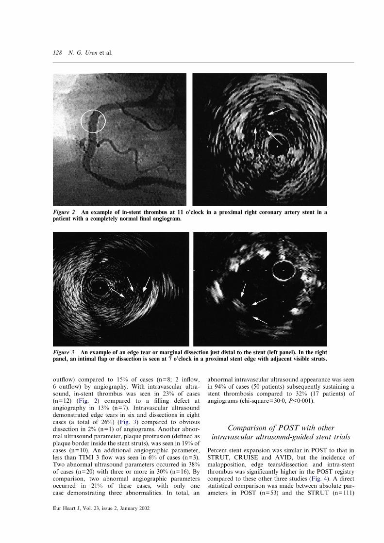
outflow) compared to 15% of cases (n=8; 2 inflow,6 outflow) by angiography. With intravascular ultra-sound, in-stent thrombus was seen in 23% of cases(n=12) (Fig. 2) compared to a filling defect atangiography in 13% (n=7). Intravascular ultrasounddemonstrated edge tears in six and dissections in eightcases (a total of 26%) (Fig. 3) compared to obviousdissection in 2% (n=1) of angiograms. Another abnor-mal ultrasound parameter, plaque protrusion (defined asplaque border inside the stent struts), was seen in 19% ofcases (n=10). An additional angiographic parameter,less than TIMI 3 flow was seen in 6% of cases (n=3).Two abnormal ultrasound parameters occurred in 38%of cases (n=20) with three or more in 30% (n=16). Bycomparison, two abnormal angiographic parametersoccurred in 21% of these cases, with only onecase demonstrating three abnormalities. In total, an
Eur Heart J, Vol. 23, issue 2, January 2002
abnormal intravascular ultrasound appearance was seenin 94% of cases (50 patients) subsequently sustaining astent thrombosis compared to 32% (17 patients) ofangiograms (chi-square=30·0, P<0·001).
Figure 2 An example of in-stent thrombus at 11 o’clock in a proximal right coronary artery stent in apatient with a completely normal final angiogram.
Figure 3 An example of an edge tear or marginal dissection just distal to the stent (left panel). In the rightpanel, an intimal flap or dissection is seen at 7 o’clock in a proximal stent edge with adjacent visible struts.
Comparison of POST with otherintravascular ultrasound-guided stent trials
Percent stent expansion was similar in POST to that inSTRUT, CRUISE and AVID, but the incidence ofmalapposition, edge tears/dissection and intra-stentthrombus was significantly higher in the POST registrycompared to these other three studies (Fig. 4). A directstatistical comparison was made between absolute par-ameters in POST (n=53) and the STRUT (n=111)

Stent thrombosis 129
registry. No difference in stent under-expansion (<80%reference) was seen, but malapposition (chi-square=11·72, P<0·001), edge tears or dissection (chi-square=5·60, P<0·05), and thrombus (chi-square=26·75,P<0·0001) were significantly more common in POSTthan in STRUT.
0
100
Expansion
Per
cen
t
80
60
40
20
PO
ST
ST
RU
T
CR
UIS
E
AV
ID
0
25
Edge tears/dissection
20
15
10
5
PO
ST
ST
RU
T
CR
UIS
E
AV
ID
0
50
Malapposition
40
30
20
10
PO
ST
ST
RU
T
CR
UIS
E
AV
ID
0
25
In-stent thrombus
20
15
10
5
PO
ST
ST
RU
T
CR
UIS
E
AV
ID
*
**
Figure 4 A direct comparison of POST with existing ultrasound-guided stent deployment registries(STRUT) and ultrasound-guided stent deployment studies (CRUISE, AVID) with respect to percent stentexpansion, edge tear/dissection, malapposition, and thrombus (*P<0·05 vs STRUT, CRUISE, and AVID).
Table 5 Comparison of acute and subacute stent thrombosis
Acute thrombosis Subacute thrombosis P value
Left ventricular function 62�11% 51�17% 0·04Stents/artery 2·0�1·3 1·5�0·8 nsMinimum stent area (mm2) 7·38�2·82 7·86�2·80 nsProximal reference lumen area (mm2) 9·77�2·60 10·7�6·0 nsDistal reference lumen area (mm2) 7·95�3·2 9·64�5·0 nsPercent expansionproximal 73·7�27·7% 76·7�22·5% nsPercent expansionaverage 80·9�18·1% 81·7�23·0% nsPercent expansiondistal 106�24% 87�27% 0·02Proximal reference intimal area (mm2) 9·89�6·66 7·78�4·20 nsDistal reference intimal area (mm2) 6·27�3·1 7·05�4·55 ns
Comparison of acute with subacute stentthrombosis
Subgroup analysis was done comparing acute stentthrombosis (within the first 24 h) with the remainingsubacute cases. Overall, the average time to thrombosiswas 132 h (range 0–600) or approximately 6 days afterstenting. Eleven patients (21%) had acute thrombosis ata mean of 12�6 h, and the remaining 42 patients had asubacute thrombosis at a mean of 164�121 h (into the7th day). In the acute group, the clinical presentationwas unstable/post-infarction angina in 63%, compared
to 55% in the subacute group. There was no significantdifference in the stent number per artery or the minimumstent area although left ventricular function was betterin the acute group (Table 5). Percent stent expansionwas no different between groups when measured withrespect to proximal reference area or to average refer-ence area, but stents were better expanded with respectto the distal reference in acute thrombosis compared tothe subacute thrombosis group. Under-expansion <80%of average reference area was similar, 36% acute vs 50%subacute, although with respect to the distal vessel,under-expansion <90% of distal vessel was seen in 18%acute vs 64% in the subacute group (chi-square=5·73,P=0·017). Malapposition was 64% in acute thrombosisvs 45% in subacute thrombosis although this did notachieve statistical significance. No significant differencein inflow/outflow disease or jailed side branches wasnoted. An increased number of edge tears/dissection wasseen in the subacute thrombosis group, 47% vs 27% and
Eur Heart J, Vol. 23, issue 2, January 2002

130 N. G. Uren et al.
in intra-stent thrombus 24% vs 9% compared to theacute stent thrombosis group, although both failed toachieve statistical significance mainly due to sample size.
Treatment and outcome of stent thrombosis
A total of 85% of patients underwent emergencycoronary intervention: 36% had balloon angioplastyalone, 20% had angioplasty plus additional therapy(thrombolysis, abciximab or both) and 29% received afurther stent, some of these also receiving intracoronarythrombolysis. Six percent had an emergency CABG and9% had no revascularization. Two-thirds of the patientssuffered a myocardial infarction by enzyme criteria andthere was an overall mortality of 15%.
Discussion
The POST registry is the largest retrospective collectionto date of patients sustaining a stent thrombosis whounderwent an intravascular ultrasound study after stentdeployment. The registry was not controlled for stentdeployment without intravascular ultrasound guidanceor for the ultrasound appearance of stents deployedwithout subsequent stent thrombosis, and thus compari-son was made with existing studies. In POST, manystents sustaining acute and subacute thrombosis wereunder-expanded, which is consistent with previous dataon stent expansion from other multicentre ultrasoundregistries of stent deployment[7–9], suggesting that stentexpansion itself is not a predictor of subacute thrombo-sis. Edge tears and dissection were found to a greaterextent in the STRUT registry[7], and to a greater extentthan in the prospective studies CRUISE[8], and AVID[9].However, almost one half of patients sustaining stentthrombosis had malapposition, which is considerablyhigher than in these other series[7–9]. Also, up to aquarter had in-stent thrombus seen at ultrasound. Thesedata from POST confirm the importance of morphologicvariables documented by intravascular ultrasound evenwhere stents may be relatively well-expanded by existingstandards.
Stent thrombosis has become a less frequent eventwith the advent of high-pressure dilatation and anti-platelet therapy[4,10,11]. Initial use of ultrasound duringtraditional stent deployment showed that 80% ofstents were under-expanded (<70% of balloon cross-sectional area) and led to the hypothesis that stentthrombosis might be decreased as a result of optimalstent placement with high-pressure balloon dila-tation under ultrasound guidance without the need foranticoagulation[12,13].
To examine clinical predictors of subacute stentthrombosis, a study of 19 cases from 1001 consecutivepatients from 1993–5 was reported[14]. Following high-pressure dilatation to achieve less than 20% residualdiameter stenosis, intravascular ultrasound was used
Eur Heart J, Vol. 23, issue 2, January 2002
in 72% to guide final deployment. Patients with anunsuccessful angiographic outcome were treated withwarfarin; otherwise, a combination of aspirin/ticlopidineor aspirin alone was used. Indications for stenting, siteand complexity of the lesion were no different betweenthe groups. Slow flow at angiography, but not residualdissection post intervention, was a strong predictor ofthrombosis. With intravascular ultrasound, a smallerminimum stent area was seen in the stent thrombosisgroup, 5·9�1·7 vs 7·8�2·5 mm2 (c.f. 7·7�2·8 mm2 inthe POST registry). These data confirmed that bailoutstenting, a factor predicting stent thrombosis in earlierstudies[15,16], only carries a high risk for subsequentthrombosis if the underlying angiographic problem isnot fully corrected. In another review of 10 thrombosiscases in 215 intravascular ultrasound-guided stents,stent thrombosis occurred in smaller reference vessels,2·7�0·5 vs 3·2�0·6 mm (c.f. 3·2�0·8 mm in POST).The only independent predictors of risk were initial stentlumen area, 4·80�1·33 mm2 vs 6·86�2·08 mm2, andfinal stent percent plaque area, 70·1�6·1% vs58·4�9·8%[17]. In contrast to POST, morphologicvariables were not examined in this retrospective study
In the ISAR (Intracoronary Stenting and Antithrom-botic Regimen) trial, a composite risk assessment foradverse outcome was devised from a list of 18 clinical,lesion-related, and procedural variables (stent length,residual dissection, residual thrombus and stent overlap)in 517 patients[18]. Stent thrombosis occurred in 5·9% ofhigh-risk (�4 criteria), 2·7% of intermediate risk (3criteria), and 0% of low risk (�2 criteria) patients. Incontrast to the low- and intermediate-risk groups, inhigh-risk patients, the stent thrombosis rate was 11·5%with anticoagulant therapy and 0% with anti-platelettherapy (P<0·001). A wider review of stent outcomeswas described by the same group in 2894 proceduresfrom 1992–7 in whom 80% received aspirin/ticlopidinewith a documented stent thrombosis rate of 2·3%[19].Residual dissection after stenting by angiography andthe use of ticlopidine after day 3 were the two majorinfluences on stent thrombosis. Although the stentthrombosis rate in standard interventional practice islow, an increased risk of late thrombosis (in up to 9% ofstents) following intracoronary stenting and intracoro-nary irradiation indicates the potential value of intra-vascular ultrasound in guiding successful intervention inhigher risk situations[20].
Limitations
The POST registry is retrospective and does not have acomparable control group of ultrasound-guided stentdeployment cases without thrombosis. At present, stentthrombosis is a rare event and prospective comparativedata will only come from analysis of the large multi-centre ultrasound-guided stent deployment trials.Despite the selection bias of POST, the observationsremain valid and can still be compared with other stentregistries where intravascular ultrasound was used to

Stent thrombosis 131
guide deployment and where significant differences wereobserved with respect to POST.
It is still a limitation of ultrasound to diagnosethrombus with complete specificity. However, severalqualitative criteria such as a scintillating lobulatedappearance, microchannels and fluctuation in adirection opposite to the vessel wall were used. Thedefinitions applied in this study to inflow or outflowdisease by ultrasound or angiography are arbitrary butare consistent with the historical observation that suc-cessful stenting should be performed from normal ornear-normal segments from proximal to distal artery[1].
The POST registry came from a period of evolution ofantiplatelet therapy. However, the majority of patientsstill received aspirin and ticopidine with only three of 53patients receiving no aspirin. Although this may beconsidered an additional risk factor for stent thrombo-sis, the discrepancy between angiography-guided andultrasound-guided stent abnormalities still remainswhen excluding these patients from analysis.
Clinical implications
Intravascular ultrasound is significantly more sensitivein defining suboptimal stent deployment leading tothrombosis compared to angiography. Althoughretrospective, the POST registry suggests that stentmalapposition, in-stent thrombus and edge tears/dissection are important determinants of stent thrombo-sis. It is not a cost-effective strategy to performintravascular ultrasound in every stent deployment toidentify risk of subsequent thrombosis given the low ratein current interventional practice. However, in caseswhere the clinical consequences of stent occlusion aregreat or where problems with antiplatelet therapy areanticipated, intravascular ultrasound is a useful adjunctto angiography in fully defining the optimal deploymentof intracoronary stents and in the identification ofpatients at risk of stent thrombosis.
We thank Mr Rob Elton, Medical Statistics, University ofEdinburgh, U.K., for statistical advice. NGU was a British HeartFoundation International Fellow.
References
[1] Colombo A, Hall P, Nakamura S et al. Intravascular stentingwithout anticoagulation accomplished with intravascularultrasound guidance. Circulation 1995; 91: 1891–3.
[2] Karrillon GJ, Morice MC, Beneviste E et al. Intracoronarystent implantation without ultrasound guidance and withreplacement of conventional anticoagulation by antiplatelettherapy: 30 day clinical outcome of the French multicenterregistry. Circulation 1996; 94: 1519–27.
[3] Hall P, Nakamura S, Maiello L et al. A randomized compari-son of combined ticlopidine and aspirin therapy versus aspirintherapy alone after successful intravascular ultrasound-guidedstent implantation. Circulation 1996; 93: 215–22.
[4] Schomig A, Neumann F-J, Kastrati A et al. A randomizedcomparison of antiplatelet and anticoagulant therapy after theplacement of coronary-artery stents. N Engl J Med 1996; 334:1084–9.
[5] Serruys PW, van Hout B, Bonnier H et al., for the BenestentStudy Group. Randomised comparison of implantation ofheparin-coated stents with balloon angioplasty in selectedpatients with coronary artery disease (Benestent II). Lancet1998; 352: 673–81.
[6] de Jaegere P, Mudra H, Figulla H et al. for the MUSIC StudyInvestigators. Intravascular ultrasound-guided optimizedstent deployment. Immediate and 6 months clinical andangiographic results from the Multicenter UltrasoundStenting in Coronaries Study (MUSIC study). Eur Heart J1998; 19: 1214–23.
[7] Metz JA, Mooney MR, Walter PD et al. Significance of edgetears in coronary stenting: initial observations from theSTRUT registry (Abstr). Circulation 1995; 92 (Suppl): I-546.
[8] Fitzgerald PJ, Oshima A, Hayase M et al. Final results of theCan Routine Ultrasound Influence Stent Expansion(CRUISE) study? Circulation 2000; 102: 523–30.
[9] Russo RJ, Attubato MS, Davidson CJ et al. Angiographyversus intravascular ultrasound-directed stent placement: finalresults from AVID (Abstr). Circulation 1999; 100 (Suppl I):I-234.
[10] Barragan P, Sainsous J, Silvestri M et al. Ticlopidine andsubcutaneous heparin as an alternative regimen followingcoronary stenting. Cathet Cardiovasc Diag 1994; 32: 133–8.
[11] Morice MC, Zemour G, Beneviste E et al. Intracoronarystenting without coumadin: one month results of a Frenchmulticenter study. Cathet Cardiovasc Diag 1995; 35: 1–7.
[12] Nakamura S, Colombo A, Gaglione S et al. Intracoronaryultrasound observations during stent implantation.Circulation 1994; 89: 2026–34.
[13] Goldberg Sl, Colombo A, Nakamura S, Almagor Y, MaielloL, Tobis JM. The benefit of intracoronary ultrasound in thedeployment of Palmaz–Schatz stents. J Am Coll Cardiol 1994;24: 996–1003.
[14] Moussa I, di Mario C, Reimers B, Akiyama T, Tobis J,Colombo A. Subacute stent thrombosis in the era of intra-vascular ultrasound-guided coronary stenting without anti-coagulation: frequency, predictors, and clinical outcome.J Am Coll Cardiol 1997; 29: 6–12.
[15] Nath FC, Muller DWM, Ellis SG et al. Thrombosis of flexiblecoil coronary stent: frequency, predictors and clinicaloutcome. J Am Coll Cardiol 1993; 21: 622–7.
[16] Agrawal LS, Ho D, Liu M, Iser S et al. Predictors ofthrombotic complications after placement of the flexible coilstent. Am J Cardiol 1994; 73: 1216–21.
[17] Werner GS, Gastmann O, Ferrari M et al. Risk factors foracute and subacute stent thrombosis after high-pressure stentimplantation: a study by intracoronary ultrasound. Am HeartJ 1998; 135: 300–9.
[18] Schuhlen H, Hadamitzky M, Walter H, Ulm K, Schomig A.Major benefit from antiplatelet therapy for patients at highrisk for adverse cardiac events after coronary Palmaz–Schatzstent placement: analysis of a prospective risk stratificationprotocol in the Intracoronary Stenting and AntithromboticRegimen (ISAR) trial. Circulation 1997; 95: 2015–21.
[19] Schuhlen H, Kastrati A, Dirschinger J et al. Intracoronarystenting and risk for major adverse cardiac events during thefirst month. Circulation 1998; 98: 104–11.
[20] Costa MA, Sabate M, van der Giessen WJ et al.Late coronary occlusion after intracoronary brachytherapy.Circulation 1999; 100: 789–92.
Appendix
The list of contributing centres and individuals is asfollows (case number in parentheses):
North AmericaAbbott Northwestern, Minneapolis, MN. MichaelMooney MD (1)
Eur Heart J, Vol. 23, issue 2, January 2002

132 N. G. Uren et al.
Emory University Hospital, Atlanta, GA. NicolasChronos MRCP (1)Georgetown University, Washington, DC. NealWeissman MD (1)Northwestern University, Chicago, IL. CharlesDavidson MD (2)Laurel Cardiology, Vancouver, Canada. Ian Penn MD(1)Mayo Clinic, Rochester, MN. Stuart Higano MD (3)Scripps Clinic, La Jolla, CA. Robert Russo MD (5)Stanford University, Stanford, CA. Paul Yock MD (4)Washington Hospital Center, Washington, DC. GaryMintz MD (5)
EuropeCentro Cuore Columbus, Milano, Italy. Carlo di MarioMD (12)
Eur Heart J, Vol. 23, issue 2, January 2002
Essen University, Essen, Germany. Gunter Gorge MD(2)Georg-August University, Gottingen, Germany. GeraldWerner MD (7)Hospital Gregorio Maranon, Madrid, Spain. Javier BotasMD (1)Ludwig Maximillian Universitat, Munchen, Germany.Harald Mudra MD (2)Medizinische Hochschule, Hannover, Germany. DirkHausmann MD (2)Western General Hospital, Edinburgh, U.K. DavidNorthridge MRCP (1)
JapanNational Toyohashi Higashi Hospital, Toyohashi.Takahiko Suzuki MD (2)Ogaki Municipal Hospital, Ogaki. Takahito Sone MD (1)