
Preanalytical errors in blood gas testing - radiometer.com · Transport Anticoagulans ... change in...
63
Preanalytical errors in blood gas testing Ana-Maria Simundic Zagreb, Croatia Clinical Institute of Chemistry, Clinical unit for Medical Biochemistry and Toxicology, University Hospital Center "Sestre milosrdnice" Faculty of Pharmacy and Biochemistry, Zagreb University Biochemia Medica EFLM WG-Preanalytical Phase
Transcript of Preanalytical errors in blood gas testing - radiometer.com · Transport Anticoagulans ... change in...

Preanalytical errors in blood gas testing Ana-Maria Simundic Zagreb, Croatia
Clinical Institute of Chemistry, Clinical unit for Medical Biochemistry and Toxicology, University Hospital Center "Sestre milosrdnice"
Faculty of Pharmacy and Biochemistry, Zagreb University
Biochemia Medica
EFLM WG-Preanalytical Phase

Bol, island Brač, Croatia

I will talk about… Errors in medicine Laboratory responsibility Blood gas testing ◦ Patient condition ◦ ID errors ◦ Sampling procedure / errors ◦ Sample type ◦ Transport ◦ Anticoagulans ◦ Safety
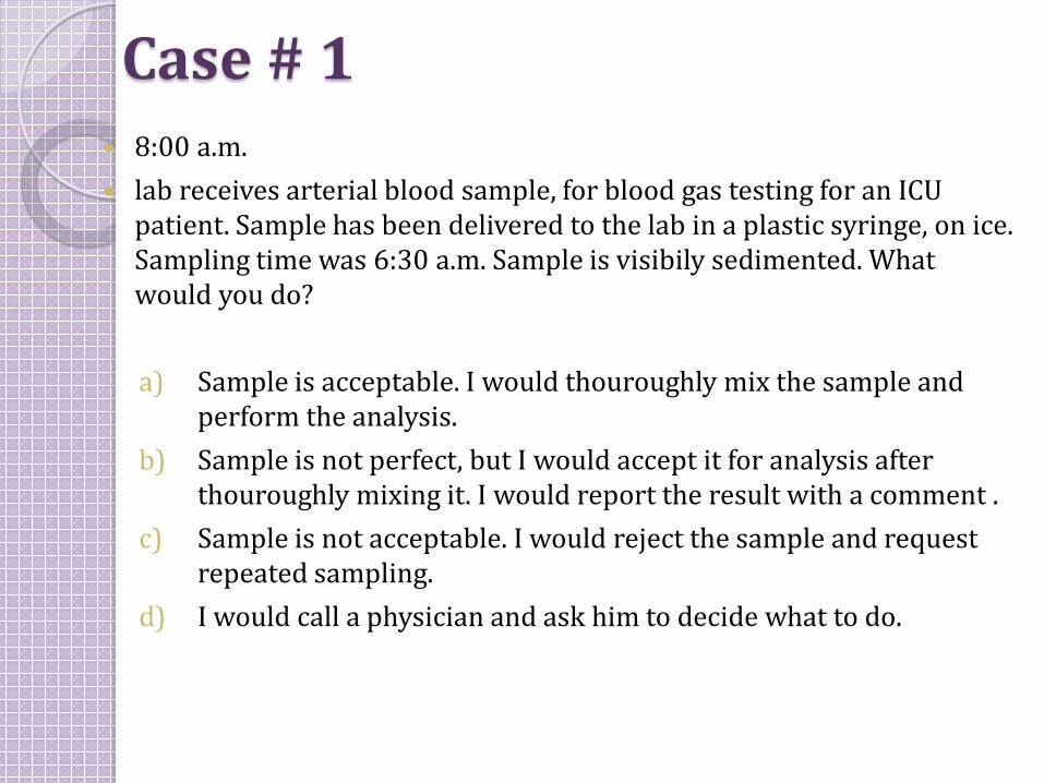
Case # 1 8:00 a.m. lab receives arterial blood sample, for blood gas testing for an ICU
patient. Sample has been delivered to the lab in a plastic syringe, on ice. Sampling time was 6:30 a.m. Sample is visibily sedimented. What would you do?
a) Sample is acceptable. I would thouroughly mix the sample and
perform the analysis. b) Sample is not perfect, but I would accept it for analysis after
thouroughly mixing it. I would report the result with a comment . c) Sample is not acceptable. I would reject the sample and request
repeated sampling. d) I would call a physician and ask him to decide what to do.
Healthcare system
Healthcare is a system that frequently harms and routinely fails to deliver the appropriate standard of care.
Davis K, et al. (2002). Room for improvement: Patients report on the quality of their health care. New York: The Commonwealth Fund.
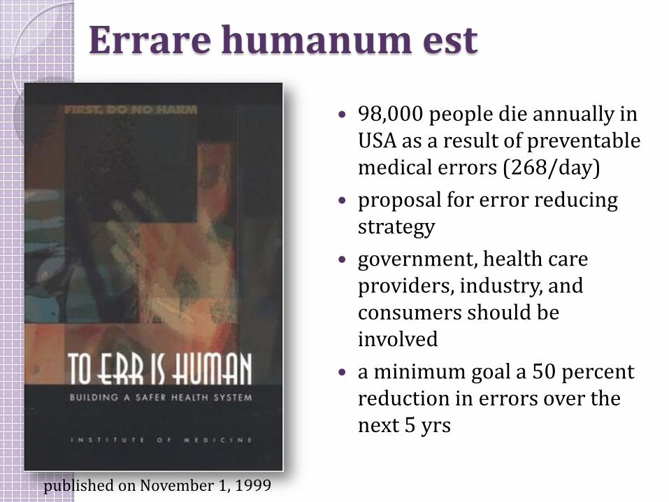
98,000 people die annually in USA as a result of preventable medical errors (268/day)
proposal for error reducing strategy
government, health care providers, industry, and consumers should be involved
a minimum goal a 50 percent reduction in errors over the next 5 yrs
published on November 1, 1999
Errare humanum est
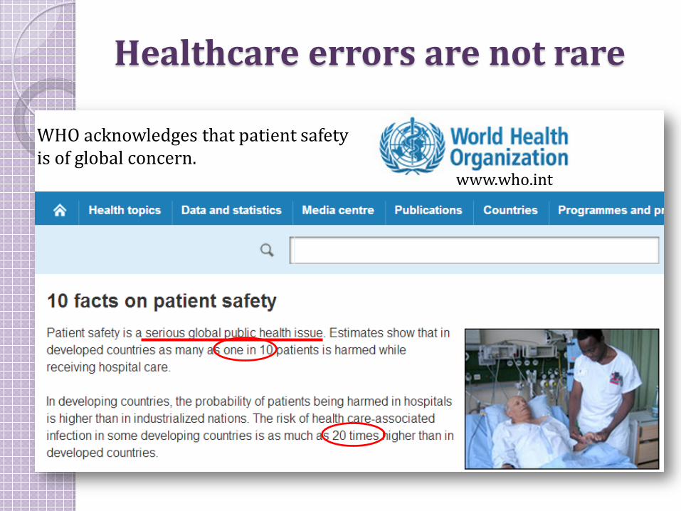
Healthcare errors are not rare
WHO acknowledges that patient safety is of global concern.
www.who.int
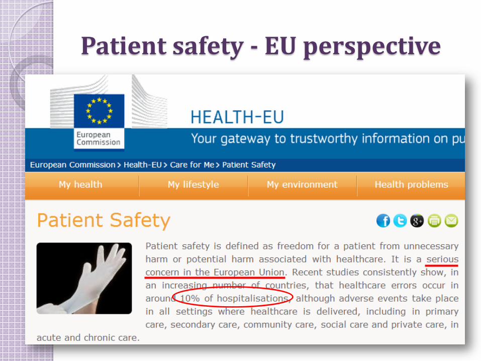
Patient safety - EU perspective

Key factors contributing to this problem:
the failure of health care providers to: ◦ define the safe practice standards ◦ consistently enforce compliance
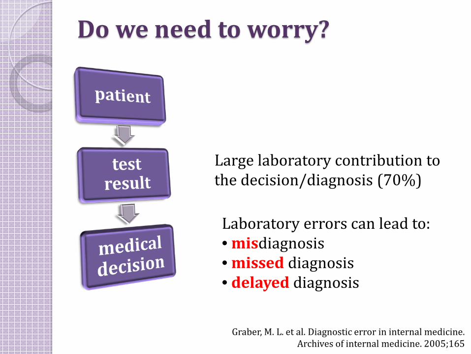
Do we need to worry?
Large laboratory contribution to the decision/diagnosis (70%)
Graber, M. L. et al. Diagnostic error in internal medicine. Archives of internal medicine. 2005;165
Laboratory errors can lead to: • misdiagnosis • missed diagnosis • delayed diagnosis

Date of download: 5/30/2013 Copyright © 2012 American Medical Association. All rights reserved.
From: Diagnostic Error in Medicine: Analysis of 583 Physician-Reported Errors
Arch Intern Med. 2009;169(20):1881-1887. doi:10.1001/archinternmed.2009.333
Classification of diagnostic errors in 583 physician-reported cases using the Diagnostic Error Evaluation and Research project tool to localize where in the diagnostic process error occurred.
Figure Legend:
The most common were radiology and
laboratory errors
44%

Preanalytical phase
68% 13%
19%
preanalytical phase analytical phase postanalytical phase
Plebani M, Carraro P. Mistakes in a stat laboratory: types and frequency. Clin Chem. 1997;43(8Pt 1):1348-51.

Case #2 Lab receives arterial blood sample from emergency department. Blood
gas testing is requested. Sample was transported by pneumatic tube within 10 minutes from sampling. You notice an air bubble in the syringe.
What would you do?
a) Sample is acceptable. I would expel the bubble and perform the analysis.
b) Sample is not perfect, I would expel the bubble and perform the analysis. I would report the result with a comment.
c) Sample is not acceptable. I would reject the sample and request repeated sampling.
d) I would call a physician and ask him to decide what to do.

Why is preanalytical phase so vulnerable?
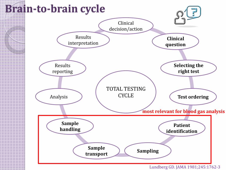
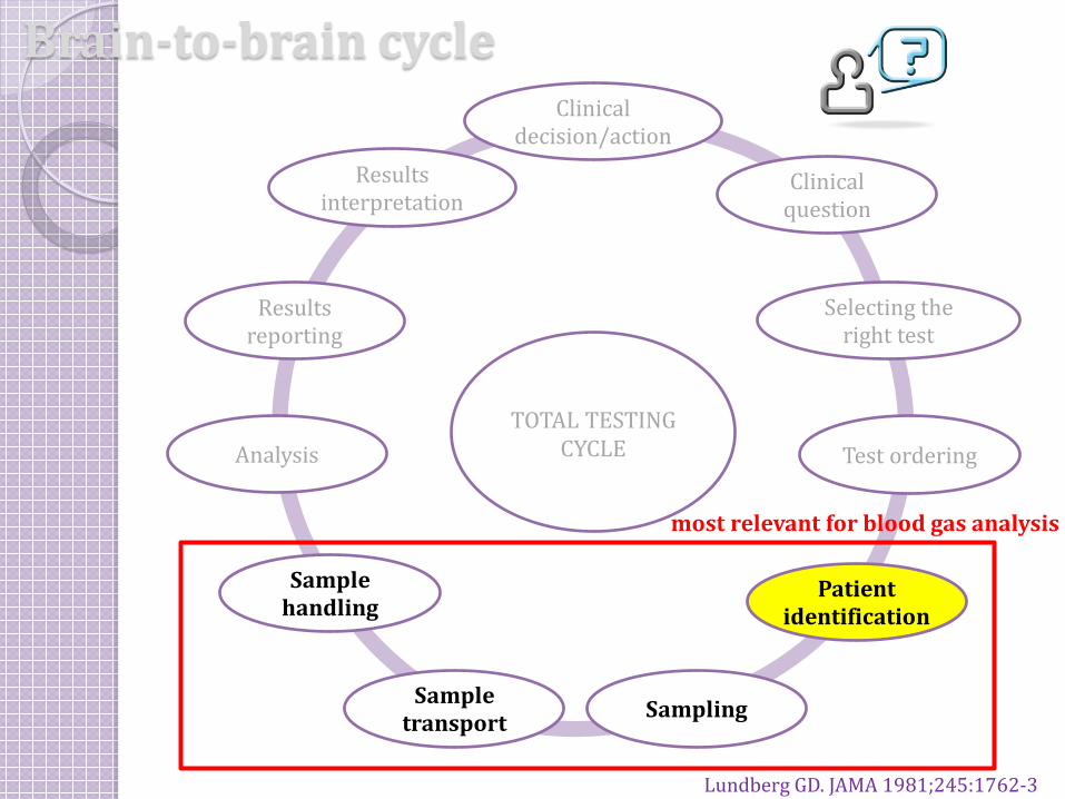
Brain-to-brain cycle
TOTAL TESTING CYCLE
Clinical decision/action
Clinical question
Selecting the right test
Test ordering
Patient identification
Sampling Sample transport
Sample handling
Analysis
Results reporting
Results interpretation
Lundberg GD. JAMA 1981;245:1762-3
most relevant for blood gas analysis
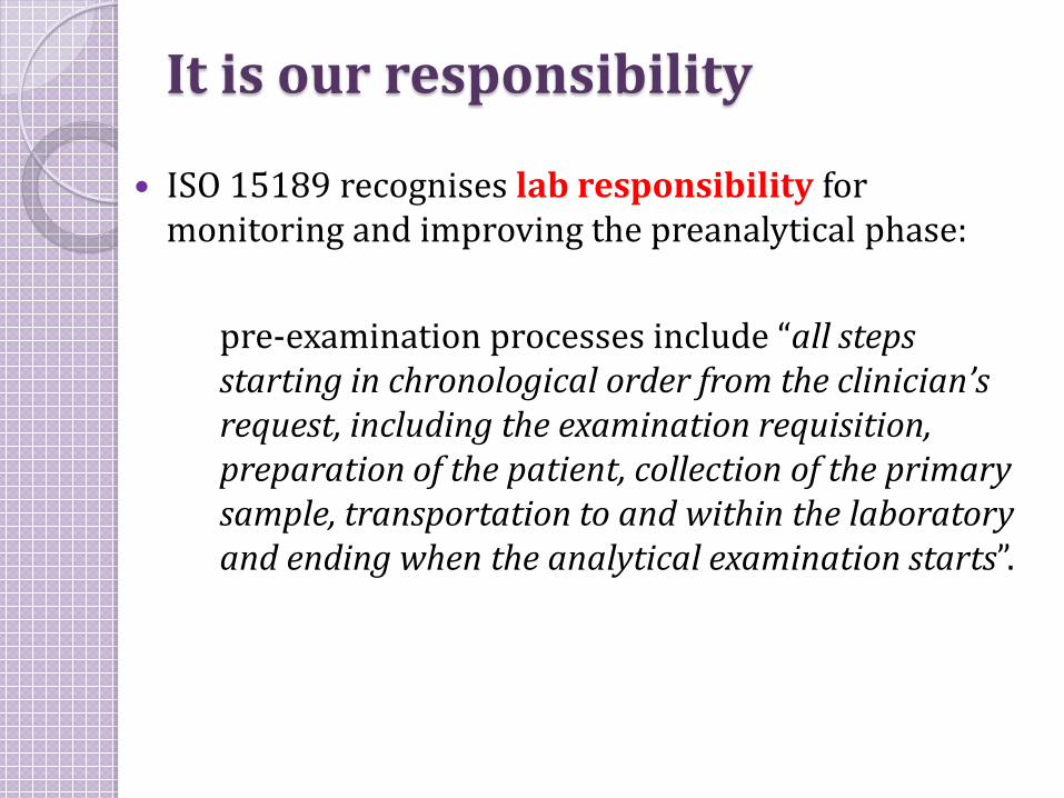
It is our responsibility
ISO 15189 recognises lab responsibility for monitoring and improving the preanalytical phase:
pre-examination processes include “all steps starting in chronological order from the clinician’s request, including the examination requisition, preparation of the patient, collection of the primary sample, transportation to and within the laboratory and ending when the analytical examination starts”.
define safe practice standards consistently enforce compliance
How?
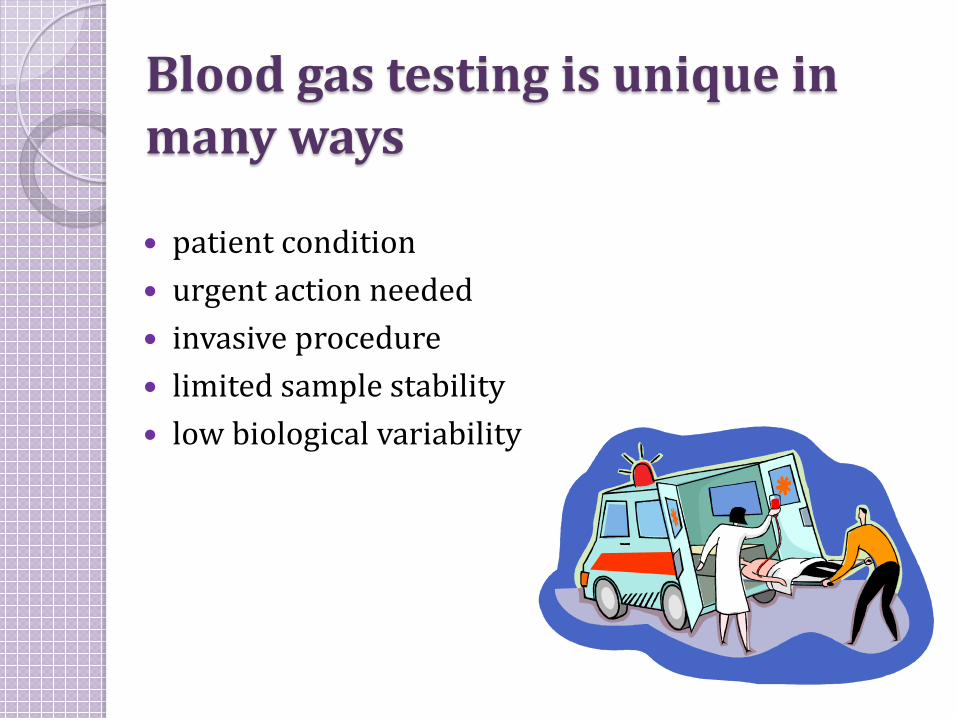
Blood gas testing is unique in many ways
patient condition urgent action needed invasive procedure limited sample stability low biological variability

Low biological variability
Lippi G, et al. Influence of spurious hemolysis on blood gas analysis. CCLM 2013;51:1651-4.

Case #3
parameter value unit Ref range Repeated sample - OK
pO2 14.6 ↑ kPa 11 - 14.4 13.1
pCO2 3.70 ↓ kPa 4.7 - 6.4 4.8
K 3.2 ↓ mmol/L 3.5 – 5.0 4.3
Na 148 ↑ mmol/L 136 – 146 139
Cl 111 ↑ mmol/L 98 – 106 102
Glu 2.8 ↓ mmol/L 3.9 – 5.8 5.6
a) Sample hemolyzed. b) Sample dilution. c) Air bubble. d) Clotted sample.
1st sample
Brain-to-brain cycle
TOTAL TESTING CYCLE
Clinical decision/action
Clinical question
Selecting the right test
Test ordering
Patient identification
Sampling Sample transport
Sample handling
Analysis
Results reporting
Results interpretation
Lundberg GD. JAMA 1981;245:1762-3
most relevant for blood gas analysis

Patient identification errors ID error frequency: ◦ 0.1-1% in laboratory medicine ◦ 0.05% in transfusion medicine
underreported (most go undetected) major healthcare issue potentially associated with serious adverse
consequences zero tolerance!
Lippi G, et al. Preanalytical quality improvement: from dream to reality. CCLM 2011;49(7):1113–1126
Any potentially mislabeled or misidentified specimen should be rejected.
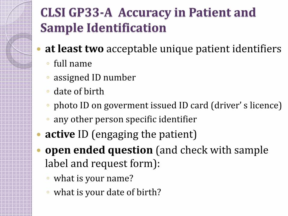
CLSI GP33-A Accuracy in Patient and Sample Identification at least two acceptable unique patient identifiers ◦ full name ◦ assigned ID number ◦ date of birth ◦ photo ID on goverment issued ID card (driver’ s licence) ◦ any other person specific identifier
active ID (engaging the patient) open ended question (and check with sample
label and request form): ◦ what is your name? ◦ what is your date of birth?

CLSI GP33-A Accuracy in Patient and Sample Identification
if any discrepancies are identified, do not collect samples until issues are resolved
if patient is not able to identify himself, ask a nurse, a friend or a relative to do that and record their names.
to minimize the error risk: ◦ use ID bracelets with barcodes or radiofrequency
identifier devices (RFID) are recommended ◦ use barcoded sample identifiers ◦ generate labels at the time and site of collection ◦ label the sample in the presence of the patient

Brain-to-brain cycle
TOTAL TESTING CYCLE
Clinical decision/action
Clinical question
Selecting the right test
Test ordering
Patient identification
Sampling Sample transport
Sample handling
Analysis
Results reporting
Results interpretation
Lundberg GD. JAMA 1981;245:1762-3
most relevant for blood gas analysis
Sampling patient condition sample type sampling site anticoagulant
Patient condition
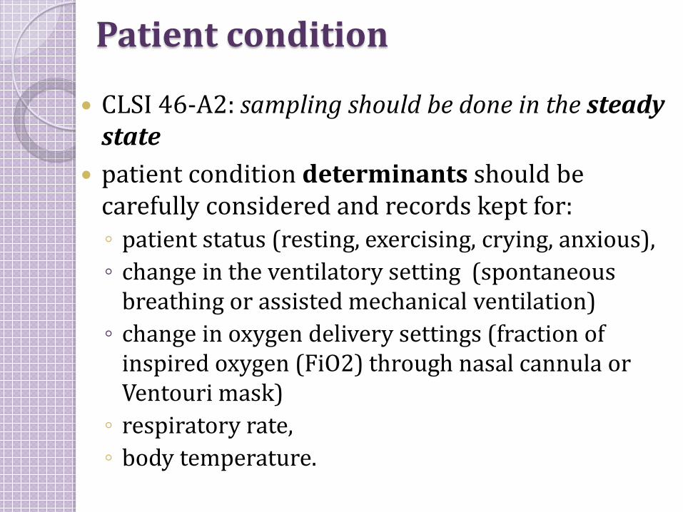
CLSI 46-A2: sampling should be done in the steady state
patient condition determinants should be carefully considered and records kept for: ◦ patient status (resting, exercising, crying, anxious), ◦ change in the ventilatory setting (spontaneous
breathing or assisted mechanical ventilation) ◦ change in oxygen delivery settings (fraction of
inspired oxygen (FiO2) through nasal cannula or Ventouri mask) ◦ respiratory rate, ◦ body temperature.
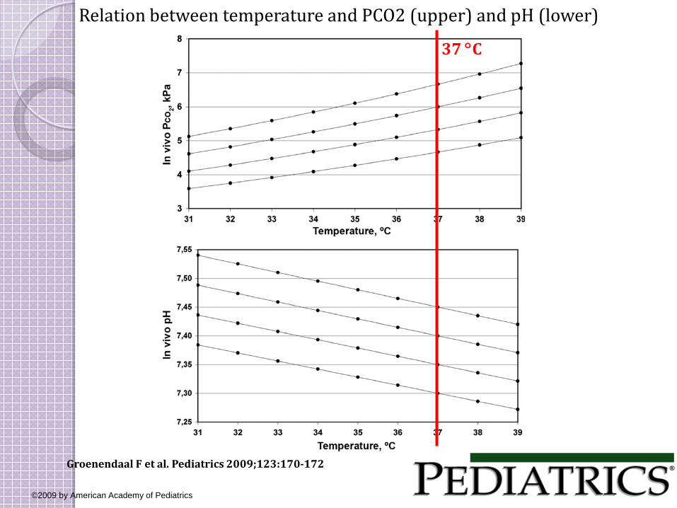
Relation between temperature and PCO2 (upper) and pH (lower)
Groenendaal F et al. Pediatrics 2009;123:170-172
©2009 by American Academy of Pediatrics
37 °C
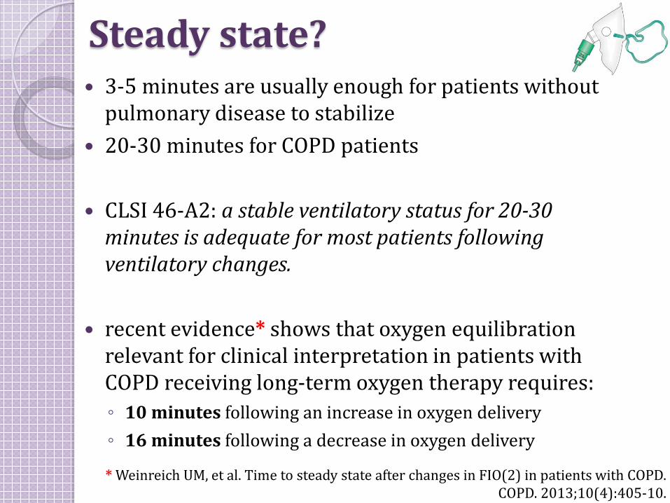
Steady state? 3-5 minutes are usually enough for patients without
pulmonary disease to stabilize 20-30 minutes for COPD patients CLSI 46-A2: a stable ventilatory status for 20-30
minutes is adequate for most patients following ventilatory changes.
recent evidence* shows that oxygen equilibration relevant for clinical interpretation in patients with COPD receiving long-term oxygen therapy requires: ◦ 10 minutes following an increase in oxygen delivery ◦ 16 minutes following a decrease in oxygen delivery
* Weinreich UM, et al. Time to steady state after changes in FIO(2) in patients with COPD. COPD. 2013;10(4):405-10.

Time and site of sampling Exact time of the blood collection and the site of
sampling should always be recorded and reported on the test report.
difficulties during blood collection
Ashok Deorari. Blood gas analysis. All India Institute of Medical Sciences. 2008

Difficulties with blood sampling Male patient, 82 years, chest pain, addmited to ED 1. sample – capillary - difficulties during blood collection 2. sample – arterial blood, after 10 minutes

Sample type
CLSI 46-A2 guideline state: “Blood gas measurement for the purpose of evaluating
the gass exchange function of the lungs (pO2 and pCO2) should be performed on arterial blood only. ...
The blood should be collected under anaerobic conditions, mixed immediately to dissolve heparin anticoagulant and promptly analysed.”

Alternative? CLSI 46-A2 guideline state: “if arterial blood can not be collected directly, peripheral
capillary blood may be collected using an arterialization technique (warming the skin to 40-45°C with a warm towel or vasodilating cream).
...blood gas results may differ, especially those for pO2, sO2, FO2Hb and ctO2.”
earlobe is better than a fingertip there is really no substitute for arterial blood if
accuracy of pO2 measurement is important (oxygen therapy)
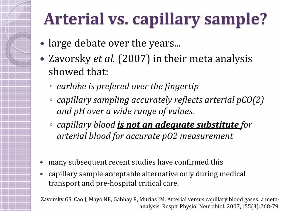
Arterial vs. capillary sample? large debate over the years... Zavorsky et al. (2007) in their meta analysis
showed that: ◦ earlobe is prefered over the fingertip ◦ capillary sampling accurately reflects arterial pCO(2)
and pH over a wide range of values. ◦ capillary blood is not an adequate substitute for
arterial blood for accurate pO2 measurement
many subsequent recent studies have confirmed this capillary sample acceptable alternative only during medical
transport and pre-hospital critical care.
Zavorsky GS, Cao J, Mayo NE, Gabbay R, Murias JM. Arterial versus capillary blood gases: a meta-analysis. Respir Physiol Neurobiol. 2007;155(3):268-79.

Arterial vs. capillary sample?
Higgins C. Capillary-blood gases: To arterialize or not. MLO. November 2008:42-47

Arterial sample vs. arterialized earlobe?
Vaquer S, et al. Earlobe arterialized capillary blood gas analysis in the intensive care unit: a pilot study. Ann Intensive Care. 2014;4:11.
Results: Poor PO2 concordance (CCC = 0.45; CI 95% = 0.26 to 0.6) of arterialized earlobe with arterial blood. Mean PO2 difference was12 mmHg (P< 0.001) (Figure 1). The higher the arterial PO2, the greater the difference (slope = 0.54).
vasodilation cream (2% nitro-glycerin cream)
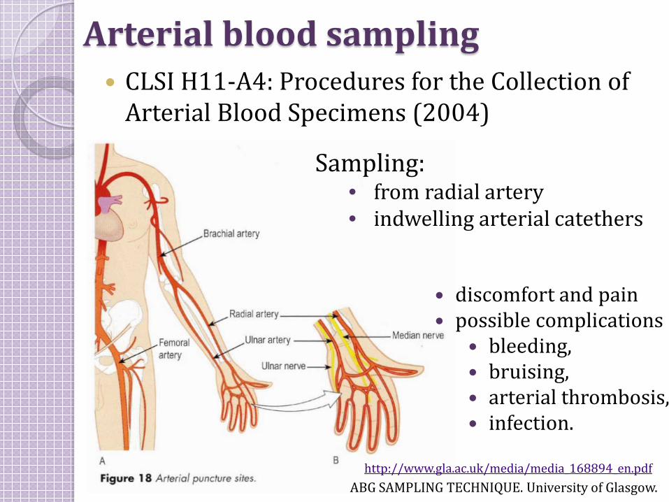
Arterial blood sampling CLSI H11-A4: Procedures for the Collection of
Arterial Blood Specimens (2004)
ABG SAMPLING TECHNIQUE. University of Glasgow. http://www.gla.ac.uk/media/media_168894_en.pdf
discomfort and pain possible complications
bleeding, bruising, arterial thrombosis, infection.
Sampling: • from radial artery • indwelling arterial catethers

Sample contamination with flush solution
during sampling from arterial catheters, there is a risk of diluting the sample with flush solution.
discard at least 3 times the dead space when sampling from catheter check catheter package for the exact volume of dead space
To avoid errors:
↑ pO2 ↓ pCO2 ↓ K+ ↑ Na+ ↑ Cl– ↓ Ca2+
↓ cGlu ↓ cLac ↓ ctHb

Case #3 results
parameter value unit Ref range Repeated sample - OK
pO2 14.6 ↑ kPa 11 - 14.4 13.1
pCO2 3.70 ↓ kPa 4.7 - 6.4 4.8
K 3.2 ↓ mmol/L 3.5 – 5.0 4.3
Na 148 ↑ mmol/L 136 – 146 139
Cl 111 ↑ mmol/L 98 – 106 102
Glu 2.8 ↓ mmol/L 3.9 – 5.8 5.6
a) Sample hemolyzed. b) Sample dilution. c) Air bubble. d) Clotted sample.
1st sample

Sample contamination with venous blood during arterial blood sampling, there is a risk of
accidentaly puncturing the vein and contaminating the sample with venous blood.
use self-filling syringes – they fill readily when puncturing an artery but not when hitting a vein
use short-bevelled needles – easier to position inside the artery make the puncture at an angle of 45°
To avoid errors: ↓ pO2 ↓ sO2 ↑ pCO2
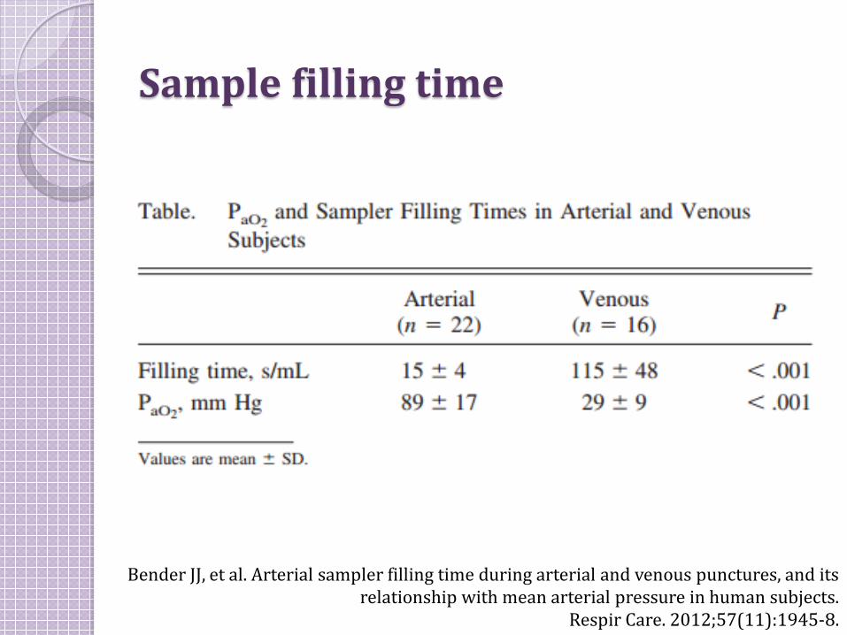
Sample filling time
Bender JJ, et al. Arterial sampler filling time during arterial and venous punctures, and its relationship with mean arterial pressure in human subjects.
Respir Care. 2012;57(11):1945-8.
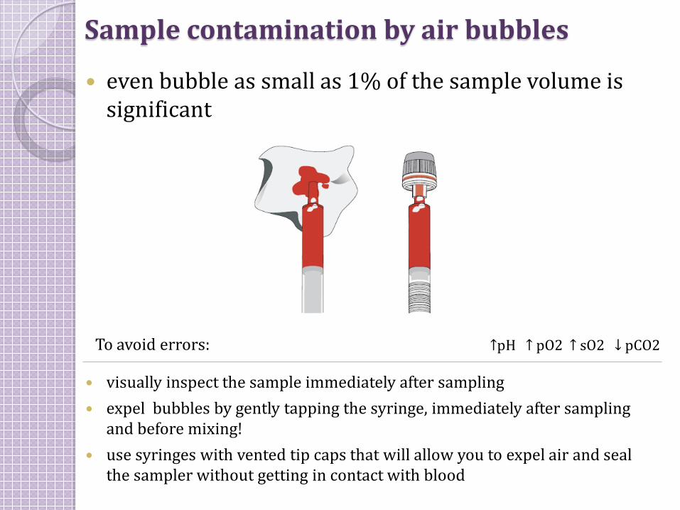
Sample contamination by air bubbles
even bubble as small as 1% of the sample volume is significant
visually inspect the sample immediately after sampling expel bubbles by gently tapping the syringe, immediately after sampling
and before mixing! use syringes with vented tip caps that will allow you to expel air and seal
the sampler without getting in contact with blood
To avoid errors: ↑pH ↑ pO2 ↑ sO2 ↓ pCO2

Case #2 - results Lab receives arterial blood sample from emergency department. Blood
gas testing is requested. Sample was transported by pneumatic tube within 10 minutes from sampling. You notice an air bubble in the syringe.
What would you do?
a) Sample is acceptable. I would expel the bubble and perform the analysis.
b) Sample is not perfect, I would expel the bubble and perform the analysis. I would report the result with a comment.
c) Sample is not acceptable. I would reject the sample and request repeated sampling.
d) I would call a physician and ask him to decide what to do.
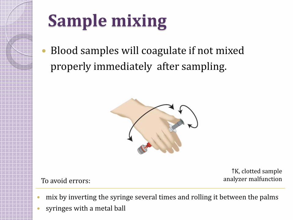
Sample mixing Blood samples will coagulate if not mixed
properly immediately after sampling.
mix by inverting the syringe several times and rolling it between the palms syringes with a metal ball
To avoid errors: ↑K, clotted sample
analyzer malfunction
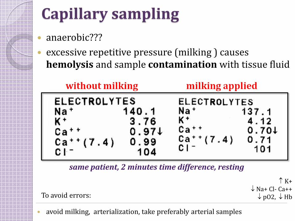
Capillary sampling
↑ K+ ↓ Na+ Cl- Ca++ ↓ pO2, ↓ Hb
anaerobic??? excessive repetitive pressure (milking ) causes
hemolysis and sample contamination with tissue fluid
same patient, 2 minutes time difference, resting
avoid milking, arterialization, take preferably arterial samples
To avoid errors:
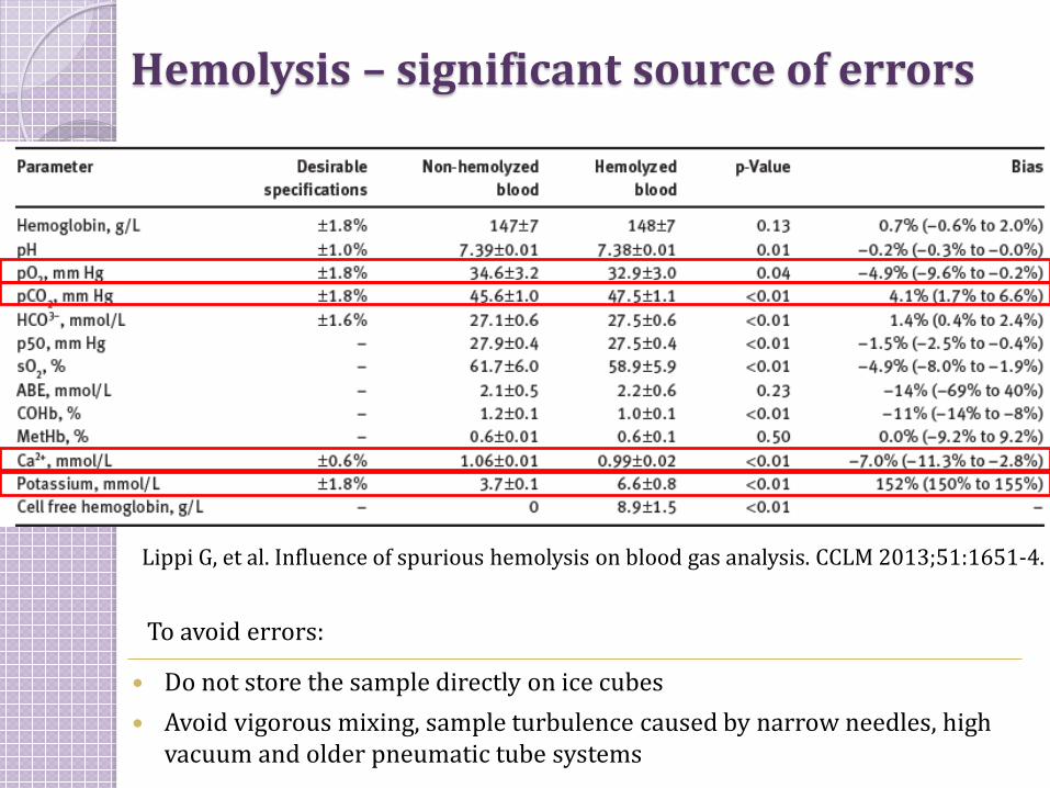
Hemolysis – significant source of errors
Lippi G, et al. Influence of spurious hemolysis on blood gas analysis. CCLM 2013;51:1651-4.
Do not store the sample directly on ice cubes Avoid vigorous mixing, sample turbulence caused by narrow needles, high
vacuum and older pneumatic tube systems
To avoid errors:

Anticoagulant lyofilized balanced Li-heparin is recommended caution! ◦ dilution of electrolytes, HCO3-, pCO2 by liquid heparin ◦ liquid heparin has atmospheric pO2 (150 mmHg/20 kPa)
and affects pO2 results ◦ Na-heparin falsely elevates sodium ◦ heparin binds cations (Ca++, Na+, K+) ◦ CLSI 46-A2 states that final sample heparin concentration
should be 20 IU/mL blood (flushing with therapeutic heparin is not recommended – it contains high heparin concentration and my alter sample pH and electrolytes) ◦ mix as soon as possible to ensure proper anticoagulation
and avoid clot formation
Baird G. Preanalytical considerations in blood gas analysis. Biochemia Medica 2013;23(1):19-27.

Dilution by liquid sodium heparin – 1/4
Tuncay Küme, Ali Rıza Şişman, Ahmet Solak, Birsen Tuğlu, Burcu Çinkooğlu, Canan Çoker. The effects of different syringe volume, needle size and sample volume on blood gas analysis in
syringes washed with heparin. Biochemia Medica 2012;22(2):189-201.
pH pCO2
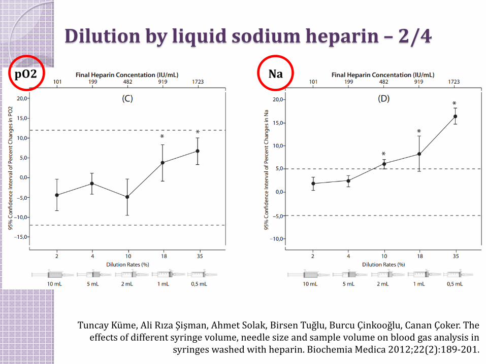
Dilution by liquid sodium heparin – 2/4
Tuncay Küme, Ali Rıza Şişman, Ahmet Solak, Birsen Tuğlu, Burcu Çinkooğlu, Canan Çoker. The effects of different syringe volume, needle size and sample volume on blood gas analysis in
syringes washed with heparin. Biochemia Medica 2012;22(2):189-201.
pO2 Na
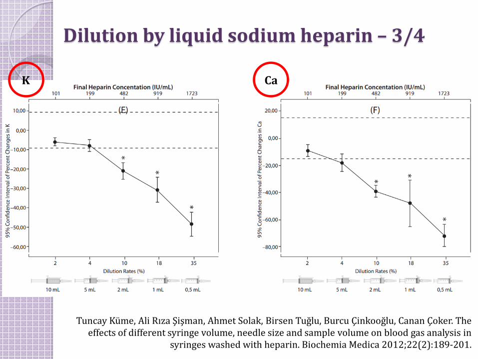
Dilution by liquid sodium heparin – 3/4
Tuncay Küme, Ali Rıza Şişman, Ahmet Solak, Birsen Tuğlu, Burcu Çinkooğlu, Canan Çoker. The effects of different syringe volume, needle size and sample volume on blood gas analysis in
syringes washed with heparin. Biochemia Medica 2012;22(2):189-201.
K Ca

Dilution by liquid sodium heparin – 4/4
Tuncay Küme, Ali Rıza Şişman, Ahmet Solak, Birsen Tuğlu, Burcu Çinkooğlu, Canan Çoker. The effects of different syringe volume, needle size and sample volume on blood gas analysis in
syringes washed with heparin. Biochemia Medica 2012;22(2):189-201.
Mg

Brain-to-brain cycle
TOTAL TESTING CYCLE
Clinical decision/action
Clinical question
Selecting the right test
Test ordering
Patient identification
Sampling Sample transport
Sample handling
Analysis
Results reporting
Results interpretation
Lundberg GD. JAMA 1981;245:1762-3
most relevant for blood gas analysis

Time is the key to sample quality
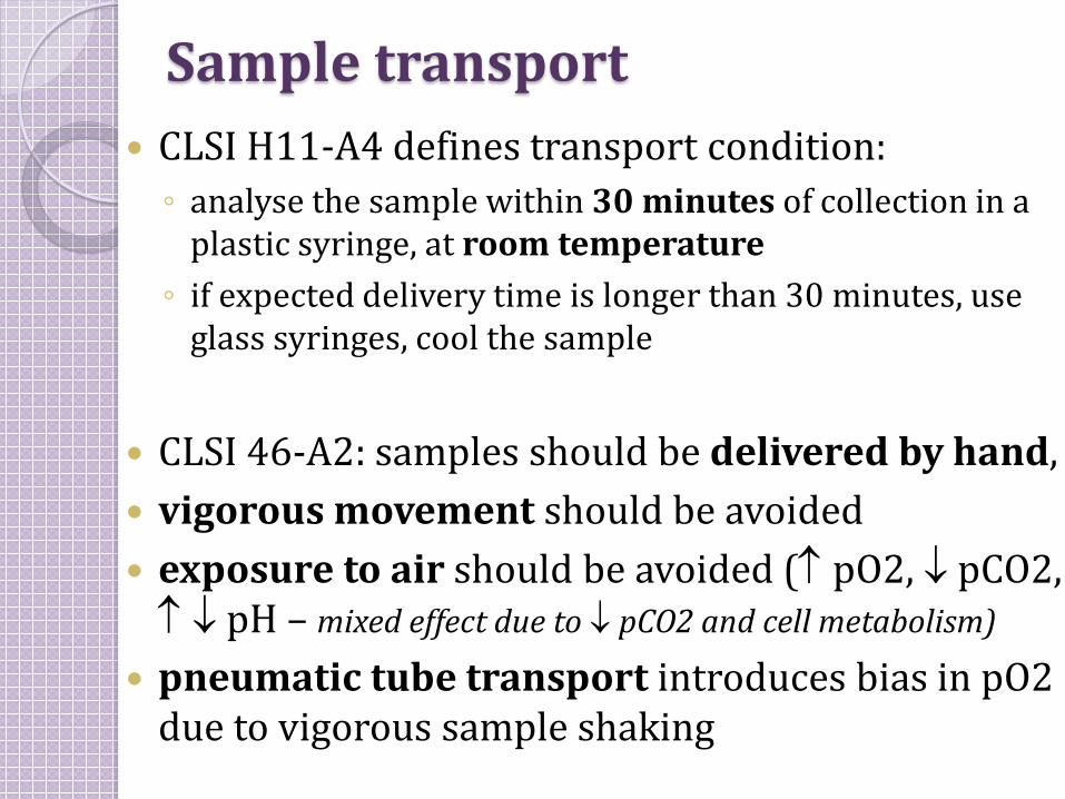
Sample transport CLSI H11-A4 defines transport condition: ◦ analyse the sample within 30 minutes of collection in a
plastic syringe, at room temperature ◦ if expected delivery time is longer than 30 minutes, use
glass syringes, cool the sample
CLSI 46-A2: samples should be delivered by hand, vigorous movement should be avoided exposure to air should be avoided (↑ pO2, ↓ pCO2, ↑ ↓ pH – mixed effect due to ↓ pCO2 and cell metabolism)
pneumatic tube transport introduces bias in pO2 due to vigorous sample shaking

Case # 1 - results 8:00 a.m. lab receives arterial blood sample, for blood gas testing for an ICU
patient. Sample has been delivered to the lab in a plastic syringe, on ice. Sampling time was 6:30 a.m. Sample is visibily sedimented. What would you do?
a) Sample is acceptable. I would thouroughly mix the sample and
perform the analysis. b) Sample is not perfect, but I would accept it for analysis after
thouroughly mixing it. I would report the result with a comment . c) Sample is not acceptable. I would reject the sample and request
repeated sampling. d) I would call a physician and ask him to decide what to do.

Brain-to-brain cycle
TOTAL TESTING CYCLE
Clinical decision/action
Clinical question
Selecting the right test
Test ordering
Patient identification
Sampling Sample transport
Sample handling
Analysis
Results reporting
Results interpretation
Lundberg GD. JAMA 1981;245:1762-3
most relevant for blood gas analysis

Sample handling Proper sample mixing prior to analysis to obtain
a homogeneous sample
mix by inverting the syringe several times and rolling it between the palms have a written policy and procedure for mixing mix gentle to avoid hemolysis!
To avoid errors:

Safety issues
Needle-stick injury and unwanted contact with patient blood are everyday daily risks
in 2000 occupational HCWs exposure has led to: ◦ 16,000 HCV, ◦ 66,000 HBV, ◦ 1,000 HIV infections *.
To avoid risks: ◦ Use a safety devices (contact with patient blood is limited) ◦ Use a protection device for the safe removal of needles ◦ Lab has a procedure for operator safety and lab staff is
compliant with the procedure
* Prüss-Üstün, A., et al. Estimation of the global burden of disease attributable to contaminated sharps injuries among health-care workers. Am J Ind Med, 2005;48: 482–90.

measure must be taken to specify and implement safe procedures for using and disposing of sharp medical instruments and contaminated waste.
... providing medical devices incorporating safety-engineered protection mechanisms.

Tips for safer blood gas testing: patient properly identified patient is in a steady state proper sampling site self-filling plastic syringes with short-beveled needles,
vented caps and balanced dry heparine 45° aspiration visually inspect the sample, expel any bubbles, mix the sample, deliver on room temperature, analyse within 30 minutes if visibly sedimented, mix >5’
Quality management
standardize procedures provide written instructions enforce compliance educate yourself and educate others monitor the quality continuous improvement No result is always better than the
wrong result!
lab responsibility!

Rovinj, Croatia

![Preanalytical phase and patient outcome - unimi.it · Preanalytical phase and patient outcome Ana-Maria Šimundić ... Ppt0000001 [Sola lettura] Author: tesi Created Date: 11/30/2018](https://static.fdocuments.us/doc/165x107/5f04f79b7e708231d410989e/preanalytical-phase-and-patient-outcome-unimiit-preanalytical-phase-and-patient.jpg)


