Pre University Examination (2017-2018) B.Sc.-II (CBZ ... filePre University Examination (2017-2018)...
18
Pre University Examination (2017-2018) B.Sc.-II (CBZ) Paper III- Pteridophytes, Gymnosperms and Paleobotany Set A Time: 3:00 Hours Max. Marks: 33 1. Answer the following (not more than 20 marks) ½ mark each (a) Write name of two pteridophytes. Answer: Lycopodium, Selaginella, Marsilea and Equisetum. (b) How many cotyledons are present in the seed of Ephedra? Answer: 2 (c) Whatcause sulphur like yellow dust in the Himalayas in pine forests in the month of April? Answer: As the pollen cones mature, they discharge large quantities of pollen grains into the air and it appears as a yellow cloud of yellow of winged pollen grains in Pinus. This is called Sulphur Shower and occurs during spring. Pollen grains are released and carried by wind to the female cones. (d) Define Protostele. Answer: The most primitive form of stele, consisting of a solid core of xylem encased by phloem or of xylem interspersed with phloem. The roots of all vascular plants, as well as the stems of lycophytes, haveprotosteles. (e) Negatively geotropic roots are found in which genera? Answer: Cycads. (f) Which pteridophyte is used as gold indicator? Answer: Equisetum accumulates. (g) Draw a diagram of mixed protostele.
Transcript of Pre University Examination (2017-2018) B.Sc.-II (CBZ ... filePre University Examination (2017-2018)...
Pre University Examination (2017-2018)
B.Sc.-II (CBZ)
Paper III- Pteridophytes, Gymnosperms and Paleobotany
Set A
Time: 3:00 Hours Max. Marks: 33
1. Answer the following (not more than 20 marks) ½ mark each
(a) Write name of two pteridophytes.
Answer: Lycopodium, Selaginella, Marsilea and Equisetum.
(b) How many cotyledons are present in the seed of Ephedra?
Answer: 2
(c) Whatcause sulphur like yellow dust in the Himalayas in pine forests in the month of
April?
Answer: As the pollen cones mature, they discharge large quantities of pollen grains into the
air and it appears as a yellow cloud of yellow of winged pollen grains in Pinus. This is called
Sulphur Shower and occurs during spring. Pollen grains are released and carried by wind to
the female cones.
(d) Define Protostele.
Answer: The most primitive form of stele, consisting of a solid core of xylem encased by
phloem or of xylem interspersed with phloem. The roots of all vascular plants, as well as the
stems of lycophytes, haveprotosteles.
(e) Negatively geotropic roots are found in which genera?
Answer: Cycads.
(f) Which pteridophyte is used as gold indicator?
Answer: Equisetum accumulates.
(g) Draw a diagram of mixed protostele.

(h) Define apospory.
Answer: Apospory is the development of 2n gametophytes, without meiosis and spores, from
vegetative, or non-reproductive, cells of the sporophyte.
(i) Which component of xylem is absent in Ephedra.
Answer: vessels.
(j) Which pteridophyte show secondary growth?
Answer: Psilotum.
(k) Name the drug obtained by Ephedra.
Answer: Ephedrine.
(l) What is Eusporangiate development of sporangia?
Answer: Sporangia develop from more than one initial cell. Each sporangium contains an
indefinite large number of spores.
(m) Write the name of gymnospermic plant whose female cone never develops.
Answer: Cycas.
(n) Spike moss is the common name of which pteridophyte?
Answer: Selaginella.
(o) Who discovered fossil genus Williamsonia?
Answer: William Crawford Williamson.
(p) Chilgoza, a dry fruit, is actually seed of which plant.
Answer: Pinus.
(q) A drug for the treatment of asthma is obtained from which plant?
Answer: Ephedra.
(r) What is petrification?
Answer: Petrification is the process of turning living organic material into stone. This
process takes place when the molecules in an organism are replaced with the molecules of a
mineral.

Unit I
2. Write short notes on following-
(i) Apogamy and Apospory. 1½ + 1½
Answer: Apogamy: Development of sporophyte from the gametophytic tissue without
involving fusion of gametes is known as apogamy. Such sporophytes grow vegetatively by
budding from the gametophyte, hence they have the same level of ploidy as gametophytes.
Apogamy first discovered by Farlow (1874) in Pteris cretica, and this term was introduced
by de Bary in 1878. There are more than 50 sp of ferns in which apogamous sporophytes are
known to occur naturally due to te absence of or nonfunctional nature of one or both sex
organs, such apogamy is known as obligate apogamy.
When functional sex organs can be induced apogamous sporophyte, it called facultative
apogamy.
Species of Dryopteris, Adiantum, Diplazium, Pteris, Polystichum, Osmunda and Asplenium
are some examples of obligate apogamy. Lang (1896) was the first to induce apogamy
successfully in many varieties of ferns. According to him 3 factors play an important role in
the induction of apogamy: (1) ability of prothallial cell to become meristematic, (2)
prevention of fertilization and (3) exposure of prothallus to direct illumination.
Factors:
1. Nutritional factor: maximum no. of apogamous sporophyte are obtained if the
gametophytes are placed in 4 % sucrose solution.
2.Light quality: far red light (705nm) has been found to be the most effective in inducing
apogamous sporophyte followed by blue (445nm) and wight light.
3. Hormones: presence of hormones like 2,4-D, GA3, IAA, and tryptophan stimulate
apogamous sporophyte at very low concentration (0.5 %) of carbohydrates.
Apospory: development of gametophyte directly from vegetative cell of the sporophyte
without the formation of spores is known as apospory. Therefore, aposporous gametophytes
have the same no. of chromosomes as sporophytes. Apospory first demonstrated by Druery
(1884) in Athyrium filix-femina. Aposporous gametophytes have also been observed on the
young leaves of Osmunda regalis, and O. javanica.
Factors:
1. Nutrition: complete absence of carbohydrate in growth medium induced development of
aposporous gametophyte, 1 % carbohydrate induced callus like structure formed structure
which was partly gametophytes and partly sporophytes (gametosporophytes), while 2 %
carbohydrate induced only callus formed only sporophyte.

2. Light intensity: low light intensity favors development of aposporous gametophyte.
3. Age of sporophytic cells: young cells of shoot apex or leaf tip form gametophytes,
whereas basal parts of shoot or leaf regenerate sporophytic tissue.
(ii) Economic importance of Pteridophytes.
Answer: pteridophytes have many economic importance like as follows:
(1) Food:Like other plants, pteridophytes constitute a good source of food to animals.
Sporocarps of Marsilea, a water fern, yield starch that is cooked and eaten by certain tribal.
(2) Soil Binding:By their growth pteridophytes bind the soil even along hill slopes. The soil
is protected from erosion.
(3) Scouring:Equisetum stems have been used in scouring (cleaning of utensils) and
polishing of metals. Equisetum species are, therefore, also called scouring rushes.
(4) Nitrogen Fixation:Azolla (a water fern) has a symbiotic association with nitrogen fixing
cyanobacterium Anabaena azollae. It is inoculated to paddy fields to function as biofertilizer.
(5) Medicines:An anthelmintic drug is obtained from rhizomes of Dryopteris (Male Shield
Fern).
(6) Ornamentals:Ferns are grown as ornamental plants for their delicate and graceful leaves.
(7) Indusrial use- Club mosses are used as a dry industrial lubricant since it microscopic
spore contain non-volatile oil.
(8) Forensic Investigation- Spores of mosses are use as finger print powder in forensic
investigation. Also use as photography flash powder.
OR
Give a detailed account of classification of pteridophytes given by Smith. 6
Answer: According to Smith (1955) the classification of the pteridophytes is as follows:

The characteristic features of these divisions are as follows:
Psilophyta:
The sporophyte is differentiated into a rhizoid bearing subterranean rhizome and an aerial
portion. The aerial portion is branched. The vascular system is of protostelic type. Leaf gaps
are absent from vascular cylinder. The terminal sporangia are borne singly at the tips of short
or long branches. The gametophyte is subterranean and colourless. They are homosporous.
Antherozoids are multiciliate.
Lycophyta:
The sporophyte is differentiated into stem, roots, and leaves. The leaves are microphyllous,
and with a single vein. The vascular strands or steles may be protostelic, siphonostelic, or
polystelic. The leaf-gaps are always absent; sporophylls produce a single sporangium on the
adaxial side near its base. The sporophylls are borne in strobili. They are homosporous or
heterosporous. The antherozoids are biflagellate or multiciliate.
Arthrophyta:
The sporophyte is differentiated into stem, roots and leaves. The stem possesses distinct
ridges and furrows. The foliage leaves are borne in transverse whorls upon stems and their
branches. The vascular cylinder is protostelic or siphonostelic. The leaf-gaps are absent. The
sporangia are produced upon a specialized structure, the sporangiophores present at the apex
of the stem. The antherozoids are multiciliate. They are homosporous.
Filicophyta:
The sporophyte is differentiated into stem, leaves and roots. With the exception of protostelic
forms, the other siphonostelic forms possess leaf-gaps in their vascular cylinders. The leaves
are macrophyllous. The leaf bears many sporangia on either the margin or the abaxial face of
the leaf lamina. They are homosporous. The antherozoids are multiciliate. The sex organs are
found on the ventral surface of the heart-shaped prothallus (gametophyte).
Unit II
3. Write short notes on following-
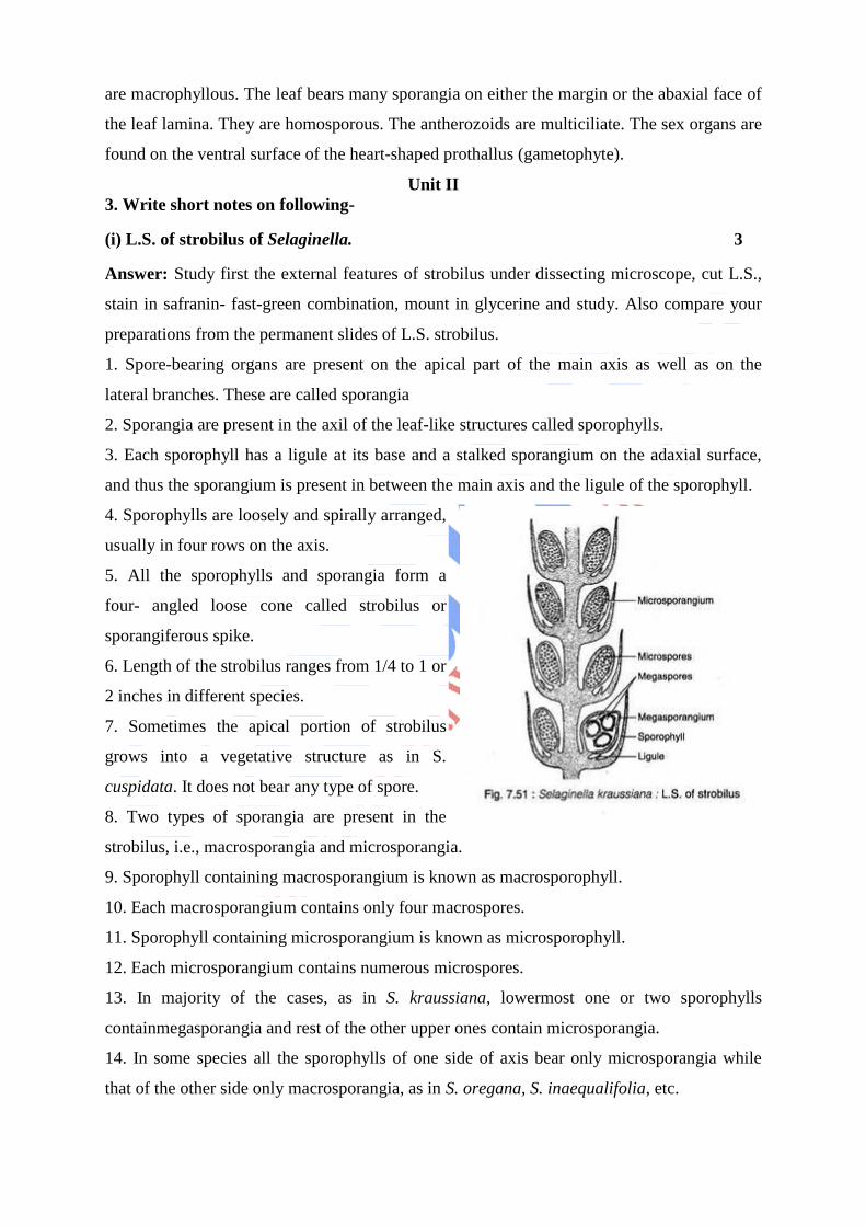
(i) L.S. of strobilus of Selaginella. 3
Answer: Study first the external features of strobilus under dissecting microscope, cut L.S.,
stain in safranin- fast-green combination, mount in glycerine and study. Also compare your
preparations from the permanent slides of L.S. strobilus.
1. Spore-bearing organs are present on the apical part of the main axis as well as on the
lateral branches. These are called sporangia
2. Sporangia are present in the axil of the leaf-like structures called sporophylls.
3. Each sporophyll has a ligule at its base and a stalked sporangium on the adaxial surface,
and thus the sporangium is present in between the main axis and the ligule of the sporophyll.
4. Sporophylls are loosely and spirally arranged,
usually in four rows on the axis.
5. All the sporophylls and sporangia form a
four- angled loose cone called strobilus or
sporangiferous spike.
6. Length of the strobilus ranges from 1/4 to 1 or
2 inches in different species.
7. Sometimes the apical portion of strobilus
grows into a vegetative structure as in S.
cuspidata. It does not bear any type of spore.
8. Two types of sporangia are present in the
strobilus, i.e., macrosporangia and microsporangia.
9. Sporophyll containing macrosporangium is known as macrosporophyll.
10. Each macrosporangium contains only four macrospores.
11. Sporophyll containing microsporangium is known as microsporophyll.
12. Each microsporangium contains numerous microspores.
13. In majority of the cases, as in S. kraussiana, lowermost one or two sporophylls
containmegasporangia and rest of the other upper ones contain microsporangia.
14. In some species all the sporophylls of one side of axis bear only microsporangia while
that of the other side only macrosporangia, as in S. oregana, S. inaequalifolia, etc.
15. In S. gracilis, a strobilus bears either microsporangia or macrosporangia.
16. Both the types of sporangia are stalked structures surrounded by a sporangial wall,
consisting of two outer jacket layers and an innermost layer of tapetum. Tapetum is very clear
in young sporangia.
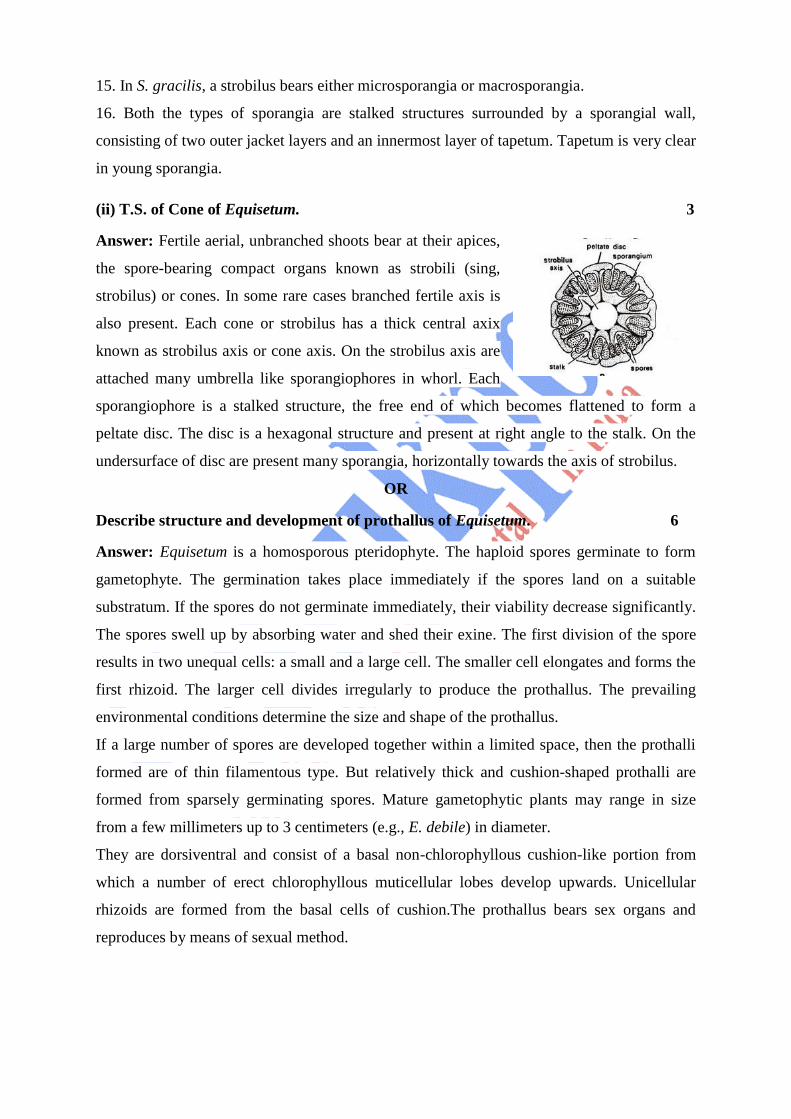
(ii) T.S. of Cone of Equisetum. 3
Answer: Fertile aerial, unbranched shoots bear at their apices,
the spore-bearing compact organs known as strobili (sing,
strobilus) or cones. In some rare cases branched fertile axis is
also present. Each cone or strobilus has a thick central axix
known as strobilus axis or cone axis. On the strobilus axis are
attached many umbrella like sporangiophores in whorl. Each
sporangiophore is a stalked structure, the free end of which becomes flattened to form a
peltate disc. The disc is a hexagonal structure and present at right angle to the stalk. On the
undersurface of disc are present many sporangia, horizontally towards the axis of strobilus.
OR
Describe structure and development of prothallus of Equisetum. 6
Answer: Equisetum is a homosporous pteridophyte. The haploid spores germinate to form
gametophyte. The germination takes place immediately if the spores land on a suitable
substratum. If the spores do not germinate immediately, their viability decrease significantly.
The spores swell up by absorbing water and shed their exine. The first division of the spore
results in two unequal cells: a small and a large cell. The smaller cell elongates and forms the
first rhizoid. The larger cell divides irregularly to produce the prothallus. The prevailing
environmental conditions determine the size and shape of the prothallus.
If a large number of spores are developed together within a limited space, then the prothalli
formed are of thin filamentous type. But relatively thick and cushion-shaped prothalli are
formed from sparsely germinating spores. Mature gametophytic plants may range in size
from a few millimeters up to 3 centimeters (e.g., E. debile) in diameter.
They are dorsiventral and consist of a basal non-chlorophyllous cushion-like portion from
which a number of erect chlorophyllous muticellular lobes develop upwards. Unicellular
rhizoids are formed from the basal cells of cushion.The prothallus bears sex organs and
reproduces by means of sexual method.

Unit III
4. Write short notes on following-
(i) Male and female cone of Pinus. 1½ + 1½
Answer: Male cone: The male cones develop in clusters in the axil of scaly leaves on long
shoot. They replace the dwarf shoots of the long shoot. Each male
cone is ovoid in shape and ranges from 1.5 to 2.5 cm. in length. A
male cone consists of a large number of microsporophylls arranged
spirally on the cone axis.Each microsporophyll is small,
membranous, brown-coloured structure. A microsporophyll is
comparable with the stamen of the flower of angiosperms because it
consists of a stalk (filament) with a terminal leafy expansion (
anther), the tip of which is projected upwards and called apophysis. Two pouch-like
microsporangia (pollen sacs) are present on the
abaxial or undersurface of each microsporophyll.
In each microsporangium are present many
microspores (pollen grains). Each microspore or
pollen grain is a rounded and yellow-coloured,
light, uninucleate structure with two outer
coverings, i.e., thick outer exine and thin inner
intine. The exine protrudes out on two sides in the
form of two balloon-shaped wings. Wings help in

floating and dispersal of pollen grains. Wings help in floating and dispersal of pollen grains.
A few microsporophylls of lower side of cone are sterile. Sporangia are also not present on
the adaxial surface of each microsporophyll of the male cone.
Female cone:
Observe the external features and longitudinal section of a
young female cone and also study 1st year, 2nd year and 3rd
year female cones. Female cone develops either solitary or
in groups of 2 to 4. They also develop in the axil of scaly
leaves on long shoots like male cones.
Each female cone is an ovoid, structure when young but
becomes elongated or cylindrical at maturity.
In the centre is present a cone axis. Many megasporophylls are arranged spirally on the cone
axis. A few megasporophylls, present at the base and at the apex of strobilus, are
sterile.Megasporophylls present in the middle of the strobilus are very large and they
decrease in size towards the base and apex.
Each megasporophyll consists of two types of scales, known as bract scales and ovuliferous
scales. Bract scales are thin, dry, membranous, brown- coloured structures having fringed
upper part. These are also called carpellary scales. An ovuliferous scale is present on the
upper surface of each bract scale. Each oruliferous scale is woody, bigger and stouter than
bract scale and it is triangular in shape. A broad sterile structure, with pointed tip, is present
at the apex of these scales. This is called apophysis. At the base of upper surface of each
ovuliferous scale are present two sessile and naked ovules. Micropyle of each ovule faces

towards the cone axis. Each ovule is orthotropous, and it remains surrounded by a single
integument, consisting of an outer fleshy, a middle stony and an inner fleshy layer. It opens
with a mouth opening called micropyle. Integument surrounds the megasporangium or
nucellus. Just opposite the micropyle is present a pollen chamber. In the endosperm or female
gametophyte are present 2 to 5 archegonia.
(ii) Pinus needle. 3
Answer: The typical needle-shaped leaf is found in all species of the Pinaceae family and it
is the arrangement of these needles in bundles or fascicles that is the most characteristic
feature of the genus Pinus. Actually pines have three kinds of leaves. The first appear after
the seed germinates and are called cotyledons or "seed leaves." These are small soft needle-
shaped leaves and their number varies from 3 or more (P. contorta, banksiana and sylvestris)
to eighteen or more (P. lambertiana, sabiniana and maximartinezii). As soon as the emerge,
they are capable of respiration and photosynthesis. Shortly after the cotyledons come the
juvenile leaves which are shaped like the cotyledons and are solitary and arranged in a spiral.
These are usually shed in several weeks after the adult leaves, which have basal sheath and
fascicular arrangement characteristic of the genus pinus, make their appearance.
The outer layer of the adult leaf is the waxy cuticle which protects the leaf from drying. In the
cuticle are minute openings known as stomata and these permit the movement of carbon
dioxide into and oxygen from the
leaf. In most haploxylon pines the
stomata are on the ventral (lower)
surfaces and the diploxylon pines
have stomata on both ventral and
dorsal surfaces. These stomata
often form fine white streaks
running along the length of the
leaf. The internal structure of the
leaf is complex and includes a
photosynthesizing parenchyma
("chlorenchyma" or mesophyll) and resin canals which may be located just beneath the
cuticle (often in the haploxlon pines) or varyingly deeper within the needle (often in the
diploxylon pines). Centrally there are fibrovascular bundles, which form the basis of
classification of the genus pinus into the subgenera Strobus ( the"soft" or "white" pines)

with one (haploxylon) fibrovascular bundle and Pinus (the "hard" or "yellow" pines)
with two (diploxylon) fibrovascular bundles. The resin canals connect with the stomata are
involved in gas exchange and the fibrovascular bundles connect ultimately with the xylem
involved with the transport of nutrients, sugars and water between the top of the tree and the
roots.
OR
Describe economic importance of Gymnosperms. What is difference between
gymnosperms and angiosperms? 6
Answer: Economic Importance of Gymnosperms:
1. Ornamental value:
A number of gymnosperms are grown as ornamental plants, e.g., Cycas, Araucaria, Thuja
etc.
2. Food Value:
i. ‘Sago’ starch obtained from pith and cortex of stem of C. revolute, C. rumphi etc.
ii. ‘Seed starch’ obtained from seeds of Cycas rumphii, Dioon edule etc. It is prepared into
flour and cooked before eating.
iii. Seeds of Pinus gerardiana (chilgoza) are edible.
iv. ‘Kaffir bread’ prepared from the stem pith of Encephalartos.
v. Young leaves of Cycas cooked as vegetables.
3. Medicinal value:
i. Ephedrine (alkaloid) extracted from Ephedra used in treating asthma, cough, cold,
bronchitis etc.
ii. Tincture of Ephedra is a cardiac stimulant.
iii. The juice extracted from young leaves of Cycas revoluta is used for curing blood
vomiting and flatulence.
4. Industrial Use:
i. Gum-Cycas gum used as adhesive, antidote for snake bites and using malignant ulcers.
ii. Tannins – Tannins extracted from bark of Araucaria, Pinus, Sequoia etc. used in leather
industry.
iii. Canada balsam – It is turpentine obtained from Abies balsamea and used as a mounting
medium in biological preparations.
iv. Amber (fossil resin) – obtained from Pinus succinifera. Wood of Pinus is used for doors,
poles, beams, railway wagon flooring etc.
v. Plywood prepared from Podocarpus.

vi. Papers like newsprints, writing and printing papers are being prepared from the wood pulp
of Pinus, Picea,Abeis, Gnetum etc.
vii. The leaves of cycads are used for preparing baskets, mats, hats, brooms etc.
viii. The fibres obtained from the leaves of Cycas and Macrozamia are used for stuffing
pillows and making mattresses.
5. Source of oils:
i. Oils extracted from seeds of C. revoluta, Macrozamia reidlei, Pinus cembra and
Cephalotaxus drupacea are used as edible oils.
ii. Red cedar wood oil extracted from the heart wood of Juniperus virginiana is used for
cleaning microscopic preparations and for oil immersion lenses.
iii. Oils obtained from Cedrus deodara, Ciyptomeria japonica and Cupressus serm-perivirens
are used in preparations of perfumes.
Unit IV
4. Write short notes on following-
(i) Reconstructed plant Lepidodendron. 3
Answer: Like other ancient lycopods, Lepidodendron was also tree like in habit (Fig.55). In
general appearance it was not unlike that of
present day Lycopodium. But in size the
genus enormously exceeded the herbaceous
Lycopodium. The petrified trunks were
sometimes as long as 100 feet. Judging from
this it may be safely assumed that the plant
reached a height of over 120 feet.The stem
was erect and did not branch up to some
distance from the ground. The branching of
the stem was typically dichotomous. The
ultimate dichotomies produced the leaves.
The branches and the foliage formed a sort of
crown at the apex of the stem.
The leaves which clad the young stems and

branches were acicular or linear in shape having a length of 5-9 inches. The arrangement of
the leaves was spiral or very rarely they showed a whorled arrangement. The leaves were
ligulate.
Each leaf had a single vein with the stomata situated in two bands on the ventral surface. The
leaves were deciduous. Upon abscission a flat rhomboidal scar persisted on the stem
resembling a small cushion. The base of the stem had a stigmarian type of root system.
1. Stem:In majority of the species, secondary growth is characteristic. But some species seem
to lack a cambium. A transverse section of the trunk, of L. vasculare shows three regions,
stele, cortex and a periderm.
In the primary structure there was an epidermis but soon i.e. even before the initiation of
secondary growth in the vasculature, it was replaced by the periderm.
The periderm was produced by a phellogen which produced phelloderm towards the interior
and phellem towards the exterior. The outline of the bark was wavy due to the presence of
leaf bases (Fig. 56).
It consisted of four regions viz.:
(1) Outer cortex consisting of alternating bands of sclerotic and parenchymatous cells,
(2) A middle cortex having a homogenous
mass of parenchyma cells. Interspersed with
the parenchyma cells were the leaf traces,
(3) Secretory zone consisting of glandular
cells which were filled with a dark coloured
substance. They probably secreted the waxy
material which covered the surface of the
stem,
(4) An inner cortex having parenchyma cells.
The central region of the stem was occupied
by the stele which was either protostelic or
siphonostelic. The protoxylem was exarch and polyarch. In many species (L. vasculare) there
was a secondary growth initiated by the cambium.
This produced secondary xylem to the interior and secondary phloem to the exterior. The
cambial activity was not uniform; as a result there was a tendency for the formation of an
eccentric vascular ring. The secondary xylem had radial rows of tracheitis separated by xylem
rays which were uniseriate.

2. Leaf:Anatomically the leaves showed a single vascular bundle flanked on either side by
parichnos cavities. These are believed to be aerating organs.
Reproduction in Lepidodendron:
The strobili of Lepidodendron are given the name Lepidostrobus. In general structure they
had a central axis bearing spirally arranged or whorled sporophylls. The sporophylls were
ligulate and somewhat peltate bearing a single, sessile, elongate sporangium on their adaxial
face.It is quite possible that some sporangia were trabeculate, the trabeculae being concerned
with nutrition. The strobili were heterosporous with the megasporophylls aggregated towards
the base.
(ii) Fossil plant Rhynia. 3
Answer: The plants of Rhynia were herbaceous. R. major was 50 cm. in height and 1.5 to 6
mm in diameter whereas R. gwynne-vaughani was only about 20 cm. in height and 1 to 3 mm
in diameter.
The plant body was differentiated into a subterranean rhizome with an abruptly turned
upright photosynthetic aerial shoots. Roots were absent but at places rhizome was provided
with tufts of unicellular rhizoids.The
aerial shoots were cylindrical and
leafless with a tapering dichotomously
branched system.
In R. major the aerial shoots were
smooth but in case of R. gwynne-
vaughani many adventitious branches
were present on the aerial shoots as well
as rhizome. These branches perhaps help
in vegetative propagation.
The tip of the aerial branch usually bears
a solitary terminal sporangium which
was about 12 mm in length and about 4 mm in diameter.
Internal Structure of Rhynia:
Transverse section (T.S.) of Aerial shoot and Rhizome:
Anatomically, the aerial shoots and rhizome are almost similar. T. S. of aerial shoot can be
differentiated into three parts: epidermis, cortex and stele.

(a) Epidermis: It was the outer-most surrounding layer. It was one cell thick and covered by
thin cuticle. In aerial shoots it
was interrupted at certain places by
stomata but stomata (Fig. 2 B)
were absent in rhizome.
(b) Cortex: Epidermis was
followed by cortex. It is
differentiated into outer cortex
and inner cortex. The outer cortex was only 1-4 cells thick, thin walled and without
intercellular spaces. The inner cortex had large intercellular spaces and its cells had
chloroplast. It is thought that this was the chief photosynthetic region of the plant. The
endodermis and pericycle layers were absent.
Stele: The centre of the aerial shoot/rhizome was occupied by stele. The stele was a
protostele (haplostele). The xylem was made up of annular tracheids and there were no sieve
plates in phloem.
Reproductive Structures of Rhynia:
The sporangia were borne singly on the apices of some aerial branches, each sporangium
being oval or slightly cylindrical structure with a little greater diameter than that of aerial
branch on which it is developed. They were 12 mm long and 4 mm in breadth in R. major and
4 mm long and 1 mm broad in R. gwynne-vaughani.
A longitudinal section (L.S.) of sporangium shows that it had a five cells thick wall. The
outermost layer was 1 cell thick cuticularized epidermis. It was followed by 3 cells thick
middle layers of thin walled cells.The inner-most layer was 1 cell thick tapetum. The wall
was surrounding a spacious sporangial cavity
which was without columella and contained large
number of spores. The spores were of same size
and measured upto 60 µ in diameter.
It means that Rhynia was homosporous. In many
specimens the sporangium contained tetrahedral
tetrads of spores (Fig. 3 B, C) which suggest that
they were formed by reduction division and the
plant bearing them represented the sporophytic
generation.There was no special mechanism of
sporangium dehiscence. The liberation of spores seems to have taken place by disintegration
of the sporangial wall. Nothing definite about the gametophyte of Rhynia is known.
OR
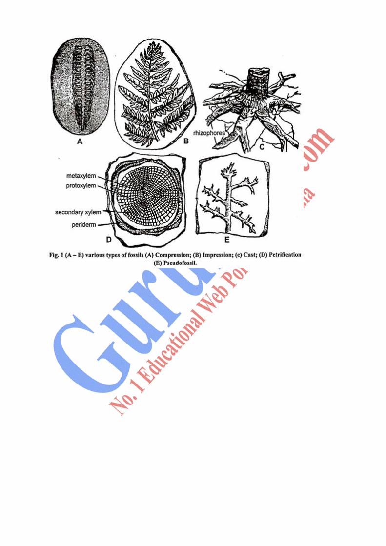
Describe fossilization and its types. 6
Answer: Fossilization is the process that preserves evidence of life in earth's rock record. This
evidence of past life is called a fossil. The word "fossil" is derived from the Latin fossilis,
something dug up. During the Middle Ages, the term "fossil" was used for any sample
recovered from the earth, including rocks and minerals.[1]
Today, the use of "fossil" is limited
to the record of ancient life. Fossilization can preserve actual remnants of an organism, or
evidence of their presence in an ecosystem
1. Petrified Fossils:The word petrifaction means turning into stones. The fossils form when
minerals replace all or the parts of the organisms. Water is full of dissolved minerals. It seeps
through the layer of sediments to reach the dead organism. When water evaporates only the
hardened, materials are left behind. There is molecule by molecule replacement of plant parts
by minerals such as iron, pyrites, silicates, carbonates, sulphates etc. These minerals get
deposited and impregnated inside the cells and the tissues of the plant. This type of fossil can
be studied by preparing the sections and are most suitable for the study of structural details
(Fig. 1D). Petrified plant organs roughly spherical in shape are known as coal balls.
2. Molds and Casts:A mold forms when hard parts of an organism are buried in the sediment
such as sand, silt or clay. The hard part completely dissolves overtime, leaving behind a
hollow area of organism shape. A cast forms as a result of the mold. Water with dissolved
minerals and sediments fills the mold’s empty space or cavity. The cavity is known as
incrustation and the mineral sediments that are left in the mold make a cast (Fig. 1C). A cast
is opposite to its mold. These fossils are suitable for the study of the morphology of fossil
plants.
3. Carbon Films:All living things contain an element carbon. When an organism dies and is
buried in sediment, the materials that make the organism break down and eventually only the
carbon remains. The thin layer of carbon left behind can show an organism’s delicate parts
like leaves or plant e.g. fern fossil 300 million years old.
4. Trace Fossils:These fossils show the activities of the organisms. An animal makes a foot
print when it steps in sand. Overtime the foot print is buried in layers of sediment. Then the
sediment becomes solid rock.
5. Preserved Remains:Some organisms are preserved in or close to their original states.
These fossils are called preserved remains e.g., an organism such as an insect is trapped in a
tree’s sticky resin and dies. More resin covers it sealing the insect inside. It hardens into
amber. Some organisms such as a wooly mammoth dies in a very cold region. Its body is
frozen in ice which preserves organism even its hair.
6. Compression:This type of fossil is common in the sedimentary deposits of rocks. It is a
sort of impression where most of the organic remains of the plant remain in the fossil state.
The plant or plant part gets buried and the sediments go on accumulating over the plant. The
growing pressure of the sedimentary rocks removes the air and the watery contents of the
fragment out and causes the plant tissue to compress. The compression shows the original
outline of the plant or plant parts but the original thickness of the plant material cannot be
determined. The buried part becomes flat due to compression or overlying pressure of the
sediments (Fig. 1 A).
7. Impression:These fossils are just impression of plants or plant parts on sediments. These
fossils are useful in studying the external features of various plant parts and venation pattern
of leaves (Fig. 1B).
8. Pseudofossils:Sometimes watery solutions of various minerals speed through the
sediments and it takes the shape of some plant part or animal. Their study shows that they are
neither plants nor animals. Such fossils are called pseudofossils (Fig. 1E).