Pre-anaesthetic metomidate sedation delays the stress response after caudal artery cannulation in...
11
Pre-anaesthetic metomidate sedation delays the stress response after caudal artery cannulation in Atlantic cod (Gadus morhua) Anders Karlsson • Bjørn Olav Rosseland • Jean-Charles Massabuau • Anders Kiessling Received: 21 May 2010 / Accepted: 19 May 2011 / Published online: 3 June 2011 Ó Springer Science+Business Media B.V. 2011 Abstract Recovery from caudal artery cannulation with and without pre-anaesthesia metomidate sedation was assessed in Atlantic cod (Gadus morhua). The levels of plasma cortisol, glucose, electrolytes and acid–base parameters were compared between sedated and unsedated cod and to those in uncannulated individuals, where the samples were obtained by sacrificial sampling (reference level). Metomidate sedation delayed the stress response, causing sedated cod plasma cortisol to return to the reference level more slowly [day 4 post surgery (PS)] than in unsedated cod (day 2 PS). Plasma glucose was elevated in both sedated and unsedated cod up to and including day 5 PS. Plasma K ? was lower and pH was higher in cannulated cod than in the reference from 24 h PS until the end of experimentation, indicating a stress effect of sacrificial sampling on plasma K ? and pH that was likely caused by an acute stress response. Metomidate sedation delayed the stress response following CA cannulation and should therefore not be used as a pre- anaesthetic sedation in Atlantic cod. The caudal artery cannulation can be a useful tool in obtaining repeated blood samples from Atlantic cod given an adequate recovery time, which was determined to be 6 days irrespective of pre-anaesthesia sedation status. Keywords Metacain Á Acute stress response Á Fish Á Gadoid Á Blood physiology Á Cortisol Á Glucose Introduction Cannulation is the process of catheterising a blood vessel and is usually performed in experimental animals to draw blood samples, inject substances directly into the blood stream or measure blood pressure without handling the animal. The cannula- tion techniques have proved to be valuable research tools in physiological experiments with fish for almost half a century (Conte et al. 1963). The various cannulation methods allow repeated sampling of blood from the same individual, providing an A. Karlsson Department of Plant and Environmental Sciences, Norwegian University of Life Sciences, P.O. Box 5003, 1432 A ˚ s, Norway B. O. Rosseland Department of Ecology and Natural Resource Management, Norwegian University of Life Sciences, P.O. Box 5003, 1432 A ˚ s, Norway J.-C. Massabuau Station Marine, Universite ´ Bordeaux 1, CNRS, UMR 5805 EPOC, Place du Dr Peyneau, 33120 Arcachon, France Present Address: A. Karlsson (&) Á A. Kiessling Aquaculture Protein Centre, Department of Animal and Aquacultural Sciences, Norwegian University of Life Sciences, P.O. Box 5003, 1432 A ˚ s, Norway e-mail: [email protected] 123 Fish Physiol Biochem (2012) 38:401–411 DOI 10.1007/s10695-011-9516-x
-
Upload
anders-karlsson -
Category
Documents
-
view
214 -
download
1
Transcript of Pre-anaesthetic metomidate sedation delays the stress response after caudal artery cannulation in...

Pre-anaesthetic metomidate sedation delays the stressresponse after caudal artery cannulation in Atlantic cod(Gadus morhua)
Anders Karlsson • Bjørn Olav Rosseland •
Jean-Charles Massabuau • Anders Kiessling
Received: 21 May 2010 / Accepted: 19 May 2011 / Published online: 3 June 2011
� Springer Science+Business Media B.V. 2011
Abstract Recovery from caudal artery cannulation
with and without pre-anaesthesia metomidate sedation
was assessed in Atlantic cod (Gadus morhua). The
levels of plasma cortisol, glucose, electrolytes and
acid–base parameters were compared between sedated
and unsedated cod and to those in uncannulated
individuals, where the samples were obtained by
sacrificial sampling (reference level). Metomidate
sedation delayed the stress response, causing sedated
cod plasma cortisol to return to the reference level more
slowly [day 4 post surgery (PS)] than in unsedated cod
(day 2 PS). Plasma glucose was elevated in both
sedated and unsedated cod up to and including day 5
PS. Plasma K? was lower and pH was higher in
cannulated cod than in the reference from 24 h PS until
the end of experimentation, indicating a stress effect of
sacrificial sampling on plasma K? and pH that was
likely caused by an acute stress response. Metomidate
sedation delayed the stress response following CA
cannulation and should therefore not be used as a pre-
anaesthetic sedation in Atlantic cod. The caudal artery
cannulation can be a useful tool in obtaining repeated
blood samples from Atlantic cod given an adequate
recovery time, which was determined to be 6 days
irrespective of pre-anaesthesia sedation status.
Keywords Metacain � Acute stress response � Fish �Gadoid � Blood physiology � Cortisol � Glucose
Introduction
Cannulation is the process of catheterising a blood
vessel and is usually performed in experimental
animals to draw blood samples, inject substances
directly into the blood stream or measure blood
pressure without handling the animal. The cannula-
tion techniques have proved to be valuable research
tools in physiological experiments with fish for
almost half a century (Conte et al. 1963). The various
cannulation methods allow repeated sampling of
blood from the same individual, providing an
A. Karlsson
Department of Plant and Environmental Sciences,
Norwegian University of Life Sciences, P.O. Box 5003,
1432 As, Norway
B. O. Rosseland
Department of Ecology and Natural Resource
Management, Norwegian University of Life Sciences,
P.O. Box 5003, 1432 As, Norway
J.-C. Massabuau
Station Marine, Universite Bordeaux 1, CNRS, UMR
5805 EPOC, Place du Dr Peyneau, 33120 Arcachon,
France
Present Address:A. Karlsson (&) � A. Kiessling
Aquaculture Protein Centre, Department of Animal
and Aquacultural Sciences, Norwegian University of Life
Sciences, P.O. Box 5003, 1432 As, Norway
e-mail: [email protected]
123
Fish Physiol Biochem (2012) 38:401–411
DOI 10.1007/s10695-011-9516-x
alternative to sacrificial sampling when studying
changes in blood variables. Several blood vessels
have been targeted, e.g., the dorsal aorta (DA) (Conte
et al. 1963; Djordjevic et al. 2011; Kiessling et al.
2003; Smith and Bell 1964; Soivio et al. 1975; Sunde
et al. 2003), the ventral aorta (Axelsson et al. 1994),
the hepatic portal vein (Eliason et al. 2007; McLean
and Ash 1989) and the caudal artery (CA) (Forgue
et al. 1989; Karlsson et al. 2011) with duration of
experimental period varying from hours to several
weeks. Using cannulated fish in a repeated sampling
setup allows a reduction in the number of experi-
mental animals as all treatments can be applied to all
animals, thereby eliminating the need for one group
of animals per treatment. It can also reduce exper-
imental variability as it allows internal paired com-
parison as reference; control, test and recovery data
can be measured in the same individual. A final
advantage over sacrificial sampling is reduced vari-
ability due to sampling stress and handling; the fish
can be practically undisturbed by the sampling
procedure and experimental setup (Djordjevic et al.
2011). Therefore, when studying fish blood parame-
ters that may be affected by gill ventilation or
immediate stress responses, e.g., plasma pH, HCO3-
or partial pressures of respiratory gases, using
cannulated individuals and repeated sampling is a
necessity.
The DA cannulation, as it is described by Soivio
et al. (1975), has been one of the most frequently
used cannulation methods, possibly because of its
comparatively uncomplicated surgical procedure.
The Atlantic cod (Gadus morhua) has an anatomy
that complicates cannulation of the DA through the
roof of the mouth. The gill efferent blood vessels
merge to form the DA very close to the pharyngeal
sphincter (visual inspection by dissection). A catheter
piercing the skin in or close to the pharyngeal
sphincter would likely severely disturb the cod and
may cause stress or suffering. This would undermine
the justification for cannulation and produce unreli-
able and possibly ambiguous results.
Most previous studies involving cannulation of
Atlantic cod report occlusive cannulation of a gill
afferent and efferent blood vessel, effectively remov-
ing one gill arch (Axelsson and Nilsson 1986; Perry
et al. 1991; Smith et al. 1985). The occlusive and
invasive nature of these procedures may cause ambig-
uous results, especially so in long-term studies of
weeks or months, where tissue necrosis may be a
significant concern. An alternative artery to cannulate
in the Atlantic cod is the CA. The CA cannulation was
first described by Forgue et al. (1989) in Wels catfish
(Silurus glanis), using open surgery, and later, using
non-invasive methods, in other teleosts (Forsman et al.
2005; Karlsson et al. 2011). The present study is the
first evaluation and detailed description of a non-
invasive CA cannulation technique for use on gadoid
fishes.
Metomidate has previously been shown to prevent
cortisol release in Atlantic salmon (Olsen et al. 1995)
and has been used as a sedative prior to anaesthesia
and subsequent cannulation in several studies by
Kiessling and co-workers throughout the past decade
(Djordjevic et al. 2011; Kiessling et al. 2006, 2003;
Kristensen et al. 2010; Sunde et al. 2003). The pre-
anaesthesia sedation, in combination with other
improvements, has reduced the general stress level
in salmonids after DA cannulation to such an extent
that most blood variables stabilize within 1–3 h post
surgery (PS), and all parameters measured were
stable 24–72 h PS (Djordjevic et al. 2011). Atlantic
cod pre-anaesthesia sedated with metomidate was
recently reported to require a lower anaesthetic
concentration and have a faster recovery time from
anaesthesia than unsedated congeners (Zahl et al.
2009). However, it is not presently known whether
the beneficial effects of metomidate sedation
observed in cannulated salmonids and Atlantic cod
during recovery from anaesthesia are transferrable to
a quicker recovery from cannulation in Atlantic cod.
The objectives of this study were to evaluate the
non-invasive CA cannulation technique in terms of
response magnitude and recovery time of blood
variables and to evaluate the use of metomidate
sedation prior to anaesthesia and CA cannulation of
Atlantic cod.
Materials and methods
Animals and experimental procedure
Atlantic cod, average weight 700 g, were purchased
from a commercial cod farm (Profunda AS; Bardstad-
vik, Norway) in early February 2009 and transported
by truck to the Norwegian Institute for Water Research
(NIVA)—Marine Research Station at Solbergstrand
402 Fish Physiol Biochem (2012) 38:401–411
123
(NIVA-MFS), Norway. No cod died during or after
transportation, and the fish were in good health upon
arrival (veterinary approval). Finally, no signs of ill
health were observed prior to, or during, the experi-
ment. The cod were housed in a circular, 6-m diameter
holding tank at a density of up to 2.5 kg/M3, fed a
commercial cod diet (BioMar AS, Norway) to satia-
tion 5 days a week and visually inspected at least once
daily. All cod were kept in the holding tank for a
minimum of 5 weeks to acclimate to their new
surroundings before being transferred to the experi-
mental unit.
Cod were cannulated (as described below) and
recovery from cannulation followed in three trials. In
all three trials, the cod were transferred from the
holding tank to the experiment tanks (square 1 m
tanks, 500 L water volume) and given 7 days to
acclimate to the new environment in groups of 4–5
individuals per tank. The tanks were purposely
designed to hold individual, cannulated fish and
create a low stress environment as described in detail
by Djordjevic et al. (2011). At least 24 h prior to, and
throughout acclimation and experimentation, cod
were deprived of food. In trial 1, (Mar–Apr 2009)
all cod were sedated with metomidate in their tanks
(sedated) prior to anaesthesia with metacain in a
separate bath before cannulation. In trials 2 (Sep–Oct
2009) and 3 (Nov–Dec 2009), cod were either
sedated with metomidate in their tanks (sedated) or
left undisturbed (unsedated) prior to metacain anaes-
thesia and cannulation.
To follow recovery from cannulation, 22 cod,
average weight 1041 ± 288 g and length 44 ± 3 cm
[± standard deviation (SD)], were sampled for blood
through the CA cannula at the following times:
directly PS (0 h), one hour PS (1 h), three hours PS
(3 h), 24 h PS (24 h), daily after 24 h until day 7 PS
(Day 2–7) and at days 9, 11 and 14 PS. To establish a
comparison/reference level, 7 Atlantic cod, average
weight 734 ± 75 g and length 41 ± 2 cm (±SD),
from the same population as the experimental fish,
were placed in the tanks described above (1 cod per
tank). The fish were left undisturbed for 14 days and
then sacrificially sampled for blood from the caudal
artery/vein using aspiration with a disposable syringe
and needle.
Upon finishing the experiment, all cod with
cannulas still attached (20 of 22 fish, 2 fish lost their
cannulas during the experiment) were dissected to
determine the placement of the cannula. Sixteen cod
were cannulated in the CA, three cod were cannulated
in the caudal vein (CV) and cannula placement could
not be determined in one cod. Due to the possible
differences in pH, PCO2 and HCO3- between arterial
and venous blood, results from individuals cannulat-
ed in the CV or with unknown cannula placement (6
cod in total) were removed from statistical analysis of
pH, PCO2 and HCO3- data.
Throughout the experiments, full strength seawater
from a depth of 60 m and with a temperature of
7.2–10.6�C and a salinity of 31.8–34.5 parts per
thousand (ppt) was used. The water flow to the tanks
was kept at 2 L/min. The flow through system
secured good water quality throughout. Typical water
qualities in the facility include: oxygen tension
18.1 kPa; pH 7.84; alkalinity 2.41 mmol/L; turbidity
0.36 FNU; total nitrogen 173 lg/L; ammonium
13 lg/L; dissolved carbon dioxide 0.44 mg/L.
Surgical procedure
The catheters were made from PE50 polyethylene
tubing (Intramedic�; Becton–Dickinson, New Jer-
sey, USA), and trocars/cannulas were made from
PL013 steel guitar wire. The PE50 was heated and
then stretched and narrowed at the penetrating end,
and a bubble was made by precision heating *5 cm
up the cannula; so that the catheter could be attached
to a suture without sliding. The catheter was then
flushed with heparinised physiological saline (NaCl,
9 g/L; Na-heparin, 150 IU/mL, injection quality)
(saline) and cut at the narrowing end to fit tightly
around the trocar. Finally, two small holes were made
on the catheter tip (one on each side) to prevent the
catheter from suctioning onto the vessel wall. The
catheters were stored immersed in 70% ethanol
without the trocar. Immediately prior to surgery, the
trocar was prepared by cutting it at an angle as low as
possible with a pair of wire cutters to produce a sharp
point with a cutting edge. Finally, the trocar was
inserted into the catheter so that only the cutting edge
and point was protruding from the PE50 tubing, and
the catheter was immersed in antiseptic fluid (Chlorh-
exidine, 0.5 g/L; Fresenius Kabi, Uppsala, Sweden).
Each fish was either sedated with metomidate
(0.5 mg/L) until it stopped responding to visual
stimuli, or not disturbed before it was transferred
from its’ respective tank to an anaesthetic bath
Fish Physiol Biochem (2012) 38:401–411 403
123
containing *30 L of aerated seawater and 0.08 g/L
metacain (Norwegian Medical Depot, Norway).
When the fish no longer responded to touch, weight
and length were measured before it was placed
upside-down in a purposely designed surgical cradle
and surrounding bath and covered with a wet cloth.
Throughout surgery, the gills were ventilated with
aerated seawater maintained at equal temperature as
in the tank (8–9�C) at *15 L/min containing a
maintenance dose of metacain (0.04 g/L).
Using a disposable insulin syringe with needle,
0.3–0.7 mL of lidocaine analgesia was injected at the
point of incision and suture placement through the
skin [10 or 20 g/L, with adrenaline (5 mg/L)]. An
#11-blade and a #3-scalpel was used to make a
horizontal incision (*5 mm) through the skin, ver-
tically positioned at approximately the same distance
from the anal fin and lateral line, and horizontally
approximately half way down the length of the anal
fin.
The ‘closed’ cannulation was performed by insert-
ing a pre-made catheter with trocar into the incision,
at approximately 45� horizontally, and pushed
through muscle and membrane tissue into the CA.
The trocar was then retracted, and the catheter was
securely placed inside the vessel by pushing it
2–3 cm into the vessel. To prevent clotting, the
catheter was filled with saline. The catheter was
secured to the fish using a single stitch of sterile, non-
absorbable suture (Supramid�, 3-0 USP; AgnTho’s
AB, Sweden) directly behind the insertion point of
the catheter. The catheter end was then melted shut,
and the surgery wound covered with Stomahesive�
paste (ConvaTec Norway AS, Oslo, Norway).
Finally, the fish received an injection of Oxytetracy-
cline antibiotic (100 g/L; Ceva Sante Animale,
Libourne, France) into the abdominal cavity to
prevent variation in condition caused by accidental
bacterial infection.
Sample collection, preparation and analysis
Prior to sampling blood from the CA catheter,
previously injected saline and a few drops of fresh
blood were discarded to ensure a pristine sample. By
light suction using a 1-mL disposable syringe with
blunted tip inserted into the catheter, 0.2–0.35 mL
blood samples were collected. The amount of sample
extracted was volume adjusted based on visual
observation of haematocrit, in order to obtain enough
plasma and draw as little blood as possible. After the
required amount of blood was sampled, saline was
injected into the catheter until blood was no longer
visible. To prevent clotting, another 0.1–0.2 mL of
saline was injected to ensure no blood was left in
catheter tip. The catheter end was sealed by melting
after each sampling.
Blood samples were immediately analysed for
glucose, PCO2, pH, HCO3- and ions (Na?, K?, Cl-)
using an i-STAT� Portable Clinical Analyzer (Med-
inor AS, Norway). Results for pH, PCO2 and HCO3-
were temperature corrected using formulas supplied
by the i-STAT� manufacturer (Abbott Point of Care
Inc.; Princeton, NJ, USA).
Blood samples were immediately centrifuged for
3 min at 20009g to separate red blood cells and
plasma. Plasma was immediately frozen at -20�C
and transferred to -80�C within 3 days. Plasma
samples (50 lL) were mixed with five times the
sample volume (250 lL) of ethyl acetate using a
vortex mixer. The mix was centrifuged for 2 min at
71559g in 4�C, and the resulting supernatant stored
at -80�C until analysis. The supernatant was later
analysed for cortisol using a Radioactive Immuno
Assay (RIA) kit (Spectria� Cortisol RIA; Orion
Diagnostica AS, Asker, Norway) and a NaI-gamma
counter (Wizard�; PerkinElmer Norge AS, Oslo,
Norway) according to instructions in the RIA kit
booklet. Samples determined to be below the detec-
tion limit of the RIA kit (5 ng/mL) were set to be
5 ng/mL before further analysis of the data.
Data and statistical analysis
Plasma Cl- was above the detection limit of the
instrument (140 mmol/L) in all samples and was
consequently excluded from further analysis. Two
samples were removed from the statistical analysis
for all blood variables due to extreme observations of
plasma cortisol; 267 and 315 ng/mL whilst no other
observations were [140 ng/mL. These samples orig-
inated from the same individual at 24 and 48 h PS.
All other samples from the same individual were
within normal range and were included in the
statistical analysis.
The data were analysed with SAS v. 9.13 (Statis-
tical Analysis Software), using the mixed procedure
for repeated measurements with a heterogeneous and
404 Fish Physiol Biochem (2012) 38:401–411
123

autoregressive covariance structure. Pre-anaesthetic
sedation status, recovery time and the interaction
between pre-anaesthetic sedation status and recovery
time were tested as class variables in the model.
Individual was included as a random variable (subject
identification). No effect of pre-anaesthetic sedation
status (P = 0.6255) or interaction between recovery
time and pre-anaesthetic sedation status (P = 0.2085)
was found for plasma K?. Consequently, the two pre-
anaesthetic sedation statuses were considered to be the
same for statistical analysis of plasma K?.
An F test was used to determine statistical
significance of fixed effects. A t test with Tukey–
Kramer adjustment for multiple comparisons was
used to determine statistical differences between the
different sampling times and between sedation
statuses within sampling time. A t test with Dunnett
adjustment for multiple comparisons was used to
determine statistical difference between the reference
level and the different sampling times. Comparisons
yielding P values\0.05 after adjustment for multiple
comparisons were considered to be statistically
different. Unless otherwise stated, P \ 0.05 in all
comparisons where differences are indicated. All
values are presented as least squares mean (LS
mean) ± standard error (SE) unless stated otherwise.
Results
Plasma cortisol and glucose
Plasma cortisol and glucose were affected by recovery
time (P \ 0.0001) and the interaction between recov-
ery time and sedation status (P \ 0.05), but no effect
was found for sedation status alone (Table 1). Both
plasma cortisol and glucose levels increased initially,
peaked (at ca 90 ng/mL for cortisol and 9 mmol/L for
glucose), and then decreased with time PS irrespective
of sedation status (Fig. 1a, b). However, the unsedated
cod displayed peak plasma cortisol and glucose levels
earlier than did the sedated cod, 1–3 vs. 3–24 h PS for
cortisol and 1 vs. 24 h PS for glucose. In unsedated
cod, plasma cortisol was higher at 1 h and at 5 days
PS, and plasma glucose was lower at 24 h PS, than in
sedated cod. The sedated cod plasma cortisol levels
were elevated, compared to the reference level
(10.6 ± 9.3 ng/mL), from 1 h up to and including
72 h PS. In unsedated cod, plasma cortisol levels were
elevated compared to the reference level from directly
PS (0 h) up to and including 24 h PS, and at day 5 PS.
In both unsedated and sedated cod, plasma glu-
cose levels were elevated compared to the reference
level (2.38 ± 1.08 mmol/L), from 0 h (unsedated)
and 1 h (sedated) up to and including day 5 PS
(Fig. 1a, b).
Acid–base parameters
Plasma PCO2, HCO3- and pH were affected by
recovery time (P \ 0.0001), sedation status
(P \ 0.01) and their interaction (P \ 0.05) (Table 1).
No clear pattern could be seen for development of
plasma PCO2 PS. In sedated cod, plasma PCO2 was
lower than the reference level (0.52 ± 0.05 kPa) at
days 1, 2, 7 and 11 PS, whilst it never differed from
the reference level in unsedated cod. The sedated cod
plasma PCO2 levels were lower than in unsedated
cod at 1, 2, 7, 11 and 14 PS (Fig. 2a). In both sedated
and unsedated cod, plasma HCO3- increased from ca
4 mmol/L directly PS (not different from the refer-
ence level at 4.0 ± 0.5 mmol/L) to ca 6.5 mmol/L at
1 h PS. In unsedated cod, the plasma HCO3- did not
change from 1 h PS onwards and was higher than the
reference level until the end of experimentation.
However, in sedated cod, the plasma HCO3- dropped
after 3 h PS and was only higher than the reference
level at days 3, 4 and 9 PS thereafter. The sedated cod
plasma HCO3- concentration was lower than in
unsedated cod at days 1, 2, 7, 11 and 14 PS (Fig. 2b).
Table 1 P values for tests of fixed effects on blood variables
Cortisol Glucose Na? K? pH PCO2 HCO3-
Time post surgery \0.0001 \0.0001 0.0002 \0.0001 \0.0001 \0.0001 \0.0001
Sedation status 0.2826 0.7882 0.0465 0.6255 0.0035 0.0019 0.0076
Time 9 sedation 0.0015 0.0424 0.0366 0.2085 0.0127 \0.0001 0.0008
Fish Physiol Biochem (2012) 38:401–411 405
123
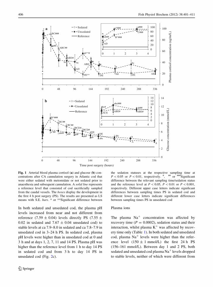
In both sedated and unsedated cod, the plasma pH
levels increased from near and not different from
reference (7.59 ± 0.04) levels directly PS (7.55 ±
0.02 in sedated and 7.67 ± 0.04 unsedated cod) to
stable levels at ca 7.9–8.0 in sedated and ca 7.8–7.9 in
unsedated cod in 3–24 h PS. In sedated cod, plasma
pH levels were higher than in unsedated cod at 0 and
3 h and at days 1, 2, 7, 11 and 14 PS. Plasma pH was
higher than the reference level from 1 h to day 14 PS
in sedated cod and from 3 h to day 14 PS in
unsedated cod (Fig. 2c).
Plasma ions
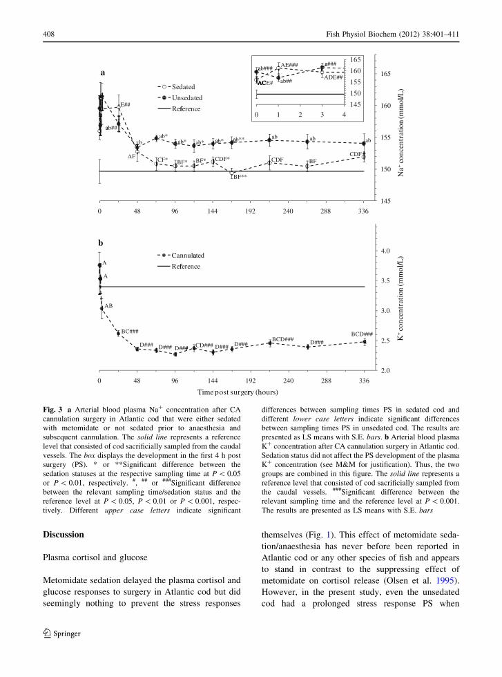
The plasma Na? concentration was affected by
recovery time (P = 0.0002), sedation status and their
interaction, whilst plasma K? was affected by recov-
ery time only (Table 1). In both sedated and unsedated
cod, plasma Na? levels were higher than the refer-
ence level (150 ± 1 mmol/L) the first 24 h PS
(156–161 mmol/L). Between day 1 and 2 PS, both
sedated and unsedated cod plasma Na? levels dropped
to stable levels, neither of which were different from
0
2
4
6
8
10
12
14
0 48 96 144 192 240 288 336
Glu
cose
con
cent
ratio
n (m
mol
/L)
Time post surgery (hours)
Sedated
Unsedated
Refeff rence
B*###
a*##
BC##BC##
BCD#BCD#
DEDE
AAA
abaa ## ab#abaa #
ab#abaa abc
abcaac
b
0
20
40
60
80
100
0 48 96 144 192 240 288 336
Plas
ma
cort
isol
con
cent
ratio
n (n
g/m
/L)
Sedated
Unsedated
Refeff rence
a
acac##
ab**#
DE**
BC###
ABC##
ABD#bbc bc
AD
ABE
DE DEAE AE
bcbcbbbc
bb
bc
0
20
40
60
80
100
0 1 2 3 4
AD
ab##
a*### a###
C###
B*###
02468101214
0 1 2 3 4
ABE#
aabaa c#abaa c###
abaa ##
ABE#ACE
b
Fig. 1 Arterial blood plasma cortisol (a) and glucose (b) con-
centrations after CA cannulation surgery in Atlantic cod that
were either sedated with metomidate or not sedated prior to
anaesthesia and subsequent cannulation. A solid line represents
a reference level that consisted of cod sacrificially sampled
from the caudal vessels. The boxes display the development in
the first 4 h post surgery (PS). The results are presented as LS
means with S.E. bars. * or **Significant difference between
the sedation statuses at the respective sampling time at
P \ 0.05 or P \ 0.01, respectively. #, ## or ###Significant
difference between the relevant sampling time/sedation status
and the reference level at P \ 0.05, P \ 0.01 or P \ 0.001,
respectively. Different upper case letters indicate significant
differences between sampling times PS in sedated cod and
different lower case letters indicate significant differences
between sampling times PS in unsedated cod
406 Fish Physiol Biochem (2012) 38:401–411
123

the reference level. However, the sedated cod Na?
level (149–153 mmol/L) was lower than that of
unsedated cod (153–155 mmol/L) at days 3–7 PS
(Fig. 3a). In cannulated cod, the plasma K? concen-
tration dropped from 3.76 ± 0.22 to 2.62 ± 0.05
mmol/L in the first 24 h PS and was then lower than
in the reference cod (3.40 ± 0.13 mmol/L) until the
end of the experiment. From day 2 PS onwards, the
plasma K? concentration in cannulated cod did not
change throughout the experiment (Fig. 3b).
7.50
7.60
7.70
7.80
7.90
8.00
8.10
0 48 96 144 192 240 288 336
pH
Time post surgey (hours)
Sedated
Unsedated
Refefef rence
cd**######
BBE*###
CF**###
bc*###
CEF*###
c*##
c
BF*###
c*###
CDEF*###
cd*###
BF###
BF###
BF###
CEF#####
CEF###d###cd###
bcd###cd###bbbcd####
0 48 96 144 192 240 288 336
0.0
0.1
0.2
0.3
0.4
0.5
0.6
Part
ial
pres
sure
ofC
O2
(kPa
)
Sedated
Unsedated
Reference
BCDE**#
ab*
BCDE**
ababa **
ab*ab**
BCDE*##
ab*
ab**
B**##
BCE*#
a
ABE
abab
abababa
abAE
ABE
ABE
ABE
0
1
2
3
4
5
6
7
8
9
0 48 96 144 192 240 288 336
HC
O3-
conc
entr
atio
n (m
mol
L/)
Sedated
Unsedated
b
AD*
b**###b*##
ACE*
b*##
ACE*
b*##
b*##
AD**
b##
b#b#b#b#b#b#b#b#b##
b##b##b##BDE####
AACD# ACEACE
ACE*
BDE#
7.57.67.77.87.98.0
0 3 4
a*
AA* c*##
BD*##B##
b
0 1 2 3 4
0.4
0.5
0.6
AC
A
AAAD
ab ab
345678
0
1 2
1 2 3 4
b# b##
a
A
BC## BC##
Reference
Fig. 2 Arterial blood
plasma partial pressure of
CO2 (a), bicarbonate
concentration (b) and pH
(c) after CA cannulation
surgery in Atlantic cod that
were either sedated with
metomidate or not sedated
prior to anaesthesia and
subsequent cannulation.
A solid line represents a
reference level that
consisted of cod
sacrificially sampled from
the caudal vessels. The
boxes display the
development in the first 4 h
post surgery (PS). The
results are presented as LS
means with S.E. bars. * or
**Significant difference
between the sedation
statuses at the respective
sampling time at P \ 0.05
or P \ 0.01, respectively.#, ## or ###Significant
difference between the
relevant sampling time/
sedation status and the
reference level at P \ 0.05,
P \ 0.01 or P \ 0.001,
respectively. Different
upper case letters indicate
significant differences
between sampling times PS
in sedated cod and different
lower case letters indicate
significant differences
between sampling times PS
in unsedated cod
Fish Physiol Biochem (2012) 38:401–411 407
123
Discussion
Plasma cortisol and glucose
Metomidate sedation delayed the plasma cortisol and
glucose responses to surgery in Atlantic cod but did
seemingly nothing to prevent the stress responses
themselves (Fig. 1). This effect of metomidate seda-
tion/anaesthesia has never before been reported in
Atlantic cod or any other species of fish and appears
to stand in contrast to the suppressing effect of
metomidate on cortisol release (Olsen et al. 1995).
However, in the present study, even the unsedated
cod had a prolonged stress response PS when
145
150
155
160
165
0 48 96 144 192 240 288 336
Na+
conc
entr
atio
n(m
mol
/Ll/)Sedated
Unsedated
Refeff rence
CF* CDF*
ab*
BF*BF*
abaa *ab* ab*aa *
BF**
ab*
a
ababab
b
ab#aa #
E##
AFBFCDF
CDF
2.0
2.5
3.0
3.5
4.0
0 48 96 144 192 240 288 336
K+
conc
entr
atio
n(m
mol
/Ll/)
Time post surgeryrr (hours)
Cannulataa ed
Refeff renceA
A
BC###
D###
AB
BCD###D###D###CD###D####D###
D###BCD###
b
145
150
155
160
165
0 1 2 3 4
a##a #
ab##
ab###aa
ACACACACACE#
AE###
ADE##
Fig. 3 a Arterial blood plasma Na? concentration after CA
cannulation surgery in Atlantic cod that were either sedated
with metomidate or not sedated prior to anaesthesia and
subsequent cannulation. The solid line represents a reference
level that consisted of cod sacrificially sampled from the caudal
vessels. The box displays the development in the first 4 h post
surgery (PS). * or **Significant difference between the
sedation statuses at the respective sampling time at P \ 0.05
or P \ 0.01, respectively. #, ## or ###Significant difference
between the relevant sampling time/sedation status and the
reference level at P \ 0.05, P \ 0.01 or P \ 0.001, respec-
tively. Different upper case letters indicate significant
differences between sampling times PS in sedated cod and
different lower case letters indicate significant differences
between sampling times PS in unsedated cod. The results are
presented as LS means with S.E. bars. b Arterial blood plasma
K? concentration after CA cannulation surgery in Atlantic cod.
Sedation status did not affect the PS development of the plasma
K? concentration (see M&M for justification). Thus, the two
groups are combined in this figure. The solid line represents a
reference level that consisted of cod sacrificially sampled from
the caudal vessels. ###Significant difference between the
relevant sampling time and the reference level at P \ 0.001.
The results are presented as LS means with S.E. bars
408 Fish Physiol Biochem (2012) 38:401–411
123

compared to similar or more invasive surgeries
performed in Atlantic salmon (Salmo salar) (Djordj-
evic et al. 2011; Eliason et al. 2007), a species whose
plasma cortisol and glucose responses to stress have
similar time frames as Atlantic cod (Olsen et al. 2002,
2008). Due to the comparatively prolonged PS stress
response observed even in unsedated cod in the
present study, the cod likely experienced a significant
level of stress when the effects of metacain anaes-
thesia (and metomidate sedation) had worn off. In
sedated cod, this PS stress likely triggered a cortisol
release and subsequent plasma glucose increase upon
withdrawal from sedation/anaesthesia since metomi-
date had suppressed their initial cortisol response. In
unsedated cod, however, netting, anaesthesia and
cannulation likely induced a stress response that
elevated plasma cortisol and glucose above the
reference levels directly PS.
The recovery times (time PS where the parameter
is no longer elevated/lowered compared to the
reference level or has stabilized) of cod plasma
cortisol and glucose were longer than what has been
reported following stress in Atlantic cod (Olsen et al.
2008) and cannulation surgeries in Atlantic salmon
(Djordjevic et al. 2011; Eliason et al. 2007). The
comparatively long recovery times observed in the
present study is one example of how knowledge
cannot always be transferred between species or
between procedures within species. In both sedated
and unsedated cod, plasma glucose recovery time was
6 days PS. This recovery time is longer than in any
other measured parameter in the present study. Thus,
we recommend a recovery time of 6 days or more in
experiments utilising the CA cannulation technique
in Atlantic cod.
Acid–base parameters
Plasma pH, PCO2 and HCO3- concentrations dif-
fered intermittently between sedated and unsedated
cod and PCO2 and HCO3- concentrations varied
more in sedated than unsedated cod (Fig. 2). The
differences between sedation statuses and increased
variability in sedated cod suggest an effect of
metomidate sedation on respiration, metabolism or
red blood cell (RBC) ion transport in Atlantic cod
that continues to affect the cod even after 2 weeks of
recovery. However, the mechanism(s) behind these
differences remain uncertain and needs to be
examined further in order to fully understand the
effect of metomidate sedation on Atlantic cod.
Acute stress and the subsequent release of cate-
cholamines to the circulation are known to affect
several blood parameters in fishes, most of which are
directed towards securing oxygen uptake and delivery
to the tissues in stressful situations (Reid et al. 1998).
Amongst these responses are changes in RBC ion
movements directed at elevating RBC pH to increase
the oxygen carrying capacity of haemoglobin (Tho-
mas and Perry 1992). Upon stimulation from cate-
cholamines, the Na?/H? exchangers on the RBC
membrane increases Na? influx and H? efflux across
the RBC membrane, effectively increasing intracel-
lular pH and Na? concentration and reducing plasma
pH and Na? concentration (Fievet et al. 1987). In the
present study, plasma pH is reduced directly PS in
both groups of cannulated cod and in the reference
cod. This was likely caused by a release of catechol-
amines prior to or during cannulation surgery, or by
the sacrificial sampling procedure in the reference
cod. However, there was no concomitant decrease in
plasma Na? levels as could be expected if the RBC
membrane Na?/H? exchangers caused the drop in
plasma pH. In fact, the opposite result was observed
as plasma Na? levels were elevated the first 24 h PS
(see below for elaboration). Due to the plasma
acidification observed in the present study, net flux
through the Cl-/HCO3- exchanger would be
expected to decrease plasma Cl- and increase plasma
HCO3- (Fievet et al. 1988). However, the opposite
result was observed in the present study; in both
sedated and unsedated cod, plasma HCO3- levels
were lowered but not different from the reference
directly PS, compared to their stabilized level at 3 h
PS onwards.
Plasma ions
The blood plasma ions measured in the present study,
Na? and K?, stabilized within 48 h PS, and sedation
status did not affect the recovery time of these
parameters. However, in sedated cod, the plasma Na?
concentration stabilized at a lower level than in
unsedated cod from day 3 to day 7 PS (Fig. 3). The
elevated plasma Na? concentrations observed in both
sedated and unsedated cod the first 24 h PS is
opposite to what can be expected based on the plasma
acidification observed the first 24 h PS. The high
Fish Physiol Biochem (2012) 38:401–411 409
123

plasma Na? levels 0–24 h PS may have been caused
by a reduced ability to maintain osmotic balance
during and directly after surgery, causing plasma Na?
to increase, despite Na? influx to the RBC via the
Na?/H? exchanger. Another possibility is mis-
matched activity levels of RBC ion exchangers,
e.g., between the Na?/K? ATPase, the Na?/H?
exchanger and the Na?/K?/Cl- co-transporters dur-
ing cannulation. Catecholamines are known to
increase the activity of RBC Na?/K? ATPase
(Bourne and Cossins 1982) and Na?/K?/Cl- co-
transporters (Russell 2000), either of which may be
the reason for the observed high plasma Na? levels
the first 24 h PS. In cannulated cod, there was a
significant drop in plasma K? concentration from
directly PS until it stabilized at day 2 PS. The initial
(0–24 h PS) high plasma K? concentration was likely
caused by a net efflux of K? from the RBC during
cannulation, likely due to increased activity of K?/
Cl- or Na?/K?/Cl- co-transporters on the RBC
membrane (Nikinmaa 2006; Russell 2000).
Cannulated cod versus sacrificially sampled
reference cod
The cod used as reference in the present study were
netted, euthanized by a blow to the head and finally
sampled for blood from the caudal vessels. Although
the procedure lasted only a few seconds, it likely
produced an acute stress response and subsequent
catecholamine release in the cod (Perry and Bernier
1999), as plasma pH, HCO3- and K? were all
elevated or lowered compared to the stable levels
observed at day 2 PS onwards in cannulated cod
(Figs. 2b, c and 3b). These findings highlight the
importance of using undisturbed fish when studying
biological parameters that may change rapidly due to
stress and indicate that sacrificially sampled fish are,
for several blood variables, not a good point of
reference. In spite of the suggested acute stress
response and subsequent catecholamine release, the
plasma cortisol and glucose levels are low in the
reference cod. This is most likely an effect of the
short time frame of the sacrificial sampling procedure
(\1 min), as plasma cortisol and glucose levels are
known to peak 1–3 h after acute stress in Atlantic cod
(Olsen et al. 2008).
The cod used as reference in the present study
were smaller than the cannulated cod. The difference
in body weight between the cannulated and the
reference cod could potentially have affected
the results. However, in the present study, none of
the regressions between plasma cortisol or glucose (at
day 14 in cannulated cod) and body weight or length
were significant. Thus, the difference in body weight
between cannulated and reference cod most likely did
not affect the results in the present study.
Conclusions and general remarks
The delayed stress response in metomidate sedated
cod observed in the present study has many possible
implications for aquaculture operating procedures
and experiments with fish. Therefore, the effects of
sedation or anaesthesia using metomidate should be
carefully evaluated for each specific application prior
to use, in order to prevent stressed and possibly
suffering fish and the possibility of producing unre-
liable or ambiguous results from experiments. In both
sedated and unsedated cod, all measured parameters
had stabilized or returned to the reference level at day
6 PS. Thus, we conclude that the CA cannulation is a
valid technique for obtaining arterial blood samples
from undisturbed Atlantic cod, given a recovery time
of 6 days or more is applied. Due to the absence of an
improved recovery from CA cannulation when using
metomidate, we recommend that metomidate not be
used as a sedative prior to anaesthesia and subsequent
handling of Atlantic cod.
Acknowledgments This study was funded by the Norwegian
Research Council (NFR) through the research projects
PROCOD (NFR project number 172263) and MODSMO
(NFR project number 172514). We would like to thank the
staff at NIVA-MFS for their technical assistance and care for
the fish prior to and during the experiment, and an anonymous
reviewer for the critical comments and suggestions on how to
improve the manuscript.
References
Axelsson M, Nilsson S (1986) Blood pressure control during
exercise in the Atlantic cod, Gadus morhua. J Exp Biol
126:225–236
Axelsson M, Davison B, Forster M, Nilsson S (1994) Blood
pressure control in the Antarctic fish Pagothenia bor-chgrevinki. J Exp Biol 190:265–279
Bourne PK, Cossins AR (1982) On the instability of K? influx
in erythrocytes of the rainbow trout, Salmo gairdneri, and
410 Fish Physiol Biochem (2012) 38:401–411
123
the role of catecholamine hormones in maintaining in vivo
influx activity. J Exp Biol 101:93–104
Conte FP, Wagner HH, Harris TO (1963) Measurement of
blood volume in the fish (Salmo gairdneri gairdneri). Am
J Physiol 205(3):533–540
Djordjevic B, Kristensen T, Øverli Ø, Rosseland BO, Kiessling
A (2011) Effect of nutritional status and sampling inten-
sity on recovery after dorsal aorta cannulation in free-
swimming Atlantic salmon (Salmo salar L.). Fish Physiol
Biochem Published online. doi:10.1007/s10695-009-
9362-2
Eliason EJ, Kiessling A, Karlsson A, Djordjevic B, Farrell AP
(2007) Validation of the hepatic portal vein cannulation
technique using Atlantic salmon Salmo salar L. J Fish
Biol 71(1):290–297. doi:10.1111/j.1095-8649.2007.014
89.x
Fievet B, Motais R, Thomas S (1987) Role of adrenergic
dependent H? release from red cells in acidosis induced
by hypoxia in trout. Am J Physiol 252(2):R269–R275
Fievet B, Claireaux G, Thomas S, Motais R (1988) Adaptive
respiratory responses of trout to acute hypoxia. III. Ion
movements and pH changes in the red blood cell. Respir
Physiol 74(1):99–113
Forgue J, Burtin B, Massabuau JC (1989) Maintenance of
oxygen consumption in resting Silurus glanis at different
levels of ambient oxygenation. J Exp Biol 143:305–319
Forsman A, Kiessling A, Ruohonen K, Vasquez J, Grove D
(2005) EU-project report ‘‘Selfish’’ Q5CA-2001-00989
Karlsson A, Sørlie Heier L, Rosseland BO, Salbu B, Kiessling
A (2011) Changes in arterial PO2, physiological blood
parameters and intracellular antioxidants in free-swim-
ming Atlantic cod (Gadus morhua) exposed to varying
levels of hyperoxia. Fish Physiol Biochem 37(1):249–258.
doi:10.1007/s10695-010-9438-z
Kiessling A, Olsen RE, Buttle L (2003) Given the same dietary
carotenoid inclusion, Atlantic salmon, Salmo salar (L.)
display higher blood levels of canthaxanthin than asta-
xanthin. Aquacult Nutr 9(4):253–261
Kiessling A, Dosanjh B, Koppe W, Higgs D (2006) Relation-
ship between blood and muscle levels of astaxanthin in
dorsal aorta cannulated Atlantic salmon. Aquaculture
254(1–4):653–657
Kristensen T, Rosseland BO, Kiessling A, Djordjevic B,
Massabau JC (2010) Lack of arterial PO2 downregulation
in Atlantic salmon (Salmo salar L.) during long-term
normoxia and hyperoxia. Fish Physiol Biochem 36(4):
1087–1095. doi:10.1007/s10695-010-9386-7
McLean E, Ash R (1989) Chronic cannulation of the hepatic
portal vein in rainbow trout, Salmo gairdneri: a pre-
requisite to net absorption studies. Aquaculture 78(2):
195–205
Nikinmaa M (2006) Gas transport. In: Evans DH, Claiborne JB
(eds) The physiology of fishes, 3rd edn. CRC Press, Boca
Raton, FL, p 601
Olsen YA, Einarsdottir IE, Nilssen KJ (1995) Metomidate
anesthesia in Atlantic salmon, Salmo salar, prevents
plasma cortisol increase during stress. Aquaculture
134(1–2):155–168
Olsen RE, Sundell K, Hansen T, Hemre GI, Myklebust R,
Mayhew TM, Ringo E (2002) Acute stress alters the
intestinal lining of Atlantic salmon, Salmo salar L.: An
electron microscopical study. Fish Physiol Biochem
26(3):211–221
Olsen RE, Sundell K, Ringø E, Myklebust R, Hemre GI, Hansen
T, Karlsen O (2008) The acute stress response in fed and
food deprived Atlantic cod, Gadus morhua L. Aquaculture
280(1–4):232–241. doi:10.1016/j.aquaculture.2008.05.006
Perry SF, Bernier NJ (1999) The acute humoral adrenergic
stress response in fish: facts and fiction. Aquaculture
177(1–4):285–295
Perry SF, Fritsche R, Kinkead R, Nilsson S (1991) Control of
catecholamine release in vivo and in situ in the Atlantic
cod (Gadus morhua) during hypoxia. J Exp Biol 155:
549–566
Reid SG, Bernier NJ, Perry SF (1998) The adrenergic stress
response in fish: control of catecholamine storage and
release. Comp Biochem Physiol C: Toxicol Pharmacol
120(1):1–27
Russell JM (2000) Sodium-potassium-chloride cotransport.
Physiol Rev 80(1):211–276
Smith LS, Bell GR (1964) A technique for prolonged blood
sampling in free-swimming salmon. J Fish Res Board Can
21(4):711–717
Smith DG, Nilsson S, Wahlqvist I, Eriksson BM (1985) Ner-
vous control of the blood pressure in the Atlantic cod,
Gadus morhua. J Exp Biol 117:335–347
Soivio A, Nyholm K, Westman K (1975) A technique for
repeated sampling of the blood of individual resting fish.
J Exp Biol 62:207–217
Sunde J, Kiessling A, Higgs D, Opstvedt J, Venturini G,
Rungruangsak-Torrissen K (2003) Evaluation of feed
protein quality by measuring plasma free amino acids in
Atlantic salmon (Salmo salar L.) after dorsal aorta can-
nulation. Aquacult Nutr 9(6):351–360
Thomas S, Perry SF (1992) Control and consequences of
adrenergic activation of red blood cell Na?/H? exchange
on blood oxygen and carbon dioxide transport in fish.
J Exp Zool 263(2):160–175
Zahl IH, Kiessling A, Samuelsen OB, Hansen MK (2009)
Anaesthesia of Atlantic cod (Gadus morhua)—Effect of
pre-anaesthetic sedation, and importance of body weight,
temperature and stress. Aquaculture 295(1–2):52–59. doi:
10.1016/j.aquaculture.2009.06.019
Fish Physiol Biochem (2012) 38:401–411 411
123