Practice Test This section is present to help the student understand what a Radiologist’s...
5
Practice Test This section is present to help the student understand what a Radiologist’s interpretation should include: 1. The part of the body X-rayed and how many views were obtained. 2. Anatomical Location of the injury 2. Descriptive adjectives of the injury 3. Eponym if one exists
-
date post
20-Dec-2015 -
Category
Documents
-
view
215 -
download
0
Transcript of Practice Test This section is present to help the student understand what a Radiologist’s...
Practice TestThis section is present to help the student understand what a Radiologist’s interpretation should include:
1. The part of the body X-rayed and how many views were obtained.2. Anatomical Location of the injury2. Descriptive adjectives of the injury3. Eponym if one exists
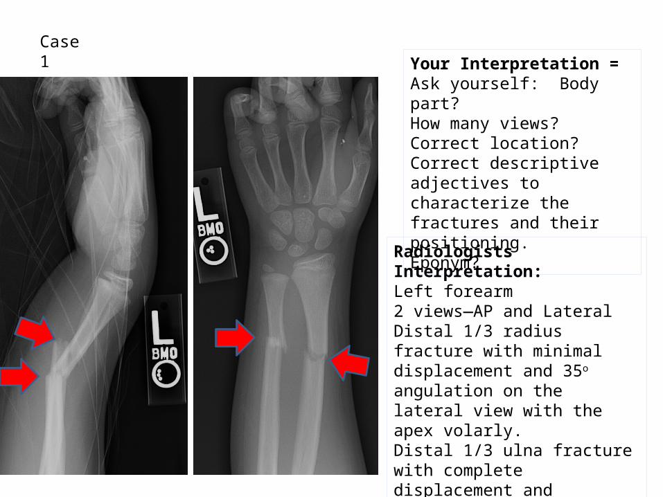
Case 1
Radiologists Interpretation:Left forearm2 views—AP and LateralDistal 1/3 radius fracture with minimal displacement and 35o angulation on the lateral view with the apex volarly.Distal 1/3 ulna fracture with complete displacement and shortening less than 1 cm. It also has 35o angulation with the apex volarly.
Your Interpretation =Ask yourself: Body part?How many views?Correct location?Correct descriptive adjectives to characterize the fractures and their positioning.Eponym?
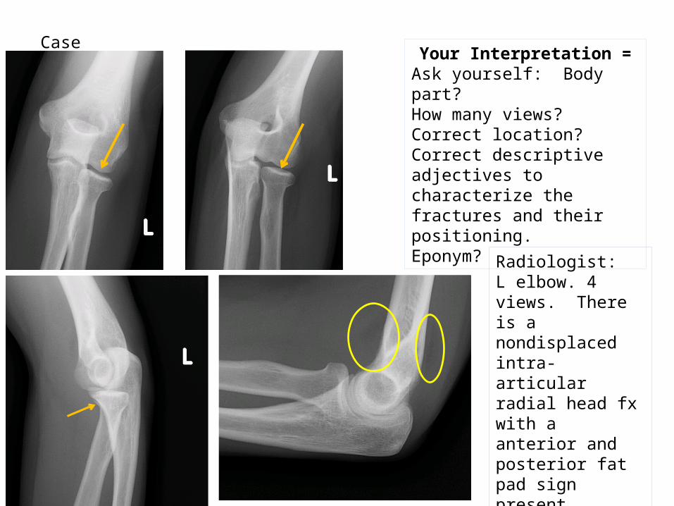
Case 2Your Interpretation =
Ask yourself: Body part?How many views?Correct location?Correct descriptive adjectives to characterize the fractures and their positioning.Eponym?
Radiologist: L elbow. 4 views. There is a nondisplaced intra-articular radial head fx with a anterior and posterior fat pad sign present.

Case 3
Your Interpretation =Ask yourself: Body part?How many views?Correct location?Correct descriptive adjectives to characterize the fractures and their positioning. Eponym?
Radiologist: R tib/fib, 2 views, oblique fracture of the prox diaphysis at the fibular neck, minimally displaced.

Case 4Your Interpretation =
Ask yourself: Body part?How many views?Correct location?Correct descriptive adjectives to characterize the fractures and their positioning.Eponym?
Radiologist: Left 2nd finger, distal phalanx. There is a comminuted tuft and shaft fracture with mild to moderate displacement of the distal fragments.


![The Value of Clinical Interventional Radiology · The growing use of interventional radiology techniques signals a need to redefine the radiologist’s role in patient management…[we]](https://static.fdocuments.us/doc/165x107/5e7989d00b513b2c0448544e/the-value-of-clinical-interventional-radiology-the-growing-use-of-interventional.jpg)