Practice Guidelines for Perioperative Transesophageal...
13
SPECIAL ARTICLES Anesthesiology 2010; 112:1–1 Copyright © 2010, the American Society of Anesthesiologists, Inc. Lippincott Williams & Wilkins Practice Guidelines for Perioperative Transesophageal Echocardiography An Updated Report by the American Society of Anesthesiologists and the Society of Cardiovascular Anesthesiologists Task Force on Transesophageal Echocardiography* P RACTICE Guidelines are systematically developed rec- ommendations that assist the practitioner and the patient in making decisions about health care. These recommendations may be adopted, modified, or rejected according to clinical needs and constraints and are not intended to replace local in- stitutional policies. In addition, Practice Guidelines developed by the American Society of Anesthesiologists (ASA) are not in- tended as standards or absolute requirements, and their use can- not guarantee any specific outcome. Practice Guidelines are sub- ject to revision as warranted by the evolution of medical knowledge, technology, and practice. They provide basic rec- ommendations that are supported by a synthesis and analysis of the current literature, expert and practitioner opinion, open fo- rum commentary, and clinical feasibility data. This update includes data published since the Practice Guidelines for Perioperative Transesophageal Echocardiog- raphy were adopted by the ASA and the Society of Cardio- vascular Anesthesiologists in 1995 and published in 1996. 1 Methodology Definition of Perioperative Transesophageal Echocardiography For these Guidelines, perioperative transesophageal echocardi- ography (TEE) refers to TEE performed on surgical patients before, during, or immediately after surgery, including the crit- ical care setting. Evidence of effectiveness is discussed relative to specific settings where perioperative TEE is customarily used (e.g., cardiac surgery, noncardiac surgery, and critical care). Purposes of the Guidelines The purposes of these Guidelines are (1) to assist the physi- cian in determining the appropriate application of TEE and (2) to improve the outcomes of surgical patients by defining the utility of perioperative TEE based on the strength of supporting evidence. Focus These Guidelines focus on the application of TEE in surgical patients and potential surgical patients in the setting of car- diac surgery, noncardiac surgery, and postoperative critical care. The Guidelines do not apply to the assessment of non- surgical patients or to postdischarge follow-up assessment of surgical patients. The Task Force believes that physician proficiency in the use of perioperative TEE is of paramount importance due to the risk of adverse outcomes resulting from incorrect interpretation. The Guidelines do not address training, certification, creden- tialing, and quality assurance, which are addressed elsewhere. 2–5 Application These Guidelines are intended for anesthesiologists and other physicians (e.g., cardiologists, surgeons, and intensiv- ists) who use TEE in the perioperative setting. Recommen- dations to perform TEE are not applicable when the proce- * Developed by the American Society of Anesthesiologists Task Force on Perioperative Transesophageal Echocardiography: Daniel M. Thys, M.D., Chair, New York, New York; Martin D. Abel, M.B.B.Ch., Rochester, Minnesota; Robert F. Brooker, M.D., Wausau, Wisconsin; Michael K. Cahalan, M.D., Salt Lake City, Utah; Richard T. Connis, Ph.D., Woodinville, Washington; Peggy G. Duke, M.D., Atlanta, Geor- gia; David G. Nickinovich, Ph.D., Bellevue, Washington; Scott T. Reeves, M.D., Charleston, South Carolina; Marc A. Rozner, Ph.D., M.D., Houston, Texas; Isobel A. Russell, M.D., San Francisco, California; Scott C. Streckenbach, M.D., Boston, Massachusetts; Pamela Sears- Rogan, M.D., Washington, DC (American Society of Echocardiogra- phy); and William J. Stewart, M.D., Cleveland, Ohio (American College of Cardiology). Received from American Society of Anesthesiologists, Park Ridge, Illinois. Submitted for publication October 22, 2009. Accepted for publication October 22, 2009. Supported by the American Society of Anesthesiologists and developed under the direction of the Com- mittee on Standards and Practice Parameters, Jeffrey L. Apfelbaum, M.D. (Chair). Approved by the ASA House of Delegates on October 21, 2009, and the SCA Board of Directors on October 21, 2009. A complete bibliography used to develop these Guidelines, arranged alphabetically, is available as Supplemental Digital Content 1, http://links.lww.com/ALN/A567. Address correspondence to The American Society of Anesthesi- ologists: 520 N. Northwest Highway, Park Ridge, Illinois 60068- 2573. This Practice Guideline, as well as all ASA Practice Parameters, may be obtained at no cost through the Journal Web site, www.anesthesiology.org. Supplemental digital content is available for this article. Direct URL citations appear in the printed text and are available in both the HTML and PDF versions of this article. Links to the digital files are provided in the HTML text of this article on the Journals Web site (www.anesthesiology.org). Anesthesiology, V 112 • No 5 1 May 2010
-
Upload
hoangxuyen -
Category
Documents
-
view
225 -
download
0
Transcript of Practice Guidelines for Perioperative Transesophageal...

SPECIAL ARTICLES Anesthesiology 2010; 112:1–1
Copyright © 2010, the American Society of Anesthesiologists, Inc. Lippincott Williams & Wilkins
Practice Guidelines for Perioperative TransesophagealEchocardiography
An Updated Report by the American Society of Anesthesiologists and the Society ofCardiovascular Anesthesiologists Task Force on Transesophageal Echocardiography*
P RACTICE Guidelines are systematically developed rec-ommendations that assist the practitioner and the patient
in making decisions about health care. These recommendationsmay be adopted, modified, or rejected according to clinicalneeds and constraints and are not intended to replace local in-stitutional policies. In addition, Practice Guidelines developedby the American Society of Anesthesiologists (ASA) are not in-tended as standards or absolute requirements, and their use can-not guarantee any specific outcome. Practice Guidelines are sub-ject to revision as warranted by the evolution of medicalknowledge, technology, and practice. They provide basic rec-ommendations that are supported by a synthesis and analysis ofthe current literature, expert and practitioner opinion, open fo-rum commentary, and clinical feasibility data.
This update includes data published since the PracticeGuidelines for Perioperative Transesophageal Echocardiog-raphy were adopted by the ASA and the Society of Cardio-vascular Anesthesiologists in 1995 and published in 1996.1
Methodology
Definition of Perioperative TransesophagealEchocardiographyFor these Guidelines, perioperative transesophageal echocardi-ography (TEE) refers to TEE performed on surgical patientsbefore, during, or immediately after surgery, including the crit-ical care setting. Evidence of effectiveness is discussed relative tospecific settings where perioperative TEE is customarily used(e.g., cardiac surgery, noncardiac surgery, and critical care).
Purposes of the GuidelinesThe purposes of these Guidelines are (1) to assist the physi-cian in determining the appropriate application of TEE and(2) to improve the outcomes of surgical patients by definingthe utility of perioperative TEE based on the strength ofsupporting evidence.
FocusThese Guidelines focus on the application of TEE in surgicalpatients and potential surgical patients in the setting of car-diac surgery, noncardiac surgery, and postoperative criticalcare. The Guidelines do not apply to the assessment of non-surgical patients or to postdischarge follow-up assessment ofsurgical patients.
The Task Force believes that physician proficiency in the useof perioperative TEE is of paramount importance due to the riskof adverse outcomes resulting from incorrect interpretation.The Guidelines do not address training, certification, creden-tialing, and quality assurance, which are addressed elsewhere.2–5
ApplicationThese Guidelines are intended for anesthesiologists andother physicians (e.g., cardiologists, surgeons, and intensiv-ists) who use TEE in the perioperative setting. Recommen-dations to perform TEE are not applicable when the proce-
* Developed by the American Society of Anesthesiologists TaskForce on Perioperative Transesophageal Echocardiography: DanielM. Thys, M.D., Chair, New York, New York; Martin D. Abel, M.B.B.Ch.,Rochester, Minnesota; Robert F. Brooker, M.D., Wausau, Wisconsin;Michael K. Cahalan, M.D., Salt Lake City, Utah; Richard T. Connis,Ph.D., Woodinville, Washington; Peggy G. Duke, M.D., Atlanta, Geor-gia; David G. Nickinovich, Ph.D., Bellevue, Washington; Scott T.Reeves, M.D., Charleston, South Carolina; Marc A. Rozner, Ph.D., M.D.,Houston, Texas; Isobel A. Russell, M.D., San Francisco, California;Scott C. Streckenbach, M.D., Boston, Massachusetts; Pamela Sears-Rogan, M.D., Washington, DC (American Society of Echocardiogra-phy); and William J. Stewart, M.D., Cleveland, Ohio (AmericanCollege of Cardiology).
Received from American Society of Anesthesiologists, Park Ridge,Illinois. Submitted for publication October 22, 2009. Accepted forpublication October 22, 2009. Supported by the American Society ofAnesthesiologists and developed under the direction of the Com-mittee on Standards and Practice Parameters, Jeffrey L. Apfelbaum,M.D. (Chair). Approved by the ASA House of Delegates on October21, 2009, and the SCA Board of Directors on October 21, 2009. Acomplete bibliography used to develop these Guidelines, arrangedalphabetically, is available as Supplemental Digital Content 1,http://links.lww.com/ALN/A567.
Address correspondence to The American Society of Anesthesi-ologists: 520 N. Northwest Highway, Park Ridge, Illinois 60068-2573. This Practice Guideline, as well as all ASA Practice Parameters,may be obtained at no cost through the Journal Web site,www.anesthesiology.org.
� Supplemental digital content is available for this article. DirectURL citations appear in the printed text and are available inboth the HTML and PDF versions of this article. Links to thedigital files are provided in the HTML text of this article on theJournal�s Web site (www.anesthesiology.org).
Anesthesiology, V 112 • No 5 1 May 2010
dure cannot be performed properly or safely nor do theyapply when TEE equipment or skilled examiners are unavail-able. The recommendations in this report are based on con-sideration of the risk benefit ratio for individual patients.
Task Force Members and ConsultantsThe ASA and Society of Cardiovascular Anesthesiologistsjointly appointed a task force of 13 members, including an-esthesiologists in both private and academic practice fromvarious geographic areas of the United States, two cardiolo-gists (one representing the American College of Cardiologyand the other representing the American Society of Echocar-diography), and two consulting methodologists from theASA Committee on Standards and Practice Parameters.
The Task Force developed the Guidelines by means of aseven-step process. First, they reached consensus on the criteriafor evidence. Second, original published research studies frompeer-reviewed journals relevant to TEE were reviewed and eval-uated. Third, expert consultants were asked (1) to participate inopinion surveys on the effectiveness of TEE imaging and (2) toreview and comment on a draft of the Guidelines developed bythe Task Force. Fourth, opinions about the Guidelines recom-mendations were solicited from a sample of active members ofthe ASA who personally perform TEE as a part of their practice.Fifth, the Task Force held an open forum at a major interna-tional meeting† to solicit input on its draft recommendations.Sixth, the consultants were surveyed to assess their opinions onthe feasibility of implementing the Guidelines. Seventh, allavailable information was used to build consensus within theTask Force to finalize the Guidelines (appendix 1).
Availability and Strength of EvidencePreparation of these Guidelines followed a rigorous method-ologic process (appendix 2). Evidence was obtained from twoprincipal sources: scientific evidence and opinion-basedevidence.
Scientific Evidence
Study findings from scientific literature published after1994 (not excluding sentinel articles published prior to1994) were aggregated and reported in summary form byevidence category, as described later. All literature (e.g.,randomized controlled trials, observational studies, andcase reports) relevant to each topic was considered whenevaluating the findings. For reporting purposes in thisdocument, only the highest level of evidence (i.e., levels 1,2, or 3 identified below) within each category (i.e., A, B,or C) is included in the summary.
Category A: Supportive LiteratureRandomized controlled trials report statistically significant(P � 0.01) differences between clinical interventions for aspecified clinical outcome.
Level 1. The literature contains multiple randomized controlledtrials, and the aggregated findings are supported by meta-analysis.‡
Level 2. The literature contains multiple randomized con-trolled trials, but there is an insufficient number of studiesto conduct a viable meta-analysis for the purpose of theseGuidelines.
Level 3. The literature contains a single randomized con-trolled trial.
Category B: Suggestive LiteratureInformation from observational studies permits inference ofbeneficial or harmful relationships among clinical interven-tions and clinical outcomes.
Level 1. The literature contains observational comparisons(e.g., cohort and case–control research designs) of two ormore clinical interventions or conditions and indicatesstatistically significant differences between clinical inter-ventions for a specified clinical outcome.
Level 2. The literature contains noncomparative observa-tional studies with associative (e.g., relative risk, correla-tion) or descriptive statistics.
Level 3. The literature contains case reports.
Category C: Equivocal LiteratureThe literature cannot determine whether there are beneficialor harmful relationships among clinical interventions andclinical outcomes.
Level 1. Meta-analysis did not find significant differencesamong groups or conditions.
Level 2. There is an insufficient number of studies toconduct meta-analysis, and (1) randomized controlledtrials have not found significant differences amonggroups or conditions, or (2) randomized controlled tri-als report inconsistent findings.
Level 3. Observational studies report inconsistent findings or donot permit inference of beneficial or harmful relationships.
Category D: Insufficient Evidence from LiteratureThe lack of scientific evidence in the literature is described bythe following conditions.
(1) No identified studies address the specified relationshipsamong interventions and outcomes.
(2) The available literature cannot be used to assess the rela-tionships among clinical interventions and clinical out-comes. The literature either does not meet the criteria forcontent as defined in the “Focus” of the Guidelines ordoes not permit a clear interpretation of findings due tomethodologic concerns (e.g., confounding in study de-sign or implementation).
† Society of Cardiovascular Anesthesiologists, 30th Annual Meet-ing, Vancouver, British Columbia, Canada, June 20, 2008.
‡ All meta-analyses are conducted by the ASA methodologygroup. Meta-analyses from other sources are reviewed but notincluded as evidence in this document.
2 Practice Guidelines
Anesthesiology, V 112 • No 5 • May 2010 Practice Guidelines
Opinion-based EvidenceAll opinion-based evidence relevant to each topic (e.g., surveydata, open-forum testimony, Internet-based comments, letters,and editorials) was considered in the development of theseGuidelines. However, only the findings obtained from formalsurveys are reported.
Opinion surveys were developed by the Task Force toaddress each clinical intervention identified in the docu-ment. Identical surveys were distributed to two groups ofrespondents: expert consultants and ASA members.
Category A: Expert OpinionSurvey responses from Task Force–appointed expert consult-ants are reported in summary form in the text. A completelisting of consultant survey responses is reported in a table inappendix 2.
Category B: Membership OpinionSurvey responses from a sample of members of the ASA arereported in summary form in the text. A complete listing of ASAmember survey responses is reported in a table in appendix 2.
Expert consultant and ASA membership survey responsesare recorded using a 5-point scale and summarized based onmedian values.§
Strongly agree: median score of 5 (at least 50% of the re-sponses are 5).
Agree: median score of 4 (at least 50% of the responses are 4or 4 and 5).
Equivocal: median score of 3 (at least 50% of the responsesare 3, or no other response category or combination ofsimilar categories contain at least 50% of the responses).
Disagree: median score of 2 (at least 50% of responses are 2or 1 and 2).
Strongly disagree: median score of 1 (at least 50% of re-sponses are 1).
Category C: Informal OpinionOpen-forum testimony, Internet-based comments, letters,and editorials are all informally evaluated and discussed dur-ing the development of Guidelines recommendations. Whenwarranted, the Task Force may add educational informationor cautionary notes based on this information.
Guidelines
Cardiac and Thoracic Aortic ProceduresCardiac and thoracic aortic procedures consist of cardiac andthoracic aortic surgery, and catheter-based intracardiac procedures.
Cardiac and thoracic aortic surgery: For cardiac or thoracicaortic surgery patients, the literature reports variations insensitivity, specificity, or positive and negative predictive val-
ues for the detection of abnormalities relating to valvular,coronary, aortic, congenital, and other cardiovascular disease(table 1 in appendix 2). Examples of these abnormalitiesinclude mitral valve abnormalities, valvular abscesses, myo-cardial ischemia, aortic dissection, and atrial septal defect(Category B2 evidence). The literature also reports a range ofsensitivity, specificity, and positive and negative predictivevalues for the confirmation or refinement by TEE of thepreoperative diagnosis (table 1 in appendix 2). Examples in-clude aortic dissection, aortic intramural hemorrhage, andvalvular or mural infective endocarditis lesions (Category B2evidence). The ASA members agree and the consultantsstrongly agree that TEE should be used for all cardiac orthoracic aortic surgery patients.Recommendations for cardiac and thoracic aortic sur-gery. For adult patients without contraindications, TEEshould be used in all open heart (e.g., valvular procedures)and thoracic aortic surgical procedures and should be con-sidered in coronary artery bypass graft surgeries to: (1) con-firm and refine the preoperative diagnosis, (2) detect new orunsuspected pathology, (3) adjust the anesthetic and surgicalplan accordingly, and (4) assess the results of surgical inter-vention. In small children, the use of TEE should be consid-ered on a case-by-case basis because of risks unique to thesepatients (e.g., bronchial obstruction).
Catheter-based intracardiac procedures: Studies with ob-servational findings confirm the utility of TEE or intra-cardiac echocardiography for guiding management ofcatheter-based intracardiac procedures (e.g., occluder de-vice placement, percutaneous valvular procedures, and in-tracardiac ablation procedures) (Category B2 evidence). Inaddition, studies with observational findings report thedetection of unsuspected abnormalities by TEE, such asaortic root abscess, atrial thrombi, atrial septal aneurysm,shunting, mitral valve/annular calcification and regurgi-tation, wall motion abnormalities, and tamponade (Cate-gory B2 evidence). The detection of pericardial effusion isalso reported (Category B3 evidence).
Both the consultants and ASA members agree that TEEshould be used for patients undergoing transcatheter intracar-diac procedures when general anesthesia is provided and intra-cardiac ultrasound is not used. The ASA members agree and theconsultants strongly agree that TEE should be used for septaldefect closure or atrial appendage obliteration. Both the consult-ants and ASA members strongly agree that TEE should be usedduring catheter-based valve replacement and repair. Finally,both the consultants and ASA members are equivocal regardingthe use of TEE during dysrhythmia treatment.Recommendations for catheter-based intracardiac pro-cedures. For patients undergoing transcatheter intracardiacprocedures, TEE may be used.
Noncardiac SurgeryFor noncardiac surgery patients, studies with observational find-ings or case reports note the detection of the following abnor-malities by TEE: (1) venous air embolism and patent foramen
§ When an equal number of responses are obtained, the medianvalue is determined by calculating the arithmetic mean of the twomiddle values. Ties are determined by a predetermined formula.
3SPECIAL ARTICLES
Practice Guidelines Anesthesiology, V 112 • No 5 • May 2010
ovale in neurosurgery (Category B2 evidence); (2) pericardial ef-fusion and compression of the cardiac chambers in liver trans-plantation (Category B3 evidence); (3) intracardiac emboli andpatent foramen ovale (Category B2 evidence), mitral regurgita-tion, left ventricular hypertrophy, and left ventricular outflowtract obstruction in orthopedic surgery (Category B3 evidence),(4) left ventricular segmental wall motion abnormalities (Cate-gory B2 evidence), aortic lesions and atrial tumors in vascularsurgery (Category B3 evidence), and (5) atrial septal defect, myo-cardial ischemia, hypovolemia, pericardial tamponade, throm-boembolic events (Category B2 evidence), pericardial effusion,tamponade, and intrapulmonary emboli in other major surgery(i.e., lung, renal, abdominal, and head/neck/chest wall surgeries)(Category B3 evidence).
The consultants and ASA members agree that TEE shouldbe used for noncardiac surgical patients when the patient hasknown or suspected cardiovascular pathology that might resultin hemodynamic, pulmonary, or neurologic compromise. Theconsultants and ASA members both strongly agree that TEEshould be used during unexplained persistent hypoten-sion. Further, both the consultants and ASA membersagree that TEE should be used when persistent unex-plained hypoxemia occurs. The ASA members agree andthe consultants strongly agree that TEE should be usedwhen life-threatening hypotension is anticipated.
Both the consultants and ASA members agree that TEEshould be used during either lung transplantation or major ab-dominal or thoracic trauma. The consultants agree although theASA members are equivocal regarding the use of TEE duringopen abdominal aortic procedures and liver transplantation.Both the consultants and ASA members are equivocal regardingthe use of TEE during: (1) endovascular aortic procedures, (2)neurosurgery in the sitting position, and (3) percutaneous car-diovascular interventions (e.g., femoral artery stenting). Finally,the consultants and ASA members both disagree with the asser-tion that TEE should be used during orthopedic surgery.Recommendations for noncardiac surgery. TEE may beused when the nature of the planned surgery or the pa-tient’s known or suspected cardiovascular pathologymight result in severe hemodynamic, pulmonary, or neu-rologic compromise. If equipment and expertise are avail-able, TEE should be used when unexplained life-threat-ening circulatory instability persists despite correctivetherapy.
Critical CareStudies with observational findings for critically ill patients withan unexplained adverse postoperative clinical course report TEEdetection for the following abnormalities: regurgitant valvularlesions, aortic or mitral valve vegetation, aortic dissection, intra-cardiac mass, tamponade, ventricular failure, and hypovolemia(Category B2 evidence). Case reports of critically ill postoperativepatients indicate that TEE detects abnormalities such as aorticroot abscess, pericardial hematoma, atherosclerotic debris in the
thoracic aorta, left ventricular hypertrophy, wall motion abnor-malities, and ventricular masses (Category B3 evidence).
Both the consultants and ASA members strongly agreethat TEE should be used for critical care patients whendiagnostic information expected to alter managementcannot be obtained by transthoracic echocardiography orother modalities in a timely manner. The ASA membersagree and the consultants strongly agree that TEE shouldbe used during unexplained persistent hypotension. Theyboth agree that TEE should be used when persistent un-explained hypoxemia occurs.Recommendations for critical care. For critical care pa-tients, TEE should be used when diagnostic information thatis expected to alter management cannot be obtained by trans-thoracic echocardiography or other modalities in a timelymanner.
Contraindications for the Use of TEEStudies with observational findings and case reports indicatethat, although rare, potential complications associated withTEE may include esophageal perforation, esophageal injury,hematoma, laryngeal palsy, dysphagia, dental injury, ordeath (Category B2 evidence). However, there is insufficientliterature to assess whether there are contraindications for theuse of TEE (Category D evidence).
Both the consultants and ASA members are equivocal withregard to whether there are no absolute contraindications toTEE other than previous esophagectomy or esophagogastrec-tomy. Those consultants and ASA members who do not agreethat there are no absolute contraindications other than previousesophagectomy or esophagogastrectomy do agree that the fol-lowing four conditions should be absolute contraindications toTEE: esophageal stricture, tracheoesophageal fistula,postesophageal surgery, and esophageal trauma. Both the con-sultants and ASA members disagree that the following four con-ditions should be absolute contraindications to TEE: Barrettesophagus, hiatal hernia, large descending aortic aneurysm, andunilateral vocal cord paralysis. Finally, both the consultants andASA members are equivocal with regard to whether the follow-ing three conditions should be absolute contraindications toTEE: esophageal varices, postradiation therapy, and previousbariatric surgery. The consultants agree but the ASA membersare equivocal that Zenker diverticulum and colonic interposi-tion are absolute contraindications. Finally, the ASA membersdisagree and the consultants are equivocal that dysphagia is anabsolute contraindication to TEE.Recommendations. TEE may be used for patients with oral,esophageal, or gastric disease, if the expected benefit outweighsthe potential risk, provided the appropriate precautions are ap-plied. These precautions may include the following: consideringother imaging modalities (e.g., epicardial echocardiography),obtaining a gastroenterology consultation using a smaller probe,limiting the examination, avoiding unnecessary probe manipu-lation, and using the most experienced operator.
4 Practice Guidelines
Anesthesiology, V 112 • No 5 • May 2010 Practice Guidelines
References
1. American Society of Anesthesiologists: Practice guidelinesfor perioperative transesophageal echocardiography. AN-ESTHESIOLOGY 1996; 84:986 –1006
2. Beïque F, Ali M, Hynes M, MacKenzie S, Denault A, Mar-tineau A, MacAdams C, Sawchuk C, Hirsch K, Lampa M,Murphy P, Honos G, Munt B, Sanfilippo A, Duke P: Car-diovascular Section of the Canadian Anesthesiologists’ So-ciety; Canadian Society of Echocardiography: Canadianguidelines for training in adult perioperative transesopha-geal echocardiography. Recommendations of the Cardio-vascular Section of the Canadian Anesthesiologists’ Societyand the Canadian Society of Echocardiography. Can J An-aesth 2006; 53:1044 – 60
3. Cahalan MK, Abel M, Goldman M, Pearlman A, Sears-RoganP, Russell I, Shanewise J, Stewart W, Troianos C: AmericanSociety of Echocardiography; Society of CardiovascularAnesthesiologists, American Society of Echocardiography,and Society of Cardiovascular Anesthesiologists task forceguidelines for training in perioperative echocardiography.Anesth Analg 2002; 94:1384 – 8
4. Mathew JP, Glas K, Troianos CA, Sears-Rogan P, Savage R,Shanewise J, Kisslo J, Aronson S, Shernan S: Council forIntraoperative Echocardiography of the American Societyof Echocardiography: ASE/Society of Cardiovascular Anes-thesiologists recommendations and guidelines for contin-uous quality improvement in perioperative echocardiogra-phy. Anesth Analg 2006; 103:1416 –25
5. Swanevelder J, Chin D, Kneeshaw J, Chambers J, BennettS, Smith D, Nihoyannopoulos P: Accreditation in transoe-sophageal echocardiography: Statement from the Associa-tion of Cardiothoracic Anaesthetists and the British Societyof Echocardiography Joint TOE Accreditation Committee.Br J Anaesth 2003; 91:469 –72
6. De Castro S, Cartoni D, d’Amati G, Beni S, Yao J, Fiorell M,Gallo P, Fedele F, Pandian NG: Diagnostic accuracy oftransthoracic and multiplane transesophageal echocardi-ography for valvular perforation in acute infective endo-carditis: Correlation with anatomic findings. Clin InfectDis 2000; 30:825– 6
7. Espinal M, Fuisz AR, Nanda NC, Aaluri SR, Mukhtar O,Sekar PC: Sensitivity and specificity of transesophagealechocardiography for determination of aortic valve mor-phology. Am Heart J 2000; 139:1071– 6
8. Hellemans IM, Pieper EG, Ravelli AC, Hamer JP, Jaarsma W,van den Brink RB, Peels CH, van Swieten H, Tijssen JG,Visser CA: Comparison of transthoracic and transesopha-geal echocardiography with surgical findings in mitral re-gurgitation. Am J Cardiol 1996; 77:728 –33
9. Muller S, Muller L, Laufer G, Alber H, Dichtl W, Frick M, Pach-inger O, Bartel T: Comparison of three-dimensional imaging totransesophageal echocardiography for preoperative evaluationin mitral valve prolapse. Am J Cardiol 2006; 98:243–8
10. Grewal KS, Malkowski MJ, Kramer CM, Dianzumba S,Reichek N: Multiplane transesophageal echocardiographicidentification of the involved scallop in patients with flailmitral valve leaflet: Intraoperative correlation. J Am SocEchocardiogr 1998; 11:966 –71
11. Senni M, Merlo M, Sangiorgi G, Gamba A, Procopio A,Glauber M, Ferrazzi P: Mitral valve repair and transesoph-ageal echocardiographic findings in a high-risk subgroupof patients with active, acute infective endocarditis.J Heart Valve Dis 2001; 10:72–7
12. Morguet AJ, Werner GS, Andreas S, Kreuzer H: Diagnosticvalue of transesophageal compared with transthoracicechocardiography in suspected prosthetic valve endocar-ditis. Herz 1995; 20:390 – 8
13. San Roman JA, Vilacosta I, Sarria C, de la Fuente L, Sanz O,Vega JL, Ronderos R, Gonzalez Pinto A, Jesus Rollan M,Graupner C, Batlle E, Lahulla F, Stoermann W, Portis M,Fernandez-Aviles F: Clinical course, microbiologic profile,and diagnosis of periannular complications in prostheticvalve endocarditis. Am J Cardiol 1999; 83:1075–9
14. Comunale ME, Body SC, Ley C, Koch C, Roach G, MathewJP, Herskowitz A, Mangano DT: The concordance of intra-operative left ventricular wall-motion abnormalities andelectrocardiographic S-T segment changes: Associationwith outcome after coronary revascularization. Multi-center Study of Perioperative Ischemia (McSPI) ResearchGroup. ANESTHESIOLOGY 1998; 88:945–54
15. Kodolitsch Y, Krause N, Spielmann R, Nienaber CA: Diag-nostic potential of combined transthoracic echocardiogra-phy and x-ray computed tomography in suspected aorticdissection. Clin Cardio 1999; 22:345–52
16. Laissy JP, Blanc F, Soyer P, Assayag P, Sibert A, Tebboune D,Arrive L, Brochet E, Hvass U, Langlois J, Menu Y: Thoracic aorticdissection: Diagnosis with transesophageal echocardiographyversus MR imaging. Radiology 1995; 194:331–6
17. Patel S, Alam M, Rosman H: Pitfalls in the echocardio-graphic diagnosis of aortic dissection. Angiology 1997;48:939 – 46
18. Sommer T, Fehske W, Holzknecht N, Smekal AV, Keller E,Lutterbey G, Kreft B, Kuhl C, Gieseke J, Abu RD, Schild H:Aortic dissection: A comparative study of diagnosis withspiral CT, multiplanar transesophageal echocardiography,and MR imaging. Radiology 1996; 199:347–52
19. Konstadt SN, Reich DL, Kahn R, Viggiani RF: Transesoph-ageal echocardiography can be used to screen for ascend-ing aortic atherosclerosis. Anesth Analg 1995; 81:225– 8
20. Minard G, Schurr MJ, Croce MA, Gavant ML, Kudsk KA, TaylorMJ, Pritchard FE, Fabian TC: A prospective analysis of trans-esophageal echocardiography in the diagnosis of traumatic dis-ruption of the aorta. J Trauma 1996; 40:225–30
21. Vignon P, Gueret P, Vedrinne JM, Lagrange P, Cornu E, AbrieuO, Gastinne H, Bensaid J, Lang RM: Role of transesophagealechocardiography in the diagnosis and management of trau-matic aortic disruption. Circulation 1995; 92:2959–68
22. Singh GK, Shiota T, Cobanoglu A, Droukas P, Rice MJ, SahnDJ: Diagnostic accuracy and role of intraoperative biplanetransesophageal echocardiography in pediatric patientswith left ventricle outflow tract lesions. J Am Soc Echo-cardiogr 1998; 11:47–56
23. Rosenberger P, Shernan SK, Body SC, Eltzschig HK: Utilityof intraoperative transesophageal echocardiography fordiagnosis of pulmonary embolism. Anesth Analg 2004;99:12– 6
24. Attenhofer CH, Vogt PR, von Segesser LK, Dirsch OR,Ritter M, Jenni R: Leaking giant aneurysm of the aortic rootdue to cystic medial necrosis with pericardial tamponademimicking type-A aortic dissection. Thorac CardiovascSurg 1996; 44:103– 4
25. Theodore S, Vaidyanathan K, Jagannath BR, Nainar M, Krish-namoorthy J, Cherian KM: Takayasu’s arteritis mimicking acuteaortic dissection. Ann Thorac Surg 2007; 83:1876–8
26. Berdat PA, Carrel T: Aortic dissection limited to the as-cending aorta mimicking intramural hematoma. Eur J Car-diothorac Surg 1999; 15:108 –9
27. Vuille C, Trigo-Trindade P, Lerch R, Kalangos A: Commoncoronary ostium mimicking an aortic abscess in a case ofbacterial endocarditis. J Am Soc Echocard 1998; 11:80 –2
28. Kupersmith AC, Belkin RN, McClung JA, Moggio RA: Aor-tic valve commissural tear mimicking type A aortic dissec-tion. J Am Soc Echocardiogr 2002; 15:658 – 60
29. Akowuah EF, Onyeaka CV, Cooper GJ: A subtle sign ofaortic outflow obstruction in an infected 29 year oldStarr-Edward’s valve. Heart 2001; 85:384
30. Sherwood JT, Gill IS: Missed acute ascending aortic dissec-tion. J Card Surg 2001; 16:86 – 8
31. Kaneko Y, Furuse A, Takeshita M, Miyaji K, Yagyu K: Fibroustissue overgrowth and prosthetic valve endocarditis: report of acase. Thorac Cardiovasc Surg 1997; 45:150–2
32. Park P, Khawly JA, Kearney DL, Altman CA, Yen KG: Bilateralendogenous endophthalmitis secondary to endocarditis withnegative transesophageal echocardiogram. Am J Ophthalmol2004; 138:151–3
33. Dalal A, Asirvatham SJ, Chandrasekaran K, Seward JB, Tajik AJ:
5SPECIAL ARTICLES
Practice Guidelines Anesthesiology, V 112 • No 5 • May 2010
Intracardiac echocardiography in the detection of pacemakerlead endocarditis. J Am Soc Echocard 2002; 15:1027–8
34. Flachskampf FA, Banbury M, Smedira N, Thomas JD, Garcia M:Transesophageal echocardiography diagnosis of intramural he-matoma of the ascending aorta: A word of caution. J Am SocEchocardiogr 1999; 12:866–70
35. Lick SD, Zwischenberger JB, Mileski WJ, Ahmad M: Torn as-cending aorta missed by transesophageal echocardiography.Ann Thorac Surg 1997; 63:1768–70
36. Kang DH, Song JK, Song MG, Lee IS, Song H, Lee JW, Park SW,Kim YH, Lim TH, Park SJ: Clinical and echocardiographic out-comes of aortic intramural hemorrhage compared with acuteaortic dissection. Am J Cardiol 1998; 81:202–6
37. Beauchesne LM, Veinot JP, Brais MP, Burwash IG, Chan KL:Acute aortic intimal tear without a mobile flap mimicking anintramural hematoma. J Am Soc Echocardiogr 2003; 16:285–8
38. Bauer M, Redzepagic S, Weng Y, Hetzer R: Successful surgicaltreatment of a giant aneurysm of the right coronary artery.Thorac Cardiovasc Surg 1998; 46:152–4
39. Kunzli A, von Segesser LK, Vogt PR, Spahn DR, Schneider J,Jenni R, Turina MI: Inflammatory aneurysm of the ascendingaorta. Ann Thorac Surg 1998; 65:1132–3
40. Paolillo V, Gastaldo D, Barretta A, Guerra F: Idiopathic orga-nized thrombus of the tricuspid valve mimicking valvular tu-mor. Tex Heart Inst J 2004; 31:192–3
41. Konishi H, Fukuda M, Kato M, Misawa Y, Fuse K: Organizedthrombus of the tricuspid valve mimicking valvular tumor. AnnThorac Surg 2001; 71:2022–4
Appendix 1: Summary of Recommendations
Cardiac and Thoracic Aortic Procedures
● Cardiac and Thoracic Aortic Surgery• For adult patients without contraindications, TEE should be
used in all open heart (e.g., valvular procedures) and thoracicaortic surgical procedures and should be considered in CABGsurgeries as well• to confirm and refine the preoperative diagnosis,• to detect new or unsuspected pathology,• to adjust the anesthetic and surgical plan accordingly, and• to assess the results of the surgical intervention.
• In small children, the use of TEE should be considered on acase-by-case basis because of risks unique to these patients (e.g.,bronchial obstruction).
● Catheter-Based Intracardiac Procedures• For patients undergoing transcatheter intracardiac procedures,
TEE may be used.
Noncardiac Surgery
● TEE may be used when the nature of the planned surgery orthe patient’s known or suspected cardiovascular pathologymight result in severe hemodynamic, pulmonary, or neuro-logic compromise.
● If equipment and expertise are available, TEE should be usedwhen unexplained life-threatening circulatory instability persistsdespite corrective therapy.
Critical Care
● For critical care patients, TEE should be used when diagnosticinformation that is expected to alter management cannot be ob-tained by transthoracic echocardiography or other modalities in atimely manner.
Contraindications for the Use of TEE
● TEE may be used for patients with oral, esophageal, or gastricdisease, if the expected benefit outweighs the potential risk, pro-vided the appropriate precautions are applied. These precautionsmay include:
• considering other imaging modalities (e.g., epicardial echocar-diography)
• obtaining a gastroenterology consultation• using a smaller probe• limiting the examination• avoiding unnecessary probe manipulation• using the most experienced operator
Appendix 2: Methods and Analyses
State of the LiteratureFor these Guidelines, a literature review was used in combinationwith opinions obtained from expert consultants and other sources(e.g., ASA members, open forums, Internet postings). Both theliterature review and opinion data were based on evidence linkagesor statements regarding potential relationships between clinical in-terventions and outcomes. The efficacy and outcomes from the useof TEE were examined for the following procedures:
1. cardiac and thoracic aortic surgery2. transcatheter intracardiac procedures3. pacemaker and implanted cardioverter defibrillator lead
extraction4. neurosurgery5. liver transplantation6. orthopedic surgery7. vascular/endovascular surgery8. other major surgery (i.e., lung, renal, abdominal, and head/neck/
chest wall)9. postoperative critical care
The impact of the use of perioperative TEE was assessed on thebasis of the following:
1. perioperative detection or diagnosis of new or unsuspectedpathology
2. confirming or refinement of previous perioperative diagnoses3. preoperative or intraoperative refinement of a surgical plan4. detecting complications during surgery5. assessing surgery outcomes6. planning and confirming device placement7. beneficial or adverse patient outcomes from the use of TEE
For the literature review, potentially relevant clinical studies pub-lished after 1994 were identified via electronic and manual searches ofthe literature. The electronic and manual searches covered a 16-yr pe-riod from 1994 through 2009. More than 8000 citations were initiallyidentified, yielding a total of 861 nonoverlapping articles thataddressed topics related to the evidence linkages. After review ofthe articles, 404 studies did not provide direct evidence and weresubsequently eliminated. A total of 457 articles contained directlinkage-related evidence. A complete bibliography used to de-velop these Guidelines, organized by section, is available as Sup-plemental Digital Content 2, http://links.lww.com/ALN/A568.
Literature reporting the detection of new abnormalities by TEEwas summarized, followed by a summary of literature reporting theconfirmation of previously diagnosed abnormalities by TEE. The
6 Practice Guidelines
Anesthesiology, V 112 • No 5 • May 2010 Practice Guidelines
sensitivity, specificity, and positive and negative predictive valuesfor the efficacy of TEE in detecting new abnormalities and in con-firming or redefining previous diagnoses were also obtained (table1). Study findings reporting the misdiagnosis or limited effective-ness of TEE to detect pathology are also listed in table 1.
Interobserver agreement among Task Force members and twomethodologists was established by interrater reliability testing.Agreement levels using a � statistic for two-rater agreement pairswere as follows: (1) type of study design, � � 0.50–1.00; (2) type ofanalysis, � � 0.50–0.83; (3) evidence linkage assignment, � �0.75–1.00; and (4) literature inclusion for database, � � 0.78–1.00. Three-rater chance-corrected agreement values were as fol-lows: (1) study design, Sav � 0.66, Var (Sav) � 0.006; (2) type ofanalysis, Sav � 0.66, Var (Sav) � 0.007; (3) linkage assignment,Sav � 0.83, Var (Sav) � 0.005; and (4) literature database inclu-sion, Sav � 0.84, Var (Sav) � 0.046. These values represent mod-erate to high levels of agreement.
Consensus-based EvidenceConsensus was obtained from multiple sources, including (1) surveyopinion from consultants who were selected based on their knowledgeor expertise in the perioperative use of TEE, (2) survey opinions solic-ited from active members of the ASA who personally perform TEE aspart of their practice, (3) testimony from attendees of a publicly held
open forum at an international anesthesia meeting, (4) Internetcommentary, and (5) Task Force opinion and interpretation.The survey rate of return was 53% (n � 55 of 103) for theconsultants, and 818 surveys were received from active ASAmembers who indicated that they personally performed TEE aspart of their practice. Results of the surveys are reported in tables2 and 3 and summarized in the text of the Guidelines.
The consultants were asked to indicate which, if any,of the recommendations would change their clinical practices if theGuidelines were instituted. The rate of return was 14% (n � 14 of103). The percent of responding consultants expecting a change intheir practice associated with each linkage topic was as follows: (1)major cardiac and thoracic aortic surgery, 7%; (2) transcatheter intra-cardiac procedures, 0%; (3) pacemaker and implanted cardioverterdefibrillator lead extraction, 7% (4); neurosurgery, 7% (5); liver trans-plantation, 0% (6); orthopedic surgery, 7% (7); vascular/endovascularsurgery, 7%, (8) other major surgery (i.e., lung, renal, abdominal, andhead/neck/chest wall), 14%; and (9) postoperative critical care, 21%.Eighty-six percent indicated that their clinical practice will not neednew equipment, supplies, or training to implement the Practice Guide-lines. Eighty-six percent indicated that the Guidelines would not re-quire ongoing changes in their practice which will affect costs. Onehundred percent of the respondents indicated that the Guidelineswould have no effect on the amount of time spent on a typical case.
7SPECIAL ARTICLES
Practice Guidelines Anesthesiology, V 112 • No 5 • May 2010

Table 1. Sensitivity, Specificity, and Predictive Values for Perioperative TEE
Detection/Diagnosis of PathologySensitivity
(%)Specificity
(%) PPV NPV
Valvular disease:Aortic, mitral, or tricuspid valvular perforation (confirmed by surgery
or autopsy)695 98 * *
Abnormal bicuspid and tricuspid aortic valve morphology (confirmedby surgery)7
Biplane TEE 66 56 * *Multiplane TEE 87 91 * *
Chordal rupture (confirmed by surgery)8 79 96 * *Mitral valve annular dilatation (confirmed by surgery)8 78 50 * *Mitral valve leaflet degeneration (confirmed by surgery)8 41 87 * *Mitral valve prolapse/flail (confirmed by surgery)9
Bileaflet involvement or combined lesion including the commissures 20 93 * *Single leaflet but multiscallop involvement 57 96 * *Commissure involvement 11 98 * *
Mitral valve/flail leaflet scallop (confirmed by surgery)10 78 92 * *Mitral valve regurgitation (confirmed by surgery)11 87 100 100% 92%Mitral vegetation (confirmed by surgery)11 90 100 100% 75%Prosthetic valve endocarditis (pathoanatomic confirmation)12 92 97 * *Prosthetic valve fistula (confirmed by surgery or necropsy)13 100 100 * *Valvular abscess (confirmed by surgery or necropsy)13 90 100 * *
Coronary disease:Myocadial infarction (confirmed by creatine kinase-MB level �100
ng/ml within 12 h after operation or new Q waves on arrival in ICUor on morning of postoperative day 1)14
45 73 27% 86%
Pseudonaneurysm (confirmed by surgery or necropsy)13 100 98 * *Aortic disease:
Aortic dissection (confirmed by aortography, surgery, or necropsy)15 67 70 * *Aortic dissection (confirmed by double-blind readings of the
images)1686 67 * *
Aortic dissection—type I or III (confirmed by CT/MRI, surgery, orautopsy)17
100 * * *
Aortic dissection—thoracic (confirmed by angiography, surgery, orautopsy)18
100 94 * *
Atherosclerosis of the ascending aorta (confirmed by epiaorticscanning)19
100 60 34% 100%
Traumatic disruption of the aorta (confirmed by aortography, clinicalfindings, or both)20
57 91 * *
Traumatic disruption of the aorta (confirmed by surgery)21 91 100 * *Other cardiovascular diseases:
Left ventricular outflow tract lesions (confirmed by surgery, catheterfindings)22
94 100 * *
Pulmonary embolus (confirmed by surgery)23
Anywhere within the pulmonary arterial circulation 46 * * *At one of three specific localizations 26 95 93% 32%
False positives/negatives:Preoperative TEE detected aneurysm and pericardial effusion; neither confirmed at surgery24
Preoperative TEE detected aortic dissection; surgery revealed Takayasu arteritis25
Preoperative TEE detected intramural hematoma; surgery revealed aortic dissection26
Preoperative TEE detected mass consistent with periannular abscess; surgery revealed coronary ostium27
Preoperative TEE detected type A aortic dissection; surgery revealed aortic valve commissural tear28
Preoperative TEE did not detect aortic outflow obstruction; surgery revealed occluded valve orifice29
Preoperative TEE did not detect ascending aortic dissection; revealed at surgery30
Preoperative TEE did not detect calcified fibrous tissue obstructing mechanical valve inflow; detected at surgery31
Preoperative TEE did not detect endocarditis, aortic root abscess; revealed at surgery32
Preoperative TEE did not detect endocarditis; detected by intracardiac echocardiography33
Preoperative TEE did not detect hematoma of ascending aorta; detected by CT34
Preoperative TEE did not detect torn ascending aorta, detected by aortography35
(continued)
8 Practice Guidelines
Anesthesiology, V 112 • No 5 • May 2010 Practice Guidelines

Table 1. Continued
Detection/Diagnosis of PathologySensitivity
(%)Specificity
(%) PPV NPV
Confirming/refining diagnosis:Aortic intramural hemorrhage (confirmed by surgery or follow-up
changes)36100 91 * *
False positives/negatives:Preoperative emergency TEE confirmed intramural hematoma; surgery revealed acute aortic intimal tear without a
mobile flap37
Preoperative emergency TEE confirmed pericardial cyst; surgery revealed coronary arterial aneurysm38
Preoperative TEE confirmed ascending aorta dissection; surgery revealed chronic inflammatory aneurysm39
Preoperative TEE confirmed tricuspid valve mass; surgery revealed thrombus40
Preoperative TEE confirmed valvular tumor; surgery revealed organized thrombus when resected41
* No available data.CT � computed tomography; ICU � intensive care unit; MRI � magnetic resonance imaging; NPV � negative predictive value; PPV �positive predictive value; TEE � transesophageal echocardiography.
9SPECIAL ARTICLES
Practice Guidelines Anesthesiology, V 112 • No 5 • May 2010
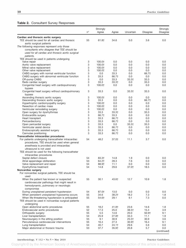
Table 2. Consultant Survey Responses
n*StronglyAgree Agree Uncertain Disagree
StronglyDisagree
Cardiac and thoracic aortic surgeryTEE should be used for all cardiac and thoracic
aortic surgical patients55 61.8† 34.6 0.0 3.6 0.0
The following responses represent only thoseconsultants who disagree that TEE should beused for all cardiac and thoracic aortic surgicalpatients.
TEE should be used in patients undergoingValve repair 3 100.0† 0.0 0.0 0.0 0.0Aortic valve replacement 3 100.0† 0.0 0.0 0.0 0.0Mitral valve replacement 3 100.0† 0.0 0.0 0.0 0.0Other valve replacement 3 100.0† 0.0 0.0 0.0 0.0CABG surgery with normal ventricular function 3 0.0 33.3 0.0 66.7† 0.0CABG surgery with abnormal ventricular function 3 33.3 66.7† 0.0 0.0 0.0Off-pump CABG 3 0.0 33.3 33.3† 33.3 0.0Redo cardiac surgery 3 33.3 33.3† 0.0 33.3 0.0Congenital heart surgery with cardiopulmonary
bypass3 100.0† 0.0 0.0 0.0 0.0
Congenital heart surgery without cardiopulmonarybypass
3 33.3 0.0 33.3† 33.3 0.0
Ascending thoracic aortic surgery 3 100.0† 0.0 0.0 0.0 0.0Descending thoracic aortic surgery 3 33.3 0.0 0.0 66.7† 0.0Hypertrophic cardiomyopathy surgery 3 100.0† 0.0 0.0 0.0 0.0Resection of cardiac mass 3 100.0† 0.0 0.0 0.0 0.0Ventricular remodeling surgery 3 100.0† 0.0 0.0 0.0 0.0Open surgery for dysrhythmias 3 33.3 33.3† 0.0 33.3 0.0Endocarditis surgery 3 66.7† 33.3 0.0 0.0 0.0Heart transplant 3 33.3 66.7† 0.0 0.0 0.0Pericardiectomy 3 33.3 66.7† 0.0 0.0 0.0Open pericardial surgery 3 33.3 0.0 33.3† 33.3 0.0Ventricular assist device 3 33.3 66.7† 0.0 0.0 0.0Endoscopically assisted surgery 3 33.3 66.7† 0.0 0.0 0.0Cannulae positioning 3 33.3 66.7† 0.0 0.0 0.0
Transcatheter intracardiac proceduresFor patients undergoing transcatheter intracardiac
procedures, TEE should be used when generalanesthesia is provided and intracardiacultrasound is not used
54 48.2 37.0† 11.1 3.7 0.0
TEE should be used for the following transcatheterintracardiac procedures
Septal defect closure 54 83.3† 14.8 1.9 0.0 0.0Atrial appendage obliteration 53 64.2† 28.3 7.6 0.0 0.0Valve replacement and repair 54 88.9† 11.1 0.0 0.0 0.0Dysrhythmia treatment 54 14.8 29.6 37.0† 18.5 0.0
Noncardiac surgeryFor noncardiac surgical patients, TEE should be
usedWhen the patient has known or suspected
cardiovascular pathology that might result inhemodynamic, pulmonary or neurologiccompromise
55 30.1 43.6† 12.7 10.9 1.8
During unexplained persistent hypotension 54 87.0† 13.0 0.0 0.0 0.0When persistent unexplained hypoxemia occurs 55 34.6 38.2† 18.2 7.3 1.8When life-threatening hypotension is anticipated 55 54.6† 29.1 9.1 7.3 0.0
TEE should be used in noncardiac surgical patientsundergoing
Open abdominal aortic procedures 55 18.2 41.8† 23.6 14.6 1.8Endovascular aortic procedures 52 17.3 25.0 38.5† 15.4 3.9Orthopedic surgery 55 5.5 14.6 20.0 50.9† 9.1Liver transplantation 54 25.9 27.8† 33.3 11.1 1.9Neurosurgery in the sitting position 55 9.1 30.9 25.5† 30.9 3.6Percutaneous cardiovascular interventions 55 16.4 27.3 30.9† 16.4 9.1Lung transplantation 54 40.7 24.1† 29.6 5.6 0.0Major abdominal or thoracic trauma 53 37.7 35.9† 20.8 5.7 0.0
(continued)
10 Practice Guidelines
Anesthesiology, V 112 • No 5 • May 2010 Practice Guidelines

Table 2. Continued
n*StronglyAgree Agree Uncertain Disagree
StronglyDisagree
Critical careFor critical care patients, TEE should be used
When diagnostic information expected to altermanagement cannot be obtained by TTE orother modalities in a timely manner
53 83.0† 15.1 1.9 0.0 0.0
During unexplained persistent hypotension 53 67.9† 32.1 0.0 0.0 0.0When persistent unexplained hypoxemia occurs 54 33.3 38.9† 22.2 5.6 0.0
ContraindicationsThere are no absolute contraindications to TEE
other than prior esophagectomy oresophagogastrectomy
54 22.2 25.9 1.9† 40.7 9.3
The following conditions should be absolutecontraindications to TEE
Esophageal varices 29 10.3 37.9 13.8† 34.5 3.5Esophageal stricture 29 20.7 55.2† 6.9 17.2 0.0Barrett esophagus 29 3.5 24.1 17.2 44.8† 10.3Zenker diverticulum 29 20.7 31.0† 17.2 31.0 0.0Postradiation therapy 29 3.5 13.8 44.8† 37.9 0.0Hiatal hernia 29 0.0 3.5 13.8 62.1† 20.7Previous bariatric surgery 29 0.0 20.7 31.0† 41.4 6.9Large descending aortic aneurysm 29 6.9 3.5 10.3 65.5† 13.8Dysphagia 29 6.9 17.2 37.9† 31.0 6.9Tracheoesophageal fistula 29 20.7 58.6† 17.2 3.5 0.0Postesophageal surgery 29 13.8 69.0† 13.8 3.5 0.0Esophageal trauma 29 48.3 34.5† 13.8 3.5 0.0Unilateral vocal cord paralysis 29 0.0 0.0 20.7 75.9† 3.5Colonic interposition 29 13.8 48.3† 20.7 13.8 3.5
* n is the number of consultants who responded to each item. All other numbers in the table represent the percentage of consultants who selected thedesignated response category. † Median response falls within the designated response category.CABG � coronary artery bypass graft; TEE � transesophageal echocardiography.
11SPECIAL ARTICLES
Practice Guidelines Anesthesiology, V 112 • No 5 • May 2010

Table 3. ASA Members Survey Responses
n*StronglyAgree Agree Uncertain Disagree
StronglyDisagree
Cardiac and thoracic aortic surgeryTEE should be used for all cardiac and thoracic
aortic surgical patients818 45.2 30.7† 6.5 15.8 1.8
The following responses represent only those ASAmembers who are uncertain or disagree thatTEE should be used for all cardiac and thoracicaortic surgical patients.
TEE should be used in patients undergoingValve repair 194 77.3† 22.2 0.5 0.0 0.0Aortic valve replacement 194 64.9† 32.0 2.6 0.5 0.0Mitral valve replacement 193 74.1† 24.9 1.0 0.0 0.0Other valve replacement 186 61.8† 31.7 5.9 0.5 0.0CABG surgery with normal ventricular function 191 1.6 10.5 23.0 56.5† 8.4CABG surgery with abnormal ventricular function 193 14.5 54.4† 22.8 7.8 0.5Off-pump CABG 193 8.8 27.5 35.8† 24.9 3.1Redo cardiac surgery 192 15.6 37.0† 33.9 13.0 0.5Congenital heart surgery with cardiopulmonary
bypass181 43.6 33.2† 23.2 0.0 0.0
Congenital heart surgery without cardiopulmonarybypass
182 26.4 29.1† 37.9 6.6 0.0
Ascending thoracic aortic surgery 191 51.3† 36.7 7.3 4.2 0.5Descending thoracic aortic surgery 192 18.2 34.4† 31.8 15.6 0.0Hypertrophic cardiomyopathy surgery 192 50.0† 35.9 13.0 0.5 0.5Resection of cardiac mass 193 56.5† 35.8 5.2 2.1 0.5Ventricular remodeling surgery 192 50.0† 40.6 8.3 0.5 0.5Open surgery for dysrhythmias 192 9.4 22.4 43.2† 24.5 0.5Endocarditis surgery 189 39.7 36.5† 19.6 4.2 0.0Heart transplant 190 33.7 31.1† 31.6 3.7 0.0Pericardiectomy 192 12.5 31.3 30.7† 24.0 1.6Open pericardial surgery 193 8.3 29.0 35.8† 25.9 1.0Ventricular assist device 189 39.7 33.9† 20.1 5.8 0.5Endoscopically assisted surgery 185 28.1 26.0† 39.5 6.0 0.6Cannulae positioning 193 14.5 28.0 39.4† 17.6 0.5
Transcatheter intracardiac proceduresFor patients undergoing transcatheter intracardiac
procedures, TEE should be used when generalanesthesia is provided and intracardiacultrasound is not used
777 22.3 35.1† 37.5 5.0 0.1
TEE should be used for the following transcatheterintracardiac procedures
Septal defect closure 776 49.5 34.3† 14.8 1.4 0.0Atrial appendage obliteration 776 36.7 39.4† 21.8 2.1 0.0Valve replacement and repair 776 68.8† 23.1 8.1 0.0 0.0Dysrhythmia treatment 776 7.7 18.8 49.7† 21.1 2.6
Noncardiac surgeryFor noncardiac surgical patients, TEE should be used
When the patient has known or suspectedcardiovascular pathology that might result inhemodynamic, pulmonary or neurologiccompromise
789 22.2 47.0† 20.7 9.8 0.4
During unexplained persistent hypotension 789 50.1† 43.7 4.9 1.1 0.1When persistent unexplained hypoxemia occurs 786 21.3 42.4† 28.1 8.1 0.1When life-threatening hypotension is anticipated 791 30.7 37.2† 23.1 8.6 0.4
TEE should be used in noncardiac surgical patientsundergoing
Open abdominal aortic procedures 786 12.2 29.1 31.0† 25.2 2.4Endovascular aortic procedures 785 5.4 14.1 32.0† 41.0 7.6Orthopedic surgery 786 0.1 6.2 26.7 55.5† 11.5Liver transplantation 779 15.5 30.6 40.8† 11.6 1.5Neurosurgery in the sitting position 783 9.8 32.4 33.1† 22.0 2.7Percutaneous cardiovascular interventions 784 5.0 13.4 32.9† 40.4 8.3Lung transplantation 780 23.1 34.6† 36.0 6.0 0.3Major abdominal or thoracic trauma 785 20.5 37.1† 28.7 12.4 1.4
(continued)
12 Practice Guidelines
Anesthesiology, V 112 • No 5 • May 2010 Practice Guidelines
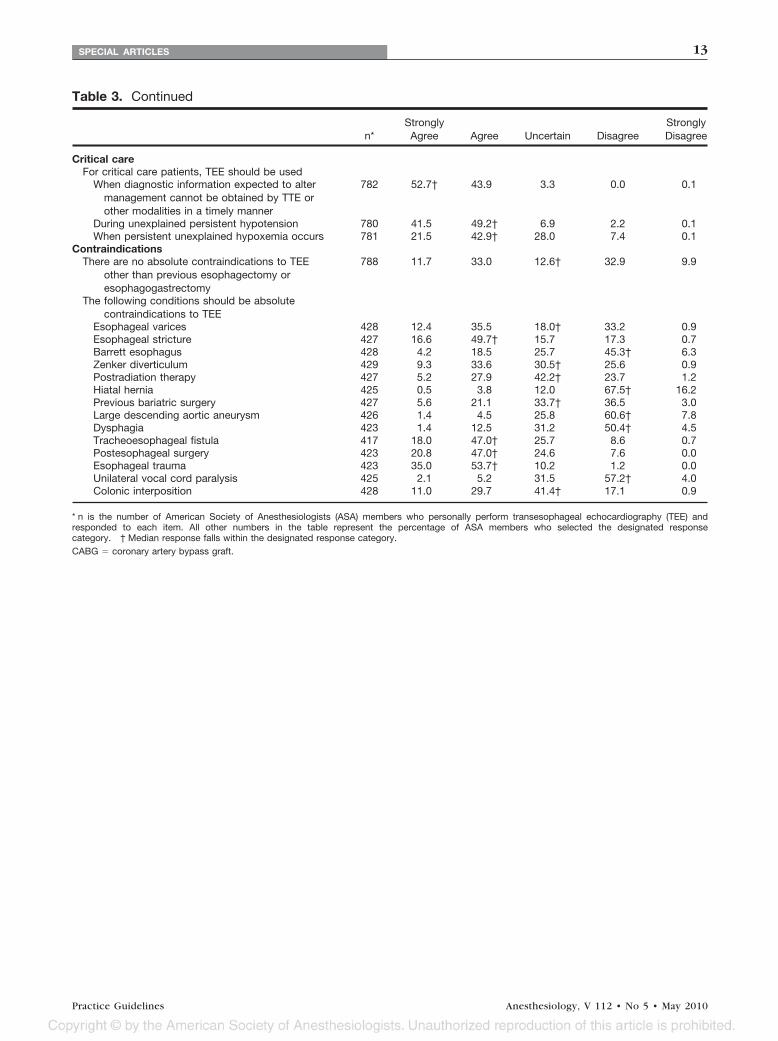
Table 3. Continued
n*StronglyAgree Agree Uncertain Disagree
StronglyDisagree
Critical careFor critical care patients, TEE should be used
When diagnostic information expected to altermanagement cannot be obtained by TTE orother modalities in a timely manner
782 52.7† 43.9 3.3 0.0 0.1
During unexplained persistent hypotension 780 41.5 49.2† 6.9 2.2 0.1When persistent unexplained hypoxemia occurs 781 21.5 42.9† 28.0 7.4 0.1
ContraindicationsThere are no absolute contraindications to TEE
other than previous esophagectomy oresophagogastrectomy
788 11.7 33.0 12.6† 32.9 9.9
The following conditions should be absolutecontraindications to TEE
Esophageal varices 428 12.4 35.5 18.0† 33.2 0.9Esophageal stricture 427 16.6 49.7† 15.7 17.3 0.7Barrett esophagus 428 4.2 18.5 25.7 45.3† 6.3Zenker diverticulum 429 9.3 33.6 30.5† 25.6 0.9Postradiation therapy 427 5.2 27.9 42.2† 23.7 1.2Hiatal hernia 425 0.5 3.8 12.0 67.5† 16.2Previous bariatric surgery 427 5.6 21.1 33.7† 36.5 3.0Large descending aortic aneurysm 426 1.4 4.5 25.8 60.6† 7.8Dysphagia 423 1.4 12.5 31.2 50.4† 4.5Tracheoesophageal fistula 417 18.0 47.0† 25.7 8.6 0.7Postesophageal surgery 423 20.8 47.0† 24.6 7.6 0.0Esophageal trauma 423 35.0 53.7† 10.2 1.2 0.0Unilateral vocal cord paralysis 425 2.1 5.2 31.5 57.2† 4.0Colonic interposition 428 11.0 29.7 41.4† 17.1 0.9
* n is the number of American Society of Anesthesiologists (ASA) members who personally perform transesophageal echocardiography (TEE) andresponded to each item. All other numbers in the table represent the percentage of ASA members who selected the designated responsecategory. † Median response falls within the designated response category.CABG � coronary artery bypass graft.
13SPECIAL ARTICLES
Practice Guidelines Anesthesiology, V 112 • No 5 • May 2010