Practice Guidelines for Central Venous Access 2020
54
Embargoed for release until approved by ASA House of Delegates. No part of this document may be released, distributed or reprinted until approved. Any unauthorized copying, reproduction, appropriation or communication of the contents of this document without the express written consent of the American Society of Anesthesiologists is subject to civil and criminal prosecution to the fullest extent possible, including punitive damages. Practice Guidelines for Central Venous Access 2020 An Updated Report by the American Society of Anesthesiologists Task Force on Central Venous Access * PRACTICE Guidelines are systematically developed recommendations that assist the practitioner and patient 1 in making decisions about health care. These recommendations may be adopted, modified, or rejected according 2 to clinical needs and constraints, and are not intended to replace local institutional policies. In addition, Practice 3 Guidelines developed by the American Society of Anesthesiologists (ASA) are not intended as standards or 4 absolute requirements, and their use cannot guarantee any specific outcome. Practice Guidelines are subject to 5 revision as warranted by the evolution of medical knowledge, technology, and practice. They provide basic 6 recommendations that are supported by a synthesis and analysis of the current literature, expert and practitioner 7 opinion, open forum commentary, and clinical feasibility data. 8 This document updates the “Practice Guidelines for Central Venous Access: A Report by the American 9 Society of Anesthesiologists Task Force on Central Venous Access,” adopted by the ASA in 2011 and published 10 in 2012. 1 11 Methodology 12 Definition of Central Venous Access 13 For these Guidelines, central venous access is defined as placement of a catheter such that the catheter is 14 inserted into a venous great vessel. The venous great vessels include the superior vena cava, inferior vena cava, 15 brachiocephalic veins, internal jugular veins, subclavian veins, iliac veins, and common femoral veins. † Excluded 16 are catheters that terminate in a systemic artery. 17 Purposes of the Guidelines 18 The purposes of these Guidelines are to (1) provide guidance regarding placement and management of 19 central venous catheters, (2) reduce infectious, mechanical, thrombotic, and other adverse outcomes associated 20 with central venous catheterization, and (3) improve management of arterial trauma or injury arising from central 21 * Updated by the American Society of Anesthesiologists Task Force on Central Venous Access: Jeffrey L. Apfelbaum, M.D. (Committee Chair), Chicago, Illinois, Stephen M. Rupp, M.D., Seattle, Washington (Co-Chair); Avery Tung, M.D. (Co-Chair), Wilmette, Illinois; Richard T. Connis, Ph.D., Woodinville, Washington (Chief Methodologist); Karen B. Domino, M.D., M.P.H., Seattle, Washington; Mark D. Grant, M.D., Ph.D., Schaumburg, Illinois (Senior Methodologist); Jonathan B. Mark, M.D., Durham, North Carolina. Received from the American Society of Anesthesiologists, Schaumburg, Illinois. Submitted for publication October __, 2019. Accepted for publication October __, 2019. Supported by the American Society of Anesthesiologists and developed under the direction of the Committee on Standards and Practice Parameters, Jeffrey L. Apfelbaum, M.D. (Chair). Approved by the ASA House of Delegates on October __, 2019 A complete bibliography used to develop this updated Advisory, arranged alphabetically by author, is available as Supplemental Digital Content 1, http://links.lww.com/ALN/XXXXXXXX. Address correspondence to the American Society of Anesthesiologists: 1061 American Lane, Schaumburg, Illinois 60173. This Practice Advisory, as well as all published ASA Practice Parameters, may be obtained at no cost through the Journal Web site, www.anesthesiology.org. † This description of the venous great vessels is consistent with the venous subset for central lines defined by the National Healthcare Safety Network (NHSN).
Transcript of Practice Guidelines for Central Venous Access 2020
Embargoed for release until approved by ASA House of Delegates. No part of this document may be released, distributed or reprinted until approved. Any unauthorized copying, reproduction, appropriation or communication of the contents of this document without the express written consent of the American Society of Anesthesiologists is subject to civil and criminal prosecution to the fullest extent possible, including punitive damages.
Practice Guidelines for Central Venous Access 2020
An Updated Report by the American Society of Anesthesiologists Task Force on Central
Venous Access*
PRACTICE Guidelines are systematically developed recommendations that assist the practitioner and patient 1
in making decisions about health care. These recommendations may be adopted, modified, or rejected according 2
to clinical needs and constraints, and are not intended to replace local institutional policies. In addition, Practice 3
Guidelines developed by the American Society of Anesthesiologists (ASA) are not intended as standards or 4
absolute requirements, and their use cannot guarantee any specific outcome. Practice Guidelines are subject to 5
revision as warranted by the evolution of medical knowledge, technology, and practice. They provide basic 6
recommendations that are supported by a synthesis and analysis of the current literature, expert and practitioner 7
opinion, open forum commentary, and clinical feasibility data. 8
This document updates the “Practice Guidelines for Central Venous Access: A Report by the American 9
Society of Anesthesiologists Task Force on Central Venous Access,” adopted by the ASA in 2011 and published 10
in 2012.1 11
Methodology 12
Definition of Central Venous Access 13
For these Guidelines, central venous access is defined as placement of a catheter such that the catheter is 14
inserted into a venous great vessel. The venous great vessels include the superior vena cava, inferior vena cava, 15
brachiocephalic veins, internal jugular veins, subclavian veins, iliac veins, and common femoral veins.† Excluded 16
are catheters that terminate in a systemic artery. 17
Purposes of the Guidelines 18
The purposes of these Guidelines are to (1) provide guidance regarding placement and management of 19
central venous catheters, (2) reduce infectious, mechanical, thrombotic, and other adverse outcomes associated 20
with central venous catheterization, and (3) improve management of arterial trauma or injury arising from central 21
* Updated by the American Society of Anesthesiologists Task Force on Central Venous Access: Jeffrey L. Apfelbaum, M.D.
(Committee Chair), Chicago, Illinois, Stephen M. Rupp, M.D., Seattle, Washington (Co-Chair); Avery Tung, M.D. (Co-Chair), Wilmette, Illinois; Richard T. Connis, Ph.D., Woodinville, Washington (Chief Methodologist); Karen B. Domino, M.D., M.P.H., Seattle, Washington; Mark D. Grant, M.D., Ph.D., Schaumburg, Illinois (Senior Methodologist); Jonathan B. Mark, M.D., Durham, North Carolina.
Received from the American Society of Anesthesiologists, Schaumburg, Illinois. Submitted for publication October __, 2019. Accepted for publication October __, 2019. Supported by the American Society of Anesthesiologists and developed under the direction of the Committee on Standards and Practice Parameters, Jeffrey L. Apfelbaum, M.D. (Chair). Approved by the ASA House of Delegates on October __, 2019
A complete bibliography used to develop this updated Advisory, arranged alphabetically by author, is available as
Supplemental Digital Content 1, http://links.lww.com/ALN/XXXXXXXX. Address correspondence to the American Society of
Anesthesiologists: 1061 American Lane, Schaumburg, Illinois 60173. This Practice Advisory, as well as all published ASA
Practice Parameters, may be obtained at no cost through the Journal Web site, www.anesthesiology.org. † This description of the venous great vessels is consistent with the venous subset for central lines defined by the National Healthcare Safety Network (NHSN).
PRACTICE GUIDELINES
venous catheterization. 22
Focus 23
These Guidelines apply to patients undergoing elective central venous access procedures performed by 24
anesthesiologists or health care professionals under the direction/supervision of anesthesiologists. The Guidelines 25
do not address (1) clinical indications for placement of central venous catheters, (2) emergency placement of 26
central venous catheters, (3) patients with peripherally inserted central catheters, (4) placement and residence of 27
a pulmonary artery catheter, (5) insertion of tunneled central lines (e.g., permacaths, portacaths, Hickman®, 28
Quinton®, (6) methods of detection or treatment of infectious complications associated with central venous 29
catheterization, (7) removal of central venous catheters,‡ (8) diagnosis and management of central venous 30
catheter-associated trauma or injury (e.g., pneumothorax or air embolism), with the exception of carotid arterial 31
injury, (9) management of peri-insertion coagulopathy, and (10) competency assessment for central line insertion. 32
Application 33
These Guidelines are intended for use by anesthesiologists and individuals under the supervision of an 34
anesthesiologist. They also may serve as a resource for other physicians (e.g., surgeons, radiologists), nurses, or 35
health care providers who manage patients with central venous catheters. 36
Task Force Members 37
The original Guidelines were developed by an ASA appointed task force of 12 members, consisting of 38
anesthesiologists in private and academic practices from various geographic areas of the United States and two 39
methodologists from the ASA Committee on Standards and Practice Parameters. In 2017, the ASA Committee on 40
Standards and Practice Parameters requested that these Guidelines be updated. This update is a revision 41
developed by an ASA-appointed task force of 7 members, including 5 anesthesiologists and two methodologists. 42
Conflict of interest documentation regarding current or potential financial and other interests pertinent to the 43
practice guideline were disclosed by all task force members and managed. 44
Process and Evaluation of Evidence 45
These updated Guidelines were developed by means of a five-step process. First, consensus was reached 46
on the criteria for evidence. Second, original published articles from peer-reviewed journals relevant to the 47
perioperative management of central venous catheters were evaluated and added to literature included in the 48
original Guidelines. Third, consultants who had expertise or interest in central venous catheterization, and who 49
practiced or worked in various settings (e.g., private and academic practice) were asked to participate in opinion 50
surveys addressing the appropriateness, completeness, and feasibility of implementation of the draft 51
recommendations, and to review and comment on a draft of the Guidelines. Fourth, additional opinions were 52
‡ Although catheter removal is not addressed by these guidelines (and is not typically performed by anesthesiologists), the risk
of venous air embolism upon removal is a serious concern. Suggestions for minimizing such risk are those directed at raising
central venous pressure during and immediately after catheter removal, and following a defined nursing protocol. These
suggestions include, but are not limited to positioning the patient in the Trendelenburg position, using the Valsalva
maneuver, applying direct pressure to the puncture site, using air-occlusive dressings, and monitoring the patient for a
reasonable period of time after catheter removal.
PRACTICE GUIDELINES
solicited from random samples of active ASA members. Fifth, all available information was used to build 53
consensus to finalize the Guidelines. A summary of recommendations can be found in appendix 1. 54
Preparation of these updated Guidelines followed a rigorous methodological process. Evidence was obtained 55
from two principal sources: scientific evidence and opinion-based evidence. Detailed descriptions of the ASA 56
process and methodology used in these Guidelines may be found in other related publications.2-5 Appendix 1 57
contains a footnote indicating where information may be found on the evidence model, literature search process, 58
literature findings, and survey results for these Guidelines. 59
Within the text of these Guidelines, literature classifications are reported for each intervention using the 60
following: Category A level 1, meta-analysis of randomized controlled trials (RCTs); Category A level 2, multiple 61
RCTs; and Category A level 3, a single RCT. Category B level 1, nonrandomized studies with group comparisons; 62
Category B level 2, nonrandomized studies with associative findings; Category B level 3, nonrandomized studies 63
with descriptive findings, and Category B level 4, case series or case reports. Statistically significant outcomes (P 64
< 0.01) are designated as either beneficial (B) or harmful (H) for the patient; statistically nonsignificant findings are 65
designated as equivocal (E). Survey findings from task force–appointed expert consultants and a random sample 66
of the ASA membership are fully reported in the text of these Guidelines. Survey responses for each 67
recommendation are reported using a 5-point scale based on median values from strongly agree to strongly 68
disagree. 69
Guidelines 70
Resource Preparation 71
Resource preparation topics include (1) assessing the physical environment where central venous 72
catheterization is planned to determine the feasibility of using aseptic techniques, (2) availability of a standardized 73
equipment set, (3) use of a checklist or protocol for central venous catheter placement and maintenance, and (4) 74
use of an assistant for central venous catheterization. 75
Literature Findings. The literature is insufficient to evaluate the effect of the physical environment for aseptic 76
catheter insertion, availability of a standardized equipment set, or the use of an assistant on outcomes associated 77
with central venous catheterization. An observational study reports that implementation of a trauma intensive care 78
unit multidisciplinary checklist is associated with reduced catheter-related infection rates (Category B2-B 79
evidence).6 Observational studies report that central line-associated or catheter-related bloodstream infection 80
rates are reduced when intensive care unit-wide bundled protocols are implemented7-36 (Category B2-B evidence); 81
evidence from fewer observational studies is equivocal37-55 (Category B2-E evidence); other observational 82
studies56-71 do not report levels of statistical significance or lacked sufficient data to calculate them. These studies 83
do not permit assessing the effect of any single component of a checklist or bundled protocol on infection rates. 84
Survey Findings. The consultants and ASA members strongly agree with the recommendation to perform 85
central venous catheterization in an environment that permits use of aseptic techniques and to assure that a 86
standardized equipment set is available for central venous access. The consultants strongly agree and ASA 87
members agree with the recommendation to use a checklist or protocol for placement and maintenance of central 88
PRACTICE GUIDELINES
venous catheters. The consultants and ASA members agree with the recommendation to use an assistant during 89
placement of a central venous catheter. 90
Recommendations for Resource Preparation. 91
• Perform central venous catheterization in an environment that permits use of aseptic techniques. 92
• Assure that a standardized equipment set is available for central venous access.§ 93
• Use a checklist or protocol for placement and maintenance of central venous catheters.** 94
• Use an assistant during placement of a central venous catheter.†† 95
Prevention of Infectious Complications 96
Interventions intended to prevent infectious complications associated with central venous access include, but 97
are not limited to (1) intravenous antibiotic prophylaxis, (2) aseptic preparation of practitioner, staff and patients, 98
(3) selection of antiseptic solution, (4) selection of catheters containing antimicrobial agents, (5) selection of 99
catheter insertion site, (6) catheter fixation method, (7) insertion site dressings, (8) catheter maintenance 100
procedures, and (9) aseptic techniques using an existing central venous catheter for injection or aspiration. 101
Intravenous Antibiotic Prophylaxis. 102
Literature Findings. The literature is insufficient to evaluate outcomes associated with the routine use of 103
intravenous prophylactic antibiotics. 104
Survey Findings. The consultants strongly agree and ASA members agree with the recommendation to not 105
routinely administer intravenous antibiotic prophylaxis. The consultants and ASA members are equivocal 106
regarding the administration of intravenous antibiotic prophylaxis for immunocompromised patients and high-risk 107
neonates. 108
Aseptic Preparation of Practitioner, Staff, and Patients. 109
Literature Findings. An RCT comparing maximal barrier precautions (i.e., mask, cap, gloves, gown, large 110
full-body drape) with a control group (i.e., gloves and small drape) reports equivocal findings for reduced 111
colonization and catheter-related septicemia (Category A3-E evidence).72 A majority of observational studies 112
reporting or with calculable levels of statistical significance report that “bundles” of aseptic protocols (e.g., 113
combinations of hand washing, sterile full-body drapes, sterile gloves, caps and masks) reduce the frequency of 114
central-line associated or catheter-related bloodstream infections (Category B2-B evidence).7-36 These studies do 115
not permit assessing the effect of any single component of a bundled protocol on infection rates. 116
Survey Findings. The consultants and ASA members strongly agree with the recommendation to use aseptic 117
techniques (e.g., hand washing) and maximal barrier precautions (e.g., sterile gowns, sterile gloves, caps, masks 118
covering both mouth and nose, and full-body patient drapes) in preparation for the placement of central venous 119
catheters. 120
§ Refer to appendix 2 for an example of a list of standardized equipment for adult patients. ** Refer to appendix 3 for an example of a checklist or protocol. †† Refer to appendix 4 for an example of a list of duties per-formed by an assistant
PRACTICE GUIDELINES
Selection of antiseptic solution. 121
Literature Findings. One RCT comparing chlorhexidine (2% aqueous solution without alcohol) with 122
povidone-iodine (10% without alcohol) for skin preparation reports equivocal findings for catheter colonization and 123
catheter-related bacteremia (Category A3-E evidence).73 An RCT comparing chlorhexidine (2% with 70% 124
isopropyl alcohol) with povidone-iodine (5% with 69% ethanol) with or without scrubbing finds lower rates of 125
catheter colonization (Category A3-B evidence) and equivocal evidence for decreased catheter-related 126
bloodstream infection (Category A3-E evidence).74 A third RCT compared two chlorhexidine concentrations (0.5% 127
or 1.0% in 79% ethanol) with povidone-iodine (10% without alcohol) reporting equivocal evidence for colonization 128
(Category A3-E evidence) and catheter-related blood stream infection (Category A3-E evidence).75 A quasi-129
experimental study (secondary analysis of an RCT) reports a lower rate of catheter-related blood stream infection 130
with chlorhexidine (2% with 70% alcohol) than povidone-iodine (5% with 69% alcohol) (Category B1-B 131
evidence).76 132
The literature is insufficient to evaluate the safety of antiseptic solutions containing chlorhexidine in neonates, 133
infants and children.‡‡ 134
Comparative studies are insufficient to evaluate the efficacy of chlorhexidine and alcohol compared with 135
chlorhexidine without alcohol for skin preparation during central venous catheterization. An RCT of 5% povidone-136
iodine with 70% alcohol compared with 10% povidone-iodine alone indicates that catheter tip colonization is 137
reduced with alcohol containing solutions (Category A3-B evidence); equivocal findings are reported for catheter-138
related bloodstream infection and clinical signs of infection (Category A3-E evidence).77 139
Survey Findings. The consultants and ASA members strongly agree with the recommendation to use a 140
chlorhexidine-containing solution for skin preparation in adults, infants, and children. For neonates, the 141
consultants and ASA members agree with the recommendation to determine the use of chlorhexidine-containing 142
solutions for skin preparation based on clinical judgment and institutional protocol. If there is a contraindication to 143
chlorhexidine, the consultants strongly agree and ASA members agree with the recommendation that povidone-144
iodine or alcohol may be used. The consultants and ASA members agree with the recommendation to use skin 145
preparation solutions containing alcohol unless contraindicated. 146
Catheters Containing Antimicrobial Agents. 147
Literature Findings. Meta-analyses of RCTs comparing antibiotic coated with uncoated catheters indicates 148
that antibiotic coated catheters are associated with reduced catheter colonization78-85 and catheter-related 149
bloodstream infection80,81,83,85,86 (Category A1-B evidence). Meta-analyses of RCTs comparing silver or silver-150
platinum-carbon impregnated catheters with uncoated catheters yield equivocal findings for catheter 151
colonization87-97 (Category A1-E evidence), but a decreased risk of catheter-related bloodstream infection87-94,96-99 152
(Category A1-B evidence). Meta-analyses of RCTs indicate that catheters coated with chlorhexidine and silver 153
sulfadiazine reduce catheter colonization compared with uncoated catheters83,95,100-118 (Category A1-B evidence), 154
but are equivocal for catheter-related bloodstream infection83,100-102,104-110,112-117,119,120 (Category A1-E evidence). 155
Cases of anaphylactic shock are reported after placement of a catheter coated with chlorhexidine and silver 156
sulfadiazine (Category B4-H evidence).121-129 157
‡‡ See 2017 FDA warning on chlorhexidine allergy.
PRACTICE GUIDELINES
Survey Findings. The consultants and ASA members agree with the recommendation to use catheters 158
coated with antibiotics or a combination of chlorhexidine and silver sulfadiazine based on infectious risk and 159
anticipated duration of catheter use for selected patients. The consultants strongly agree and ASA members 160
agree with the recommendation to not use catheters containing antimicrobial agents as a substitute for additional 161
infection precautions. 162
Selection of Catheter Insertion Site. 163
Literature Findings. RCTs comparing subclavian and femoral insertion sites report higher rates of catheter 164
colonization at the femoral site (Category A2-H evidence); findings for catheter-related sepsis or catheter-related 165
bloodstream infection are equivocal (Category A2-E evidence).130,131 An RCT finds a higher rate of catheter 166
colonization for internal jugular compared with subclavian insertion (Category A3-H evidence) and for femoral 167
compared with internal jugular insertion (Category A3-H evidence); evidence is equivocal for catheter-related 168
bloodstream infection for either comparison (Category A3-E evidence).131 169
A nonrandomized comparative study of burn patients reports that catheter colonization and catheter-related 170
bloodstream infection occur more frequently with an insertion site closer to the burn location (Category B1-H 171
evidence).132 172
Survey Findings. The consultants and ASA members strongly agree with the recommendations to (1) 173
determine catheter insertion site selection based on clinical need, (2) select an insertion site that is not 174
contaminated or potentially contaminated (e.g., burned or infected skin, inguinal area, adjacent to tracheostomy or 175
open surgical wound), and (3) select an upper body insertion site when possible to minimize the risk of infection in 176
adults. 177
Catheter Fixation. 178
Literature Findings. The literature is insufficient to evaluate whether catheter fixation with sutures, staples or 179
tape is associated with a higher risk for catheter-related infections. 180
Survey Findings. The consultants strongly agree and ASA members agree with the recommendation to 181
determine the use of sutures, staples, or tape for catheter fixation on a local or institutional basis. The consultants 182
and ASA members both strongly agree with the recommendation to minimize the number of needle punctures of 183
the skin. 184
Insertion Site Dressings. 185
Literature Findings. The literature is insufficient to evaluate the efficacy of transparent bio-occlusive 186
dressings to reduce the risk of infection. Pooled estimates from RCTs are consistent with lower rates of catheter 187
colonization with chlorhexidine sponge dressings compared with standard polyurethane90,133-138 (Category A1-B 188
evidence), but equivocal for catheter-related bloodstream infection90,133-140 (Category A1-E evidence). An RCT 189
reports a higher frequency of severe localized contact dermatitis in neonates with chlorhexidine-impregnated 190
dressings compared with povidone-iodine impregnated dressings133 (Category A3-H evidence); findings 191
concerning dermatitis from RCTs in adults are equivocal (Category A2-E evidence).90,134,136,137,141 192
Survey Findings. The consultants and ASA members both strongly agree with the recommendations to use 193
transparent bio-occlusive dressings to protect the site of central venous catheter insertion from infection. The 194
consultants and ASA members both agree with the recommendation that dressings containing chlorhexidine may 195
be used in adults, infants, and children unless contraindicated. For neonates, the consultants and ASA members 196
PRACTICE GUIDELINES
agree with the recommendation to determine the use of transparent or sponge dressings containing chlorhexidine 197
based on clinical judgment and institutional protocol. If a chlorhexidine containing dressing is used, the 198
consultants and ASA members both strongly agree with the recommendation to observe the site daily for signs of 199
irritation, allergy or, necrosis. 200
Catheter Maintenance. 201
Catheter maintenance consists of: (1) determining the optimal duration of catheterization, (2) conducting 202
catheter site inspections, (3) periodically changing catheters, and (4) changing catheters using a guidewire instead 203
of selecting a new insertion site. 204
Literature Findings. Nonrandomized comparative studies indicate that longer catheterization is associated 205
with higher catheter colonization rates, infection, and sepsis (Category B1-H evidence).21,142-145 The literature is 206
insufficient to evaluate whether time intervals between catheter site inspections are associated with the risk for 207
catheter-related infection. RCTs report equivocal findings for catheter tip colonization when catheters are changed 208
at 3 versus 7-day intervals (Category A2-E evidence).146,147 RCTs report equivocal findings for catheter tip 209
colonization when guidewires are used to change catheters compared with new insertion sites (Category A2-E 210
evidence).148-150 211
Survey Findings. The consultants and ASA members strongly agree with the following recommendations: (1) 212
determine the duration of catheterization based on clinical need, (2) assess the clinical need for keeping the 213
catheter in place on a daily basis, (3) remove catheters removed promptly when no longer deemed clinically 214
necessary, (4) inspect the catheter insertion site daily for signs of infection, (5) change or remove the catheter 215
when catheter insertion site infection is suspected, and (6) when a catheter related infection is suspected, replace 216
the catheter using a new insertion site rather than changing the catheter over a guidewire. 217
Aseptic Techniques Using an Existing Central Venous Catheter for Injection or Aspiration 218
Aseptic techniques using an existing central venous catheter for injection or aspiration consist of (1) wiping 219
the port with an appropriate antiseptic, (2) capping stopcocks or access ports, and (3) use of needleless catheter 220
connectors or access ports. 221
Literature Findings. The literature is insufficient to evaluate whether wiping ports or capping stopcocks 222
when using an existing central venous catheter for injection or aspiration decreases the risk of catheter-related 223
infections. RCTs comparing needleless connectors with standard caps indicate lower rates of microbial 224
contamination of stopcock entry ports with needleless connectors151-153 (Category A2-B evidence) but findings for 225
catheter-related bloodstream infection are equivocal151,154 (Category A2-E evidence). 226
Survey Findings. The consultants and ASA members strongly agree with the recommendations to wipe 227
catheter access ports with an appropriate antiseptic (e.g., alcohol) before each access when using an existing 228
central venous catheter for injection or aspiration, and to cap central venous catheter stopcocks or access ports 229
when not in use. The consultants and ASA members agree that needleless catheter access ports may be used 230
on a case-by-case basis 231
Recommendations for Prevention of Infectious Complications. 232
Intravenous Antibiotic Prophylaxis. 233
• Do not routinely administer intravenous antibiotic prophylaxis, 234
PRACTICE GUIDELINES
Aseptic Preparation. 235
• In preparation for the placement of central venous catheters, use aseptic techniques (e.g., hand washing) 236
and maximal barrier precautions (e.g., sterile gowns, sterile gloves, caps, masks covering both mouth and 237
nose, and full-body patient drapes). 238
Selection of Antiseptic Solution. 239
• Use a chlorhexidine-containing solution for skin preparation in adults, infants, and children. 240
o For neonates, determine the use of chlorhexidine-containing solutions for skin preparation based 241
on clinical judgment and institutional protocol. 242
• If there is a contraindication to chlorhexidine, povidone-iodine or alcohol may be used. 243
• Unless contraindicated, use skin preparation solutions containing alcohol. 244
Catheters Containing Antimicrobial Agents. 245
• For selected patients, use catheters coated with antibiotics, a combination of chlorhexidine and silver 246
sulfadiazine, or silver-platinum-carbon impregnated catheters based on risk of infection and anticipated 247
duration of catheter use. 248
o Do not use catheters containing antimicrobial agents as a substitute for additional infection 249
precautions. 250
Selection of Catheter Insertion Site. 251
• Determine catheter insertion site selection based on clinical need. 252
• Select an insertion site that is not contaminated or potentially contaminated (e.g., burned or infected skin, 253
inguinal area, adjacent to tracheostomy or open surgical wound). 254
• In adults, select an upper body insertion site when possible to minimize the risk of infection. 255
Catheter Fixation. 256
• Determine the use of sutures, staples, or tape for catheter fixation on a local or institutional basis. 257
• Minimize the number of needle punctures of the skin. 258
Insertion Site Dressings. 259
• Use transparent bio-occlusive dressings to protect the site of central venous catheter insertion from 260
infection. 261
• Unless contraindicated, dressings containing chlorhexidine may be used in adults, infants, and children. 262
• For neonates, determine the use of transparent or sponge dressings containing chlorhexidine based on 263
clinical judgment and institutional protocol. 264
• If a chlorhexidine containing dressing is used, observe the site daily for signs of irritation, allergy or 265
necrosis 266
Catheter Maintenance. 267
• Determine the duration of catheterization based on clinical need. 268
• Assess the clinical need for keeping the catheter in place on a daily basis. 269
• Remove catheters promptly when no longer deemed clinically necessary. 270
• Inspect the catheter insertion site daily for signs of infection 271
PRACTICE GUIDELINES
• Change or remove the catheter when catheter insertion site infection is suspected. 272
• When a catheter related infection is suspected, replace the catheter using a new insertion site rather than 273
changing the catheter over a guidewire. 274
Aseptic Techniques Using an Existing Central Venous Catheter for Injection or Aspiration. 275
• Wipe catheter access ports with an appropriate antiseptic (e.g., alcohol) before each access when using 276
an existing central venous catheter for injection or aspiration. 277
• Cap central venous catheter stopcocks or access ports when not in use. 278
• Needleless catheter access ports may be used on a case-by-case basis 279
Prevention of Mechanical Trauma or Injury 280
Interventions intended to prevent mechanical trauma or injury associated with central venous access include 281
but are not limited to: (1) selection of catheter insertion site, (2) positioning the patient for needle insertion and 282
catheter placement, (3) needle insertion, wire placement and catheter placement, (4) guidance for needle, 283
guidewire, and catheter placement, and (5) verification of needle, wire and catheter placement. 284
Selection of Catheter Insertion Site. 285
Literature Findings. RCTs comparing subclavian and femoral insertion sites report that the femoral site 286
has a higher risk of thrombotic complications in adult patients130,131 (Category A2-H evidence); one RCT131 287
concludes that thrombosis risk is higher with internal jugular than subclavian catheters (Category A3-H evidence) 288
while for femoral versus internal jugular catheters, findings are equivocal (Category A3-E evidence). RCTs report 289
equivocal findings for successful venipuncture when the internal jugular site is compared with the subclavian site 290
(Category A2-E evidence).131,155,156 Equivocal finding are also reported for the femoral versus subclavian site130,131 291
(Category A2-E evidence), and the femoral versus internal jugular site131 (Category A3-E evidence). RCTs 292
examining mechanical complications (primarily arterial injury, hematoma, and pneumothorax) report equivocal 293
findings for the femoral versus subclavian site130,131 (Category A2-E evidence) as well as the internal jugular 294
versus subclavian or femoral sites131 (Category A3-E evidence). 295
Survey Findings. The consultants and ASA members strongly agree with the recommendation to 296
determine catheter insertion site selection based on clinical need and practitioner judgment, experience, and skill. 297
The consultants agree and ASA members strongly agree with the recommendations to select an upper body 298
insertion site to minimize the risk of thrombotic complications relative to the femoral site. 299
Positioning the Patient for Needle Insertion and Catheter Placement. 300
Literature Findings. Observational studies report that Trendelenburg positioning (i.e., head down from 301
supine) increases the right internal jugular vein diameter or cross-sectional area in healthy adult volunteers157-161 302
(Category B2-B evidence), but findings are equivocal in studies enrolling adult patients (Category B2-E 303
evidence).158,162-164 Observational studies comparing the Trendelenburg position and supine position in pediatric 304
patients report increased right internal jugular vein diameter or cross-sectional area165-167 (Category B2-B 305
evidence), and one observational study of newborns reported similar findings168 (Category B2-B evidence). The 306
literature is insufficient to evaluate whether Trendelenburg positioning improves insertion success rates or 307
decreases the risk of mechanical complications. 308
PRACTICE GUIDELINES
Survey Findings. The consultants and ASA members strongly agree with the recommendation to 309
perform central venous access in the neck or chest with the patient in the Trendelenburg position when clinically 310
appropriate and feasible. 311
Needle Insertion, Wire Placement, and Catheter Placement. Needle insertion, wire placement, and catheter 312
placement includes: (1) selection of catheter size and type, (2) use of a wire-through-thin-wall needle technique (i.e., 313
Seldinger technique) versus a catheter-over-the-needle-then-wire-through-the-catheter technique (i.e., modified 314
Seldinger technique), (3) limiting the number of insertion attempts, and (4) introducing two catheters in the same 315
central vein. 316
Literature Findings. 317
Case reports describe severe injury (e.g., hemorrhage, hematoma, pseudoaneurysm, arteriovenous fistula, 318
arterial dissection, neurologic injury including stroke, and severe or lethal airway obstruction) when unintentional 319
arterial cannulation occurs with large bore catheters (Category B4-H evidence).169-178 320
An RCT comparing a thin-wall needle technique versus a catheter-over-the-needle for right internal jugular 321
vein insertion in adults reports equivocal findings for first-attempt success rates and frequency of complications 179 322
(Category A3-E evidence); for right-sided subclavian insertion in adults an RCT reports first-attempt success more 323
likely and fewer complications with a thin-wall needle technique (Category A3-B evidence).180 One RCT reports 324
equivocal findings for first-attempt success rates and frequency of complications when comparing a thin-wall 325
needle with catheter-over-the-needle technique for internal jugular vein insertion (preferentially right) in neonates 326
(Category A3-E evidence).181 Observational studies report a greater frequency of complications occurring with 327
increasing number of insertion attempts (Category B3-H evidence).182-184 One nonrandomized comparative study 328
reports a higher frequency of dysrhythmia when two central venous catheters are placed in the same vein (right 329
internal jugular) compared with placement of one catheter in the vein (Category B1-H evidence); differences in 330
carotid artery punctures or hematomas were not noted (Category B1-E evidence).185 331
Survey Findings. The consultants and ASA members strongly agree with the recommendation to select 332
catheter size (i.e., outside diameter) and type based on the clinical situation and skill/experience of the operator. 333
The consultants and ASA members agree with the recommendations to (1) select the smallest size catheter 334
appropriate for the clinical situation, (2) for the subclavian approach select a thin-wall needle (i.e., Seldinger) 335
technique versus a catheter-over-the-needle (i.e., modified Seldinger) technique, (3) for the jugular or femoral 336
approach, select a thin-wall needle or catheter-over-the-needle technique based on the clinical situation and the 337
skill/experience of the operator, and (4) base the decision to use a thin-wall needle technique or a catheter-over-338
the-needle technique least in part on the method used to confirm that the wire resides in the vein before a dilator 339
or large-bore catheter is threaded. The consultants agree and ASA members strongly agree that the number of 340
insertion attempts should be based on clinical judgment and the decision to place two catheters in a single vein 341
should be made on a case-by-case basis. 342
Guidance for Needle, Wire, and Catheter Placement. Guidance for needle, wire, and catheter placement 343
includes (1) real-time or dynamic ultrasound for vessel localization and guiding the needle to its intended venous 344
location, and (2) static ultrasound imaging for the purpose of prepuncture vessel localization. 345
PRACTICE GUIDELINES
Literature Findings. 346
Meta-analyses of RCTs comparing real-time ultrasound guided venipuncture of the internal jugular with an 347
anatomical landmark approach report higher first insertion attempt success rates,186-197 higher overall success 348
rates,186,187,189-192,194-204 lower rates of arterial puncture,186-188,190-201,203,205 and fewer insertion attempts188,190,191,194-349
197,199,200,203-205 (Category A1-B evidence). RCTs also indicate reduced access time or times to cannulation with 350
ultrasound compared to a landmark approach (Category A2-B evidence).188,191,194-196,199,200,202-205 351
For the subclavian vein, RCTs report fewer insertion attempts with real-time ultrasound guided 352
venipuncture206,207 (Category A2-B evidence), and higher overall success rates206-208 (Category A2-B evidence). 353
When compared with a landmark approach, findings are equivocal for arterial puncture207,208 and hematoma207,208 354
(Category A2-E evidence). For the femoral vein, an RCT reports a higher first-attempt success rate and fewer 355
needle passes with real-time ultrasound guided venipuncture compared with the landmark approach in pediatric 356
patients (Category A3-B evidence).209 357
Meta-analyses of RCTs comparing static ultrasound with a landmark approach yields equivocal evidence for 358
improved overall success for internal jugular insertion190,202,210-212 (Category A1-E evidence), overall success 359
irrespective of insertion site182,190,202,210-212 (Category A1-E evidence), or impact on arterial puncture rates190,202,210-360
212 (Category A1-E evidence). RCTs comparing static ultrasound with a landmark approach for locating the 361
internal jugular vein report a higher first insertion attempt success rate with static ultrasound (Category A3-B 362
evidence).190,212 The literature is equivocal regarding overall success for subclavian vein access182 (Category A3-E 363
evidence) or femoral vein access202 (Category A3-E evidence). 364
Survey Findings. The consultants and ASA members strongly agree with the recommendation to use real-365
time ultrasound guidance for vessel localization and venipuncture when the internal jugular vein is selected for 366
cannulation. The consultants and ASA members agree that when feasible, real-time ultrasound may be used 367
when the subclavian or femoral vein is selected. The consultants strongly agree and ASA members agree with 368
the recommendation to use static ultrasound imaging before prepping and draping for prepuncture identification of 369
anatomy to determine vessel localization and patency when the internal jugular vein is selected for cannulation. 370
The consultants and ASA members agree that static ultrasound may also be used when the subclavian or femoral 371
vein is selected. 372
Verification of Needle, Wire, and Catheter Placement. Verification of needle, wire, and catheter placement 373
includes: (1) confirming that the catheter or thin-wall needle resides in the vein, (2) confirming venous residence of 374
the wire, and (3) confirming residence of the catheter in the venous system and final catheter tip position.§§ 375
Literature Findings. 376
A retrospective observational study reports that manometry can detect arterial punctures not identified by 377
blood flow and color (Category B3-B evidence).213 The literature is insufficient to address ultrasound, pressure-378
waveform analysis, blood gas analysis, blood color, or the absence of pulsatile flow as effective methods of 379
confirming catheter or thin-wall needle venous access. 380
Two observational studies indicate that ultrasound can confirm venous placement of the wire before dilation or 381
final catheterization (Category B3-B evidence).214,215 Observational studies also demonstrate that transthoracic 382
§§ Verification methods for needle, wire, or catheter placement may include any one or more of the following: ultrasound,
manometry, pressure waveform analysis, venous blood gas, fluoroscopy, continuous electrocardiography, transesophageal echocardiography, and chest radiography.
PRACTICE GUIDELINES
ultrasound can confirm residence of the guidewire in the venous system (Category B3-B evidence).216-219 One 383
observational study indicates that transesophageal echocardiography can be used to identify guidewire position220 384
(Category B3-B evidence), and case reports document similar findings221,222 (Category B4-B evidence). 385
Observational studies indicate that transthoracic ultrasound can confirm correct catheter tip position (Category 386
B2-B evidence).216,217,223-240 ***††† Observational studies also indicate that fluoroscopy241,242 and chest 387
radiography243,244 can identify the position of the catheter (Category B2-B evidence). RCTs comparing continuous 388
electrocardiographic guidance for catheter placement with no electrocardiography indicate that continuous 389
electrocardiography is more effective in identifying proper catheter tip placement (Category A2-B evidence).245-247 390
Case reports document unrecognized retained guidewires resulting in complications including embolization and 391
fragmentation,248 infection,249 arrhythmia,250 cardiac perforation,248 stroke,251 and migration through soft-tissue252 392
(Category B-4H evidence). 393
Survey Findings. The consultants and ASA members strongly agree with the recommendation to confirm 394
venous access after insertion of a catheter that went over the needle or a thin-wall needle, and with the 395
recommendation to not rely on blood color or absence of pulsatile flow for confirming that the catheter or thin-wall 396
needle resides in the vein. The consultants strongly agree and ASA members agree with the recommendation to 397
confirm venous residence of the wire after the wire is threaded when using the thin-wall needle technique. The 398
consultants are equivocal and ASA members agree that when using the catheter-over-the-needle technique, 399
confirmation that the wire resides in the vein may not be needed (1) if the catheter enters the vein easily and 400
manometry or pressure waveform measurement provides unambiguous confirmation of venous location of the 401
catheter; and (2) if the wire passes through the catheter and enters the vein without difficulty. The consultants and 402
ASA members strongly agree with the recommendation to confirm venous residence of the wire after the wire is 403
threaded if there is any uncertainty that the catheter or wire resides in the vein and insertion of a dilator or large-404
bore catheter may then proceed. The consultants and ASA members strongly agree with the following 405
recommendations, (1) after final catheterization and before use, confirm residence of the catheter in the venous 406
system as soon as clinically appropriate, (2) confirm the final position of the catheter tip as soon as clinically 407
appropriate, (3) for central venous catheters placed in the operating room, perform a chest radiograph no later 408
than the early postoperative period to confirm the position of the catheter tip, (4) verify that the wire has not been 409
retained in the vascular system at the end of the procedure by confirming the presence of the removed wire in the 410
procedural field, and (5) if the complete guidewire is not found in the procedural field, order chest radiography to 411
determine whether the guidewire has been retained in the patient's vascular system. 412
Recommendations for Prevention of Mechanical Trauma or Injury 413
Catheter Insertion Site Selection. 414
• Determine catheter insertion site selection based on clinical need and practitioner judgment, experience, 415
and skill. 416
• Select an upper body insertion site when possible to minimize the risk of thrombotic complications relative 417
to the femoral site. 418
*** Studies also report high specificities of transthoracic ultrasound for excluding the presence of a pneumothorax.216,218,219,227-
229,232,233,236,238,240 ††† Chest radiography was used as a reference standard for these studies.
PRACTICE GUIDELINES
Positioning the Patient for Needle Insertion and Catheter Placement 419
• Perform central venous access in the neck or chest with the patient in the Trendelenburg position when 420
clinically appropriate and feasible. 421
Needle Insertion, Wire Placement, and Catheter Placement. 422
• Select catheter size (i.e., outside diameter) and type based on the clinical situation and skill/experience of 423
the operator. 424
• Select the smallest size catheter appropriate for the clinical situation. 425
• For the subclavian approach select a thin-wall needle (i.e., Seldinger) technique versus a catheter-over-426
the-needle (i.e., modified Seldinger) technique 427
• For the jugular or femoral approach, select a thin-wall needle or catheter-over-the-needle technique based 428
on the clinical situation and the skill/experience of the operator. 429
• For accessing the vein before threading a dilator or large-bore catheter, base the decision to use a thin-430
wall needle technique or a catheter-over-the-needle technique at least in part on the method used to 431
confirm that the wire resides in the vein (figure 1).‡‡‡ 432
• The number of insertion attempts should be based on clinical judgment. 433
• The decision to place two catheters in a single vein should be made on a case-by-case basis. 434
Guidance of Needle, Wire, and Catheter Placement 435
• Use real time ultrasound guidance for vessel localization and venipuncture when the internal jugular vein 436
is selected for cannulation (see fig. 1). 437
o When feasible, real-time ultrasound may be used when the subclavian or femoral vein is 438
selected.§§§ 439
• Use static ultrasound imaging before prepping and draping for prepuncture identification of anatomy to 440
determine vessel localization and patency when the internal jugular vein is selected for cannulation. 441
o Static ultrasound may also be used when the subclavian or femoral vein is selected. 442
Verification of Needle, Wire, and Catheter Placement 443
• After insertion of a catheter that went over the needle or a thin-wall needle, confirm venous access.****†††† 444
o Do not rely on blood color or absence of pulsatile flow for confirming that the catheter or thin-wall 445
needle resides in the vein. 446
• When using the thin-wall needle technique, confirm venous residence of the wire after the wire is 447
threaded. 448
o When using the catheter-over-the-needle technique, confirmation that the wire resides in the vein 449
may not be needed (1) when the catheter enters the vein easily and manometry or pressure 450
‡‡‡ The catheter over-the-needle technique may provide more stable venous access if manometry is used for venous
confirmation. §§§ This approach may not be feasible in emergency circumstances or in the presence of other clinical constraints. **** For neonates, infants, and children, confirmation of venous placement may take place after the wire is threaded. †††† Methods for confirming that the catheter or thin-wall needle resides in the vein include, but are not limited to, ultrasound, manometry, or pressure-waveform analysis measurement
PRACTICE GUIDELINES
waveform measurement provides unambiguous confirmation of venous location of the catheter; 451
and (2) when the wire passes through the catheter and enters the vein without difficulty. 452
o If there is any uncertainty that the catheter or wire resides in the vein, confirm venous residence 453
of the wire after the wire is threaded. Insertion of a dilator or large-bore catheter may then 454
proceed.‡‡‡‡ 455
• After final catheterization and before use, confirm residence of the catheter in the venous system as soon 456
as clinically appropriate.§§§§ 457
• Confirm the final position of the catheter tip as soon as clinically appropriate.***** 458
o For central venous catheters placed in the operating room, perform a chest radiograph no later 459
than the early postoperative period to confirm the position of the catheter tip. 460
• Verify that the wire has not been retained in the vascular system at the end of the procedure by confirming 461
the presence of the removed wire in the procedural field. 462
o If the complete guidewire is not found in the procedural field, order chest radiography to 463
determine whether the guidewire has been retained in the patient's vascular system. 464
Management of Arterial Trauma or Injury Arising from Central Venous Catheterization 465
Literature Findings. Case reports of adult patients with arterial puncture by a large bore catheter/vessel 466
dilator during attempted central venous catheterization indicate severe complications (e.g., cerebral infarction, 467
arteriovenous fistula, hemothorax) after immediate catheter removal172,176,253 (Category B4-H evidence); 468
complications are uncommonly reported for adult patients whose catheters were left in place before surgical 469
consultation and repair172,176,254 (Category B4-E evidence). 470
Survey Findings. The consultants and ASA members strongly agree that when unintended cannulation of an 471
arterial vessel with a dilator or large-bore catheter occurs, leave the dilator or catheter in place and immediately 472
consult a general surgeon, a vascular surgeon, or an interventional radiologist regarding surgical or nonsurgical 473
catheter removal for adults. The consultants and ASA members strongly agree that for neonates, infants, and 474
children, determine on a case-by-case basis whether to leave the catheter in place and obtain consultation or to 475
remove the catheter nonsurgically. The consultants strongly agree and ASA members agree with the 476
recommendation that after the injury has been evaluated and a treatment plan has been executed, confer with the 477
surgeon regarding relative risks and benefits of proceeding with the elective surgery versus deferring surgery to 478
allow for a period of patient observation. 479
Recommendations for management of arterial trauma or injury arising from central venous access. 480
• When unintended cannulation of an arterial vessel with a dilator or large-bore catheter occurs, leave the 481
dilator or catheter in place and immediately consult a general surgeon, a vascular surgeon, or an 482
interventional radiologist regarding surgical or nonsurgical catheter removal for adults. 483
‡‡‡‡ Methods for confirming that the wire resides in the vein include, but are not limited to, ultrasound (identification of the wire in the vein) or transesophageal echocardiography (identification of the wire in the superior vena cava or right atrium), continuous electrocardiography (identification of narrow-complex ectopy), or fluoroscopy. §§§§ Methods for confirming that the catheter is still in the venous system after catheterization and before use include manometry, pressure waveform measurement, or contrast-enhanced ultrasound. ***** Methods for confirming the position of the catheter tip include chest radiography, fluoroscopy, or point-of-care transthoracic echocardiography or continuous electrocardiography.
PRACTICE GUIDELINES
• For neonates, infants, and children, determine on a case-by-case basis whether to leave the catheter in 484
place and obtain consultation or to remove the catheter nonsurgically. 485
• After the injury has been evaluated and a treatment plan has been executed, confer with the surgeon 486
regarding relative risks and benefits of proceeding with the elective surgery versus deferring surgery to 487
allow for a period of patient observation. 488
489
____________________________________
References
1. American Society of Anesthesiologists Task Force on Central Venous A, Rupp SM, Apfelbaum JL, Blitt C, Caplan RA, Connis RT, Domino KB, Fleisher LA, Grant S, Mark JB, Morray JP, Nickinovich DG, Tung A: Practice guidelines for central venous access: a report by the American Society of Anesthesiologists Task Force on Central Venous Access. Anesthesiology 2012; 116:539-73
2. Connis RT, Nickinovich DG, Caplan RA, Arens JF: The development of evidence-based clinical practice guidelines. Integrating medical science and practice. Int J Technol Assess Health Care 2000; 16:1003-12
3. Apfelbaum JL, Connis RT, Nickinovich DG: 2012 Emery A. Rovenstine Memorial Lecture: the genesis, development, and future of the American Society of Anesthesiologists evidence-based practice parameters. Anesthesiology 2013; 118:767-8
4. Apfelbaum JL, Connis RT: The American Society of Anesthesiologists Practice Parameter Methodology. Anesthesiology 2019; 130:367-384
5. Connis RT, Nickinovich DG, Caplan RA, Apfelbaum JL: Evaluation and classification of evidence for the ASA clinical practice guidelines, Miller’s Anesthesia, 8th edition. Edited by MIller RD. Philadelphia, PA, Elsevier Saunders, 2015, pp 3257-70
6. Chua C, Wisniewski T, Ramos A, Schlepp M, Fildes JJ, Kuhls DA: Multidisciplinary trauma intensive care unit checklist: impact on infection rates. J Trauma Nurs 2010; 17:163-6
7. Allen GB, Miller V, Nicholas C, Hess S, Cordes MK, Fortune JB, Blondin J, Ashikaga T, Ricci M: A multitiered strategy of simulation training, kit consolidation, and electronic documentation is associated with a reduction in central line-associated bloodstream infections. Am J Infect Control 2014; 42:643-8
8. Almeida CC, Pissarra da Silva SMS, Flor de Lima Caldas de Oliveira FSD, Guimaraes Pereira Areias MHF: Nosocomial sepsis: evaluation of the efficacy of preventive measures in a level-III neonatal intensive care unit. J Matern Fetal Neonatal Med 2017; 30:2036-2041
9. Berenholtz SM, Pronovost PJ, Lipsett PA, Hobson D, Earsing K, Farley JE, Milanovich S, Garrett-Mayer E, Winters BD, Rubin HR, Dorman T, Perl TM: Eliminating catheter-related bloodstream infections in the intensive care unit. Crit Care Med 2004; 32:2014-20
10. Bion J, Richardson A, Hibbert P, Beer J, Abrusci T, McCutcheon M, Cassidy J, Eddleston J, Gunning K, Bellingan G, Patten M, Harrison D, Matching Michigan C, Writing C: 'Matching Michigan': a 2-year stepped interventional programme to minimise central venous catheter-blood stream infections in intensive care units in England. BMJ Qual Saf 2013; 22:110-23
11. Burrell AR, McLaws ML, Murgo M, Calabria E, Pantle AC, Herkes R: Aseptic insertion of central venous lines to reduce bacteraemia: The central line associated bacteraemia in NSW intensive care units (CLAB ICU) collaborative. Medical Journal of Australia 2011; 194:583-587
12. Harris BD, Hanson C, Christy C, Adams T, Banks A, Willis TS, Maciejewski ML: Strict hand hygiene and other practices shortened stays and cut costs and mortality in a pediatric intensive care unit. Health Aff (Millwood) 2011; 30:1751-61
13. Higuera F, Rosenthal VD, Duarte P, Ruiz J, Franco G, Safdar N: The effect of process control on the incidence of central venous catheter-associated bloodstream infections and mortality in intensive care units in Mexico. Crit Care Med 2005; 33:2022-7
PRACTICE GUIDELINES
14. Hocking C, Pirret AM: Using a combined nursing and medical approach to reduce the incidence of central line associated bacteraemia in a New Zealand critical care unit: a clinical audit. Intensive Crit Care Nurs 2013; 29:137-46
15. Hsin HT, Hsu MS, Shieh JS: The long-term effect of bundle care for catheter-related blood stream infection: 5-year follow-up. Postgrad Med J 2017; 93:133-137
16. Kim JS, Holtom P, Vigen C: Reduction of catheter-related bloodstream infections through the use of a central venous line bundle: epidemiologic and economic consequences. Am J Infect Control 2011; 39:640-646
17. Klintworth G, Stafford J, O'Connor M, Leong T, Hamley L, Watson K, Kennon J, Bass P, Cheng AC, Worth LJ: Beyond the intensive care unit bundle: Implementation of a successful hospital-wide initiative to reduce central line-associated bloodstream infections. Am J Infect Control 2014; 42:685-7
18. Lin WP, Chang YC, Wu UI, Hung MC, Chuang PY, Wang JT, Sheng WH, Chen YC, Chang SC: Multimodal interventions for bundle implementation to decrease central line-associated bloodstream infections in adult intensive care units in a teaching hospital in Taiwan, 2009-2013. J Microbiol Immunol Infect 2018; 51:644-651
19. Longmate AG, Ellis KS, Boyle L, Maher S, Cairns CJ, Lloyd SM, Lang C: Elimination of central-venous-catheter-related bloodstream infections from the intensive care unit. BMJ Qual Saf 2011; 20:174-80
20. Martinez-Morel HR, Sanchez-Paya J, Garcia-Shimizu P, Mendoza-Garcia JL, Tenza-Iglesias I, Rodriguez-Diaz JC, Merino DELE, Nolasco A: Effectiveness of a programme to reduce the burden of catheter-related bloodstream infections in a tertiary hospital. Epidemiol Infect 2016; 144:2011-7
21. McLaws ML, Burrell AR: Zero risk for central line-associated bloodstream infection: are we there yet? Crit Care Med 2012; 40:388-93
22. McMullan C, Propper G, Schuhmacher C, Sokoloff L, Harris D, Murphy P, Greene WH: A multidisciplinary approach to reduce central line-associated bloodstream infections. Jt Comm J Qual Patient Saf 2013; 39:61-9
23. Miller K, Briody C, Casey D, Kane JK, Mitchell D, Patel B, Ritter C, Seckel M, Wakai S, Drees M: Using the Comprehensive Unit-based Safety Program model for sustained reduction in hospital infections. Am J Infect Control 2016; 44:969-76
24. Miller MR, Griswold M, Harris JM, 2nd, Yenokyan G, Huskins WC, Moss M, Rice TB, Ridling D, Campbell D, Margolis P, Muething S, Brilli RJ: Decreasing PICU catheter-associated bloodstream infections: NACHRI's quality transformation efforts. Pediatrics 2010; 125:206-13
25. Miller MR, Niedner MF, Huskins WC, Colantuoni E, Yenokyan G, Moss M, Rice TB, Ridling D, Campbell D, Brilli RJ, National Association of Children's H, Related Institutions Pediatric Intensive Care Unit Central Line-Associated Bloodstream Infection Quality Transformation T: Reducing PICU central line-associated bloodstream infections: 3-year results. Pediatrics 2011; 128:e1077-83
26. Munoz-Price LS, Dezfulian C, Wyckoff M, Lenchus JD, Rosalsky M, Birnbach DJ, Arheart KL: Effectiveness of stepwise interventions targeted to decrease central catheter-associated bloodstream infections. Crit Care Med 2012; 40:1464-9
27. Padilla Fortunatti CF: Impact of two bundles on central catheter-related bloodstream infection in critically ill patients. Rev Lat Am Enfermagem 2017; 25:e2951
28. Palomar M, Alvarez-Lerma F, Riera A, Diaz MT, Torres F, Agra Y, Larizgoitia I, Goeschel CA, Pronovost PJ, Bacteremia Zero Working G: Impact of a national multimodal intervention to prevent catheter-related bloodstream infection in the ICU: the Spanish experience. Crit Care Med 2013; 41:2364-72
29. Pronovost P, Needham D, Berenholtz S, Sinopoli D, Chu H, Cosgrove S, Sexton B, Hyzy R, Welsh R, Roth G, Bander J, Kepros J, Goeschel C: An intervention to decrease catheter-related bloodstream infections in the ICU. N Engl J Med 2006; 355:2725-32
30. Reddy KK, Samuel A, Smiley KA, Weber S, Hon H: Reducing Central Line-Associated Bloodstream Infections in Three ICUs at a Tertiary Care Hospital in the United Arab Emirates. Jt Comm J Qual Patient Saf 2014; 40:559-1
PRACTICE GUIDELINES
31. Render ML, Hasselbeck R, Freyberg RW, Hofer TP, Sales AE, Almenoff PL, Group VICA: Reduction of central line infections in Veterans Administration intensive care units: an observational cohort using a central infrastructure to support learning and improvement. BMJ Qual Saf 2011; 20:725-32
32. Richards GA, Brink AJ, Messina AP, Feldman C, Swart K, van den Bergh D, Netcare Antimicrobial S, Infection Prevention Study A: Stepwise introduction of the 'Best Care Always' central-line-associated bloodstream infection prevention bundle in a network of South African hospitals. J Hosp Infect 2017; 97:86-92
33. Rodriguez-Creixems M, Munoz P, Martin-Rabadan P, Cercenado E, Guembe M, Bouza E: Evolution and aetiological shift of catheter-related bloodstream infection in a whole institution: the microbiology department may act as a watchtower. Clin Microbiol Infect 2013; 19:845-51
34. Salama MF, Jamal WY, Mousa HA, Al-Abdulghani KA, Rotimi VO: The effect of hand hygiene compliance on hospital-acquired infections in an ICU setting in a Kuwaiti teaching hospital. J Infect Public Health 2013; 6:27-34
35. Schulman J, Stricof R, Stevens TP, Horgan M, Gase K, Holzman IR, Koppel RI, Nafday S, Gibbs K, Angert R, Simmonds A, Furdon SA, Saiman L, New York State Regional Perinatal Care C: Statewide NICU central-line-associated bloodstream infection rates decline after bundles and checklists. Pediatrics 2011; 127:436-44
36. Shepherd EG, Kelly TJ, Vinsel JA, Cunningham DJ, Keels E, Beauseau W, McClead RE, Jr.: Significant Reduction of Central-Line Associated Bloodstream Infections in a Network of Diverse Neonatal Nurseries. J Pediatr 2015; 167:41-6 e1-3
37. Al-Tawfiq JA, Amalraj A, Memish ZA: Reduction and surveillance of device-associated infections in adult intensive care units at a Saudi Arabian hospital, 2004-2011. Int J Infect Dis 2013; 17:e1207-11
38. Balla KC, Rao SP, Arul C, Shashidhar A, Prashantha YN, Nagaraj S, Suresh G: Decreasing Central Line-associated Bloodstream Infections Through Quality Improvement Initiative. Indian Pediatr 2018; 55:753-756
39. Dumyati G, Concannon C, van Wijngaarden E, Love TM, Graman P, Pettis AM, Greene L, El-Daher N, Farnsworth D, Quinlan G, Karr G, Ward L, Knab R, Shelly M: Sustained reduction of central line-associated bloodstream infections outside the intensive care unit with a multimodal intervention focusing on central line maintenance. Am J Infect Control 2014; 42:723-30
40. Esteban E, Ferrer R, Urrea M, Suarez D, Rozas L, Balaguer M, Palomeque A, Jordan I: The impact of a quality improvement intervention to reduce nosocomial infections in a PICU. Pediatr Crit Care Med 2013; 14:525-32
41. Exline MC, Ali NA, Zikri N, Mangino JE, Torrence K, Vermillion B, St Clair J, Lustberg ME, Pancholi P, Sopirala MM: Beyond the bundle--journey of a tertiary care medical intensive care unit to zero central line-associated bloodstream infections. Crit Care 2013; 17:R41
42. Hong AL, Sawyer MD, Shore A, Winters BD, Masuga M, Lee H, Mathews SC, Weeks K, Goeschel CA, Berenholtz SM, Pronovost PJ, Lubomski LH, On the CSBSIP: Decreasing central-line-associated bloodstream infections in Connecticut intensive care units. J Healthc Qual 2013; 35:78-87
43. Jeong IS, Park SM, Lee JM, Song JY, Lee SJ: Effect of central line bundle on central line-associated bloodstream infections in intensive care units. Am J Infect Control 2013; 41:710-6
44. Kellie SP, Scott MJ, Cavallazzi R, Wiemken TL, Goss L, Parker D, Saad M: Procedural and educational interventions to reduce ventilator-associated pneumonia rate and central line-associated blood stream infection rate. J Intensive Care Med 2014; 29:165-74
45. Mazi W, Begum Z, Abdulla D, Hesham A, Maghari S, Assiri A, Senok A: Central line-associated bloodstream infection in a trauma intensive care unit: impact of implementation of Society for Healthcare Epidemiology of America/Infectious Diseases Society of America practice guidelines. Am J Infect Control 2014; 42:865-7
46. Mueller JT, Wright AJ, Fedraw LA, Murad MH, Brown DR, Thompson KM, Flick R, Seville MT, Huskins WC: Standardizing central line safety: lessons learned for physician leaders. Am J Med Qual 2014; 29:191-9
47. Pageler NM, Longhurst CA, Wood M, Cornfield DN, Suermondt J, Sharek PJ, Franzon D: Use of electronic medical record-enhanced checklist and electronic dashboard to decrease CLABSIs. Pediatrics 2014; 133:e738-46
PRACTICE GUIDELINES
48. Park SW, Ko S, An HS, Bang JH, Chung WY: Implementation of central line-associated bloodstream infection prevention bundles in a surgical intensive care unit using peer tutoring. Antimicrob Resist Infect Control 2017; 6:103
49. Paula AP, Oliveira PR, Miranda EP, Felix CS, Lorigados CB, Giovani AM, Lima AL: The long-term impact of a program to prevent central line-associated bloodstream infections in a surgical intensive care unit. Clinics (Sao Paulo) 2012; 67:969-70
50. Remington L, Faraklas I, Gauthier K, Carper C, Wiggins JB, Lewis GM, Cochran A: Assessment of a Central Line-Associated Bloodstream Infection Prevention Program in a Burn-Trauma Intensive Care Unit. JAMA Surg 2016; 151:485-6
51. Sacks GD, Diggs BS, Hadjizacharia P, Green D, Salim A, Malinoski DJ: Reducing the rate of catheter-associated bloodstream infections in a surgical intensive care unit using the Institute for Healthcare Improvement Central Line Bundle. Am J Surg 2014; 207:817-23
52. Salama MF, Jamal W, Al Mousa H, Rotimi V: Implementation of central venous catheter bundle in an intensive care unit in Kuwait: Effect on central line-associated bloodstream infections. J Infect Public Health 2016; 9:34-41
53. Tang HJ, Lin HL, Lin YH, Leung PO, Chuang YC, Lai CC: The impact of central line insertion bundle on central line-associated bloodstream infection. BMC Infect Dis 2014; 14:356
54. Warren DK, Cosgrove SE, Diekema DJ, Zuccotti G, Climo MW, Bolon MK, Tokars JI, Noskin GA, Wong ES, Sepkowitz KA, Herwaldt LA, Perl TM, Solomon SL, Fraser VJ, Prevention Epicenter P: A multicenter intervention to prevent catheter-associated bloodstream infections. Infect Control Hosp Epidemiol 2006; 27:662-9
55. Wu PP, Liu CE, Chang CY, Huang HC, Syu SS, Wang CH, Huang YC: Decreasing catheter-related bloodstream infections in the intensive care unit: interventions in a medical center in central Taiwan. J Microbiol Immunol Infect 2012; 45:370-6
56. Ahmed SS, McCaskey MS, Bringman S, Eigen H: Catheter-associated bloodstream infection in the pediatric intensive care unit: a multidisciplinary approach. Pediatr Crit Care Med 2012; 13:e69-72
57. Berenholtz SM, Lubomski LH, Weeks K, Goeschel CA, Marsteller JA, Pham JC, Sawyer MD, Thompson DA, Winters BD, Cosgrove SE, Yang T, Louis TA, Meyer Lucas B, George CT, Watson SR, Albert-Lesher MI, St Andre JR, Combes JR, Bohr D, Hines SC, Battles JB, Pronovost PJ, On the CSBSIp: Eliminating central line-associated bloodstream infections: a national patient safety imperative. Infect Control Hosp Epidemiol 2014; 35:56-62
58. Ceballos K, Waterman K, Hulett T, Makic MB: Nurse-driven quality improvement interventions to reduce hospital-acquired infection in the NICU. Adv Neonatal Care 2013; 13:154-63; quiz 164-5
59. Eggimann P, Harbarth S, Constantin MN, Touveneau S, Chevrolet JC, Pittet D: Impact of a prevention strategy targeted at vascular-access care on incidence of infections acquired in intensive care. Lancet 2000; 355:1864-8
60. Gray J, Proudfoot S, Power M, Bennett B, Wells S, Seddon M: Target CLAB Zero: A national improvement collaborative to reduce central line-associated bacteraemia in New Zealand intensive care units. N Z Med J 2015; 128:13-21
61. Hansen S, Schwab F, Schneider S, Sohr D, Gastmeier P, Geffers C: Time-series analysis to observe the impact of a centrally organized educational intervention on the prevention of central-line-associated bloodstream infections in 32 German intensive care units. J Hosp Infect 2014; 87:220-6
62. Henderson DM, Staiger TO, Peterson GN, Sinanan MN, Angiulo CL, Makarewicz VA, Wild LM, Whimbey EE: A collaborative, systems-level approach to eliminating healthcare-associated MRSA, central-line-associated bloodstream infections, ventilator-associated pneumonia, and respiratory virus infections. J Healthc Qual 2012; 34:39-47; quiz 48-9
63. Kuo SH, Lin WR, Lin JY, Huang CH, Jao YT, Yang PW, Tsai JR, Wang WH, Chen YH, Hung CT, Lu PL: The epidemiology, antibiograms and predictors of mortality among critically-ill patients with central line-associated bloodstream infections. J Microbiol Immunol Infect 2018; 51:401-410
64. Latif A, Kelly B, Edrees H, Kent PS, Weaver SJ, Jovanovic B, Attallah H, de Grouchy KK, Al-Obaidli A, Goeschel CA, Berenholtz SM: Implementing a multifaceted intervention to decrease central line-associated
PRACTICE GUIDELINES
bloodstream infections in SEHA (Abu Dhabi Health Services Company) intensive care units: the Abu Dhabi experience. Infect Control Hosp Epidemiol 2015; 36:816-22
65. Lin DM, Weeks K, Bauer L, Combes JR, George CT, Goeschel CA, Lubomski LH, Mathews SC, Sawyer MD, Thompson DA, Watson SR, Winters BD, Marsteller JA, Berenholtz SM, Pronovost PJ, Pham JC: Eradicating central line-associated bloodstream infections statewide: the Hawaii experience. Am J Med Qual 2012; 27:124-9
66. Lin DM, Weeks K, Holzmueller CG, Pronovost PJ, Pham JC: Maintaining and sustaining the On the CUSP: stop BSI model in Hawaii. Jt Comm J Qual Patient Saf 2013; 39:51-60
67. Seddon ME, Hocking CJ, Bryce EA, Hillman J, McCoubrie V: From ICU to hospital-wide: extending central line associated bacteraemia (CLAB) prevention. N Z Med J 2014; 127:60-71
68. Seddon ME, Hocking CJ, Mead P, Simpson C: Aiming for zero: decreasing central line associated bacteraemia in the intensive care unit. N Z Med J 2011; 124:9-21
69. Southworth SL, Henman LJ, Kinder LA, Sell JL: The journey to zero central catheter-associated bloodstream infections: culture change in an intensive care unit. Crit Care Nurse 2012; 32:49-54
70. Wallace MC, Macy DL: Reduction of Central Line-Associated Bloodstream Infection Rates in Patients in the Adult Intensive Care Unit. J Infus Nurs 2016; 39:47-55
71. Zingg W, Cartier V, Inan C, Touveneau S, Theriault M, Gayet-Ageron A, Clergue F, Pittet D, Walder B: Hospital-wide multidisciplinary, multimodal intervention programme to reduce central venous catheter-associated bloodstream infection. PLoS One 2014; 9:e93898
72. Raad, II, Hohn DC, Gilbreath BJ, Suleiman N, Hill LA, Bruso PA, Marts K, Mansfield PF, Bodey GP: Prevention of central venous catheter-related infections by using maximal sterile barrier precautions during insertion. Infect Control Hosp Epidemiol 1994; 15:231-8
73. Maki DG, Ringer M, Alvarado CJ: Prospective randomised trial of povidone-iodine, alcohol, and chlorhexidine for prevention of infection associated with central venous and arterial catheters. Lancet 1991; 338:339-43
74. Mimoz O, Lucet JC, Kerforne T, Pascal J, Souweine B, Goudet V, Mercat A, Bouadma L, Lasocki S, Alfandari S, Friggeri A, Wallet F, Allou N, Ruckly S, Balayn D, Lepape A, Timsit JF, investigators Ct: Skin antisepsis with chlorhexidine-alcohol versus povidone iodine-alcohol, with and without skin scrubbing, for prevention of intravascular-catheter-related infection (CLEAN): an open-label, multicentre, randomised, controlled, two-by-two factorial trial. Lancet 2015; 386:2069-2077
75. Yasuda H, Sanui M, Abe T, Shime N, Komuro T, Hatakeyama J, Matsukubo S, Kawano S, Yamamoto H, Andoh K, Seo R, Inoue K, Noda E, Saito N, Nogami S, Okamoto K, Fuke R, Gushima Y, Kobayashi A, Takebayashi T, Lefor AK, for Japanese Society of Education for P, Trainees in Intensive Care Clinical Trial G: Comparison of the efficacy of three topical antiseptic solutions for the prevention of catheter colonization: a multicenter randomized controlled study. Crit Care 2017; 21:320
76. Pages J, Hazera P, Megarbane B, du Cheyron D, Thuong M, Dutheil JJ, Valette X, Fournel F, Mermel LA, Mira JP, Daubin C, Parienti JJ, Group SS: Comparison of alcoholic chlorhexidine and povidone-iodine cutaneous antiseptics for the prevention of central venous catheter-related infection: a cohort and quasi-experimental multicenter study. Intensive Care Med 2016; 42:1418-26
77. Parienti JJ, du Cheyron D, Ramakers M, Malbruny B, Leclercq R, Le Coutour X, Charbonneau P, Members of the NSG: Alcoholic povidone-iodine to prevent central venous catheter colonization: A randomized unit-crossover study. Crit Care Med 2004; 32:708-13
78. Bach A, Darby D, Bottiger B, Bohrer H, Motsch J, Martin E: Retention of the antibiotic teicoplanin on a hydromer-coated central venous catheter to prevent bacterial colonization in postoperative surgical patients. Intensive Care Med 1996; 22:1066-9
79. Kamal GD, Pfaller MA, Rempe LE, Jebson PJ: Reduced intravascular catheter infection by antibiotic bonding. A prospective, randomized, controlled trial. JAMA 1991; 265:2364-8
80. Leon C, Ruiz-Santana S, Rello J, de la Torre MV, Valles J, Alvarez-Lerma F, Sierra R, Saavedra P, Alvarez-Salgado F, Cabana Study G: Benefits of minocycline and rifampin-impregnated central venous catheters. A prospective, randomized, double-blind, controlled, multicenter trial. Intensive Care Med 2004; 30:1891-9
PRACTICE GUIDELINES
81. Raad I, Darouiche R, Dupuis J, Abi-Said D, Gabrielli A, Hachem R, Wall M, Harris R, Jones J, Buzaid A, Robertson C, Shenaq S, Curling P, Burke T, Ericsson C: Central venous catheters coated with minocycline and rifampin for the prevention of catheter-related colonization and bloodstream infections. A randomized, double-blind trial. The Texas Medical Center Catheter Study Group. Ann Intern Med 1997; 127:267-74
82. Thornton J, Todd NJ, Webster NR: Central venous line sepsis in the intensive care unit. A study comparing antibiotic coated catheters with plain catheters. Anaesthesia 1996; 51:1018-20
83. Marik PE, Abraham G, Careau P, Varon J, Fromm RE, Jr.: The ex vivo antimicrobial activity and colonization rate of two antimicrobial-bonded central venous catheters. Crit Care Med 1999; 27:1128-31
84. Raad, II, Darouiche RO, Hachem R, Abi-Said D, Safar H, Darnule T, Mansouri M, Morck D: Antimicrobial durability and rare ultrastructural colonization of indwelling central catheters coated with minocycline and rifampin. Crit Care Med 1998; 26:219-24
85. Yücel N, Lefering R, Maegele M, Max M, Rossaint R, Koch A, Schwarz R, Korenkov M, Beuth J, Bach A, Schierholz J, Pulverer G, Neugebauer EA: Reduced colonization and infection with miconazole-rifampicin modified central venous catheters: a randomized controlled clinical trial. J Antimicrob Chemother 2004; 54:1109-15
86. Gilbert RE, Mok Q, Dwan K, Harron K, Moitt T, Millar M, Ramnarayan P, Tibby SM, Hughes D, Gamble C, investigators Ct: Impregnated central venous catheters for prevention of bloodstream infection in children (the CATCH trial): a randomised controlled trial. Lancet 2016; 387:1732-42
87. Bong JJ, Kite P, Wilco MH, McMahon MJ: Prevention of catheter related bloodstream infection by silver iontophoretic central venous catheters: a randomised controlled trial. J Clin Pathol 2003; 56:731-5
88. Boswald M, Lugauer S, Regenfus A, Braun GG, Martus P, Geis C, Scharf J, Bechert T, Greil J, Guggenbichler JP: Reduced rates of catheter-associated infection by use of a new silver-impregnated central venous catheter. Infection 1999; 27 Suppl 1:S56-60
89. Kalfon P, de Vaumas C, Samba D, Boulet E, Lefrant JY, Eyraud D, Lherm T, Santoli F, Naija W, Riou B: Comparison of silver-impregnated with standard multi-lumen central venous catheters in critically ill patients. Crit Care Med 2007; 35:1032-9
90. Arvaniti K, Lathyris D, Clouva-Molyvdas P, Haidich AB, Mouloudi E, Synnefaki E, Koulourida V, Georgopoulos D, Gerogianni N, Nakos G, Matamis D, Catheter-Related Infections in ICUG: Comparison of Oligon catheters and chlorhexidine-impregnated sponges with standard multilumen central venous catheters for prevention of associated colonization and infections in intensive care unit patients: a multicenter, randomized, controlled study. Crit Care Med 2012; 40:420-9
91. Antonelli M, De Pascale G, Ranieri VM, Pelaia P, Tufano R, Piazza O, Zangrillo A, Ferrario A, De Gaetano A, Guaglianone E, Donelli G: Comparison of triple-lumen central venous catheters impregnated with silver nanoparticles (AgTive(R)) vs conventional catheters in intensive care unit patients. J Hosp Infect 2012; 82:101-7
92. Moretti EW, Ofstead CL, Kristy RM, Wetzler HP: Impact of central venous catheter type and methods on catheter-related colonization and bacteraemia. J Hosp Infect 2005; 61:139-45
93. Bach A, Eberhardt H, Frick A, Schmidt H, Bottiger BW, Martin E: Efficacy of silver-coating central venous catheters in reducing bacterial colonization. Crit Care Med 1999; 27:515-21
94. Corral L, Nolla-Salas M, Ibanez-Nolla J, Leon MA, Diaz RM, Cruz Martin M, Iglesia R, Catalan R: A prospective, randomized study in critically ill patients using the Oligon Vantex catheter. J Hosp Infect 2003; 55:212-9
95. Dunser MW, Mayr AJ, Hinterberger G, Florl CL, Ulmer H, Schmid S, Friesenecker B, Lorenz I, Hasibeder WR: Central venous catheter colonization in critically ill patients: a prospective, randomized, controlled study comparing standard with two antiseptic-impregnated catheters. Anesth Analg 2005; 101:1778-84
96. Goldschmidt H, Hahn U, Salwender HJ, Haas R, Jansen B, Wolbring P, Rinck M, Hunstein W: Prevention of catheter-related infections by silver coated central venous catheters in oncological patients. Zentralbl Bakteriol 1995; 283:215-23
97. Stoiser B, Kofler J, Staudinger T, Georgopoulos A, Lugauer S, Guggenbichler JP, Burgmann H, Frass M: Contamination of central venous catheters in immunocompromised patients: a comparison between two different types of central venous catheters. J Hosp Infect 2002; 50:202-6
PRACTICE GUIDELINES
98. Hagau N, Studnicska D, Gavrus RL, Csipak G, Hagau R, Slavcovici AV: Central venous catheter colonization and catheter-related bloodstream infections in critically ill patients: a comparison between standard and silver-integrated catheters. Eur J Anaesthesiol 2009; 26:752-8
99. Harter C, Salwender HJ, Bach A, Egerer G, Goldschmidt H, Ho AD: Catheter-related infection and thrombosis of the internal jugular vein in hematologic-oncologic patients undergoing chemotherapy: a prospective comparison of silver-coated and uncoated catheters. Cancer 2002; 94:245-51
100. Bach A, Schmidt H, Bottiger B, Schreiber B, Bohrer H, Motsch J, Martin E, Sonntag HG: Retention of antibacterial activity and bacterial colonization of antiseptic-bonded central venous catheters. J Antimicrob Chemother 1996; 37:315-22
101. Brun-Buisson C, Doyon F, Sollet JP, Cochard JF, Cohen Y, Nitenberg G: Prevention of intravascular catheter-related infection with newer chlorhexidine-silver sulfadiazine-coated catheters: a randomized controlled trial. Intensive Care Med 2004; 30:837-43
102. Ciresi DL, Albrecht RM, Volkers PA, Scholten DJ: Failure of antiseptic bonding to prevent central venous catheter-related infection and sepsis. Am Surg 1996; 62:641-6
103. Collin GR: Decreasing catheter colonization through the use of an antiseptic-impregnated catheter: a continuous quality improvement project. Chest 1999; 115:1632-40
104. George SJ, Vuddamalay P, Boscoe MJ: Antiseptic-impregnated central venous catheters reduce the incidence of bacterial colonization and associated infection in immunocompromised transplant patients. Eur J Anaesthesiol 1997; 14:428-31
105. Hannan M, Juste RN, Umasanker S, Glendenning A, Nightingale C, Azadian B, Soni N: Antiseptic-bonded central venous catheters and bacterial colonisation. Anaesthesia 1999; 54:868-72
106. Heard SO, Wagle M, Vijayakumar E, McLean S, Brueggemann A, Napolitano LM, Edwards LP, O'Connell FM, Puyana JC, Doern GV: Influence of triple-lumen central venous catheters coated with chlorhexidine and silver sulfadiazine on the incidence of catheter-related bacteremia. Arch Intern Med 1998; 158:81-7
107. Maki DG, Stolz SM, Wheeler S, Mermel LA: Prevention of central venous catheter-related bloodstream infection by use of an antiseptic-impregnated catheter. A randomized, controlled trial. Ann Intern Med 1997; 127:257-66
108. Ostendorf T, Meinhold A, Harter C, Salwender H, Egerer G, Geiss HK, Ho AD, Goldschmidt H: Chlorhexidine and silver-sulfadiazine coated central venous catheters in haematological patients--a double-blind, randomised, prospective, controlled trial. Support Care Cancer 2005; 13:993-1000
109. Rupp ME, Lisco SJ, Lipsett PA, Perl TM, Keating K, Civetta JM, Mermel LA, Lee D, Dellinger EP, Donahoe M, Giles D, Pfaller MA, Maki DG, Sherertz R: Effect of a second-generation venous catheter impregnated with chlorhexidine and silver sulfadiazine on central catheter-related infections: a randomized, controlled trial. Ann Intern Med 2005; 143:570-80
110. Tennenberg S, Lieser M, McCurdy B, Boomer G, Howington E, Newman C, Wolf I: A prospective randomized trial of an antibiotic- and antiseptic-coated central venous catheter in the prevention of catheter-related infections. Arch Surg 1997; 132:1348-51
111. van Heerden PV, Webb SA, Fong S, Golledge CL, Roberts BL, Thompson WR: Central venous catheters revisited--infection rates and an assessment of the new Fibrin Analysing System brush. Anaesth Intensive Care 1996; 24:330-3
112. Camargo LF, Marra AR, Buchele GL, Sogayar AM, Cal RG, de Sousa JM, Silva E, Knobel E, Edmond MB: Double-lumen central venous catheters impregnated with chlorhexidine and silver sulfadiazine to prevent catheter colonisation in the intensive care unit setting: a prospective randomised study. J Hosp Infect 2009; 72:227-33
113. Jaeger K, Zenz S, Juttner B, Ruschulte H, Kuse E, Heine J, Piepenbrock S, Ganser A, Karthaus M: Reduction of catheter-related infections in neutropenic patients: a prospective controlled randomized trial using a chlorhexidine and silver sulfadiazine-impregnated central venous catheter. Ann Hematol 2005; 84:258-62
114. Mer M, Duse AG, Galpin JS, Richards GA: Central venous catheterization: a prospective, randomized, double-blind study. Clin Appl Thromb Hemost 2009; 15:19-26
PRACTICE GUIDELINES
115. Osma S, Kahveci SF, Kaya FN, Akalin H, Ozakin C, Yilmaz E, Kutlay O: Efficacy of antiseptic-impregnated catheters on catheter colonization and catheter-related bloodstream infections in patients in an intensive care unit. J Hosp Infect 2006; 62:156-62
116. Sheng WH, Ko WJ, Wang JT, Chang SC, Hsueh PR, Luh KT: Evaluation of antiseptic-impregnated central venous catheters for prevention of catheter-related infection in intensive care unit patients. Diagn Microbiol Infect Dis 2000; 38:1-5
117. Theaker C, Juste R, Lucas N, Tallboys C, Azadian B, Soni N: Comparison of bacterial colonization rates of antiseptic impregnated and pure polymer central venous catheters in the critically ill. J Hosp Infect 2002; 52:310-2
118. van Vliet J, Leusink JA, de Jongh BM, de Boer A: A comparison between two types of central venous catheters in the prevention of catheter-related infections: The importance of performing all the relevant cultures. Clinical Intensive Care 2001; 12:135-140
119. Logghe C, Van Ossel C, D'Hoore W, Ezzedine H, Wauters G, Haxhe JJ: Evaluation of chlorhexidine and silver-sulfadiazine impregnated central venous catheters for the prevention of bloodstream infection in leukaemic patients: a randomized controlled trial. J Hosp Infect 1997; 37:145-56
120. Pemberton LB, Ross V, Cuddy P, Kremer H, Fessler T, McGurk E: No difference in catheter sepsis between standard and antiseptic central venous catheters. A prospective randomized trial. Arch Surg 1996; 131:986-9
121. Oda T, Hamasaki J, Kanda N, Mikami K: Anaphylactic shock induced by an antiseptic-coated central venous [correction of nervous] catheter. Anesthesiology 1997; 87:1242-4
122. Stephens R, Mythen M, Kallis P, Davies DW, Egner W, Rickards A: Two episodes of life-threatening anaphylaxis in the same patient to a chlorhexidine-sulphadiazine-coated central venous catheter. Br J Anaesth 2001; 87:306-8
123. Terazawa E, Shimonaka H, Nagase K, Masue T, Dohi S: Severe anaphylactic reaction due to a chlorhexidine-impregnated central venous catheter. Anesthesiology 1998; 89:1296-8
124. Faber M, Leysen J, Bridts C, Sabato V, De Clerck LS, Ebo DG: Allergy to chlorhexidine: beware of the central venous catheter. Acta Anaesthesiol Belg 2012; 63:191-4
125. Guleri A, Kumar A, Morgan RJ, Hartley M, Roberts DH: Anaphylaxis to chlorhexidine-coated central venous catheters: a case series and review of the literature. Surg Infect (Larchmt) 2012; 13:171-4
126. Khoo A, Oziemski P: Chlorhexidine impregnated central venous catheter inducing an anaphylatic shock in the intensive care unit. Heart Lung Circ 2011; 20:669-70
127. Qin Z, Zeng Z: Anaphylaxis to chlorhexidine in a chlorhexidine-coated central venous catheter during general anaesthesia. Anaesth Intensive Care 2016; 44:297-8
128. Toomey M: Preoperative chlorhexidine anaphylaxis in a patient scheduled for coronary artery bypass graft: a case report. AANA J 2013; 81:209-14
129. Wang ML, Chang CT, Huang HH, Yeh YC, Lee TS, Hung KY: Chlorhexidine-related refractory anaphylactic shock: a case successfully resuscitated with extracorporeal membrane oxygenation. J Clin Anesth 2016; 34:654-7
130. Merrer J, De Jonghe B, Golliot F, Lefrant JY, Raffy B, Barre E, Rigaud JP, Casciani D, Misset B, Bosquet C, Outin H, Brun-Buisson C, Nitenberg G, French Catheter Study Group in Intensive C: Complications of femoral and subclavian venous catheterization in critically ill patients: a randomized controlled trial. JAMA 2001; 286:700-7
131. Parienti JJ, Mongardon N, Megarbane B, Mira JP, Kalfon P, Gros A, Marque S, Thuong M, Pottier V, Ramakers M, Savary B, Seguin A, Valette X, Terzi N, Sauneuf B, Cattoir V, Mermel LA, du Cheyron D, Group SS: Intravascular Complications of Central Venous Catheterization by Insertion Site. N Engl J Med 2015; 373:1220-9
132. Ramos GE, Bolgiani AN, Patino O, Prezzavento GE, Guastavino P, Durlach R, Fernandez Canigia LB, Benaim F: Catheter infection risk related to the distance between insertion site and burned area. J Burn Care Rehabil 2002; 23:266-71
133. Garland JS, Alex CP, Mueller CD, Otten D, Shivpuri C, Harris MC, Naples M, Pellegrini J, Buck RK, McAuliffe TL, Goldmann DA, Maki DG: A randomized trial comparing povidone-iodine to a chlorhexidine gluconate-
PRACTICE GUIDELINES
impregnated dressing for prevention of central venous catheter infections in neonates. Pediatrics 2001; 107:1431-6
134. Levy I, Katz J, Solter E, Samra Z, Vidne B, Birk E, Ashkenazi S, Dagan O: Chlorhexidine-impregnated dressing for prevention of colonization of central venous catheters in infants and children: a randomized controlled study. Pediatr Infect Dis J 2005; 24:676-9
135. Roberts B, Cheung D: Biopatch--a new concept in antimicrobial dressings for invasive devices. Aust Crit Care 1998; 11:16-9
136. Timsit JF, Schwebel C, Bouadma L, Geffroy A, Garrouste-Orgeas M, Pease S, Herault MC, Haouache H, Calvino-Gunther S, Gestin B, Armand-Lefevre L, Leflon V, Chaplain C, Benali A, Francais A, Adrie C, Zahar JR, Thuong M, Arrault X, Croize J, Lucet JC, Dressing Study G: Chlorhexidine-impregnated sponges and less frequent dressing changes for prevention of catheter-related infections in critically ill adults: a randomized controlled trial. JAMA 2009; 301:1231-41
137. Timsit JF, Mimoz O, Mourvillier B, Souweine B, Garrouste-Orgeas M, Alfandari S, Plantefeve G, Bronchard R, Troche G, Gauzit R, Antona M, Canet E, Bohe J, Lepape A, Vesin A, Arrault X, Schwebel C, Adrie C, Zahar JR, Ruckly S, Tournegros C, Lucet JC: Randomized controlled trial of chlorhexidine dressing and highly adhesive dressing for preventing catheter-related infections in critically ill adults. Am J Respir Crit Care Med 2012; 186:1272-8
138. Duzkaya DS, Sahiner NC, Uysal G, Yakut T, Citak A: Chlorhexidine-Impregnated Dressings and Prevention of Catheter-Associated Bloodstream Infections in a Pediatric Intensive Care Unit. Crit Care Nurse 2016; 36:e1-e7
139. Biehl LM, Huth A, Panse J, Kramer C, Hentrich M, Engelhardt M, Schafer-Eckart K, Kofla G, Kiehl M, Wendtner CM, Karthaus M, Ullmann AJ, Hellmich M, Christ H, Vehreschild MJ: A randomized trial on chlorhexidine dressings for the prevention of catheter-related bloodstream infections in neutropenic patients. Ann Oncol 2016; 27:1916-22
140. Ruschulte H, Franke M, Gastmeier P, Zenz S, Mahr KH, Buchholz S, Hertenstein B, Hecker H, Piepenbrock S: Prevention of central venous catheter related infections with chlorhexidine gluconate impregnated wound dressings: a randomized controlled trial. Ann Hematol 2009; 88:267-72
141. Pedrolo E, Danski MT, Vayego SA: Chlorhexidine and gauze and tape dressings for central venous catheters: a randomized clinical trial. Rev Lat Am Enfermagem 2014; 22:764-71
142. Gil RT, Kruse JA, Thill-Baharozian MC, Carlson RW: Triple- vs single-lumen central venous catheters. A prospective study in a critically ill population. Arch Intern Med 1989; 149:1139-43
143. Moro ML, Vigano EF, Cozzi Lepri A: Risk factors for central venous catheter-related infections in surgical and intensive care units. The Central Venous Catheter-Related Infections Study Group. Infect Control Hosp Epidemiol 1994; 15:253-64
144. Bicudo D, Batista R, Furtado GH, Sola A, Medeiros EA: Risk factors for catheter-related bloodstream infection: a prospective multicenter study in Brazilian intensive care units. Braz J Infect Dis 2011; 15:328-31
145. Timsit JF, L'Heriteau F, Lepape A, Francais A, Ruckly S, Venier AG, Jarno P, Boussat S, Coignard B, Savey A: A multicentre analysis of catheter-related infection based on a hierarchical model. Intensive Care Med 2012; 38:1662-72
146. Bonawitz SC, Hammell EJ, Kirkpatrick JR: Prevention of central venous catheter sepsis: a prospective randomized trial. Am Surg 1991; 57:618-23
147. Kowalewska-Grochowska K, Richards R, Moysa GL, Lam K, Costerton JW, King EG: Guidewire catheter change in central venous catheter biofilm formation in a burn population. Chest 1991; 100:1090-5
148. Snyder RH, Archer FJ, Endy T, Allen TW, Condon B, Kaiser J, Whatmore D, Harrington G, McDermott CJ: Catheter infection. A comparison of two catheter maintenance techniques. Ann Surg 1988; 208:651-3
149. Michel LA, Bradpiece HA, Randour P, Pouthier F: Safety of central venous catheter change over guidewire for suspected catheter-related sepsis. A prospective randomized trial. Int Surg 1988; 73:180-6
150. Kealey GP, Chang P, Heinle J, Rosenquist MD, Lewis RW, 2nd: Prospective comparison of two management strategies of central venous catheters in burn patients. J Trauma 1995; 38:344-9
PRACTICE GUIDELINES
151. Lucet JC, Hayon J, Bruneel F, Dumoulin JL, Joly-Guillou ML: Microbiological evaluation of central venous catheter administration hubs. Infect Control Hosp Epidemiol 2000; 21:40-2
152. Casey AL, Burnell S, Whinn H, Worthington T, Faroqui MH, Elliott TS: A prospective clinical trial to evaluate the microbial barrier of a needleless connector. J Hosp Infect 2007; 65:212-8
153. Casey AL, Worthington T, Lambert PA, Quinn D, Faroqui MH, Elliott TS: A randomized, prospective clinical trial to assess the potential infection risk associated with the PosiFlow needleless connector. J Hosp Infect 2003; 54:288-93
154. Yebenes JC, Vidaur L, Serra-Prat M, Sirvent JM, Batlle J, Motje M, Bonet A, Palomar M: Prevention of catheter-related bloodstream infection in critically ill patients using a disinfectable, needle-free connector: a randomized controlled trial. Am J Infect Control 2004; 32:291-5
155. Kaiser CW, Koornick AR, Smith N, Soroff HS: Choice of route for central venous cannulation: subclavian or internal jugular vein? A prospective randomized study. J Surg Oncol 1981; 17:345-54
156. Kocum A, Sener M, Caliskan E, Bozdogan N, Atalay H, Aribogan A: An alternative central venous route for cardiac surgery: supraclavicular subclavian vein catheterization. J Cardiothorac Vasc Anesth 2011; 25:1018-23
157. Karaaslan P, Gokay BV, Karakaya MA, Darcin K, Karakaya AD, Ormeci T, Kose EA: Comparison of the Trendelenburg position versus upper-limb tourniquet on internal jugular vein diameter. Ann Saudi Med 2017; 37:308-312
158. Suarez T, Baerwald JP, Kraus C: Central venous access: the effects of approach, position, and head rotation on internal jugular vein cross-sectional area. Anesth Analg 2002; 95:1519-24, table of contents
159. Parry G: Trendelenburg position, head elevation and a midline position optimize right internal jugular vein diameter. Can J Anaesth 2004; 51:379-81
160. Bellazzini MA, Rankin PM, Gangnon RE, Bjoernsen LP: Ultrasound validation of maneuvers to increase internal jugular vein cross-sectional area and decrease compressibility. Am J Emerg Med 2009; 27:454-9
161. Armstrong PJ, Sutherland R, Scott DH: The effect of position and different manoeuvres on internal jugular vein diameter size. Acta Anaesthesiol Scand 1994; 38:229-31
162. Samy Modeliar S, Sevestre MA, de Cagny B, Slama M: Ultrasound evaluation of central veinsin the intensive care unit:effects of dynamic manoeuvres. Intensive Care Med 2008; 34:333-8
163. Nassar B, Deol GRS, Ashby A, Collett N, Schmidt GA: Trendelenburg position does not increase cross-sectional area of the internal jugular vein predictably. Chest 2013; 144:177-182
164. Gok F, Sarkilar G, Kilicaslan A, Yosunkaya A, Uzun ST: Comparison of the effect of the Trendelenburg and passive leg raising positions on internal jugular vein size in critically ill patients. Int J Clin Exp Med 2015; 8:19037-43
165. Sayin MM, Mercan A, Koner O, Ture H, Celebi S, Sozubir S, Aykac B: Internal jugular vein diameter in pediatric patients: are the J-shaped guidewire diameters bigger than internal jugular vein? An evaluation with ultrasound. Paediatr Anaesth 2008; 18:745-51
166. Kim HY, Choi JM, Lee YH, Lee S, Yoo H, Gwak M: Effects of the Trendelenburg Position and Positive End-Expiratory Pressure on the Internal Jugular Vein Cross-Sectional Area in Children With Simple Congenital Heart Defects. Medicine (Baltimore) 2016; 95:e3525
167. Dincyurek GN, Mogol EB, Turker G, Yavascaoglu B, Gurbet A, Kaya FN, Moustafa BR, Yazici T: The effects of the Trendelenburg position and the Valsalva manoeuvre on internal jugular vein diameter and placement in children. Singapore Med J 2015; 56:468-71
168. Karaaslan P, Darcin K, Ormeci T, Karakaya MA, Ince A, Aslan NA, Tastekin A: Effects of varying entry points and trendelenburg positioning degrees in internal jugular vein area measurements of newborns. Niger J Clin Pract 2018; 21:514-518
169. Brown CQ: Inadvertent prolonged cannulation of the carotid artery. Anesth Analg 1982; 61:150-2
170. Digby S: Fatal respiratory obstruction following insertion of a central venous line. Anaesthesia 1994; 49:1013-4
PRACTICE GUIDELINES
171. Farhat K, Nakhjavan K, Cope C, Yazdanfar S, Fernandez J, Gooch A, Goldberg H: Iatrogenic arteriovenous fistula: a complication of percutaneous subclavian vein puncture. Chest 1975; 67:480-2
172. Guilbert MC, Elkouri S, Bracco D, Corriveau MM, Beaudoin N, Dubois MJ, Bruneau L, Blair JF: Arterial trauma during central venous catheter insertion: Case series, review and proposed algorithm. J Vasc Surg 2008; 48:918-25; discussion 925
173. Kulvatunyou N, Heard SO, Bankey PE: A subclavian artery injury, secondary to internal jugular vein cannulation, is a predictable right-sided phenomenon. Anesth Analg 2002; 95:564-6, table of contents
174. Maschke SP, Rogove HJ: Cardiac tamponade associated with a multilumen central venous catheter. Crit Care Med 1984; 12:611-3
175. Nicholson T, Ettles D, Robinson G: Managing inadvertent arterial catheterization during central venous access procedures. Cardiovasc Intervent Radiol 2004; 27:21-5
176. Shah PM, Babu SC, Goyal A, Mateo RB, Madden RE: Arterial misplacement of large-caliber cannulas during jugular vein catheterization: case for surgical management. J Am Coll Surg 2004; 198:939-44
177. Sloan MA, Mueller JD, Adelman LS, Caplan LR: Fatal brainstem stroke following internal jugular vein catheterization. Neurology 1991; 41:1092-5
178. Zaidi NA, Khan M, Naqvi HI, Kamal RS: Cerebral infarct following central venous cannulation. Anaesthesia 1998; 53:186-91
179. Lee YH, Kim TK, Jung YS, Cho YJ, Yoon S, Seo JH, Jeon Y, Bahk JH, Hong DM: Comparison of Needle Insertion and Guidewire Placement Techniques During Internal Jugular Vein Catheterization: The Thin-Wall Introducer Needle Technique Versus the Cannula-Over-Needle Technique. Crit Care Med 2015; 43:2112-6
180. Kim E, Kim BG, Lim YJ, Jeon YT, Hwang JW, Kim HC, Choi YH, Park HP: A prospective randomised trial comparing insertion success rate and incidence of catheterisation-related complications for subclavian venous catheterisation using a thin-walled introducer needle or a catheter-over-needle technique. Anaesthesia 2016; 71:1030-6
181. Song IK, Kim EH, Lee JH, Jang YE, Kim HS, Kim JT: Seldinger vs modified Seldinger techniques for ultrasound-guided central venous catheterisation in neonates: a randomised controlled trial. Br J Anaesth 2018; 121:1332-1337
182. Mansfield PF, Hohn DC, Fornage BD, Gregurich MA, Ota DM: Complications and failures of subclavian-vein catheterization. N Engl J Med 1994; 331:1735-8
183. Calvache JA, Rodriguez MV, Trochez A, Klimek M, Stolker RJ, Lesaffre E: Incidence of Mechanical Complications of Central Venous Catheterization Using Landmark Technique: Do Not Try More Than 3 Times. J Intensive Care Med 2016; 31:397-402
184. Lefrant JY, Muller L, De La Coussaye JE, Prudhomme M, Ripart J, Gouzes C, Peray P, Saissi G, Eledjam JJ: Risk factors of failure and immediate complication of subclavian vein catheterization in critically ill patients. Intensive Care Med 2002; 28:1036-41
185. Reeves ST, Roy RC, Dorman BH, Fishman RL, Pinosky ML: The incidence of complications after the double-catheter technique for cannulation of the right internal jugular vein in a university teaching hospital. Anesth Analg 1995; 81:1073-6
186. Bansal R, Agarwal SK, Tiwari SC, Dash SC: A prospective randomized study to compare ultrasound-guided with nonultrasound-guided double lumen internal jugular catheter insertion as a temporary hemodialysis access. Ren Fail 2005; 27:561-4
187. Cajozzo M, Quintini G, Cocchiera G, Greco G, Vaglica R, Pezzano G, Barbera V, Modica G: Comparison of central venous catheterization with and without ultrasound guide. Transfus Apher Sci 2004; 31:199-202
188. Dolu H, Goksu S, Sahin L, Ozen O, Eken L: Comparison of an ultrasound-guided technique versus a landmark-guided technique for internal jugular vein cannulation. J Clin Monit Comput 2015; 29:177-82
189. Mallory DL, McGee WT, Shawker TH, Brenner M, Bailey KR, Evans RG, Parker MM, Farmer JC, Parillo JE: Ultrasound guidance improves the success rate of internal jugular vein cannulation. A prospective, randomized trial. Chest 1990; 98:157-60
PRACTICE GUIDELINES
190. Milling TJ, Jr., Rose J, Briggs WM, Birkhahn R, Gaeta TJ, Bove JJ, Melniker LA: Randomized, controlled clinical trial of point-of-care limited ultrasonography assistance of central venous cannulation: the Third Sonography Outcomes Assessment Program (SOAP-3) Trial. Crit Care Med 2005; 33:1764-9
191. Shrestha BR, Gautam B: Ultrasound versus the landmark technique: a prospective randomized comparative study of internal jugular vein cannulation in an intensive care unit. JNMA J Nepal Med Assoc 2011; 51:56-61
192. Slama M, Novara A, Safavian A, Ossart M, Safar M, Fagon JY: Improvement of internal jugular vein cannulation using an ultrasound-guided technique. Intensive Care Med 1997; 23:916-9
193. Teichgraber UK, Benter T, Gebel M, Manns MP: A sonographically guided technique for central venous access. AJR Am J Roentgenol 1997; 169:731-3
194. Troianos CA, Jobes DR, Ellison N: Ultrasound-guided cannulation of the internal jugular vein. A prospective, randomized study. Anesth Analg 1991; 72:823-6
195. Agarwal A, Singh DK, Singh AP: Ultrasonography: a novel approach to central venous cannulation. Indian J Crit Care Med 2009; 13:213-6
196. Denys BG, Uretsky BF, Reddy PS: Ultrasound-assisted cannulation of the internal jugular vein. A prospective comparison to the external landmark-guided technique. Circulation 1993; 87:1557-62
197. Palepu GB, Deven J, Subrahmanyam M, Mohan S: Impact of ultrasonography on central venous catheter insertion in intensive care. Indian J Radiol Imaging 2009; 19:191-8
198. Verghese ST, McGill WA, Patel RI, Sell JE, Midgley FM, Ruttimann UE: Comparison of three techniques for internal jugular vein cannulation in infants. Paediatr Anaesth 2000; 10:505-11
199. Verghese ST, McGill WA, Patel RI, Sell JE, Midgley FM, Ruttimann UE: Ultrasound-guided internal jugular venous cannulation in infants: a prospective comparison with the traditional palpation method. Anesthesiology 1999; 91:71-7
200. Karakitsos D, Labropoulos N, De Groot E, Patrianakos AP, Kouraklis G, Poularas J, Samonis G, Tsoutsos DA, Konstadoulakis MM, Karabinis A: Real-time ultrasound-guided catheterisation of the internal jugular vein: a prospective comparison with the landmark technique in critical care patients. Crit Care 2006; 10:R162
201. Grebenik CR, Boyce A, Sinclair ME, Evans RD, Mason DG, Martin B: NICE guidelines for central venous catheterization in children. Is the evidence base sufficient? Br J Anaesth 2004; 92:827-30
202. Airapetian N, Maizel J, Langelle F, Modeliar SS, Karakitsos D, Dupont H, Slama M: Ultrasound-guided central venous cannulation is superior to quick-look ultrasound and landmark methods among inexperienced operators: a prospective randomized study. Intensive Care Med 2013; 39:1938-44
203. Turker G, Kaya FN, Gurbet A, Aksu H, Erdogan C, Atlas A: Internal jugular vein cannulation: an ultrasound-guided technique versus a landmark-guided technique. Clinics (Sao Paulo) 2009; 64:989-92
204. Zanolla GR, Baldisserotto M, Piva J: How useful is ultrasound guidance for internal jugular venous access in children? J Pediatr Surg 2018; 53:789-793
205. Sulek CA, Blas ML, Lobato EB: A randomized study of left versus right internal jugular vein cannulation in adults. J Clin Anesth 2000; 12:142-5
206. Gualtieri E, Deppe SA, Sipperly ME, Thompson DR: Subclavian venous catheterization: greater success rate for less experienced operators using ultrasound guidance. Crit Care Med 1995; 23:692-7
207. Fragou M, Gravvanis A, Dimitriou V, Papalois A, Kouraklis G, Karabinis A, Saranteas T, Poularas J, Papanikolaou J, Davlouros P, Labropoulos N, Karakitsos D: Real-time ultrasound-guided subclavian vein cannulation versus the landmark method in critical care patients: a prospective randomized study. Crit Care Med 2011; 39:1607-12
208. Oh AY, Jeon YT, Choi EJ, Ryu JH, Hwang JW, Park HP, Do SH: The influence of the direction of J-tip on the placement of a subclavian catheter: real time ultrasound-guided cannulation versus landmark method, a randomized controlled trial. BMC Anesthesiol 2014; 14:11
209. Aouad MT, Kanazi GE, Abdallah FW, Moukaddem FH, Turbay MJ, Obeid MY, Siddik-Sayyid SM: Femoral vein cannulation performed by residents: a comparison between ultrasound-guided and landmark technique in infants and children undergoing cardiac surgery. Anesth Analg 2010; 111:724-8
PRACTICE GUIDELINES
210. Alderson PJ, Burrows FA, Stemp LI, Holtby HM: Use of ultrasound to evaluate internal jugular vein anatomy and to facilitate central venous cannulation in paediatric patients. Br J Anaesth 1993; 70:145-8
211. Hayashi H, Amano M: Does ultrasound imaging before puncture facilitate internal jugular vein cannulation? Prospective randomized comparison with landmark-guided puncture in ventilated patients. J Cardiothorac Vasc Anesth 2002; 16:572-5
212. Armstrong PJ, Cullen M, Scott DH: The 'SiteRite' ultrasound machine--an aid to internal jugular vein cannulation. Anaesthesia 1993; 48:319-23
213. Ezaru CS, Mangione MP, Oravitz TM, Ibinson JW, Bjerke RJ: Eliminating arterial injury during central venous catheterization using manometry. Anesth Analg 2009; 109:130-4
214. Gillman LM, Blaivas M, Lord J, Al-Kadi A, Kirkpatrick AW: Ultrasound confirmation of guidewire position may eliminate accidental arterial dilatation during central venous cannulation. Scand J Trauma Resusc Emerg Med 2010; 18:39
215. Bowdle A, Jelacic S, Togashi K, Ferreira R: Ultrasound identification of the guidewire in the brachiocephalic vein for the prevention of inadvertent arterial catheterization during internal jugular central venous catheter placement. Anesth Analg 2016; 123:896-900
216. Amir R, Knio ZO, Mahmood F, Oren-Grinberg A, Leibowitz A, Bose R, Shaefi S, Mitchell JD, Ahmed M, Bardia A, Talmor D, Matyal R: Ultrasound as a Screening Tool for Central Venous Catheter Positioning and Exclusion of Pneumothorax. Crit Care Med 2017; 45:1192-1198
217. Arellano R, Nurmohamed A, Rumman A, Day AG, Milne B, Phelan R, Tanzola R: The utility of transthoracic echocardiography to confirm central line placement: an observational study. Can J Anaesth 2014; 61:340-6
218. Bedel J, Vallee F, Mari A, Riu B, Planquette B, Geeraerts T, Genestal M, Minville V, Fourcade O: Guidewire localization by transthoracic echocardiography during central venous catheter insertion: a periprocedural method to evaluate catheter placement. Intensive Care Med 2013; 39:1932-7
219. Kim SC, Graff I, Sommer A, Hoeft A, Weber S: Ultrasound-guided supraclavicular central venous catheter tip positioning via the right subclavian vein using a microconvex probe. J Vasc Access 2016; 17:435-9
220. Yoshimura M, Nakanishi T, Sakamoto S, Toriumi T: Confirmation of optimal guidewire length for central venous catheter placement using transesophageal echocardiography. J Clin Anesth 2016; 35:58-61
221. Mahmood F, Sundar S, Khabbaz K: Misplacement of a guidewire diagnosed by transesophageal echocardiography. J Cardiothorac Vasc Anesth 2007; 21:420-1
222. Sawchuk C, Fayad A: Confirmation of internal jugular guide wire position utilizing transesophageal echocardiography. Can J Anaesth 2001; 48:688-90
223. Alonso-Quintela P, Oulego-Erroz I, Rodriguez-Blanco S, Muniz-Fontan M, Lapena-Lopez-de Armentia S, Rodriguez-Nunez A: Location of the Central Venous Catheter Tip With Bedside Ultrasound in Young Children: Can We Eliminate the Need for Chest Radiography? Pediatr Crit Care Med 2015; 16:e340-5
224. Baviskar AS, Khatib KI, Bhoi S, Galwankar SC, Dongare HC: Confirmation of endovenous placement of central catheter using the ultrasonographic "bubble test". Indian J Crit Care Med 2015; 19:38-41
225. Blans MJ, Endeman H, Bosch FH: The use of ultrasound during and after central venous catheter insertion versus conventional chest X-ray after insertion of a central venous catheter. Neth J Med 2016; 74:353-357
226. Cortellaro F, Mellace L, Paglia S, Costantino G, Sher S, Coen D: Contrast enhanced ultrasound vs chest x-ray to determine correct central venous catheter position. Am J Emerg Med 2014; 32:78-81
227. Duran-Gehring PE, Guirgis FW, McKee KC, Goggans S, Tran H, Kalynych CJ, Wears RL: The bubble study: ultrasound confirmation of central venous catheter placement. Am J Emerg Med 2015; 33:315-9
228. Galante O, Slutsky T, Fuchs L, Smoliakov A, Mizrakli Y, Novack V, Brotfein E, Klein M, Frenkel A, Koifman L, Almog Y: Single-Operator Ultrasound-Guided Central Venous Catheter Insertion Verifies Proper Tip Placement. Crit Care Med 2017; 45:e994-e1000
229. Gekle R, Dubensky L, Haddad S, Bramante R, Cirilli A, Catlin T, Patel G, D'Amore J, Slesinger TL, Raio C, Modayil V, Nelson M: Saline Flush Test: Can Bedside Sonography Replace Conventional Radiography for Confirmation of Above-the-Diaphragm Central Venous Catheter Placement? J Ultrasound Med 2015; 34:1295-9
PRACTICE GUIDELINES
230. Kamalipour H, Ahmadi S, Kamali K, Moaref A, Shafa M, Kamalipour P: Ultrasound for Localization of Central Venous Catheter: A Good Alternative to Chest X-Ray? Anesth Pain Med 2016; 6:e38834
231. Korsten P, Mavropoulou E, Wienbeck S, Ellenberger D, Patschan D, Zeisberg M, Vasko R, Tampe B, Muller GA: The "rapid atrial swirl sign" for assessing central venous catheters: Performance by medical residents after limited training. PLoS One 2018; 13:e0199345
232. Lanza C, Russo M, Fabrizzi G: Central venous cannulation: are routine chest radiographs necessary after B-mode and colour Doppler sonography check? Pediatr Radiol 2006; 36:1252-6
233. Maury E, Guglielminotti J, Alzieu M, Guidet B, Offenstadt G: Ultrasonic examination: an alternative to chest radiography after central venous catheter insertion? Am J Respir Crit Care Med 2001; 164:403-5
234. Meggiolaro M, Scatto A, Zorzi A, Roman-Pognuz E, Lauro A, Passarella C, Bonaccorso G: Confirmation of correct central venous catheter position in the preoperative setting by echocardiographic "bubble-test". Minerva Anestesiol 2015; 81:989-1000
235. Park YH, Lee JH, Byon HJ, Kim HS, Kim JT: Transthoracic echocardiographic guidance for obtaining an optimal insertion length of internal jugular venous catheters in infants. Paediatr Anaesth 2014; 24:927-32
236. Vezzani A, Brusasco C, Palermo S, Launo C, Mergoni M, Corradi F: Ultrasound localization of central vein catheter and detection of postprocedural pneumothorax: an alternative to chest radiography. Crit Care Med 2010; 38:533-8
237. Weekes AJ, Johnson DA, Keller SM, Efune B, Carey C, Rozario NL, Norton HJ: Central vascular catheter placement evaluation using saline flush and bedside echocardiography. Acad Emerg Med 2014; 21:65-72
238. Weekes AJ, Keller SM, Efune B, Ghali S, Runyon M: Prospective comparison of ultrasound and CXR for confirmation of central vascular catheter placement. Emerg Med J 2016; 33:176-80
239. Wilson SP, Assaf S, Lahham S, Subeh M, Chiem A, Anderson C, Shwe S, Nguyen R, Fox JC: Simplified point-of-care ultrasound protocol to confirm central venous catheter placement: A prospective study. World J Emerg Med 2017; 8:25-28
240. Kim YI, Ryu JH, Min MK, Park MR, Park SC, Yeom SR, Han SK, Park SW, Lee SH: Usefulness of ultrasonography for the evaluation of catheter misplacement and complications after central venous catheterization. Clin Exp Emerg Med 2018; 5:71-75
241. Janik JE, Cothren CC, Janik JS, Hendrickson RJ, Bensard DD, Partrick DA, Karrer FM: Is a routine chest x-ray necessary for children after fluoroscopically assisted central venous access? J Pediatr Surg 2003; 38:1199-202
242. Pang H, Chen Y, He X, Zeng Q, Ye P: Fluoroscopy-guided subclavian vein catheterization in 203 children with hematologic disease. Medicine (Baltimore) 2018; 97:e13527
243. Andropoulos DB, Stayer SA, Bent ST, Campos CJ, Bezold LI, Alvarez M, Fraser CD: A controlled study of transesophageal echocardiography to guide central venous catheter placement in congenital heart surgery patients. Anesth Analg 1999; 89:65-70
244. Wirsing M, Schummer C, Neumann R, Steenbeck J, Schmidt P, Schummer W: Is traditional reading of the bedside chest radiograph appropriate to detect intraatrial central venous catheter position? Chest 2008; 134:527-533
245. Francis KR, Picard DL, Fajardo MA, Pizzi WF: Avoiding complications and decreasing costs of central venous catheter placement utilizing electrocardiographic guidance. Surg Gynecol Obstet 1992; 175:208-11
246. Gebhard RE, Szmuk P, Pivalizza EG, Melnikov V, Vogt C, Warters RD: The accuracy of electrocardiogram-controlled central line placement. Anesth Analg 2007; 104:65-70
247. McGee WT, Ackerman BL, Rouben LR, Prasad VM, Bandi V, Mallory DL: Accurate placement of central venous catheters: a prospective, randomized, multicenter trial. Crit Care Med 1993; 21:1118-23
248. Perez-Diez D, Salgado-Fernandez J, Vazquez-Gonzalez N, Calvino-Santos R, Vazquez-Rodriguez JM, Aldama-Lopez G, Garcia-Barreiro JJ, Castro-Beiras A: Images in cardiovascular medicine. Percutaneous retrieval of a lost guidewire that caused cardiac tamponade. Circulation 2007; 115:e629-31
249. Lin YN, Chou JW, Chen YH, Liu CY, Ho CM: A 20-year retained guidewire, should it be removed? QJM 2013; 106:373-4
PRACTICE GUIDELINES
250. Gunduz Y, Vatan MB, Osken A, Cakar MA: A delayed diagnosis of a retained guidewire during central venous catheterisation: a case report and review of the literature. BMJ Case Rep 2012; 2012
251. Bugnicourt JM, Belhomme D, Bonnaire B, Constans JM, Manaouil C: Posterior Cerebral Infarction following Loss of Guide Wire. Case Rep Neurol Med 2013; 2013:164710
252. Hemsinli D, Mutlu H, Altun G, Pulathan Z, Ozdemir AC: An unexpected image on a chest radiograph. Scott Med J 2017; 62:156-158
253. Ho L, Spanger M, Hayward P, McNicol L, Weinberg L: Missed carotid artery cannulation: a line crossed and lessons learnt. Anaesth Intensive Care 2014; 42:793-800
254. Abeysinghe V, Xu JH, Sieunarine K: Iatrogenic injury of vertebral artery resulting in stroke after central venous line insertion. BMJ Case Rep 2017; 2017
255. Jackson D, Turner R: Power analysis for random-effects meta-analysis. Res Synth Methods 2017; 8:290-302
256. Team RC: R: A Language and Environment for Statistical Computing. 2019
257. Schwarzer G: meta: An R package for meta-analysis (4.9-4). R News 2007; 7:40--45
258. Schwarzer G, Carpenter JR, Rücker G: metasens: Advanced Statistical Methods to Model and Adjust for Bias in Meta-Analysis. 2017
490
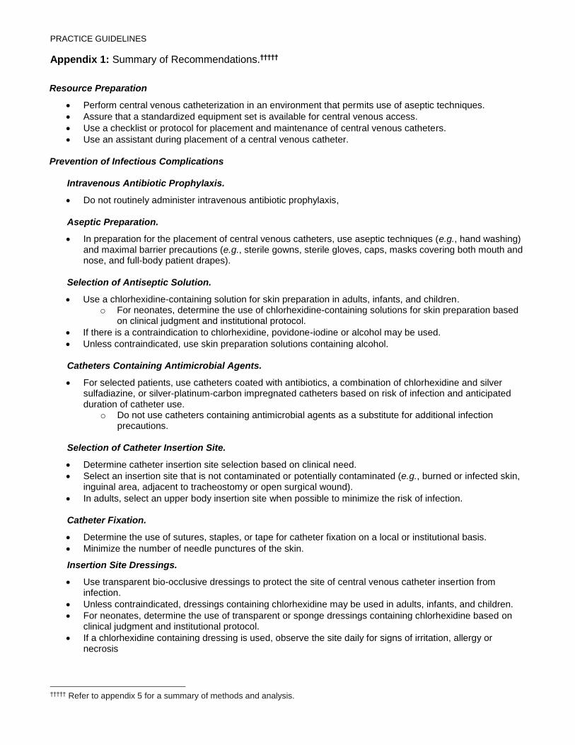
PRACTICE GUIDELINES
Appendix 1: Summary of Recommendations.†††††
Resource Preparation
• Perform central venous catheterization in an environment that permits use of aseptic techniques.
• Assure that a standardized equipment set is available for central venous access.
• Use a checklist or protocol for placement and maintenance of central venous catheters.
• Use an assistant during placement of a central venous catheter.
Prevention of Infectious Complications
Intravenous Antibiotic Prophylaxis.
• Do not routinely administer intravenous antibiotic prophylaxis,
Aseptic Preparation.
• In preparation for the placement of central venous catheters, use aseptic techniques (e.g., hand washing) and maximal barrier precautions (e.g., sterile gowns, sterile gloves, caps, masks covering both mouth and nose, and full-body patient drapes).
Selection of Antiseptic Solution.
• Use a chlorhexidine-containing solution for skin preparation in adults, infants, and children. o For neonates, determine the use of chlorhexidine-containing solutions for skin preparation based
on clinical judgment and institutional protocol.
• If there is a contraindication to chlorhexidine, povidone-iodine or alcohol may be used.
• Unless contraindicated, use skin preparation solutions containing alcohol.
Catheters Containing Antimicrobial Agents.
• For selected patients, use catheters coated with antibiotics, a combination of chlorhexidine and silver sulfadiazine, or silver-platinum-carbon impregnated catheters based on risk of infection and anticipated duration of catheter use.
o Do not use catheters containing antimicrobial agents as a substitute for additional infection precautions.
Selection of Catheter Insertion Site.
• Determine catheter insertion site selection based on clinical need.
• Select an insertion site that is not contaminated or potentially contaminated (e.g., burned or infected skin, inguinal area, adjacent to tracheostomy or open surgical wound).
• In adults, select an upper body insertion site when possible to minimize the risk of infection.
Catheter Fixation.
• Determine the use of sutures, staples, or tape for catheter fixation on a local or institutional basis.
• Minimize the number of needle punctures of the skin.
Insertion Site Dressings.
• Use transparent bio-occlusive dressings to protect the site of central venous catheter insertion from infection.
• Unless contraindicated, dressings containing chlorhexidine may be used in adults, infants, and children.
• For neonates, determine the use of transparent or sponge dressings containing chlorhexidine based on clinical judgment and institutional protocol.
• If a chlorhexidine containing dressing is used, observe the site daily for signs of irritation, allergy or necrosis
††††† Refer to appendix 5 for a summary of methods and analysis.
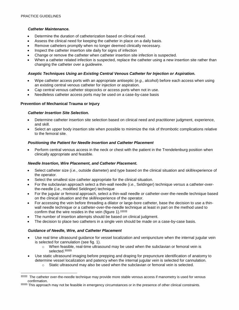
PRACTICE GUIDELINES
Catheter Maintenance.
• Determine the duration of catheterization based on clinical need.
• Assess the clinical need for keeping the catheter in place on a daily basis.
• Remove catheters promptly when no longer deemed clinically necessary.
• Inspect the catheter insertion site daily for signs of infection
• Change or remove the catheter when catheter insertion site infection is suspected.
• When a catheter related infection is suspected, replace the catheter using a new insertion site rather than changing the catheter over a guidewire.
Aseptic Techniques Using an Existing Central Venous Catheter for Injection or Aspiration.
• Wipe catheter access ports with an appropriate antiseptic (e.g., alcohol) before each access when using an existing central venous catheter for injection or aspiration.
• Cap central venous catheter stopcocks or access ports when not in use.
• Needleless catheter access ports may be used on a case-by-case basis
Prevention of Mechanical Trauma or Injury
Catheter Insertion Site Selection.
• Determine catheter insertion site selection based on clinical need and practitioner judgment, experience, and skill.
• Select an upper body insertion site when possible to minimize the risk of thrombotic complications relative to the femoral site.
Positioning the Patient for Needle Insertion and Catheter Placement
• Perform central venous access in the neck or chest with the patient in the Trendelenburg position when clinically appropriate and feasible.
Needle Insertion, Wire Placement, and Catheter Placement.
• Select catheter size (i.e., outside diameter) and type based on the clinical situation and skill/experience of the operator.
• Select the smallest size catheter appropriate for the clinical situation.
• For the subclavian approach select a thin-wall needle (i.e., Seldinger) technique versus a catheter-over-the-needle (i.e., modified Seldinger) technique
• For the jugular or femoral approach, select a thin-wall needle or catheter-over-the-needle technique based on the clinical situation and the skill/experience of the operator.
• For accessing the vein before threading a dilator or large-bore catheter, base the decision to use a thin-wall needle technique or a catheter-over-the-needle technique at least in part on the method used to confirm that the wire resides in the vein (figure 1).‡‡‡‡‡
• The number of insertion attempts should be based on clinical judgment.
• The decision to place two catheters in a single vein should be made on a case-by-case basis.
Guidance of Needle, Wire, and Catheter Placement
• Use real time ultrasound guidance for vessel localization and venipuncture when the internal jugular vein is selected for cannulation (see fig. 1).
o When feasible, real-time ultrasound may be used when the subclavian or femoral vein is selected.§§§§§
• Use static ultrasound imaging before prepping and draping for prepuncture identification of anatomy to determine vessel localization and patency when the internal jugular vein is selected for cannulation.
o Static ultrasound may also be used when the subclavian or femoral vein is selected.
‡‡‡‡‡ The catheter over-the-needle technique may provide more stable venous access if manometry is used for venous
confirmation. §§§§§ This approach may not be feasible in emergency circumstances or in the presence of other clinical constraints.

PRACTICE GUIDELINES
Verification of Needle, Wire, and Catheter Placement
• After insertion of a catheter that went over the needle or a thin-wall needle, confirm venous access.******††††††
o Do not rely on blood color or absence of pulsatile flow for confirming that the catheter or thin-wall needle resides in the vein.
• When using the thin-wall needle technique, confirm venous residence of the wire after the wire is threaded.
o When using the catheter-over-the-needle technique, confirmation that the wire resides in the vein may not be needed (1) when the catheter enters the vein easily and manometry or pressure waveform measurement provides unambiguous confirmation of venous location of the catheter; and (2) when the wire passes through the catheter and enters the vein without difficulty.
o If there is any uncertainty that the catheter or wire resides in the vein, confirm venous residence of the wire after the wire is threaded. Insertion of a dilator or large-bore catheter may then proceed.‡‡‡‡‡‡
• After final catheterization and before use, confirm residence of the catheter in the venous system as soon as clinically appropriate.§§§§§§
• Confirm the final position of the catheter tip as soon as clinically appropriate.******* o For central venous catheters placed in the operating room, perform a chest radiograph no later
than the early postoperative period to confirm the position of the catheter tip.
• Verify that the wire has not been retained in the vascular system at the end of the procedure by confirming the presence of the removed wire in the procedural field.
o If the complete guidewire is not found in the procedural field, order chest radiography to determine whether the guidewire has been retained in the patient's vascular system.
Management of Arterial Trauma or Injury Arising from Central Venous Catheterization
• When unintended cannulation of an arterial vessel with a dilator or large-bore catheter occurs, leave the dilator or catheter in place and immediately consult a general surgeon, a vascular surgeon, or an interventional radiologist regarding surgical or nonsurgical catheter removal for adults.
• For neonates, infants, and children, determine on a case-by-case basis whether to leave the catheter in place and obtain consultation or to remove the catheter nonsurgically.
• After the injury has been evaluated and a treatment plan has been executed, confer with the surgeon regarding relative risks and benefits of proceeding with the elective surgery versus deferring surgery to allow for a period of patient observation.
****** For neonates, infants, and children, confirmation of venous placement may take place after the wire is threaded. †††††† Methods for confirming that the catheter or thin-wall needle resides in the vein include, but are not limited to, ultrasound, manometry, or pressure-waveform analysis measurement ‡‡‡‡‡‡ Methods for confirming that the wire resides in the vein include, but are not limited to, ultrasound (identification of the wire in the vein) or transesophageal echocardiography (identification of the wire in the superior vena cava or right atrium), continuous electrocardiography (identification of narrow-complex ectopy), or fluoroscopy. §§§§§§ Methods for confirming that the catheter is still in the venous system after catheterization and before use include manometry or pressure waveform measurement ******* Methods for confirming the position of the catheter tip include chest radiography, fluoroscopy, or point-of-care transthoracic echocardiography or continuous electrocardiography.
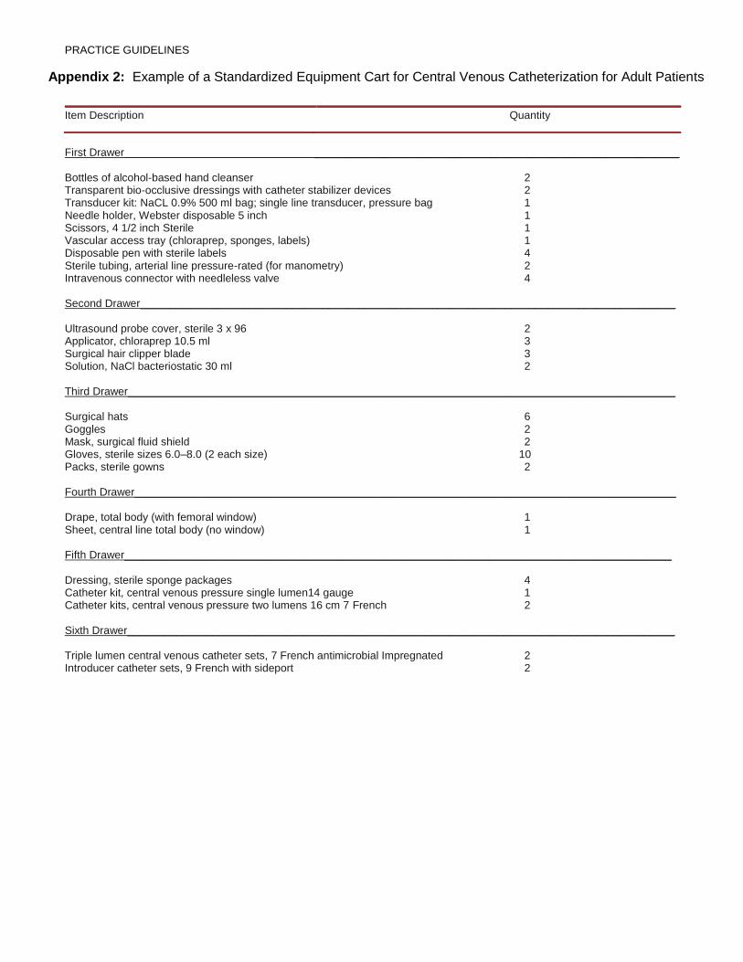
PRACTICE GUIDELINES
Appendix 2: Example of a Standardized Equipment Cart for Central Venous Catheterization for Adult Patients
Item Description Quantity
First Drawer ____________________________________________________________ Bottles of alcohol-based hand cleanser 2 Transparent bio-occlusive dressings with catheter stabilizer devices 2 Transducer kit: NaCL 0.9% 500 ml bag; single line transducer, pressure bag 1 Needle holder, Webster disposable 5 inch 1 Scissors, 4 1/2 inch Sterile 1 Vascular access tray (chloraprep, sponges, labels) 1 Disposable pen with sterile labels 4 Sterile tubing, arterial line pressure-rated (for manometry) 2 Intravenous connector with needleless valve 4
Second Drawer________________________________________________________________________________________ Ultrasound probe cover, sterile 3 x 96 2 Applicator, chloraprep 10.5 ml 3 Surgical hair clipper blade 3 Solution, NaCl bacteriostatic 30 ml 2
Third Drawer__________________________________________________________________________________________ Surgical hats 6 Goggles 2 Mask, surgical fluid shield 2 Gloves, sterile sizes 6.0–8.0 (2 each size) 10 Packs, sterile gowns 2 Fourth Drawer_________________________________________________________________________________________ Drape, total body (with femoral window) 1 Sheet, central line total body (no window) 1 Fifth Drawer__________________________________________________________________________________________ Dressing, sterile sponge packages 4 Catheter kit, central venous pressure single lumen14 gauge 1 Catheter kits, central venous pressure two lumens 16 cm 7 French 2 Sixth Drawer__________________________________________________________________________________________ Triple lumen central venous catheter sets, 7 French antimicrobial Impregnated 2 Introducer catheter sets, 9 French with sideport 2
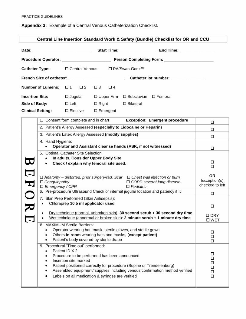
PRACTICE GUIDELINES
Appendix 3: Example of a Central Venous Catheterization Checklist.
Central Line Insertion Standard Work & Safety (Bundle) Checklist for OR and CCU
Date: __________________________ Start Time: ________________ End Time: _______________
Procedure Operator: ______________________ Person Completing Form: ______________________
Catheter Type: Central Venous PA/Swan-Ganz™
French Size of catheter: _______________ . Catheter lot number: _______________
Number of Lumens: 1 2 3 4
Insertion Site: Jugular Upper Arm Subclavian Femoral
Side of Body: Left Right Bilateral
Clinical Setting: Elective Emergent
1. Consent form complete and in chart Exception: Emergent procedure
2. Patient’s Allergy Assessed (especially to Lidocaine or Heparin)
3. Patient’s Latex Allergy Assessed (modify supplies)
4. Hand Hygiene:
• Operator and Assistant cleanse hands (ASK, if not witnessed)
5. Optimal Catheter Site Selection:
• In adults, Consider Upper Body Site
• Check / explain why femoral site used: ________________________________
Anatomy – distorted, prior surgery/rad. Scar Chest wall infection or burn Coagulopathy COPD severe/ lung disease Emergency / CPR Pediatric
OR Exception(s)
checked to left
6. Pre-procedure Ultrasound Check of internal jugular location and patency if IJ
7. Skin Prep Performed (Skin Antisepsis):
• Chloraprep 10.5 ml applicator used
• Dry technique (normal, unbroken skin): 30 second scrub + 30 second dry time
• Wet technique (abnormal or broken skin): 2 minute scrub + 1 minute dry time
DRY WET
8. MAXIMUM Sterile Barriers:
• Operator wearing hat, mask, sterile gloves, and sterile gown
• Others in room wearing hats and masks, (except patient)
• Patient’s body covered by sterile drape
9. Procedural “Time out” performed:
• Patient ID X 2
• Procedure to be performed has been announced
• Insertion site marked
• Patient positioned correctly for procedure (Supine or Trendelenburg)
• Assembled equipment/ supplies including venous confirmation method verified
• Labels on all medication & syringes are verified
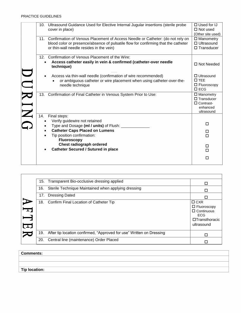
PRACTICE GUIDELINES
10. Ultrasound Guidance Used for Elective Internal Jugular insertions (sterile probe cover in place)
Used for IJ Not used (Other site used)
11. Confirmation of Venous Placement of Access Needle or Catheter: (do not rely on blood color or presence/absence of pulsatile flow for confirming that the catheter or thin-wall needle resides in the vein)
Manometry Ultrasound Transducer
12. Confirmation of Venous Placement of the Wire:
• Access catheter easily in vein & confirmed (catheter-over needle technique)
• Access via thin-wall needle (confirmation of wire recommended)
• or ambiguous catheter or wire placement when using catheter-over-the-needle technique
Not Needed
Ultrasound TEE Fluoroscopy ECG
13. Confirmation of Final Catheter in Venous System Prior to Use: Manometry Transducer
Contrast-
enhanced ultrasound
14. Final steps:
• Verify guidewire not retained
• Type and Dosage (ml / units) of Flush: _____________
• Catheter Caps Placed on Lumens
• Tip position confirmation: Fluoroscopy Chest radiograph ordered
• Catheter Secured / Sutured in place
15. Transparent Bio-occlusive dressing applied
16. Sterile Technique Maintained when applying dressing
17. Dressing Dated
18. Confirm Final Location of Catheter Tip CXR
Fluoroscopy
Continuous ECG
Transthoracic
ultrasound
19. After tip location confirmed, “Approved for use” Written on Dressing
20. Central line (maintenance) Order Placed
Comments:
Tip location:
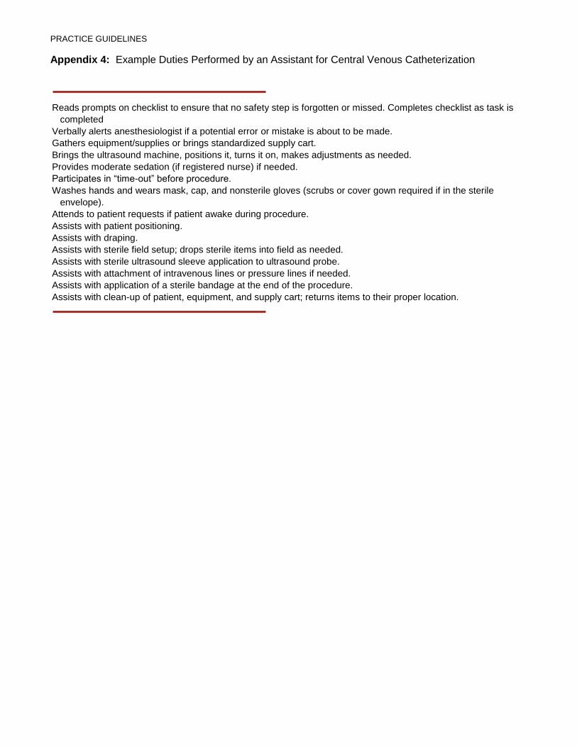
PRACTICE GUIDELINES
Appendix 4: Example Duties Performed by an Assistant for Central Venous Catheterization
Reads prompts on checklist to ensure that no safety step is forgotten or missed. Completes checklist as task is
completed
Verbally alerts anesthesiologist if a potential error or mistake is about to be made.
Gathers equipment/supplies or brings standardized supply cart.
Brings the ultrasound machine, positions it, turns it on, makes adjustments as needed.
Provides moderate sedation (if registered nurse) if needed.
Participates in “time-out” before procedure.
Washes hands and wears mask, cap, and nonsterile gloves (scrubs or cover gown required if in the sterile
envelope).
Attends to patient requests if patient awake during procedure.
Assists with patient positioning.
Assists with draping.
Assists with sterile field setup; drops sterile items into field as needed.
Assists with sterile ultrasound sleeve application to ultrasound probe.
Assists with attachment of intravenous lines or pressure lines if needed.
Assists with application of a sterile bandage at the end of the procedure.
Assists with clean-up of patient, equipment, and supply cart; returns items to their proper location.
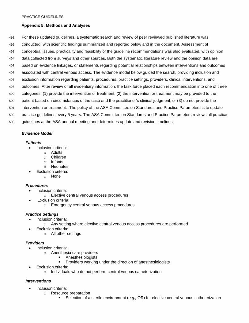
PRACTICE GUIDELINES
Appendix 5: Methods and Analyses
For these updated guidelines, a systematic search and review of peer reviewed published literature was 491
conducted, with scientific findings summarized and reported below and in the document. Assessment of 492
conceptual issues, practicality and feasibility of the guideline recommendations was also evaluated, with opinion 493
data collected from surveys and other sources. Both the systematic literature review and the opinion data are 494
based on evidence linkages, or statements regarding potential relationships between interventions and outcomes 495
associated with central venous access. The evidence model below guided the search, providing inclusion and 496
exclusion information regarding patients, procedures, practice settings, providers, clinical interventions, and 497
outcomes. After review of all evidentiary information, the task force placed each recommendation into one of three 498
categories: (1) provide the intervention or treatment, (2) the intervention or treatment may be provided to the 499
patient based on circumstances of the case and the practitioner’s clinical judgment, or (3) do not provide the 500
intervention or treatment. The policy of the ASA Committee on Standards and Practice Parameters is to update 501
practice guidelines every 5 years. The ASA Committee on Standards and Practice Parameters reviews all practice 502
guidelines at the ASA annual meeting and determines update and revision timelines. 503
Evidence Model
Patients
• Inclusion criteria: o Adults o Children o Infants o Neonates
• Exclusion criteria: o None
Procedures
• Inclusion criteria: o Elective central venous access procedures
• Exclusion criteria: o Emergency central venous access procedures
Practice Settings
• Inclusion criteria: o Any setting where elective central venous access procedures are performed
• Exclusion criteria: o All other settings
Providers
• Inclusion criteria: o Anesthesia care providers
▪ Anesthesiologists ▪ Providers working under the direction of anesthesiologists
• Exclusion criteria: o Individuals who do not perform central venous catheterization
Interventions
• Inclusion criteria: o Resource preparation
▪ Selection of a sterile environment (e.g., OR) for elective central venous catheterization
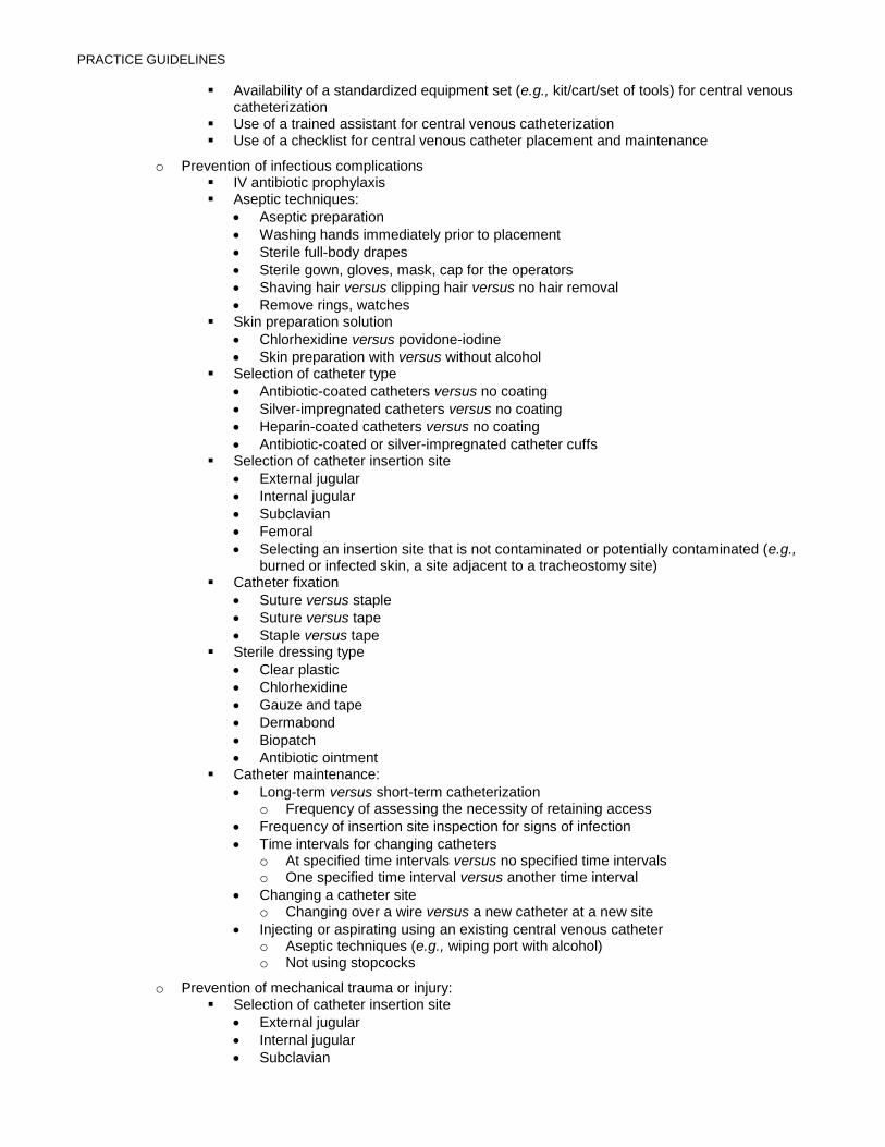
PRACTICE GUIDELINES
▪ Availability of a standardized equipment set (e.g., kit/cart/set of tools) for central venous catheterization
▪ Use of a trained assistant for central venous catheterization ▪ Use of a checklist for central venous catheter placement and maintenance
o Prevention of infectious complications ▪ IV antibiotic prophylaxis ▪ Aseptic techniques:
• Aseptic preparation
• Washing hands immediately prior to placement
• Sterile full-body drapes
• Sterile gown, gloves, mask, cap for the operators
• Shaving hair versus clipping hair versus no hair removal
• Remove rings, watches ▪ Skin preparation solution
• Chlorhexidine versus povidone-iodine
• Skin preparation with versus without alcohol ▪ Selection of catheter type
• Antibiotic-coated catheters versus no coating
• Silver-impregnated catheters versus no coating
• Heparin-coated catheters versus no coating
• Antibiotic-coated or silver-impregnated catheter cuffs ▪ Selection of catheter insertion site
• External jugular
• Internal jugular
• Subclavian
• Femoral
• Selecting an insertion site that is not contaminated or potentially contaminated (e.g., burned or infected skin, a site adjacent to a tracheostomy site)
▪ Catheter fixation
• Suture versus staple
• Suture versus tape
• Staple versus tape ▪ Sterile dressing type
• Clear plastic
• Chlorhexidine
• Gauze and tape
• Dermabond
• Biopatch
• Antibiotic ointment ▪ Catheter maintenance:
• Long-term versus short-term catheterization o Frequency of assessing the necessity of retaining access
• Frequency of insertion site inspection for signs of infection
• Time intervals for changing catheters o At specified time intervals versus no specified time intervals o One specified time interval versus another time interval
• Changing a catheter site o Changing over a wire versus a new catheter at a new site
• Injecting or aspirating using an existing central venous catheter o Aseptic techniques (e.g., wiping port with alcohol) o Not using stopcocks
o Prevention of mechanical trauma or injury: ▪ Selection of catheter insertion site
• External jugular
• Internal jugular
• Subclavian
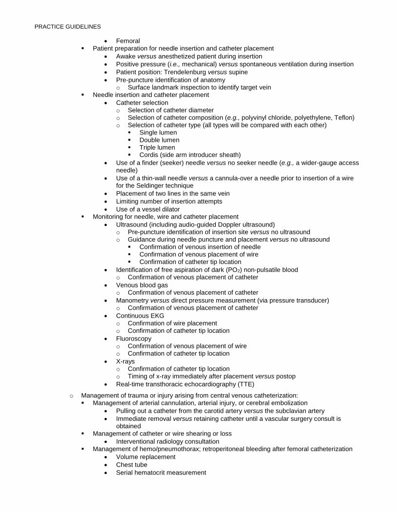
PRACTICE GUIDELINES
• Femoral ▪ Patient preparation for needle insertion and catheter placement
• Awake versus anesthetized patient during insertion
• Positive pressure (i.e., mechanical) versus spontaneous ventilation during insertion
• Patient position: Trendelenburg versus supine
• Pre-puncture identification of anatomy o Surface landmark inspection to identify target vein
▪ Needle insertion and catheter placement
• Catheter selection o Selection of catheter diameter o Selection of catheter composition (e.g., polyvinyl chloride, polyethylene, Teflon) o Selection of catheter type (all types will be compared with each other)
▪ Single lumen ▪ Double lumen ▪ Triple lumen ▪ Cordis (side arm introducer sheath)
• Use of a finder (seeker) needle versus no seeker needle (e.g., a wider-gauge access needle)
• Use of a thin-wall needle versus a cannula-over a needle prior to insertion of a wire for the Seldinger technique
• Placement of two lines in the same vein
• Limiting number of insertion attempts
• Use of a vessel dilator ▪ Monitoring for needle, wire and catheter placement
• Ultrasound (including audio-guided Doppler ultrasound) o Pre-puncture identification of insertion site versus no ultrasound o Guidance during needle puncture and placement versus no ultrasound
▪ Confirmation of venous insertion of needle ▪ Confirmation of venous placement of wire ▪ Confirmation of catheter tip location
• Identification of free aspiration of dark (PO2) non-pulsatile blood o Confirmation of venous placement of catheter
• Venous blood gas o Confirmation of venous placement of catheter
• Manometry versus direct pressure measurement (via pressure transducer) o Confirmation of venous placement of catheter
• Continuous EKG o Confirmation of wire placement o Confirmation of catheter tip location
• Fluoroscopy o Confirmation of venous placement of wire o Confirmation of catheter tip location
• X-rays o Confirmation of catheter tip location o Timing of x-ray immediately after placement versus postop
• Real-time transthoracic echocardiography (TTE)
o Management of trauma or injury arising from central venous catheterization: ▪ Management of arterial cannulation, arterial injury, or cerebral embolization
• Pulling out a catheter from the carotid artery versus the subclavian artery
• Immediate removal versus retaining catheter until a vascular surgery consult is obtained
▪ Management of catheter or wire shearing or loss
• Interventional radiology consultation ▪ Management of hemo/pneumothorax; retroperitoneal bleeding after femoral catheterization
• Volume replacement
• Chest tube
• Serial hematocrit measurement
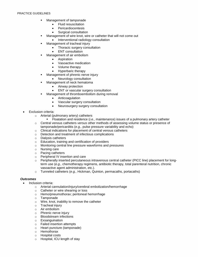
PRACTICE GUIDELINES
▪ Management of tamponade
• Fluid resuscitation
• Pericardiocentesis
• Surgical consultation ▪ Management of wire knot, wire or catheter that will not come out
• Interventional radiology consultation ▪ Management of tracheal injury
• Thoracic surgery consultation
• ENT consultation ▪ Management of air embolism
• Aspiration
• Vasoactive medication
• Volume therapy
• Hyperbaric therapy ▪ Management of phrenic nerve injury
• Neurology consultation ▪ Management of neck hematoma
• Airway protection
• ENT or vascular surgery consultation ▪ Management of thromboembolism during removal
• Anticoagulation
• Vascular surgery consultation
• Neurosurgery surgery consultation
• Exclusion criteria: o Arterial (pulmonary artery) catheters
▪ Floatation and residence (i.e., maintenance) issues of a pulmonary artery catheter o Central venous catheters versus other methods of assessing volume status or presence of
tamponade/pericarditis (e.g., pulse pressure variability and echo) o Clinical indications for placement of central venous catheters o Detection and treatment of infectious complications o Dialysis catheters o Education, training and certification of providers o Monitoring central line pressure waveforms and pressures o Nursing care o Pacing catheters o Peripheral IV insertion and care o Peripherally inserted percutaneous intravenous central catheter (PICC line) placement for long-
term use (e.g., chemotherapy regimens, antibiotic therapy, total parenteral nutrition, chronic vasoactive agent administration, etc.).
o Tunneled catheters (e.g., Hickman, Quinton, permacaths, portacaths)
Outcomes
• Inclusion criteria: o Arterial cannulation/injury/cerebral embolization/hemorrhage o Catheter or wire shearing or loss o Hemo/pneumothorax; peritoneal hemorrhage o Tamponade o Wire, knot, inability to remove the catheter o Tracheal injury o Air embolism o Phrenic nerve injury o Bloodstream infections o Exsanguination o Failed insertion attempts o Heart puncture (tamponade) o Hemothorax o Hospital costs o Hospital, ICU length of stay
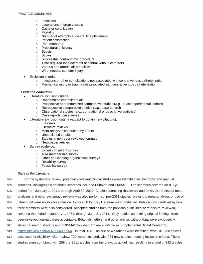
PRACTICE GUIDELINES
o Infections o Lacerations of great vessels o Catheter colonization o Mortality o Number of attempts at central line placement o Patient satisfaction o Pneumothorax o Procedural efficiency o Sepsis o Stroke o Successful, nontraumatic procedure o Time required for placement of central venous catheters o Venous and arterial air embolism o Wire, needle, catheter injury
• Exclusion criteria: o Infections or other complications not associated with central venous catheterization o Mechanical injury or trauma not associated with central venous catheterization
Evidence collection
• Literature inclusion criteria: o Randomized controlled trials o Prospective nonrandomized comparative studies (e.g., quasi-experimental, cohort) o Retrospective comparative studies (e.g., case-control) o Observational studies (e.g., correlational or descriptive statistics) o Case reports, case series
• Literature exclusion criteria (except to obtain new citations): o Editorials o Literature reviews o Meta-analyses conducted by others o Unpublished studies o Studies in non-peer reviewed journals o Newspaper articles
• Survey evidence: o Expert consultant survey o ASA membership survey o Other participating organization surveys o Reliability survey o Feasibility survey
State of the Literature.
For the systematic review, potentially relevant clinical studies were identified via electronic and manual 504
searches. Bibliographic database searches included PubMed and EMBASE. The searches covered an 8.3-yr 505
period from January 1, 2011, through April 30, 2019. Citation searching (backward and forward) of relevant meta-506
analyses and other systematic reviews was also performed; pre-2011 studies relevant to meta-analyses or use of 507
ultrasound were eligible for inclusion. No search for grey literature was conducted. Publications identified by task 508
force members were also considered. Accepted studies from the previous guidelines were also re-reviewed, 509
covering the period of January 1, 1971, through June 31, 2011. Only studies containing original findings from 510
peer-reviewed journals were acceptable. Editorials, letters, and other articles without data were excluded. A 511
literature search strategy and PRISMA* flow diagram are available as Supplemental Digital Content 2, 512
http://links.lww.com/ALN/XXXXXXXX. In total, 4,491 unique new citations were identified, with 1013 full articles 513
assessed for eligibility. After review, 729 were excluded, with 284 new studies meeting inclusion criteria. These 514
studies were combined with 258 pre-2011 articles from the previous guidelines, resulting in a total of 542 articles 515
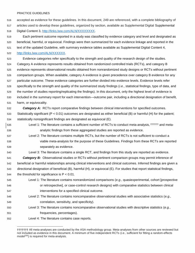
PRACTICE GUIDELINES
accepted as evidence for these guidelines. In this document, 249 are referenced, with a complete bibliography of 516
articles used to develop these guidelines, organized by section, available as Supplemental Digital Supplemental 517
Digital Content 3, http://links.lww.com/ALN/XXXXXXXX. 518
Each pertinent outcome reported in a study was classified by evidence category and level and designated as 519
beneficial, harmful, or equivocal. Findings were then summarized for each evidence linkage and reported in the 520
text of the updated Guideline, with summary evidence tables available as Supplemental Digital Content 4, 521
http://links.lww.com/ALN/XXXXXX. 522
Evidence categories refer specifically to the strength and quality of the research design of the studies. 523
Category A evidence represents results obtained from randomized controlled trials (RCTs), and category B 524
evidence represents observational results obtained from nonrandomized study designs or RCTs without pertinent 525
comparison groups. When available, category A evidence is given precedence over category B evidence for any 526
particular outcome. These evidence categories are further divided into evidence levels. Evidence levels refer 527
specifically to the strength and quality of the summarized study findings (i.e., statistical findings, type of data, and 528
the number of studies reporting/replicating the findings). In this document, only the highest level of evidence is 529
included in the summary report for each intervention—outcome pair, including a directional designation of benefit, 530
harm, or equivocality. 531
Category A: RCTs report comparative findings between clinical interventions for specified outcomes. 532
Statistically significant (P < 0.01) outcomes are designated as either beneficial (B) or harmful (H) for the patient; 533
statistically nonsignificant findings are designated as equivocal (E). 534
Level 1: The literature contains a sufficient number of RCTs to conduct meta-analysis, ††††††† and meta-535
analytic findings from these aggregated studies are reported as evidence. 536
Level 2: The literature contains multiple RCTs, but the number of RCTs is not sufficient to conduct a 537
viable meta-analysis for the purpose of these Guidelines. Findings from these RCTs are reported 538
separately as evidence. 539
Level 3: The literature contains a single RCT, and findings from this study are reported as evidence. 540
Category B: Observational studies or RCTs without pertinent comparison groups may permit inference of 541
beneficial or harmful relationships among clinical interventions and clinical outcomes. Inferred findings are given a 542
directional designation of beneficial (B), harmful (H), or equivocal (E). For studies that report statistical findings, 543
the threshold for significance is P < 0.01. 544
Level 1: The literature contains nonrandomized comparisons (e.g., quasiexperimental, cohort [prospective 545
or retrospective], or case-control research designs) with comparative statistics between clinical 546
interventions for a specified clinical outcome. 547
Level 2: The literature contains noncomparative observational studies with associative statistics (e.g., 548
correlation, sensitivity, and specificity). 549
Level 3: The literature contains noncomparative observational studies with descriptive statistics (e.g., 550
frequencies, percentages). 551
Level 4: The literature contains case reports. 552
††††††† All meta-analyses are conducted by the ASA methodology group. Meta-analyses from other sources are reviewed but not included as evidence in this document. A minimum of five independent RCTs (i.e., sufficient for fitting a random-effects model255) is required for meta-analysis.
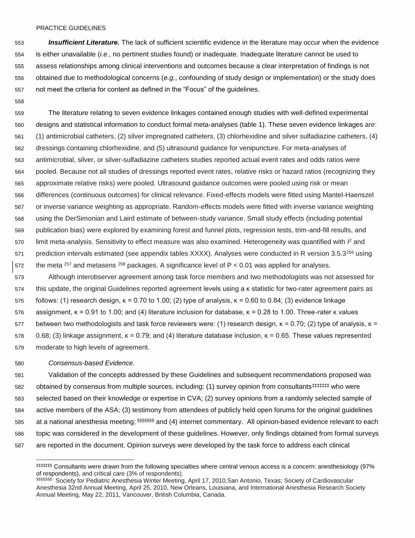
PRACTICE GUIDELINES
Insufficient Literature. The lack of sufficient scientific evidence in the literature may occur when the evidence 553
is either unavailable (i.e., no pertinent studies found) or inadequate. Inadequate literature cannot be used to 554
assess relationships among clinical interventions and outcomes because a clear interpretation of findings is not 555
obtained due to methodological concerns (e.g., confounding of study design or implementation) or the study does 556
not meet the criteria for content as defined in the “Focus” of the guidelines. 557
558
The literature relating to seven evidence linkages contained enough studies with well-defined experimental 559
designs and statistical information to conduct formal meta-analyses (table 1). These seven evidence linkages are: 560
(1) antimicrobial catheters, (2) silver impregnated catheters, (3) chlorhexidine and silver sulfadiazine catheters, (4) 561
dressings containing chlorhexidine, and (5) ultrasound guidance for venipuncture. For meta-analyses of 562
antimicrobial, silver, or silver-sulfadiazine catheters studies reported actual event rates and odds ratios were 563
pooled. Because not all studies of dressings reported event rates, relative risks or hazard ratios (recognizing they 564
approximate relative risks) were pooled. Ultrasound guidance outcomes were pooled using risk or mean 565
differences (continuous outcomes) for clinical relevance. Fixed-effects models were fitted using Mantel-Haenszel 566
or inverse variance weighting as appropriate. Random-effects models were fitted with inverse variance weighting 567
using the DerSimonian and Laird estimate of between-study variance. Small study effects (including potential 568
publication bias) were explored by examining forest and funnel plots, regression tests, trim-and-fill results, and 569
limit meta-analysis. Sensitivity to effect measure was also examined. Heterogeneity was quantified with I2 and 570
prediction intervals estimated (see appendix tables XXXX). Analyses were conducted in R version 3.5.3256 using 571
the meta 257 and metasens 258 packages. A significance level of P < 0.01 was applied for analyses. 572
Although interobserver agreement among task force members and two methodologists was not assessed for 573
this update, the original Guidelines reported agreement levels using a κ statistic for two-rater agreement pairs as 574
follows: (1) research design, κ = 0.70 to 1.00; (2) type of analysis, κ = 0.60 to 0.84; (3) evidence linkage 575
assignment, κ = 0.91 to 1.00; and (4) literature inclusion for database, κ = 0.28 to 1.00. Three-rater κ values 576
between two methodologists and task force reviewers were: (1) research design, κ = 0.70; (2) type of analysis, κ = 577
0.68; (3) linkage assignment, κ = 0.79; and (4) literature database inclusion, κ = 0.65. These values represented 578
moderate to high levels of agreement. 579
Consensus-based Evidence. 580
Validation of the concepts addressed by these Guidelines and subsequent recommendations proposed was 581
obtained by consensus from multiple sources, including: (1) survey opinion from consultants‡‡‡‡‡‡‡ who were 582
selected based on their knowledge or expertise in CVA; (2) survey opinions from a randomly selected sample of 583
active members of the ASA; (3) testimony from attendees of publicly held open forums for the original guidelines 584
at a national anesthesia meeting; §§§§§§§ and (4) internet commentary. All opinion-based evidence relevant to each 585
topic was considered in the development of these guidelines. However, only findings obtained from formal surveys 586
are reported in the document. Opinion surveys were developed by the task force to address each clinical 587
‡‡‡‡‡‡‡ Consultants were drawn from the following specialties where central venous access is a concern: anesthesiology (97% of respondents), and critical care (3% of respondents). §§§§§§§ Society for Pediatric Anesthesia Winter Meeting, April 17, 2010,San Antonio, Texas; Society of Cardiovascular Anesthesia 32nd Annual Meeting, April 25, 2010, New Orleans, Louisiana, and International Anesthesia Research Society Annual Meeting, May 22, 2011, Vancouver, British Columbia, Canada.
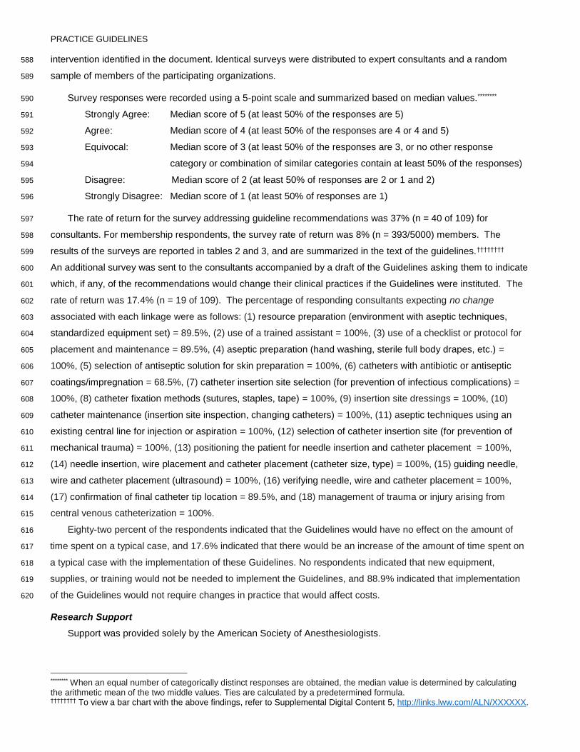
PRACTICE GUIDELINES
intervention identified in the document. Identical surveys were distributed to expert consultants and a random 588
sample of members of the participating organizations. 589
Survey responses were recorded using a 5-point scale and summarized based on median values.******** 590
Strongly Agree: Median score of 5 (at least 50% of the responses are 5) 591
Agree: Median score of 4 (at least 50% of the responses are 4 or 4 and 5) 592
Equivocal: Median score of 3 (at least 50% of the responses are 3, or no other response 593
category or combination of similar categories contain at least 50% of the responses) 594
Disagree: Median score of 2 (at least 50% of responses are 2 or 1 and 2) 595
Strongly Disagree: Median score of 1 (at least 50% of responses are 1) 596
The rate of return for the survey addressing guideline recommendations was 37% (n = 40 of 109) for 597
consultants. For membership respondents, the survey rate of return was 8% (n = 393/5000) members. The 598
results of the surveys are reported in tables 2 and 3, and are summarized in the text of the guidelines.†††††††† 599
An additional survey was sent to the consultants accompanied by a draft of the Guidelines asking them to indicate 600
which, if any, of the recommendations would change their clinical practices if the Guidelines were instituted. The 601
rate of return was 17.4% (n = 19 of 109). The percentage of responding consultants expecting no change 602
associated with each linkage were as follows: (1) resource preparation (environment with aseptic techniques, 603
standardized equipment set) = 89.5%, (2) use of a trained assistant = 100%, (3) use of a checklist or protocol for 604
placement and maintenance = 89.5%, (4) aseptic preparation (hand washing, sterile full body drapes, etc.) = 605
100%, (5) selection of antiseptic solution for skin preparation = 100%, (6) catheters with antibiotic or antiseptic 606
coatings/impregnation = 68.5%, (7) catheter insertion site selection (for prevention of infectious complications) = 607
100%, (8) catheter fixation methods (sutures, staples, tape) = 100%, (9) insertion site dressings = 100%, (10) 608
catheter maintenance (insertion site inspection, changing catheters) = 100%, (11) aseptic techniques using an 609
existing central line for injection or aspiration = 100%, (12) selection of catheter insertion site (for prevention of 610
mechanical trauma) = 100%, (13) positioning the patient for needle insertion and catheter placement = 100%, 611
(14) needle insertion, wire placement and catheter placement (catheter size, type) = 100%, (15) guiding needle, 612
wire and catheter placement (ultrasound) = 100%, (16) verifying needle, wire and catheter placement = 100%, 613
(17) confirmation of final catheter tip location = 89.5%, and (18) management of trauma or injury arising from 614
central venous catheterization = 100%. 615
Eighty-two percent of the respondents indicated that the Guidelines would have no effect on the amount of 616
time spent on a typical case, and 17.6% indicated that there would be an increase of the amount of time spent on 617
a typical case with the implementation of these Guidelines. No respondents indicated that new equipment, 618
supplies, or training would not be needed to implement the Guidelines, and 88.9% indicated that implementation 619
of the Guidelines would not require changes in practice that would affect costs. 620
Research Support
Support was provided solely by the American Society of Anesthesiologists.
******** When an equal number of categorically distinct responses are obtained, the median value is determined by calculating the arithmetic mean of the two middle values. Ties are calculated by a predetermined formula. †††††††† To view a bar chart with the above findings, refer to Supplemental Digital Content 5, http://links.lww.com/ALN/XXXXXX.

PRACTICE GUIDELINES
Competing Interests
The authors declare no competing interests.
Correspondence
Address correspondence to the American Society of Anesthesiologists: 1061 American Lane, Schaumburg,
Illinois 60173. These updated Practice Guidelines, and all ASA Practice Parameters, may be obtained at no cost
through the Journal Web site, www.anesthesiology.org.
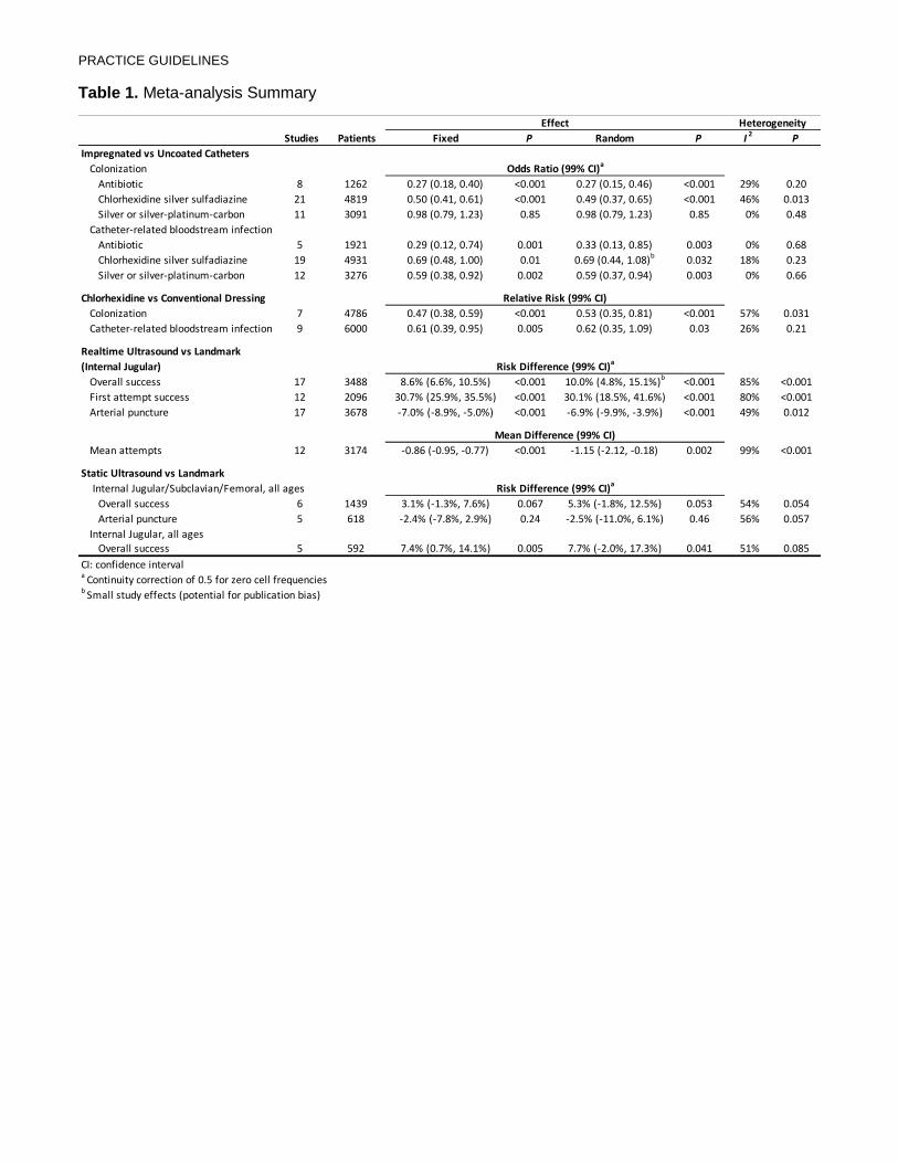
PRACTICE GUIDELINES
Table 1. Meta-analysis Summary
Studies Patients Fixed P Random P I2
P
Impregnated vs Uncoated Catheters
Colonization
Antibiotic 8 1262 0.27 (0.18, 0.40) <0.001 0.27 (0.15, 0.46) <0.001 29% 0.20
Chlorhexidine silver sulfadiazine 21 4819 0.50 (0.41, 0.61) <0.001 0.49 (0.37, 0.65) <0.001 46% 0.013
Silver or silver-platinum-carbon 11 3091 0.98 (0.79, 1.23) 0.85 0.98 (0.79, 1.23) 0.85 0% 0.48
Catheter-related bloodstream infection
Antibiotic 5 1921 0.29 (0.12, 0.74) 0.001 0.33 (0.13, 0.85) 0.003 0% 0.68
Chlorhexidine silver sulfadiazine 19 4931 0.69 (0.48, 1.00) 0.01 0.69 (0.44, 1.08)b
0.032 18% 0.23
Silver or silver-platinum-carbon 12 3276 0.59 (0.38, 0.92) 0.002 0.59 (0.37, 0.94) 0.003 0% 0.66
Chlorhexidine vs Conventional Dressing
Colonization 7 4786 0.47 (0.38, 0.59) <0.001 0.53 (0.35, 0.81) <0.001 57% 0.031
Catheter-related bloodstream infection 9 6000 0.61 (0.39, 0.95) 0.005 0.62 (0.35, 1.09) 0.03 26% 0.21
Overall success 17 3488 8.6% (6.6%, 10.5%) <0.001 10.0% (4.8%, 15.1%)b
<0.001 85% <0.001
First attempt success 12 2096 30.7% (25.9%, 35.5%) <0.001 30.1% (18.5%, 41.6%) <0.001 80% <0.001
Arterial puncture 17 3678 -7.0% (-8.9%, -5.0%) <0.001 -6.9% (-9.9%, -3.9%) <0.001 49% 0.012
Mean attempts 12 3174 -0.86 (-0.95, -0.77) <0.001 -1.15 (-2.12, -0.18) 0.002 99% <0.001
Static Ultrasound vs Landmark
Overall success 6 1439 3.1% (-1.3%, 7.6%) 0.067 5.3% (-1.8%, 12.5%) 0.053 54% 0.054
Arterial puncture 5 618 -2.4% (-7.8%, 2.9%) 0.24 -2.5% (-11.0%, 6.1%) 0.46 56% 0.057
Internal Jugular, all agesOverall success 5 592 7.4% (0.7%, 14.1%) 0.005 7.7% (-2.0%, 17.3%) 0.041 51% 0.085
Effect Heterogeneity
Realtime Ultrasound vs Landmark
(Internal Jugular) Risk Difference (99% CI)a
Mean Difference (99% CI)
Odds Ratio (99% CI)a
CI: confidence intervala
Continuity correction of 0.5 for zero cell frequenciesb Small study effects (potential for publication bias)
Relative Risk (99% CI)
Risk Difference (99% CI)a
Internal Jugular/Subclavian/Femoral, all ages
PRACTICE GUIDELINES
Table 2. Expert Consultant Survey Results ‡‡‡‡‡‡‡‡
Recommendations N Strongly Agree (%)
Agree (%)
Equivocal (%)
Disagree (%)
Strongly Disagree (%)
Resource Preparation
1. Perform central venous catheterization in an environment that permits use of aseptic techniques. 40 95* 5 0 0 0 2. Assure that a standardized equipment set is available for central venous access. 39 77* 18 5 0 0 3. Use a checklist or protocol for placement and maintenance of central venous catheters. 38 60* 26 8 5 0 4. Use an assistant during placement of a central venous catheter. 40 42 28* 23 8 0
Prevention of Infections Complications
Intravenous Antibiotic Prophylaxis
5. For immunocompromised patients and high-risk neonates, administer intravenous antibiotic prophylaxis on a case-by case basis.
40 12 30 40* 18 0
6. Do not routinely administer intravenous antibiotic prophylaxis.
40 55* 38 8 0 0
Aseptic Preparation
7. In preparation for the placement of central venous catheters, use aseptic techniques (e.g., hand washing) and maximal barrier precautions (e.g., sterile gowns, sterile gloves, caps, masks covering both mouth and nose, and full-body patient drapes).
39 74* 21 3 3 0
Selection of Antiseptic Solution
8. Use a chlorhexidine-containing solution for skin preparation in adults, infants, and children. 39 85* 15 0 0 0 9. For neonates, determine the use of chlorhexidine-containing solutions for skin preparation based on clinical judgment and institutional protocol. 38 42 40* 18 0 0 10. If there is a contraindication to chlorhexidine, povidone-iodine or alcohol may be used. 39 59* 36 5 0 0 11. Unless contraindicated, use skin preparation solutions containing alcohol. 39 28 39* 23 10 0
Catheters Containing Antimicrobial Agents
12. For selected patients, use catheters coated with antibiotics or a combination of chlorhexidine and silver sulfadiazine based on infectious risk, cost, and anticipated duration of catheter use.
38 29 40* 24 8 0
13. Do not use catheters containing antimicrobial agents as a substitute for additional infection precautions.
38 63* 18 16 3 0
Selection of Catheter Insertion Site 14. Determine catheter insertion site selection based on clinical need. 38 71* 29 0 0 0 15. Select an insertion site that is not contaminated or potentially contaminated (e.g., burned or infected skin, inguinal area, adjacent to tracheostomy or open surgical wound). 38 82* 16 3 0 0 16. To minimize the risk of infection in adults, select an upper body insertion site when possible. 38 76* 21 3 0 0
Catheter Fixation
17. Determine the use of sutures, staples, or tape for catheter fixation on a local or institutional basis.
38 47* 32 8 13 0
18. Minimize the number of needle punctures of the skin. 38 61* 32 8 0 0
‡‡‡‡‡‡‡‡ N = number of members who responded to each item. An asterisk beside a percentage score indicates the median.
PRACTICE GUIDELINES
Insertion Site Dressings
19. Use transparent bio-occlusive dressings to protect the site of central venous catheter insertion from infection.
38 71* 29 0 0 0
20. Unless contraindicated, dressings containing chlorhexidine may be used in adults, infants, and children.
38 45 37* 18 0 0
21. For neonates, determine the use of transparent or sponge dressings containing chlorhexidine based on clinical judgment and institutional protocol.
38 37 47* 13 3 0
22. If a chlorhexidine containing dressing is used, observe the site daily for signs of irritation, allergy or necrosis.
38 66* 26 8 0 0
Catheter Maintenance
23. Determine the duration of catheterization based on clinical need. 38 79* 21 0 0 0 24. Assess the clinical need for keeping the catheter in place on a daily basis. 38 87* 13 0 0 0 25. Remove catheters removed promptly when no longer deemed clinically necessary. 38 90* 11 0 0 0 26. Inspect the catheter insertion site daily for signs of infection. 38 92* 8 0 0 0 27. Change or remove the catheter when catheter insertion site infection is suspected. 38 66* 32 0 3 0 28. When a catheter related infection is suspected, replace the catheter using a new insertion site rather than changing the catheter over a guidewire. 38 61* 34 3 3 0
Aseptic Techniques Using an Existing Central Venous Catheter for Injection or Aspiration
29. Wipe catheter access ports with an appropriate antiseptic (e.g., alcohol) before each access when using an existing central venous catheter for injection or aspiration.
38 90* 8 3 0 0
30. Cap central venous catheter stopcocks or access ports when not in use. 38 79* 18 3 0 0 31. Needleless catheter access ports may be used on a case-by-case basis. 38 24 53* 13 11 0
Prevention of Mechanical Trauma or Injury
Catheter Insertion Site Selection
32. Determine catheter insertion site selection based on clinical need and practitioner judgment, experience, and skill.
38 68* 26 3 3 0
33. Selection of an upper body insertion site may minimize the risk of thrombotic complications relative to the femoral site.
38 21 47* 26 5 0
Positioning the Patient for Needle Insertion and Catheter Placement 34. When clinically appropriate and feasible, perform central venous access in the neck or chest with the patient in the Trendelenburg position.
38 53* 34 11 3 0
Needle Insertion, Wire Placement, and Catheter Placement
35. Select catheter size (i.e., outside diameter) and type based on the clinical situation and skill/experience of the operator.
38 63* 32 5 0 0
36. Select the smallest size catheter appropriate for the clinical situation.
38 40 42* 18 0 0
37. For the subclavian approach select a thin-wall needle (i.e., Seldinger) technique versus a catheter-over-the-needle (i.e., modified Seldinger) technique.
38 32 50* 18 0 0
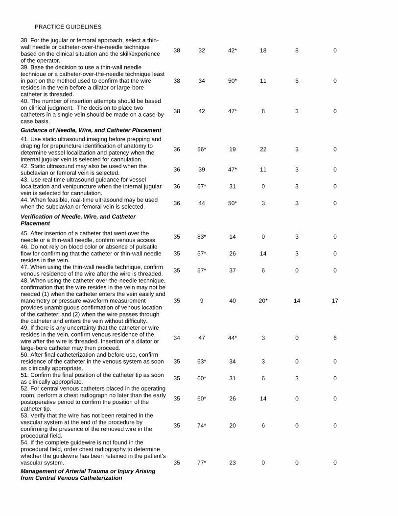
PRACTICE GUIDELINES
38. For the jugular or femoral approach, select a thin-wall needle or catheter-over-the-needle technique based on the clinical situation and the skill/experience of the operator.
38 32 42* 18 8 0
39. Base the decision to use a thin-wall needle technique or a catheter-over-the-needle technique least in part on the method used to confirm that the wire resides in the vein before a dilator or large-bore catheter is threaded.
38 34 50* 11 5 0
40. The number of insertion attempts should be based on clinical judgment. The decision to place two catheters in a single vein should be made on a case-by-case basis.
38 42 47* 8 3 0
Guidance of Needle, Wire, and Catheter Placement
41. Use static ultrasound imaging before prepping and draping for prepuncture identification of anatomy to determine vessel localization and patency when the internal jugular vein is selected for cannulation.
36 56* 19 22 3 0
42. Static ultrasound may also be used when the subclavian or femoral vein is selected.
36 39 47* 11 3 0
43. Use real time ultrasound guidance for vessel localization and venipuncture when the internal jugular vein is selected for cannulation.
36 67* 31 0 3 0
44. When feasible, real-time ultrasound may be used when the subclavian or femoral vein is selected.
36 44 50* 3 3 0
Verification of Needle, Wire, and Catheter Placement
45. After insertion of a catheter that went over the needle or a thin-wall needle, confirm venous access.
35 83* 14 0 3 0
46. Do not rely on blood color or absence of pulsatile flow for confirming that the catheter or thin-wall needle resides in the vein.
35 57* 26 14 3 0
47. When using the thin-wall needle technique, confirm venous residence of the wire after the wire is threaded.
35 57* 37 6 0 0
48. When using the catheter-over-the-needle technique, confirmation that the wire resides in the vein may not be needed (1) when the catheter enters the vein easily and manometry or pressure waveform measurement provides unambiguous confirmation of venous location of the catheter; and (2) when the wire passes through the catheter and enters the vein without difficulty.
35 9 40 20* 14 17
49. If there is any uncertainty that the catheter or wire resides in the vein, confirm venous residence of the wire after the wire is threaded. Insertion of a dilator or large-bore catheter may then proceed.
34 47 44* 3 0 6
50. After final catheterization and before use, confirm residence of the catheter in the venous system as soon as clinically appropriate.
35 63* 34 3 0 0
51. Confirm the final position of the catheter tip as soon as clinically appropriate.
35 60* 31 6 3 0
52. For central venous catheters placed in the operating room, perform a chest radiograph no later than the early postoperative period to confirm the position of the catheter tip.
35 60* 26 14 0 0
53. Verify that the wire has not been retained in the vascular system at the end of the procedure by confirming the presence of the removed wire in the procedural field.
35 74* 20 6 0 0
54. If the complete guidewire is not found in the procedural field, order chest radiography to determine whether the guidewire has been retained in the patient's vascular system. 35 77* 23 0 0 0
Management of Arterial Trauma or Injury Arising from Central Venous Catheterization
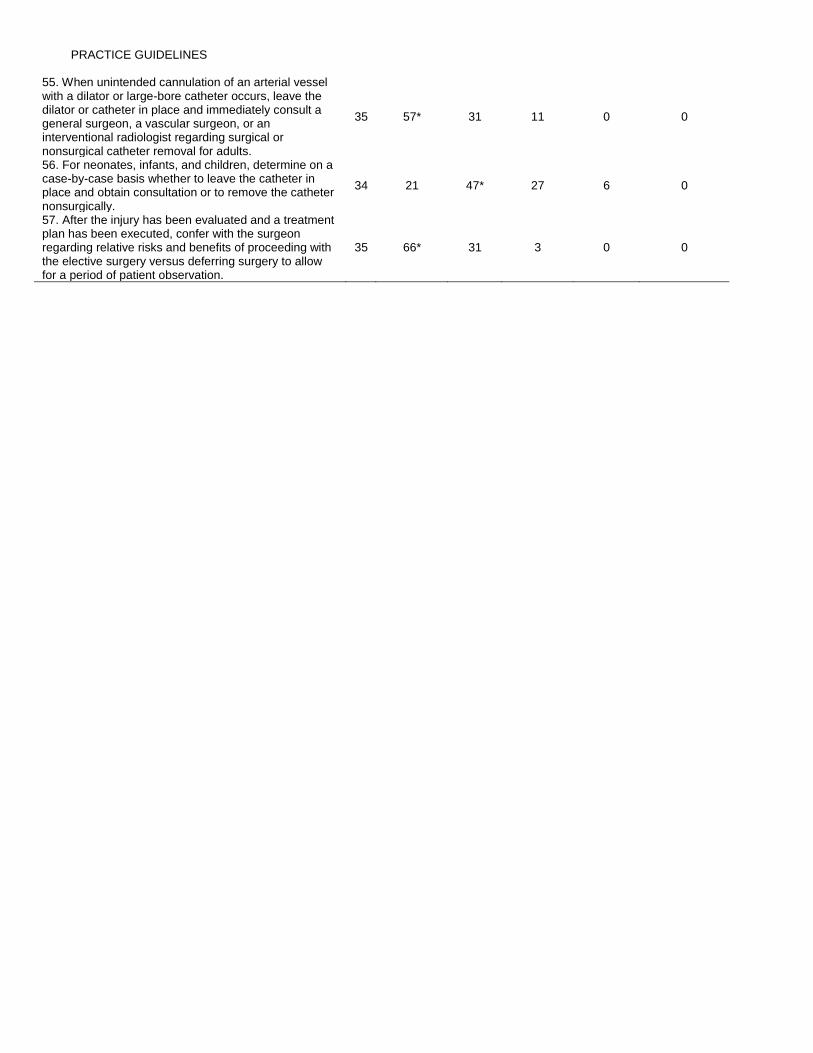
PRACTICE GUIDELINES
55. When unintended cannulation of an arterial vessel with a dilator or large-bore catheter occurs, leave the dilator or catheter in place and immediately consult a general surgeon, a vascular surgeon, or an interventional radiologist regarding surgical or nonsurgical catheter removal for adults.
35 57* 31 11 0 0
56. For neonates, infants, and children, determine on a case-by-case basis whether to leave the catheter in place and obtain consultation or to remove the catheter nonsurgically.
34 21 47* 27 6 0
57. After the injury has been evaluated and a treatment plan has been executed, confer with the surgeon regarding relative risks and benefits of proceeding with the elective surgery versus deferring surgery to allow for a period of patient observation.
35 66* 31 3 0 0
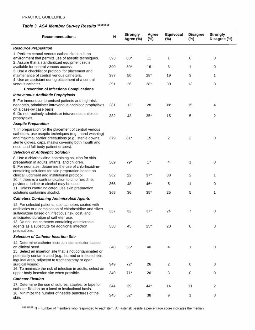
PRACTICE GUIDELINES
Table 3. ASA Member Survey Results §§§§§§§§
Recommendations N Strongly Agree (%)
Agree (%)
Equivocal (%)
Disagree (%)
Strongly Disagree (%)
Resource Preparation
1. Perform central venous catheterization in an environment that permits use of aseptic techniques. 393 88* 11 1 0 0 2. Assure that a standardized equipment set is available for central venous access. 390 80* 16 3 1 0 3. Use a checklist or protocol for placement and maintenance of central venous catheters. 387 50 28* 19 3 1 4. Use an assistant during placement of a central venous catheter. 391 26 28* 30 13 3
Prevention of Infections Complications
Intravenous Antibiotic Prophylaxis
5. For immunocompromised patients and high-risk neonates, administer intravenous antibiotic prophylaxis on a case-by case basis.
381 13 28 39* 15 4
6. Do not routinely administer intravenous antibiotic prophylaxis.
382 43 35* 15 5 2
Aseptic Preparation
7. In preparation for the placement of central venous catheters, use aseptic techniques (e.g., hand washing) and maximal barrier precautions (e.g., sterile gowns, sterile gloves, caps, masks covering both mouth and nose, and full-body patient drapes).
379 81* 15 2 2 0
Selection of Antiseptic Solution
8. Use a chlorhexidine-containing solution for skin preparation in adults, infants, and children. 369 79* 17 4 1 0 9. For neonates, determine the use of chlorhexidine-containing solutions for skin preparation based on clinical judgment and institutional protocol. 362 22 37* 38 2 1 10. If there is a contraindication to chlorhexidine, povidone-iodine or alcohol may be used. 366 48 46* 5 1 0 11. Unless contraindicated, use skin preparation solutions containing alcohol. 368 36 35* 25 5 1
Catheters Containing Antimicrobial Agents
12. For selected patients, use catheters coated with antibiotics or a combination of chlorhexidine and silver sulfadiazine based on infectious risk, cost, and anticipated duration of catheter use.
357 32 37* 24 7 0
13. Do not use catheters containing antimicrobial agents as a substitute for additional infection precautions.
358 45 25* 20 8 3
Selection of Catheter Insertion Site 14. Determine catheter insertion site selection based on clinical need. 348 55* 40 4 1 0 15. Select an insertion site that is not contaminated or potentially contaminated (e.g., burned or infected skin, inguinal area, adjacent to tracheostomy or open surgical wound). 349 72* 26 2 0 0 16. To minimize the risk of infection in adults, select an upper body insertion site when possible. 349 71* 26 3 0 0
Catheter Fixation
17. Determine the use of sutures, staples, or tape for catheter fixation on a local or institutional basis.
344 29 44* 14 11 2
18. Minimize the number of needle punctures of the skin.
345 52* 38 9 1 0
§§§§§§§§ N = number of members who responded to each item. An asterisk beside a percentage score indicates the median.
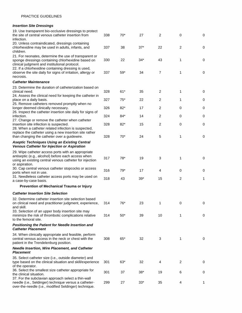
PRACTICE GUIDELINES
Insertion Site Dressings
19. Use transparent bio-occlusive dressings to protect the site of central venous catheter insertion from infection.
338 70* 27 2 0 0
20. Unless contraindicated, dressings containing chlorhexidine may be used in adults, infants, and children.
337 38 37* 22 2 0
21. For neonates, determine the use of transparent or sponge dressings containing chlorhexidine based on clinical judgment and institutional protocol.
330 22 34* 43 1 0
22. If a chlorhexidine containing dressing is used, observe the site daily for signs of irritation, allergy or necrosis.
337 59* 34 7 1 0
Catheter Maintenance
23. Determine the duration of catheterization based on clinical need. 328 61* 35 2 1 0 24. Assess the clinical need for keeping the catheter in place on a daily basis. 327 75* 22 2 1 0 25. Remove catheters removed promptly when no longer deemed clinically necessary. 326 82* 17 2 0 0 26. Inspect the catheter insertion site daily for signs of infection. 324 84* 14 2 0 0 27. Change or remove the catheter when catheter insertion site infection is suspected. 328 82* 15 2 0 0 28. When a catheter related infection is suspected, replace the catheter using a new insertion site rather than changing the catheter over a guidewire. 328 70* 24 5 1 0
Aseptic Techniques Using an Existing Central Venous Catheter for Injection or Aspiration
29. Wipe catheter access ports with an appropriate antiseptic (e.g., alcohol) before each access when using an existing central venous catheter for injection or aspiration.
317 78* 19 3 1 0
30. Cap central venous catheter stopcocks or access ports when not in use.
316 79* 17 4 0 0
31. Needleless catheter access ports may be used on a case-by-case basis.
318 43 39* 15 2 1
Prevention of Mechanical Trauma or Injury
Catheter Insertion Site Selection
32. Determine catheter insertion site selection based on clinical need and practitioner judgment, experience, and skill.
314 76* 23 1 0 0
33. Selection of an upper body insertion site may minimize the risk of thrombotic complications relative to the femoral site.
314 50* 39 10 1 0
Positioning the Patient for Needle Insertion and Catheter Placement 34. When clinically appropriate and feasible, perform central venous access in the neck or chest with the patient in the Trendelenburg position.
308 65* 32 3 1 0
Needle Insertion, Wire Placement, and Catheter Placement
35. Select catheter size (i.e., outside diameter) and type based on the clinical situation and skill/experience of the operator.
301 63* 32 4 2 0
36. Select the smallest size catheter appropriate for the clinical situation.
301 37 38* 19 6 0
37. For the subclavian approach select a thin-wall needle (i.e., Seldinger) technique versus a catheter-over-the-needle (i.e., modified Seldinger) technique.
299 27 33* 35 4 1
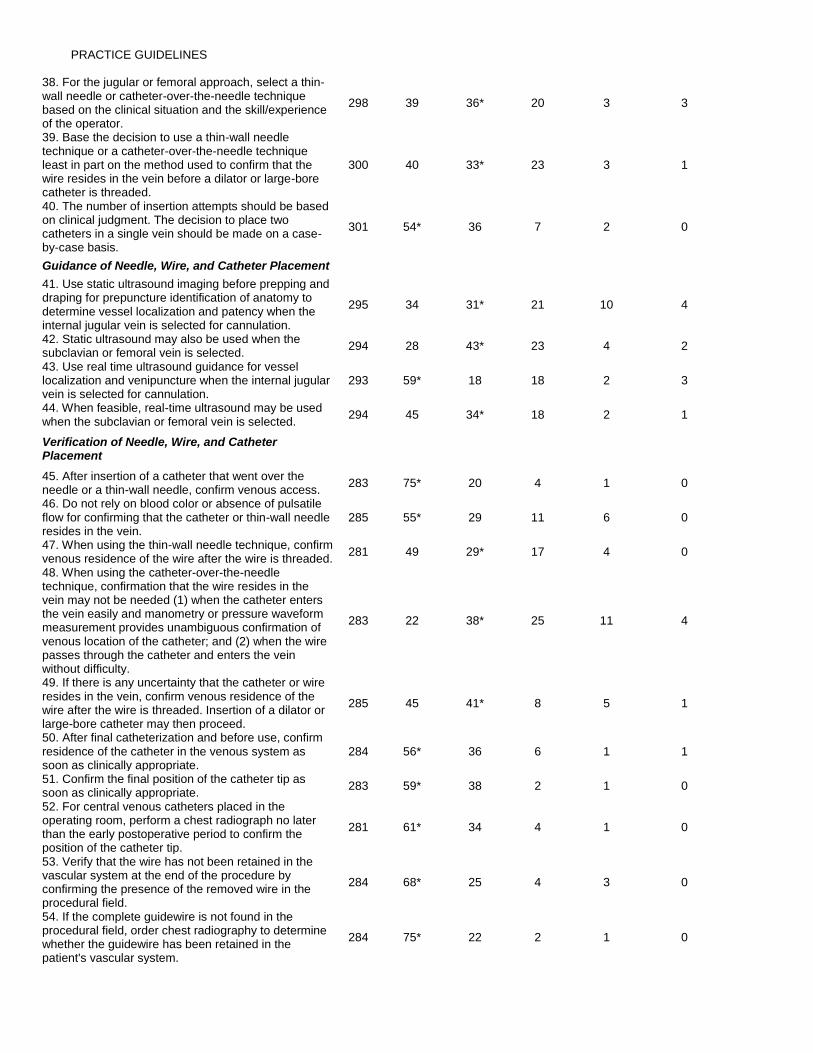
PRACTICE GUIDELINES
38. For the jugular or femoral approach, select a thin-wall needle or catheter-over-the-needle technique based on the clinical situation and the skill/experience of the operator.
298 39 36* 20 3 3
39. Base the decision to use a thin-wall needle technique or a catheter-over-the-needle technique least in part on the method used to confirm that the wire resides in the vein before a dilator or large-bore catheter is threaded.
300 40 33* 23 3 1
40. The number of insertion attempts should be based on clinical judgment. The decision to place two catheters in a single vein should be made on a case-by-case basis.
301 54* 36 7 2 0
Guidance of Needle, Wire, and Catheter Placement
41. Use static ultrasound imaging before prepping and draping for prepuncture identification of anatomy to determine vessel localization and patency when the internal jugular vein is selected for cannulation.
295 34 31* 21 10 4
42. Static ultrasound may also be used when the subclavian or femoral vein is selected.
294 28 43* 23 4 2
43. Use real time ultrasound guidance for vessel localization and venipuncture when the internal jugular vein is selected for cannulation.
293 59* 18 18 2 3
44. When feasible, real-time ultrasound may be used when the subclavian or femoral vein is selected.
294 45 34* 18 2 1
Verification of Needle, Wire, and Catheter Placement
45. After insertion of a catheter that went over the needle or a thin-wall needle, confirm venous access.
283 75* 20 4 1 0
46. Do not rely on blood color or absence of pulsatile flow for confirming that the catheter or thin-wall needle resides in the vein.
285 55* 29 11 6 0
47. When using the thin-wall needle technique, confirm venous residence of the wire after the wire is threaded.
281 49 29* 17 4 0
48. When using the catheter-over-the-needle technique, confirmation that the wire resides in the vein may not be needed (1) when the catheter enters the vein easily and manometry or pressure waveform measurement provides unambiguous confirmation of venous location of the catheter; and (2) when the wire passes through the catheter and enters the vein without difficulty.
283 22 38* 25 11 4
49. If there is any uncertainty that the catheter or wire resides in the vein, confirm venous residence of the wire after the wire is threaded. Insertion of a dilator or large-bore catheter may then proceed.
285 45 41* 8 5 1
50. After final catheterization and before use, confirm residence of the catheter in the venous system as soon as clinically appropriate.
284 56* 36 6 1 1
51. Confirm the final position of the catheter tip as soon as clinically appropriate.
283 59* 38 2 1 0
52. For central venous catheters placed in the operating room, perform a chest radiograph no later than the early postoperative period to confirm the position of the catheter tip.
281 61* 34 4 1 0
53. Verify that the wire has not been retained in the vascular system at the end of the procedure by confirming the presence of the removed wire in the procedural field.
284 68* 25 4 3 0
54. If the complete guidewire is not found in the procedural field, order chest radiography to determine whether the guidewire has been retained in the patient's vascular system.
284 75* 22 2 1 0
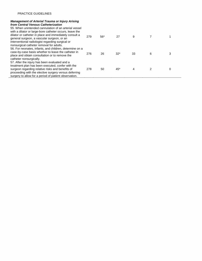
PRACTICE GUIDELINES
Management of Arterial Trauma or Injury Arising from Central Venous Catheterization 55. When unintended cannulation of an arterial vessel with a dilator or large-bore catheter occurs, leave the dilator or catheter in place and immediately consult a general surgeon, a vascular surgeon, or an interventional radiologist regarding surgical or nonsurgical catheter removal for adults.
279 56* 27 9 7 1
56. For neonates, infants, and children, determine on a case-by-case basis whether to leave the catheter in place and obtain consultation or to remove the catheter nonsurgically.
276 26 32* 33 6 3
57. After the injury has been evaluated and a treatment plan has been executed, confer with the surgeon regarding relative risks and benefits of proceeding with the elective surgery versus deferring surgery to allow for a period of patient observation.
278 50 45* 4 2 0