Practically Useful: What the R Protein Modeling Suite Can ...
12
pubs.acs.org/Biochemistry Published on Web 03/17/2010 r 2010 American Chemical Society Biochemistry 2010, 49, 2987–2998 2987 DOI: 10.1021/bi902153g Practically Useful: What the ROSETTA Protein Modeling Suite Can Do for You Kristian W. Kaufmann, † Gordon H. Lemmon, † Samuel L. DeLuca, † Jonathan H. Sheehan, † and Jens Meiler* Department of Chemistry, Vanderbilt University, 7330 Stevenson Center, Station B 351822, Nashville, Tennessee 37235. † These authors contributed equally to this work. Received December 15, 2009; Revised Manuscript Received February 25, 2010 ABSTRACT: The objective of this review is to enable researchers to use the software package ROSETTA for biochemical and biomedicinal studies. We provide a brief review of the six most frequent research problems tackled with ROSETTA. For each of these six tasks, we provide a tutorial that illustrates a basic ROSETTA protocol. The ROSETTA method was originally developed for de novo protein structure prediction and is regularly one of the best performers in the community-wide biennial Critical Assessment of Structure Prediction. Predictions for protein domains with fewer than 125 amino acids regularly have a backbone root-mean-square deviation of better than 5.0 A ˚ . More impressively, there are several cases in which ROSETTA has been used to predict structures with atomic level accuracy better than 2.5 A ˚ . In addition to de novo structure prediction, ROSETTA also has methods for molecular docking, homology modeling, determining protein structures from sparse experimental NMR or EPR data, and protein design. ROSETTA has been used to accurately design a novel protein structure, predict the structure of protein-protein complexes, design altered specificity protein-protein and protein-DNA interactions, and stabilize proteins and protein complexes. Most recently, ROSETTA has been used to solve the X-ray crystallographic phase problem. ROSETTA is a unified software package for protein structure prediction and functional design. It has been used to predict protein structures with and without the aid of sparse experi- mental data, perform protein-protein and protein-small mole- cule docking, design novel proteins, and redesign existing proteins for altered function. ROSETTA allows for rapid tests of hypotheses in biomedical research which would be impossible or exorbitantly expensive to perform via traditional experimental methods. Thereby, ROSETTA methods are becoming increasingly important in the interpretation of biological findings, e.g., from genome projects and in the engineering of therapeutics, probe molecules, and model systems in biomedical research. ROSETTA, like all structure prediction algorithms, must perform two tasks. First, ROSETTA must explore or sample the relevant conformational space, and in the case of design, sequence space. Second, ROSETTA must accurately rank or evaluate the energy of the resulting structural models. For this purpose, ROSETTA implements (mostly) knowledge-guided Metropolis Monte Carlo sampling approaches coupled with (mostly) knowledge-based energy functions. Knowledge-based energy functions assume that most molecular properties can be derived from available informa- tion, in this case the Protein Data Bank (PDB) (1, 2). While other reviews have focused on a specific ROSETTA functionality, this work briefly reviews the approaches to sam- pling and scoring used by each of the major ROSETTA protocols (protein structure prediction, protein docking, ligand docking, and protein design). Additionally, in the Supporting Informa- tion, we provide tutorials demonstrating six of the protocols introduced in this review. The tutorials we provide are intended as starting points and are therefore as basic as possible. ROSETTA provides experienced users the option of extending and tailoring these protocols to their biomedical research question. Some of the calculations described below can be run on a standard workstation in a reasonable time, while others require small computer clusters found at most universities. ROSETTA CONFORMATIONAL SAMPLING STRA- TEGIES The majority of conformational sampling protocols in ROSETTA use the Metropolis Monte Carlo algorithm to guide sampling. Gradient-based minimization is often employed for the last step of refinement of initial models. Since each ROSETTA protocol allows degrees of freedom specific for the task, ROSETTA can perform a diverse set of protein modeling tasks (3). Sampling Strategies for Backbone Degrees of Freedom. ROSETTA separates large backbone conformational sampling from local backbone refinement. Large backbone conforma- tional changes are modeled by exchanging the backbone con- formations of nine or three amino acid peptide fragments. Peptide conformations are collected from the PDB for homo- logous stretches of sequence (4) that capture the structural bias for the local sequence (5). For local refinement of protein models, ROSETTA utilizes Metropolis Monte Carlo sampling of φ and ψ angles that are calculated not to disturb the global fold of the protein. Rohl (6) provides a review of the fragment selection and backbone refinement algorithms in ROSETTA. Sampling Strategies for Side Chain Degrees of Freedom. Systematic sampling of side chain degrees of freedom of even short peptides quickly becomes intractable (7). ROSETTA drasti- cally reduces the number of conformations sampled through the use of discrete conformations of side chains observed in the PDB (8, 9). These “rotamers” capture allowed combinations between side chain torsion angles, as well as the backbone φ and ψ angles, and thereby reduce the amount of conformational *To whom correspondence should be addressed. Telephone: (615) 936-5662. Fax: (615) 936-2211. E-mail: [email protected].
Transcript of Practically Useful: What the R Protein Modeling Suite Can ...

pubs.acs.org/BiochemistryPublished on Web 03/17/2010r 2010 American Chemical Society
Biochemistry 2010, 49, 2987–2998 2987
DOI: 10.1021/bi902153g
Practically Useful: What the ROSETTA Protein Modeling Suite Can Do for You
Kristian W. Kaufmann,† Gordon H. Lemmon,† Samuel L. DeLuca,† Jonathan H. Sheehan,† and Jens Meiler*
Department of Chemistry, Vanderbilt University, 7330 Stevenson Center, Station B 351822, Nashville, Tennessee 37235.†These authors contributed equally to this work.
Received December 15, 2009; Revised Manuscript Received February 25, 2010
ABSTRACT: The objective of this review is to enable researchers to use the software package ROSETTA forbiochemical and biomedicinal studies. We provide a brief review of the six most frequent research problemstackled with ROSETTA. For each of these six tasks, we provide a tutorial that illustrates a basic ROSETTA
protocol. The ROSETTA method was originally developed for de novo protein structure prediction and isregularly one of the best performers in the community-wide biennial Critical Assessment of StructurePrediction. Predictions for protein domains with fewer than 125 amino acids regularly have a backboneroot-mean-square deviation of better than 5.0 A. More impressively, there are several cases in which ROSETTA
has been used to predict structures with atomic level accuracy better than 2.5 A. In addition to de novostructure prediction, ROSETTA also has methods for molecular docking, homology modeling, determiningprotein structures from sparse experimental NMRor EPR data, and protein design. ROSETTA has been used toaccurately design a novel protein structure, predict the structure of protein-protein complexes, design alteredspecificity protein-protein and protein-DNA interactions, and stabilize proteins and protein complexes.Most recently, ROSETTA has been used to solve the X-ray crystallographic phase problem.
ROSETTA is a unified software package for protein structureprediction and functional design. It has been used to predictprotein structures with and without the aid of sparse experi-mental data, perform protein-protein and protein-small mole-cule docking, design novel proteins, and redesign existingproteins for altered function. ROSETTA allows for rapid tests ofhypotheses in biomedical research which would be impossible orexorbitantly expensive to perform via traditional experimentalmethods. Thereby, ROSETTA methods are becoming increasinglyimportant in the interpretation of biological findings, e.g., fromgenome projects and in the engineering of therapeutics, probemolecules, and model systems in biomedical research.
ROSETTA, like all structure prediction algorithms, must performtwo tasks. First, ROSETTA must explore or sample the relevantconformational space, and in the case of design, sequence space.Second, ROSETTA must accurately rank or evaluate the energy ofthe resulting structural models. For this purpose, ROSETTA
implements (mostly) knowledge-guided Metropolis Monte Carlosampling approaches coupled with (mostly) knowledge-basedenergy functions. Knowledge-based energy functions assume thatmost molecular properties can be derived from available informa-tion, in this case the Protein Data Bank (PDB) (1, 2).
While other reviews have focused on a specific ROSETTA
functionality, this work briefly reviews the approaches to sam-pling and scoring used by each of the major ROSETTA protocols(protein structure prediction, protein docking, ligand docking,and protein design). Additionally, in the Supporting Informa-tion, we provide tutorials demonstrating six of the protocolsintroduced in this review. The tutorials we provide are intendedas starting points and are therefore as basic as possible. ROSETTA
provides experienced users the option of extending and tailoring
these protocols to their biomedical research question. Some ofthe calculations described below can be run on a standardworkstation in a reasonable time, while others require smallcomputer clusters found at most universities.
ROSETTA CONFORMATIONAL SAMPLING STRA-
TEGIES
Themajority of conformational sampling protocols inROSETTA
use the Metropolis Monte Carlo algorithm to guide sampling.Gradient-based minimization is often employed for the last stepof refinement of initial models. Since each ROSETTA protocolallows degrees of freedom specific for the task, ROSETTA canperform a diverse set of protein modeling tasks (3).Sampling Strategies for Backbone Degrees of Freedom.
ROSETTA separates large backbone conformational samplingfrom local backbone refinement. Large backbone conforma-tional changes are modeled by exchanging the backbone con-formations of nine or three amino acid peptide fragments.Peptide conformations are collected from the PDB for homo-logous stretches of sequence (4) that capture the structural biasfor the local sequence (5). For local refinement of proteinmodels,ROSETTA utilizes Metropolis Monte Carlo sampling of φ and ψangles that are calculated not to disturb the global fold of theprotein. Rohl (6) provides a review of the fragment selection andbackbone refinement algorithms in ROSETTA.Sampling Strategies for Side Chain Degrees of Freedom.
Systematic sampling of side chain degrees of freedom of evenshort peptides quickly becomes intractable (7). ROSETTA drasti-cally reduces the number of conformations sampled through theuse of discrete conformations of side chains observed in thePDB (8, 9). These “rotamers” capture allowed combinationsbetween side chain torsion angles, as well as the backbone φ andψ angles, and thereby reduce the amount of conformational
*To whom correspondence should be addressed. Telephone: (615)936-5662. Fax: (615) 936-2211. E-mail: [email protected].

2988 Biochemistry, Vol. 49, No. 14, 2010 Kaufmann et al.
space (9). A Metropolis Monte Carlo simulated annealing run isused to search for the combination of rotamers occupying theglobal minimum in the energy function (8, 10). This generalapproach is extended to protein design via replacement of arotamer of amino acid A with a rotamer of amino acid B in theMonte Carlo step.
ROSETTA ENERGY FUNCTION
Simulations with ROSETTA can be classified on the basis ofwhether amino acid side chains are represented by super atoms orcentroids in the low-resolution mode or at atomic detail in thehigh-resolution mode. Both modes come with tailored energyfunctions that have been reviewed previously by Rohl (6).ROSETTA Knowledge-Based Centroid Energy Function.
The low-resolution energy function treats the side chains ascentroids (4, 11). The energy function models solvation, electro-statics, hydrogen bonding between β strands, and steric clashes.Solvation effects are modeled as the probability of seeing aparticular amino acidwith a given number ofR carbons within anamino acid-dependent cutoff distance. Electrostatic interactionsare modeled as the probability of observing a given distancebetween centroids of amino acids. Hydrogen bonding between βstrands is evaluated on the basis of the relative geometricarrangement of strand fragments. Backbone atom and side chaincentroid overlap is penalized and thus provides the repulsivecomponent of a van derWaals force. A radius of gyration term isused to model the effect of van der Waals attraction. Allprobability profiles have been derived using Bayesian statisticson crystal structures from the PDB. The lower resolution of thiscentroid-based energy function smoothes the energy landscape atthe expense of its accuracy. The smoother energy landscapeallows structures that are close to the true global minima tomaintain a low energy even with structural defects that a fullatom energy function would stiffly penalize.Knowledge-Based All Atom Energy Function. The all
atom high-resolution energy function used by ROSETTA wasoriginally developed for protein design (8, 12). It combines the6-12 Lennard-Jones potential for van der Waals forces, asolvation approximation (13), an orientation-dependent hydro-gen bonding potential (14), a knowledge-based electrostaticsterm, and a knowledge-based conformation-dependent aminoacid internal free energy term (9). An important consideration inthe construction of this potential was that all energy terms arepairwise decomposable. The pairwise decomposition of each ofthe terms limits the total number of energy contributions to1/2N(N - 1) when N is the number of atoms within the system.This limitation allows precomputation and storage of many ofthese energy contributions in the computer memory, which isnecessary for rapid execution of the Metropolis Monte Carlosampling strategies employed by ROSETTA during protein designand atomic-detail protein structure prediction.
PROTEIN STRUCTURE PREDICTION
The central tenet of structural biology is that structuredetermines function. Thus, the structure of a protein is criticalfor a full understanding of its function. Experimental structuredetermination techniques such as X-ray crystallography, nuclearmagnetic resonance, electron paramagnetic resonance, and elec-tron microscopy require significant investments of effort toproduce structures of a molecule. Conversely, the advent of thegenomic revolution created an explosion in the number of known
sequences for biopolymers. For example, from October 2008 toMarch 2009, approximately 8 million (!) new, nonredundantsequences were added to the BLAST database. ROSETTA reme-dies the shortfall in structural information by predicting high-probability structures for a given amino acid sequence.De Novo Folding Simulation. The “protein folding pro-
blem” (given an amino acid sequence, predict the tertiarystructure into which it folds) is considered the greatest challengein computational structural biology. The ROSETTA de novostructure prediction algorithm has been reviewed and describedin detail elsewhere (4, 6, 11, 15). Briefly, ROSETTA begins with anextended peptide chain. Insertion of backbone fragments rapidly“folds” the protein using the low-resolution energy function andsampling approaches (Figure 1). ROSETTA attempts approxi-mately 30000 nine-residue fragment insertions followed by an-other 10000 three-residue fragment insertions to generatea protein model (6). Usually, 20000-50000 models are foldedfor each individual protein (15). The resulting models canundergo atomic-detail refinement, or if computational expenseis an issue, clustering based on the CR root-mean-square devia-tion (rmsd) (16, 17) can reduce the number of models beforeperforming refinement. The clustering parameters are chosen bythe user to generate clusters that maintain the same overall fold(i.e., CR rmsd< 5 A) while maximizing coverage of the structurespace sampled. The lowest-energy models and the structures atthe center of the clusters enter atomic-detail refinement (readbelow). The “Protein Folding” and “Refinement” tutorials in thesupplement cover many aspects of this protocol.
In 2009, Das et al. implemented an addition to the existing denovo protein folding protocol that allowed for accurate predic-tion of homomeric proteins (18). They combined elements ofROSETTA de novo structure prediction (19) with protein-proteindocking (20) to develop ROSETTAFOLD-AND-DOCK. FOLD-AND-DOCK alternates between cycles of symmetric fragment insertionas in ROSETTA de novo prediction and rigid-body docking bet-ween the partially assembled monomers. Following complexassembly, the entire complex undergoes full atom refinement.
FOLD-AND-DOCK assumes that secondary structural elements ofa homomer are symmetric and inserts the same fragments intoevery subunit. As the interface between a homomer is largelyburied, docking while folding allows this region to be protectedand stabilized during the folding simulation, which greatlyimproved the accuracy. Using this method, the structure of aK138Amutant of the Rab6-GTP 3GCC185 Rab binding domaincomplex (PDB entry 3bbp) (21) was predicted within 1 A rmsd ofthe experimental structure in a blind prediction test.
To further improve resolution, sparse NMR-derived chemicalshift restraints were added, yielding models with rmsd values of<1 A. Typically, structure elucidation for homomers withNMR-derived restraints would have required extensive data setsof RDCs, NOEs, chemical shifts, and scalar couplings.Comparative Modeling. Comparative modeling in ROSETTA
starts after the alignment of a target sequence with a templateprotein, using sequence-sequence or sequence-structure align-ment tools as described by Raman et al. (19). The quality of thealignment determines the aggressiveness of the sampling inROSETTA (19). In a case with a high degree of sequence homology(>50% identical sequence), the protein backbone is only rebuiltin regions surrounding insertions and deletions in the sequencealignment (19, 22). Consequently, ROSETTA starts with thetemplate structure and builds in missing loops using fragmentinsertion or randomization of φ and ψ angles followed by one of

Current Topic/Perspective Biochemistry, Vol. 49, No. 14, 2010 2989
the loop closure algorithms such as cyclic coordinate descent orkinematic closure (23-25). In the case of a medium to low degreeof sequence identity between the template and target, Raman etal. applied a more aggressive iterative stochastic rebuild andrefine protocol that allowed the complete rebuilding of largeregions of the protein, which in some cases included entiresecondary structure elements (19).
Mandell et al. (24) recently developed a Loop Closure algo-rithm in ROSETTA that achieves rmsd values of better than 1 A.Their adaptation of Kinematic closure (KIC) first selects threeCR atoms as pivots. Next, nonpivot (j and ψ) torsion angles aresampled, leading to a chain break at the middle pivot. Finally,KIC is used to determine j and ψ torsion angles for the pivotatoms that close the loop. For a data set of 25 loops containing 12residues each,ROSETTA achieved amedian accuracy of 0.8 A rmsd(see Figure 2). This demonstrates an improvement over both thestandard ROSETTA cyclic coordinate descent protocol and a state-of-the-art molecular dynamics protocol (median accuracies of2.0 and 1.2 A rmsd, respectively). The “LoopModeling” tutorialdemonstrates the kinematic loop closure protocol.Model Relaxation and Refinement.After construction of a
protein backbone via de novo protein folding or comparativemodeling, the model enters atom-detail refinement (15, 26, 27).During the iterative relaxation protocol, φ and ψ angles of thebackbone are perturbed slightly while the overall global con-formation of the protein is maintained. The side chains of the
protein are adjusted using a simulated annealing MetropolisMonte Carlo search of the rotamer space. Finally, gradientminimization is applied to all torsional degrees of freedom(φ,ψ,ω, and χ). The repulsive portion of van derWaals potentialis increased incrementally, moving the structure to the nearestenergy minimum. Extensive use of the all atommodel refinementhas proven integral to the success ofROSETTA in the recentCriticalAssessment of Structure Prediction (CASP) experiments. A basicrefinement protocol is introduced in the “Refinement” tutorial.
Recently,Qian et al. applied the refinement protocol to proteinstructures determined de novo, via comparative modeling, orusing NMR-derived restraints (26). In this protocol, proteinmodels derived fromNMR constraints or comparative modelingwere used as a basis for solving the crystallographic phaseproblem. The model was initially minimized using ROSETTA’sall atom Monte Carlo Refinement protocol. The results of thisrefinement were used to identify regions likely to deviate from thenative structure. In this context, it was demonstrated that regionsof high variability between refinedmodels often correlate to areasof deviation from the native structure. The conformational spacein these areas was sampled extensively using the fragmentreplacement approach used by ROSETTA’s de novo structureprediction protocol. The resulting models are then subjected toanother round of all atom refinement. This cycle of refinementand conformational sampling is performed iteratively, each timeusing only the lowest-energy models from the previous round
FIGURE 1: De novo folding algorithm.ROSETTA starts from (a) fragment libraries with sequence-dependent (j andψ) angles that capture the localconformational space accessible to a sequence. (b) Combining different fragments from the libraries folds the protein through optimization ofnon-local contacts. The low-resolution energy functiondepicted inpanel c smoothes the rough energy surface, resulting in a deep, broadminimumfor the native conformation. Metropolis Monte Carlo minimization drives the structure toward the global minimum.

2990 Biochemistry, Vol. 49, No. 14, 2010 Kaufmann et al.
of refinement, until the system converges. The final models werethen used in molecular replacement to solve the crystallographicphase problem. In a blind test, this ab initio phase solutionmethod resulted in significant improvement in structural resolu-tion compared to that of the original unrefined models. Theprotocol can also be applied for the refinement of models derivedfrom medium-resolution NMR data.ROSETTA’s Performance in the CASP Experiment. RO-
SETTA has displayed remarkable success in de novo structureprediction in the last several blind CASP experiments; this isevidenced by the method’s ranking among the top structureprediction groups (16, 19, 22, 28-31). During CASP, sequencesof proteins not yet reported in the PDB are distributed amongparticipating laboratories. Within a given time limit, predictionsare collected and assessed on the basis of the experimentalstructure that is typically available shortly after the CASPexperiment (http://www.predictioncenter.org). Generally, ROSET-
TA has superseded competing approaches at predicting thestructure of small to moderately sized protein domains withfewer than 200 amino acids de novo. Shortly after the CASP6(held in 2004), Bradley et al. showed that for a benchmark ofsmall proteins ROSETTA de novo structure prediction foundmodels at atomic detail accuracy in an encouraging eight of16 cases (15, 29). In that same year, Misura et al. found thathomology models built with ROSETTA can be more accuratethan their templates (15). During CASP 7, with the assistanceof the ROSETTA@Home distributed computer network, severalmoderately sized domains were predicted to atomic-detail accu-racy within 2 A of the experimental structure for the firsttime (16). On the basis of the performance of ROSETTA inimproving models over the best template structures available(see Figure 3) (19), Raman et al. suggest that the limitationof the ROSETTA structure prediction protocol remains in thesampling algorithms rather than the energy function. For thisreason, prediction of larger domains becomes possible upon
introduction of experimental data which restricts the conforma-tional space.ROSETTA Leverages Sparse Experimental Data from
NMR and EPR Experiments. ROSETTA allows incorporationof many types of experimental restraints. ROSETTA’s ability todeal with restraints derived from nuclear magnetic resonance(NMR) spectroscopy is themost developed (32). NMR restraintshave two entry points into the ROSETTA protein structure predic-tion routine. Chemical shift assignments for backbone atoms canbe converted to φ and ψ backbone angle restraints (33) and areused during the selection of the fragment libraries. Distance andorientation restraints [e.g., nuclear Overhauser effect (NOE)restraints and residual dipolar couplings (RDCs), respectively]
FIGURE 2: Kinematic loop closure. (a) The kinematic loop closure algorithm samplesj andψ angles at the cyanCR spheres froma residue specificRamachandran map. The j and ψ angles at green CR spheres are determined analytically to close the loop. (b) The energy vs rmsd plot showsaccuracies for the prediction of loop conformation better than 1 A achieved through the improved sampling offered by the kinematic closureprotocol. (c) The kinematic closure prediction (blue) closely resembles the crystallographic conformation (cyan). From (24) reprinted withpermission from Nature Methods.
FIGURE 3: Comparative modeling CASP performance. Raman andcolleagues demonstrated that comparative models refined with RO-
SETTA improved upon the best template structure available for severalCASP targets, for example, (a) T0492 and (b) T0464. The nativestructure is colored blue, the best submitted ROSETTA model red, andthe best template structure green. The ROSETTA models for T0492resulted in atomic-level accuracy for side chains in the core of theprotein. For T0464, a 25-residue insertion was predicted whichresulted in models that were significantly improved over the besttemplates available. One of the models was further improved in themodel refinement category. From (19) reprinted with permissionfrom Proteins.
Current Topic/Perspective Biochemistry, Vol. 49, No. 14, 2010 2991
are incorporated into the scoring function used during folding.Bowers et al. showed that a sparse mixture of short- and long-rangeNOE restraints (approximately one restraint per residue) inaddition to the backbone chemical shifts enables ROSETTA tobuild models at atomic-detail accuracy (34). Rohl and Baker (35)likewise demonstrated that limited RDC measurements (appro-ximately one per residue) in combination with backbone chemi-cal shifts identify the correct fold. Meiler and Baker presented aprotocol that uses unassigned NMR restraints for rapid proteinfold determination (36).More recently, Shen et al. showed the useof a modified fragment selection protocol in ROSETTA to generatestructures of a quality comparable to those from traditionalNMR structure determination methods (37). Furthermore, Shenfound that ROSETTA sampling can compensate for the incompleteand incorrect NMR restraints (38). A major point to note is thatin each of these examples ROSETTA is used to complementstructure restraints obtained early in the structure determinationprocess. Consistently, ROSETTAmodels are accurate at the atomiclevel of detail that would only be apparent from either signifi-cantly more or higher-resolution experimental information. Forexample, Rohl and Baker found that ROSETTA produced ubiqui-tin models with an rmsd of <4 A of the experimental structureusing RDC restraints that were also consistent with models thathave an rmsd of >20 A (35).
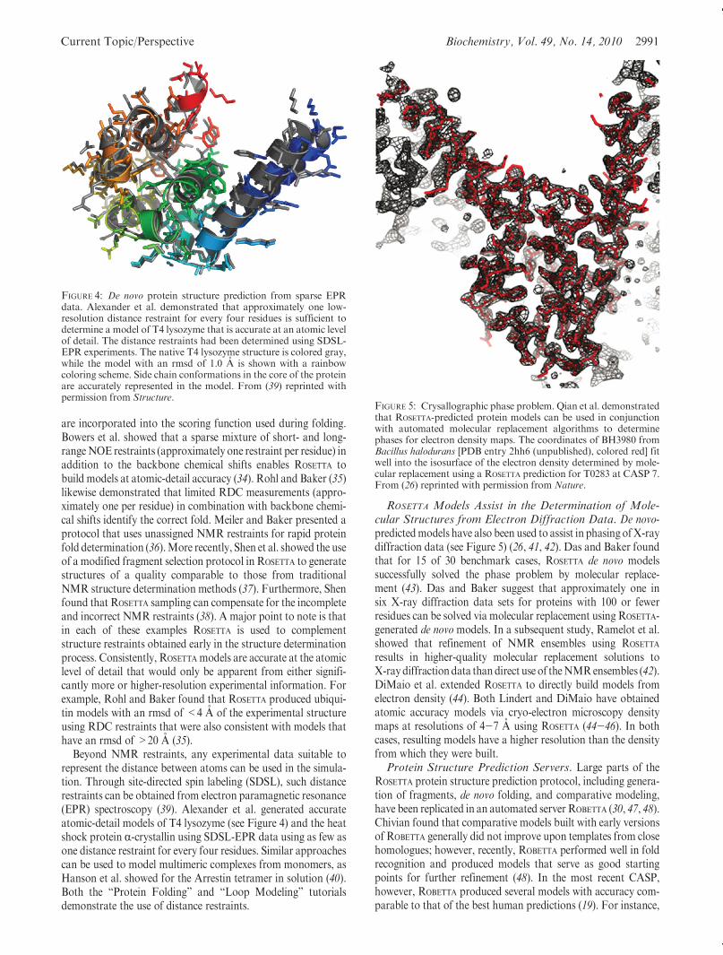
Beyond NMR restraints, any experimental data suitable torepresent the distance between atoms can be used in the simula-tion. Through site-directed spin labeling (SDSL), such distancerestraints can be obtained from electron paramagnetic resonance(EPR) spectroscopy (39). Alexander et al. generated accurateatomic-detail models of T4 lysozyme (see Figure 4) and the heatshock protein R-crystallin using SDSL-EPR data using as few asone distance restraint for every four residues. Similar approachescan be used to model multimeric complexes from monomers, asHanson et al. showed for the Arrestin tetramer in solution (40).Both the “Protein Folding” and “Loop Modeling” tutorialsdemonstrate the use of distance restraints.
ROSETTA Models Assist in the Determination of Mole-cular Structures from Electron Diffraction Data. De novo-predictedmodels have also been used to assist in phasing ofX-raydiffraction data (see Figure 5) (26, 41, 42). Das and Baker foundthat for 15 of 30 benchmark cases, ROSETTA de novo modelssuccessfully solved the phase problem by molecular replace-ment (43). Das and Baker suggest that approximately one insix X-ray diffraction data sets for proteins with 100 or fewerresidues can be solved via molecular replacement using ROSETTA-generated de novomodels. In a subsequent study, Ramelot et al.showed that refinement of NMR ensembles using ROSETTA
results in higher-quality molecular replacement solutions toX-raydiffractiondata thandirect use of theNMRensembles (42).DiMaio et al. extended ROSETTA to directly build models fromelectron density (44). Both Lindert and DiMaio have obtainedatomic accuracy models via cryo-electron microscopy densitymaps at resolutions of 4-7 A using ROSETTA (44-46). In bothcases, resulting models have a higher resolution than the densityfrom which they were built.Protein Structure Prediction Servers. Large parts of the
ROSETTA protein structure prediction protocol, including genera-tion of fragments, de novo folding, and comparative modeling,have been replicated in an automated server ROBETTA (30, 47, 48).Chivian found that comparative models built with early versionsof ROBETTA generally did not improve upon templates from closehomologues; however, recently, ROBETTA performed well in foldrecognition and produced models that serve as good startingpoints for further refinement (48). In the most recent CASP,however, ROBETTA produced several models with accuracy com-parable to that of the best human predictions (19). For instance,
FIGURE 4: De novo protein structure prediction from sparse EPRdata. Alexander et al. demonstrated that approximately one low-resolution distance restraint for every four residues is sufficient todetermine a model of T4 lysozyme that is accurate at an atomic levelof detail. The distance restraints had been determined using SDSL-EPR experiments. The native T4 lysozyme structure is colored gray,while the model with an rmsd of 1.0 A is shown with a rainbowcoloring scheme. Side chain conformations in the core of the proteinare accurately represented in the model. From (39) reprinted withpermission from Structure.
FIGURE 5: Crysallographic phase problem. Qian et al. demonstratedthat ROSETTA-predicted protein models can be used in conjunctionwith automated molecular replacement algorithms to determinephases for electron density maps. The coordinates of BH3980 fromBacillus halodurans [PDB entry 2hh6 (unpublished), colored red] fitwell into the isosurface of the electron density determined by mole-cular replacement using a ROSETTA prediction for T0283 at CASP 7.From (26) reprinted with permission from Nature.

2992 Biochemistry, Vol. 49, No. 14, 2010 Kaufmann et al.
ROBETTA’s top model for the server only target, T0513 domain 2,had an rmsd of 0.84 A. In general, the performance of ROBETTA
compared to that of other servers increases as the quality of thetemplate structures decreases (19). ROBETTA is publicly accessibleat http://robetta.bakerlab.org.
PROTEIN-PROTEIN DOCKING
While protein function is often determined by interactionswithother proteins, most structures found in the PDB contain singlechains. Because of the difficulties in determining structures ofprotein complexes, computational methods for predicting protein-protein interactions are important. ROSETTADOCK provides toolsfor predicting the interaction of two proteins (49). ROSETTADOCK
employs first a low-resolution rigid-body docking. The secondhigh-resolution refinement stage provides for side chain confor-mational sampling and backbone relaxation.
The ROSETTADOCK algorithm begins with random reorienta-tion of both proteins (49). Next, one protein slides into contactwith the other. The following low-resolution docking conforma-tional search involves 500 Monte Carlo rigid-body movements.These moves rotate and translate one protein around the surfaceof the other with movements chosen from a Gaussian distribu-tion centered at 5� and 0.7 A. Each conformation is scored usingthe low-resolution energy function based on residue pair inter-action statistics, residue environment statistics, and van derWaals attractive and repulsive terms. In this low-resolution step,side chains are represented by their centroids.
Next, 50 cycles of high-resolution refinement at the atomiclevel of detail are performed. Each cycle consists of a 0.1 Arandom rigid-body translation, Monte Carlo-based side chainrotamer sampling (packing), and gradient-based rigid-bodyminimization to find a local energy minimum. Finally, backboneflexibility is introduced around the protein interface. The “Pro-tein-ProteinDocking” tutorial demonstrates the entire protocol.ROSETTADOCK is available as a web server (http://rosettadock.graylab.jhu.edu) but is limited to complexes for which theapproximate binding orientation is known. The server produces1000 structures and returns details for the 10 lowest-energymodels (50).
ROSETTADOCK successfully recovered the native structures of42 of 54 benchmark targets from which side chains had beenremoved (49). Starting with randomly placed proteins, ROSETTA-
DOCK predicts more than 50% of the interface contacts for 23 of32 benchmark targets (49). These results have improved with theaddition of backbone flexibility (3) and conformational sam-pling (51).
ROSETTADOCK has been used to predict the structures ofanthrax protective antigen (52) and epidermal growth factor (53)bound to monoclonal antibodies. Both docking studies led topredicted interface structures consistent with known mutantbinding properties and were used to select residues for site-directed mutagenesis. The antibody modeling protocol has beenmade accessible through a web server (http://antibody.graylab.jhu.edu).
ROSETTADOCK was benchmarked in the Critical Assessment ofPRediction of Interactions (CAPRI) experiment (Figure 6),where it achieved full-atom rmsds of better than 1.6 A for mosttargets (54). Its success has been attributed to advances such asthe inclusion of gradient-based energyminimization of side chaintorsion angles (54), incorporation of biochemical data (55), andcoupling of docking with loop modeling (55).
Sircar and Gray recently reported on an extension of theROSETTADOCK algorithm that allows for accurate modeling ofantibody-antigen complexes in the absence of an antibodycrystal structure (56). SNUGDOCK simultaneously samples therigid-body antibody-antigen positions, the orientation of anti-body light and heavy chains, and the conformations of the sixcomplementary determining loops. Additionally, antibody con-formational ensembles can be provided to mimic conformationalselection. As in ROSETTADOCK, side chain rotamers are sampledduring high-resolution refinement.
SNUGDOCK was compared with ROSETTADOCK in a blindprediction of human MCP-1 binding 11k2 antibody (PDBentry 2bdn) (57). While the lowest-energy structure producedby ROSETTADOCK is incorrect, the model produced by SNUG-
DOCK meets the CAPRI acceptable criterion of having morethan 30% of the residue-residue contacts predicted correctly.When combined with ensemble sampling, five of the 10 lowest-energy models meet the CAPRI medium-quality criterion ofcorrectly predicting more than 50% residue-residue contacts.Similar results were seen in a benchmark of 15 antibody-antigencomplexes.
PROTEIN-LIGAND DOCKING
Ligand docking seeks to predict the interaction between aprotein and a small molecule. Most ligand docking applicationsstruggle to correctly predict conformational selection or induced-fit effects (58) resulting from ligand and protein flexibility. Asapplications were originally designed for protein-ligand dock-ing, flexibility is often a feature added as an afterthought. On theother hand, ROSETTA was originally developed for de novostructure prediction. As such, it was designed from its inceptionto efficiently model flexibility. While protein flexibility is well-defined by side chain rotamers and backbone φ and ψ anglechanges, small molecule flexibility was newly introduced intoROSETTA (59). Modeling ligand flexibility using knowledge-basedscore functions is especially challenging since the available smallmolecule crystal structures pale in comparison to the possiblechemical diversity available to small molecules.
ROSETTALIGAND is an application for docking a small moleculein the binding pocket of a protein that considers both ligand andprotein flexibility (60). The ROSETTALIGAND algorithm is amodification of the ROSETTADOCK algorithm. First, a ligand
FIGURE 6: Protein interface prediction. High-resolution CAPRI pre-diction of the colicin D-immunity protein D interface. Both rigid-body orientation and side chain conformation were modeled. Thecrystal structure is colored red and orange, and the ROSETTAmodel iscolored blue. (a)Whole protein complex. (b) The interface shows theside chains of catalytic residue H611 and additional positivelycharged residues that are thought to bind to the RNA, as well astheir matching negatively charged residues in the immunity protein.From (54) reprinted with permission from Proteins: Structure, Func-tion, and Bioinformatics.

Current Topic/Perspective Biochemistry, Vol. 49, No. 14, 2010 2993
conformer is chosen randomly from a user-provided ligandconformational ensemble. Second, the ligand is moved to auser-defined putative binding site. A low-resolution shape com-plementarity search translates and rotates the ligand optimizingattractive and repulsive score terms. In the high-resolution phase,cycles of Monte Carlo minimization perturb the ligand pose andoptimize amino acid side chain rotamers and ligand conformers.Lastly, all torsion degrees of freedom in the ligand and proteinundergo gradient minimization, and the model is output. The“Small Molecule Docking” tutorial demonstrates this protocol.
In a benchmark, ROSETTALIGAND successfully recovered thenative structure of 80 of 100 protein-ligand complexes with anrmsd better than 2.0 A. When docking ligands into experimentalprotein structures determined with different binding partners(cross-docking), ROSETTALIGAND recovered the native structurein 14 of 20 cases. Comparing binding energy predictions with 229experimentally determined binding energies from the Ligand-ProteinDatabase (http://lpdb.chem.lsa.umich.edu) (61),ROSETTA-LIGAND achieved an overall correlation coefficient of 0.63, whichis comparable to the best scoring functions available for pro-tein-ligand interfaces (62).
Recently, backbone flexibility was added to the dockingalgorithm which led to improved predictions, including lowerrmsds among top scoring ligands (63). Backbone flexibilityallows prediction of induced-fit effects that occur upon ligandbinding. When ROSETTALIGAND was tested in a blind study on aset of lead-like compounds, its performance was comparable tothose of other commercially available docking programs (64).The authors caution, however, that current docking programsfail 70% of the time on at least one of the receptors in the test set.
Often researchers seek to understand the interaction of a smallmolecule with a target protein whose structure has not yet beendetermined. In such cases, docking studies utilize comparativemodels. ROSETTALIGAND was recently used by Kaufmannet al. (65) to dock serotonin into comparative models of humanand Drosophila serotonin transporters (hSERT and dSERT,respectively). The models were based on the leucine transporter(LeuTAa) structure reported by Yamashita et al. (66) which has
an overall sequence similarity of 17%and a binding site similarityof 50% with SERT. Using these models alone, ROSETTALIGAND
predicted a binding mode that places serotonin deep in thebinding pocket of SERT (see Figure 7). This binding mode isconsistent with site-directed mutagenesis studies and substitutedcysteine accessibilitymethod (SCAM) data and retains the amineplacement seen in the LeuTAa structure. Additionally, bindingenergy predictions of serotonin analogues agree with experimen-tal data (R = 0.72).
Kaufmann and Meiler find that ROSETTALIGAND successfullydocks a variety of small molecules into comparative models(unpublished results). ROSETTALIGAND identified the bindingmode within 2 A rmsd for six of nine protein-ligand complexesin which models had been submitted in the eighth CASPcompetition. In seven additional examples, Kaufmann andMeiler observe that ROSETTALIGAND samples the correct bindingmode in at least one template formost ligands, yielding an overallsuccess rate of better than 70%. This success rate is comparableto ROSETTALIGAND’s performance with an experimental structurefor the protein partner and can be attributed toROSETTALIGAND’sability to sample protein conformational changes.
PROTEIN DESIGN
All protocols discussed up to this point relate to proteinstructure prediction and seek to determine the position of aminoacid atoms in space. Protein design, on the other hand, seeks todetermine an amino acid sequence that folds into a given proteinstructure or performs a given function. In this context, the proteindesign problem (to find a sequence that folds into a given tertiarystructure) is also known as the “inverse protein folding problem”.The ROSETTADESIGN algorithm (12) is an iterative process thatenergetically optimizes both the structure and sequence of aprotein. ROSETTADESIGN alternates between rounds of fixedbackbone sequence optimization and flexible backbone energyminimization (12). During the sequence optimization step, aMonte Carlo simulated annealing search is used to sample thesequence space. Every amino acid is considered at each positionin the sequence, and rotamers are constrained to the DunbrackLibrary (67). After each round of Monte Carlo sequence optimi-zation, the backbone is relaxed to accommodate the designedamino acids (12). The practical uses of ROSETTADESIGN can bedivided into five basic categories: design of novel folds (12),redesign of existing proteins (68), protein interface design (69),enzyme design (70), and prediction of fibril-forming regions inproteins (71). The “De Novo Protein Design” tutorial demon-strates the complete redesign of the protein ubiquitin.De Novo Protein Design. The ROSETTADESIGN method has
been used for the de novo design of a fold that was not (yet)represented in the PDB. A starting backbonemodel consisting ofa five-strand β sheet and two packed R helices was constructedwith the ROSETTA de novo protocol using distance constraintsderived from a two-dimensional sketch (12). The sequence wasiteratively designed with five simulation trials of 15 cycles each.The final sequence was expressed, and the structure was deter-mined using X-ray crystallography. The experimental structurehas an rmsd with respect to the computational design of <1.1 A(see Figure 8) (12).
Similarly, a molecular switch that folded into a trimeric coiledcoil in the absence of zinc, and a monomeric zinc finger in thepresence of zinc, was designed by extending ROSETTADESIGN tosimultaneously optimize a sequence in two different folds. The
FIGURE 7: Complex of the human serotonin transporter with itssubstrate. The color scheme of serotonin displays the differentialsensitivity of human and Drosophila serotonin transporter (SERT)for serotonin derivatives as dervied from a QSAR study. Blueindicates a higher sensitivity in dSERT, while red indicates a highersensitivity in hSERT. The QSAR data indicate that the docking posepredicted by ROSETTALIGAND is plausible. From (65) reprinted withpermission from Proteins.

2994 Biochemistry, Vol. 49, No. 14, 2010 Kaufmann et al.
sequence of an existing zinc finger domain was aligned with acoiled-coil hemaglutinin domain. During the design protocol, thesequence was optimized to fold into both tertiary structures (72).Redesign of Existing Proteins. When nine globular pro-
teins were stripped of all side chains and then redesigned usingROSETTADESIGN, the average sequence recovery was 35% for allresidues (73). In four of nine cases, the protein stability improvedas measured by chemical denaturation. The structure of a rede-signed human procarboxypeptidase (PDB entry 1aye) (74) wasdetermined experimentally. ROSETTADESIGNwas then used to syste-matically identify mutations of procarboxypeptidase that wouldimprove the stability of the protein. All of the tested mutants weremore stable than thewild-type protein, with the top-scoringmutanthaving a reduction of free energy of 5.2 kcal/mol (75).
The ROSETTADESIGN server (http://rosettadesign.med.unc.edu) (76) is a Web-based interface to the fixed backbone designmodule of ROSETTA that allows design of proteins with up to 200residues. The average design takes 5-30 min to complete.Interface Design. Computational design techniques have
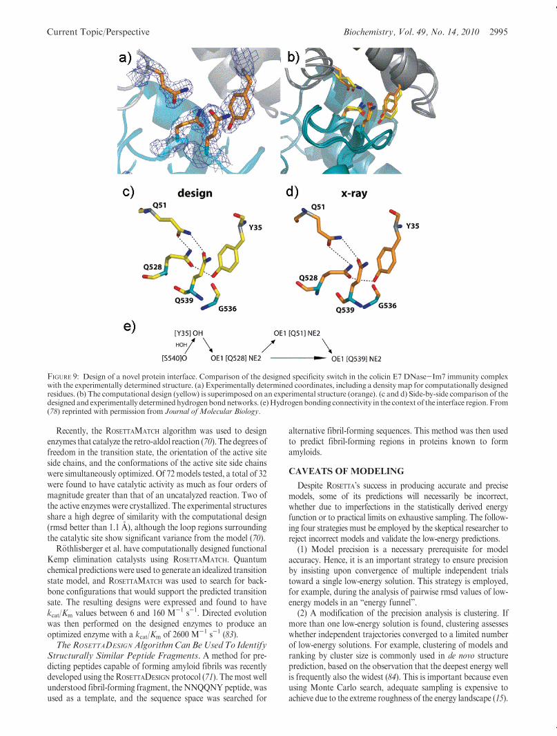
been used to engineer an endonuclease with altered specificity.A 1400 A2 interface was designed between individual domains oftwo homodimeric endonucleases (I-DomI and I-CreI). The de-sign retained specificity and catalytic activity and crystallizedwith an rmsd of 0.8 A with respect to the model (77). Similarly, ahighly effective specificity switch was designed into the colicin E7DNase-Im7 immunity complex through the design of a novelhydrogen bond network (Figure 9). This designed networkexhibited a 300-fold increase in specificity (78). ROSETTA’s alaninescanning application simulates experimental alanine scanning insilico. Each residue in the protein complex is iteratively mutatedto an alanine, and the change in binding free energy is calculated.In silico alanine scanning has been implemented in the currentversion of ROSETTA and is available through a Web-based inter-face (http://robetta.bakerlab.org) (69). More recently, multispe-
cific designs have been generated in which a single proteininterface sequence is simultaneously optimized to bind to multi-ple targets, producing a so-called “hub” protein (79).
Protein design approaches have enhanced our knowledge ofhow protein sequence relates to protein structure. For instance,the finding that designed protein sequences are highly similar tothe native sequence suggests that native protein sequences areoptimal for their structure (8). Recently, Babor and Kortemmeinvestigated the antibody sequence-structure relationship usingROSETTA protein design. They demonstrated that native sequencesof antibody H3 loops are optimal for conformational flexibi-lity (80). The authors collected pairs of unbound and antigen-bound antibody structures. They used multiconstraint designto find low-scoring sequences that were consistent with bothunbound and bound structures. The sequences predicted bymulticonstraint design were more similar to the native sequencesthan the sequences predicted to preferentially bind either theunbound or bound conformations. Next, they collected pairs ofantibody structures differing only in their degree of maturation.They used protein design in ROSETTA to show that mature anti-body sequences are optimized for the bound conformation.
A current major challenge in protein interface design is thede novo design of a novel protein-protein interface. So far, themost successful attempts at de novo interface design have beenrelatively modest, focusing on small proteins and yieldingmicromolar affinity (20, 81).Enzyme Design. The ROSETTAMATCH algorithm (82) starts
from the protein backbone and attempts to build toward thespecified transition state geometry. In this method, all possibleactive site positions are defined for the protein scaffold, androtamers from the Dunbrack library (67) are placed at eachsequence position in the catalytic site. The sequence of the areasurrounding the catalytic site is then designed using the ROSET-
TADESIGN method (82).
FIGURE 8: Design of a novel protein fold. (a) The experimentally determined structure of the Top7 (red) fold displays an rmsd of 1.17 A withrespect to the model that had been previously designed for this protein (blue). (b) In the core of the protein, side chain conformations have beendesigned to atomic-detail accuracy. From (12) reprinted with permission from AAAS.
Current Topic/Perspective Biochemistry, Vol. 49, No. 14, 2010 2995
Recently, the ROSETTAMATCH algorithm was used to designenzymes that catalyze the retro-aldol reaction (70). The degrees offreedom in the transition state, the orientation of the active siteside chains, and the conformations of the active site side chainswere simultaneously optimized. Of 72 models tested, a total of 32were found to have catalytic activity as much as four orders ofmagnitude greater than that of an uncatalyzed reaction. Two ofthe active enzymes were crystallized. The experimental structuresshare a high degree of similarity with the computational design(rmsd better than 1.1 A), although the loop regions surroundingthe catalytic site show significant variance from the model (70).
R€othlisberger et al. have computationally designed functionalKemp elimination catalysts using ROSETTAMATCH. Quantumchemical predictions were used to generate an idealized transitionstate model, and ROSETTAMATCH was used to search for back-bone configurations that would support the predicted transitionsate. The resulting designs were expressed and found to havekcat/Km values between 6 and 160 M-1 s-1. Directed evolutionwas then performed on the designed enzymes to produce anoptimized enzyme with a kcat/Km of 2600 M-1 s-1 (83).TheROSETTADESIGN AlgorithmCan BeUsed To Identify
Structurally Similar Peptide Fragments. A method for pre-dicting peptides capable of forming amyloid fibrils was recentlydeveloped using the ROSETTADESIGN protocol (71). The most wellunderstood fibril-forming fragment, the NNQQNYpeptide, wasused as a template, and the sequence space was searched for
alternative fibril-forming sequences. This method was then usedto predict fibril-forming regions in proteins known to formamyloids.
CAVEATS OF MODELING
Despite ROSETTA’s success in producing accurate and precisemodels, some of its predictions will necessarily be incorrect,whether due to imperfections in the statistically derived energyfunction or to practical limits on exhaustive sampling. The follow-ing four strategies must be employed by the skeptical researcher toreject incorrect models and validate the low-energy predictions.
(1) Model precision is a necessary prerequisite for modelaccuracy. Hence, it is an important strategy to ensure precisionby insisting upon convergence of multiple independent trialstoward a single low-energy solution. This strategy is employed,for example, during the analysis of pairwise rmsd values of low-energy models in an “energy funnel”.
(2) A modification of the precision analysis is clustering. Ifmore than one low-energy solution is found, clustering assesseswhether independent trajectories converged to a limited numberof low-energy solutions. For example, clustering of models andranking by cluster size is commonly used in de novo structureprediction, based on the observation that the deepest energy wellis frequently also the widest (84). This is important because evenusing Monte Carlo search, adequate sampling is expensive toachieve due to the extreme roughness of the energy landscape (15).
FIGURE 9: Design of a novel protein interface. Comparison of the designed specificity switch in the colicin E7 DNase-Im7 immunity complexwith the experimentally determined structure. (a) Experimentally determined coordinates, including a density map for computationally designedresidues. (b) The computational design (yellow) is superimposed on an experimental structure (orange). (c and d) Side-by-side comparison of thedesignedand experimentally determinedhydrogenbondnetworks. (e)Hydrogenbonding connectivity in the context of the interface region. From(78) reprinted with permission from Journal of Molecular Biology.

2996 Biochemistry, Vol. 49, No. 14, 2010 Kaufmann et al.
(3) Every mode of ROSETTA described in this review has beenbenchmarked on a set of test cases. Before these protocols areapplied to a system that falls outside the scope of the test cases, it isnecessary to apply the protocol to a closely related systemof knownstructure. This experiment serves as a positive control for themethod. Even if the application falls within the scope of the originalbenchmark, it is advisible to reproduce the benchmark results toensure the operator-independent performance of the respectiveversion of the software and accurate application of the protocol.
(4) It is insufficient to rely solely on the ROSETTA energyfunction to discriminate goodmodels from bad. The reliability ofthe result can be improved by incorporating the scores fromdisparate structure evaluators such as PROCHECK (85) and theDOPE scoring function implemented in Modeler (86).
All of the preceding avenues are available without a departurefrom purely computational methodology. However, the mostpowerful and only conclusive method to ensure the reliability ofcomputational models is the incorporation of experimental data.There are three strategies to incorporate experimental data intoa modeling project: (a) Experimental restraints can be appliedduring the simulation (compare protein structure determinationfrom NMR/EPR restraints); (b) Experimental restraints candiscriminate inaccurate models in a post-simulation filtering;(c) Experimental restraints can be recorded to verify a computa-tional model or hypothesis. More broadly, ROSETTA is mostvaluable as an integrated component of a research program inwhich initial structural models are used to guide hypothesisgeneration, and then data from experimental testing of thesepredictions are used to select and refine supported models in aniterative process.
CONCLUSION
The ROSETTA proteinmodeling suite provides a variety of toolsfor protein structure prediction and functional design. Thesetechniques have been used in conjunction with traditionalmolecular and biochemical techniques to make predictions thatwould be prohibitively expensive or time-consuming via non-computational methods. The quality of predictions has reachedatomic-detail accuracy in many examples and is a practical toolfor biochemical and biomedical research. ROSETTA’s de novofolding protocol is applicable if the protein of interest has nodetectable homologues in the PDB and is fewer than 100 residuesin length. For comparative models based on medium to distanthomologues (25 and 50% identical sequence), ROSETTA’s com-parative modeling protocols offer the ability of remodelingvariable regions and regions of poor alignment. ROSETTA’sknowledge-based energy function and large-scale sampling stra-tegies allow for construction of models from incomplete orlimited experimental data sets. ROSETTA shows the capability ofsupplying structural detail in regions of the models underdeter-mined by the experimental data. ROSETTA’s protein-protein andprotein-ligand docking protocols have proven to be particularlyhelpful if induced fit and conformational selection play a criticalrole in the interaction. Specialized protocols make ROSETTA anattractive option for antibody modeling. While de novo proteindesign remains a challenging problem, ROSETTA can routinely beapplied when searching for thermo-stabilizing mutations, whenredesigning protein-protein interfaces, and when performingin silico mutagenesis studies such as alanine scanning.Installation and Licensing. The ROSETTA licenses are avail-
able at http://www.rosettacommons.org/software free of charge for
noncommercial use. ROSETTA is compatible with most Unix-basedoperating systems and is distributed as source code. A user manualdescribing compilation, installation, and usage for the currentrelease can be found at http://www.rosettacommons.org/manuals/rosetta3_user_guide. Interested developers can join the ROSETTA-
COMMONS setup to contribute to the ROSETTA software package.
ADDITIONAL FEATURES
Several ROSETTA Methods under Development HaveBeenExcluded fromThis Review. In addition to the protocolsdescribed above, several additional methods are currently indevelopment. These methods have been excluded from thisreview as they are not yet fully implemented in the release versionof the software. ROSETTAMEMBRANE is a transmembrane helixscoring potential that allows ROSETTA to predict and designmembrane bound proteins at atomic detail. In 2007, Barthet al. used this potential to predict the structure of small trans-membrane helices at up to 2.5 A rmsd (87). The ROSETTADESIGN
protocol has also been adapted to model DNA-protein inter-actions. In 2002, Chevalier et al. used a DNA-protein interac-tion scoring function in combination with ROSETTADESIGN toproduce a novel endonuclease with high specificity (77). Inaddition to DNA-protein interactions, scoring potentials havebeen developed to score RNA-RNA interactions and allow forde novo prediction of RNA tertiary structure. This method wasdeveloped by Das et al. and uses the ROSETTA fragment-baseddesign approach in conjunction with a knowledge-based poten-tial for modeling RNA interactions. Through the use of thismethod, RNA structures have been predicted with a 4.0 A rmsdwith respect to the backbone (16). Sheffler et al. implemented aspace filling VDWmodel calledROSETTAHOLES that detects voidsand packing errors in protein structures (88). Extensions to theexperimental modes available for docking small molecule ligandsare also under development. These extensions will allow users tosimultaneously dock multiple ligands and perform fragment-based design based on a scaffold and a library of small chemicalfragments.ROSETTA Interfaces. ROSETTA provides several optional user
interfaces for interacting with the ROSETTA library. In addition tothe standard command line interface, pyROSETTA (http://pyro-setta.org) has been developed. It contains a set of Pythonbindings to the ROSETTA libraries which integrates many aspectsof ROSETTA into Python scripts. A simple XML-based scriptinglanguage is available which allows users without programmingexperience to quickly generate custom protocols consisting ofexisting ROSETTA movers and filters. In addition to these con-ventional interfaces, the “FoldIt” game has been developed inwhich the player manually alters the protein conformation toidentify energy minima using the ROSETTA scoring function(http://www.fold.it).
ACKNOWLEDGMENT
We thank LauraMizoue for discussions about themanuscript.
SUPPORTING INFORMATION AVAILABLE
We provide six tutorials that demonstrate basic usage ofROSETTA: (1) protein folding, (2) refinement, (3) loop modeling,(4) protein-protein docking, (5) small molecule docking, and (6)protein design. This material is available free of charge via theInternet at http://pubs.acs.org.

Current Topic/Perspective Biochemistry, Vol. 49, No. 14, 2010 2997
REFERENCES
1. Berman, H. M., Battistuz, T., Bhat, T. N., Bluhm, W. F., Bourne,P. E., Burkhardt, K., Feng, Z., Gilliland, G. L., Iype, L., Jain, S.,Fagan, P., Marvin, J., Padilla, D., Ravichandran, V., Schneider, B.,Thanki, N., Weissig, H., Westbrook, J. D., and Zardecki, C. (2002)The Protein Data Bank. Acta Crystallogr. D58, 899–907.
2. Bernstein, F. C., Koetzle, T. F.,Williams,G. J.,Meyer, E. F., Jr., Brice,M. D., Rodgers, J. R., Kennard, O., Shimanouchi, T., and Tasumi,M.(1977) The Protein Data Bank: A computer-based archival file formacromolecular structures. J. Mol. Biol. 112, 535–542.
3. Wang, C., Bradley, P., and Baker, D. (2007) Protein-protein dockingwith backbone flexibility. J. Mol. Biol. 373, 503–519.
4. Simons, K. T., Kooperberg, C., Huang, E., and Baker, D. (1997)Assembly of protein tertiary structures from fragments with similarlocal sequences using simulated annealing and Bayesian scoringfunctions. J. Mol. Biol. 268, 209–225.
5. Bystroff, C., Simons, K. T., Han, K. F., and Baker, D. (1996) Localsequence-structure correlations in proteins. Curr. Opin. Biotechnol. 7,417–421.
6. Rohl, C. A., Strauss, C. E., Misura, K. M., and Baker, D. (2004)Protein structure prediction using Rosetta. Methods Enzymol. 383,66–93.
7. Levinthal, C. (1968) Are there pathways for protein folding. J. Chim.Phys. Phys.-Chim. Biol. 65, 44–45.
8. Kuhlman, B., and Baker, D. (2000)Native protein sequences are closeto optimal for their structures. Proc. Natl. Acad. Sci. U.S.A. 97,10383–10388.
9. Dunbrack, R. L., Jr., and Karplus, M. (1993) Backbone-dependentrotamer library for proteins. Application to side-chain prediction.J. Mol. Biol. 230, 543–574.
10. Leaver-Fay, A., Kuhlman, B., and Snoeyink, J. (2005) Rotamer-PairEnergy Calculations Using a Trie Data Structure. Lect. Notes Com-put. Sci. 3692, 389–400.
11. Simons, K. T., Ruczinski, I., Kooperberg, C., Fox, B. A., Bystroff, C.,and Baker, D. (1999) Improved recognition of native-like proteinstructures using a combination of sequence-dependent and sequence-independent features of proteins. Proteins 34, 82–95.
12. Kuhlman, B., Dantas, G., Ireton, G. C., Varani, G., Stoddard, B. L.,and Baker, D. (2003) Design of a novel globular protein fold withatomic-level accuracy. Science 302, 1364–1368.
13. Lazaridis, T., and Karplus, M. (1999) Effective energy function forproteins in solution. Proteins 35, 133–152.
14. Kortemme, T.,Morozov, A. V., and Baker, D. (2003) An orientation-dependent hydrogen bonding potential improves prediction of speci-ficity and structure for proteins and protein-protein complexes.J. Mol. Biol. 326, 1239–1259.
15. Bradley, P., Misura, K. M., and Baker, D. (2005) Toward high-resolution de novo structure prediction for small proteins. Science309, 1868–1871.
16. Das, R., Qian, B., Raman, S., Vernon, R., Thompson, J., Bradley, P.,Khare, S., Tyka, M. D., Bhat, D., Chivian, D., Kim, D. E., Sheffler,W.H.,Malmstrom,L.,Wollacott,A.M.,Wang,C.,Andre, I., andBaker,D. (2007) Structure prediction for CASP7 targets using extensive all-atomrefinement with Rosetta@home. Proteins 69 (Suppl. 8), 118–128.
17. Bonneau, R., Strauss, C. E., Rohl, C. A., Chivian, D., Bradley, P.,Malmstrom, L., Robertson, T., and Baker, D. (2002) De novoprediction of three-dimensional structures for major protein families.J. Mol. Biol. 322, 65–78.
18. Das, R., Andre, I., Shen, Y., Wu, Y., Lemak, A., Bansal, S., Arrow-smith, C. H., Szyperski, T., and Baker, D. (2009) Simultaneousprediction of protein folding and docking at high resolution. Proc.Natl. Acad. Sci. U.S.A. 106, 18978–18983.
19. Raman, S., Vernon, R., Thompson, J., Tyka,M., Sadreyev, R., Pei, J.,Kim, D., Kellogg, E., Dimaio, F., Lange, O., Kinch, L., Sheffler, W.,Kim, B. H., Das, R., Grishin, N. V., and Baker, D. (2009) Structureprediction for CASP8 with all-atom refinement using Rosetta. Pro-teins 77 (Suppl. 9), 89–99.
20. Mandell, D. J., and Kortemme, T. (2009) Computer-aided design offunctional protein interactions. Nat. Chem. Biol. 5, 797–807.
21. Burguete, A. S., Fenn, T. D., Brunger, A. T., and Pfeffer, S. R. (2008)Rab and Arl GTPase family members cooperate in the localization ofthe golgin GCC185. Cell 132, 286–298.
22. Rohl, C. A., Strauss, C. E., Chivian, D., and Baker, D. (2004)Modeling structurally variable regions in homologous proteins withRosetta. Proteins 55, 656–677.
23. Canutescu, A. A., and Dunbrack, R. L., Jr. (2003) Cyclic coordinatedescent: A robotics algorithm for protein loop closure.Protein Sci. 12,963–972.
24. Mandell, D. J., Coutsias, E. A., and Kortemme, T. (2009) Sub-angstrom accuracy in protein loop reconstruction by robotics-inspired conformational sampling. Nat. Methods 6, 551–552.
25. Coutsias, E. A., Seok, C., Jacobson, M. P., and Dill, K. A. (2004) Akinematic view of loop closure. J. Comput. Chem. 25, 510–528.
26. Qian, B., Raman, S., Das, R., Bradley, P., McCoy, A. J., Read, R. J.,and Baker, D. (2007) High-resolution structure prediction and thecrystallographic phase problem. Nature 450, 259–264.
27. Misura, K. M., Chivian, D., Rohl, C. A., Kim, D. E., and Baker, D.(2006) Physically realistic homologymodels built withROSETTA canbe more accurate than their templates. Proc. Natl. Acad. Sci. U.S.A.103, 5361–5366.
28. Bonneau, R., Tsai, J., Ruczinski, I., Chivian, D., Rohl, C., Strauss,C. E., and Baker, D. (2001) Rosetta in CASP4: Progress in ab initioprotein structure prediction. Proteins 5 (Suppl.), 119–126.
29. Bradley, P., Malmstrom, L., Qian, B., Schonbrun, J., Chivian, D.,Kim, D. E., Meiler, J., Misura, K. M., and Baker, D. (2005) Freemodeling with Rosetta in CASP6. Proteins 61 (Suppl. 7), 128–134.
30. Chivian, D., Kim, D. E., Malmstrom, L., Bradley, P., Robertson, T.,Murphy, P., Strauss, C. E., Bonneau, R., Rohl, C. A., and Baker, D.(2003) Automated prediction of CASP-5 structures using the Robettaserver. Proteins 53 (Suppl. 6), 524–533.
31. Bradley, P., Chivian, D., Meiler, J., Misura, K. M., Rohl, C. A.,Schief, W. R., Wedemeyer, W. J., Schueler-Furman, O., Murphy, P.,Schonbrun, J., Strauss, C. E., and Baker, D. (2003) Rosetta predic-tions in CASP5: Successes, failures, and prospects for completeautomation. Proteins 53 (Suppl. 6), 457–468.
32. Rohl, C. A. (2005) Protein structure estimation from minimal re-straints using Rosetta. Methods Enzymol. 394, 244–260.
33. Cornilescu, G., Delaglio, F., and Bax, A. (1999) Protein backboneangle restraints from searching a database for chemical shift andsequence homology. J. Biomol. NMR 13, 289–302.
34. Bowers, P. M., Strauss, C. E., and Baker, D. (2000) De novo proteinstructure determination using sparse NMR data. J. Biomol. NMR 18,311–318.
35. Rohl, C. A., and Baker, D. (2002) De novo determination of proteinbackbone structure from residual dipolar couplings using Rosetta.J. Am. Chem. Soc. 124, 2723–2729.
36. Meiler, J., and Baker, D. (2003) Rapid protein fold determinationusing unassigned NMR data. Proc. Natl. Acad. Sci. U.S.A. 100,15404–15409.
37. Shen, Y., Lange, O., Delaglio, F., Rossi, P., Aramini, J. M., Liu, G.,Eletsky, A., Wu, Y., Singarapu, K. K., Lemak, A., Ignatchenko, A.,Arrowsmith, C. H., Szyperski, T., Montelione, G. T., Baker, D., andBax, A. (2008) Consistent blind protein structure generation fromNMR chemical shift data. Proc. Natl. Acad. Sci. U.S.A. 105, 4685–4690.
38. Shen, Y., Vernon, R., Baker, D., and Bax, A. (2009) De novo proteinstructure generation from incomplete chemical shift assignments.J. Biomol. NMR 43, 63–78.
39. Alexander, N., Bortolus, M., Al-Mestarihi, A., McHaourab, H., andMeiler, J. (2008) De novo high-resolution protein structure deter-mination from sparse spin-labeling EPR data. Structure 16, 181–195.
40. Hanson, S. M., Dawson, E. S., Francis, D. J., Van Eps, N., Klug,C. S., Hubbell, W. L., Meiler, J., and Gurevich, V. V. (2008) A modelfor the solution structure of the rod arrestin tetramer. Structure 16,924–934.
41. Das, R., and Baker, D. (2008) Macromolecular modeling withRosetta. Annu. Rev. Biochem. 77, 363–382.
42. Ramelot, T. A., Raman, S., Kuzin, A. P., Xiao, R., Ma, L. C., Acton,T. B., Hunt, J. F., Montelione, G. T., Baker, D., and Kennedy, M. A.(2009) Improving NMR protein structure quality by Rosetta refine-ment: A molecular replacement study. Proteins 75, 147–167.
43. Das, R., and Baker, D. (2009) Prospects for de novo phasing with denovo protein models. Acta Crystallogr. D65, 169–175.
44. DiMaio, F., Tyka, M. D., Baker, M. L., Chiu, W., and Baker, D.(2009) Refinement of protein structures into low-resolution densitymaps using Rosetta. J. Mol. Biol. 392, 181–190.
45. Lindert, S., Staritzbichler, R., Wotzel, N., Karakas, M., Stewart,P. L., and Meiler, J. (2009) EM-fold: De novo folding of R-helicalproteins guided by intermediate-resolution electron microscopy den-sity maps. Structure 17, 990–1003.
46. Lindert, S., Stewart, P. L., and Meiler, J. (2009) Hybrid approaches:Applying computational methods in cryo-electron microscopy. Curr.Opin. Struct. Biol. 19, 218–225.
47. Kim, D. E., Chivian, D., and Baker, D. (2004) Protein structureprediction and analysis using the Robetta server. Nucleic Acids Res.32, W526–W531.

2998 Biochemistry, Vol. 49, No. 14, 2010 Kaufmann et al.
48. Chivian, D., Kim, D. E., Malmstrom, L., Schonbrun, J., Rohl, C. A.,andBaker,D. (2005) Prediction of CASP6 structures using automatedRobetta protocols. Proteins 61 (Suppl. 7), 157–166.
49. Gray, J. J., Moughon, S., Wang, C., Schueler-Furman, O., Kuhlman,B., Rohl, C. A., and Baker, D. (2003) Protein-protein docking withsimultaneous optimization of rigid-body displacement and side-chainconformations. J. Mol. Biol. 331, 281–299.
50. Lyskov, S., and Gray, J. J. (2008) The RosettaDock server for localprotein-protein docking. Nucleic Acids Res. 36, W233–W238.
51. Chaudhury, S., and Gray, J. J. (2008) Conformer selection andinduced fit in flexible backbone protein-protein docking using com-putational and NMR ensembles. J. Mol. Biol. 381, 1068–1087.
52. Sivasubramanian, A., Maynard, J. A., and Gray, J. J. (2008) Model-ing the structure of mAb 14B7 bound to the anthrax protectiveantigen. Proteins 70, 218–230.
53. Sivasubramanian, A., Chao, G., Pressler, H. M., Wittrup, K. D., andGray, J. J. (2006) Structural model of the mAb 806-EGFR complexusing computational docking followed by computational and experi-mental mutagenesis. Structure 14, 401–414.
54. Schueler-Furman, O., Wang, C., and Baker, D. (2005) Progress inprotein-protein docking: Atomic resolution predictions in the CAPRIexperiment using RosettaDock with an improved treatment of side-chain flexibility. Proteins: Struct., Funct., Bioinf. 60, 187–194.
55. Chaudhury, S., Sircar, A., Sivasubramanian, A., Berrondo, M., andGray, J. J. (2007) Incorporating biochemical information and back-bone flexibility in RosettaDock for CAPRI rounds 6-12.Proteins 69,793–800.
56. Sircar, A., and Gray, J. J. (2010) SnugDock: Paratope structuraloptimization during antibody-antigen docking compensates for er-rors in antibody homology models. PLoS Comput. Biol. 6, e1000644.
57. Reid, C., Rushe, M., Jarpe, M., van Vlijmen, H., Dolinski, B., Qian,F., Cachero, T. G., Cuervo, H., Yanachkova, M., Nwankwo, C.,Wang, X., Etienne, N., Garber, E., Bailly, V., de Fougerolles, A., andBoriack-Sjodin, P. A. (2006) Structure activity relationships of mono-cyte chemoattractant proteins in complex with a blocking antibody.Protein Eng., Des. Sel. 19, 317–324.
58. Taylor, R. D., Jewsbury, P. J., and Essex, J. W. (2002) A review ofprotein-small molecule dockingmethods. J. Comput.-AidedMol. Des.16, 151–166.
59. Kaufmann, K., Glab, K., Mueller, R., and Meiler, J. (2008) SmallMolecule Rotamers Enable Simultaneous Optimization of SmallMolecule and Protein Degrees of Freedom in ROSETTALIGANDDocking. German Conference on Bioinformatics (Beyer, A., andSchroeder, M., Eds.) pp 148-157, Dresden.
60. Meiler, J., and Baker, D. (2006) ROSETTALIGAND: Protein-smallmolecule docking with full side-chain flexibility.Proteins 65, 538–548.
61. Roche, O., Kiyama, R., and Brooks, C. L., III (2001) Ligand-proteindatabase: Linking protein-ligand complex structures to binding data.J. Med. Chem. 44, 3592–3598.
62. Ferrara, P., Gohlke, H., Price, D. J., Klebe, G., and Brooks, C. L., III(2004) Assessing scoring functions for protein-ligand interactions.J. Med. Chem. 47, 3032–3047.
63. Davis, I. W., and Baker, D. (2009) RosettaLigand docking with fullligand and receptor flexibility. J. Mol. Biol. 385, 381–392.
64. Davis, I. W., Raha, K., Head, M. S., and Baker, D. (2009) Blinddocking of pharmaceutically relevant compounds using RosettaLi-gand. Protein Sci. 18, 1998–2002.
65. Kaufmann, K.W., Dawson, E. S., Henry, L. K., Field, J. R., Blakely,R. D., and Meiler, J. (2009) Structural determinants of species-selective substrate recognition in human and Drosophila serotonintransporters revealed through computational docking studies. Pro-teins 74, 630–642.
66. Yamashita, A., Singh, S. K., Kawate, T., Jin, Y., and Gouaux, E.(2005) Crystal structure of a bacterial homologue of Naþ/Cl--depen-dent neurotransmitter transporters. Nature 437, 215–223.
67. Dunbrack, R. L., and Karplus, M. (1993) Backbone-DependentRotamer Library for Proteins: Application to Side-Chain Prediction.J. Mol. Biol. 230, 543–574.
68. Korkegian, A., Black, M. E., Baker, D., and Stoddard, B. L. (2005)Computational thermostabilization of an enzyme. Science 308, 857–860.
69. Kortemme, T., Joachimiak, L. A., Bullock, A. N., Schuler, A. D.,Stoddard, B. L., and Baker, D. (2004) Computational redesign ofprotein-protein interaction specificity. Nat. Struct. Mol. Biol. 11, 371–379.
70. Jiang, L., Althoff, E. A., Clemente, F. R., Doyle, L., R€othlisberger,D., Zanghellini, A., Gallaher, J. L., Betker, J. L., Tanaka, F., Barbas,C. F., Hilvert, D., Houk, K. N., Stoddard, B. L., and Baker, D. (2008)De novo computational design of retro-aldol enzymes. Science 319,1387–1391.
71. Thompson, M. J., Sievers, S. A., Karanicolas, J., Ivanova, M. I.,Baker, D., and Eisenberg, D. (2006) The 3D profile method foridentifying fibril-forming segments of proteins. Proc. Natl. Acad.Sci. U.S.A. 103, 4074–4078.
72. Ambroggio, X. I., andKuhlman, B. (2006) Computational design of asingle amino acid sequence that can switch between two distinctprotein folds. J. Am. Chem. Soc. 128, 1154–1161.
73. Dantas, G., Kuhlman, B., Callender, D., Wong, M., and Baker, D.(2003) A large scale test of computational protein design: Folding andstability of nine completely redesigned globular proteins. J.Mol. Biol.332, 449–460.
74. Garcia-Saez, I., Reverter, D., Vendrell, J., Aviles, F. X., and Coll, M.(1997) The three-dimensional structure of human procarboxypepti-dase A2. Deciphering the basis of the inhibition, activation andintrinsic activity of the zymogen. EMBO J. 16, 6906–6913.
75. Dantas, G., Corrent, C., Reichow, S. L., Havranek, J. J., Eletr, Z. M.,Isern, N. G., Kuhlman, B., Varani, G., Merritt, E. A., and Baker, D.(2007) High-resolution structural and thermodynamic analysis ofextreme stabilization of human procarboxypeptidase by computa-tional protein design. J. Mol. Biol. 366, 1209–1221.
76. Liu, Y., and Kuhlman, B. (2006) RosettaDesign server for proteindesign. Nucleic Acids Res. 34, W235–W238.
77. Chevalier, B. S., Kortemme, T., Chadsey, M. S., Baker, D., Monnat,R. J., and Stoddard, B. L. (2002) Design, activity, and structure of ahighly specific artificial endonuclease. Mol. Cell 10, 895–905.
78. Joachimiak, L. A., Kortemme, T., Stoddard, B. L., and Baker, D.(2006) Computational design of a new hydrogen bond network and atleast a 300-fold specificity switch at a protein-protein interface.J. Mol. Biol. 361, 195–208.
79. Humphris, E. L., andKortemme, T. (2007)Design ofmulti-specificityin protein interfaces. PLoS Comput. Biol. 3, e164.
80. Babor,M., andKortemme, T. (2009)Multi-constraint computationaldesign suggests that native sequences of germline antibody H3 loopsare nearly optimal for conformational flexibility. Proteins 75, 846–858.
81. Huang, P.-S., Love, J. J., and Mayo, S. L. (2007) A de novo designedprotein protein interface. Protein Sci. 16, 2770–2774.
82. Zanghellini, A., Jiang, L., Wollacott, A. M., Cheng, G., Meiler, J.,Althoff, E. A., R€othlisberger, D., and Baker, D. (2006) New algo-rithms and an in silico benchmark for computational enzyme design.Protein Sci. 15, 2785–2794.
83. R€othlisberger, D., Khersonsky, O., Wollacott, A. M., Jiang, L.,DeChancie, J., Betker, J., Gallaher, J. L., Althoff, E. A., Zanghellini,A., Dym, O., Albeck, S., Houk, K. N., Tawfik, D. S., and Baker, D.(2008) Kemp elimination catalysts by computational enzyme design.Nature 453, 190–195.
84. Shortle, D., Simons, K. T., and Baker, D. (1998) Clustering of low-energy conformations near the native structures of small proteins.Proc. Natl. Acad. Sci. U.S.A. 95, 11158–11162.
85. Laskowski, R. A., Macarthur, M. W., Moss, D. S., and Thornton,J. M. (1993) Procheck: A Program to Check the StereochemicalQuality of Protein Structures. J. Appl. Crystallogr. 26, 283–291.
86. Shen, M. Y., and Sali, A. (2006) Statistical potential for assess-ment and prediction of protein structures. Protein Sci. 15, 2507–2524.
87. Barth, P., Schonbrun, J., and Baker, D. (2007) Toward high-resolu-tion prediction and design of transmembrane helical protein struc-tures. Proc. Natl. Acad. Sci. U.S.A. 104, 15682–15687.
88. Sheffler,W., and Baker, D. (2009) RosettaHoles: Rapid assessment ofprotein core packing for structure prediction, refinement, design, andvalidation. Protein Sci. 18, 229–239.


![In-Database Learning with Sparse Tensors · Rendle introduced factorization machines on relational data [28], a practically useful model that factorizes the parameter space to better](https://static.fdocuments.us/doc/165x107/5ed7521d20ba17332f267eee/in-database-learning-with-sparse-rendle-introduced-factorization-machines-on-relational.jpg)