Practical Pathology and Geneti cs of Hirs chsprun g...
15
Practical Pathology and Genetics of Hirschsprung Disease Raj Paul Kapur, MD, Ph.D., Children's Hospital & Regional Medical Center Dept of Laboratories, Seattle, WA The only obligate diagnostic feature of the malformation, known as Hirschsprung disease (HSCR), is absence of ganglion cells in the myenteric and submucosal plexuses of the terminal rectum. Hypoganglionosis (paucity, but not complete loss of ganglion cells), segmental aganglionosis that does not involve the terminal rectum, and various forms of neuronal dysmorphogenesis in which ganglion cells are present, but dysplastic, should not be regarded as HSCR, even though the clinical presentation may be similar. It is very likely that the pathogenesis of these conditions is distinct from HSCR. The pathology of some of these other conditions is reviewed elsewhere. 1-3 Submucosal Biopsies Aganglionosis can be diagnosed by evaluation of either submucosal or myenteric ganglia. Since, in either instance, the pathologist must establish absence of ganglion cells, adequate sampling is essential and the recognition of ancillary histopathological findings that correlate with aganglionosis can be helpful. In most centers, initial diagnosis of HSCR is based on histopathological study of suction rectal biopsies. Diagnostic biopsies must be taken at least 1- 1.5 cm proximal to the anorectal squamo-columnar junction, measure >2 mm in diameter, and include sufficient submucosa. When properly oriented and sectioned adequately (75-100 levels), H&E-stained, paraffin-embedded sections are generally sufficient to exclude the presence of submucosal ganglion cells and suggest the diagnosis of HSCR. The presence of hypertrophic nerve fibers (>40 !m diameter) is observed in many, but not all cases, and helps establish the diagnosis. Similarly, the frequent, but not invariable, presence of abnormally thick and numerous AChE-stained nerve fibers in the muscularis mucosa +/- lamina propria can be used to confirm the diagnosis, particularly when neither ganglion cells nor hypertrophic nerves are observed. The hypertrophic nerves that exist in most patients with HSCR arise from extrinsic autonomic and sensory fibers, which enter along with vessels from perirectal region and project for a finite distance rostrally. It is the number and diameter of these fibers that increases in HSCR, giving rise to the “hypertrophic” nerves that are frequently, but not always, observed in the myenteric plexus, submucosa, and mucosa of HSCR patients. Because these fibers only project for a finite distance proximal to the rectum, hypertrophic innervation may not be observed in biopsies taken rostrally in long-segment disease. Furthermore, extrinsic nerve hypertrophy of the rectum may not be observed in patients with combined deficiency of intrinsic ganglion cells and other peripheral ganglia (more common with long-segment HSCR) or very premature infants with delayed extrinsic innervation. The average submucosal biopsy may be 3 mm across and the average distance between submucosal ganglia varies with age, but is probably about 500 microns and contains 2-7 ganglion cells, each of which measures 20-30 microns in diameter. With 3-5 um-thick paraffin sections, ganglia are missed in many random sections. Therefore, it is important to evaluate alot of sections (>75). With an adequate, well-oriented, not crushed, H&E-stained biopsy one can be fairly confident about the diagnosis of HSCR simply by careful examination of each section. In this situation, the presence of certain ancillary features of HSCR can be quite helpful, but is not required. The problem is that very often the biopsies are suboptimal due to paucity of submucosa, crush, or both. It is in these cases, where AChE-staining can be particularly valuable. 1 AChE staining and interpretation Histochemical staining for AChE activity is a useful adjunct for the diagnosis of HSCR. The procedure is only performed with frozen sections and therefore requires a second suction biopsy, if paraffin sections are also going to be evaluated. The traditional protocol for AChE staining requires approximately 90 min, 3 but rapid procedures have been developed that require 5-10 minutes. 4, 5 AChE staining in the rectum of normal children includes staining of nerves in the submucosa and small fibers in the muscularis mucosa. Usually the latter are confined to the inner half of the muscularis mucosa. 6 If fibers are positively stained in the lamina propria, they are extremely thin and few. Most, but not all, of the rectal submucosal biopsies from HSCR patients contain more densely packed, large, AChE-positive fibers through the full thickness of the muscularis mucosa. In addition, prominent AChE-positive fibers are often present in the lamina propria. The latter finding should not be relied upon too heavily because hypertrophic nerves are confined to the muscularis mucosa in biopsies from a significant subset of HSCR patients, particularly young infants. Histochemical diagnostic approach to HSCR Considerable variation exists in the approaches taken by clinicians and pathologists to diagnose HSCR. In particular, the number of biopsies, number and section thickness, and use of AChE- staining differ between groups. In some parts of the world, diagnosis of HSCR is based solely on histochemically stained frozen sections of suction rectal biopsies; paraffin sections are not required. 3 The latter protocols generally stain separate sections from each biopsy for AChE and one or two markers of ganglion cell bodies, such as lactate dehydrogenase (LDH) and succinate dehydrogenase (SDH). A potential added value of this histochemical battery is that it may disclose subtle forms of enteric dysganglionosis (e.g., ganglion cell dysmaturity), apart from HSCR, which may explain the HSCR-like symptoms in a patient with ganglion cells. 3 With regard to diagnosis of HSCR, either the H&E/paraffin-based or histochemistry/frozen-based technique is valid, but experience with a given method is undoubtedly the most important variable that influences diagnostic accuracy. The pathologist must be secure with what constitutes adequate specimen, slides, orientation, etc. Above all, he/she should have the confidence to distinguish diagnostic from equivocal findings and clearly communicate the results to the clinician. In some instances, repeat biopsy is indicated, particularly if the specimen is inadequate or equivocal results are obtained. Intraoperative Seromuscular Biopsies Many surgical approaches to HSCR are currently employed with a trend toward one-step procedures that are often transanal. In other instances, diagnosis based on suction rectal biopsy is followed by a two-stage procedure that begins with placement of an ostomy proximal to the aganglionic segment. Intraoperative seromuscular biopsies are important to determine that ganglion cells are present at the level where the ostomy or anastomosis will be placed. During ostomy placement, aganglionic gut is frequently not biopsied intraoperatively. However, whether the definitive surgery is done in one or two stages, it is helpful if the transitional zone between aganglionic and ganglionic gut is mapped intraoperatively prior to resection of any bowel. Generally this is accomplished by sequential seromuscular biopsies, from distal-to-proximal. Seromuscular biopsies should be a minimum of 1 cm in length and extend for a depth of 3-5 mm, so as to include the longitudinal and most of the circular layers of the muscularis propria. Proper orientation of the biopsy for frozen sections greatly facilitates sampling and identification of ganglion cells. The goal is to cut perpendicular to the serosal surface, thereby visualizing the both muscle layers and their interface in the histological sections. With a well-oriented biopsy, 2 two-to-five sections are generally sufficient to confirm/exclude aganglionosis. Recognition of ganglion cells in usually not difficult, although inflammation sometimes obscures their cytological features. Analysis of HSCR Resections The pathologist’s goals in analyzing resected aganglionic gut from an HSCR patient are to confirm the diagnosis of HSCR, map the transitional zone, and assess the integrity of the nervous system at the proximal end of the resection. Confirmation that the distal gut is aganglionic is straightforward. A map of the transition zone can be completed by sampling multiple areas along the length of the resected segment to document the presence/absence of submucosal and myenteric ganglia. Some pathologists prepare rolls from longitudinal strips of the entire length of the resection. 3 I prefer to use transverse sections because the circumferential interface between aganglionic and ganglionic gut is often irregular, which will not be apparent in any single longitudinal strip. 7 Full-circumference sections are needed, particularly if the aganglionic zone appears extend within two cm of the proximal resection margin. Construction of a decent map may require repeated sampling sessions. Evaluation of the integrity of the proximal gut is the most challenging aspect of HSCR pathology. In principle, the genetic etiologies that produce distal aganglionosis may have more subtle effects on the number, distribution, circuitry, and/or differentiation of proximal neurons. Certainly, most HSCR patients harbor a transitional zone of variable length, in which the density of myenteric ganglia is obviously less than in normal gut. However, mild-to-moderate changes in neuronal density are extremely difficult to diagnose by routine analysis and yet may correlate with continued HSCR-like symptoms postoperatively. Incorporation of hypoganglionic transitional zone reportedly increases the likelihood of persistent post-surgical constipation that may necessitate a repeat operation. However, the pathological criteria used to define the transitional zone were not provided in these studies. In addition to hypoganglionosis, submucosal hyperganglionosis (intestinal neuronal dysplasia, type B; IND) has been reported in the proximal gut of HSCR patients. IND is a controversial form of submucosal hyperganglionosis that has been defined entirely based on histochemically stained biopsies. 8 Similar changes have been observed as isolated “neuropathy” in some children with HSCR-like symptoms, as well as in contexts of other primary disorders of dysmotility, including the transitional zone of HSCR. The significance of Hirschsprung-associated IND is hotly debated in the literature; some authors have advocated screening for IND with frozen sections at the time of surgery so as to extend the resection proximal to the affected area. However, no compelling data exists to suggest that IND-like changes in HSCR either predict a poor outcome or should be managed any differently from isolated HSCR. A variety of other poorly understood histopathological findings are observed in aganglionic bowel and or the transitional zone. Some of these are summarize in Table 1. While most of these findings have no established clinical and/or genetic significance, their potential correlations with specific genetic defects and / or post-operative complications have not been adequately studied. 3 TABLE 1: MISCELLANEOUS HISTOPATHOLOGICAL FINDINGS IN HSCR Histopathological Finding Location References Eosinophilic neural infiltrates Aganglionic and transitional zones; transmural including nerve plexuses 9 Submucosal arterial fibromuscular dysplasia Transitional zone 10 Loss of c-kit immunoreactive interstitial cells of Cajal Conflicting data 11-13 Peripheral nerve pattern of laminin expression Aganglionic and transitional zones 14 Loss of nNOS innervation in muscularis propria Aganglionic zone 15-17 Skip Lesions and Zonal Aganglionosis It is important to be aware of two rare forms of congenital aganglionosis that deviate from the classic pattern, in which the distal rectum and uninterrupted contiguous bowel are devoid of nerve cell bodies. In the intestinal tracts of persons with “zonal” (“segmental”) aganglionosis, ganglion cells are present in the distal rectum, but are absent from a proximal segment of gut. In contrast, “skip areas” are ganglion cell-containing segments of large intestine, flanked proximally and distally by aganglionic gut. Zonal aganglionosis is considered to be an acquired lesion (disruption) that results when ganglion cells (or their precursors) in a fully colonized segment of gut die due to ischemic, viral, immunologic, or other types of injury. The aganglionic segment can occur in small or large intestine. In some cases, a specific etiology is suggested by history (e.g., necrotizing enterocolitis) or other pathological findings (e.g., viral cytopathy). Alternatively, it has been suggested that zonal aganglionosis might result from failure of vagal and sacral crest cells to converge in the gut wall. At the time this hypothesis was introduced, it was less certain that sacral crest cells naturally adopt an enteric neural fate. Given recent evidence that a subset of colonic neurons derive normally from the sacral crest, the proposal is more tenable. However, evidence for neuron formation in the distal colon does not occur in experimental models in which hindgut colonization by vagal crest cells is prevented (discussed above). Some colleagues and I reviewed the subject of skip areas in 1995 and found that only eleven cases had been reported. Since then, I have been informed of several other cases that were encountered by colleagues, and I suspect the entity may be more common than the literature might suggest. With one exception, skip areas are located in the large intestine and bracketed by aganglionic areas that invariably include both the distal rectum and appendix plus variable lengths of contiguous large and small intestine. Pathologists and surgeons must be cognizant of skip areas, and not use biopsies of the appendix as a means to diagnose total colonic aganglionosis since relatively large skip areas can be present in which ganglion cells exist. In at least one patient, the skip area was recognized, preserved, and used to establish a functional anastomosis between small intestine and anus. Ultrashort-segment Hirschsprung Disease Ultrashort-segment HSCR is a controversial entity that has been defined differently by various investigators. Initially, the term “ultrashort-segment HSCR” was reserved to describe patients with clinical and radiological findings similar to those of HSCR, but with ganglion cells in their rectal biopsies. Others reserved the diagnosis for only those biopsies that contain a normal density of ganglion cells and demonstrate an HSCR-like AChE-staining patter. In either case, the underlying assumption is that an extremely short aganglionic zone in the distal rectum, inferior to 4
Transcript of Practical Pathology and Geneti cs of Hirs chsprun g...

Practical Pathology and Genetics of Hirschsprung Disease Raj Paul Kapur, MD, Ph.D., Children's Hospital & Regional Medical Center
Dept of Laboratories, Seattle, WA
The only obligate diagnostic feature of the malformation, known as Hirschsprung disease
(HSCR), is absence of ganglion cells in the myenteric and submucosal plexuses of the terminal
rectum. Hypoganglionosis (paucity, but not complete loss of ganglion cells), segmental
aganglionosis that does not involve the terminal rectum, and various forms of neuronal
dysmorphogenesis in which ganglion cells are present, but dysplastic, should not be regarded as
HSCR, even though the clinical presentation may be similar. It is very likely that the
pathogenesis of these conditions is distinct from HSCR. The pathology of some of these other
conditions is reviewed elsewhere.1-3
Submucosal Biopsies
Aganglionosis can be diagnosed by evaluation of either submucosal or myenteric ganglia. Since,
in either instance, the pathologist must establish absence of ganglion cells, adequate sampling is
essential and the recognition of ancillary histopathological findings that correlate with
aganglionosis can be helpful. In most centers, initial diagnosis of HSCR is based on
histopathological study of suction rectal biopsies. Diagnostic biopsies must be taken at least 1-
1.5 cm proximal to the anorectal squamo-columnar junction, measure >2 mm in diameter, and
include sufficient submucosa. When properly oriented and sectioned adequately (75-100 levels),
H&E-stained, paraffin-embedded sections are generally sufficient to exclude the presence of
submucosal ganglion cells and suggest the diagnosis of HSCR. The presence of hypertrophic
nerve fibers (>40 !m diameter) is observed in many, but not all cases, and helps establish the
diagnosis. Similarly, the frequent, but not invariable, presence of abnormally thick and numerous
AChE-stained nerve fibers in the muscularis mucosa +/- lamina propria can be used to confirm
the diagnosis, particularly when neither ganglion cells nor hypertrophic nerves are observed.
The hypertrophic nerves that exist in most patients with HSCR arise from extrinsic autonomic
and sensory fibers, which enter along with vessels from perirectal region and project for a finite
distance rostrally. It is the number and diameter of these fibers that increases in HSCR, giving
rise to the “hypertrophic” nerves that are frequently, but not always, observed in the myenteric
plexus, submucosa, and mucosa of HSCR patients. Because these fibers only project for a finite
distance proximal to the rectum, hypertrophic innervation may not be observed in biopsies taken
rostrally in long-segment disease. Furthermore, extrinsic nerve hypertrophy of the rectum may
not be observed in patients with combined deficiency of intrinsic ganglion cells and other
peripheral ganglia (more common with long-segment HSCR) or very premature infants with
delayed extrinsic innervation.
The average submucosal biopsy may be 3 mm across and the average distance between
submucosal ganglia varies with age, but is probably about 500 microns and contains 2-7 ganglion
cells, each of which measures 20-30 microns in diameter. With 3-5 um-thick paraffin sections,
ganglia are missed in many random sections. Therefore, it is important to evaluate alot of
sections (>75). With an adequate, well-oriented, not crushed, H&E-stained biopsy one can be
fairly confident about the diagnosis of HSCR simply by careful examination of each section. In
this situation, the presence of certain ancillary features of HSCR can be quite helpful, but is not
required. The problem is that very often the biopsies are suboptimal due to paucity of
submucosa, crush, or both. It is in these cases, where AChE-staining can be particularly valuable.
1
AChE staining and interpretation
Histochemical staining for AChE activity is a useful adjunct for the diagnosis of HSCR. The
procedure is only performed with frozen sections and therefore requires a second suction biopsy,
if paraffin sections are also going to be evaluated. The traditional protocol for AChE staining
requires approximately 90 min,3 but rapid procedures have been developed that require 5-10
minutes.4, 5 AChE staining in the rectum of normal children includes staining of nerves in the
submucosa and small fibers in the muscularis mucosa. Usually the latter are confined to the inner
half of the muscularis mucosa.6 If fibers are positively stained in the lamina propria, they are
extremely thin and few. Most, but not all, of the rectal submucosal biopsies from HSCR patients
contain more densely packed, large, AChE-positive fibers through the full thickness of the
muscularis mucosa. In addition, prominent AChE-positive fibers are often present in the lamina
propria. The latter finding should not be relied upon too heavily because hypertrophic nerves are
confined to the muscularis mucosa in biopsies from a significant subset of HSCR patients,
particularly young infants.
Histochemical diagnostic approach to HSCR
Considerable variation exists in the approaches taken by clinicians and pathologists to diagnose
HSCR. In particular, the number of biopsies, number and section thickness, and use of AChE-
staining differ between groups. In some parts of the world, diagnosis of HSCR is based solely on
histochemically stained frozen sections of suction rectal biopsies; paraffin sections are not
required.3 The latter protocols generally stain separate sections from each biopsy for AChE and
one or two markers of ganglion cell bodies, such as lactate dehydrogenase (LDH) and succinate
dehydrogenase (SDH). A potential added value of this histochemical battery is that it may
disclose subtle forms of enteric dysganglionosis (e.g., ganglion cell dysmaturity), apart from
HSCR, which may explain the HSCR-like symptoms in a patient with ganglion cells.3
With regard to diagnosis of HSCR, either the H&E/paraffin-based or histochemistry/frozen-based
technique is valid, but experience with a given method is undoubtedly the most important
variable that influences diagnostic accuracy. The pathologist must be secure with what
constitutes adequate specimen, slides, orientation, etc. Above all, he/she should have the
confidence to distinguish diagnostic from equivocal findings and clearly communicate the results
to the clinician. In some instances, repeat biopsy is indicated, particularly if the specimen is
inadequate or equivocal results are obtained.
Intraoperative Seromuscular Biopsies
Many surgical approaches to HSCR are currently employed with a trend toward one-step
procedures that are often transanal. In other instances, diagnosis based on suction rectal biopsy is
followed by a two-stage procedure that begins with placement of an ostomy proximal to the
aganglionic segment. Intraoperative seromuscular biopsies are important to determine that
ganglion cells are present at the level where the ostomy or anastomosis will be placed. During
ostomy placement, aganglionic gut is frequently not biopsied intraoperatively. However, whether
the definitive surgery is done in one or two stages, it is helpful if the transitional zone between
aganglionic and ganglionic gut is mapped intraoperatively prior to resection of any bowel.
Generally this is accomplished by sequential seromuscular biopsies, from distal-to-proximal.
Seromuscular biopsies should be a minimum of 1 cm in length and extend for a depth of 3-5 mm,
so as to include the longitudinal and most of the circular layers of the muscularis propria. Proper
orientation of the biopsy for frozen sections greatly facilitates sampling and identification of
ganglion cells. The goal is to cut perpendicular to the serosal surface, thereby visualizing the
both muscle layers and their interface in the histological sections. With a well-oriented biopsy,
2
two-to-five sections are generally sufficient to confirm/exclude aganglionosis. Recognition of
ganglion cells in usually not difficult, although inflammation sometimes obscures their
cytological features.
Analysis of HSCR Resections
The pathologist’s goals in analyzing resected aganglionic gut from an HSCR patient are to
confirm the diagnosis of HSCR, map the transitional zone, and assess the integrity of the nervous
system at the proximal end of the resection. Confirmation that the distal gut is aganglionic is
straightforward. A map of the transition zone can be completed by sampling multiple areas along
the length of the resected segment to document the presence/absence of submucosal and
myenteric ganglia. Some pathologists prepare rolls from longitudinal strips of the entire length of
the resection.3 I prefer to use transverse sections because the circumferential interface between
aganglionic and ganglionic gut is often irregular, which will not be apparent in any single
longitudinal strip.7 Full-circumference sections are needed, particularly if the aganglionic zone
appears extend within two cm of the proximal resection margin. Construction of a decent map
may require repeated sampling sessions.
Evaluation of the integrity of the proximal gut is the most challenging aspect of HSCR pathology.
In principle, the genetic etiologies that produce distal aganglionosis may have more subtle effects
on the number, distribution, circuitry, and/or differentiation of proximal neurons. Certainly, most
HSCR patients harbor a transitional zone of variable length, in which the density of myenteric
ganglia is obviously less than in normal gut. However, mild-to-moderate changes in neuronal
density are extremely difficult to diagnose by routine analysis and yet may correlate with
continued HSCR-like symptoms postoperatively. Incorporation of hypoganglionic transitional
zone reportedly increases the likelihood of persistent post-surgical constipation that may
necessitate a repeat operation. However, the pathological criteria used to define the transitional
zone were not provided in these studies.
In addition to hypoganglionosis, submucosal hyperganglionosis (intestinal neuronal dysplasia,
type B; IND) has been reported in the proximal gut of HSCR patients. IND is a controversial
form of submucosal hyperganglionosis that has been defined entirely based on histochemically
stained biopsies.8 Similar changes have been observed as isolated “neuropathy” in some children
with HSCR-like symptoms, as well as in contexts of other primary disorders of dysmotility,
including the transitional zone of HSCR. The significance of Hirschsprung-associated IND is
hotly debated in the literature; some authors have advocated screening for IND with frozen
sections at the time of surgery so as to extend the resection proximal to the affected area.
However, no compelling data exists to suggest that IND-like changes in HSCR either predict a
poor outcome or should be managed any differently from isolated HSCR.
A variety of other poorly understood histopathological findings are observed in aganglionic
bowel and or the transitional zone. Some of these are summarize in Table 1. While most of these
findings have no established clinical and/or genetic significance, their potential correlations with
specific genetic defects and / or post-operative complications have not been adequately studied.
3
TABLE 1: MISCELLANEOUS HISTOPATHOLOGICAL FINDINGS IN HSCR
Histopathological Finding Location References
Eosinophilic neural infiltrates Aganglionic and
transitional zones;
transmural including
nerve plexuses
9
Submucosal arterial fibromuscular dysplasia Transitional zone 10
Loss of c-kit immunoreactive interstitial cells of
Cajal
Conflicting data 11-13
Peripheral nerve pattern of laminin expression Aganglionic and
transitional zones
14
Loss of nNOS innervation in muscularis propria Aganglionic zone 15-17
Skip Lesions and Zonal Aganglionosis
It is important to be aware of two rare forms of congenital aganglionosis that deviate from the
classic pattern, in which the distal rectum and uninterrupted contiguous bowel are devoid of nerve
cell bodies. In the intestinal tracts of persons with “zonal” (“segmental”) aganglionosis, ganglion
cells are present in the distal rectum, but are absent from a proximal segment of gut. In contrast,
“skip areas” are ganglion cell-containing segments of large intestine, flanked proximally and
distally by aganglionic gut.
Zonal aganglionosis is considered to be an acquired lesion (disruption) that results when ganglion
cells (or their precursors) in a fully colonized segment of gut die due to ischemic, viral,
immunologic, or other types of injury. The aganglionic segment can occur in small or large
intestine. In some cases, a specific etiology is suggested by history (e.g., necrotizing
enterocolitis) or other pathological findings (e.g., viral cytopathy). Alternatively, it has been
suggested that zonal aganglionosis might result from failure of vagal and sacral crest cells to
converge in the gut wall. At the time this hypothesis was introduced, it was less certain that
sacral crest cells naturally adopt an enteric neural fate. Given recent evidence that a subset of
colonic neurons derive normally from the sacral crest, the proposal is more tenable. However,
evidence for neuron formation in the distal colon does not occur in experimental models in which
hindgut colonization by vagal crest cells is prevented (discussed above).
Some colleagues and I reviewed the subject of skip areas in 1995 and found that only eleven
cases had been reported. Since then, I have been informed of several other cases that were
encountered by colleagues, and I suspect the entity may be more common than the literature
might suggest. With one exception, skip areas are located in the large intestine and bracketed by
aganglionic areas that invariably include both the distal rectum and appendix plus variable
lengths of contiguous large and small intestine. Pathologists and surgeons must be cognizant of
skip areas, and not use biopsies of the appendix as a means to diagnose total colonic
aganglionosis since relatively large skip areas can be present in which ganglion cells exist. In at
least one patient, the skip area was recognized, preserved, and used to establish a functional
anastomosis between small intestine and anus.
Ultrashort-segment Hirschsprung Disease
Ultrashort-segment HSCR is a controversial entity that has been defined differently by various
investigators. Initially, the term “ultrashort-segment HSCR” was reserved to describe patients
with clinical and radiological findings similar to those of HSCR, but with ganglion cells in their
rectal biopsies. Others reserved the diagnosis for only those biopsies that contain a normal
density of ganglion cells and demonstrate an HSCR-like AChE-staining patter. In either case, the
underlying assumption is that an extremely short aganglionic zone in the distal rectum, inferior to
4

the submucosal biopsy site, is responsible for obstructive symptoms. Another possibility is that
such patients have distal hypoganglionosis, which is extremely difficult to document because
ganglion cells are normally sparse or absent in the terminal 2-3 cm of rectum.
Patients who fulfill revised criteria for “ultrashort-segment HSCR” probably constitute a
heterogeneous set of disorders that may include aganglionosis, hypoganglionosis and anal
achalasia (internal sphincter achalasia).18 In those patients with aganglionosis, the HSCR cannot
be established by suction rectal biopsy because the aganglionic myenteric plexus does not extend
proximal to the zone of physiological submucosal hypoganglionosis (2-3 cm from the dentate
line). In patients that do not have aganglionosis, anatomic changes may not be evident and the
diagnosis is based on physiological or histochemical criteria. Regulation of anal sphincter tone is
a complex process, which is influenced by both the central and peripheral nervous systems.
Heightened sphincter tone could be caused by psychogenic, myogenic, or neurogenic etiologies.
A major stimulus for sphincter relaxation is nitric oxide (NO) release by efferent nerve fibers in
the smooth muscle. Reduced nicotinamide-adenine dinucleotide phosphate (NADPH)-diaphorase
activity (nitric oxide synthase), a marker for NO-producing nerve fibers, has been observed in the
internal sphincter of patients with anal achalasia. In some cases, the symptoms resolve after
sphincter myotomy.
Immunohistochemistry and the diagnosis of HSCR
An unbelievable number of papers have been published which essentially describe some neuron-
specific marker that the authors suggest could be used to facilitate the diagnosis of HSCR.
Although most of these antibodies are fairly specific, none is used widely in practice because
pathologists who regularly search for ganglion cells in H&E-stained sections or LDH/SDH
histochemically stained sections are quite good at discriminating neuron cell bodies without the
need for special stains. False positive diagnoses result either from inadequate sampling or
observer inexperience.
As opposed to most of the published immunohistochemical targets which are expressed in
ganglionic gut, a marker specifically expressed in aganglionic gut would be much more valuable.
The fundamental problem in the diagnosis of HSCR is sampling and the fact that the primary
feature is absence of ganglion cells, which are sparsely distributed. A reliable marker is needed
that detects changes related to the absence of ganglion cells. AChE histochemistry does this to
some degree, but the hypertrophic nerve fibers revealed by this technique represent a quantitative
change that is subject to individual interpretation. Also, as noted above, abnormal AChE staining
is not present in all cases and is age-dependent. Recently, laminin immunohistochemistry was
shown to highlight changes in submucosal nerves that may distinguish aganglionic bowel from
ganglionic bowel.14 However, the sensitivity and specificity of this method have not been
established.
Molecular genetics and HSCR
Hirschsprung disease (HSCR, intestinal aganglionosis) affects an estimated 1:5000 liveborn
infants. The actual incidence of this malformation may much higher since intestinal
aganglionosis is frequently associated with other anomalies, and many embryos with multiple
defects might die in utero. It is now clear that most, if not all, cases of HSCR have a genetic
basis and that HSCR is a complex multigene disorder characterized by incomplete penetrance and
variable associated anomalies.19 Mutations in more than 11 different genes have been implicated
in the pathogenesis of HSCR (Table 2); many were first recognized in murine models for this
condition. However, according to most estimates, mutations in one or more of these genes only
can be detected in about half of all HSCR cases. Therefore, other genetic and/or environmental
factors are likely to be involved. RET is the gene in which mutations are most frequently
5
detected in patients with non-syndromic HSCR. In addition, recent studies indicate that non-
coding polymorphisms (base-pair differences that do not affect protein structure and which are
present in >1% of the normal population) in the proximal portions of the RET gene pose a
significant risk for HSCR, possibly by reducing RET expression.20-22
6
TABLE 2: SUSCEPTIBILITY GENES IN ISOLATED AND SYNDROMIC HSCR
SYNDROME
GENETIC
DEFECT
% WITH
HSCR COMMON PHENOTYPIC FEATURES
Isolated HSCRa
RET
EDNRBb
EDN3b
100%
TTF-1 NE TTF-1 mutations have been associated with
hypothyroidism and cleft palate, but the only
reported HSCR patient with a TTF-1 mutation had
isolated HSCR.
Syndromic HSCR
MEN2A RETc <1% 23 Medullary thyroid carcinoma, pheochromocytoma,
parathyroid hyperplasia
Smith-Lemli-Opitz DHCR7 16% 24 Growth retardation, pedal syndactyly, mental
retardation, hypospadius, dysmorphic facies
Down Trisomy 21 2-9% 25 Prominent epicanthal folds, upslanting palpebrae,
hypotonia, mental retardation, flat midface, single
transverse palmar crease
Waardenburg-Shah SOX10
EDN3b
EDNRBb
100%d Deafness, piebaldism, other neurological deficits
Mowat-Wilson ZHFX1B 62% 26 Abnormal facies, cardiac malformations, mental
retardation, genitourinary anomalies
Haddad PHOX2Be 100%f Congenital central hypoventilation, neuroblastoma
Goldberg-Shprintzen KIAA1279 100% Microcephaly, various brain malformations, cleft
palate
X-linked hydrocephalus L1CAM NE Cerebral aqueductal stenosis, hydrocephalus,
absent corpus callosum
Cartilage-Hair
HypoplasiagRMRP NE Skeletal dysplasia, sparse blond hair,
immunodeficiency, anemia
Bardet-Biedl BBS1,
BBS2,
BBS4,
BBS6h,
BBS7,BBS8
2%27 Obesity, retinal degeneration, polydactyly, gonadal
and renal malformation
Kauffman-McKusick MKKSh NE Polydactyly, congenital heart defect,
hydrometrocolpos aSince HSCR most often presents shortly after birth features of some syndromes (e.g., MEN2A) may not be
obvious at the time of diagnosis. bIsolated HSCR is more commonly associated with heterozygous mutations; Waardenburg-Shah syndrome is
more commonly associated with mutations in both alleles. cMissense mutations affecting one of three cysteine codons (Cys609, Cys 618, or Cys620) 32. dHSCR is a diagnostic feature that differentiates Waardenburg-Shah syndrome from other variants of
Waardenburg syndrome. eCongenital central hypoventilation syndrome has also been associated with RET, GDNF, and EDN3 mutations
in rare patients, but concurrent HSCR has only been associated with PHOX2B mutations 49. fHSCR is a diagnostic feature of Haddad syndrome, but PHOX2B mutations also occur in patients with isolated
congenital central hypoventilation 49. gMany patients have been presented with a HSCR and a variety of limb defects different from cartilage-hair
hypoplasia 37. hBBS6 and MKKS are the same gene 50, 51.
NE, not established.
7
The gene products encoded by all established “HSCR genes” are either expressed by neural crest-
derived enteric neural progenitors or by adjacent cells in the colonization pathway that
progenitors follow. Many of these gene products are intercellular signaling components that
influence proliferation, survival, migration, and/or differentiation of enteric neural crest cells and,
in some cases, other cell types as well. Recent research using avian, zebrafish, and murine
models is providing insight into when, where, and how these gene products influence enteric
neurodevelopment. Although much is unknown about the molecular and cell biology of each
HSCR gene product in normal enteric neurodevelopment, it appears that defects in these genes
lead to intestinal aganglionosis by interfering with events at very different points in the
colonization route that neural crest cells negotiate en route to colonization of the entire gut. At
the same time, studies murine and human studies are beginning to elucidate complex interactions
that exist between polymorphic alleles of these genes.
It is important to realize that intestinal aganglionosis is a malformation, analogous to other birth
defects like absent radii, cleft lip, congenital heart malformations, etc. HSCR can occur as an
isolated anomaly or in the context of multiple malformations, which in many instances constitute
recognizable syndromes. Genotype-phenotype correlation in patients with HSCR is often not
sufficient to suggest mutation in a particular candidate gene because considerable phenotypic
overlap exists among HSCR patients with mutations in different genes. However, complete
clinical evaluation and family history should be obtained as these details sometimes provide clues
to the nature of the genetic defect.
A particularly challenging problem in the field of Hirschsprung genetics is the phenomenon of
"incomplete penetrance". Incomplete penetrance refers to the fact that only a subset of
individuals who carry a particular mutation in one of the genes listed in Table 2 actually exhibit
the HSCR phenotype (intestinal aganglionosis). Other persons, even immediate family members,
with the same mutation are unaffected. Incomplete penetrance is observed in many other human
genetic disorders and is usually attributed to one of three variables - genetic modifiers,
environmental modifiers, or stochastic events. Substantial evidence has been gathered to suggest
that genetic modifiers are responsible for some of the variable penetrance of HSCR mutations. In
some cases, interactions between alleles of two different genes listed in Table 2 determine
whether enteric neurodevelopment is perturbed. For example, subtle polymorphisms that alter the
nucleotide sequence of the RET gene, but not the amino acid sequence of the RET protein, have
been shown to influence penetrance of a particular missense mutation in the EDNRB gene.28 In
addition, genetic loci that influence penetrance of RET mutations, but do not correlate with any of
the genes listed in the table, have been mapped. Thus, HSCR is a complex genetic disorder that
challenges the comprehension of scientists and clinicians, let alone the families with an affected
child.
At this time, the genetic data listed in Table 2 has limited practical value. Mutational analysis for
many of the genes listed is not easily obtained and does not affect clinical management or
counseling, except in rare contexts. One possible exception is testing for MEN2A mutations of
the RET gene in patients with sporadic HSCR. Despite the fact that most patients with MEN2A
do not have HSCR, some studies have reported relatively high rates (2.5-5%) of MEN2A-
associated mutations in patients with what initially appears to be sporadic HSCR.29 Early
diagnosis of MEN2A allows better surveillance for neoplasia in the affect individual and
screening of relatives who may also carry the same mutation.
8

References
1. Kapur RP: Motor disorders. In: Russo P, Ruchelli E, Piccoli DA, editors: Pathology of Pediatric
Gastrointestinal and Liver Disease, ed. New York: 2004, Springer-Verlag. p. 128.
2. Holschneider AM, Puri P: Hirschsprung's disease and allied disorders, ed 2nd, Amsterdam, 2000,
Harwood Academic Publishers.
3. Meier-Ruge W, Bruder E: Pathology of chronic constipation in pediatric and adult coloproctology,
Pathobiology 72:1, 2005.
4. Kobayashi H, O'Briain DS, Hirakawa H, Wang Y, Puri P: A rapid technique of acetylcholinesterase
staining, Arch Pathol Lab Med 118:1127, 1994.
5. Martucciello G, Favre A, Torre M, Pini Prato A, Jasonni V: A new rapid acetylcholinesterase
histochemical method for the intraoperative diagnosis of Hirschsprung's disease and intestinal neuronal
dysplasia, Eur J Pediatr Surg 11:300, 2001.
6. Moore SW, Johnson G: Acetylcholinesterase in Hirschsprung's disease, Pediatr Surg Int 21:255, 2005.
7. White FV, Langer JC: Circumferential distribution of ganglion cells in the transition zone of children
with Hirschsprung disease, Pediatr Dev Pathol 3:216, 2000.
8. Kapur RP: Neuronal dysplasia: a controversial pathological correlate of intestinal pseudo-obstruction,
Am J Med Genet 122A:287, 2003.
9. Lowichik A, Weinberg AG: Eosinophilic infiltration of the enteric neural plexuses in Hirschsprung's
disease, Ped Pathol Lab Med 17:885, 1997.
10. Taguchi T, Suita S, Hirata Y, Hirose R, Yamada T, Toyohara T: Abnormally shaped arteries in the
intestine of children with Hirschsprung's disease: etiological considerations related to ischemic theory,
J Pediatr Gastroenterol Nutr 18:200, 1994.
11. Vanderwinden J-M, Rumessen JJ, Liu H, Descamps D, De Laet M-H, Vanderhaeghen J-J: Interstitial
cells of Cajal in human colon and in Hirschsprung's disease, Gastroenterology 111:901, 1996.
12. Taguchi T, Suita S, Masumoto K, Nagasaki A: An abnormal distribution of C-kit positive cells in the
normoganglionic segment can predict a poor clinical outcome in patients with Hirschsprung's disease,
Eur J Pediatr Surg 15:153, 2005.
13. Newman CJ, Laurini RN, Lesbros Y, Reinberg O, Meyrat BJ: Interstitial cells of Cajal are normally
distributed in both ganglionated and aganglionic bowel in Hirschsprung's disease, Pediatr Surg Int
19:662, 2003.
14. Alpy F, Ritie L, Jaubert F, Becmeur F, Mechine-Neuville A, Lefebvre O, et al.: The expression pattern
of laminin isoforms in Hirschsprung disease reveals a distal peripheral nerve differentiation, Hum
Pathol 36:1055, 2005.
15. Bealer JF, Natuzzi ES, Buscher C, Ursell PC, Flake AW, Adzick NS, et al.: Nitric oxide synthase is
deficient in the aganglionic colon of patients with Hirschsprung's disease, Pediatrics 93:647, 1994.
16. Kobayashi H, Hirakawa H, Puri P: NADPH-diaphorase histochemistry: a reliable test for the
intraoperative diagnosis of Hirschsprung's disease, J Pediatr Surg 31:1552, 1996.
17. O'Kelly TJ, Davies JR, Tam PKH, Brading AF, Mortensen NJMC: Abnormalities of nitric-oxide-
producing neurons in Hirschsprung's disease: morphology and implications, J Pediatr Surg 29:294,
1994.
18. Neilson IR, Yazbeck S: Ultrashort Hirschsprung's disease: myth or reality, J Pediatr Surg 25:1135,
1990.
19. Kapur RP: Mendelian and complex diseases in gastroenterology and hepatology: multiple endocrine
neoplasia type 2B and Hirschsprung's disease, Clin Gastroenterol Hepatol in press2005.
20. Borrego S, Wright FA, Fernandez RM, Williams N, Lopez-Alonso M, Davuluri R, et al.: A founding
locus within the RET proto-oncogene may account for a large proportion of apparently sporadic
Hirschsprung disease and a subset of cases of sporadic medullary thyroid carcinoma, Am J Hum Genet
72:88, 2003.
21. Burzynski GM, Nolte IM, Osinga J, Ceccherini I, Twight B, Maas S, et al.: Localizing a putative
mutation as the major contributor to the development of sporadic Hirschsprung disease to the RET
genomic sequence between the promoter region and exon 2, Eur J Hum Genet 12:604, 2004.
22. Emison ES, McCallion AS, Kashuk CS, Bush RT, Grice E, Lin S, et al.: A common sex-dependent
mutation in a RET enhancer underlies Hirschsprung disease risk, Nature 434:857, 2005.
23. Yip L, Cote GJ, Shapiro SE, Ayers GD, Herzog CE, Sellin RV, et al.: Multiple endocrine neoplasia
type 2: evaluation of the genotype-phenotype relationship, Arch Surgery 138:409, 2003.
9
24. Jira PE, Waterham HR, Wanders RJ, Smeitink JA, Sengers RC, Wevers RA: Smith-Lemli-Opitz
syndrome and the DHCR7 gene, Ann Hum Genet 67:269, 2003.
25. Levy J: Gastrointestinal concerns. In: Pueschel SM, Pueschel JK, editors: Biomedical Concerns In
Persons With Down Syndrome, ed. Baltimore: 1992, Paul H. Brookes Publishing Co. p. 119.
26. Mowat D, Wilson MJ, Goossens M: Mowat-Wilson syndrome, J Med Genet 40:305, 2003.
27. Beales PL, Elcioglu N, Woolf AS, Parker D, Flinter FA: New criteria for improved diagnosis of
Bardet-Biedl syndrome: results of a population survey, J Med Genet 36:437, 1999.
28. McCallion AS, Stames E, Conlon RA, Chakravarti A: Phenotypic variation in two-locus mouse models
of Hirschsprung disease: tissue-specific interaction between Ret and Ednrb, Proc Natl Acad Sci USA
100:1826, 2003.
29. Sijmons RH, Hofstra RM, Wijburg FA, Links TP, Zwierstra RP, Vermey A, et al.: Oncological
implications of RET gene mutations in Hirschsprung's disease, Gut 43:542, 1998.
10
1
Pediatric Inflammatory Bowel Disease: New Insights and Controversies
Donald A. Antonioli, M.D.
Beth Israel Deaconess Medical Center and Harvard Medical School
Boston, MA
Introduction
The category of “inflammatory bowel disease” (IBD) has been traditionally
defined as encompassing two entities, ulcerative colitis (UC) and Crohn’s disease (CD).
The etiology of these two conditions is as yet unknown; therefore, there is no gold
standard for their diagnosis. The pathogenesis of both UC and CD is not yet well defined
but involves an interplay of generic susceptibility, abnormalities in the gut lining
epithelium, imbalances between beneficial and pathogenic gut bacteria, and host immune
response dysregulation (1). Diagnosis is achieved by (1) eliminating known causes of
acute, self-limited enteritis and colitis (e.g., infections, allergies, medication-related
injury) and (2) recognizing clinical, imaging, endoscopic, and histologic features
characteristic of these two conditions. Although the classic forms of pediatric UC and
CD are extensively described in the pathology literature, recent advances in imaging
techniques, the increasing use of upper gastrointestinal endoscopy and colonoscopy to
evaluate these conditions, the use of video capsule endoscopy to directly visualize the
entire small intestine, and the implementation of diagnostic procedures early in the course
of disease have resulted in the recognition of new findings and deviations from “classic”
teachings that may confound the diagnosis of UC and CD and even raise the possibility
of other types of IBD. In this presentation, the findings of classic UC and CD in the
colon will be reviewed, but the emphasis is on atypical and newly described aspects
detected in mucosal biopsies that may cause diagnostic difficulties in pediatric patients.
Histologic Features of Mucosal Colitis
Distinguishing an acute self-limited colitis (ASLC) from IBD is important because of
the long-term psychological and medical management aspects of IBD. This distinction may
be problematic in some cases for the following reason. The usual cases of ASLC are
diagnosed by a combination of clinical, epidemiologic, and/or microbiological findings.
Only the unusual cases with a prolonged clinical course, atypical laboratory findings, and/or
negative infectious workups eventuate in colonoscopy. Colonic mucosal biopsy specimens
from such patients may contain indeterminate findings that are difficult to categorize with
certainty. The histologic features of active and chronic colitis in mucosal specimens are
summarized below, followed by a discussion of the distinction between ASLC and IBD.
Active colitis
Activity is characteristic of both ASLC and IBD. The features of activity are well
known:
2
1) Neutrophils in the lamina propria;
2) Cryptitis and crypt abscesses;
3) Erosions and ulcers.
Marginated neutrophils (i.e., neutrophils confined to the endothelial layer of small
vessels) may be the result of the bowel cleansing process or the colonoscopic procedure
itself; it is, therefore, not included as a manifestation of active colitis. These findings may be
focal or diffuse; in my opinion, their pattern of distribution cannot reliably be used to
distinguish infectious colitis from IBD. Inflammatory pseudomembranes, increased
eosinophilia of the lamina propria, and mucin-depleted (‘withered”) crypts associated with a
relative paucity of neutrophils are findings most suggestive of an ischemic injury or infection
with toxin-producing bacteria such as C. difficile and enterohemorrhagic E. coli (2-6).
Chronic colitis
The histologic features of chronic colitis are summarized below:
1) Increased mononuclear cell inflammation in the lamina propria;
2) Crypt distortion and atrophy;
3) Surface villiform change;
4) Basal plasmacytosis in colonic mucosa distal to the cecum;
5) Basal lymphoid aggregates;
6) Paneth cells identified distal to the transverse colon.
Determination of increased mononuclear cell inflammation in the lamina propria is
the most subjective and probably the least reliable criterion of chronic injury. This analysis
is hampered by the fact that mononuclear cells are normally more numerous in the right than
in the left colon, by variations in section thickness that may give the false impression of
increased cellularity, and by the fact that increased mononuclear cell inflammation is typical
of the resolving phase of infectious colitides (5,7). Precision in diagnosis is important
because the pathologist’s diagnosis of “increased mononuclear inflammation” in colonic
mucosa may be translated by clinicians to mean IBD (7). In most cases of established IBD,
other features to support chronicity are present, particularly if multiple specimens are
submitted. Basal plasmacytosis distal to the cecum is an excellent marker of chronicity but
may be absent in the early phase of IBD and may diminish over time in inactive disease (8).
These changes of chronicity, however, are not synonymous with IBD. They may
develop with the healing of any severe mucosal injury such as necrotizing enterocolitis of
newborn infants or the colitis associated with Hirschsprung’s disease. The diagnosis of IBD,
therefore, must be supported by appropriate clinical, imaging, endoscopic, and, in some
cases, serologic data. If the pathologist has no information about the patient, it is best to
diagnose such specimens descriptively as “findings compatible with healed injury” rather
than “chronic inactive colitis” because the latter phrase will typically be interpreted as IBD
by clinicians.

3
Changes of chronicity may be focal or diffuse within a mucosal specimen or among
specimens. In addition, finding even a single abnormality among those summarized earlier is
sufficient to support a diagnosis of chronic injury. Multiple abnormalities secondary to
chronicity may be infrequent in pediatric IBD, particularly in the earlier phases of disease
(see later discussion) and/or with limited mucosal sampling (9).
In both ASLC and IBD, inflammatory changes may be focal or diffuse within a
mucosal specimen. In IBD, active inflammation is distributed throughout the lamina propria,
whereas it tends to be confined to the upper half of the lamina propria in ASLC (7); however,
this feature may not be evident if only one specimen is available for diagnosis. As expected,
the major discriminator between ASLC and IBD is the presence of unequivocal features of
chronicity in initial or follow-up biopsy specimens (3-5,7,10). Once chronicity is
established, the clinician can correlate this finding with the remainder of the patient’s profile
to exclude known causes of chronic injury (such as Yersinia infections and certain parasitic
diseases) before establishing a diagnosis of IBD.
Artifacts That May Confound the Interpretation of Colonic Mucosal Inflammation
The colonoscope itself may be associated with minor mucosal damage. Since
biopsies are obtained as the last procedure of the endoscopy, the colonoscope may have had
time to irritate the mucosa, resulting in vascular congestion as well as edema and fresh
hemorrhage in the lamina propria. When unaccompanied by epithelial damage or an
inflammatory infiltrate, these findings, which are most prevalent in the distal colon, are best
interpreted as iatrogenic in origin (2,11,12).
The colonic cleansing procedure may also induce iatrogenic lesions. Oral non-
absorbable lavage agents and isotonic saline enemas are gentle and do not injure colonic
mucosa; therefore, they are favored for use in infants and young children (13). The cleansing
process, however, may be prolonged, resulting in poor compliance and inadequate cleaning.
Therefore, a more vigorous but shorter cleansing protocol using sodium phosphate (taken
orally and/or as enemas), oral magnesium citrate, and/or bisacodyl enemas and suppositories
is often employed, particularly in older children and adolescents. These agents may produce
mucosal lesions.
Sodium phosphate preparations (and, to a lesser extent, magnesium citrate) may
produce aphthous ulcers with these characteristics:
1) Endoscopy: Discrete, small (1-3 mm), sharply defined erosions, surrounded by an
erythematous halo. Most frequent in the rectosigmoid. Adjacent mucosa
typically unremarkable;
2) Histology: Either an erosion overlying a lymphoid follicle or a focal, superficial,
ischemic-type lesion with mucin-depleted crypts, modest active inflammation,
and fibrinous exudate (14).
4
The prevalence of this change varies from 6% to 24% in recent clinical series (13-16).
In a pediatric patient being evaluated for the cause of diarrhea, the differential diagnosis of
these findings includes infectious colitis, drug-related injury (e.g., NSAID’s), and low-grade
IBD, particularly Crohn’s disease. Recognition of the possible iatrogenic origin of the
finding plus integration with all clinical data should permit correct categorization.
Another very common effect of oral/enema sodium phosphate preparations is the
development, in otherwise unremarkable mucosa, of discrete foci of basally localized
neutrophil cryptitis associated with apoptosis and, in some cases, an infiltrate of eosinophils.
This constellation of findings is unaccompanied by crypt destruction (crypt abscesses), crypt
architectural distortion, or increased mononuclear cell inflammation and macrophages in the
surrounding lamina propria (13,15-17).
In the strictest sense, this constellation of findings does represent a “focal active
colitis”. Given the fact that it is typically iatrogenic in origin, however, I recommend using a
more descriptive term such as “non-specific basal cryptitis”, with a comment that such
changes may be the result of bowel cleansing agents. The reason for this usage is that, based
on my experience, the unqualified term “focal active colitis” will be interpreted by clinicians
as representing either an infectious colitis or IBD, especially Crohn’s disease (7). As
discussed by Xin and colleagues (18), focal active colitis that is likely caused by disease is
characterized by involvement of a longer length of the crypts by inflammation than in
iatrogenic injury, by the development of crypt abscesses, and by an increased concentration
of lymphocytes and macrophages (or even mucin granulomas) in the adjacent lamina propria.
Some biopsies may contain changes intermediate between typical iatrogenic effect and the
classic features of true focal active colitis just described. In this situation, a descriptive
diagnosis is again justified to alert the clinician that the differential diagnosis for the
histologic findings includes infections, IBD, and bowel prep effect.
Inflammatory Bowel Disease
Accurate categorization of IBD cases as UC and CD has become increasingly
important for several reasons:
1) Surgery is considered curative for UC but not for CD;
2) Patients with UC but not CD are candidates for creation of ileoanal pouches;
3) More specific treatments to treat the two conditions are emerging. For example,
infliximab (Remicade), a chimeric monoclonal antibody to tumor necrosis factor-
alpha has been successful in inducing remission in many patients with therapy-
resistant CD. Proper patient selection is crucial because use of infliximab has
been associated with a significant risk of serious infections, including reactivation
of latent tuberculosis (19).
5
Clinical features
IBD is common in young patients: about 10% of cases develop before age 15 years
and 30% of cases before the age of 25 years (20). It develops more commonly in Caucasians
than in other races, and recent data from this country suggest that CD is becoming more
common than UC (21). The clinical features of pediatric UC and CD are, in general, similar
to those in adults. Features unique to pediatric IBD, however, are delayed puberty and
growth failure. The latter is twice as prevalent in CD as in UC, is related more to disease
activity than to corticosteroid therapy, and is especially associated with ileal disease (20,22).
A family history of IBD is noted in 10% to 30% of patients (21).
Unlike the situation in adults, pediatric patients with certain congenital disorders
may present with signs and symptoms that mimic IBD. The major conditions having this
presentation include:
1) Glycogen storage disease, type Ib: terminal ileitis/colitis; perianal disease
(mimics CD);
2) Chronic granulomatous disease: chronic active colitis with non-necrotic
granulomas (mimics CD);
3) Wiskott-Aldrich syndrome: a UC-like colitis (21).
These entities are characterized by immunodeficiencies and impaired processing of
infectious agents. In infants and young children with chronic colitis, these conditions
should be ruled out; true idiopathic IBD is rare before the age of 2-3 years (21).
The findings of several recent studies also support the notion of an increased risk of
IBD in celiac disease, with rates of IBD 3 to 8 times greater than in controls (23). This
association suggests that patients not responding to adequate treatment for celiac disease
should be evaluated for ileocolitis; conversely, treated IBD patients with persistent diarrhea
should raise the possibility of concurrent but unrecognized celiac disease. The various
possibilities can be sorted out by clinical, endoscopic, histologic and serologic findings. For
example, increased villous intraepithelial lymphocytes are not characteristic of the duodenitis
associated with IBD; their presence should at least raise the possibility of undiagnosed celiac
disease (23,24).
Natural history
Pediatric IBD is characterized by exacerbations and remissions, but the natural
histories of UC and CD are somewhat different (22). At first presentation, UC is
characterized by more extensive endoscopic and histologic disease in children than adults. In
recent series, pancolitis has been documented 50% or more of children compared to 8% to
20% of adults; conversely, proctitis or localized left-sided disease is present in approximately
25% of children but in up to half of adults at initial evaluation (20-22,25-27). Over time,
progression to more extensive disease, including pancolitis, occurs in 50% to 70% of
children with initially limited disease (compared to 10% to 30% of adults), especially those
with an early age of onset and/or severe disease (20,28-31). At least one relapse will have
6
been documented in about 90% of pediatric UC patients after 10 years of follow-up, and
approximately one-third will have had a colectomy, chiefly because of unresponsiveness to
medical therapy, prepubertal growth failure or fulminant colitis (“toxic megacolon”) (20).
Since by current definition UC is a disease confined to the colon, proctocolectomy is
curative, and the patients are excellent candidates for creation of an ileoanal pouch.
Pediatric CD first presents as small bowel disease in 40% of patients, as ileocolitis
in 30%, and colitis in 30%. Of interest, CD tends to present in the colon in very young
patients, with ileal disease rarely documented before the age of 8 to 10 years. Ileal
disease then increases in prevalence throughout adolescence. The reason for this finding
is unclear, but may be related to immunologic changes associated with lymphoid follicle
development and enlargement (Peyer’s patches) in the second decade of life (32).
Early surgery is much more common than in UC: up to 50% of pediatric patients
with CD will have surgery for complications of the disease within 5 years of diagnosis
and up to 90% will have an operation within 15 years (22). Surgery is performed for
reasons like those for UC and for additional CD-related indications including treatment of
intestinal fistulas and abscesses, gut obstruction, and intractable perianal disease (20,33).
Since CD is a condition that may involve the entire gastrointestinal tract, surgery is
limited to removal of clinically significant lesions and is usually not curative.
Asymptomatic recurrences have been documented in up to 70% of patients within one
year of surgery, and symptomatic recurrent disease is common within five years.
Recurrences typically occur proximal to sites of previous anastomoses or ostomies.
Patients with CD are usually not considered good candidates for ileoanal pouches
because of the risk of recurrent disease in the pouch (20,22).
Pathologic findings
The pathologic features of UC and CD are identical in children and adults and
have been codified in many recent reviews (6,7,34). The distinguishing features of
classic UC and colonic CD are summarized in Table 1.
Classic UC begins in the rectum and, if it progresses, does so in a continuous
retrograde fashion. Although by definition UC is confined to the colon, it may be
accompanied by superficial mild non-specific mucosal inflammation in the terminal
ileum (“backwash ileitis”) (see later discussion). At microscopy, the inflammatory
process in UC is superficial (confined to the mucosa and submucosa) and is diffuse
within the mucosa of the involved segment (34).
In contrast, colonic CD typically begins as localized right-sided or multifocal
disease and progresses in a patchy fashion, with “skip areas” of uninvolved mucosa.
Characteristic histologic findings include granulomas, deep or transmural inflammation
(often characterized by the presence of lymphoid aggregates in the muscularis propria
and/or at the muscularis/serosal interface), deep mural fissures or fistulas, and, in a
minority of patients, a necrotizing or giant cell vasculitis. In the mucosa, the
inflammatory lesions are often focal rather than diffuse (34,35).

7
Although diagnostically important, granulomas are not invariably present in
otherwise typical cases of Crohn’s colitis. Even with serial sectioning, granulomas are
detected in only approximately one-third of mucosal biopsy specimens from patients with
Crohn’s colitis. In children with Crohn’s colitis, granulomas tend to be more common in
the rectosigmoid than elsewhere in the colon. Also, prospective data suggest that the
prevalence of granulomas decreases with increasing duration of disease, perhaps due to
the effects of medical therapy; thus, granulomas may be more often detected in children
than in adults (36).
The sarcoid-like granulomas characteristic of CD must be distinguished from
foreign body-type granulomas and from the non-specific mucin granulomas that may be
present in both UC and CD. Mucin granulomas are typically adjacent to or in direct
contact with inflamed or ruptured crypts, tend to be poorly formed, and often contain
giant cells (2,34). Their true nature can be determined by detection of intracytoplasmic
mucin using stains such as the alcian blue-PAS with diastase pretreatment.
In mucosal biopsy specimens from untreated patients, the presence of non-mucin
granulomas and focality of colitis are the best discriminators for CD (6,7). Focality of
colitis in the rectosigmoid in children, however, should be interpreted with caution since
it may be present at the onset of UC in this population (see later discussion). In adults,
villiform surface change, crypt architectural distortion and crypt epithelial mucin
depletion are said to be more common in UC than CD (6,7), but I have not found these to
be particularly helpful distinguishing characteristics in pediatric IBD. The pathologist
can offer the largest amount of useful information if, at the onset of pediatric IBD,
colonoscopy (rather than flexible sigmoidoscopy) with protocol sampling of even
endoscopically unremarkable mucosa is performed. Of 42 pediatric patients ultimately
proven to have IBD in one recent study, 10 had normal rectosigmoid biopsy specimens.
Additional, more proximal, sampling confirmed a diagnosis of CD in 60%; the remaining
four patients were later classified as either UC or indeterminate colitis (26). Finally, as
noted earlier, ileal disease is rare in CD before the age of 8 to 10 years. Therefore,
untreated idiopathic colitis without distinguishing features (granulomas, focality) in very
young patients can only be tentatively classified as UC; some of these cases may declare
themselves to be CD later in their clinical course by the development of unequivocal ileal
involvement (27,32).
8
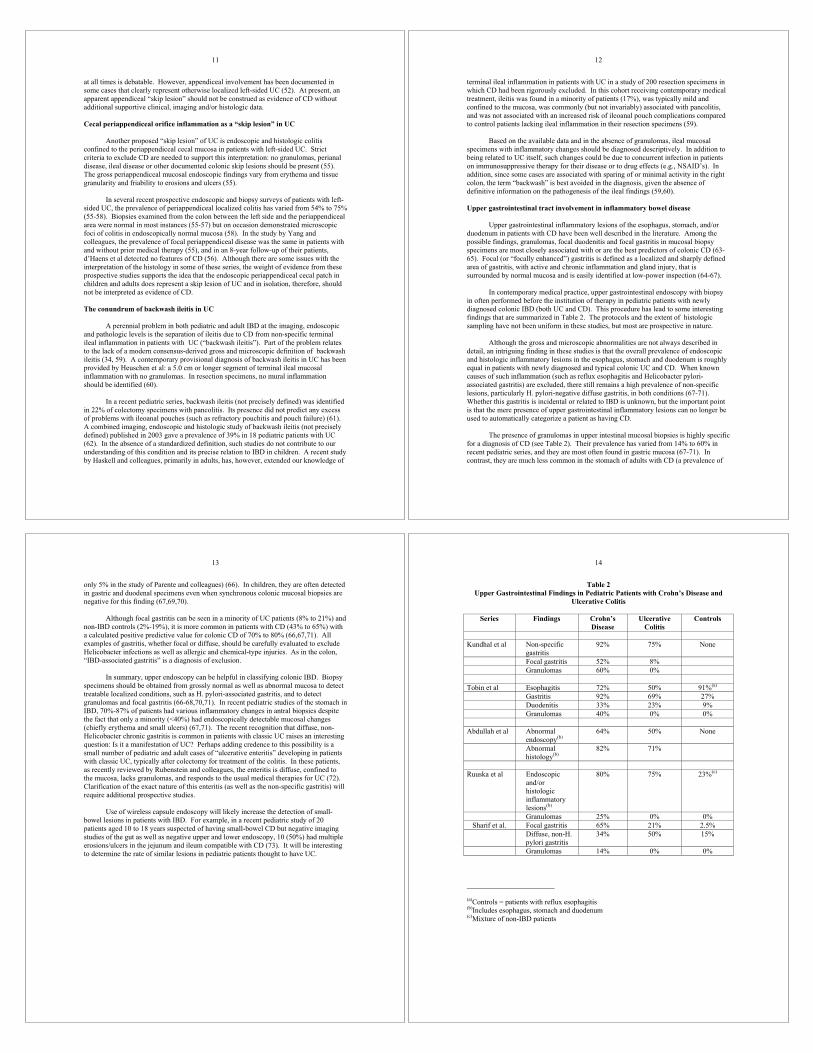
Table 1
Classic Distinguishing Features of Ulcerative Colitis (UC) and Crohn’s Colitis (CC)
UC CC
Gross features
Isolated right-sided
colitis
No Yes
Rectal involvement Yes Variable
Distribution Diffuse Diffuse or focal
Method of extension Continuous(a)
Often discontinuous (“skip
areas” of normal colon)
Involvement of gut
proximal to colon
No(b)
Common
Fistulas No Occasional
“Creeping” serosal fat No Common
Thickened bowel wall No Yes
Strictures Rare Occasional
Microscopic features
Inflammation confined
to mucosa and
submucosa
Yes Uncommon
Transmural
inflammation
No(c)
Common
Fissuring ulcers No(c)
Yes
Fistulas No Yes
Sarcoid-like granulomas No Yes
Distribution of
inflammatory changes
in mucosal specimens
Diffuse Focal or diffuse
Vasculitis No Yes
(a)Recent studies document appendiceal or cecal involvement in some cases of localized
left-sided UC (b)
Possible upper gastrointestinal involvement by otherwise classic UC is a subject of
current debate (see text for details) (c)
May be present in fulminant colitis/toxic megacolon
9
Atypical Features and Controversial Aspects of Pediatric IBD
Unusual features in rectosigmoid mucosal biopsies at the onset of pediatric UC
At first presentation and before therapy, the majority of adult patients with UC
(>90%) will have diffuse active colitis, usually with features of chronicity, in
rectosigmoid mucosal specimens (25,37). Initial rectosigmoid specimens in children
ultimately shown to have UC, however, demonstrate focal colitis and/or the absence of
chronic changes in approximately one-third of patients and are completely normal in 4%
to 8% (25,37,38).
These atypical findings are not specifically related to the patients’ ages at the
onset of colitis (although they are predominantly found in patients younger than 10
years), the duration of symptoms before endoscopy, the symptoms themselves, or the
ultimate evolution of UC (i.e., development of diffuse distal disease, proximal
progression over time) (9,25,38). The reasons for these findings are unknown. One
suggestion is that children may be evaluated earlier in the course of UC than adults (25-
37); however, it is also clear that changes of chronicity may develop within a few weeks
or months of symptom onset (5,9).
This presentation of distal UC with focal disease and a paucity or lack of features
of chronicity in pediatric patients raises several diagnostic possibilities and stresses the
need for a complete evaluation of the patient. First, it should be recognized that
ulcerative colitis is not excluded by these findings (18,25,37,38). Second, they may
represent a non-relapsing, infectious-type colitis, which often is patchy and may have
rectal sparing (39). Crohn’s disease also enters the differential diagnosis; detection of
focal proximal colitis, granulomas, ileal disease or perianal disease would support that
diagnosis.
The predictive value of focal active colitis for development or recognition of CD
once the confounding conditions discussed in the preceding paragraph have been
eliminated has recently been examined. In a cohort of 29 pediatric patients with focal
active colitis, 8 (28%) developed CD; most of the remainder had either infectious colitis
or remained idiopathic (18). In contrast, focal active colitis in adults evolved into a
diagnosis of CD over time in fewer than 15% of patients (40,41). One possible reason for
the difference in outcome between the two populations is that unlike the case in adults,
colonoscopy in children is typically performed for evaluation of abdominal pain, diarrhea
or hematochezia rather than cancer surveillance, thus creating a bias towards detection of
inflammatory diseases. As in children, many cases of “focal active colitis” (particularly
those with minor abnormalities) in adults are likely secondary to effects of bowel
cleansing agents such as sodium phosphate (18,40,41).
10
Effects of medical therapy on the histology of UC in colonic mucosal biopsy specimens
The classic teaching has been that quiescent UC heals with fixed morphologic
changes that permit continued recognition of the colonic mucosa as injured. In 1993,
however, Odze and colleagues demonstrated that medical therapy of left-sided UC with
topical 5-aminosalycylic acid caused reversion of colonic mucosa to a normal appearance
in 64% of patients (42). Since that time, several authors have confirmed and extended
this observation. The results of these studies document that in patients with established
extensive or pancolitis receiving contemporary medical therapy, histologic diffuse
disease has become focal within the colon in up to 54% of patients and the rectum has
become unremarkable in up to 34% on one or more occasions during follow-up (8,43-
47). These results were not related to the duration of disease or the type of therapy
employed (systemic or topical) (47).
Rectal sparing and focal colitis are typical of CD. Thus, to avoid diagnostic
confusion, it is important to know the medication history of patients with presumed UC
in whom such findings are documented. In both children and adults with chronic IBD,
unfortunately, such information is often not available at the time of biopsy specimen
interpretation. In this situation and in the absence of granulomas, focality and rectal
sparing should be described but not interpreted, with the comment that prior medical
therapy may have affected the histologic findings.
Appendiceal involvement as a “skip lesion” in UC
In several retrospective studies of colectomy specimens, the authors have
attempted to determine the prevalence of appendiceal inflammation in patients with UC
of various extents. Appendiceal involvement by UC must be distinguished from
incidental acute appendicitis and has been defined as a lesion confined to the mucosa
with architectural and cellular features of chronicity that may or may not be accompanied
by activity (48). Not unexpectedly, the prevalence of chronic appendicitis in patients
with pancolitis (whether due to UC or CD) is high, occurring in approximately 60% of
cases (48,49).
Of greater interest is the prevalence of this finding in UC patients with less than
pancolitis (i.e., with at least cecal/right-sided sparing in the colectomy specimen). In this
group, appendicitis has been reported in 15% to almost 100% of pediatric and adult
patients, giving rise to the notion of appendicitis as a “skip lesion” in otherwise classic
UC (48,50-53). In a follow-up study of ileoanal anastomoses performed on patients with
appendicitis as a presumed “skip lesion” of UC, there were no cases with outcomes
suggesting that a diagnosis of CD had been missed (54).
A criticism of these studies relates to their retrospective nature (49,55). The
extent of sampling of the cecum and right colon has not been performed in a standardized
fashion, so that the extent of sampling has been either limited or unknown. Also,
information about prior medical therapy and its possible effects on damaged mucosa has
not been provided. Thus, whether or not this portion of the colon has been truly “normal”
11
at all times is debatable. However, appendiceal involvement has been documented in
some cases that clearly represent otherwise localized left-sided UC (52). At present, an
apparent appendiceal “skip lesion” should not be construed as evidence of CD without
additional supportive clinical, imaging and/or histologic data.
Cecal periappendiceal orifice inflammation as a “skip lesion” in UC
Another proposed “skip lesion” of UC is endoscopic and histologic colitis
confined to the periappendiceal cecal mucosa in patients with left-sided UC. Strict
criteria to exclude CD are needed to support this interpretation: no granulomas, perianal
disease, ileal disease or other documented colonic skip lesions should be present (55).
The gross periappendiceal mucosal endoscopic findings vary from erythema and tissue
granularity and friability to erosions and ulcers (55).
In several recent prospective endoscopic and biopsy surveys of patients with left-
sided UC, the prevalence of periappendiceal localized colitis has varied from 54% to 75%
(55-58). Biopsies examined from the colon between the left side and the periappendiceal
area were normal in most instances (55-57) but on occasion demonstrated microscopic
foci of colitis in endoscopically normal mucosa (58). In the study by Yang and
colleagues, the prevalence of focal periappendiceal disease was the same in patients with
and without prior medical therapy (55), and in an 8-year follow-up of their patients,
d’Haens et al detected no features of CD (56). Although there are some issues with the
interpretation of the histology in some of these series, the weight of evidence from these
prospective studies supports the idea that the endoscopic periappendiceal cecal patch in
children and adults does represent a skip lesion of UC and in isolation, therefore, should
not be interpreted as evidence of CD.
The conundrum of backwash ileitis in UC
A perennial problem in both pediatric and adult IBD at the imaging, endoscopic
and pathologic levels is the separation of ileitis due to CD from non-specific terminal
ileal inflammation in patients with UC (“backwash ileitis”). Part of the problem relates
to the lack of a modern consensus-derived gross and microscopic definition of backwash
ileitis (34, 59). A contemporary provisional diagnosis of backwash ileitis in UC has been
provided by Heuschen et al: a 5.0 cm or longer segment of terminal ileal mucosal
inflammation with no granulomas. In resection specimens, no mural inflammation
should be identified (60).
In a recent pediatric series, backwash ileitis (not precisely defined) was identified
in 22% of colectomy specimens with pancolitis. Its presence did not predict any excess
of problems with ileoanal pouches (such as refractory pouchitis and pouch failure) (61).
A combined imaging, endoscopic and histologic study of backwash ileitis (not precisely
defined) published in 2003 gave a prevalence of 39% in 18 pediatric patients with UC
(62). In the absence of a standardized definition, such studies do not contribute to our
understanding of this condition and its precise relation to IBD in children. A recent study
by Haskell and colleagues, primarily in adults, has, however, extended our knowledge of
12
terminal ileal inflammation in patients with UC in a study of 200 resection specimens in
which CD had been rigorously excluded. In this cohort receiving contemporary medical
treatment, ileitis was found in a minority of patients (17%), was typically mild and
confined to the mucosa, was commonly (but not invariably) associated with pancolitis,
and was not associated with an increased risk of ileoanal pouch complications compared
to control patients lacking ileal inflammation in their resection specimens (59).
Based on the available data and in the absence of granulomas, ileal mucosal
specimens with inflammatory changes should be diagnosed descriptively. In addition to
being related to UC itself, such changes could be due to concurrent infection in patients
on immunosuppressive therapy for their disease or to drug effects (e.g., NSAID’s). In
addition, since some cases are associated with sparing of or minimal activity in the right
colon, the term “backwash” is best avoided in the diagnosis, given the absence of
definitive information on the pathogenesis of the ileal findings (59,60).
Upper gastrointestinal tract involvement in inflammatory bowel disease
Upper gastrointestinal inflammatory lesions of the esophagus, stomach, and/or
duodenum in patients with CD have been well described in the literature. Among the
possible findings, granulomas, focal duodenitis and focal gastritis in mucosal biopsy
specimens are most closely associated with or are the best predictors of colonic CD (63-
65). Focal (or “focally enhanced”) gastritis is defined as a localized and sharply defined
area of gastritis, with active and chronic inflammation and gland injury, that is
surrounded by normal mucosa and is easily identified at low-power inspection (64-67).
In contemporary medical practice, upper gastrointestinal endoscopy with biopsy
in often performed before the institution of therapy in pediatric patients with newly
diagnosed colonic IBD (both UC and CD). This procedure has lead to some interesting
findings that are summarized in Table 2. The protocols and the extent of histologic
sampling have not been uniform in these studies, but most are prospective in nature.
Although the gross and microscopic abnormalities are not always described in
detail, an intriguing finding in these studies is that the overall prevalence of endoscopic
and histologic inflammatory lesions in the esophagus, stomach and duodenum is roughly
equal in patients with newly diagnosed and typical colonic UC and CD. When known
causes of such inflammation (such as reflux esophagitis and Helicobacter pylori-
associated gastritis) are excluded, there still remains a high prevalence of non-specific
lesions, particularly H. pylori-negative diffuse gastritis, in both conditions (67-71).
Whether this gastritis is incidental or related to IBD is unknown, but the important point
is that the mere presence of upper gastrointestinal inflammatory lesions can no longer be
used to automatically categorize a patient as having CD.
The presence of granulomas in upper intestinal mucosal biopsies is highly specific
for a diagnosis of CD (see Table 2). Their prevalence has varied from 14% to 60% in
recent pediatric series, and they are most often found in gastric mucosa (67-71). In
contrast, they are much less common in the stomach of adults with CD (a prevalence of
13
only 5% in the study of Parente and colleagues) (66). In children, they are often detected
in gastric and duodenal specimens even when synchronous colonic mucosal biopsies are
negative for this finding (67,69,70).
Although focal gastritis can be seen in a minority of UC patients (8% to 21%) and
non-IBD controls (2%-19%), it is more common in patients with CD (43% to 65%) with
a calculated positive predictive value for colonic CD of 70% to 80% (66,67,71). All
examples of gastritis, whether focal or diffuse, should be carefully evaluated to exclude
Helicobacter infections as well as allergic and chemical-type injuries. As in the colon,
“IBD-associated gastritis” is a diagnosis of exclusion.
In summary, upper endoscopy can be helpful in classifying colonic IBD. Biopsy
specimens should be obtained from grossly normal as well as abnormal mucosa to detect
treatable localized conditions, such as H. pylori-associated gastritis, and to detect
granulomas and focal gastritis (66-68,70,71). In recent pediatric studies of the stomach in
IBD, 70%-87% of patients had various inflammatory changes in antral biopsies despite
the fact that only a minority (<40%) had endoscopically detectable mucosal changes
(chiefly erythema and small ulcers) (67,71). The recent recognition that diffuse, non-
Helicobacter chronic gastritis is common in patients with classic UC raises an interesting
question: Is it a manifestation of UC? Perhaps adding credence to this possibility is a
small number of pediatric and adult cases of “ulcerative enteritis” developing in patients
with classic UC, typically after colectomy for treatment of the colitis. In these patients,
as recently reviewed by Rubenstein and colleagues, the enteritis is diffuse, confined to
the mucosa, lacks granulomas, and responds to the usual medical therapies for UC (72).
Clarification of the exact nature of this enteritis (as well as the non-specific gastritis) will
require additional prospective studies.
Use of wireless capsule endoscopy will likely increase the detection of small-
bowel lesions in patients with IBD. For example, in a recent pediatric study of 20
patients aged 10 to 18 years suspected of having small-bowel CD but negative imaging
studies of the gut as well as negative upper and lower endoscopy, 10 (50%) had multiple
erosions/ulcers in the jejunum and ileum compatible with CD (73). It will be interesting
to determine the rate of similar lesions in pediatric patients thought to have UC.
14
Table 2
Upper Gastrointestinal Findings in Pediatric Patients with Crohn’s Disease and
Ulcerative Colitis
Series Findings Crohn’s
Disease
Ulcerative
Colitis
Controls
Kundhal et al Non-specific
gastritis
92% 75% None
Focal gastritis 52% 8%
Granulomas 60% 0%
Tobin et al Esophagitis 72% 50% 91%(a)
Gastritis 92% 69% 27%
Duodenitis 33% 23% 9%
Granulomas 40% 0% 0%
Abdullah et al Abnormal
endoscopy(b)
64% 50% None
Abnormal
histology(b)
82% 71%
Ruuska et al Endoscopic
and/or
histologic
inflammatory
lesions(b)
80% 75% 23%(c)
Granulomas 25% 0% 0%
Sharif et al. Focal gastritis 65% 21% 2.5%
Diffuse, non-H.
pylori gastritis
34% 50% 15%
Granulomas 14% 0% 0%
(a)
Controls = patients with reflux esophagitis (b)
Includes esophagus, stomach and duodenum (c)
Mixture of non-IBD patients

15
Summary
Because of the complexity of diagnosing and accurately classifying patients with
pediatric IBD, it is clear that any one examination technique, such as histologic analysis
of colonic mucosal biopsies, will seldom give a precise diagnosis. Rather, integration of
all clinical, imaging, laboratory and morphologic data is necessary to achieve diagnostic
accuracy. Pathologists, who usually lack the full range of clinical information, should not
feel pressured to render specific diagnoses based on examination of limited colonic
mucosal material. Our task is first to determine whether or not colitis is present. If it is,
we can state whether it is active, chronic or both and, in untreated patients, give its
distribution (focal or diffuse). Finally, the presence or absence of more specific features
such as granulomas should be mentioned. Our reports will then be an important
contribution to the clinician who must integrate all the data to obtain the most appropriate
diagnosis.
References
1. Hampton T. Scientists explore pathogenesis of IBD. JAMA 2004, 292:2708-
2713.
2. Antonioli DA. Pediatric inflammatory bowel disease. Pediatr Dev Pathol 2005;
8:2-19.
3. Nostrant TT, Kumar NB, Appelman HD. Histopathology differentiates acute
self-limited colitis from ulcerative colitis. Gastroenterology 1987; 92:318-328.
4. Jenkins D, Balsitis M, Gallivan MF, et al. Guidelines for the initial biopsy
diagnosis of suspected chronic idiopathic inflammatory bowel disease: the British
Society of Gastroenterology initiative. J Clin Pathol 1997; 50:93-105.
5. Tanaka M, Riddell R, Saito H, et al. Morphologic criteria applicable to biopsy
specimens for effective distinction of inflammatory bowel disease from other
forms of colitis and of Crohn’s disease from ulcerative colitis. Scand J
Gastroenterol 1999; 34:55-67.
6. LeBerre N, Heresbach D, Kerbaol M, et al. Histological discrimination of
idiopathic inflammatory bowel disease from the other types of colitis. J Clin
Pathol 1995; 48:749-753.
7. Tsang P, Rotterdam H. Biopsy diagnosis of colitis. Am J Surg Pathol 1999;
23:423-430.
8. Geboes K, Dalle I. Influence of treatment on morphologic features of mucosal
inflammation. Gut 2002; 50:S37-S42.
9. Robert ME, Tang L, Hao LM, Reyes-Mugica M. Patterns of inflammation in
mucosal biopsies of ulcerative colitis: perceived differences in pediatric
populations are limited to children younger than 10 years. Am J Surg Pathol
2004; 28:183-189.
10. Odze RD, Wershil BK, Leichtner AM, Antonioli DA. Allergic colitis in infants.
J Pediatr 1995; 126:163-170.
16
11. Goldman H. Interpretation of large intestinal mucosal biopsy specimens. Hum
Pathol 1994; 25:1150-1159.
12. Levine DS, Haggitt RC. Colon. In: Sternberg SS, ed. Histology for Pathologists,
Second Edition. Philadelphia, Lippincott-Raven, 1997, pp 519-537.
13. Watts DA, Lessells AM, Penman ID, Ghosh S. Endoscopic and histologic
features of sodium phosphate bowel preparation-induced colonic ulceration: case
report and review. Gastrointest Endosc 2002; 55:584-587.
14. Berkelhammer C, Ekambaram A, Silva RG. Low-volume oral colonoscopy
bowel preparation: sodium phosphate and magnesium citrate. Gastrointest
Endosc 2002; 56:89-94.
15. Driman DK, Preiksaitis HG. Colorectal inflammation and increased cell
proliferation associated with oral sodium phosphate bowel preparation solutions.
Hum Pathol 1998; 29:972-978.
16. Zwas FR, Cirillo NW, El-Serag HB, et al. Colonic mucosal abnormalities
associated with oral sodium phosphate for colonoscopy preparation. Gastrointest
Endosc 1998; 48:276-278.
17. Wang NACS, Penman ID, Campbell S, et al. Microscopic focal cryptitis
associated with oral sodium phosphate preparation. Histopathology 2000;
36:476-478.
18. Xin W, Brown PI, Greenson JK. The clinical significance of focal active colitis
in pediatric patients. Am J Surg Pathol 2003; 27:1134-1138.
19. Keane J, Gershon S, Wise R, et al. Tuberculosis associated with infliximab, a
tumour necrosis alpha neutralizing factor. N Engl J Med 2001; 345: 1098-1104.
20. Langholz E, Munkholm P, Krasnilikoff PA, et al. Inflammatory bowel diseases
with onset in childhood. Clinical features, morbidity, and mortality in a regional
cohort. Scand J Gastroenterol 1997; 32:139-147.
21. Hait L, Bousvaros A, Grand R. Pediatric inflammatory bowel disease: what
children can teach adults. Inflamm Bowel Dis 2005; 11:519-527.
22. Israel EJ, Kleinman RE. Inflammatory bowel disease: diagnosis and treatment.
Semin Gastrointest Dis 1994; 5:95-105.
23. Yang A, Chen Y, Scherl E, et al. Inflammatory bowel disease in patients with
celiac disease. Inflamm Bowel Dis 2005; 11:528-532.
24. Peters U, Askling J, Gridley G, et al. Causes of death in patients with celiac
disease in a population-based Swedish cohort. Arch Intern Med 2003; 163:1566-
1572.
25. Glickman JN, Bousvaros A, Farraye FA, et al. Pediatric patients with untreated
ulcerative colitis may present initially with unusual morphologic findings. Am J
Surg Pathol 2004; 28:190-197.
26. Escher J, ten Kate F, Lichtenbelt K, et al. Value of rectosigmoidoscopy with
biopsies for diagnosis of inflammatory bowel disease in children. Inflamm Bowel
Dis 2002; 8:16-22.
27. Mamula P, Telega GW, Markowitz JE, et al. Inflammatory bowel disease in
children 5 years of age and younger. Am J Gastroenterol 2002; 97:2005-2010.
28. Powell-Tuck J, Richie JK, Lennard-Jones JE. The prognosis of idiopathic
proctitis. Scand J Gastroenterol 1977; 12:727-732.
17
29. Farmer RG. Long-term prognosis for patients with ulcerative proctosigmoiditis
(ulcerative colitis confined to the rectum and sigmoid colon). J Clin
Gastroenterol 1979; 1:47-50.
30. Mir-Madjlessi SH, Michener WM, Farmer RG. Course and prognosis of
idiopathic ulcerative proctosigmoiditis in young patients. J Pediatr Gastroenterol
Nutr 1986; 5:570-575.
31. Hyams J, Davis P, Lerer T, et al. Clinical outcome of ulcerative proctitis in
children. J Pediatr Gastroenterol Nutr 1997; 25:149-152.
32. Meinzer U, Ideström M, Alberti C, et al. Ileal involvement is age dependent in
pediatric Crohn’s disease. Inflamm Bowel Dis 2005; 11:639-644.
33. Tolia V. Perianal Crohn’s disease in children and adolescents. Am J
Gastroenterol 1996; 91:922-926.
34. Odze R. Diagnostic problems and advances in inflammatory bowel disease. Mod
Pathol 2003; 16:347-358.
35. Wakefield AJ, Sankey EA, Dhillon AP, et al. Granulomatous vasculitis in
Crohn’s disease. Gastroenterology 1991; 100:1279-1287.
36. Schmitz-Moorman P, Schag M. Histology of the lower intestinal tract in Crohn’s
disease of children and adolescents: multicentric paediatric Crohn’s disease study.
Pathol Res Pract 1990; 186:479-484.
37. Washington K, Greenson JK, Montgomery E, et al. Histopathology of ulcerative
colitis in initial rectal biopsy in children. Am J Surg Pathol 2002; 26:1441-1449.
38. Markowitz J, Kahn E, Grandcher K, et al. Atypical rectosigmoid histology in
children with newly diagnosed ulcerative colitis. Am J Gastroenterol 1993;
88:2034-2037.
39. Mantzaris GJ, Hatzis A, Archavlis E, et al. The role of colonoscopy in the
differential diagnosis of acute, severe hemorrhagic colitis. Endoscopy 1995;
27:645-653.
40. Greenson JK, Robert SA, Carpenter SL, et al. The clinical significance of focal
active colitis. Hum Pathol 1997; 28:729-733.
41. Volk EE, Shapiro BD, Easley KA, et al. The clinical significance of a biopsy-
based diagnosis of focal active colitis: a clinicopathologic study of 31 cases. Mod
Pathol 1998; 11:789-794.
42. Odze R, Antonioli D, Peppercorn M, Goldman H. Effect of topical 5-
aminosalicylic acid (5-ASA) therapy on rectal mucosal morphology in chronic
ulcerative colitis. Am J Surg Pathol 1993; 17:869-875.
43. Moum B, Ekboom A, Vatn MH, et al. Changes in the extent of colonoscopic and
histological involvement in ulcerative colitis over time. Am J Gastroenterol 1999;
94:1564-1569.
44. Bernstein CN, Shanahan F, Anton PA, Weinstein WM. Patchiness of mucosal
inflammation in treated ulcerative colitis: a prospective study. Gastrointest
Endosc 1995; 42:232-237.
45. Levine TS, Tzardi M, Mitchell S, et al. Diagnostic difficulty arising from rectal
recovery in ulcerative colitis. J Clin Pathol 1996; 49:319-323.
46. Kleer CG, Appelman H. Ulcerative colitis: patterns of involvement in colorectal
biopsies and changes with time. Am J Surg Pathol 1998; 22:983-989.
18
47. Kim B, Barnett JL, Kleer CG, et al. Endoscopic and histological patchiness in
treated ulcerative colitis. Am J Gastroenterol 1999; 94:3258-3262.
48. Scott IS, Sheaff M, Coumbe A, et al. Appendiceal involvement in ulcerative
colitis. Histopathology 1998; 33:168-173.
49. Goldblum JR, Appelman HD. Appendiceal involvement in ulcerative colitis.
Mod Pathol 1992; 5:607-610.
50. Davison AM, Dixon MF. The appendix is a “skip lesion” in ulcerative colitis.
Histopathology 1990; 16:93-95.
51. Kahn E, Markowitz J, Daum F. The appendix in inflammatory bowel disease in
children. Mod Pathol 1992; 5:380-383.
52. Groisman GM, George J, Harpaz N. Ulcerative appendicitis in universal and
nonuniversal ulcerative colitis. Mod Pathol 1994; 7:322-325.
53. Kroft SH, Stryker SJ, Rao MS. Appendiceal involvement as a skip lesion in
ulcerative colitis. Mod Pathol 1994; 7:912-914.
54. Perry WB, Opelka FG, Smith D, et al. Discontinuous appendiceal involvement in
ulcerative colitis: pathology and clinical correlation. Gastrointest Surg 1999;
3:141-144.
55. Yang SK, et al. Appendiceal orifice inflammation as a skip lesion in ulcerative
colitis: an analysis in relation to medical therapy and disease extent. Gastrointest
Endosc 1999; 49:743-747.
56. d’Haens G, Geboes K, Ponette E, et al. Patchy cecal inflammation associated
with distal ulcerative colitis: a prospective endoscopic study. Am J Gastroenterol
1997; 92:1275-1279.
57. Okawa K, Aoki T, Sano K, et al. Ulcerative colitis with skip lesions at the mouth
of the appendix: a clinical study. Am J Gastroenterol 1998; 93:2405-2410.
58. Matsumoto T, Nakamura S, Shimizu M, Iida M. Significance of appendiceal
involvement in patients with ulcerative colitis. Gastrointest Endosc 2002; 55:180-
185.
59. Haskell H, Andrews CW, Reddy SI, et al. Pathologic features and clinical
significance of “backward” ileitis in ulcerative colitis. Am J Surg Pathol 2005;
29:1472-1481.
60. Heuschen UA, Hinz U, Allemeyer EH, et al. Backwas ileitis is strongly
associated with colorectal carcinoma in ulcerative colitis. Gastroenterology 2001;
120:841-847.
61. Alexander F, Sarigol S, DiFiore J, et al. Fate of the pouch in 151 pediatric
patients after ileal pouch anal anastomosis. J Pediatr Surg 2003; 38:78-82.
62. Laghi A, Borrelli P, Paolantonio P, et al. Contrast enhanced magnetic resonance
imaging of the terminal ileum in children with Crohn’s disease. Gut 2003;
52:393-397.
63. Pascasio JM, Hammond S, Qualman SJ. Recognition of Crohn’s disease on
incidental gastric biopsy in childhood. Pediatr Dev Pathol 2003; 6: 209-214.
64. Oberhuber G, Puspok A, Oesterreicher C, et al. Focally enhanced gastritis: a
frequent type of gastritis in patients with Crohn’s disease. Gastroenterology
1997; 112:698-706.
65. Wright CL, Ridell RH. Histology of the stomach and duodenum in Crohn’s
disease. Am J Surg Pathol 1998; 22:383-390.

19
66. Parente F, Cucino C, Bollani S, et al. Focal gastric inflammatory infiltrates in
inflammatory bowel diseases: prevalence, immunohistochemical characteristics,
and diagnostic role. Am J Gastroenterol 2000; 95:705-711.
67. Kundhal PS, Stormon MQ, Zacho M, et al. Gastric antral biopsy in the
differentiation of pediatric colitides. Am J Gastroenterol 2003; 98:557-561.
68. Ruuska T, Vaajalahti P, Arajärvi P, Mäki M. Prospective evaluation of upper
gastrointestinal mucosal lesions in children with ulcerative colitis and Crohn’s
disease. J Pediatr Gastroenterol Nutr 1994; 19:181-186.
69. Tobin JM, Sinha B, Ramani P, et al. Upper gastrointestinal mucosal disease in
pediatric Crohn’s disease and ulcerative colitis: a blinded, controlled study. J
Pediatr Gastroenterol Nutr 2001; 32:443-448.
70. Abdullah BA, Gupta SK, Croffie JM, et al. The role of
esophagogastroduodenoscopy in the initial evaluation of childhood inflammatory
bowel disease: a 7-year study. J Pediatr Gastroenterol Nutr 2002; 35:636-640.
71. Sharif F, McDermott M, Dillon M, et al. Focally enhanced gastritis in children
with Crohn’s disease and ulcerative colitis. Am J Gastroenterol 2002; 97:1415-
1420.
72. Rubenstein J, Sherif A, Appelman H, Chey WD. Ulcerative colitis-associated
enteritis: is ulcerative colitis always confined to the colon? J Clin Gastroenterol
2004; 38:46-51.
73. Sant’Anna AMGA, Dubois J, Miron M-C, Seidman EG. Wireless capsule
endoscopy for obscure small-bowel disorders: final results of the first pediatric
controlled trial. Clin Gastroenterol Hepatol 2005; 3:264-270.
ENTEROPATHIES OF INFANCY
PIERRE RUSSO, MD
The Children’s Hospital of Philadelphia and the University of Pennsylvania
INTRODUCTION
Acute diarrhea, in the industrialized world, is usually self-limited, infectious in nature,
and rarely requires biopsy evaluation. Chronic diarrhea can cause failure to thrive, frequently
results in intestinal biopsy and may require parenteral nutrition and other forms of therapy.
Protracted diarrhea has replaced the older term “intractable diarrhea” in the GI literature and
denotes a syndrome of severe diarrhea, usually occurring in infants less than a year of age and
requiring aggressive nutritional and sometimes immunosuppressive management. Though not
strictly synonymous with chronic diarrhea, the two are frequently used interchangeably. Several
recent reviews on the subject of protracted diarrhea in childhood are available[1-4].
Disorders resulting in chronic diarrhea of childhood can be grouped into several major
pathophysiologic categories:
I. Congenital transport disorders and disaccharidase deficiencies
II. Disorders of enterocyte development
III. Inflammatory and Immune-mediated disorders
IV. Systemic disorders and Tumors
V. Motility disorders
VI. Endocrine disorders – congenital adrenal hyperplasia
VII. Infections – “blind loop”, Giardia, HIV
VIII. Anatomic disorders – Malrotation, short gut, lymphangiectasia
IX. Pancreatic disorders – Cystic fibrosis, Shwachman disease
During the past twenty years, enteric infections and food intolerance have been replaced
by rarer congenital conditions, partly due to greater awareness of these entities as well as due to
improvements in diagnosis and management
1977-1993 1993-1996 1997-2001
(N=38) (N=32) (N=61)
ETIOLOGY n(%) n(%) n(%)
Enteric infection 18 (48) 4 (12) 2 (3)
Food intolerance 8 (22) 3 (10) 10 (17)
Autoimmune enteropathy 2 (5) 8 (25) 7 (12)
Structural enterocyte defects 2 (5) 7 (22) 16 (26)
Celiac disease 1 (2.5) 0 (0) 0 (0)
Eosinophilic enteropathy 1 (2.5) 1 (3) 0 (0)
Lymphangiectasia 1 (2.5) 1 (3) 2 (3)
Motility disorders 2 (5) 3 (9) 16 (26)
Munchausen syndrome by proxy 0 (0) 0 (0) 1 (15)
Unknown 3 (7.5) 5 (16) 7 (11)
From: Guarino A and DeMarco G. Persistent Diarrhea, chapter 10, in Pediatric Gastrointestinal Disease, 4th edition
(2004)
In evaluating intestinal biopsies, it is helpful to remember that disorders resulting in
severe diarrhea tend to cluster in different age groups:
Infant Toddler
" Microvillus inclusion disease " Irritable colon of childhood
" Autoimmune enteropathy " Protracted infectious enteritis
" Milk and soy protein intolerance " Giardiasis
" Protracted infectious enteritis " Sucrase-isomaltase deficiency
" Hirschsprung’s disease " Tumors
" Congenital transport defects " Celiac disease
" Munchausen syndrome by proxy " Inflammatory bowel disease
Modified from Vanderhoof J.A. p.32-42 ch 3 in Pediatric Gastrointestinal Disease, Wyllie R. And Hyams J.S., eds W.B.
Saunders, 1999
The type of diarrhea can also offer clues: Osmotic diarrhea results from an increased
osmotic load in the distal small bowel and colon, with an osmotic “gap” in stool electrolytes, and
usually stops when oral feeding is discontinued. It is usually associated with disaccharidase
deficiencies and transport disorders, decreased absorptive surface (microvillous inclusion
disorder) and hypermotility disorders. On the other hand, secretory diarrhea is characterized by
an exudate of mucous, protein or blood, and a variable response to withholding of oral feeds.
Disorders usually associated with secretory diarrhea include congenital fluid transport disorders,
inflammatory disorders and increased secretion of gastrointestinal activating substances.
In evaluating intestinal biopsies, disorders that cause protracted diarrhea in early
life can be broadly divided into those in which villus architecture is maintained, and those
characterized by villus atrophy:
Normal villous morphology
Congenital chloride diarrhea
Congenital carbohydrate malabsorption
Congenital sucrose isomaltase deficiency
Congenital bile acid malabsorption
Lipid-filled enterocytes
Abetalipoproteinemia
Chylomicron retention disorder
Villous atrophy
Pauci-inflammatory
Microvillous inclusion disease
Epithelial dysplasia
Inflammatory
Autoimmune enteropathy
IPEX
Enterocolitis with dysmorphic features
Congenital immunodeficiency disorders
Milk intolerance
Gluten-sensitive enteropathy
Most transport disorders and diasaccharidase deficiencies do not result in appreciable
histologic changes on intestinal biopsies. In fact, normal findings on an intestinal biopsy should
prompt the clinician to search for one of these entities.

Lipid transport disorders such as Abetalipoproteinemia, homozygous
hypolipoproteinemia and chylomicron retention disease (Anderson disease) are characterized by
accumulation of lipid within the enterocytes and clinical characteristics, such as fat
malabsorption, low levels of serum lipids, failure to thrive in childhood, neurologic and visual
problems resulting from malabsorption of fat-soluble vitamins. These conditions are associated
with disorders of apolipoproteins, which reside on the surface of chylomicrons. Apoliproproteins
native to the intestine are apolipoprotein (apo) A-I, apo A-IV, and apoB, which has two forms:
apo B-100 and apo B-48, both encoded by the same gene located on chromosome 2[5].
Abetalipoproteinemia is an autosomal recessive disorder characterized by the absence of
Microsomal Triglyceride Transfer protein (MTP), responsible for assembly of lipoprotein
particles, and for the proper folding of ApoB, preventing its premature degradation[6]. Patients
have diarrhea and fat malabsorption usually appearing within the first few months of life, with
acanthocytosis, and deficiencies in fat-soluble vitamins resulting in retinitis pigmentosa and
neurologic symptoms. Serum levels of cholesterol and triglycerides are typically low, and do not
rise after a fatty meal. Fat-filled enterocytes are noted on intestinal biopsies of fasting patients,
which on electron microscopy are irregular in size and generally non-membrane-bound. No lipid
is noted in the extracellular space. Chylomicron retention (Anderson) disease is similar to
abetalipoproteinemia in its gastrointestinal manifestations and impact on growth, though
acanthocytosis is usually absent and neurologic and ocular abnormalities are much less severe.
Also, in contrast to abetalipoproteinemia, serum fasting triglyceride levels are normal and
hypocholesterolemia is less marked. The basis for the disorder appears to be an inability to
export chylomicrons from the enterocyte into lacteals[7]. Fat-filled enterocytes are also seen in
small bowel biopsies. On ultrastructure, lipid droplets are more uniform in size than the droplets
in abetalipoproteinemia, and some appear to be bounded by the membranes of the endoplasmic
reticulum[8].
Microvillus Inclusion Disease ( MVID)
Initially described by Davidson et al in 1978, and subsequently by investigators in other
parts of the world, MVID is characterized by refractory secretory diarrhea occurring within the
first week of life[9, 10]. There is a roughly equal sex incidence. Birth history is usually
unremarkable, without evidence of polyhydramnios, as is seen in congenital chloride diarrhea.
Tests of small intestinal function are diffusely abnormal[11]. The pattern of inheritance appears
to be autosomal recessive[10].
Small bowel biopsies are characterized by severe villus atrophy, with little or no crypt
hyperplasia, and without significant inflammation. The pathognomonic features are present on
ultrastructural study and include absent or small stubby microvilli, vesicular structures located
towards the apex of the enterocytes containing microvilli, and granules containing dense
amorphous material. Even without an electron microscope, this disease can be strongly suspected
on paraffin sections by the absence of a distinct brush border on the periodic-acid-Schiff (PAS)
stain, and by the presence of PAS-positive diastase-resistant densities at the apex of the
enterocytes. Similar observations are noted using immunohistochemical staining for alkaline
phosphatase, CEA, and more recently, with anti-CD 10, a membrane-associated neutral
peptidase[12]. Microvillus inclusions have also been reported in the colon, gallbladder and renal
tubular epithelium in these patients[11]. These inclusions can sometimes be difficult to detect,
and several biopsies may be required to identify them. It appears that MVID represents a
spectrum of entities, ranging from cases with typical early presentation and diagnostic inclusions
on EM, to cases in which the clinical presentation is atypical, or cases in which inclusions either
cannot be demonstrated (microvillus dystrophy), or the inclusions are “atypical”[13-16].
In a multicenter survey of 23 patients with MVID, the one-year survival was less than
25%[10]. Medical therapy in these patients has generally proven to be ineffective and small
intestinal transplantation currently is the best treatment[10, 17, 18]. Instances of apparent
resolution of the disease with improvement of the mucosal features on long-term TPN have been
reported[19].
Tufting Enteropathy (Epithelial dysplasia )
First described by Reifen et al, then by Goulet and colleagues, patients with tufting
enteropathy present in the neonatal period with a watery diarrhea. Prenatal history is uneventful
and the disease appears to be inherited in an autosomal recessive fashion, as suggested by the
finding of other affected siblings and frequent parental consanguinity[20, 21].
Its histological hallmarks are severe villus atrophy with the formation of “tufts” of
rounded, tear-drop shaped enterocytes which appear to shed into the lumen. There may be a mild
increase in the lamina propria inflammatory cells, but intraepithelial lymphocytes (IELs) do not
appear to be significantly increased. Transmission electron microscopy has revealed a normal
brush border. Abnormalities in immunohistochemical staining for basement membrane laminin
and heparan sulfate proteoglycan, ultrastructural irregularities of desmosomes, and abnormal
distribution of #2$1 integrin have been observed in patients with this disorder, suggesting that
the early onset of the disease and its characteristic histomorphological changes may result from
abnormal cell-cell and cell-matrix interaction[22, 23]. Dietary manipulations, antibiotic and
immunosuppression have proven ineffective in these patients, and long-term TPN and small-
bowel transplantation are the current treatment options for these patients[11].
A probably related disorder resulting from the absence of intestinal epithelial #6$4
integrin has been described. The salient features are severe watery diarrhea in the first two weeks
of life in association with pyloric atresia, with extensive sloughing of the epithelium observed on
biopsies[24].
Autoimmune Enteropathy
The term “autoimmune enteropathy”(AIE) was proposed by Unsworth and
Walker-Smith to describe a syndrome of protracted diarrhea with the presence of autoantibodies
against gut epithelium[25].
More than 100 cases have since been reported in the past twenty years[26], most cases
occurring in infancy or the first year of life. There is a strong male preponderance, family history
of other affected siblings, frequent extra-intestinal involvement and various circulating
autoantibodies[27, 28]. However, this entity has also been reported in older children[29] and
even adults[30-32], in whom it may be responsible for a proportion of cases referred to as
“refractory sprue”. Intestinal biopsies in most of the reported cases are characterized by severe
villus atrophy and variable crypt hyperplasia. Concomitant colitis and gastritis are present in the
majority of cases. Marked inflammatory destruction of intestinal crypts with extensive apoptosis
is a feature noted in many of the cases. These findings are similar to those noted in severe
intestinal graft-versus-host disease, and suggest an abnormal immune-mediated attack against
intestinal epithelium.
The hallmark of this entity is the presence of circulating anti-enterocyte antibodies
manifest as positive staining in a linear pattern along the apex and baso-lateral border of the

enterocyte by indirect immunofluorescence[27]. The antibodies are predominantly IgG, and have
been described as complement-fixing, though IgM and IgA have also been described[33].
Antibodies reacting against mucus or goblet cells have also been described in occasional patients
with an apparently related entity, and intestinal biopsies in these cases have shown a marked
depletion of goblet cells[29, 34].
Extra-intestinal disease in these patients includes insulin-dependent diabetes,
glomerulopathy and hemolytic anemia, as well as pulmonary and dermatologic
manifestations[26]. Numerous autoantibodies have been detected in these patients, including
anti-smooth muscle, anti-endoplasmic reticulum, anti-mitochondrial, anti-parietal, anti-adrenal,
as well as anti-DNA and anti-ANA antibodies.
AIE has been associated with with several immunodeficiencies and autoimmune diseases:
Autoimmune Enteropathy: Associated Conditions
" IPEX Immunodysregulation / polyendocrinopathy / enteropathy / X-linked.
" Immunoosseous dysplasia, Schimke type
" Usher syndrome (hyperinsulinism with enteropathy and deafness)
" Autoimmune Polyendocrinopathy, Candidiasis and Ectodermal dysplasia
Perhaps the most well- recognized such association is with the X-linked syndrome of
Immunodysregulation, Polyendocrinopathy and Enteropathy (IPEX) syndrome, initially
described by Powell[35]. This syndrome has been related to mutations of the FOX P3 gene
(Xp11.23-Xq13.3)[36, 37], and is the human equivalent of scurfy in the mouse[38]. The protein
encoded by the gene, Foxp3, is expressed in regulatory CD4+/CD25+ T cells and is essential for
immune homeostasis[39]. Scurfy mice are characterized by failure to thrive, overexpression of
CD4+/CD8+ lymphocytes, extensive multiorgan inflammatory infiltration and over production
of cytokines[40].
Mortality in cases of AIE has been high, occurring in up to one-third of reported cases, but
more frequent in the older descriptions. Diet changes and steroids alone have been largely
ineffective, and the use of immunosuppressants appears to be necessary in most cases.
Cyclosporine has induced a remission in several studies, though some remained resistant. A
positive response to Tacrolimus has been reported in patients refractory to other therapies[41,
42].
“Syndromatic Diarrhea”
Girault et al described a group of patients with dysmorphic features, consisting of a
prominent forehead, broad nose, hypertelorism and wooly, easily removable abnormal hair
(trichorrhexis nodosa), in association with intractable diarrhea and immunodeficiency, which
they termed “syndromatic intractable diarrhea”, because of the constellation of extra-intestinal
manifestations[43]. Most of the patients reported were of Middle Eastern origin, and there was
parental consanguinity and a history of similarly affected siblings. The immunodeficiency was
characterized by impaired T-cell and antibody responses despite normal immunoglobulin levels.
Intestinal biopsies revealed moderate villous atrophy without a conspicuous increase in
inflammatory cells.
Other syndromes associated with early severe diarrhea include:
" Epidermolysis Bullosa with Pyloric atresia
" Tricho-hepato-enteric syndrome
" Infantile Systemic Hyalinosis
" Phosphomannose Isomerase Deficiency (CDG type Ib)
" Hypohidrotic Ectodermal Dysplasia
" Progressive Familial Cholestasis, PFIC I (ATP8B1)
" Lysinuric Protein Intolerance
Hair abnormalities can be seen in association with chronic diarrhea in other disorders,
typically in Menke’s disease, Zinc deficiency and Lysinuric protein intolerance. Distinct hair
shaft anomalies have been reported in two siblings as the tricho-hepato-enteric syndrome, in
which thin sparse hair (trichomalacia) was seen in association with hypertelorism, chronic
diarrhea, a progressive hypermethioninemia and a hemochromatosis phenotype[44].
Congenital Immunodeficiencies
Over 50% of children with immunodeficiencies present with chronic diarrhea, and the
gastrointestinal tract is the second most common site of infection in these cases, after the
pulmonary system. Immunodeficiency states may predispose to various abnormal intestinal
immune reactions because of increased intestinal permeability, resulting in heightened exposure
to dietary or microbial antigens.
Primary Immunodeficiency Disorders with Prominent Gastrointestinal Manifestations
Antibody Deficiencies
! X-linked agammaglobulinemia
! Common variable Immunodeficiency
! IgA deficiency
! Immunodeficiency with elevated IgM (hyper-IgM syndrome)
! Transient hypogammaglobulinemia of infancy
! IgG subclass deficiency
Combined Immunodeficiencies
! Severe combined immunodeficiency
! Major histocompatibility complex Class II deficiency
T-Cell Disorders
! Wiskott-Aldrich syndrome
! Ataxia telangiectasia
! DiGeorge syndrome
Disorders of Phagocyte Functions
! Chronic granulomatous disease
! CD11 / CD18 Leukocyte adhesion molecule deficiency
! Hermansky Pudlak syndrome
! Glycogen storage type IB
Other Disorders
! Chronic mucocutaneous candidiasis
! Autoimmune Polyendocrinopathy/candidiasis/ectodermal dystrophy
! IPEX (Immunodeficiency, Polyendocrinopathy, Enteropathy, X-linked)
Common Variable Immunodeficiency(CVID) is one of the more common primary
immunodeficiencies. Chronic diarrhea is frequent in these patients, and other gastrointestinal
manifestations include chronic active hepatitis and hemorrhagic gastritis[45, 46]. Histologic
findings in the intestines fall into four major categories: infections (frequently Giardia), nodular
lymphoid hyperplasia (NLH), celiac-like villous atrophy, and a severe Crohn’s-like enterocolitis.
There is an absence of germinal follicles in the lymphoid tissues of the GI tract, and near absence
of plasma cells in the lamina propria.
Chronic diarrhea refractory to a gluten-free diet in association with villus atrophy has
been observed in patients with CVID, and major differentiating features from celiac disease
include a crypt-destructive inflammatory process in association with prominent apoptosis,
reminiscent of autoimmune enteropathy[47]. An accumulation of foamy macrophages in the
lamina propria, resembling Whipple’s disease or chronic granulomatous disease, has been
noted[47].
IgA deficiency is the most common primary immunodeficiency, with a prevalence of
approximately 1 in 300[48]. Gastrointestinal disorders include Giardia infection, celiac disease,
chronic inflammatory bowel disease, atrophic gastritis with pernicious anemia, food allergies and
nodular lymphoid hyperplasia[49]. There is a 10-20-fold increase in celiac disease in patients
with IgA deficiency[50, 51], and the clinical presentation, histologic changes and response to
gluten are similar to that of non-IgA- deficient patients, the distinguishing feature being an
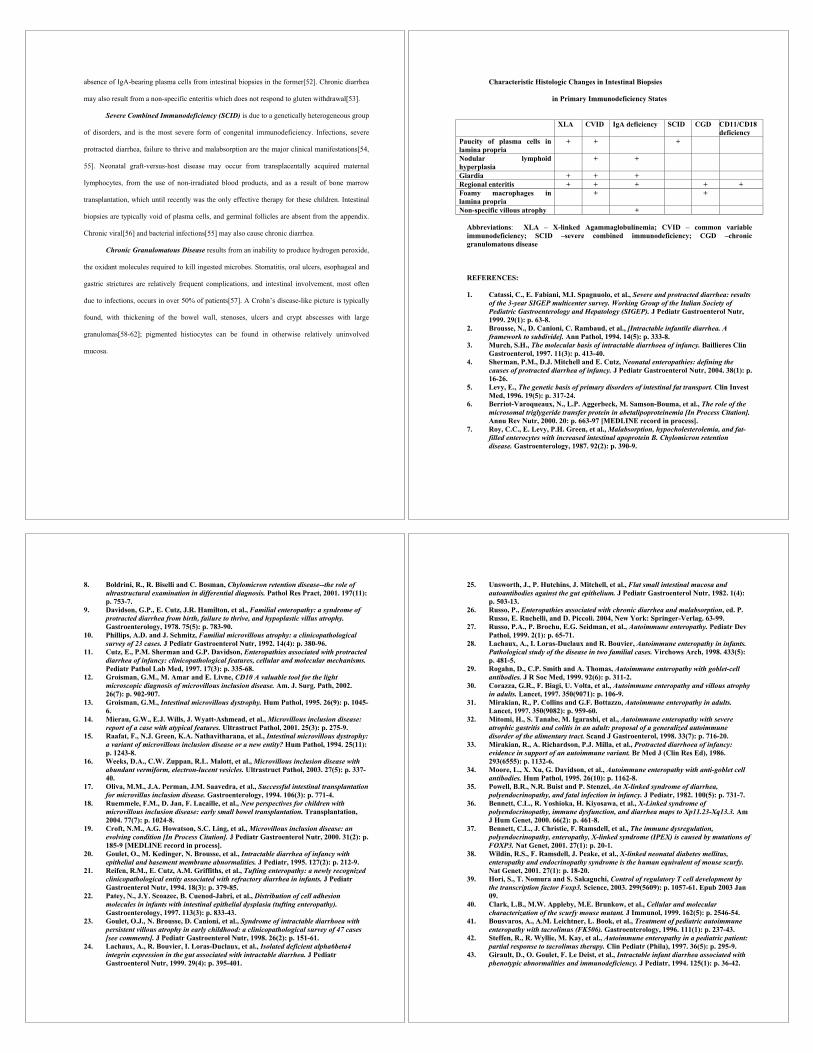
absence of IgA-bearing plasma cells from intestinal biopsies in the former[52]. Chronic diarrhea
may also result from a non-specific enteritis which does not respond to gluten withdrawal[53].
Severe Combined Immunodeficiency (SCID) is due to a genetically heterogeneous group
of disorders, and is the most severe form of congenital immunodeficiency. Infections, severe
protracted diarrhea, failure to thrive and malabsorption are the major clinical manifestations[54,
55]. Neonatal graft-versus-host disease may occur from transplacentally acquired maternal
lymphocytes, from the use of non-irradiated blood products, and as a result of bone marrow
transplantation, which until recently was the only effective therapy for these children. Intestinal
biopsies are typically void of plasma cells, and germinal follicles are absent from the appendix.
Chronic viral[56] and bacterial infections[55] may also cause chronic diarrhea.
Chronic Granulomatous Disease results from an inability to produce hydrogen peroxide,
the oxidant molecules required to kill ingested microbes. Stomatitis, oral ulcers, esophageal and
gastric strictures are relatively frequent complications, and intestinal involvement, most often
due to infections, occurs in over 50% of patients[57]. A Crohn’s disease-like picture is typically
found, with thickening of the bowel wall, stenoses, ulcers and crypt abscesses with large
granulomas[58-62]; pigmented histiocytes can be found in otherwise relatively uninvolved
mucosa.
Characteristic Histologic Changes in Intestinal Biopsies
in Primary Immunodeficiency States
XLA CVID IgA deficiency SCID CGD CD11/CD18
deficiency
Paucity of plasma cells in
lamina propria
+ + +
Nodular lymphoid
hyperplasia
+ +
Giardia + + +
Regional enteritis + + + + +
Foamy macrophages in
lamina propria
+ +
Non-specific villous atrophy +
Abbreviations: XLA – X-linked Agammaglobulinemia; CVID – common variable
immunodeficiency; SCID –severe combined immunodeficiency; CGD –chronic
granulomatous disease
REFERENCES:
1. Catassi, C., E. Fabiani, M.I. Spagnuolo, et al., Severe and protracted diarrhea: results
of the 3-year SIGEP multicenter survey. Working Group of the Italian Society of
Pediatric Gastroenterology and Hepatology (SIGEP). J Pediatr Gastroenterol Nutr,
1999. 29(1): p. 63-8.
2. Brousse, N., D. Canioni, C. Rambaud, et al., [Intractable infantile diarrhea. A
framework to subdivide]. Ann Pathol, 1994. 14(5): p. 333-8.
3. Murch, S.H., The molecular basis of intractable diarrhoea of infancy. Baillieres Clin
Gastroenterol, 1997. 11(3): p. 413-40.
4. Sherman, P.M., D.J. Mitchell and E. Cutz, Neonatal enteropathies: defining the
causes of protracted diarrhea of infancy. J Pediatr Gastroenterol Nutr, 2004. 38(1): p.
16-26.
5. Levy, E., The genetic basis of primary disorders of intestinal fat transport. Clin Invest
Med, 1996. 19(5): p. 317-24.
6. Berriot-Varoqueaux, N., L.P. Aggerbeck, M. Samson-Bouma, et al., The role of the
microsomal triglygeride transfer protein in abetalipoproteinemia [In Process Citation].
Annu Rev Nutr, 2000. 20: p. 663-97 [MEDLINE record in process].
7. Roy, C.C., E. Levy, P.H. Green, et al., Malabsorption, hypocholesterolemia, and fat-
filled enterocytes with increased intestinal apoprotein B. Chylomicron retention
disease. Gastroenterology, 1987. 92(2): p. 390-9.
8. Boldrini, R., R. Biselli and C. Bosman, Chylomicron retention disease--the role of
ultrastructural examination in differential diagnosis. Pathol Res Pract, 2001. 197(11):
p. 753-7.
9. Davidson, G.P., E. Cutz, J.R. Hamilton, et al., Familial enteropathy: a syndrome of
protracted diarrhea from birth, failure to thrive, and hypoplastic villus atrophy.
Gastroenterology, 1978. 75(5): p. 783-90.
10. Phillips, A.D. and J. Schmitz, Familial microvillous atrophy: a clinicopathological
survey of 23 cases. J Pediatr Gastroenterol Nutr, 1992. 14(4): p. 380-96.
11. Cutz, E., P.M. Sherman and G.P. Davidson, Enteropathies associated with protracted
diarrhea of infancy: clinicopathological features, cellular and molecular mechanisms.
Pediatr Pathol Lab Med, 1997. 17(3): p. 335-68.
12. Groisman, G.M., M. Amar and E. Livne, CD10 A valuable tool for the light
microscopic diagnosis of microvillous inclusion disease. Am. J. Surg. Path, 2002.
26(7): p. 902-907.
13. Groisman, G.M., Intestinal microvillous dystrophy. Hum Pathol, 1995. 26(9): p. 1045-
6.
14. Mierau, G.W., E.J. Wills, J. Wyatt-Ashmead, et al., Microvillous inclusion disease:
report of a case with atypical features. Ultrastruct Pathol, 2001. 25(3): p. 275-9.
15. Raafat, F., N.J. Green, K.A. Nathavitharana, et al., Intestinal microvillous dystrophy:
a variant of microvillous inclusion disease or a new entity? Hum Pathol, 1994. 25(11):
p. 1243-8.
16. Weeks, D.A., C.W. Zuppan, R.L. Malott, et al., Microvillous inclusion disease with
abundant vermiform, electron-lucent vesicles. Ultrastruct Pathol, 2003. 27(5): p. 337-
40.
17. Oliva, M.M., J.A. Perman, J.M. Saavedra, et al., Successful intestinal transplantation
for microvillus inclusion disease. Gastroenterology, 1994. 106(3): p. 771-4.
18. Ruemmele, F.M., D. Jan, F. Lacaille, et al., New perspectives for children with
microvillous inclusion disease: early small bowel transplantation. Transplantation,
2004. 77(7): p. 1024-8.
19. Croft, N.M., A.G. Howatson, S.C. Ling, et al., Microvillous inclusion disease: an
evolving condition [In Process Citation]. J Pediatr Gastroenterol Nutr, 2000. 31(2): p.
185-9 [MEDLINE record in process].
20. Goulet, O., M. Kedinger, N. Brousse, et al., Intractable diarrhea of infancy with
epithelial and basement membrane abnormalities. J Pediatr, 1995. 127(2): p. 212-9.
21. Reifen, R.M., E. Cutz, A.M. Griffiths, et al., Tufting enteropathy: a newly recognized
clinicopathological entity associated with refractory diarrhea in infants. J Pediatr
Gastroenterol Nutr, 1994. 18(3): p. 379-85.
22. Patey, N., J.Y. Scoazec, B. Cuenod-Jabri, et al., Distribution of cell adhesion
molecules in infants with intestinal epithelial dysplasia (tufting enteropathy).
Gastroenterology, 1997. 113(3): p. 833-43.
23. Goulet, O.J., N. Brousse, D. Canioni, et al., Syndrome of intractable diarrhoea with
persistent villous atrophy in early childhood: a clinicopathological survey of 47 cases
[see comments]. J Pediatr Gastroenterol Nutr, 1998. 26(2): p. 151-61.
24. Lachaux, A., R. Bouvier, I. Loras-Duclaux, et al., Isolated deficient alpha6beta4
integrin expression in the gut associated with intractable diarrhea. J Pediatr
Gastroenterol Nutr, 1999. 29(4): p. 395-401.
25. Unsworth, J., P. Hutchins, J. Mitchell, et al., Flat small intestinal mucosa and
autoantibodies against the gut epithelium. J Pediatr Gastroenterol Nutr, 1982. 1(4):
p. 503-13.
26. Russo, P., Enteropathies associated with chronic diarrhea and malabsorption, ed. P.
Russo, E. Ruchelli, and D. Piccoli. 2004, New York: Springer-Verlag. 63-99.
27. Russo, P.A., P. Brochu, E.G. Seidman, et al., Autoimmune enteropathy. Pediatr Dev
Pathol, 1999. 2(1): p. 65-71.
28. Lachaux, A., I. Loras-Duclaux and R. Bouvier, Autoimmune enteropathy in infants.
Pathological study of the disease in two familial cases. Virchows Arch, 1998. 433(5):
p. 481-5.
29. Rogahn, D., C.P. Smith and A. Thomas, Autoimmune enteropathy with goblet-cell
antibodies. J R Soc Med, 1999. 92(6): p. 311-2.
30. Corazza, G.R., F. Biagi, U. Volta, et al., Autoimmune enteropathy and villous atrophy
in adults. Lancet, 1997. 350(9071): p. 106-9.
31. Mirakian, R., P. Collins and G.F. Bottazzo, Autoimmune enteropathy in adults.
Lancet, 1997. 350(9082): p. 959-60.
32. Mitomi, H., S. Tanabe, M. Igarashi, et al., Autoimmune enteropathy with severe
atrophic gastritis and colitis in an adult: proposal of a generalized autoimmune
disorder of the alimentary tract. Scand J Gastroenterol, 1998. 33(7): p. 716-20.
33. Mirakian, R., A. Richardson, P.J. Milla, et al., Protracted diarrhoea of infancy:
evidence in support of an autoimmune variant. Br Med J (Clin Res Ed), 1986.
293(6555): p. 1132-6.
34. Moore, L., X. Xu, G. Davidson, et al., Autoimmune enteropathy with anti-goblet cell
antibodies. Hum Pathol, 1995. 26(10): p. 1162-8.
35. Powell, B.R., N.R. Buist and P. Stenzel, An X-linked syndrome of diarrhea,
polyendocrinopathy, and fatal infection in infancy. J Pediatr, 1982. 100(5): p. 731-7.
36. Bennett, C.L., R. Yoshioka, H. Kiyosawa, et al., X-Linked syndrome of
polyendocrinopathy, immune dysfunction, and diarrhea maps to Xp11.23-Xq13.3. Am
J Hum Genet, 2000. 66(2): p. 461-8.
37. Bennett, C.L., J. Christie, F. Ramsdell, et al., The immune dysregulation,
polyendocrinopathy, enteropathy, X-linked syndrome (IPEX) is caused by mutations of
FOXP3. Nat Genet, 2001. 27(1): p. 20-1.
38. Wildin, R.S., F. Ramsdell, J. Peake, et al., X-linked neonatal diabetes mellitus,
enteropathy and endocrinopathy syndrome is the human equivalent of mouse scurfy.
Nat Genet, 2001. 27(1): p. 18-20.
39. Hori, S., T. Nomura and S. Sakaguchi, Control of regulatory T cell development by
the transcription factor Foxp3. Science, 2003. 299(5609): p. 1057-61. Epub 2003 Jan
09.
40. Clark, L.B., M.W. Appleby, M.E. Brunkow, et al., Cellular and molecular
characterization of the scurfy mouse mutant. J Immunol, 1999. 162(5): p. 2546-54.
41. Bousvaros, A., A.M. Leichtner, L. Book, et al., Treatment of pediatric autoimmune
enteropathy with tacrolimus (FK506). Gastroenterology, 1996. 111(1): p. 237-43.
42. Steffen, R., R. Wyllie, M. Kay, et al., Autoimmune enteropathy in a pediatric patient:
partial response to tacrolimus therapy. Clin Pediatr (Phila), 1997. 36(5): p. 295-9.
43. Girault, D., O. Goulet, F. Le Deist, et al., Intractable infant diarrhea associated with
phenotypic abnormalities and immunodeficiency. J Pediatr, 1994. 125(1): p. 36-42.

44. Verloes, A., J. Lombet, Y. Lambert, et al., Tricho-hepato-enteric syndrome: further
delineation of a distinct syndrome with neonatal hemochromatosis phenotype,
intractable diarrhea, and hair anomalies. Am J Med Genet, 1997. 68(4): p. 391-5.
45. Conley, M.E., C.L. Park and S.D. Douglas, Childhood common variable
immunodeficiency with autoimmune disease. J Pediatr, 1986. 108(6): p. 915-22.
46. Hausser, C., J.L. Virelizier, D. Buriot, et al., Common variable
hypogammaglobulinemia in children. Clinical and immunologic observations in 30
patients. Am J Dis Child, 1983. 137(9): p. 833-7.
47. Washington, K., T.T. Stenzel, R.H. Buckley, et al., Gastrointestinal pathology in
patients with common variable immunodeficiency and X-linked agammaglobulinemia.
Am J Surg Pathol, 1996. 20(10): p. 1240-52.
48. Buckley, R.H., Chapter 123 T, B, and NK-Cell Systems, in Nelson Textbook of
pediatrics, R.E. Behrman, R.M. Kliegman, and H.B. Jenson, Editors. 2000, W. B.
Saunders company: Philadelphia. p. 590-606.
49. Seidman, E. and O. Goulet, Primary Immunodeficiency Diseases, in Pediatric
Gastrointestinal Disease, A.M. Walker, et al., Editors. 2004, BC Decker: Hamilton,
Ontario. p. 707-733.
50. Meini, A., N.M. Pillan, V. Villanacci, et al., Prevalence and diagnosis of celiac disease
in IgA-deficient children. Ann Allergy Asthma Immunol, 1996. 77(4): p. 333-6.
51. Heneghan, M.A., F.M. Stevens, E.M. Cryan, et al., Celiac sprue and
immunodeficiency states: a 25-year review. J Clin Gastroenterol, 1997. 25(2): p. 421-
5.
52. Savilahti, E., IgA deficiency in children. Immunoglobulin-containing cells in the
intestinal mucosa, immunoglobulins in secretions and serum IgA levels. Clin Exp
Immunol, 1973. 13(3): p. 395-406.
53. Lai Ping So, A. and L. Mayer, Gastrointestinal manifestations of primary
immunodeficiency disorders. Semin Gastrointest Dis, 1997. 8(1): p. 22-32.
54. Stephan, J.L., V. Vlekova, F. Le Deist, et al., Severe combined immunodeficiency: a
retrospective single-center study of clinical presentation and outcome in 117 patients. J
Pediatr, 1993. 123(4): p. 564-72.
55. Seidman, E.G., Gastrointestinal Manifestations of Primary Immunodeficiency
Diseases, in Pediatric Gastrointestinal Disease, W.A. Walker, et al., Editors. 2000,
B.C. decker: hamilton, Ontario. p. 525-547.
56. Jarvis, W.R., P.J. Middleton and E.W. Gelfand, Significance of viral infections in
severe combined immunodeficiency disease. Pediatr Infect Dis, 1983. 2(3): p. 187-92.
57. Barton, L.L., S.L. Moussa, R.G. Villar, et al., Gastrointestinal complications of
chronic granulomatous disease: case report and literature review. Clin Pediatr (Phila),
1998. 37(4): p. 231-6.
58. Mitomi, H., T. Mikami, H. Takahashi, et al., Colitis in chronic granulomatous disease
resembling Crohn's disease: comparative analysis of CD68-positive cells between two
disease entities. Dig Dis Sci, 1999. 44(2): p. 452-6.
59. Fisher, J.E., A.R. Khan, L. Heitlinger, et al., Chronic granulomatous disease of
childhood with acute ulcerative colitis: a unique association. Pediatr Pathol, 1987.
7(1): p. 91-6.
60. Isaacs, D., V.M. Wright, D.G. Shaw, et al., Chronic granulomatous disease
mimicking Crohn's disease. J Pediatr Gastroenterol Nutr, 1985. 4(3): p. 498-501.
61. Harris, B.H. and E.T. Boles, Jr., Intestinal lesions in chronic granulomatous disease
of childhood. J Pediatr Surg, 1973. 8(6): p. 955-6.
62. Werlin, S.L., M.J. Chusid, J. Caya, et al., Colitis in chronic granulomatous disease.
Gastroenterology, 1982. 82(2): p. 328-31.
Eosinophilic Disorders of Childhood
Eduardo Ruchelli, MD
The Children’s Hospital of Philadelphia and the University of Pennsylvania School of
Medicine
Introduction
Allergy and eosinophilia in the gastrointestinal (GI) tract are not equivalent. GI
eosinophilia is associated with infections (parasites), nutritional deficiencies,
inflammatory processes, neoplasias, and systemic disorders. However, in addition to
eosinophilia, other histologic changes usually accompany the increased number of
eosinophils in most of these conditions. In contrast, there is a group of GI disorders that
are primarily or exclusively characterized by tissue eosinophilia in the absence of known
causes for eosinophilia. These primary eosinophilic GI disorders include eosinophilic
esophagitis (EoE), allergic proctocolitis, and eosinophilic gastroenteritis. Several lines of
evidence support an allergic cause for these disorders but in many patients a specific
allergen is not easily identified..
Baseline eosinophils are normally found in the GI tract, specifically in the
stomach, small bowel, and large intestine. Eosinophils are present prenatally. They
convey antiparasite immunity, release cytotoxic granule proteins and, by interacting with
T-cells, aid in antigen presentation, proliferation, and maturation and regulate T-cell
responses. Normal levels in the GI tract range from ~5 eosinophils/high-powered field
(HPF) in the stomach antrum and fundus to ~35 eosinophils/HPF in the cecum.
Geographic variations have been found in the level of eosinophils in the GI mucosa. In
addition to their number (subject to the known geographic variation), factors that
determine whether eosinophils found in the GI tract should be considered “abnormal”
include, their location (superficial vs deep; intraepithelial vs lamina propria), the finding
of free granules, and the presence of associated pathologic abnormalities, such as
epithelial changes, neutrophilia, and lymphocytosis.
Food allergy is believed to affect 2% to 8% of children younger than 2 years of
age. It has been estimated to be the most frequent cause of chronic diarrhea in infancy in
the U.S. The foreign antigenic substances are mainly cow’s milk in the infant and egg
and soy protein in the older child. There is evidence that minute amounts of substances in
breast milk may act as allergens in the infant. Many of these allergic reactions are
temporary and appear to wane with age. Cross-reactivity between substances causing
gastrointestinal and respiratory allergic reactions has been demonstrated. Predisposing
factors in the child include a family history of atopy, IgA deficiency, early exposure to
the antigen, and preceding gastrointestinal infection, possibly even including infection by
Helicobacter pylori.
Food allergy can be mediated by type I, type III, or type IV immune reactions,
singly or in combination. Type I reactions are of immediate onset and are associated with
mast cell degranulation and elevated IgE antibodies. Though infrequently documented on
biopsy samples mucosal reactions are characterized by edema and altered motility and
permeability. Type III (immune-complex) reactions are not believed to play a major role
in food allergy, though they have been reported on occasion. Type IV, or cell-mediated
reactions, result in the production of increased levels of IL-4, IL-5, and interferon-
gamma, eosinophil chemoattractants secreted by activated lymphocytes and by
eosinophils in an autocrine fashion all of which modulate eosinophil recruitment to the
gut, the salient histologic feature of allergic enteropathy. In general, allergic disorders
affecting the gut are non-IgE-mediated.
A clinical classification of food hypersensitivity reactions has been recently
proposed and is presented in Table 1.
TABLE 1. Classification of Gastrointestinal Food Hypersensitivity.
IgE-mediated disorders
• Immediate gastrointestinal hypersensitivity
• Oral allergy syndrome
Mixed IgE and non-IgE-mediated disorders
• Eosinophilic esophagitis
• Eosinophilic gastroenteritis
Non-IgE-mediated disorders
• Dietary protein (allergic) colitis and proctitis
• Dietary protein enteropathy
• Celiac disease
Source: Adapted from Sampson HA and Anderson JA.
Eosinophilic Disorders of the GI Tract
Eosinophilic Esophagitis
Over the last decade, it has been recognized that not all cases that present with
symptoms of GER and have associated esophageal eosinophilia represent
gastroesophageal reflux disease. A number of studies have described a unique group of
patients with eosinophilic esophagitis who present with symptoms that are otherwise
indistinguishable from those secondary to reflux esophagitis but fail to respond to
conventional antireflux therapy. The degree of eosinophilia in this patient population
tends to be unusually severe, greater than 20 eosinophils per high-magnification
microscopic field. The symptoms as well as the histologic abnormalities frequently
improve with either steroid or restricted diet therapy. On the basis of these observations
and the frequent extra-intestinal allergic symptoms (asthma, eczema, and chronic rhinitis)
experienced by these patients, an allergic etiology for this type of esophagitis has been
proposed. However, one of the difficulties in establishing the actual etiology of this form
of eosinophilic esophagitis is that in many patients a specific allergen is not easily
identified. This form of esophagitis is usually referred as EoE.

While esophageal histology is essential in making the diagnosis of EoE, in some
cases, endoscopy may reveal either a ringed appearance or linear furrows; however, these
visual features are not pathognomonic for EoE. In some patients, superficial eosinophilic
microabscesses in the squamous epithelium give the esophagus an endoscopic appearance
characterized by white punctate lesions or a white exudates, which may be initially
misinterpreted as Candida esophagitis. The main features of EoE are listed in Table 2.
In patients with EoE, the histologic changes are similar to reflux esophagitis, but
the number of intraepithelial eosinophils is usually higher, frequently more than 20
eosinophils per high-magnification microscopic fields. Liacouras et al demonstrated that
the clinical and histologic features of allergic esophagitis might evolve over years. Some
patients may actually remain asymptomatic and present late in the course of the disease
with dysphagia secondary to strictures. When eosinophilic infiltration is not confined to
the esophagus but involves other segments of the gastrointestinal tract, classic
eosinophilic gastroenteritis should be considered. Whether EoE is a variant of
eosinophilic gastroenteritis or represents a different process is not clear.
TABLE 2. Eosinophilic Esophagitis.
Gastrointestinal symptoms Vomiting and regurgitation
Epigastric and chest pain
Heartburn
Nausea
Dysphagia
Extra-intestinal symptoms Asthma
Eczema
Chronic rhinitis
24-hour, pH probe testing Normal
Histology Severe esophageal eosinophilia
Treatment Unresponsive to anti-reflux therapy
Responsive to steroids and restricted diet
Because EoE has only recently been recognized, both acute and long-term
complications have been difficult to assess. However, a review of the literature suggests
that while EoE has been mainly documented in children, it also occurs in adults. Adults
identified with this disorder often suffer from significant dysphagia and the development
of esophageal strictures. Thus, if EoE is left untreated in childhood, it may cause future
signs and symptoms as have been seen in adults.
While initial reports have demonstrated that the disease responds to corticosteroid
therapy, either taken orally or by swallowed inhalation therapy, many studies have
demonstrated a high rate of recurrence after discontinuation of therapy. Recently, patients
have been found to respond to a food elimination diet using an elementary formula,
which results in complete resolution of esophageal eosinophilia and its corresponding
symptoms.
Allergic Proctocolitis
Infants with allergic proctocolitis typically present during the first 2 months of life
with blood-streaked stools, which may lead to a consideration of an anal fissure or tear.
Growth is usually not affected, and peripheral eosinophilia is variable. Breast-fed infants
may present at an older age and have less severe symptoms. The diagnosis in an infant
fed cow’s milk or commercial formulas is relatively straightforward. Biopsies are
performed to rule out other causes of bloody diarrhea in the infant such as infections and
Hirschsprung–associated colitis.
Endoscopic features include erythema, mucosal friability, focal erosions, and
nodularity, the latter suggestive of nodular lymphoid hyperplasia. Disease is usually
limited to the distal colon. Eosinophils may be observed in stool smears, which should
also be obtained to detect parasitic infection. Increased numbers of eosinophils,
frequently clustered and some times accompanied by neutrophils, are characteristically
noted in all compartments of the mucosa in biopsies. The number of eosinophils is
variable, reportedly ranging from greater than 6 to more than 20 per high power field, and
is frequently a focal finding. Eosinophil numbers may also vary as a result of dietary
withdrawal therapies often empirically attempted prior to obtaining the biopsies.
Infiltration of crypt and surface epithelium is generally readily noted and is an important
diagnostic feature. The mucosal architecture is well preserved. Significant crypt
architectural distorsion is distinctly unusual and, if present, should suggest other
disorders. Nodular lymphoid hyperplasia is a frequent concomitant finding. The
differential diagnosis includes parasitic infections such as Dientamoeba fragilis, which
can be identified in stool smears. It should be pointed out that eosinophilic infiltrates can
also be seen in cases of Hirschsprung disease and that, conversely, the clinical and
radiologic features of allergic colitis can occasionally mimic aganglionosis. The
treatment is dietary change to a hypoallergenic formula. The condition is temporary; most
patients can tolerate milk after 2 years of age.
Eosinophilic Gastroenteritis
Eosinophilic gastroenteritis involves eosinophilic infiltration throughout the GI
tract. It is a rare condition, the etiology of which is unknown, and is the most difficult to
treat eosinophilic disorder of the GI tract. Eosinophilic gastroenteritis is, in the broadest
sense, a histological observation based upon a mostly subjective impression of increased
eosinophils, usually seen in a biopsy sample of the intestine or colon where eosinophils
normally reside. Pediatric patients of all ages have been reported, as well as adults,
presenting with a bewildering array of clinical manifestations. Peripheral eosinophilia
may be, but not always, elevated. Corroborating tests such as the detection of allergen-
specific IgE antibodies, by pinprick or by in-vitro radioallergosorbent testing (RAST), are
of varying diagnostic usefulness. Numerous case reports document unusual
manifestations such as pancreatitis, pyloric stenosis, abdominal mass, intestinal
perforation, and duodenal ulcers.
This situation is complicated by the absence of well-defined criteria for an
objective assessment of increased eosinophils, especially in the intestinal and colonic
mucosa, by the fact that increased numbers of eosinophils can be observed in diseases
other than allergic, and by the difficulty in substantiating in an unequivocal manner an
allergic etiology in many cases. The difficulty in establishing the diagnosis by histologic
examination results from the variability in normal eosinophil counts, as well as by the
variability in eosinophilic infiltration even in established disease. Mucosal eosinophil
counts in the normal intestine and colon vary according to site, age, geographic location,
and even time of year. Establishing the diagnosis histologically is thus fraught with
difficulty and should not be attempted without clinical and laboratory correlation.
A greater degree of confidence in the diagnosis is justified in the presence of
marked eosinophilic infiltration or large clusters of eosinophils, with epithelial infiltration
and damage. Furthermore, increased eosinophils in the gastric body and antrum, where
eosinophil counts are usually low, may be a more reliable marker. Perhaps the greatest
value of the biopsy is to identify causes other than allergy for the patient’s symptoms.
References:
1. Onbasi K, Sin AZ, Doganavsargil B, Onder GF, Bor S, Sebik F. Eosinophil
infiltration of the oesophageal mucosa in patients with pollen allergy during the
season. Clin Exp Allergy 2005;35(11):1423-31.
2. Cury EK, Schraibman V, Faintuch S. Eosinophilic infiltration of the esophagus:
gastroesophageal reflux versus eosinophilic esophagitis in children--discussion on
daily practice. J Pediatr Surg 2004;39(2):e4-7.
3. Straumann A, Spichtin HP, Bucher KA, Heer P, Simon HU. Eosinophilic
esophagitis: red on microscopy, white on endoscopy. Digestion 2004; 70:109-16.
4. Sant'Anna AM, Rolland S, Fournet JC, Yazbeck S, Drouin E. Eosinophilic
Esophagitis in Children: Symptoms, Histology and pH Probe Results. J Pediatr
Gastroenterol Nutr 2004; 39:373-377.
5. Potter JW, Saeian K, Staff D, et al. Eosinophilic esophagitis in adults: an
emerging problem with unique esophageal features. Gastrointest Endosc 2004;
59:355-61.
6. Lim JR, Gupta SK, Croffie JM, et al. White specks in the esophageal mucosa: An
endoscopic manifestation of non-reflux eosinophilic esophagitis in children.
Gastrointest Endosc 2004; 59:835-8.
7. Liacouras CA, Ruchelli E. Eosinophilic esophagitis. Curr Opin Pediatr 2004;
16:560-6.
8. Straumann A, Spichtin HP, Grize L, Bucher KA, Beglinger C, Simon HU. Natural
history of primary eosinophilic esophagitis: a follow-up of 30 adult patients for up
to 11.5 years. Gastroenterology 2003; 125:1660-9.
9. Markowitz JE, Spergel JM, Ruchelli E, Liacouras CA. Elemental diet is an
effective treatment for eosinophilic esophagitis in children and adolescents. Am J
Gastroenterol 2003; 98:777-82.
10. Khan S, Orenstein SR, Di Lorenzo C, et al. Eosinophilic esophagitis: strictures,
impactions, dysphagia. Dig Dis Sci 2003; 48:22-9.
11. Fox VL, Nurko S, Teitelbaum JE, Badizadegan K, Furuta GT. High-resolution
EUS in children with eosinophilic "allergic" esophagitis. Gastrointest Endosc
2003; 57:30-6.
12. Croese J, Fairley SK, Masson JW, et al. Clinical and endoscopic features of
eosinophilic esophagitis in adults. Gastrointest Endosc 2003; 58:516-22.
13. Khan S, Henderson WA. Treatment of Eosinophilic Esophagitis in Children. Curr
Treat Options Gastroenterol 2002; 5:367-376.
14. Furuta GT. Eosinophilic esophagitis: an emerging clinicopathologic entity. Curr
Allergy Asthma Rep 2002; 2:67-72.
15. Fox VL, Nurko S, Furuta GT. Eosinophilic esophagitis: It's not just kid's stuff.
Gastrointest Endosc 2002; 56:260-70.
16. Rothenberg ME, Mishra A, Collins MH, Putnam PE. Pathogenesis and clinical
features of eosinophilic esophagitis. J Allergy Clin Immunol 2001; 108:891-4.
17. Furuta GT. Clinicopathologic features of esophagitis in children. Gastrointest
Endosc Clin N Am 2001; 11:683-715.
18. Orenstein SR, Shalaby TM, Di Lorenzo C, Putnam PE, Sigurdsson L, Kocoshis
SA. The spectrum of pediatric eosinophilic esophagitis beyond infancy: a clinical
series of 30 children. Am J Gastroenterol 2000; 95:1422-30.
19. Ruchelli E, Wenner W, Voytek T, Brown K, Liacouras C. Severity of esophageal
eosinophilia predicts response to conventional gastroesophageal reflux therapy.
Pediatr Dev Pathol 1999; 2:15-8.
20. Liacouras CA, Markowitz JE. Eosinophilic esophagitis: A subset of eosinophilic
gastroenteritis. Curr Gastroenterol Rep 1999; 1:253-8.
21. Liacouras CA, Wenner WJ, Brown K, Ruchelli E. Primary eosinophilic
esophagitis in children: successful treatment with oral corticosteroids. J Pediatr
Gastroenterol Nutr 1998; 26:380-5.
22. Kelly KJ, Lazenby AJ, Rowe PC, Yardley JH, Perman JA, Sampson HA.
Eosinophilic esophagitis attributed to gastroesophageal reflux: improvement with
an amino acid-based formula. Gastroenterology 1995; 109:1503-12.
23. Lake AM. Allergic bowel disease. Adolesc Med Clin 2004; 15:105-17, ix-x.
24. Walker-Smith J. Cow's milk allergy: a new understanding from immunology. Ann
Allergy Asthma Immunol 2003; 90:81-3.
25. Wal JM. Cow's milk proteins/allergens. Ann Allergy Asthma Immunol 2002;
89:3-10.
26. Kokkonen J, Karttunen TJ. Lymphonodular hyperplasia on the mucosa of the
lower gastrointestinal tract in children: an indication of enhanced immune
response? J Pediatr Gastroenterol Nutr 2002; 34:42-6.
27. Khan S, Orenstein SR. Eosinophilic gastroenteritis: epidemiology, diagnosis and
management. Paediatr Drugs 2002; 4:563-70.
28. Host A. Frequency of cow's milk allergy in childhood. Ann Allergy Asthma
Immunol 2002; 89:33-7.
29. Heine RG, Elsayed S, Hosking CS, Hill DJ. Cow's milk allergy in infancy. Curr
Opin Allergy Clin Immunol 2002; 2:217-25.

30. Vanderhoof JA, Young RJ. Allergic disorders of the gastrointestinal tract. Curr
Opin Clin Nutr Metab Care 2001; 4:553-6.
31. Rothenberg ME, Mishra A, Brandt EB, Hogan SP. Gastrointestinal eosinophils.
Immunol Rev 2001; 179:139-55.
32. Rothenberg ME, Mishra A, Brandt EB, Hogan SP. Gastrointestinal eosinophils in
health and disease. Adv Immunol 2001; 78:291-328.
33. Dupont C. GI manifestations of food allergy. Pediatr Allergy Immunol 2001; 12
Suppl 14:41-2.
34. Daher S, Tahan S, Sole D, et al. Cow's milk protein intolerance and chronic
constipation in children. Pediatr Allergy Immunol 2001; 12:339-42.
35. Khan S, Orenstein SR. Eosinophilic gastroenteritis masquerading as pyloric
stenosis. Clin Pediatr (Phila) 2000; 39:55-7.
36. Kelly KJ. Eosinophilic gastroenteritis. J Pediatr Gastroenterol Nutr 2000; 30:S28-
35.
37. Sampson HA, Anderson JA. Summary and recommendations: Classification of
gastrointestinal manifestations due to immunologic reactions to foods in infants
and young children. J Pediatr Gastroenterol Nutr 2000; 30 Suppl:S87-94.
38. Shweiki E, West JC, Klena JW, et al. Eosinophilic gastroenteritis presenting as an
obstructing cecal mass--a case report and review of the literature. Am J
Gastroenterol 1999; 94:3644-5.
39. Kokkonen J, Karttunen TJ, Niinimaki A. Lymphonodular hyperplasia as a sign of
food allergy in children [see comments]. J Pediatr Gastroenterol Nutr 1999;
29:57-62.
40. Bloom DA, Buonomo C, Fishman SJ, Furuta G, Nurko S. Allergic colitis: a
mimic of Hirschsprung disease. Pediatr Radiol 1999; 29:37-41.
41. Pascal RR, Gramlich TL, Parker KM, Gansler TS. Geographic variations in
eosinophil concentration in normal colonic mucosa. Mod Pathol 1997; 10:363-5.
42. Sampson HA. Diseases of the gastrointestinal tract of children caused by immune
reactions to foods. Monogr Allergy 1996; 32:36-48.
43. Lowichik A, Weinberg AG. A quantitative evaluation of mucosal eosinophils in
the pediatric gastrointestinal tract. Mod Pathol 1996; 9:110-4.
44. Moon A, Kleinman RE. Allergic gastroenteropathy in children. Ann Allergy
Asthma Immunol 1995; 74:5-12; quiz 12-6.
45. Machida HM, Catto Smith AG, Gall DG, Trevenen C, Scott RB. Allergic colitis
in infancy: clinical and pathologic aspects [see comments]. J Pediatr
Gastroenterol Nutr 1994; 19:22-6.
46. Lee CM, Changchien CS, Chen PC, et al. Eosinophilic gastroenteritis: 10 years
experience. Am J Gastroenterol 1993; 88:70-4.
47. Steffen RM, Wyllie R, Petras RE, et al. The spectrum of eosinophilic
gastroenteritis. Report of six pediatric cases and review of the literature. Clin
Pediatr (Phila) 1991; 30:404-11.
48. Winter HS, Antonioli DA, Fukagawa N, Marcial M, Goldman H. Allergy-related
proctocolitis in infants: diagnostic usefulness of rectal biopsy. Mod Pathol 1990;
3:5-10.
49. Talley NJ, Shorter RG, Phillips SF, Zinsmeister AR. Eosinophilic gastroenteritis:
a clinicopathological study of patients with disease of the mucosa, muscle layer,
and subserosal tissues. Gut 1990; 31:54-8.
50. Hill SM, Milla PJ. Colitis caused by food allergy in infants. Arch Dis Child 1990;
65:132-3.
51. Wilson NW, Hamburger RN. Allergy to cow's milk in the first year of life and its
prevention. Ann Allergy 1988; 61:323-7.
52. Goldman H, Proujansky R. Allergic proctitis and gastroenteritis in children.
Clinical and mucosal biopsy features in 53 cases. Am J Surg Pathol 1986; 10:75-
86.
53. Blackshaw AJ, Levison DA. Eosinophilic infiltrates of the gastrointestinal tract. J
Clin Pathol 1986; 39:1-7.
Celiac disease: Recent observations and some reflections on older ones
Cyrus E. Rubin, MD, University of Washington, Professor
Emeritus of Medicine and Pathology, Division of
Gastroenterology, Seattle, WA
Selected Bibliography
1. Gee S. On The Coeliac Affliction. St. Bartholomew’s Hospital Report 1888;24:17-20,
Quoted in Cooke WT, Holmes GTK, Livingstone C. Coeliac Disease 1984 Churchill &
Livingston, Longman Group LTD NYC
The classical clinical description of the “celiac affection” by the great British
clinician Samuel Gee. Proposed that “errors in the diet may perhaps be a cause of the
disease and that cow’s milk is the least suitable kind of food for these children.” (a
prediction of later recognition of lactase deficiency in celiac disease). Cites a child that
“wonderfully throve when fed upon a quart of the best Dutch mussels daily”. He
concludes that “if a patient be cured at all, it may be by means of diet”. )
2. Dicke WK. Doctoral Thesis, University of Utrecht , Netherlands
The origin of the gluten-free diet.
3. Van de Kamer JH, Weijers HA, Dicke WK. Coeliac disease. IV – An investigation into
the injurious constituents of wheat in connection with their action on patients with coeliac
disease. Acta Paediatr 1953; 42:223-231.
2 & 3.These two of a classic series of studies indicate that wheat, barley, and rye
as well as oats induce steatorrhea in celiac disease.
4. Paulley JW. Observations on the aetiology of idiopathic steatorrhoea. Br Med J
1954;2:1318-1321.
The first histologic observation of the flat lesion in celiac disease at laporotomy
by JW Paully – a British physician.
5. Shiner M, Doniach I. Histopathologic studies in steatorrhea. Gastroenterology
1960;38:419-440.
The first demonstration of the flat intestinal lesion by suction biopsy.
6. Rubin CE, Brandborg LL, Phelps PC, Taylor HC. Studies of Celiac Disease. I. The
Apparent Identical and Specific Nature of the Duodenal and Proximal Jejunal Lesion in
Celiac Disease and Idiopathic Sprue. Gastroenterology 1960;38(1):28-49. (Citation
Classic)
More extensive observations on the flat lesion in celiac disease in children and
idiopathic sprue in adults. This is the origin of the name celiac sprue.
7. Rubin CE, Dobbins WO. Progress in Gastroenterology, peroral biopsy of the small
intestine, a review of its diagnostic usefulness. Gastroenterology 1965;49:676-697.
1
Recognizes the characteristic, but non-diagnostic histology of the flat intestinal
lesion of untreated celiac sprue. Therefore proposed “a second criteria for a firm
diagnosis of celiac sprue – a well documented and dramatic clinical response after
institution of a strict gluten-free diet.” These two criteria remain the cornerstones for the
diagnosis of celiac sprue to this day.
8. Kaukinen K, Turjanmass K, Mäki M, Partanen J, Venäläinen R, Reunala T, & Collin
P. Intolerance to Cereals is Not Specific for Coeliac Disease. Scand J Gastroenterol
2000; 9:942-946.
Intolerance to gluten in non-celiacs is common and thus the use of a clinical
response to a gluten free diet alone is diagnostically unreliable.
9. Dieterich W, Ehnis T, Bauer M, Donner P, Volta U & et al. Identification of tissue
transglutaminase as the autoantigen of celiac disease. Nature Medicine 1997;3(7):797-
801.
10. Dieterich W, Laag E, Schöpper H, Volta U, Ferguson A & et al. Autoantibodies to
tissue transglutaminase as predictors of celiac disease. Gastroenterology 1998;115:1317-
1321.
This is the first work describing the specificity and sensitivity of serum TTG for
the diagnostic screening for celiac sprue.
11. Tesei N, Sugai E, Vazquez H, Smecuol E, Niveloni S & et al. Antibodies to human
recombinant tissue transglutaminase may detect coeliac disease patients undiagnosed by
endomysial antibodies. Aliment Pharmacol Ther 2003; 17(11):1415-1423.
12. Tonutti E, Visentini D, Bizzaro N, Caradonna M, Cerni L & et al. The role of
antitissue transglutaminase assay for the diagnosis and monitoring of coeliac disease: a
French-Italian multicentre study. J Clin Pathol 2003;56(5):389-393.
11 &12 These two papers provide further proof of the superiority of serum TTG
in screening for celiac disease.
HISTOLOGIC PROOF OF INJURIOUS PROPERTIES OF WHEAT, BARLEY & RYE
IN CELIAC SPRUE
13. Quinton WE, Flick AL, Rubin CE. The design of a hydraulic suction tube for peroral
biopsy of the human gastrointestinal tract. Gastroenterology 1962;42:281-284.
An instrument making it possible to biopsy the whole length of the bowel
repeatedly while it remains in situ.
14. Rubin CE, Brandborg LL, Flick AL, Phelps P, Parmentier C & Van Niel S. Studies
of celiac sprue – III. The effect of repeated wheat instillation inot the proximal ileum of
patients on a gluten free diet. Gastroenterology 1962;43(6):621-641.
2

First real time histologic demonstration of small bowel injury by wheat in celiac
sprue.
15. Rubin CE, Brandborg LL, Flick AL, MacDonald WC, Parkins AR & et al. Biopsy
studies on the pathogenesis of coeliac sprue. In: Ciba Foundation Study Group No. 14
Intestinal Biopsy. Little, Brown and Company, Boston MA, 1962.
Only wheat, barley, and rye destroy small bowel villi in celiac sprue. Oats do not
destroy the villi but only non-specifically cause active inflammation in both celiac sprue
and normal controls.
16.MacDonald WC, Flick AL, Brandborg LL, Trier JS, Rubin CE. Studies of celiac
sprue. IV. The response of the whole length of the small bowel to a gluten-free diet.
Gastroenterology 1964;47:573-589.
Severity of malabsorption is proportional to length of injured small bowel.
SEEKING COMPLICATIONS OF CELIAC SPRUE BY SCREENING?
17. Cellier C, Delabesse E, Helmer C, Patey N, Matuchansky C & et al. Refractory
sprue, coeliac disease, and enteropathy-associated T-cell lymphoma. The Lancet 2000;
356:203-208.
75% of cases of celiac sprue not responding to a gluten free diet have an aberrant
T-cell population similar to that seen in T-cell lymphoma complicating celiac disease.
18. Robert ME, Ament ME, Weinstein WM. The Histologic Spectrum and Clinical
Outcome of Refractory and Unclassified Sprue. Am J of Surg Path 2000;24(5):676-687.
There were histologic patterns predictive of poor outcome: 1. development of
collagenous sprue, 2. marked mucosal thinning, 3. gastric metaplasia, 4. gastric
inflammation and lymphoma.
19. Catassi C, Fabiani E, Corrao G, et al. Risk of Non-Hodgkin Lymphoma (NHL) in
Celiac Disease. JAMA 2002;1413-1419.
This is one of the best studies of NHL in celiac disease in an otherwise confusing
literature. This paper indicates that early diagnosis of celiac disease by mass screening
does not significantly reduce its impact on the general population; therefore it comes out
against mass screening to detect lymphoma in celiac disease.
20.Sategna GC, Solerio E, Scaglione N, et al. Duration of gluten exposure in adult
coeliac disease does not correlate with the risk for autoimmune disorders. Gut
2001;49:502-505.
Early treatment of celiac disease with a GFD does not decrease risk for
autoimmune disease.
21. Bagdi E, Diss TC, Munson P, Isaacson PG. Mucosal Intra-epithelial Lymphocytes in
Enteropathy-Associated T-Cell Lymphoma (EATL), Ulcerative Jejunitis, and Refractory
Celiac Disease Constitute a Neoplastic Population. Blood 1999;94(1):260-264.
3
“Loss of response to a gluten free diet (refractory sprue) and ulcerative jejunitis
are complications of celiac disease that may progress to enteropathy-associated T-cell
lymphoma (EATL). Both conditions are characterized by the presence of
nonlymphomatous monoclonal populations that show clonal identity with the lymphoma
itself is also present in the enteropathic mucosa.”
DERMATITIS HERPETIFORMIS (DH)
22. Marks J, Shuster S, Watson AJ. Small bowel changes in dermatitis herpetiformis.
Lancet 1966; 2:1280-1282.
The first description of the flat small intestinal lesion in Dermatitis Herpetiformis
(DH).
23. Brow JR, Parker F, Weinstein WM, Rubin CE. The small intestinal mucosa in
dermatitis herpetiformis – I. Severity and distribution of the small intestinal lesion and
associated malabsorption. Gastroenterology 1971; 60(3): 355-361.
Description of the varied small intestinal lesion in a series of DH patients despite
almost uniform lack of malabsorption.
24. Weinstein WM, Brow JR, Parker F, Rubin CE. The small intestinal mucosa in
dermatitis herpetiformis – II. Relationship of the small intestinal lesion to gluten.
Gastroenterology 1971;60(3)362-369.
Demonstration that the intestinal lesion in DH is related to gluten.
25. Fry L, Seah PP, Riches DJ, Hoffbrand AV. Clearance of skin lesions in dermatitis
herpetiformis after gluten withdrawal. The Lancet 1973; Feb 10;1(7798):288-91.
Demonstration that a strict gluten free diet of long duration cures the skin lesion
in dermatitis herpetiformis.
26. Weinstein WM. Latent celiac sprue. Gastroenterology 1974; 66(4):489-493.
Demonstration that rare patients with a normal proximal bowel in DH also carry
the gene for celiac sprue in a latent form that can be made to penetrate by a high gluten
diet.
GENERAL REFERENCE
27. National Institutes of Health Consensus Development Conference Statement Celiac
Disease June 28-30, 2004. http://consensus.nih.gov/cons/118/118cdc_intro.htm
A generally excellent statement but it does not describe the genetic identity of DH
and celiac sprue nor does it recognize that serum TTG is the method of choice for
screening for celiac sprue because it is reproducible easily and objectively and has a high
sensitivity and specificity.
28. Diamond Jared. Guns, Germs, and Steel, the Fate of Human Societies, WW Norton
Inc. NYC, NY 1999 (paperback)
4
A fabulous book on the origin of human societies and the domestication of plants
being the initial step in progression from a primitive hunter-gatherer society to a more
complex, sedentary agricultural society. The initial sedentary agricultural society first
occurred 8000 BCE in the Fertile Crescent of southwestern Asia, where wild cereals grew
prolifically after the end of the Pleistocene era, at the end of the ice age. Emmer wheat
and barley were the first plants in the world to be domesticated from the wild cereals in
the Fertile Crescent. They remain a major source of nutrition worldwide even though they
are two of the three are the toxic grains in celiac disease!
29. MacDonald WC, Dobbins WO, Rubin CE. Studies of the familial nature of celiac
sprue using small intestinal biopsy. New Engl J of Med 1965;272:448-456.
Family study revealing the celiac gene to be a dominant with incomplete
penetrance.
30.Van de Kamer JH. Coeliac Disease: A Historical Review. Journal of the Irish Medical
Association 1974;67(15):405-406
5