$PQZSJHIU CZ0LBZBNB6OJWFSTJUZ.FEJDBM4DIPPM (e...
5
N on-invasive prenatal genetic testing (NIPT) is indeterminate when the fetus-derived cell-free DNA concentration in the blood is low [1-4], and the mechanism thereof has not been completely elucidated. We experienced a case in which a re-examination of an “indefinite” fetus gave a positive trisomy 18 reading, the amniotic fluid chromosomal test subsequently carried out for a definite diagnosis revealed a normal karyotype, and confined placental mosaicism (CPM) was observed in a microarray analysis of the placenta. We hereby report on the issues with NIPT as well as suggestions for resolving them. Case Report e mother was a 39-year-old women, primigrav- ida. ere was nothing worthy of special mention regarding her family history and past history. Her hus- band was 46 years old. She became pregnant by intra- cytoplasmic sperm injection. Based on her older maternal age, she was referred as an outpatient to the Genetic Counseling Department in our hospital, where she underwent NIPT at 15 weeks and 2 days of preg- nancy. At 17 weeks and 5 days of pregnancy, she underwent NIPT again following post-examination counseling. e false positive trisomy 18 test results Acta Med. Okayama, 2017 Vol. 71, No. 2, pp. 181-185 CopyrightⒸ 2017 by Okayama University Medical School. http: // escholarship.lib.okayama- u.ac.jp / amo/ Case Report Discrepancy between Non-invasive Prenatal Genetic Testing (NIPT) and Amniotic Chromosomal Test due to Placental Mosaicism: A Case Report and Literature Review Kei Hayata * , Yuji Hiramatsu, Hisashi Masuyama, Eriko Eto, Takashi Mitsui, and Shoko Tamada Departments of Obstetrics and Gynecology, Okayama University Graduate School of Medicine, Dentistry and Pharmaceutical Science, Okayama 700-8558, Japan We experienced a case of advanced maternal age in which a fetus was found to be positive for trisomy 18 at re-examination following indeterminate non-invasive prenatal genetic testing (NIPT), the amniotic fluid chro- mosomal test revealed a normal karyotype, and confined placental mosaicism (CPM) was observed in an SNP microarray analysis of the placenta. e child was born with no defects or complications. In the present case, the result of the original NIPT at week 15 of pregnancy was indeterminate and the subsequent re-examination result was positive; since the definitive normal diagnosis was not reported until the latter half of week 21, the pregnant patient was subjected to psychological stress for a long period of time. e problem with NIPT is that most of the fetus-derived cell-free DNA in the maternal blood is not derived directly from the fetus but from the villus cells of the placenta, leading to indefinite diagnoses; for that reason, the pregnant patient was subjected to psychological stress for a long period of time. Of the 18,251 cases undergoing NIPT in the past 2 years in Japan, 51 had indeterminate results; this was the second case in which a subsequent re-examination gave a pos- itive result for trisomy 18. Key words: non-invasive prenatal genetic testing, massively parallel sequencing, confined placental mosaicism, genetic counseling, trisomy 18 Received August 16, 2016 ; accepted December 5, 2016. * Corresponding author. Phone : +81-86-235-7320; Fax : +81-86-225-9570 E-mail : [email protected] (K. Hayata) Conflict of Interest Disclosures: No potential conflict of interest relevant to this article was reported.
Transcript of $PQZSJHIU CZ0LBZBNB6OJWFSTJUZ.FEJDBM4DIPPM (e...
N on-invasive prenatal genetic testing (NIPT) is indeterminate when the fetus-derived cell-free
DNA concentration in the blood is low [1-4], and the mechanism thereof has not been completely elucidated. We experienced a case in which a re-examination of an “indefinite” fetus gave a positive trisomy 18 reading, the amniotic fluid chromosomal test subsequently carried out for a definite diagnosis revealed a normal karyotype, and confined placental mosaicism (CPM) was observed in a microarray analysis of the placenta. We hereby report on the issues with NIPT as well as suggestions for resolving them.
Case Report
The mother was a 39-year-old women, primigrav-ida. There was nothing worthy of special mention regarding her family history and past history. Her hus-band was 46 years old. She became pregnant by intra-cytoplasmic sperm injection. Based on her older maternal age, she was referred as an outpatient to the Genetic Counseling Department in our hospital, where she underwent NIPT at 15 weeks and 2 days of preg-nancy. At 17 weeks and 5 days of pregnancy, she underwent NIPT again following post-examination counseling. The false positive trisomy 18 test results
Acta Med. Okayama, 2017Vol. 71, No. 2, pp. 181-185CopyrightⒸ 2017 by Okayama University Medical School.
http ://escholarship.lib.okayama-u.ac.jp/amo/Case Report
Discrepancy between Non-invasive Prenatal Genetic Testing (NIPT) and Amniotic Chromosomal Test due to Placental Mosaicism:
A Case Report and Literature Review
Kei Hayata*, Yuji Hiramatsu, Hisashi Masuyama, Eriko Eto, Takashi Mitsui, and Shoko Tamada
Departments of Obstetrics and Gynecology, Okayama University Graduate School of Medicine, Dentistry and Pharmaceutical Science, Okayama 700-8558, Japan
We experienced a case of advanced maternal age in which a fetus was found to be positive for trisomy 18 at re-examination following indeterminate non-invasive prenatal genetic testing (NIPT), the amniotic fluid chro-mosomal test revealed a normal karyotype, and confined placental mosaicism (CPM) was observed in an SNP microarray analysis of the placenta. The child was born with no defects or complications. In the present case, the result of the original NIPT at week 15 of pregnancy was indeterminate and the subsequent re-examination result was positive; since the definitive normal diagnosis was not reported until the latter half of week 21, the pregnant patient was subjected to psychological stress for a long period of time. The problem with NIPT is that most of the fetus-derived cell-free DNA in the maternal blood is not derived directly from the fetus but from the villus cells of the placenta, leading to indefinite diagnoses; for that reason, the pregnant patient was subjected to psychological stress for a long period of time. Of the 18,251 cases undergoing NIPT in the past 2 years in Japan, 51 had indeterminate results; this was the second case in which a subsequent re-examination gave a pos-itive result for trisomy 18.
Key words: non-invasive prenatal genetic testing, massively parallel sequencing, confined placental mosaicism, genetic counseling, trisomy 18
Received August 16, 2016 ; accepted December 5, 2016.*Corresponding author. Phone : +81-86-235-7320; Fax : +81-86-225-9570E-mail : [email protected] (K. Hayata)
Conflict of Interest Disclosures: No potential conflict of interest relevant to this article was reported.

were revealed at 19 weeks and 4 days of pregnancy, and chromosomal testing of her amniotic fluid was carried out the following day. A fetal ultrasound was also car-ried out at this time; however, no clear findings sug-gesting chromosomal abnormalities in the fetus were found. Upon probing the company that conducted the test as to why it took so long for the results to appear, they responded that the first test had been positive for trisomy 18, but was not informative because the target chromosomal concentration level was near the standard cut-off level. Upon re-testing by NIPT, one specimen was negative, but trisomy 18 positive was observed in other specimens and so it was ultimately determined as “positive.” Fluorescence in situ hybridization (FISH) was returned at 20 weeks and 5 days of pregnancy, confirm-ing that the 18th chromosome was normal. The final report may have extended beyond 22 weeks of preg-nancy, and so the patient agreed to make a decision regarding continuing pregnancy after the interim report. At 21 weeks and 5 days of pregnancy, chromo-somal testing of the patient’s amniotic fluid revealed that the karyotype was 46 , XX (Fig. 1), and the couple was told that CPM might be the cause thereof. CPM
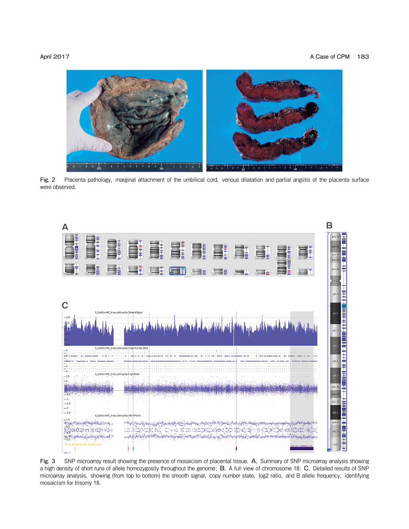
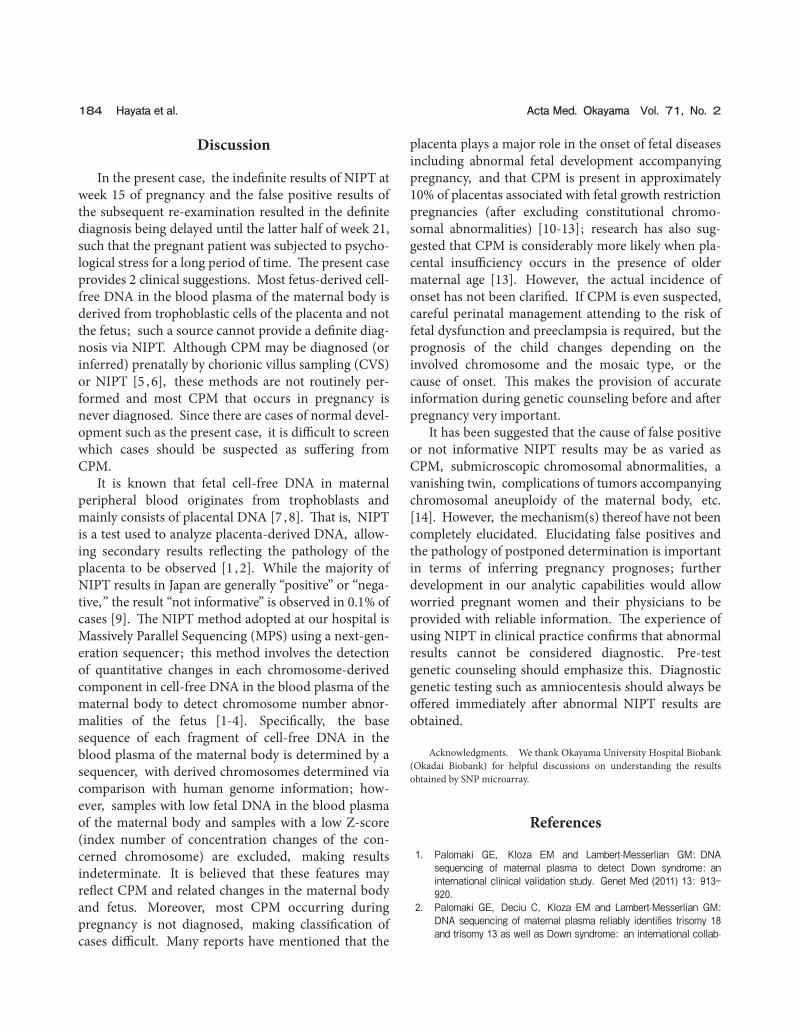
causes placental insufficiency, leading to fetal growth restriction; therefore, perinatal period management was subsequently carried out at our hospital. Fortunately, fetal development and the course of preg-nancy were both good. She went into labor at 39 weeks and 5 days of pregnancy, and delivered a 3,306 g baby girl whose Apgar scores were 8 and then 9 at 1 and then 5 min, respectively. The child was not observed with any external malformations, and the extracted placenta was subjected to pathologic and chromosomal testing. In the placenta pathology, marginal attachment of the umbilical cord, venous dilatation and partial angiitis of the placenta surface were observed (Fig. 2); however, villus growth corresponding to several weeks was observed with no clear observation of abnormal find-ings. At the same time, regarding chromosomal testing of the placenta, SNP microarray analysis was carried out and arr (18) × 2-3 was found (Fig. 3). The estimated percent mosaicism of cells with trisomy 18 is 40-50%. Because the previous amniocentesis on this pregnancy showed normal female results (46 , XX), the trisomy 18 cells in this microarray analysis likely represent con-fined placental mosaicism.
182 Hayata et al. Acta Med. Okayama Vol. 71, No. 2
6 7 8 9 10 11 12
13 14 15 16 17 18
19 20 21 22 X Y
1 2 3 4 5
Fig. 1 Conventional karyotype analysis of cultured amniocytes shows the fetal karyotype as 46 ,XX.
April 2017 A Case of CPM 183
Fig. 2 Placenta pathology, marginal attachment of the umbilical cord, venous dilatation and partial angiitis of the placenta surface were observed.
A
C
B
Fig. 3 SNP microarray result showing the presence of mosaicism of placental tissue. A, Summary of SNP microarray analysis showing a high density of short runs of allele homozygosity throughout the genome; B, A full view of chromosome 18; C, Detailed results of SNP microarray analysis, showing (from top to bottom) the smooth signal, copy number state, log2 ratio, and B allele frequency, identifying mosaicism for trisomy 18.
Discussion
In the present case, the indefinite results of NIPT at week 15 of pregnancy and the false positive results of the subsequent re-examination resulted in the definite diagnosis being delayed until the latter half of week 21, such that the pregnant patient was subjected to psycho-logical stress for a long period of time. The present case provides 2 clinical suggestions. Most fetus-derived cell-free DNA in the blood plasma of the maternal body is derived from trophoblastic cells of the placenta and not the fetus; such a source cannot provide a definite diag-nosis via NIPT. Although CPM may be diagnosed (or inferred) prenatally by chorionic villus sampling (CVS) or NIPT [5 , 6], these methods are not routinely per-formed and most CPM that occurs in pregnancy is never diagnosed. Since there are cases of normal devel-opment such as the present case, it is difficult to screen which cases should be suspected as suffering from CPM.
It is known that fetal cell-free DNA in maternal peripheral blood originates from trophoblasts and mainly consists of placental DNA [7 , 8]. That is, NIPT is a test used to analyze placenta-derived DNA, allow-ing secondary results reflecting the pathology of the placenta to be observed [1 , 2]. While the majority of NIPT results in Japan are generally “positive” or “nega-tive,” the result “not informative” is observed in 0.1% of cases [9]. The NIPT method adopted at our hospital is Massively Parallel Sequencing (MPS) using a next-gen-eration sequencer; this method involves the detection of quantitative changes in each chromosome-derived component in cell-free DNA in the blood plasma of the maternal body to detect chromosome number abnor-malities of the fetus [1-4]. Specifically, the base sequence of each fragment of cell-free DNA in the blood plasma of the maternal body is determined by a sequencer, with derived chromosomes determined via comparison with human genome information; how-ever, samples with low fetal DNA in the blood plasma of the maternal body and samples with a low Z-score (index number of concentration changes of the con-cerned chromosome) are excluded, making results indeterminate. It is believed that these features may reflect CPM and related changes in the maternal body and fetus. Moreover, most CPM occurring during pregnancy is not diagnosed, making classification of cases difficult. Many reports have mentioned that the
placenta plays a major role in the onset of fetal diseases including abnormal fetal development accompanying pregnancy, and that CPM is present in approximately 10% of placentas associated with fetal growth restriction pregnancies (after excluding constitutional chromo-somal abnormalities) [10-13]; research has also sug-gested that CPM is considerably more likely when pla-cental insufficiency occurs in the presence of older maternal age [13]. However, the actual incidence of onset has not been clarified. If CPM is even suspected, careful perinatal management attending to the risk of fetal dysfunction and preeclampsia is required, but the prognosis of the child changes depending on the involved chromosome and the mosaic type, or the cause of onset. This makes the provision of accurate information during genetic counseling before and after pregnancy very important.
It has been suggested that the cause of false positive or not informative NIPT results may be as varied as CPM, submicroscopic chromosomal abnormalities, a vanishing twin, complications of tumors accompanying chromosomal aneuploidy of the maternal body, etc. [14]. However, the mechanism(s) thereof have not been completely elucidated. Elucidating false positives and the pathology of postponed determination is important in terms of inferring pregnancy prognoses; further development in our analytic capabilities would allow worried pregnant women and their physicians to be provided with reliable information. The experience of using NIPT in clinical practice confirms that abnormal results cannot be considered diagnostic. Pre-test genetic counseling should emphasize this. Diagnostic genetic testing such as amniocentesis should always be offered immediately after abnormal NIPT results are obtained.
Acknowledgments. We thank Okayama University Hospital Biobank (Okadai Biobank) for helpful discussions on understanding the results obtained by SNP microarray.
References
1. Palomaki GE, Kloza EM and Lambert-Messerlian GM: DNA sequencing of maternal plasma to detect Down syndrome: an international clinical validation study. Genet Med (2011) 13: 913-920.
2. Palomaki GE, Deciu C, Kloza EM and Lambert-Messerlian GM: DNA sequencing of maternal plasma reliably identifies trisomy 18 and trisomy 13 as well as Down syndrome: an international collab-
184 Hayata et al. Acta Med. Okayama Vol. 71, No. 2

orative study. Genet Med (2012) 14: 296-305. 3. Mazloom AR, Dzakula Z and Oeth P: Noninvasive prenatal detec-
tion of sex chromosomal aneuploidies by sequencing circulating cell-free DNA from maternal plasma. Prenat Diagn (2013) 33: 591-597.
4. Bianchi DW, Platt LD and Goldberg JD: Genome-wide fetal aneu-ploidy detection by maternal plasma DNA sequencing. Obstet Gynecol (2012) 119: 890-901.
5. Mao J, Wang T and Wang BJ: Confined placental origin of the cir-culating cell free fetal DNA revealed by a discordant non-invasive prenatal test result in a trisomy 18 pregnancy. Clin Chim Acta (2014) 433: 190-193.
6. Grati FR, Malvestiti F and Ferreira JC: Fetoplacental mosa-icism: potential implications for false-positive and false-negative noninvasive prenatal screening results. Genet Med (2014) 16: 620-624.
7. Alberry M, Maddocks D and Jones M: Free fetal DNA in maternal plasma in anembryonic pregnancies: confirmation that the origin is the trophoblast. Prenat Diagn (2007) 27: 415-418.
8. Bianchi DW: Circulating fetal DNA: its origin and diagnostic
potential-a review. Placenta (2004) 25: S93-S101. 9. Sago H, Sekizawa A and Japan NIPT consortium: Nationwide
demonstration project of next-generation sequencing of cell-free DNA in maternal plasma in Japan: one-year experience. Prenat Diagn (2015) 35: 331-336.
10. Robinson WP, Penaherrera MS and Jiang R: Assessing the role of placental trisomy in preeclampsia and intrauterine growth restric-tion. Prenat Diagn (2010) 30: 1-8.
11. Miura K, Yoshiura K and Miura S: Clinical outcome of infants with confined placental mosaicism and intrauterine growth restriction of unknown cause. Am J Med Genet A (2006) 140A: 1827-1833.
12. Lestou VS, Desilets V and Lomax BL: Comparative genomic hybridization: a new approach to screening for intrauterine com-plete or mosaic aneuploidy. Am J Med Genet (2000) 92: 281-284.
13. Wilkins-Haug L, Quade B and Morton CC: Confined placental mosaicism as a risk factor among newborns with fetal growth restriction. Prenat Diagn (2006) 26: 428-432.
14. Wapner RJ, Martin CL and Levy B: chromosomal microarray ver-sus karyotyping for prenatal diagnosis. N Engl J Med (2012) 367: 2175-2184.
April 2017 A Case of CPM 185