PP4 and PP2A regulate Hedgehog signaling by controlling Smo and ...
10
307 RESEARCH ARTICLE INTRODUCTION The hedgehog (hh) family members control many aspects of development in both vertebrates and invertebrates (Jia and Jiang, 2006). Abnormal activation of Hh pathway has been observed in several types of human cancers (Pasca di Magliano and Hebrok, 2003; Taipale and Beachy, 2001). The Hh signal is transduced through a reception system that includes the transmembrane proteins Patched (Ptc) and Smo. The main outcome of Hh signaling is the modulation of transcriptional responses via the Ci/Gli family of Zn- finger transcription factors. Hh signaling regulates the balance between the transcriptional activator and repressor forms of Ci/Gli (Lum and Beachy, 2004). In Drosophila, the absence of Hh allows Ptc to inhibit Smo signaling activity. In the cytoplasm, the kinesin- related protein Costal 2 (Cos2) recruits multiple kinases, including PKA, CK1 and GSK3 to sequentially phosphorylate full-length Ci (Ci FL ) (Zhang et al., 2005), which creates binding sites for the SCF ubiquitin ligase containing the F-Box protein Slimb (Jia et al., 2005; Smelkinson and Kalderon, 2006), leading to Ub/proteasome- mediated processing into a truncated repressor form (Ci REP ). Ci REP functions as a repressor to block the expression of Hh responsive genes such as decapentaplegic (dpp), as well as hh itself (Aza-Blanc et al., 1997; Methot and Basler, 1999). The presence of Hh relieves the inhibition of Ptc on Smo, inducing Smo cell-surface accumulation and phosphorylation (Denef et al., 2000) by kinases including PKA and CK1 (Apionishev et al., 2005; Jia et al., 2004; Zhang et al., 2004). Smo phosphorylation by PKA and CK1 appears to activate Smo by inducing a conformational switch (Zhao et al., 2007), as well as promoting Smo cell surface accumulation(Jia et al., 2004). In addition, peak levels of Hh promote Smo hyperphosphorylation that is modulated by a feedback loop involving downstream components Cos2 and Fused (Fu) (Claret et al., 2007; Liu et al., 2007). Activated Smo blocks Ci FL phosphorylation and proteolytic processing required for generating Ci REP , and further promotes nuclear translocation and activation of accumulated Ci FL (Chen et al., 1999; Methot and Basler, 2000; Wang et al., 2000; Wang and Holmgren, 2000). The Drosophila wing disc has been used as an excellent model to study the Hh signal transduction. Posterior (P) compartment cells in the wing discs secrete Hh protein that moves into the anterior (A) compartment and induces the expression of Hh target genes, such as dpp, ptc and engrailed (en), which can be used to monitor the levels of Hh signaling activity (Jia and Jiang, 2006). Ci is produced in A- compartment cells but not P-compartment cells, whereas Smo is expressed in the whole wing but accumulated in P-compartment cells as well as A-compartment cells near the AP boundary, where there is Hh-mediated stimulation. Phosphorylation of Smo and Ci has been shown to be the major post-translational event that regulates their signaling activities, but how their phosphorylation is regulated is still poorly understood. Levels of cellular protein phosphorylation are often modulated by the opposing action of protein kinases and phosphatases. Phosphatases are typically classified into two main groups, the Serine/Threonine (Ser/Thr) protein phosphatases (STPs) and protein tyrosine phosphatases (PTPs). STPs can be subdivided into the PPP and PPM families based on distinct amino acid sequences and crystal structures (Cohen, 1997). In the Hh signaling cascade, multiple Ser/Thr kinases are involved, including PKA, GSK3 and CK1 family members. Even though PP2A has been implicated as a positive regulator in Hh signaling (Casso et al., 2008; Nybakken et al., 2005), its relevant substrates remained undetermined. Thus, it is not clear whether phosphatases are involved in regulating Smo and Ci phosphorylation, and if so which phosphatases are responsible. PP4 and PP2A regulate Hedgehog signaling by controlling Smo and Ci phosphorylation Hongge Jia, Yajuan Liu, Wei Yan and Jianhang Jia* The seven-transmembrane protein Smoothened (Smo) and Zn-finger transcription factor Ci/Gli are crucial components in Hedgehog (Hh) signal transduction that mediates a variety of processes in animal development. In Drosophila, multiple kinases have been identified to regulate Hh signaling by phosphorylating Smo and Ci; however, the phosphatase(s) involved remain obscured. Using an in vivo RNAi screen, we identified PP4 and PP2A as phosphatases that influence Hh signaling by regulating Smo and Ci, respectively. RNAi knockdown of PP4, but not of PP2A, elevates Smo phosphorylation and accumulation, leading to increased Hh signaling activity. Deletion of a PP4-interaction domain (amino acids 626-678) in Smo promotes Smo phosphorylation and signaling activity. We further find that PP4 regulates the Hh-induced Smo cell-surface accumulation. Mechanistically, we show that Hh downregulates Smo-PP4 interaction that is mediated by Cos2. We also provide evidence that PP2A is a Ci phosphatase. Inactivating PP2A regulatory subunit (Wdb) by RNAi or by loss-of-function mutation downregulates, whereas overexpressing regulatory subunit upregulates, the level and thus signaling activity of full-length Ci. Furthermore, we find that Wdb counteracts kinases to prevent Ci phosphorylation. Finally, we have obtained evidence that Wdb attenuates Ci processing probably by dephosphorylating Ci. Taken together, our results suggest that PP4 and PP2A are two phosphatases that act at different positions of the Hh signaling cascade. KEY WORDS: Smo, Ci, PP4, PP2A, Hh, Signal transduction, Drosophila Development 136, 307-316 (2009) doi:10.1242/dev.030015 Sealy Center for Cancer Cell Biology, Department of Biochemistry and Molecular Biology, University of Texas Medical Branch, Galveston, TX 77555, USA *Author for correspondence (e-mail: [email protected]) Accepted 11 November 2008 DEVELOPMENT
Transcript of PP4 and PP2A regulate Hedgehog signaling by controlling Smo and ...

307RESEARCH ARTICLE
INTRODUCTIONThe hedgehog (hh) family members control many aspects ofdevelopment in both vertebrates and invertebrates (Jia and Jiang,2006). Abnormal activation of Hh pathway has been observed inseveral types of human cancers (Pasca di Magliano and Hebrok,2003; Taipale and Beachy, 2001). The Hh signal is transducedthrough a reception system that includes the transmembrane proteinsPatched (Ptc) and Smo. The main outcome of Hh signaling is themodulation of transcriptional responses via the Ci/Gli family of Zn-finger transcription factors. Hh signaling regulates the balancebetween the transcriptional activator and repressor forms of Ci/Gli(Lum and Beachy, 2004). In Drosophila, the absence of Hh allowsPtc to inhibit Smo signaling activity. In the cytoplasm, the kinesin-related protein Costal 2 (Cos2) recruits multiple kinases, includingPKA, CK1 and GSK3 to sequentially phosphorylate full-length Ci(CiFL) (Zhang et al., 2005), which creates binding sites for the SCFubiquitin ligase containing the F-Box protein Slimb (Jia et al., 2005;Smelkinson and Kalderon, 2006), leading to Ub/proteasome-mediated processing into a truncated repressor form (CiREP). CiREP
functions as a repressor to block the expression of Hh responsivegenes such as decapentaplegic (dpp), as well as hh itself (Aza-Blancet al., 1997; Methot and Basler, 1999). The presence of Hh relievesthe inhibition of Ptc on Smo, inducing Smo cell-surfaceaccumulation and phosphorylation (Denef et al., 2000) by kinasesincluding PKA and CK1 (Apionishev et al., 2005; Jia et al., 2004;Zhang et al., 2004). Smo phosphorylation by PKA and CK1 appearsto activate Smo by inducing a conformational switch (Zhao et al.,2007), as well as promoting Smo cell surface accumulation(Jia et al.,
2004). In addition, peak levels of Hh promote Smohyperphosphorylation that is modulated by a feedback loopinvolving downstream components Cos2 and Fused (Fu) (Claretet al., 2007; Liu et al., 2007). Activated Smo blocks CiFL
phosphorylation and proteolytic processing required for generatingCiREP, and further promotes nuclear translocation and activation ofaccumulated CiFL (Chen et al., 1999; Methot and Basler, 2000;Wang et al., 2000; Wang and Holmgren, 2000).
The Drosophila wing disc has been used as an excellent model tostudy the Hh signal transduction. Posterior (P) compartment cells inthe wing discs secrete Hh protein that moves into the anterior (A)compartment and induces the expression of Hh target genes, such asdpp, ptc and engrailed (en), which can be used to monitor the levelsof Hh signaling activity (Jia and Jiang, 2006). Ci is produced in A-compartment cells but not P-compartment cells, whereas Smo isexpressed in the whole wing but accumulated in P-compartmentcells as well as A-compartment cells near the AP boundary, wherethere is Hh-mediated stimulation. Phosphorylation of Smo and Cihas been shown to be the major post-translational event thatregulates their signaling activities, but how their phosphorylation isregulated is still poorly understood.
Levels of cellular protein phosphorylation are often modulatedby the opposing action of protein kinases and phosphatases.Phosphatases are typically classified into two main groups, theSerine/Threonine (Ser/Thr) protein phosphatases (STPs) andprotein tyrosine phosphatases (PTPs). STPs can be subdividedinto the PPP and PPM families based on distinct amino acidsequences and crystal structures (Cohen, 1997). In the Hhsignaling cascade, multiple Ser/Thr kinases are involved,including PKA, GSK3 and CK1 family members. Even thoughPP2A has been implicated as a positive regulator in Hh signaling(Casso et al., 2008; Nybakken et al., 2005), its relevant substratesremained undetermined. Thus, it is not clear whetherphosphatases are involved in regulating Smo and Ciphosphorylation, and if so which phosphatases are responsible.
PP4 and PP2A regulate Hedgehog signaling by controllingSmo and Ci phosphorylationHongge Jia, Yajuan Liu, Wei Yan and Jianhang Jia*
The seven-transmembrane protein Smoothened (Smo) and Zn-finger transcription factor Ci/Gli are crucial components in Hedgehog(Hh) signal transduction that mediates a variety of processes in animal development. In Drosophila, multiple kinases have beenidentified to regulate Hh signaling by phosphorylating Smo and Ci; however, the phosphatase(s) involved remain obscured. Usingan in vivo RNAi screen, we identified PP4 and PP2A as phosphatases that influence Hh signaling by regulating Smo and Ci,respectively. RNAi knockdown of PP4, but not of PP2A, elevates Smo phosphorylation and accumulation, leading to increased Hhsignaling activity. Deletion of a PP4-interaction domain (amino acids 626-678) in Smo promotes Smo phosphorylation and signalingactivity. We further find that PP4 regulates the Hh-induced Smo cell-surface accumulation. Mechanistically, we show that Hhdownregulates Smo-PP4 interaction that is mediated by Cos2. We also provide evidence that PP2A is a Ci phosphatase. InactivatingPP2A regulatory subunit (Wdb) by RNAi or by loss-of-function mutation downregulates, whereas overexpressing regulatorysubunit upregulates, the level and thus signaling activity of full-length Ci. Furthermore, we find that Wdb counteracts kinases toprevent Ci phosphorylation. Finally, we have obtained evidence that Wdb attenuates Ci processing probably bydephosphorylating Ci. Taken together, our results suggest that PP4 and PP2A are two phosphatases that act at different positionsof the Hh signaling cascade.
KEY WORDS: Smo, Ci, PP4, PP2A, Hh, Signal transduction, Drosophila
Development 136, 307-316 (2009) doi:10.1242/dev.030015
Sealy Center for Cancer Cell Biology, Department of Biochemistry and MolecularBiology, University of Texas Medical Branch, Galveston, TX 77555, USA
*Author for correspondence (e-mail: [email protected])
Accepted 11 November 2008 DEVELO
PMENT

308
In this study, we performed an in vivo RNAi screen with theRNAi library from VDRC (Vienna Drosophila RNAi Center)(Dietzl et al., 2007) targeting the catalytic subunits of the STPs inthe Drosophila genome (Morrison et al., 2000), in which weidentified PP4 and PP2A as phosphatases that regulate Smoand CiFL phosphorylation, respectively. We found that Smophosphorylation is elevated by RNAi knockdown of PP4 or byabolishing Smo-PP4 interaction. We also found that the signalingactivity of CiFL is positively regulated by PP2A. We providedevidence that PP2A prevents CiFL proteolytic processing bydephosphorylating CiFL.
MATERIALS AND METHODSMutants and transgenesWdbIP is a hypomorphic allele (Hannus et al., 2002). smo3 is a null allele(Chen and Struhl, 1998). In vivo RNAi library has been described (Dietzl etal., 2007). MS1096, act>CD2>Gal4, ap-Gal4, UAS-P35, UAS-HA-CiFL,UAS-mC*, UAS-CRL, dpp-lacZ, ptc-lacZ and hh-lacZ have been described(Jia et al., 2003; Jia et al., 2005; Liu et al., 2007; Zhang et al., 2006)(http://flybase.bio.indiana.edu). We constructed 2XHA-UAST and 2XFlag-UAST backbones to generate UAS-HA- or UAS-Flag-tagged PP4(CG18339), PP4R (PP4 regulatory subunit, CG2890), Mts (PP2A catalyticsubunit, CG7109), Wdb (PP2A regulatory subunit, CG5643) and Tws(PP2A regulatory subunit, CG6235). We obtained full-length cDNAs eitherfrom DGRC or by RT-PCR from fly embryonic RNA. Myc-Smo, Myc-SmoCT, Myc-Smo�CT and HA-Cos2 have been described (Jia et al., 2003;Wang et al., 2000). We swapped each corresponding sequence of SmoCTtruncations and internal deletions (Jia et al., 2003) into the 3XFlag-UASTbackbone to raise the affinity for western blot. We constructed Flag-CiFL byswapping the Ci coding sequence from HA-CiFL (Wang et al., 1999) into the3XFlag-UAST. HA-PP4 transformants were generated by standard P-element mediated transformation. Multiple independent lines wereexamined. EP3559, UAS-Wdb, UAS-Tws, UAS-Mts, UAS-DN-Mts fly strainshave been described (Hannus et al., 2002; Mayer-Jaekel et al., 1993;Sathyanarayanan et al., 2004). To construct attB-UAS-Myc-Smo and attB-UAS-Myc-Smo�626-678, the attB sequence (Bateman et al., 2006) wasinserted in upstream of the UAS-binding sites in pUAST, then Myc-Smo orMyc-Smo�626-678 sequences were inserted. The vas-phi-zh2A-VK5 flies(gift from Dr Hugo Bellen) were used to generate UAS-Myc-Smo and UAS-Myc-Smo�626-678 transgenes at the 75B1 attP locus. Genotypes forgenerating clones are as follows: wdbIP clones, yw hsp-flp/+ or Y; FRT82wdbIP/FRT82 hs-GFP; smo clones expressing CiFL or co-expressing CiFL
with Wdb, y MS1096 hsp-flp1/yw or Y; smo3 FRT40/hs-GFP FRT40; UAS-HA-Ci or UAS-HA-Ci with UAS-Wdb/hh-lacZ.
Cell culture, transfection, immunoprecipitation, western blotanalysis and GST fusion protein pull-downS2 cell culture, transfection, immunoprecipitation and immunoblot analysiswere performed with standard protocols (Liu et al., 2007). Smo cell-surfaceaccumulation was detected by immunostaining with anti-SmoN antibodybefore cell permeabilization (Jia et al., 2004). The intensity of cell-surfaceor total Smo was analyzed by Metamorph software. To target eachphosphatase gene with less than 17 nucleotide contiguous off-targetsequence, we synthesized Mts, Wdb, Tws, PP4 and PP4R dsRNA againstthe cDNA regions of 301-900, 681-1236, 761-1360, 304-921 and 791-1288,respectively (Chen et al., 2007). Cos2 and GFP dsRNA synthesis and themethod for RNAi in S2 cells have been described (Liu et al., 2007). OA(Calbiochem) treatment was used to inhibit both PP4 and PP2A (Cohen etal., 1990) at a final concentration of 50 nM for 3 hours before harvesting thecells. GST-Smo557-686 fusion protein pull-down has been described (Liuet al., 2007). His-Cos2MB and His-Cos2CT were constructed by fusingCos2 corresponding sequence to the pET30 vector, expressed in E. coli, andpurified with the His resins (Clontech). Antibodies used in this study weremouse anti-Cos2 (gift from D. Robbins), anti-Flag, M2 (Sigma), anti-GFP(Chemicon), anti-HA, F7 (Santa Cruz), anti-His, 4D11 (Upstate), anti-Myc,9E10 (Santa Cruz), anti-SmoN (DSHB), anti-β-tubulin (DSHB); and rabbitanti-HA, Y-11 (Santa Cruz) and anti-GST (Santa Cruz).
Immunostaining of imaginal discsStandard protocols for immunofluorescence staining of imaginal discs wereused with the antibodies mouse anti-Myc, 9E10 (Santa Cruz), anti-HA, F7(Santa Cruz), anti-Flag, M2 (Sigma), anti-SmoN (DSHB), anti-Ptc (DSHB),anti-CD2 (Serotec); rabbit anti-Flag (ABR), anti-HA, Y-11 (Santa Cruz),anti-βGal (Cappel), anti-GFP (Clontech); and rat anti-Ci 2A (gift from R.Holmgren). MG132 (100 μM; Calbiochem) in M3 medium (Sigma) wasused to treat wing discs for up to 6 hours before immunostaining.
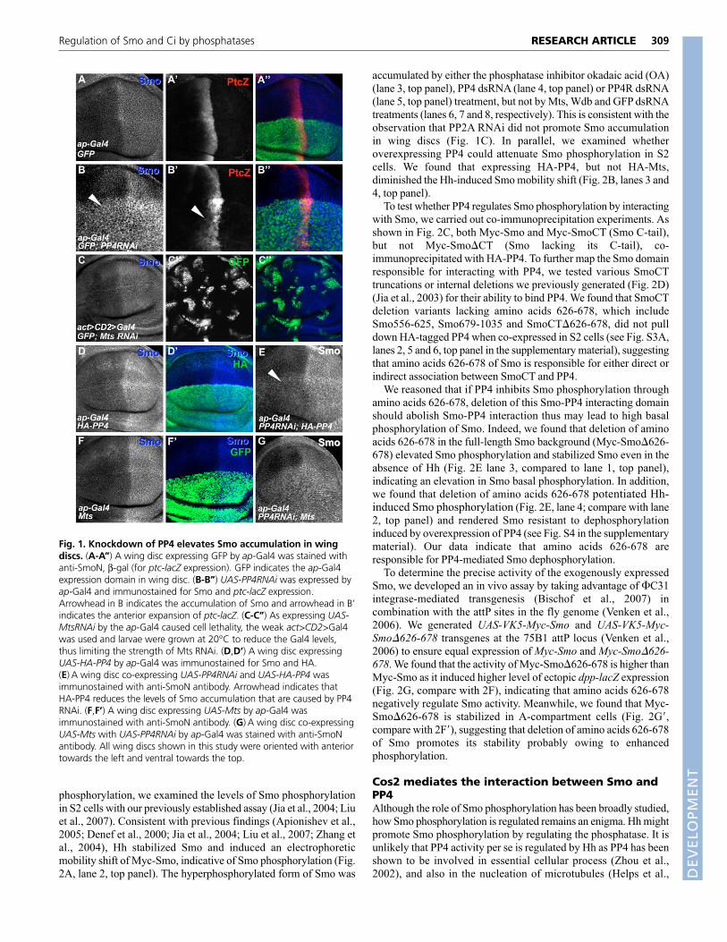
RESULTSIdentification of Smo phosphatase by in vivoRNAi screenIn response to Hh stimulation, many Hh pathway components arephosphorylated and a number of kinases have been identified.However, the phosphatase(s) involved have remained elusive. Toexplore whether phosphatases are also involved, we obtained 45RNAi lines from the VDRC, targeting 26 catalytic subunits of theSTPs (Morrison et al., 2000). We performed in vivo screening byoverexpressing individual RNAi lines via the wing-specific MS1096Gal4 to determine whether they induced any adult wing phenotypes.We found that RNAi of eight genes affected wing developmentindicated by wing phenotypes or wing blisters (data not shown). Alleight genes encode members of the PPP family of phosphatase. Thefact that increased Smo phosphorylation stabilizes Smo in wingdiscs (Denef et al., 2000; Jia et al., 2004) suggests that modulationof Smo phosphatase might alter Smo levels. We thus performed thenext round of screening with a direct approach to detect Smochanges when a specific phosphatase was knocked down by RNAi.Each UAS-RNAi line targeting the eight candidates was expressedvia the dorsal compartment-specific ap-Gal4, and wing discs fromlate third instar larvae were immunostained with anti-SmoNantibody. Knockdown of PP4 by RNAi induced Smo accumulation(Fig. 1B, compared with wild-type disc staining in Fig. 1A).Moreover, expressing UAS-PP4RNAi caused elevated expressionand anterior expansion of Hh target genes such as ptc-lacZ in dorsalbut not ventral compartment cells (Fig. 1B�), a phenotype similar tothat caused by overexpressing PKA (Jia et al., 2004; Liu et al.,2007). These observations indicate that PP4 exerts a negativeinfluence on Hh signaling in responding cells, probably by reducingSmo phosphorylation. RNAi of other phosphatases including Mtsdid not elevate Smo (Fig. 1C-C�; data not shown).
UAS-PP4 RNAi shares a 22-nucleotide contiguous sequence withCanA1 phosphatase (CG1455; see supplementary information).However, CanA1RNAi had no effects on Smo accumulation in wingdisc (see Fig. S1 in the supplementary material), suggesting theaccumulation of Smo by PP4 RNAi in wing discs (Fig. 1B) was dueto the downregulation of PP4 activity. To further examine thespecificity of PP4 RNAi, we tested whether the overexpressed PP4could rescue its RNAi phenotype. As shown in Fig. 1E, co-expressing UAS-HA-PP4 attenuated the Smo elevation caused byPP4 RNAi. By contrast, co-expressing UAS-Mts did not alleviatePP4 RNAi-induced Smo accumulation (Fig. 1G), indicating thespecificity of PP4 RNAi. Overexpression of UAS-HA-PP4 or UAS-Mts alone did not downregulate Smo accumulation in wing discs(Fig. 1D,F). Taken together, our data suggest that PP4 blocks Smoaccumulation and downregulates Hh signaling activity.
PP4 downregulates Smo phosphorylationPP4 is a highly conserved carboxymethylated protein that belongsto the PP2A family of STPs. Drosophila PP4 shares 91.5% identitywith human PP4. To further determine whether Smo elevationinduced by PP4 RNAi (Fig. 1B) was due to enhanced Smo
RESEARCH ARTICLE Development 136 (2)
DEVELO
PMENT
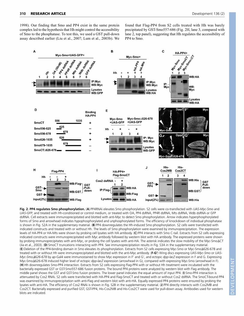
phosphorylation, we examined the levels of Smo phosphorylationin S2 cells with our previously established assay (Jia et al., 2004; Liuet al., 2007). Consistent with previous findings (Apionishev et al.,2005; Denef et al., 2000; Jia et al., 2004; Liu et al., 2007; Zhang etal., 2004), Hh stabilized Smo and induced an electrophoreticmobility shift of Myc-Smo, indicative of Smo phosphorylation (Fig.2A, lane 2, top panel). The hyperphosphorylated form of Smo was
accumulated by either the phosphatase inhibitor okadaic acid (OA)(lane 3, top panel), PP4 dsRNA (lane 4, top panel) or PP4R dsRNA(lane 5, top panel) treatment, but not by Mts, Wdb and GFP dsRNAtreatments (lanes 6, 7 and 8, respectively). This is consistent with theobservation that PP2A RNAi did not promote Smo accumulationin wing discs (Fig. 1C). In parallel, we examined whetheroverexpressing PP4 could attenuate Smo phosphorylation in S2cells. We found that expressing HA-PP4, but not HA-Mts,diminished the Hh-induced Smo mobility shift (Fig. 2B, lanes 3 and4, top panel).
To test whether PP4 regulates Smo phosphorylation by interactingwith Smo, we carried out co-immunoprecipitation experiments. Asshown in Fig. 2C, both Myc-Smo and Myc-SmoCT (Smo C-tail),but not Myc-Smo�CT (Smo lacking its C-tail), co-immunoprecipitated with HA-PP4. To further map the Smo domainresponsible for interacting with PP4, we tested various SmoCTtruncations or internal deletions we previously generated (Fig. 2D)(Jia et al., 2003) for their ability to bind PP4. We found that SmoCTdeletion variants lacking amino acids 626-678, which includeSmo556-625, Smo679-1035 and SmoCT�626-678, did not pulldown HA-tagged PP4 when co-expressed in S2 cells (see Fig. S3A,lanes 2, 5 and 6, top panel in the supplementary material), suggestingthat amino acids 626-678 of Smo is responsible for either direct orindirect association between SmoCT and PP4.
We reasoned that if PP4 inhibits Smo phosphorylation throughamino acids 626-678, deletion of this Smo-PP4 interacting domainshould abolish Smo-PP4 interaction thus may lead to high basalphosphorylation of Smo. Indeed, we found that deletion of aminoacids 626-678 in the full-length Smo background (Myc-Smo�626-678) elevated Smo phosphorylation and stabilized Smo even in theabsence of Hh (Fig. 2E lane 3, compared to lane 1, top panel),indicating an elevation in Smo basal phosphorylation. In addition,we found that deletion of amino acids 626-678 potentiated Hh-induced Smo phosphorylation (Fig. 2E, lane 4; compare with lane2, top panel) and rendered Smo resistant to dephosphorylationinduced by overexpression of PP4 (see Fig. S4 in the supplementarymaterial). Our data indicate that amino acids 626-678 areresponsible for PP4-mediated Smo dephosphorylation.
To determine the precise activity of the exogenously expressedSmo, we developed an in vivo assay by taking advantage of �C31integrase-mediated transgenesis (Bischof et al., 2007) incombination with the attP sites in the fly genome (Venken et al.,2006). We generated UAS-VK5-Myc-Smo and UAS-VK5-Myc-Smo�626-678 transgenes at the 75B1 attP locus (Venken et al.,2006) to ensure equal expression of Myc-Smo and Myc-Smo�626-678. We found that the activity of Myc-Smo�626-678 is higher thanMyc-Smo as it induced higher level of ectopic dpp-lacZ expression(Fig. 2G, compare with 2F), indicating that amino acids 626-678negatively regulate Smo activity. Meanwhile, we found that Myc-Smo�626-678 is stabilized in A-compartment cells (Fig. 2G�,compare with 2F�), suggesting that deletion of amino acids 626-678of Smo promotes its stability probably owing to enhancedphosphorylation.
Cos2 mediates the interaction between Smo andPP4Although the role of Smo phosphorylation has been broadly studied,how Smo phosphorylation is regulated remains an enigma. Hh mightpromote Smo phosphorylation by regulating the phosphatase. It isunlikely that PP4 activity per se is regulated by Hh as PP4 has beenshown to be involved in essential cellular process (Zhou et al.,2002), and also in the nucleation of microtubules (Helps et al.,
309RESEARCH ARTICLERegulation of Smo and Ci by phosphatases
Fig. 1. Knockdown of PP4 elevates Smo accumulation in wingdiscs. (A-A’’) A wing disc expressing GFP by ap-Gal4 was stained withanti-SmoN, β-gal (for ptc-lacZ expression). GFP indicates the ap-Gal4expression domain in wing disc. (B-B’’) UAS-PP4RNAi was expressed byap-Gal4 and immunostained for Smo and ptc-lacZ expression.Arrowhead in B indicates the accumulation of Smo and arrowhead in B’indicates the anterior expansion of ptc-lacZ. (C-C’’) As expressing UAS-MtsRNAi by the ap-Gal4 caused cell lethality, the weak act>CD2>Gal4was used and larvae were grown at 20°C to reduce the Gal4 levels,thus limiting the strength of Mts RNAi. (D,D’) A wing disc expressingUAS-HA-PP4 by ap-Gal4 was immunostained for Smo and HA.(E) A wing disc co-expressing UAS-PP4RNAi and UAS-HA-PP4 wasimmunostained with anti-SmoN antibody. Arrowhead indicates thatHA-PP4 reduces the levels of Smo accumulation that are caused by PP4RNAi. (F,F’) A wing disc expressing UAS-Mts by ap-Gal4 wasimmunostained with anti-SmoN antibody. (G) A wing disc co-expressingUAS-Mts with UAS-PP4RNAi by ap-Gal4 was stained with anti-SmoNantibody. All wing discs shown in this study were oriented with anteriortowards the left and ventral towards the top.
DEVELO
PMENT
310
1998). Our finding that Smo and PP4 exist in the same proteincomplex led to the hypothesis that Hh might control the accessibilityof Smo to the phosphatase. To test this, we used a GST pull-downassay described earlier (Liu et al., 2007; Lum et al., 2003b). We
found that Flag-PP4 from S2 cells treated with Hh was barelyprecipitated by GST-Smo557-686 (Fig. 2H, lane 3, compared withlane 2, top panel), suggesting that Hh regulates the accessibility ofPP4 to Smo.
RESEARCH ARTICLE Development 136 (2)
Fig. 2. PP4 regulates Smo phosphorylation. (A) PP4RNAi elevates Smo phosphorylation. S2 cells were co-transfected with UAS-Myc-Smo andUAS-GFP, and treated with Hh-conditioned or control medium, or treated with OA, PP4 dsRNA, PP4R dsRNA, Mts dsRNA, Wdb dsRNA or GFPdsRNA. Cell extracts were immunoprecipitated and blotted with anti-Myc to detect Smo phosphorylation. Arrow indicates hyperphosphorylatedforms of Smo and arrowhead indicates hypophosphorylated and unphosphorylated forms. The efficiency of knockdown of individual phosphataseis shown in Fig. S2A in the supplementary material. (B) PP4 downregulates the Hh-induced Smo phosphorylation. S2 cells were transfected withindicated constructs and treated with or without Hh. The levels of Smo phosphorylation were examined by immunoprecipitation. The expressionlevels of HA-PP4 or HA-Mts were shown by probing cell lysates with HA antibody. (C) PP4 interacts with Smo C-tail. Extracts from S2 cells expressingindicated constructs were immunoprecipitated with Myc antibody followed by western blot with HA antibody. The expressed proteins were shownby probing immunoprecipitates with anti-Myc, or probing the cell lysates with anti-HA. The asterisk indicates the slow mobility of the Myc-Smo�CT(Jia et al., 2003). (D) SmoCT truncations interacting with PP4. See immunoprecipitation results in Fig. S3A in the supplementary material.(E) Deletion of the PP4-binding domain in Smo elevates its phosphorylation. Extracts from S2 cells expressing Myc-Smo or Myc-Smo�626-678 andtreated with or without Hh were immunoprecipitated and blotted with the anti-Myc antibody. (F-G’) Wing discs expressing UAS-Myc-Smo or UAS-Myc-Smo�626-678 by ap-Gal4 were immunostained to show Myc expression in F’ and G’, and ectopic dpp-lacZ expression in F and G. ExpressingMyc-Smo�626-678 induced higher level of ectopic dpp-lacZ expression (arrowhead in G), compared with expressing Myc-Smo (arrowhead in F).(H) Hh downregulates Smo-PP4 interaction. Extracts from S2 cells expressing Flag-PP4 with or without Hh treatment were incubated with thebacterially expressed GST or GST-Smo557-686 fusion proteins. The bound PP4 proteins were analyzed by western blot with Flag antibody. Themiddle panel shows the GST and GST-Smo fusion proteins. The lower panel indicates the equal amount of input PP4. (I) Smo-PP4 interaction isattenuated by Cos2 RNAi. S2 cells were transfected with HA-PP4 and Flag-SmoCT and treated with or without Cos2 dsRNA. The SmoCT-bound PP4was examined by immunoprecipitation with anti-Flag and western blot with anti-HA. Equally expressed PP4 proteins were ensured by probing thelysates with anti-HA. The efficiency of Cos2 RNAi is shown in Fig. S2B in the supplementary material. (J) PP4 directly interacts with Cos2MB andCos2CT. Bacterially expressed and purified GST, GST-PP4, His-Cos2MB and His-Cos2CT were used for pull-down assay. Antibodies used for westernblots are indicated. D
EVELO
PMENT
The Smo domain responsible for PP4 association falls into theCos2-binding domains (amino acids 557-686) (Liu et al., 2007; Lumet al., 2003b). In addition, we have shown that Cos2 has a negativerole in Smo phosphorylation (Liu et al., 2007). These observationsraised the possibility that Cos2 may serve as a scaffold to bridge PP4and Smo. If so, removing Cos2 should decrease the amount of Smo-bound PP4, thus attenuating Smo inhibition by PP4. In support ofthis model, we have found that SmoCT-PP4 interaction is attenuatedby Cos2 RNAi (Fig. 2I, lane 2, compare with lane 1, top panel),suggesting that Cos2 may downregulate Smo phosphorylation byrecruiting the phosphatase or facilitating the interaction betweenSmo and its phosphatase. We then examined the interaction betweenCos2 and PP4 and we found that both Cos2 microtubule-binding(MB) and cargo domain (CT) bind PP4 in our immunoprecipitationassay (see Fig. S3B in the supplementary material, data not shown).We verified PP4 interaction with Cos2 by using an in vitro GST pull-down assay. As shown in Fig. 2J, GST-PP4 fusion protein pulleddown both His-Cos2MB and His-Cos2CT (lanes 2 and 4, top panel),whereas GST protein failed to pull down any His-Cos2 proteins,suggesting that PP4 directly binds Cos2 N- and C-terminal domains.
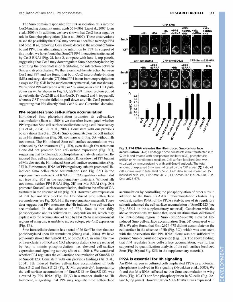
PP4 regulates Smo cell-surface accumulationHh-induced Smo phosphorylation promotes its cell-surfaceaccumulation (Jia et al., 2004); we therefore investigated whetherPP4 regulates Smo cell-surface localization using a cell-based assay(Jia et al., 2004; Liu et al., 2007). Consistent with our previousobservations (Jia et al., 2004), Smo accumulated on the cell surfaceupon Hh stimulation (Fig. 3B, compare with Fig. 3A without Hhtreatment). The Hh-induced Smo cell-surface accumulation wasenhanced by OA treatment (Fig. 3D), even though OA treatmentalone did not promote Smo cell-surface expression (Fig. 3C),suggesting that the blockade of phosphatase activity elevates the Hh-induced Smo cell-surface accumulation. Knockdown of PP4 but notof Mts elevated the Hh-induced Smo cell-surface accumulation (Fig.3F,H). Furthermore, RNAi of PP4 regulatory subunit promoted Hh-induced Smo cell-surface accumulation (see Fig. S5D in thesupplementary material) but RNAi of PP2A regulatory subunit didnot (see Fig. S5F in the supplementary material). Without Hhtreatment, neither PP4 RNAi (Fig. 3E) nor Mts RNAi (Fig. 3G)promoted Smo cell-surface accumulation, similar to the effect of OAtreatment in the absence of Hh (Fig. 3C). However, overexpressionof PP4 but not Mts blocked the Hh-induced Smo cell-surfaceaccumulation (see Fig. S5G,H in the supplementary material). Thesedata suggest that PP4 attenuates the Hh-induced Smo cell-surfaceaccumulation. In the absence of PP4, Smo is not fullyphosphorylated and its activation still depends on Hh, which mayexplain why the accumulation of Smo by PP4 RNAi in anterior-mostregions of wing disc is unable to induce ectopic ptc-lacZ expression(Fig. 1B).
Smo intracellular domain has a total of 26 Ser/Thr sites that arephosphorylated upon Hh stimulation (Zhang et al., 2004). We havepreviously shown that SmoSD12, or SmoSD123, in which the twoor three clusters of PKA and CK1 phosphorylation sites are replacedby Asp to mimic phosphorylation, has elevated cell-surfaceexpression and signaling activity (Jia et al., 2004). We wonderedwhether PP4 regulates the cell-surface accumulation of SmoSD12or SmoSD123. Consistent with our previous findings (Jia et al.,2004), Hh induced further cell-surface accumulation of bothSmoSD12 and SmoSD123 (Fig. 3J,M, respectively). Importantly,the cell-surface accumulation of SmoSD12 or SmoSD123 waselevated by PP4 RNAi (Fig. 3K,N) in a manner similar to Hhtreatment, suggesting that PP4 may regulate Smo cell-surface
accumulation by controlling the phosphorylation of other sites inaddition to the three PKA-CK1 phosphorylation clusters. Bycontrast, neither RNAi of the PP2A catalytic nor of its regulatorysubunit enhanced the cell-surface accumulation of SmoSD123 (seeFig. S5K-L in the supplementary material). Consistent with theabove observations, we found that, upon Hh stimulation, deletion ofthe PP4-binding region in Smo (Smo�626-678) elevated Hh-induced Smo cell-surface accumulation (Fig. 3P, compared with3B). We also found that Smo�626-678 did not accumulate on thecell surface in the absence of Hh (Fig. 3O), which was consistentwith the observation that PP4 RNAi alone was not sufficient topromote Smo cell-surface expression (Fig. 3E). The above finding,that PP4 regulates Smo cell-surface accumulation, was furthersupported by quantification analysis of the cell-surface localizedSmo (Fig. 3Q and Fig. S5N in the supplementary material).
PP2A is essential for Hh signalingAn RNAi screen in cultured cells implicated PP2A as a potentialphosphatase involved in Hh signaling (Nybakken et al., 2005). Wefound that Mts RNAi affected neither Smo accumulation in wingdiscs (Fig. 1C-C�) nor Smo phosphorylation in S2 cells (Fig. 2A,lane 6, top panel). However, when UAS-MtsRNAi was expressed in
311RESEARCH ARTICLERegulation of Smo and Ci by phosphatases
Fig. 3. PP4 RNAi elevates the Hh-induced Smo cell-surfaceaccumulation. (A-P) CFP-tagged Smo constructs were transfected intoS2 cells and treated with phosphatase inhibitor (OA), phosphatasedsRNA or Hh-conditioned medium. Cell-surface-localized Smo wasvisualized by immunostaining with anti-SmoN antibody. The totalamount of expressed Smo was indicated by the CFP signal. (Q) Ratio ofcell surface level to total level of Smo. Each data set was based on 15individual cells. WT, CFP-Smo; SD123, CFP-SmoSD123; �626-678, CFP-Smo �626-678.
DEVELO
PMENT

312
wing discs by the wing-specific MS1096 Gal4, CiFL levels weresignificantly reduced (Fig. 4B) and dpp-lacZ expression diminished(compare Fig. 4B,B� with 4A,A�). Similar results were obtainedwhen a dominant-negative form of Mts (DN-Mts) wasoverexpressed (see Fig. S6B-B� in the supplementary material). Inthese experiments, P35 was co-expressed to prevent cell deathowing to loss of PP2A activity (Zhang et al., 2006).
PP2A functions are mediated largely by the regulatory B subunitthat directs the phosphatase to distinct substrate (Virshup, 2000).Thus, we examined the roles of PP2A regulatory subunits inregulating Hh signaling. We found that Wdb RNAi inhibited theactivity of Hh signaling, indicated by the attenuated CiFL levels (Fig.4C) and reduced dpp-lacZ expression (Fig. 4C�). Knockdown ofTws by RNAi caused early lethality (not shown). We did not observeHh-related defects when knocking down other PP2A regulatorysubunits, including PP2A-B� (CG7913) and PP2A-B� (CG4733)(not shown).
We also used wdb mutants to examine its physiological functionin regulating CiFL. As null mutation of wdb causes cell lethality(Hannus et al., 2002), we used a wdb hypomorphic allele, WdbIP,that harbors a stop codon at amino acid 332, resulting a significantreduction of Wdb activity (Hannus et al., 2002). We examined Smoand CiFL distribution in wing discs carrying clones homozygous forWdbIP and found that CiFL levels were reduced (Fig. 4F) with Smoaccumulation unaffected (Fig. 4F�) in wdb mutant cells. Theseresults suggest that Wdb has a positive role in Hh signaling byregulating Ci and Hh target genes, which is consistent with thefindings in a genetic screen where mts was identified as a positiveregulator in Hh pathway (Casso et al., 2008).
We next performed gain-of-function study and found thatexpressing UAS-Mts or UAS-Wdb by ap-Gal4 elevated CiFL levels(Fig. 4D,E) and induced ectopic dpp-lacZ expression (Fig. 4D�,E�,respectively). By contrast, overexpressing HA-PP4 neither elevatedCiFL levels nor upregulated Hh target genes (see Fig. S7 in thesupplementary material; not shown). Although expressing UAS-Mts
or UAS-Wdb alone induced little, if any, ectopic Ptc expression thatis indicative of high level of Hh signaling activity (see Fig. S8E,F inthe supplementary material), co-expressing UAS-Mts with UAS-Wdb induced ectopic Ptc expression (see Fig. S8G in thesupplementary material). Expressing HA-CiFL induced ectopic ptc-lacZ in the P-compartment but rarely in the A-compartment (Fig.4G); co-expressing Wdb with CiFL induced ectopic ptc-lacZexpression in A-compartment cells away from the AP boundary(Fig. 4H). These results strengthen the view that PP2A-Wdbpositively regulates Hh signaling activity.
Overexpression of Wdb and Mts elevated CiFL levels (Fig. 4D,E;see Fig. S8A,B in the supplementary material) but did not influencethe Hh-induced Smo accumulation (see Fig. S8A�,B� in thesupplementary material). Knockdown of endogenous Wdb by RNAidecreased CiFL levels (see Fig. S8C in the supplementary material),which was in line with the result of Mts RNAi (see Fig. S8D in thesupplementary material). However, we did not observe changes inSmo accumulation when Wdb or Mts was knocked down (see Fig.S8C�,D� in the supplementary material). Consistently, expression ofDN-Mts did not result in Smo accumulation in wing discs (see Fig.S6B� in the supplementary material). Our data indicate that PP2A isinvolved in Hh signaling by specifically regulating CiFL.
Genetic interaction between PP2A and Ci kinasesIn the absence of Hh, CiFL is phosphorylated by multiple kinasesincluding PKA, GSK3 and CK1 (Jia et al., 2005; Smelkinson andKalderon, 2006), leading to its processing to generate CiREP thatblocks the expression of Hh target genes. To determine whetherPP2A positively regulates Hh signaling by counteracting Ci kinases,thus downregulating CiFL phosphorylation, we examined the geneticinteraction between PP2A and kinases in wing discs. Consistent withprevious findings (Jia et al., 2005; Wang et al., 1999),overexpressing the constitutively active form of PKA (mC*) byMS1096 Gal4 attenuated the levels of CiFL (Fig. 5B, compare withwild-type CiFL staining in Fig. 5A) and downregulated the
RESEARCH ARTICLE Development 136 (2)
Fig. 4. PP2A is essential for Hhsignaling. (A,A’) A wild-type wing discwas stained to show the expression ofCiFL and dpp-lacZ. The Ci antibody onlyrecognizes CiFL in wing discs. (B,B’) Awing disc co-expressing UAS-MtsRNAiwith UAS-P35 by MS1096 Gal4 wasimmunostained with Ci and β-galantibodies. Arrowhead in B indicatesthe decreased CiFL levels. Arrowhead inB’ indicates the downregulated dpp-lacZ expression. MS1096 Gal4 isexpressed at lower level in the ventralregion than in the dorsal region of thewing disc (Jia et al., 2003). (C-C’) Awing disc expressing UAS-WdbRNAi byMS1096 Gal4. Arrowhead in Cindicates the decreased CiFL andarrowhead in C� indicates thedownregulated dpp-lacZ. (D-E’) Wingdiscs expressing UAS-Mts or UAS-Wdbby ap-Gal4. Arrowheads in D and Eshow the elevation of CiFL andarrowheads in D’ and E’ show the induced ectopic dpp-LacZ expression. (F-F’’) A wing disc bearing wdbIP homozygous clones that were marked bythe lack of GFP expression was immunostained with anti-Ci and anti-SmoN antibodies. Arrowhead in F shows the reduction of CiFL. Arrowhead in F’shows the unaffected Smo accumulation in wdb mutant cells. (G,H) Wing discs expressing UAS-HA-CiFL alone, or along with UAS-Wdb by C765Gal4. Arrowhead in H indicates the ectopic Ptc-lacZ expression in A-compartment cells. D
EVELO
PMENT

expression of Hh target genes such as ptc-lacZ (Fig. 5B�, comparewith wild-type ptc-lacZ expression in Fig. 5A�). Expressing UAS-Wdb by MS1096 Gal4 elevated CiFL levels (Fig. 5C) but did notaffect ptc-lacZ expression (Fig. 5C�). Strikingly, co-expressing UAS-Wdb with UAS-mC* attenuated the effect of mC*, as evident by therescue of CiFL levels (Fig. 5D) and ptc-lacZ expression near the APboundary (Fig. 5D�), suggesting that Wdb counteracts PKA toregulate CiFL and Hh target genes. We previously generated CK1RNAi transformant, UAS-CRL, which produces dsRNA thatefficiently interferes with the activity of endogenous CK1 (Jia et al.,2005). Expressing UAS-CRL in wing discs caused accumulation ofCiFL (Fig. 5E) (Jia et al., 2005). To determine whether PP2A alsocounteracts CK1 to regulate Ci, we co-expressed UAS-CRL withUAS-MtsRNAi by MS1096 Gal4. We found that the elevation of CiFL
caused by CRL expression was severely restricted by co-expressingUAS-MtsRNAi (Fig. 5F), suggesting that PP2A also counteracts CK1activity in regulating CiFL.
PP2A dephosphorylates Ci and attenuates CiprocessingTo determine whether PP2A regulates Ci phosphorylation, we thenexamined CiFL phosphorylation status when PP2A was knockeddown by RNAi in S2 cells. As shown in Fig. 6, upon OA treatment,CiFL underwent rapid changes in phosphorylation indicated by anelectrophoretic mobility shift (Fig. 6A, lane 2, compare with lane 1,top panel). We found that Flag-CiFL showed low mobility whenendogenous Mts or Wdb was knocked down by RNAi (lanes 3 and4, top panel). By contrast, Flag-CiFL did not exhibit a mobility shiftby PP4 or GFP RNAi (lanes 5 and 6, top panel). These data suggestthat CiFL is specifically regulated by PP2A. To further determinewhether the elevated levels of CiFL produced by overexpressing Mtsor Wdb in wing discs (Fig 4D,E; see Fig. S8A,B in thesupplementary material) were due to changes in CiFL
phosphorylation, we examined whether overexpressingphosphatases could attenuate CiFL phosphorylation in S2 cells. OAtreatment consistently induced the mobility shift of CiFL (Fig. 6B,lane 2; compare with lane 1, top panel). Expressing HA-Mts (Fig.6B, lane3, top panel) or HA-Wdb (lane 4), but not HA-PP4 (lane 5),alleviated the OA-induced CiFL mobility shift, suggesting thatPP2A, but not PP4, prevents CiFL phosphorylation and, thus, theaccumulation in wing discs.
We next asked whether downregulation of Ci phosphorylationaffects its processing. The proteolytic processing of CiFL requiresthe activity of the proteasome. The levels of CiFL in wing discs wereelevated by treatment with the proteasome inhibitor MG132 (Fig.6D, compare with 6C). We found that overexpressing UAS-WdbRNAi by ap-Gal4 destabilized CiFL in dorsal compartment cells(Fig. 6E). However, downregulation of CiFL by Wdb RNAi wasprevented by the treatment with MG132 (Fig. 6F), suggesting thatPP2A acts upstream of the proteasome to dephosphorylate CiFL andpromote CiFL accumulation. Similarly, MG132 treatment preventedthe downregulation of CiFL by Mts RNAi (not shown).
To determine whether the accumulation of CiFL was due to theblockade of CiFL processing, we assessed CiFL processing using anin vivo function assay, in which UAS-HA-CiFL or UAS-HA-CiFL plusUAS-Wdb were misexpressed in the P-compartment wing discscarrying smo mutant clones and hh-lacZ reporter gene. Consistentwith previous findings (Jia et al., 2005), P-compartment smo mutantcells expressing HA-CiFL blocked hh-lacZ expression (Fig. 6G-G�),indicating that CiFL was processed to generate CiREP. By contrast, P-compartment smo mutant cells co-expressing HA-CiFL with Wdbpartially suppressed hh-lacZ expression (Fig. 6H-H�), suggestingthat HA-CiFL is partially blocked to produce CiREP in the presenceof Wdb. We also assessed CiFL processing by immunoprecipitationand western blot analysis. UAS-HA-CiFL was expressed either aloneor along with UAS-Wdb by MS1096 Gal4 in wing discs. HA-CiFL
did not give rise to detectable CiREP when Wdb was co-expressed,whereas HA-CiFL alone was partially processed into CiREP (Fig. 6I).Taken together, our data suggest that PP2A is a positive regulator inHh signaling by inhibiting the phosphorylation and processing ofCiFL.
DISCUSSIONRegulated phosphorylation of Smo and Ci are crucial events inmediating Hh signal transduction. Previous studies have identifiedmultiple Ser/Thr kinases that regulate Hh signaling byphosphorylating Smo and Ci; however, the Smo and Ciphosphatases remain unknown. We are able to identify thephosphatase that regulates Smo phosphorylation by using an in vivoscreen. Our screen differs from the previous screens because we usean in vivo assay to examine Smo expression levels, which is a moredirect readout, and because knockdown of specific phosphatase
313RESEARCH ARTICLERegulation of Smo and Ci by phosphatases
Fig. 5. PP2A counteracts kinases to regulate Ci and Hh target gene expression. (A,A’) A wild-type wing disc was immunostained to showCiFL and ptc-lacZ expression. (B,B’) A wing disc overexpressing UAS-mC* by MS1096 Gal4. Arrowhead in B shows the downregulated CiFL level andarrowhead in B’ shows the attenuated ptc-lacZ expression. (C,C’) A wing disc overexpressing UAS-Wdb by MS1096 Gal4. Arrowhead in C showsthe upregulated CiFL. (D,D’) A wing disc co-expressing UAS-mC* with UAS-Wdb by MS1096 Gal4. Arrowhead in D shows reinstated CiFL andarrowhead in D’ shows the partially restored ptc-lacZ expression. (E,F) Wing discs expressing UAS-CRL or co-expressing UAS-CRL with UAS-MtsRNAiby MS1096 Gal4 and stained to show the levels of CiFL.
DEVELO
PMENT

314
gene(s) involved in Smo dephosphorylation might not affect thepathway activity in a significant way and such gene(s) could havebeen missed in the previous RNAi screens with cultured cells (Lumet al., 2003a; Nybakken et al., 2005). In this study, we haveidentified PP4 as a novel Hh signaling component that regulatesSmo phosphorylation. Our study provides the first evidence for thephysiological Smo and Ci phosphatases, and uncovers theunderlying mechanism of Smo regulation by phosphatase (Fig. 6J).
PP4 and PP2A play distinct roles in the HhpathwayIn this study, we have identified PP4 and PP2A to be negative andpositive regulators in the Hh pathway, and we showed that they exerttheir roles through Smo and Ci, respectively. Are PP4 and PP2A theonly phosphatases in the Hh pathway? Although our data suggest thatPP4 is a phosphatase for Smo, we cannot exclude the possibility ofthe involvement of other phosphatase(s). Hh induces extensive Smophosphorylation at numerous Ser/Thr sites, and multiple kinases are
involved in these phosphorylation events (Apionishev et al., 2005;Jia et al., 2004; Zhang et al., 2004). It might be possible that multiplephosphatases could be involved. In addition, our loss-of-functionstudies on PP2A regulating Ci are not based on null mutations. Thiswas due to the fact that genetic null mutations of the catalytic andregulatory subunits cause cell lethality (Sathyanarayanan et al.,2004). Thus, the results might not be exclusive.
PP4 and regulation of Smo phosphorylationWe showed that removal of PP4 by RNAi in wing discs inducedSmo accumulation in A-compartment cells both near and away fromthe AP boundary (Fig. 1B). In addition, PP4 RNAi induced theelevation and anterior expansion of Hh target gene expression (Fig.1B�). However, the accumulated Smo caused by PP4 RNAi did notectopically activate Hh target genes in cells away from the APboundary (Fig. 1B�, data not shown). In addition, although Smophosphorylation was potentiated by knocking down PP4 orabolishing Smo-PP4 interaction (Fig. 2A,E), the elevated
RESEARCH ARTICLE Development 136 (2)
Fig. 6. PP2A downregulates Ci phosphorylation and blocks Ci proteolytic processing. (A) CiFL phosphorylation is upregulated by PP2A RNAi.S2 cells were transfected with Flag-Ci and treated with OA or indicated dsRNA. Cell extracts were subjected to direct western blot with anti-Flagantibody. Arrow indicates hyperphosphorylated forms of Ci and arrowhead indicates the hypophosphorylated or unphosphorylated forms. β-Tubulin serves as loading control. The knockdown efficiency of individual phosphatase was estimated by the method used for Fig. 2A. (B) PP2Adownregulates CiFL phosphorylation. S2 cells were transfected with Flag-Ci alone or along with indicated HA-tagged phosphatase and treated withor without OA. Cell lysates were probed with anti-Flag or anti-HA antibodies. (C) A disc shows the wild-type CiFL staining. (D) A wing disc shows theCiFL stabilization by the treatment of proteasome inhibitor MG132. (E,F) Wing discs expressing UAS-WdbRNAi by ap-Gal4 were treated with orwithout MG132 and stained to show CiFL. Arrowhead in E indicates the destabilized CiFL by Wdb RNAi. Arrowhead in F indicates that thedestabilized CiFL by Wdb RNAi was restored by MG132 treatment. (G-H’) Wing discs bearing smo3 clones and expressing UAS-HA-CiFL alone oralong with UAS-Wdb by MS1096 Gal4 were stained to show the expression of GFP (green) and hh-lacZ (red). Arrowheads in G and H indicate smo3
clones that are marked by the lack of GFP expression. Arrowheads in G’ and H’ indicate the hh-lacZ expression in smo3 cells. (I) Western blotanalysis of protein extracts from wing discs expressing UAS-HA-CiFL or co-expressing UAS-HA-CiFL with UAS-Wdb using the MS1096 Gal4. Proteinextracts were prepared from 400 wing discs, immunoprecipitated and blotted with HA antibody. (J) A model for the involvement of PP4 and PP2Ain Hh signaling. PP4 negatively regulates Hh signal transduction by antagonizing the phosphorylation of Smo. PP2A positively regulates Hh pathwayby counteracting kinases to downregulate CiFL phosphorylation and attenuate its proteasome-mediated processing.
DEVELO
PMENT

phosphorylation did not suffice to promote Smo cell-surfaceaccumulation (Fig. 3E,O). These data suggest that the basalphosphorylation of Smo regulated by PP4 is not sufficient to activateSmo, and that de novo Smo activation still depends on Hh.
Previous studies have shown that PKA and CK1 are required forHh-induced Smo accumulation and signaling activity.Phosphorylation-deficient forms of Smo (with PKA or CK1 sitesmutated to Ala) are defective in Hh signaling, whereas SmoSD123,the phosphorylation-mimicking Smo, has potent signaling activityand high level of cell-surface accumulation (Jia et al., 2004; Zhanget al., 2004). Thus, the PKA and CK1 sites are apparently crucial inmediating Smo phosphorylation and activation. Hh treatment maycause increased phosphorylation at these sites. In addition to PKAand CK1 sites, there are many other Ser/Thr residues that arephosphorylated upon Hh stimulation (Zhang et al., 2004). Althoughphosphorylation-mimicking mutations at these sites alone did nothave discernible effect on Smo, their phosphorylation couldmodulate the cell-surface accumulation and activity of Smophosphorylated at the three PKA/CK1 sites, which may at least inpart explain why cell-surface accumulation and activity ofSmoSD123 is still regulated by Hh (Jia et al., 2004) (Fig. 3M). Here,we found that removing PP4 alone promoted Smo phosphorylationbut did not elevate the cell-surface accumulation of Smo. It ispossible that high levels of basal Smo phosphorylation in theabsence of PP4 do not reach the threshold for promoting Smo cell-surface accumulation. It is also possible that basal Smophosphorylation mainly occurs at sites other than the crucialPKA/CK1 phosphorylation clusters. In support of this notion, wefound that knockdown PP4 by RNAi promoted SmoSD123 tofurther accumulate on the cell surface in the absence of Hh (Fig. 3N).
How is Smo phosphorylation regulated? Hh may regulate Smophosphorylation by regulating the accessibility of its kinase and/orphosphatase. In this study, we found that Smo interacts with PP4through amino acids 626-678, a region we previously mapped to bea Cos2-interacting domain (Liu et al., 2007). We further found thatSmo-PP4 association diminished when Cos2 was knocked down byRNAi (Fig. 2I). Our previous study revealed that Cos2 impedes Hh-induced Smo phosphorylation by interacting with amino acids 626-678 of Smo and Hh-induced phosphorylation of Cos2 at Ser572dissociates Cos2 from amino acids 626-678 of Smo, therebyalleviating its inhibition on Smo phosphorylation (Liu et al., 2007).In this study, we found that Cos2 inhibits Smo phosphorylation byrecruiting PP4 and Hh promotes Smo phosphorylation by preventingCos2-PP4 complex from binding to amino acids 626-678 of Smo.Smo�626-678, when not interacting with PP4, could still interactwith Cos2 via a Cos2-interaction domain near the Smo C terminus(Jia et al., 2003). The Cos2-binding Smo C terminus might notrecruit PP4. Taken together, our findings suggest that Hh maypromote Smo phosphorylation at least in part by reducing theaccessibility of a phosphatase.
PP2A and regulation of Ci phosphorylationAs stated in the Introduction, phosphorylation of Ci/Gli controls thebalance of its activator and repressor activity. Here, we demonstratea role of PP2A in dephosphorylating Ci and attenuating Ciprocessing. However, it is not known whether Hh regulates PP2A todephosphorylate Ci. Previous studies have shown that Hh interfereswith the Cos2-Ci-kinase protein complex (Zhang et al., 2005). It ispossibly that Hh also regulates Ci phosphatase, or the accessibilityof the phosphatase. Future studies should determine whether PP2Ainteracts with Cos2-Ci and whether such interaction is regulated byHh.
Many aspects of Smo and Ci/Gli regulation are conserved acrossspecies. For example, both Drosophila and mammalian Smoproteins undergo a conformational switch in response to Hhstimulation (Zhao et al., 2007). Ci/Gli proteolysis is mediated bythe same set of kinases and E3 ubiquitin ligases (Jiang, 2006). Inaddition, it has been shown that PP2A is involved in vertebrate Hhsignaling, probably by regulating Gli nuclear localization andactivity (Krauss et al., 2008; Rorick et al., 2007). Therefore, itwould be interesting to determine whether PP4 and PP2A playsimilar roles in regulating phosphorylation of vertebrate Smo andGli.
We thank Drs Suzanne Eaton, David Glover, Amita Sehgal, David Robbins,Robert Holmgren, C.-ting Wu and Jin Jiang for providing valuable reagents;VDRC for fly strains; DGRC for cDNA clones; and DSHB for antibodies. Wethank Dr Hugo Bellen and Koen Venken for vas-phi-zh2A-VK5 fly stock. Weare grateful to Drs Jin Jiang for comments on the manuscript, Leoncio Vergarafor assistance with cell-surface Smo quantification and Tianyan Gao for helpfuldiscussions. This work was supported by grants from the National Institute ofHealth (R01 GM 079684), the American Heart Association and the AmericanCancer Society Institutional Research Award to J.J. Deposited in PMC forrelease after 12 months.
Supplementary materialSupplementary material for this article is available athttp://dev.biologists.org/cgi/content/full/136/2/307/DC1
ReferencesApionishev, S., Katanayeva, N. M., Marks, S. A., Kalderon, D. and
Tomlinson, A. (2005). Drosophila Smoothened phosphorylation sites essentialfor Hedgehog signal transduction. Nat. Cell Biol. 7, 86-92.
Aza-Blanc, P., Ramirez-Weber, F. A., Laget, M. P., Schwartz, C. and Kornberg,T. B. (1997). Proteolysis that is inhibited by hedgehog targets Cubitusinterruptus protein to the nucleus and converts it to a repressor. Cell 89, 1043-1053.
Bateman, J. R., Lee, A. M. and Wu, C. T. (2006). Site-specific transformation ofDrosophila via phiC31 integrase-mediated cassette exchange. Genetics 173,769-777.
Bischof, J., Maeda, R. K., Hediger, M., Karch, F. and Basler, K. (2007). Anoptimized transgenesis system for Drosophila using germ-line-specific phiC31integrases. Proc. Natl. Acad. Sci. USA 104, 3312-3317.
Casso, D. J., Liu, S., Iwaki, D. D., Ogden, S. K. and Kornberg, T. B. (2008). Ascreen for modifiers of hedgehog signaling in Drosophila melanogaster identifiesswm and mts. Genetics 178, 1399-1413.
Chen, C. H., von Kessler, D. P., Park, W., Wang, B., Ma, Y. and Beachy, P. A.(1999). Nuclear trafficking of Cubitus interruptus in the transcriptionalregulation of Hedgehog target gene expression. Cell 98, 305-316.
Chen, F., Archambault, V., Kar, A., Lio, P., D’Avino, P. P., Sinka, R., Lilley, K.,Laue, E. D., Deak, P., Capalbo, L. et al. (2007). Multiple protein phosphatasesare required for mitosis in Drosophila. Curr. Biol. 17, 293-303.
Chen, Y. and Struhl, G. (1998). In vivo evidence that Patched and Smoothenedconstitute distinct binding and transducing components of a Hedgehog receptorcomplex. Development 125, 4943-4948.
Claret, S., Sanial, M. and Plessis, A. (2007). Evidence for a novel feedback loopin the hedgehog pathway involving smoothened and fused. Curr. Biol. 17, 1326-1333.
Cohen, P. T. (1997). Novel protein serine/threonine phosphatases: variety is thespice of life. Trends Biochem. Sci. 22, 245-251.
Cohen, P., Holmes, C. F. and Tsukitani, Y. (1990). Okadaic acid: a new probe forthe study of cellular regulation. Trends Biochem. Sci. 15, 98-102.
Denef, N., Neubuser, D., Perez, L. and Cohen, S. M. (2000). Hedgehog inducesopposite changes in turnover and subcellular localization of patched andsmoothened. Cell 102, 521-531.
Dietzl, G., Chen, D., Schnorrer, F., Su, K. C., Barinova, Y., Fellner, M., Gasser,B., Kinsey, K., Oppel, S., Scheiblauer, S. et al. (2007). A genome-widetransgenic RNAi library for conditional gene inactivation in Drosophila. Nature448, 151-156.
Hannus, M., Feiguin, F., Heisenberg, C. P. and Eaton, S. (2002). Planar cellpolarization requires Widerborst, a B’ regulatory subunit of protein phosphatase2A. Development 129, 3493-3503.
Helps, N. R., Brewis, N. D., Lineruth, K., Davis, T., Kaiser, K. and Cohen, P. T.(1998). Protein phosphatase 4 is an essential enzyme required for organisationof microtubules at centrosomes in Drosophila embryos. J. Cell Sci. 111, 1331-1340.
Jia, J. and Jiang, J. (2006). Decoding the Hedgehog signal in animaldevelopment. Cell Mol. Life Sci. 63, 1249-1265.
315RESEARCH ARTICLERegulation of Smo and Ci by phosphatases
DEVELO
PMENT

316
Jia, J., Tong, C. and Jiang, J. (2003). Smoothened transduces Hedgehog signal byphysically interacting with Costal2/Fused complex through its C-terminal tail.Genes Dev. 17, 2709-2720.
Jia, J., Tong, C., Wang, B., Luo, L. and Jiang, J. (2004). Hedgehog signallingactivity of Smoothened requires phosphorylation by protein kinase A and caseinkinase I. Nature 432, 1045-1050.
Jia, J., Zhang, L., Zhang, Q., Tong, C., Wang, B., Hou, F., Amanai, K. andJiang, J. (2005). Phosphorylation by Double-Time/CKIε and CKIα targetsCubitus Interruptus for Slimb/β-TRCP-mediated proteolytic processing. Dev. Cell9, 819-830.
Jiang, J. (2006). Regulation of Hh/Gli signaling by dual ubiquitin pathways. CellCycle 5, 2457-2463.
Krauss, S., Foerster, J., Schneider, R. and Schweiger, S. (2008). Proteinphosphatase 2A and rapamycin regulate the nuclear localization and activity ofthe transcription factor GLI3. Cancer Res. 68, 4658-4665.
Liu, Y., Cao, X., Jiang, J. and Jia, J. (2007). Fused Costal2 protein complexregulates Hedgehog-induced Smo phosphorylation and cell-surfaceaccumulation. Genes Dev. 21, 1949-1963.
Lum, L. and Beachy, P. A. (2004). The Hedgehog response network: sensors,switches, and routers. Science 304, 1755-1759.
Lum, L., Yao, S., Mozer, B., Rovescalli, A., Von Kessler, D., Nirenberg, M. andBeachy, P. A. (2003a). Identification of Hedgehog pathway components byRNAi in Drosophila cultured cells. Science 299, 2039-2045.
Lum, L., Zhang, C., Oh, S., Mann, R. K., von Kessler, D. P., Taipale, J., Weis-Garcia, F., Gong, R., Wang, B. and Beachy, P. A. (2003b). Hedgehog signaltransduction via Smoothened association with a cytoplasmic complex scaffoldedby the atypical kinesin, Costal-2. Mol. Cell 12, 1261-1274.
Mayer-Jaekel, R. E., Ohkura, H., Gomes, R., Sunkel, C. E., Baumgartner, S.,Hemmings, B. A. and Glover, D. M. (1993). The 55 kd regulatory subunit ofDrosophila protein phosphatase 2A is required for anaphase. Cell 72, 621-633.
Methot, N. and Basler, K. (1999). Hedgehog controls limb development byregulating the activities of distinct transcriptional activator and repressor formsof Cubitus interruptus. Cell 96, 819-831.
Methot, N. and Basler, K. (2000). Suppressor of fused opposes hedgehog signaltransduction by impeding nuclear accumulation of the activator form of Cubitusinterruptus. Development 127, 4001-4010.
Morrison, D. K., Murakami, M. S. and Cleghon, V. (2000). Protein kinases andphosphatases in the Drosophila genome. J. Cell Biol. 150, F57-F62.
Nybakken, K., Vokes, S. A., Lin, T. Y., McMahon, A. P. and Perrimon, N.(2005). A genome-wide RNA interference screen in Drosophila melanogastercells for new components of the Hh signaling pathway. Nat. Genet. 37, 1323-1332.
Pasca di Magliano, M. and Hebrok, M. (2003). Hedgehog signalling in cancerformation and maintenance. Nat. Rev. Cancer 3, 903-911.
Rorick, A. M., Mei, W., Liette, N. L., Phiel, C., El-Hodiri, H. M. and Yang, J.(2007). PP2A:B56epsilon is required for eye induction and eye field separation.Dev. Biol. 302, 477-493.
Sathyanarayanan, S., Zheng, X., Xiao, R. and Sehgal, A. (2004).Posttranslational regulation of Drosophila PERIOD protein by proteinphosphatase 2A. Cell 116, 603-615.
Smelkinson, M. G. and Kalderon, D. (2006). Processing of the Drosophilahedgehog signaling effector Ci-155 to the repressor Ci-75 is mediated by directbinding to the SCF component slimb. Curr. Biol. 16, 110-116.
Taipale, J. and Beachy, P. A. (2001). The Hedgehog and Wnt signalling pathwaysin cancer. Nature 411, 349-354.
Venken, K. J., He, Y., Hoskins, R. A. and Bellen, H. J. (2006). P[acman]: a BACtransgenic platform for targeted insertion of large DNA fragments in D.melanogaster. Science 314, 1747-1751.
Virshup, D. M. (2000). Protein phosphatase 2A: a panoply of enzymes. Curr.Opin. Cell Biol. 12, 180-185.
Wang, G., Wang, B. and Jiang, J. (1999). Protein kinase A antagonizesHedgehog signaling by regulating both the activator and repressor forms ofCubitus interruptus. Genes Dev. 13, 2828-2837.
Wang, G., Amanai, K., Wang, B. and Jiang, J. (2000). Interactions with Costal2and suppressor of fused regulate nuclear translocation and activity of cubitusinterruptus. Genes Dev. 14, 2893-2905.
Wang, Q. T. and Holmgren, R. A. (2000). Nuclear import of cubitus interruptus isregulated by hedgehog via a mechanism distinct from Ci stabilization and Ciactivation. Development 127, 3131-3139.
Zhang, C., Williams, E. H., Guo, Y., Lum, L. and Beachy, P. A. (2004). Extensivephosphorylation of Smoothened in Hedgehog pathway activation. Proc. Natl.Acad. Sci. USA 101, 17900-17907.
Zhang, L., Jia, J., Wang, B., Amanai, K., Wharton, K. A., Jr and Jiang, J.(2006). Regulation of wingless signaling by the CKI family in Drosophila limbdevelopment. Dev. Biol. 299, 221-237.
Zhang, W., Zhao, Y., Tong, C., Wang, G., Wang, B., Jia, J. and Jiang, J. (2005).Hedgehog-regulated Costal2-kinase complexes control phosphorylation andproteolytic processing of Cubitus interruptus. Dev. Cell 8, 267-278.
Zhao, Y., Tong, C. and Jiang, J. (2007). Hedgehog regulates smoothened activityby inducing a conformational switch. Nature 450, 252-258.
Zhou, G., Mihindukulasuriya, K. A., MacCorkle-Chosnek, R. A., Van Hooser,A., Hu, M. C., Brinkley, B. R. and Tan, T. H. (2002). Protein phosphatase 4 isinvolved in tumor necrosis factor-alpha-induced activation of c-Jun N-terminalkinase. J. Biol. Chem. 277, 6391-6398.
RESEARCH ARTICLE Development 136 (2)
DEVELO
PMENT


