POTTER AND PERRY’S Fundamentals of Nursing · 2019-01-15 · Communication and nursing practice...
105
Jackie Crisp Catherine Taylor Clint Douglas Geraldine Rebeiro Fundamentals of Nursing POTTER AND PERRY’S 4 th edition
Transcript of POTTER AND PERRY’S Fundamentals of Nursing · 2019-01-15 · Communication and nursing practice...
Jackie Crisp
Catherine Taylor
Clint Douglas
Geraldine Rebeiro
Fundamentalsof Nursing
POTTER AND PERRY’S
4th edition
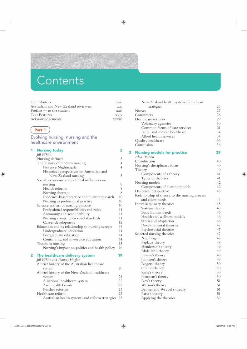
Contributors xviiAustralian and New Zealand reviewers xxiPreface — to the student xxiiText Features xxivAcknowledgements xxviii
Part 1
Evolving nursing: nursing and the healthcare environment
1 Nursing today 2Jill WhiteNursing defined 3The history of modern nursing 4
Florence Nightingale 4Historical perspectives on Australian and
New Zealand nursing 5Social, economic and political inf luences on
nursing 8Health reforms 8Nursing shortage 8Evidence-based practice and nursing research 10Nursing as professional practice 10
Science and art of nursing practice 10Professional responsibilities and roles 11Autonomy and accountability 11Nursing competencies and standards 11Career development 12
Education and its relationship to nursing careers 14Undergraduate education 14Postgraduate education 14Continuing and in-service education 14
Trends in nursing 15Nursing’s impact on politics and health policy 16
2 The healthcare delivery system 19Jill White and Frances HughesA brief history of the Australian healthcare
system 20A brief history of the New Zealand healthcare
system 21A national healthcare system 22Area health boards 22Further reforms 23
Healthcare reform 23Australian health systems and reform strategies 23
New Zealand health system and reform strategies 25
Nurses 27Consumers 28Healthcare services 29
Voluntary agencies 30Common forms of care services 31Rural and remote healthcare 34Allied health services 34
Quality healthcare 36Conclusion 36
3 Nursing models for practice 39Alan PearsonIntroduction 40Nursing’s disciplinary focus 40Theory 40
Components of a theory 41Types of theories 41
Nursing models 42Components of nursing models 42
Historical perspective 43Relationship of theory to the nursing process
and client needs 44Interdisciplinary theories 45
Systems theory 45Basic human needs 46Health and wellness models 46Stress and adaptation 46Developmental theories 47Psychosocial theories 47
Selected nursing theories 47Nightingale 47Peplau’s theory 49Henderson’s theory 49Abdellah’s theory 49Levine’s theory 49Johnson’s theory 49Rogers’ theory 50Orem’s theory 50King’s theory 50Neuman’s theory 50Roy’s theory 51Watson’s theory 51Benner and Wrubel’s theory 51Parse’s theory 51Applying the theories 52
Contents
ch00-i-xxviii-9780729541107.indd 5 31/08/12 4:19 PM

CONTENTSvi
Formulation of the nursing diagnosis 95Nursing diagnosis statement 96Support of the diagnostic statement 98Sources of diagnostic error 98
7 Planning, implementing and evaluating nursing care 100Bronwyn JonesEstablishing priorities 101Critical thinking in establishing goals and
expected outcomes 101Goals of care 101Expected outcomes 103Guidelines for writing goals and expected
outcomes 103Planning nursing care 105
Purpose of care plans 105Care plans in various settings 105Writing the nursing care plan 106Critical (or clinical) pathways 107Protocols and standing orders 107
Critical thinking in designing nursing interventions 111Types of interventions 111Selection of interventions 112
Critical thinking and the implementation process 112Reviewing and revising the existing nursing
care plan 112Organising resources and care delivery 113Implementing nursing interventions 114Achieving a client’s goals of care 115Communicating nursing interventions 116
Critical thinking skills and evaluation of care 116Evaluation of goal achievement 116Care plan revision 118Unmet goals 118
8 Managing client care 121Patricia Mary Davidson and Louise HickmanEvidence to inform nursing practice 122Preparing for complexity 123Chronic care 124Positive practice environments 124The role of the registered nurse 125Models of nursing care 126
Models of nursing care promoting wellness, autonomy and self-care 127
Building a nursing team 127Approaches to delivering nursing care 128Approaches to managing client care 129Communication among the clinical team 131Philosophy and vision for nurses managing
client care 132Leadership skills for nursing students 132Measuring outcomes of nursing care 132Quality improvement processes for nurses 133
Nursing-sensitive indicators 134Skill mix for the student nurse 134
The link between theory and knowledge development in nursing 52
4 Critical inquiry and practice development 55Brendan McCormack and Jackie CrispThree levels of nursing inquiry 56
Inquiry involving critical engagement in everyday practice 56
Inquiry involving collaborative and ongoing evaluation of local practice 58
Inquiry involving nursing research for advancement of nursing knowledge 59
Practice development 63Facilitation of practice development 65Person-centredness and person-centred
practice 66Taking a PEEP 67
Taking a PEEP at people 67Taking a PEEP at practice effects 68Taking a PEEP at impact of environment on
nursing practice 68Taking a PEEP at engagement through praxis 68
The complexity of nursing inquiry 69
Part 2
Framing nursing: critical processes in nursing practice
5 Critical thinking and nursing judgment 74Bronwyn JonesIntroduction 75Critical thinking defined 75
Ref lection 75Intuition 77
Clinical decisions in nursing practice 77Knowledge base 77
Development of critical thinking skills in nursing 79Critical thinking processes 79
Problem solving 79Decision making 80
Clinical judgment model 81Standards for critical thinking 82Critical thinking synthesis 82
6 Nursing assessment and diagnosis 85Bronwyn JonesA critical thinking approach to assessment 86
Organisation of data gathering 87Data collection 88
Types of data 88Sources of data 88Methods of data collection 89
Interview 89Nursing health history 90Physical examination 93
Data documentation 93Analysis and interpretation of data 93
ch00-i-xxviii-9780729541107.indd 6 31/08/12 4:19 PM
CONTENTS vii
Legal relationships in nursing practice 171The law of contract 171The nurse–doctor relationship 172Do no resuscitate orders 173Workload problems 174Floating 174
Legal issues in nursing specialties 175Community health nursing 175Emergency department 175Nursing children 175Medical/surgical nursing and gerontological
nursing 176Critical care nursing 176Perioperative nursing 176Mental health nursing 176Hospital in the home and outreach services 177Remote area nursing 177Professional involvement of nurses 177
11 Legal implications in nursing practice in New Zealand 179Elaine PappsRegulation of nursing in New Zealand 181
Continuing competence and annual practising certificates 181
Regulation of nurses from Australia or other countries 183
Competence notifications and review 183Health notifications 183Complaints about nurses 183Health and Disability Commissioner 184Health Practitioners Disciplinary Tribunal 184
Sources of law 185Legal liability in nursing 185
Treatment injury 185Exemplary damages 185Torts 186Negligence 186Standards of care 186The need for careful documentation 186Confidentiality and privacy 186Obtaining consent 187Use of human tissue and organ donation 188
Legal relationships in relation to employment 188The law of contract 188
Legal issues in nursing specialties 189Nursing children 189Mental health nursing 190
Professional responsibility of nurses 190
12 Communication 193Jane Stein-ParburyCommunication and nursing practice 194The context of nursing practice 194
Why nurses need to communicate 194Healthcare environments and communication 196
Patient-centred communication 196Focusing on solutions 197
Part 3
Positioning nursing: professional responsibilites in nursing practice
9 Ethics and professional practice 138Megan-Jane JohnstoneTerms and concepts 140
Ethics and morality 140Bioethics 140Nursing ethics 141Moral principles 141Moral rules 141Rights 141Moral duties 142
The importance of ethics 142Moral conduct in nursing 143
Moral accountability and responsibility 143Guides to ethical professional conduct 144
Moral theories 144Deontological ethics 144Teleological ethics 145Ethical principlism 145Moral rights theory 146Virtue ethics 147Cross-cultural ethics 147
Nursing codes of ethics 147Moral problems in nursing 148
Nursing point of view 149Distinguishing moral problems from other
kinds of problems 149Identifying and responding effectively to
moral problems in nursing 149Processes of moral decision making 151Bioethical issues in nursing 154
Conclusion 157
10 Legal implications in nursing practice in Australia 160Mary ChiarellaRegulation of nursing 161Legal and professional boundaries of nursing 161
Sources of law 162Legal liability in nursing 162
Torts 163Negligence 163Nursing students 164Standards of care 164The need for careful documentation 166Confidentiality and privacy 166Assault and battery 167The right of the patient to receive
information 168The patient’s right to refuse treatment 169Dying with dignity 169Caring for the dying 171Brain death and organ donation 171
ch00-i-xxviii-9780729541107.indd 7 31/08/12 4:19 PM

CONTENTSviii
Standards 251Types of documentation 251Charting by exception 253Case management and critical pathways 254Common record-keeping forms 254Home healthcare documentation 260Long-term healthcare documentation 261
Computerised documentation 261Reporting 262
Change-of-shift reports 262Telephone reports 264Telephone orders 264Transfer reports 264Incident reports 265
15 Developing a culture of safety and quality 267Geraldine RebeiroScientific knowledge base 268
Environmental safety 268Providing a safe patient environment 269
Nursing knowledge base 273Risks at developmental stages 273Individual risk factors 275Risks in the healthcare agency 275
Critical thinking synthesis 278Safety and the nursing process 278
Assessment 278Nursing diagnosis 279Planning 279Implementation 282Skill 15-1 Applying restraints 290Skill 15-2 Seizure precautions 297Evaluation 298
Part 4
Adapting nursing: nursing across the life span
16 Health and wellness 302Judy Yarwood and Karen BetonyHealth and wellness 303
Social determinants of health 305Wellbeing and wellness 307What determines health and wellbeing? 307
Promoting health and wellness 310Preventive care 310Health promotion at a community and
population level 312Cultural inf luences on health promotion 314
Promoting health in Australia and New Zealand 315
17 Sociocultural considerations and nursing practice 320Leonie Cox and Chris TauaThe context of nursing in Australia and
New Zealand/Aotearoa 321
Effective communication 197Communication and interpersonal relationships 197
Dynamics of interpersonal communication 197Professional nursing relationships 198
Levels of communication 200Intrapersonal communication 200Interpersonal communication 200Small-group communication 200
Forms of communication 201Verbal communication 201Non-verbal communication 201
Developing communication skills 202The need to ‘unlearn’ previous
communication patterns 202Elements of professional communication 204
Courtesy and use of names 204Privacy and confidentiality 204Trustworthiness 204
Communication within the nursing process 204Assessment 204Nursing diagnosis 207Planning 207Implementation 208Evaluation 214
13 Client education 217Trish BurtonPurposes of client education 218
Maintenance and promotion of health and illness prevention 218
Restoration of health 218Coping with impaired functioning 219
Teaching and learning 219Role of the nurse in teaching and learning 220Teaching as communication 220
Domains of learning 222Cognitive learning 222Affective learning 222Psychomotor learning 222
Basic learning principles 223Motivation to learn 223Ability to learn 226Learning environment 228
Integrating the nursing and teaching processes 228Assessment 228Nursing diagnosis 230Planning 231Implementation 235Evaluation 241
Documentation of client teaching 242
14 Documentation 244Pauline CallejaMultidisciplinary communication within the
healthcare team 245Documentation 246
Purposes of records 246Guidelines for high-quality documentation
and reporting 248
ch00-i-xxviii-9780729541107.indd 8 31/08/12 4:19 PM

CONTENTS ix
Moral development theory 370Jean Piaget’s moral development theory 370Lawrence Kohlberg’s moral development
theory 370
20 Conception to adolescence 374Jane Davey, Robyn Galway and Shaun ThompsonGrowth and development 375
Definitions 375Stages of growth and development 375
Critical periods of development 375Major factors inf luencing growth and
development 376Selecting a developmental framework for
nursing 376Conception 376
Intrauterine life 377Transition from intrauterine to extrauterine life 381
Physical changes 381Psychosocial changes 381Other health considerations during
newborn transition 382The newborn 382
Physical changes 382Cognitive changes 385Psychosocial changes 386Other health considerations for newborns 386
The infant 386Physical changes 386Cognitive changes 389Psychosocial changes 389Other health considerations during infancy 390
The toddler 395Physical changes 395Cognitive changes 395Psychosocial changes 395Other health considerations during
toddlerhood 396The preschooler 399
Physical changes 399Cognitive changes 399Psychosocial changes 400
School-age children and adolescents 401Middle childhood 403Preadolescence 409Adolescence 409
21 Young and middle adulthood 421Sue NagyYoung adulthood 423
Physical changes 423Cognitive changes 423Psychosocial changes 423Health risks 426Health concerns 427
Middle adulthood 432Physical changes 432Cognitive changes 434
What is culture? 324The inf luence of whiteness 326The inf luence of class 326Ethnicity—what is it? 327
Worldview and the lifeworld 327What is health? 328Culture shock, culture clash, culture conf lict 330
Culture shock 330Culture clash and culture conf lict 330
Power 332So what has this got to do with nursing? 333
Models of care 334So what is cultural competence? 336
Competence defined 336Ref lecting on self 337Professional nursing regulation and cultural
issues 337Communication skills 338Developing trust 339Negotiating knowledge 340Negotiating outcomes 342
18 Caring for families 346Nicola BrownWhat is a family? 347
Trends in family structure and function in Australia and New Zealand 347
Family theory and models 349Family systems theory 349Family developmental theory 349Family cycle of health and illness model 350Family theory and models: how does this
link to nursing? 350Family-centred care 350Family nursing—what is that? 353
Family as context 353Family as client 353Family as system 353
Family nursing care 354Tools used in family assessment 354
Self-care when working with families 354
19 Developmental theories 358Sue NagyGrowth versus development 359Theories of growth and development 359Biophysical development 360
Gesell’s theory of development 360Genetic theories of ageing 360Non-genetic cellular theories 361Physiological theories of ageing 361
Psychosocial theory 361Sigmund Freud’s psychoanalytical model of
psychosexual development 363Erik Erikson 364Robert Havighurst 368
Cognitive development theory 369Jean Piaget’s theory of cognitive
development 369
ch00-i-xxviii-9780729541107.indd 9 31/08/12 4:19 PM

CONTENTSx
The family’s effect on self-concept development 000
The nurse’s effect on the client’s self-concept 000Altered self-concept 000
Stressors affecting a person’s spirituality 000Spiritual healing 000
Critical thinking synthesis 000Dimensions of self and the nursing process 000
Assessment 000Nursing diagnosis 000Planning 000Implementation 000Evaluation 000
24 Sexual health 000Helen CalabrettoIntroduction 000Sexual development 000
Infancy 000Toddler/preschool period 000School-age years 000Puberty/adolescence 000Adulthood 000Older adulthood 000
Definitions of terms 000Pregnancy 000Abortion 000Current methods of contraception 000
Fertility-awareness based (FAB) methods 000Barrier methods 000Hormonal methods 000Injectable contraception 000Contraceptive implant 000Emergency contraception 000Other contraceptive methods 000Permanent methods of contraception 000
Sexually transmitted infections 000Viruses 000Bacteria 000Parasites 000Prevalence of STIs 000
Circumcision 000Female genital mutilation 000Health promotion activities 000
Testicular cancer 000Prostate cancer 000Cervix cancer (cervical cancer) 000Breast cancer 000Ovarian cancer 000
Talking to clients about sexual issues 000Impact of altered states of health on
sexuality 000Sexual history as part of nursing assessment 000
25 Loss, dying, death and grief 000John RosenbergLoss, grief, bereavement and mourning 000Categories of loss 000
Psychosocial changes 434Health concerns 435
22 Older adulthood 440Susan HuntWorking with older adults—gerontology as
a specialty area 441Older adults as part of our population 442Ageism 443Abuse of the elderly 444Towards an understanding of how we age 445Understanding normal ageing 446
Physiological changes 446Cognitive changes 449Psychosocial changes 450
Assessment of the older adult 450Risks to healthy ageing 450
Risk factors 451Health issues experienced by older people 451
Impaired cognition 451Urinary incontinence 455Constipation and faecal incontinence 455Adverse drug events 456
Successful ageing 456Service provision 457
Home (community care) 458Retirement villages or communities 458Adult day-care 458Respite care 458Subacute care/rehabilitation care 458Residential aged care 458
Health promotion and maintenance: psychosocial health concerns 459Therapeutic communication 459Touch 459Reality orientation 459Validation therapy 460Reminiscence 460Body-image interventions 460
Part 5
Relating nursing: human basis of nursing practice
23 Dimensions of self: pathways to self-identity 000Anthony WelchDimensions of the self 000
Identity 000Body image 000Self-esteem 000Role performance 000Spirituality 000
Development of self-concept 000Stages of development 000Stressors affecting self concept 000Role stressors 000
ch00-i-xxviii-9780729541107.indd 10 31/08/12 4:19 PM

CONTENTS xi
Palpation 000Percussion 000Auscultation 000Olfaction 000
Preparation for examination 000Infection control 000Environment 000Physical preparation of the patient 000Psychological preparation of the patient 000
Assessment of age groups 000Children 000Older adults 000
Organisation of the examination 000General survey 000
General appearance and behaviour 000Vital signs 000Measurement of head and chest
circumference 000Health perception–health management pattern 000
Strengths and problems related to health perception and health management 000
Nutritional–metabolic pattern 000Mouth 000Height and weight 000Skin 000Hair and scalp 000Nails 000Abdomen 000Skill 27-1 Assessment of the abdomen and
gastrointestinal tract 000Activity–exercise pattern 000
Musculoskeletal system 000Skill 27-2 Assessment of the musculoskeletal
system 000Cardiovascular assessment 000Skill 27-3 Assessment of the cardiovascular
and peripheral vascular systems 000Peripheral vascular system 000Respiratory system 000Skill 27-4 Assessment of the respiratory
system 000Cognitive–perceptual pattern 000
Mental and emotional status 000Skill 27-5 Mental state assessment 000Eyes 000Ears 000Sensory function 000Skill 27-6 Assessment of central nervous
system and level of consciousness 000Motor function 000Abnormal findings related to sensory and
motor function 000Sexuality–reproductive pattern 000
Breasts 000External genitalia 000
Value–belief pattern 000Self perception–self concept pattern 000Role–relationships pattern 000
Normal grief patterns for adults 000Grief and gender 000Individual grieving styles 000
Normal grief patterns for children 000Personality 000Social roles 000Personal values 000Perception of the deceased person’s importance 000Complicated or high-risk grief 000Nursing practice and grief 000
Supporting the grieving person 000Assessment and planning 000
The physiological, psychological, existential and social aspects of dying 000
Key approaches to care and support for the dying person 000Settings of care 000What does ‘quality of life’ mean? 000Advance care directives 000
Nursing assessment and implementation of care 000Care of the body following death 000Self-care for nurses providing end-of-life care 000
26 Sensory alterations 000Andrew ScanlonScientific knowledge base 000
Normal sensation 000Sensory alterations 000
Nursing knowledge base 000Factors affecting sensory function 000
Critical thinking synthesis 000Nursing process 000
Assessment 000Nursing diagnosis 000Planning 000Implementation 000Evaluation 000
Part 6
Practising nursing: scientific basis of nursing practice
27 Health assessment 000Helen ForbesHealth assessment and physical examination 000
Frameworks for health assessment 000Gathering a health history: subjective data
collection 000Physical examination: objective data
collection 000Developing problem statements and a
care plan 000Evaluating nursing care 000Integration of physical assessment with
nursing care 000Physical assessment techniques 000
Inspection 000
ch00-i-xxviii-9780729541107.indd 11 31/08/12 4:19 PM
CONTENTSxii
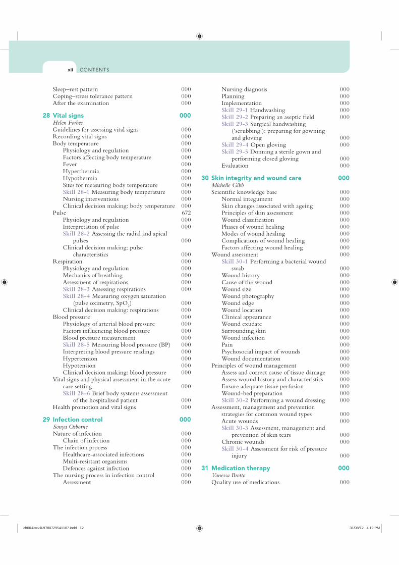
Nursing diagnosis 000Planning 000Implementation 000Skill 29-1 Handwashing 000Skill 29-2 Preparing an aseptic field 000Skill 29-3 Surgical handwashing
(‘scrubbing’): preparing for gowning and gloving 000
Skill 29-4 Open gloving 000Skill 29-5 Donning a sterile gown and
performing closed gloving 000Evaluation 000
30 Skin integrity and wound care 000Michelle GibbScientific knowledge base 000
Normal integument 000Skin changes associated with ageing 000Principles of skin assessment 000Wound classification 000Phases of wound healing 000Modes of wound healing 000Complications of wound healing 000Factors affecting wound healing 000
Wound assessment 000Skill 30-1 Performing a bacterial wound
swab 000Wound history 000Cause of the wound 000Wound size 000Wound photography 000Wound edge 000Wound location 000Clinical appearance 000Wound exudate 000Surrounding skin 000Wound infection 000Pain 000Psychosocial impact of wounds 000Wound documentation 000
Principles of wound management 000Assess and correct cause of tissue damage 000Assess wound history and characteristics 000Ensure adequate tissue perfusion 000Wound-bed preparation 000Skill 30-2 Performing a wound dressing 000
Assessment, management and prevention strategies for common wound types 000Acute wounds 000Skill 30-3 Assessment, management and
prevention of skin tears 000Chronic wounds 000Skill 30-4 Assessment for risk of pressure
injury 000
31 Medication therapy 000Vanessa BrottoQuality use of medications 000
Sleep–rest pattern 000Coping–stress tolerance pattern 000After the examination 000
28 Vital signs 000Helen ForbesGuidelines for assessing vital signs 000Recording vital signs 000Body temperature 000
Physiology and regulation 000Factors affecting body temperature 000Fever 000Hyperthermia 000Hypothermia 000Sites for measuring body temperature 000Skill 28-1 Measuring body temperature 000Nursing interventions 000Clinical decision making: body temperature 000
Pulse 672Physiology and regulation 000Interpretation of pulse 000Skill 28-2 Assessing the radial and apical
pulses 000Clinical decision making: pulse
characteristics 000Respiration 000
Physiology and regulation 000Mechanics of breathing 000Assessment of respirations 000Skill 28-3 Assessing respirations 000Skill 28-4 Measuring oxygen saturation
(pulse oximetry, SpO2) 000
Clinical decision making: respirations 000Blood pressure 000
Physiology of arterial blood pressure 000Factors inf luencing blood pressure 000Blood pressure measurement 000Skill 28-5 Measuring blood pressure (BP) 000Interpreting blood pressure readings 000Hypertension 000Hypotension 000Clinical decision making: blood pressure 000
Vital signs and physical assessment in the acute care setting 000Skill 28-6 Brief body systems assessment
of the hospitalised patient 000Health promotion and vital signs 000
29 Infection control 000Sonya OsborneNature of infection 000
Chain of infection 000The infection process 000
Healthcare-associated infections 000Multi-resistant organisms 000Defences against infection 000
The nursing process in infection control 000Assessment 000
ch00-i-xxviii-9780729541107.indd 12 31/08/12 4:19 PM

CONTENTS xiii
Skill 31-9 Adding medications to intravenous f luid containers 000
Skill 31-10 Administering medications by intravenous bolus 000
Skill 31-11 Administering intravenous medications by piggyback/tandem set-up, intermittent intravenous infusion sets and mini-infusion pumps 000
32 Complementary therapies in nursing practice 000Ysanne Chapman and Melanie BirksCommon terms and their relationships 000
Relationship of terms 000The biomedical model of healthcare 000Inf luences on contemporary healthcare
approaches 000Quantum physics 000Chaos theory 000Human energy fields and centres 000
Principles of complementary therapies 000Tracing the use of complementary therapies
in nursing practice 000Uses of complementary therapies in nursing
practice 000Classifications of complementary therapies 000Examples of complementary therapies 000
Incorporating complementary therapies into nursing practice 000Political issues and implications 000Practice issues and implications 000Educational issues and implications 000Research issues and implications 000Strategies for introducing complementary
therapies 000
Part 7
Focusing nursing: basic human needs
33 Promoting mobility 000Clint DouglasPromoting mobility and preventing immobility 000Scientific knowledge base 000
Overview of body mechanics, exercise and activity 000
Regulation of movement 000Pathological inf luences on mobility 000
Nursing knowledge base 000Complications of immobility 000Systemic effects of immobility 000Psychosocial effects 000Developmental changes 000
Critical thinking synthesis 000Nursing process for impaired mobility 000
Assessment 000Nursing diagnosis 000Planning 000
The medication team 000Scientific knowledge base 000
Application of pharmacology in nursing practice 000
Pharmacokinetics as the basis of medication actions 000
Types of medication action 000Routes of administration 000System of medication measurement 000
Medication administration 000Orders in acute care agencies 000Prescriptions 000Distribution systems 000
Critical thinking in administering medications 000Accountability and responsibility 000Safe medication administration 000
Nursing process and medication administration 000Assessment 000Nursing diagnosis 000Planning 000Implementation 000Evaluation 000
Methods of administration 000Oral administration 000Skill 31-1 Administering oral medications 000Topical medication applications 000Nasal instillation 000Skill 31-2 Administering nasal instillations 000Eye instillation 000Skill 31-3 Administering opthalmic
medications 000Ear instillation 000Vaginal instillation 000Skill 31-4 Administering vaginal
medications 000Rectal instillation 000Skill 31-5 Administering rectal suppositories 000Administering medications by inhalation 000Skill 31-6 Using metered-dose inhalers
(MDIs) 000Administering medication by irrigation 000Parenteral administration of medications 000Skill 31-7 Preparing injections 000
Mixing medications 000Mixing medications from two vials 000Mixing medications from one vial and
one ampoule 000Mixing and preparing insulin 000
Administering injections 000Skill 31-8 Administering injections 000Subcutaneous injections 000Intramuscular injections 000Intradermal injections 000
Safety in administering medications by injection 000Needleless devices 000Needle recapping 000Intravenous administration 000
ch00-i-xxviii-9780729541107.indd 13 31/08/12 4:19 PM

CONTENTSxiv
Implementation 0000Evaluation 0000
36 Nutrition 0000Trish BurtonScientific knowledge base 0000
Nutrients: the biochemical units of nutrition 0000
Anatomy and physiology of the digestive system 0000
Dietary guidelines 0000Nursing knowledge base 0000
Nutrition during human growth and development 0000
Alternative food patterns 0000Alcohol 0000
Critical thinking synthesis 0000Nursing process and nutrition 0000
Assessment 0000Nursing diagnosis 0000Planning 0000Implementation 0000Skill 36-1 Inserting a small-bore
nasoenteric tube for enteral feedings 0000Skill 36-2 Administering enteral feedings
via nasoenteric tubes 0000Skill 36-3 Administering enteral
feedings via gastrostomy or jejunostomy tube 0000
Evaluation 0000
37 Bowel elimination 0000Elizabeth WattScientific knowledge base 0000
Mouth 0000Stomach 0000Small intestine 0000Large intestine 0000Rectum 0000
Nursing knowledge base 0000Factors affecting bowel elimination 0000Common bowel elimination problems 0000
Critical thinking synthesis 0000Nursing process and bowel elimination 0000
Assessment 0000Nursing diagnosis 0000Planning 0000Implementation 0000Skill 37-1 Administering a prepared
enema 0000Skill 37-2 Pouching an ostomy 0000Skill 37-3 Inserting and maintaining a
nasogastric tube (for decompression) 0000Evaluation 0000
38 Urinary elimination 0000Elizabeth WattScientific knowledge base 0000
Implementation 000Skill 33-1 Applying elastic stockings 000Skill 33-2 Positioning patients in bed 000Skill 33-3 Transfer techniques 000Evaluation 000
34 Hygiene 000Trish BurtonScientific knowledge base 000
The skin 000The feet, hands and nails 000The oral cavity 000The hair 000The ears, eyes and nose 000The perineal area 000
Nursing knowledge base 000Social practices 000Personal preferences 000Body image 000Socioeconomic status 000Health beliefs and motivation 000Cultural variables 000Physical condition 000
Critical thinking synthesis 000Nursing process 000
Assessment 000Nursing diagnosis 000Planning 000Implementation 000Skill 34-1 Bathing a patient 000Skill 34-2 Perineal care 000Skill 34-3 Menstrual hygiene 000Skill 34-4 Administering a back rub 000Skill 34-5 Performing nail and foot care 000Skill 34-6 Providing oral hygiene 000Skill 34-7 Performing mouth care for an
unconscious or debilitated patient 000Skill 34-8 Caring for the patient with
contact lenses 000Skill 34-9 Making an occupied bed 000Evaluation 000
35 Sleep 0000Geraldine RebeiroScientific knowledge base 0000
Physiology of sleep 0000Functions of sleep 0000Physical illness 0000Sleep disorders 0000
Nursing knowledge base 0000Sleep and rest 0000Normal sleep requirements and patterns 0000Factors affecting sleep 0000
Critical thinking synthesis 0000Nursing process 0000
Assessment 0000Nursing diagnosis 0000Planning 0000
ch00-i-xxviii-9780729541107.indd 14 31/08/12 4:19 PM
CONTENTS xv
Factors affecting oxygenation 0000Alterations in cardiac functioning 0000Alterations in respiratory functioning 0000
Nursing knowledge base 0000Developmental factors 0000Lifestyle factors 0000Environmental factors 0000
Critical thinking synthesis 0000Nursing process 0000
Assessment 0000Nursing diagnosis 0000Planning 0000Implementation 0000Skill 40-1 Suctioning 0000Skill 40-2 Care of patients with chest
tubes 0000Skill 40-3 Applying a nasal cannula or
oxygen mask 0000Skill 40-4 Using home liqukd oxygen
equiment 0000Skill 40-5 Cardiopulmonary resuscitation—
basic life support 0000Evaluation 0000
41 Pain management 0000Clint Douglas and Anthony SchoenwaldPain management nursing 0000Scientific knowledge base 0000
Defining pain 0000Evolution of pain theories 0000Physiology of pain 0000Psychosocial factors inf luencing pain 0000
Critical thinking synthesis 0000Nursing process 0000
Assessment 0000Skill 41-1 Focused pain assessment 0000Nursing diagnosis 0000Planning 0000Implementation 0000Evaluation 0000
42 Stress and adaptation 0000 Patricia BarkwayScientific knowledge base 0000
Stress and stressors 0000Physiological adaptation 0000Models of stress 0000Factors inf luencing response to stress 0000
Nursing knowledge base 0000Physiological response 0000Psychological response 0000Psychological/emotional issues 0000Developmental factors 0000Intellectual factors 0000Social determinants 0000Spiritual considerations 0000
Critical thinking synthesis 0000Nursing process 0000
Urinary system 0000Pelvic f loor muscles 0000Micturition 0000
Nursing knowledge base 0000Factors affecting urinary elimination 0000Common urinary elimination problems 0000
Critical thinking synthesis 0000Nursing process and urinary elimination 0000
Assessment 0000Skill 38-1 Collecting a midstream
(clean-voided) urine specimen 0000Nursing diagnosis 0000Planning 0000Implementation 0000Skill 38-2 Inserting a straight or
indwelling catheter 0000Skill 38-3 Applying a sheath/condom
drainage device 0000Evaluation 0000
39 Fluid, electrolyte and acid–base balance 0000Karen WottonScientific knowledge base 0000
Application of knowledge of f luid and electrolyte balance to practice 0000
Distribution of body f luids 0000Composition of body f luids 0000Movement of body f luids 0000Regulation of body f luids 0000Regulation of electrolytes 0000Regulation of acid–base balance 0000Disturbances in electrolyte, f luid and
acid–base balances 0000Nursing knowledge base 0000Critical thinking synthesis 0000Nursing process 0000
Assessment 0000Nursing diagnosis 0000Planning 0000Implementation 0000Skill 39-1 Subcutaneous (SC) infusion
(hyperdermoclysis) 0000Skill 39-2 Initiating a peripheral
intravenous (IV) infusion 0000Skill 39-3 Regulating intravenous f low
rate 0000Skill 39-4 Changing intravenous solution
and infusion tubing 0000Skill 39-5 Changing a peripheral
intravenous dressing 0000Evaluation 0000
40 Oxygenation 0000Margaret WheelerScientific knowledge base 0000
Cardiovascular physiology 0000Respiratory physiology 0000
ch00-i-xxviii-9780729541107.indd 15 31/08/12 4:19 PM
CONTENTSxvi
Discharge from the PARU 0000Postoperative rehabilitation 0000
The nursing process in postoperative care 0000Assessment 0000Nursing diagnosis 0000Planning 0000Implementation 0000Evaluation 0000
The client experiencing a medical admission 0000
45 Mental health 0000Anthony O’BrienMental health scope of practice 0000History of mental health nursing 0000
Hildegard Peplau and interpersonal care 0000Recovery and mental health 0000Mental illness 0000
Mental illness and personality disorder 0000Substance use 0000Developmental disability 0000Self-harm and suicide 0000
Psychiatric diagnosis 0000Assessment in mental health nursing 0000
Practice contexts 0000Treatment modalities 0000
Individual psychotherapy 0000Cognitive therapy 0000Dialectical behaviour therapy 0000Group therapy 0000Pharmacological therapy 0000Electroconvulsive therapy 0000
Culture and mental illness 0000Stigma 0000Hearing voices 0000Mental health legislation 0000Clinical supervision in mental health nursing 0000Mental health promotion 0000Professional organisations in mental health
nursing 0000
46 Caring for the cancer survivor 0000Patsy YatesThe effects of cancer on quality of life 0000
Physical wellbeing and symptoms 0000Psychological wellbeing 0000Social wellbeing 0000Spiritual wellbeing 0000
Cancer and families 0000Family distress 0000
Implications for nursing 0000Survivor assessment 0000Client education 0000Providing resources 0000Components of survivorship care 0000Survivorship care plan 0000
Index 0000
Assessment 0000Nursing diagnosis 0000Planning 0000Implementation 0000Evaluation 0000
Part 8
Situtating nursing: contexts of care
43 Community-based nursing focusing on the older person 0000Lynn Chenoweth and Ann McKillopAustralia’s and New Zealand’s health support
for older people 0000Policy contexts 0000
Healthcare for populations as well as individuals 0000Primary healthcare 0000Community and people-focused
healthcare 0000Integrated community health services 0000Strengths-based approach 0000
Supporting the older person with chronic illness 0000Impact of chronic illness 0000Evidence-based chronic illness models 0000
The changing scope of community nursing practice 0000Advanced community nursing 0000Competencies for community nursing 0000Nursing competencies for integrated care 0000
Quality community nursing services for older people 0000Challenges for community nurses 0000Overcoming community nursing
challenges 0000 Summary 0000
44 Acute care 0000Nicole PhillipsAcute care0000The client experiencing surgery 0000Classification of surgery 0000The nursing process in the preoperative
surgical phase 0000Assessment 0000Nursing diagnosis 0000Planning 0000Implementation 0000Skill 44-1 Demonstrating postoperative
exercises 0000Evaluation 0000
Transferring the client to the operating room 0000Intraoperative phase considerations 0000
Postoperative surgical phase 0000Immediate postoperative recovery 0000
ch00-i-xxviii-9780729541107.indd 16 31/08/12 4:19 PM

Lynn Chenoweth, RN, DipRec, BA, GCert Teach/Learn, MA (Hons), MAdEd, PhDProfessor of Aged and Extended Care Nursing, University of Technology Sydney and South Eastern Sydney Local Health District, NSW
Mary Chiarella RN, RM, LLB(Hons), PhDProfessor of Nursing, Sydney Nursing School, The University of Sydney, NSW
Leonie Cox PhD, GCertHEd, RNSenior Lecturer, Queensland University of Technology, Qld
Jackie Crisp RN, PhD, FCNProfessor of Child and Adolescent NursingSydney Children’s Hospitals Network and Faculty of Nursing, Midwifery and Health, University of Technology, Sydney, NSW
Jane Davey RN, RM, BAppSc (Nsg), MN (Nurs Ed), PhDNurse Manager, Professional and Educational Development Service, Sydney Children’s Hospital, Randwick, NSWHonorary Associate (Clinical Fellow), University of Technology, Sydney, NSW
Patricia M. Davidson RN, BA, MEd, PhDProfessor and Director, Centre for Cardiovascular and Chronic Care, Faculty of Health, University of Technology, Sydney, NSW
Clint Douglas RN, PhD Lecturer, School of Nursing, Queensland University of Technology, Qld
Helen Forbes RN, PhD, MedStud, BAppSc (Adv Nsg Ed)Director of Teaching and Learning, School of Nursing and Midwifery, Deakin University, Vic
Australia and New Zealand
Patricia Barkway RN, CMHN, FACMHN, BA, MSc(PHC)Senior Lecturer, Mental Health Nursing, Flinders University, Adelaide, SA
Karen Betony RGN, MSc (Nsg)Nurse Maude Association, Christchurch, New Zealand
Melanie Birks RN, PhD, BN, MEd, FRCNADeputy Dean, CQ University, Qld
Vanessa S.A. Brotto RN, BN, BAppSc (HP), GDipAdvNurs (Crit Care), GCertHEd, MClinNursLecturer, Deakin University, Vic
Nicola Brown RN, MN (Hons), MRCNALecturer, Faculty of Nursing, Midwifery and Health, University of Technology, Sydney, NSW
Trish Burton DipAppSc, BSc, BAppSc, MEd, PhDSenior Lecturer, School of Nursing and Midwifery, Victoria University, Vic
Helen Calabretto RN, RM, DipT (Nsg Ed), BEd (Nsg Stud), MEdStud, PhDManager—Workforce Development and Resources, SHine SAAdjunct Senior Lecturer, School of Nursing and Midwifery, University of South Australia
Pauline Calleja MANP, BNSc, RN, MRCNALecturer, Simulation Coordinator, School of Nursing, Queensland University of Technology, QldVisiting Scholar, Emergency Department, Nurse Practice and Development Unit, Princess Alexandra Hospital, Qld
Ysanne Chapman RN, PhD (Adel), MSc (Hons), BEd (Nsg), GDE, DNE, DRMProfessor and Dean of Nursing and Midwifery, School of Nursing and Midwifery, Central Queensland University, Mackay, Qld
Contributors
ch00-i-xxviii-9780729541107.indd 17 31/08/12 4:19 PM

xviii CONTRIBUTORS
Ann McKillop RN, DNSenior Lecturer, School of Nursing, University of Auckland, New Zealand
Sue Nagy RN, PhD, FCNAdjunct Professor, Faculty of Nursing, Midwifery and Health, University of Technology, Sydney, NSW
Anthony J. O’Brien RN, BA, MPhil (Hons), FANZCMHN Senior Lecturer, School of Nursing, University of Auckland, New ZealandNurse Specialist, Liaison Psychiatry, Auckland District Health Board, Auckland, New Zealand
Sonya Osborne RN, PhD, MACORN, MRCNASenior Lecturer, Queensland University of Technology, Qld
Elaine Papps RN, PhDSenior Lecturer, Faculty of Health Science, Eastern Institute of Technology, Hawke’s Bay, New Zealand
Alan Pearson AM, RN, MSc, PhD, FRCNSA, FAAG, FRCN Executive Director and Professor of Evidence-Based Healthcare in the Joanna Briggs Institute at the University of Adelaide, SACoordinator of the Cochrane Nursing Care Field; Editor-in-Chief of the International Journal of Nursing PracticeMember of the South Australian Health and Medical Research Institute Scientific Advisory Committee
Nicole M. Phillips RN, BN, DipAppSci (Nsg), GDipAdvNsg (Ed), MNS, PhDSenior Lecturer in Nursing, Director of Undergraduate Studies, School of Nursing and Midwifery, Faculty of Health, Deakin University, Vic
Geraldine Rebeiro BAppSc (Adv Nsg), BEdStud, MEd, RN, MidwifeLecturer in Nursing/Clinical Coordinator (Vic), Australian Catholic University, Vic
John Rosenberg RN, PhD, MACNDirector, Calvary Centre for Palliative Care Research, Canberra, ACT
Andrew Scanlon DNP, MNurs (Nurs Pract), MNS, RN, NP, FRCNA Lecturer, La Trobe University, Clinical School of Nursing at Austin Health, School of Nursing and MidwiferyNurse Practitioner—Neurosurgery, Austin Health, Vic
Robyn Galway RN, MN, MEd, GCert Paed, GCertC&FHN, Cert IV TAANurse Educator, Sydney Children’s Hospital Randwick, NSWConjoint Associate Lecturer, University of New South Wales, NSWClinical Fellow, Faculty of Nursing, Midwifery and Health, University of Technology, Sydney, NSW
Michelle Gibb BNsg, MNsgSc (NP), M Wound Care Nurse Practitioner Wound Management, Queensland University of Technology, Qld
Louise Hickman RN, BN, MPH, PhDSenior Lecturer, Faculty of Health, University of Technology Sydney, NSW
Frances Hughes RN, DN, ONZMChief Nursing and Midwifery Officer, Nursing and Midwifery Office, Queensland Health, Qld
Susan Hunt RN, MEd, PhD, FRCNASenior Nurse Advisor, Commonwealth Department of Health and Ageing Adjunct Associate Professor, Australian University of AustraliaAdjunct Associate Professor, University of South Australia, SA
Megan-Jane Johnstone RN, PhDProfessor of Nursing and Director, Centre for Quality and Patient Safety Research (QPS), School of Nursing and Midwifery, Deakin University, Melbourne, Vic
Bronwyn E. Jones RN, BAppSci (Nsg), MAppSci (Health Stud), PhD Adjunct Associate Professor, School of Nursing and Midwifery, Edith Cowan University, WA
Brendan McCormack DPhil, BSc (Hons), PGCEA, RMN, RGNDirector, Institute of Nursing Research and Head of the Person-centred Practice Research Centre, University of Ulster, Northern IrelandAdjunct Professor of Nursing, University of Technology, SydneyAdjunct Professor of Nursing, Faculty of Medicine, Nursing and Health Care, Monash University, MelbourneVisiting Professor, School of Medicine and Dentistry, University of AberdeenProfessor II, Buskerud University College, Drammen, Norway
ch00-i-xxviii-9780729541107.indd 18 31/08/12 4:19 PM
xixCONTRIBUTORS
Patsy Yates PhD, MSocSc, BA, DipAppSc, RN, FRCNAProfessor, School of Nursing and Institute of Health and Biomedical Innovation, Queensland University of Technology, Qld
United States
Paillette M. Archer, RN, EdDProfessorSaint Francis Medical Center, College of Nursing Peoria, Illinois
Marjorie Baier, PhD, RNAssociate Professor School of NursingSouthern Illinois University—Edwardsville, Edwardsville, Illinois
Karen Balakas, PhD, RN, CNEProfessor and Director Clinical Research PartnershipsGoldfarb School of Nursing at Barnes-Jewish College, St. Louis, Missouri
Jeri Burger, PhD, RNAssistant ProfessorUniversity of Southern Indiana, Evansville, Indiana
Linda Cason, MSN, RN-BC, NE-BC, CNRNManager, Employee Education and Development Department Deaconess Hospital, Evansville, Indiana
Janice Colwell, RN, MS, CWOCN, FAANAdvance Practice Nurse, University of Chicago, Chicago, Illinois
Rhonda W. Comrie, PhD, RN, CNE, AE-CAssociate Professor, School of NursingSouthern Illinois University—Edwardsville, Edwardsville, Illinois
Ruth M. Curchoe, RN, MSN, CICDirector, Infection Prevention Unity Health System Rochester, New York
Marinetta DeMoss, RN, MSNManager of Staff DevelopmentSt. Mary’s Medical Center, Evansville, Indiana
Christine R. Durbin, PhD, JD, RNAssistant Professor School of NursingSouthern Illinois University—Edwardsville, Edwardsville, Illinois
Anthony Schoenwald MNS (Nurs Pract), GradDipEd, BNNurse Practitioner, Ipswich Hospital, Qld
Jane Stein-Parbury RN, BSN, MEd, PhD, FCNAProfessor of Mental Health Nursing, University of Technology, Sydney & South East Sydney Local Health District, NSW
Chris Taua RN, BN, MN (Distinction), PGCertMH, CertAdTch, FNZCMHNPrincipal Lecturer, Department of Nursing and Human Services, Christchurch Polytechnic Institute of Technology, Christchurch, New Zealand
Shaun Thompson Clinical Nurse Educator, Sydney Children’s Hospital, NSW
Elizabeth Watt DipN, BAppSc (Adv Nsg), MNS, CertPromCont, RN, RM, FRCNAHead, Clinical School of Nursing at Austin Health, School of Nursing and Midwifery, Faculty of Health Sciences, La Trobe University, Bundoora, Vic
Anthony Welch PhD, MEd, BEd, BN, GradDip (Counselling), DipAppSc (Nurs Ed), RN, ACMHN, MIH&SSRAssociate Professor Mental Health Nursing, Assistant Dean Community Engagement, School of Nursing and Midwifery, CQ University, QldAdjunct Associate Professor, Queensland University of Technology, Qld
Jill White AM, RN, RM, MEd, PhDProfessor of Nursing and Midwifery, Dean Sydney Nursing School, University of Sydney, NSW
Margaret Wheeler RN, RM, BN (Hon), GradDip Adult Ed & TrainingLecturer, School of Nursing, Queensland University of Technology, Qld
Karen Wotton RN, RM, BN, MEMgt, PhDSenior Lecturer, School of Nursing and Midwifery, Chair Simulation Steering Committee, Flinders University, SA
Judy Yarwood RN, MA (Hons), BHlthSc (Nsg), DipTchg (Tert), MNZCN (Aotearoa)Principal Lecturer, Department of Nursing, Christchurch Polytechnic Institute of Technology, Christchurch, New Zealand
ch00-i-xxviii-9780729541107.indd 19 31/08/12 4:19 PM
CONTENTSxx
Frank Lyerla, PhD, RNAssistant Professor School of NursingSouthern Illinois University—Edwardsville, Edwardsville, Illinois
Deborah Marshall, MSNAssistant Professor of NursingDunigan Family Department of Nursing University of Evansville, Evansville, Indiana
Jill Parsons, RN, MSN, PCCNAssistant Professor, MacMurray College, Jacksonville, Illinois
Patsy L. Ruchala, DNSc, RNDirector and Professor, University of Nevada—Reno, Reno, Nevada
Carrie Sona, RN, MSN, CCRN, ACNS, CCNSSurgical Critical Care CNS, Barnes Jewish Hospital, St. Louis, Missouri
Ann B. Tritak, EdD, MA, BSN, RNDean and Professor of NursingSchool of Nursing, Saint Peter ‘s College, Jersey City, New Jersey
Terry L. Wood, PhD, RN, CNEAssistant Clinical Professor School of NursingSouthern Illinois University—Edwardsville, Edwardsville, Illinois
Rita Wunderlich, PhD, RNAssociate ProfessorDirector Baccalaureate Program, Saint Louis University, St. Louis, Missouri
Valerie Yancey, PhD, RNAssociate Professor School of NursingSouthern Illinois University—Edwardsville, Edwardsville, Illinois
Margaret Ecker, RN, MSDirector, Nursing QualityKaiser Permanente Los Angeles Medical Center, Los Angeles, California
Linda Felver, PhD, RNAssociate Professor School of NursingOregon Health & Sciences University, Portland, Oregon
Susan Jane Fetzer, PhD, RN, MBAAssociate ProfessorUniversity of New Hampshire, Durham, New Hampshire
Victoria N. Folse, PhD, APN, PMHCNS-BC, LCPCDirector and Associate ProfessorSchool of Nursing, Illinois Wesleyan University, Bloomington, Illinois
Kay E. Gaehle, PhD, RNAssociate Professor of NursingSchool of Nursing, Southern Illinois University—Edwardsville, Edwardsville, Illinois
Lori Klingman, MSN, RNNursing Faculty and AdvisorOhio Valley General Hospital School of Nursing, McKees Rocks, Pennsylvania
Mary S. Koithan, PhD, RN, CNS-BSAssociate Professor, College of Nursing, University of Arizona, Tucson, Arizona
Karen Korem, RN-BC, MAProfessional Practice SpecialistGeriatric Nurse Clinician, OSF Saint Francis Medical Center, Peoria, Illinois
Jerrilee LaMar, PhD, RN, CNEAssistant Professor of NursingUniversity of Evansville, Evansville, Indiana
Kathy Lever, MSN, WHNP-CAssociate Professor of Nursing University of Evansville, Evansville, Indiana
ch00-i-xxviii-9780729541107.indd 20 31/08/12 4:19 PM

this may seem simple or trivial. They may even wonder why it takes an educated person to do them. We hope that as you work through these chapters, you come to realise why activities such as feeding, bathing, toileting, walking or turning patients are critically important aspects of care, recovery and rehabilitation. The clinical examples and critical thinking questions throughout this text underscore how putting this nursing knowledge and skill into practice can mean the difference between, on the one hand, patient recovery and independence—and, on the other, costly and life-threatening complications, functional decline and disability.
The profound impact of nurse staffing levels, education, workload, skill mix and the nursing work environment on patient outcomes has been well documented in a large and growing body of international research evidence over the past decade (see the box below). These results overwhelmingly support the position that the quality of
To the studentWelcome to the fourth edition of the most successful fundamental text ever to be published for nursing students across Australia and New Zealand. Within this new edition we have maintained the core function of a fundamentals book: that of providing the next generation of nurses with crucial knowledge and skills related to your chosen profession and your practice. However, we have added a goal of supporting your development of a range of critical skills and understandings that will prepare you for the ever-changing and complex world of healthcare.
As editors, we began work on this new edition with the aim of emphasising the importance and complexity of fundamental nursing care. In our experience, many people confuse these complex nursing activities with kindness or niceness. Indeed, to the general public and those new to the profession, many of the topics covered in a textbook like
Preface
BOX 1 Effect of nursing interventions on quality and safety of health care. From Australian Nursing Federation (ANF) 2009 Ensuring quality, safety and positive patient outcomes: why investing in nursing makes $ense. ANF, Melbourne. Online.
Available at http://anf.org.au/documents/reports/Issues_Ensuring_quality.pdf 27 Aug 2012.
ch00-i-xxviii-9780729541107.indd 22 31/08/12 4:19 PM
xxiiiPREFACE
We encourage you to embrace this concept of nursing as knowledge work and engage with the features of this text that aim to cultivate this approach to nursing practice.
The first part of this is to form a critically ref lective approach to self-care and development throughout your nursing career, through supporting your insight into how your own thinking around the information discussed within each chapter is evolving. We are, therefore, seeking to engage your ref lective processes to achieve deep understanding of ‘so what do I think about this now?’, and of the broader ideas around caring for self and others we work with in order to maximise the likelihood of effective workplace cultures and the best outcomes for patients/clients.
The second part is an extension of the above, and seeks to actively engage you in thinking about the content you encounter throughout the book, to facilitate deeper learning and memory and to resist the idea of rote learning. We know that one of the most effective ways of achieving this is to provide examples and stories that are meaningful, and we have taken this approach throughout the book by integrating clinical scenarios or practice examples and critical thinking questions throughout each chapter.
The third part of the approach focuses on ensuring that you are exposed to, and hopefully come to understand, the similarities and differences in patient/client/family experiences and needs, and how these vary across individuals, groups and in relation to environmental and other contextual factors. We have, therefore, moved away from a reliance on highlighting specific cultural issues or age/development stages to a more integrated approach to discussing and dealing with diversity in relation to the content of the specific chapter.
Last, we believe it is crucial that you see the dynamic and evolving nature of evidence for nursing practice—how thinking and knowledge evolve—and understand the need to see ongoing changes in practice as the norm. We also want you to see the need for all clinicians to actively engage in processes associated with their own learning, the learning of others, and the development of practice. We have continued to focus on evidence through the use of research highlights, but once again we have taken a more integrated approach to capture the most up-to-date knowledge/evidence and practices that we can.
Overall, we would like to dedicate this edition to all those students studying to become the best nurses they can be—we wish you well in your endeavours and hope this book provides a solid foundation on which to build the knowledge and expertise required to join one of the most highly regarded, and crucial, professions in the world.
REFERENCEAranda S 2007 Image, identity and voice—nursing in the
public eye. 6th Vivian Bullwinkel Oration. Royal College of Nursing, Australia.
nursing care matters—not because nurses are kind, sweet and self less, but because appropriate nursing care saves lives and improves patient outcomes, as well as patients’ experiences of their care.
As Aranda (2007) argues:
Herein lies the central point of our [nursing’s] image and identity problem—basic nursing care is not understood as skilled practice by nurses themselves or by the public … I point out that while yes we do bath and shower people and engage in work that is sometimes difficult and unpleasant, this work is a door to understanding human experiences of illness. It is through this door that opportunities to make a real difference in the quality of that experience occur.
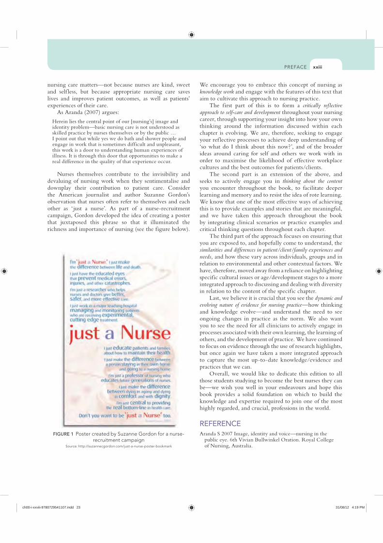
Nurses themselves contribute to the invisibility and devaluing of nursing work when they sentimentalise and downplay their contribution to patient care. Consider the American journalist and author Suzanne Gordon’s observation that nurses often refer to themselves and each other as ‘ just a nurse’. As part of a nurse-recruitment campaign, Gordon developed the idea of creating a poster that juxtaposed this phrase so that it illuminated the richness and importance of nursing (see the figure below).
FIGURE 1 Poster created by Suzanne Gordon for a nurse-recruitment campaign
Source: http://suzannecgordon.com/just-a-nurse-poster-bookmark
ch00-i-xxviii-9780729541107.indd 23 31/08/12 4:19 PM
KEY TERMS LEARNING OUTCOMES
Mastery of content will enable you to:
Describe the anatomy and physiology of the skin.
Discuss normal phases of wound healing.
Describe the modes of wound healing.
Discuss abnormal wound healing.
Outline the factors affecting wound healing.
Conduct a head-to-toe skin assessment and pressure injury risk assessment
Describe the differences between nursing care of acute and chronic wounds.
Describe the principles of wound assessment and management.
Discuss the assessment, management and prevention strategies for common wound types.
Arterial leg ulcers, p. 805Blanching, p. 794Debridement, p. 776Dehiscence, p. 767Dermis, p. 758Diabetic foot ulcers, p. 806Epidermis, p. 757Eschar, p. 776Evisceration, p. 767Exudate, p. 774Fibroblasts, p. 758Fistula, p. 763Friction, p. 793Granulation tissue, p. 766Haematoma, p. 767Haemorrhage, p. 767
Haemoserous, p. 774Haemostasis, p. 765Malignant wounds, p. 807Moist wound environment, p. 777Negative pressure wound
therapy, p. 786Pressure injury, p. 793Primary intention, p. 766Purulent, p. 774Sanguineous, p. 774Secondary intention, p. 766Serous, p. 774Shearing force, p. 793Skin tear, p. 786Venous leg ulcers, p. 804Wound, p. 762
Chapter 30
Michelle Gibb
Skin integrity and wound care
ch30-756-813-9780729541107.indd 756 31/08/12 10:00 AM
757
The skin, or the integumentary system, is the body’s largest organ. It comprises 15% of the total body weight, has an area of approximately 7600 square centimetres and receives one third of circulating blood volume in the average adult (Shores, 2007).
Maintaining skin integrity is a complex process, one that is often taken for granted until damage occurs. As is shown in Table 30-1, the skin has to perform many different functions. Having a good understanding of the layers of the skin and the functions of normal skin is important so that you are able to recognise risk factors for poor skin integrity and undertake actions to prevent skin breakdown or to improve wound healing outcomes.
The following clinical example will be used throughout this chapter for you to ref lect on the key concepts and how they apply to nursing practice.
What factors in this clinical scenario might have contributed to
the development of this skin tear?
Scientific knowledge base Normal integumentThe thickness of the skin varies depending on location, with skin thickness ranging from 0.05 to 0.3 mm. The thickest skin is on the soles of the feet and the palms of the hands. The thicker the skin, the better it is able to withstand injury. The skin consists of three layers (see Figure 30-2):
FUNCTIONS OF THE SKIN
FUNCTION OF THE SKIN EXPLANATION
Protection The skin provides a covering that is designed to protect us from damage or injury
Temperature control (thermoregulation)
Sweat evaporates and cools the skin. Blood vessels also dilate and constrict to prevent heat loss and maintain a stable body temperature
Sensation and communication
Nerve endings and receptors are found in the skin and these help us to respond to touch, pain, heat or cold
Metabolism The skin helps us to metabolise vitamin D through exposure of the skin to sunlight
Elimination The skin helps us to eliminate waste through its function of excretion and secretion
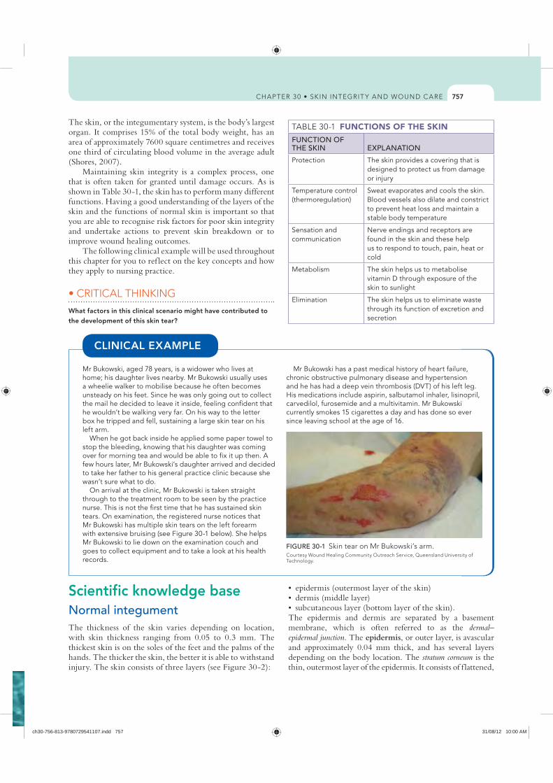
Mr Bukowski, aged 78 years, is a widower who lives at home; his daughter lives nearby. Mr Bukowski usually uses a wheelie walker to mobilise because he often becomes unsteady on his feet. Since he was only going out to collect the mail he decided to leave it inside, feeling confident that he wouldn’t be walking very far. On his way to the letter box he tripped and fell, sustaining a large skin tear on his left arm.
When he got back inside he applied some paper towel to stop the bleeding, knowing that his daughter was coming over for morning tea and would be able to fix it up then. A few hours later, Mr Bukowski’s daughter arrived and decided to take her father to his general practice clinic because she wasn’t sure what to do.
On arrival at the clinic, Mr Bukowski is taken straight through to the treatment room to be seen by the practice nurse. This is not the first time that he has sustained skin tears. On examination, the registered nurse notices that Mr Bukowski has multiple skin tears on the left forearm with extensive bruising (see Figure 30-1 below). She helps Mr Bukowski to lie down on the examination couch and goes to collect equipment and to take a look at his health records.
Mr Bukowski has a past medical history of heart failure, chronic obstructive pulmonary disease and hypertension and he has had a deep vein thrombosis (DVT) of his left leg. His medications include aspirin, salbutamol inhaler, lisinopril, carvedilol, furosemide and a multivitamin. Mr Bukowski currently smokes 15 cigarettes a day and has done so ever since leaving school at the age of 16.
CLINICAL EXAMPLE
FIGURE 30-1 Skin tear on Mr Bukowski’s arm.
Technology.
The epidermis and dermis are separated by a basement membrane, which is often referred to as the dermal–epidermal junction. The epidermis, or outer layer, is avascular and approximately 0.04 mm thick, and has several layers depending on the body location. The stratum corneum is the thin, outermost layer of the epidermis. It consists of f lattened,
ch30-756-813-9780729541107.indd 757 31/08/12 10:00 AM
758
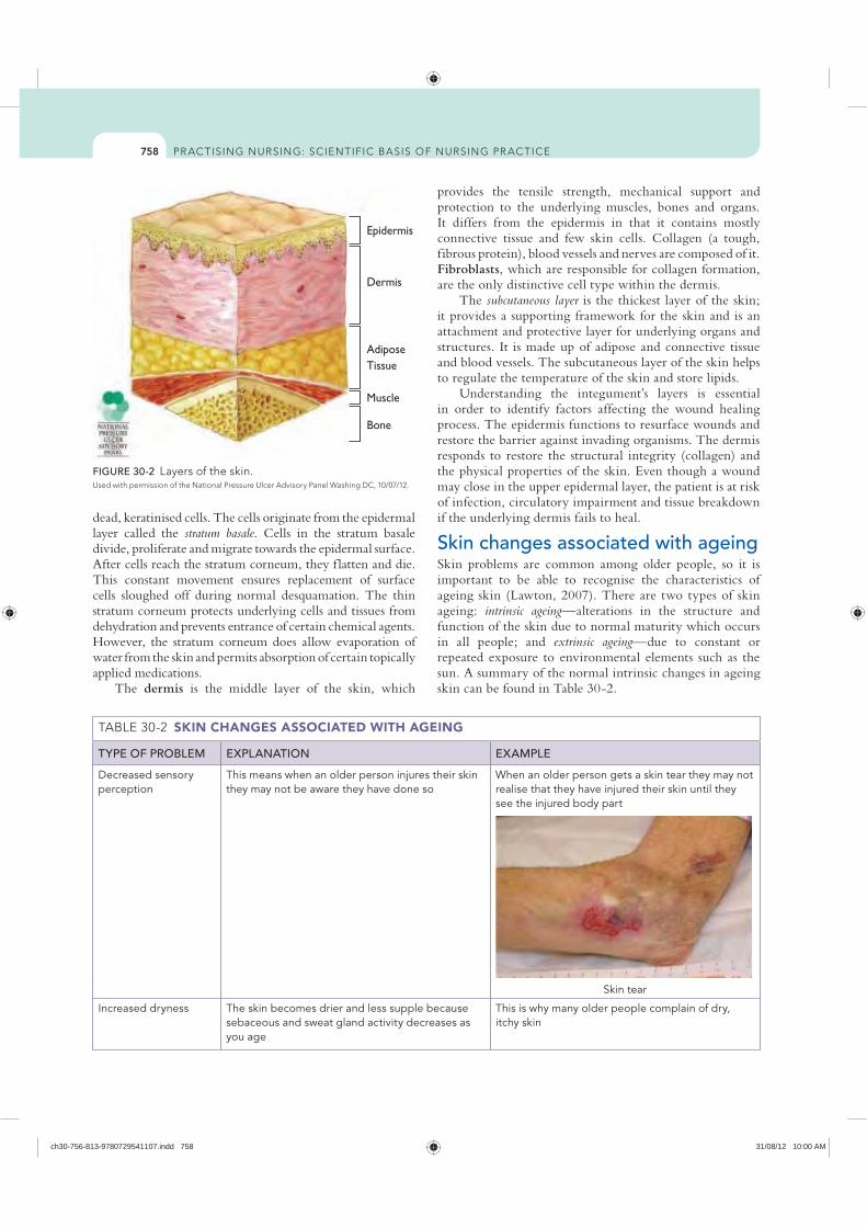
provides the tensile strength, mechanical support and protection to the underlying muscles, bones and organs. It differs from the epidermis in that it contains mostly connective tissue and few skin cells. Collagen (a tough, fibrous protein), blood vessels and nerves are composed of it. Fibroblasts, which are responsible for collagen formation, are the only distinctive cell type within the dermis.
The subcutaneous layer is the thickest layer of the skin; it provides a supporting framework for the skin and is an attachment and protective layer for underlying organs and structures. It is made up of adipose and connective tissue and blood vessels. The subcutaneous layer of the skin helps to regulate the temperature of the skin and store lipids.
Understanding the integument’s layers is essential in order to identify factors affecting the wound healing process. The epidermis functions to resurface wounds and restore the barrier against invading organisms. The dermis responds to restore the structural integrity (collagen) and the physical properties of the skin. Even though a wound may close in the upper epidermal layer, the patient is at risk of infection, circulatory impairment and tissue breakdown if the underlying dermis fails to heal.
Skin changes associated with ageingSkin problems are common among older people, so it is important to be able to recognise the characteristics of ageing skin (Lawton, 2007). There are two types of skin ageing: intrinsic ageing—alterations in the structure and function of the skin due to normal maturity which occurs in all people; and extrinsic ageing—due to constant or repeated exposure to environmental elements such as the sun. A summary of the normal intrinsic changes in ageing skin can be found in Table 30-2.
dead, keratinised cells. The cells originate from the epidermal layer called the stratum basale. Cells in the stratum basale divide, proliferate and migrate towards the epidermal surface. After cells reach the stratum corneum, they f latten and die. This constant movement ensures replacement of surface cells sloughed off during normal desquamation. The thin stratum corneum protects underlying cells and tissues from dehydration and prevents entrance of certain chemical agents. However, the stratum corneum does allow evaporation of water from the skin and permits absorption of certain topically applied medications.
The dermis is the middle layer of the skin, which
Epidermis
Dermis
AdiposeTissue
Muscle
Bone
FIGURE 30-2
SKIN CHANGES ASSOCIATED WITH AGEING
TYPE OF PROBLEM EXPLANATION EXAMPLE
Decreased sensory perception
This means when an older person injures their skin they may not be aware they have done so
When an older person gets a skin tear they may not realise that they have injured their skin until they see the injured body part
Skin tear
Increased dryness The skin becomes drier and less supple because sebaceous and sweat gland activity decreases as you age
This is why many older people complain of dry, itchy skin
ch30-756-813-9780729541107.indd 758 31/08/12 10:00 AM
759
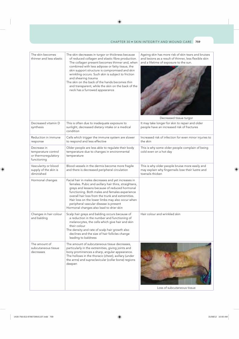
The skin becomes thinner and less elastic
The skin decreases in turgor or thickness because of reduced collagen and elastic fibre production. The collagen present becomes thinner and, when combined with less adipose or fatty tissue, the skin support structure is compromised and skin wrinkling occurs. Such skin is subject to friction and shearing trauma
The skin on the back of the hands becomes thin and transparent, while the skin on the back of the neck has a furrowed appearance
Ageing skin has more risk of skin tears and bruises and lesions as a result of thinner, less flexible skin and a lifetime of exposure to the sun.
Decreased tissue turgor
Decreased vitamin D synthesis
This is often due to inadequate exposure to sunlight, decreased dietary intake or a medical condition
It may take longer for skin to repair and older people have an increased risk of fractures
Reduction in immune response
Cells which trigger the immune system are slower to respond and less effective
Increased risk of infection for even minor injuries to the skin
Decrease in temperature control or thermoregulatory functioning
Older people are less able to regulate their body temperature due to changes in environmental temperature
This is why some older people complain of being cold even on a hot day
Vascularity or blood supply of the skin is diminished
Blood vessels in the dermis become more fragile and there is decreased peripheral circulation
This is why older people bruise more easily and may explain why fingernails lose their lustre and toenails thicken
Hormonal changes Facial hair in males decreases and yet increases in females. Pubic and axillary hair thins, straightens, greys and lessens because of reduced hormonal functioning. Both males and females experience overall hair loss from the trunk and extremities. Hair loss on the lower limbs may also occur when peripheral vascular disease is present
Hormonal changes also lead to drier skin
Changes in hair colour and balding
Scalp hair greys and balding occurs because of a reduction in the number and functioning of melanocytes, the cells which give hair and skin their colour
The density and rate of scalp hair growth also declines and the size of hair follicles change leading to baldness
Hair colour and wrinkled skin
The amount of subcutaneous tissue decreases
The amount of subcutaneous tissue decreases, particularly in the extremities, giving joints and bony prominences a sharp, angular appearance. The hollows in the thoracic (chest), axillary (under the arms) and supraclavicular (collar bone) regions deepen
Loss of subcutaneous tissue
ch30-756-813-9780729541107.indd 759 31/08/12 10:00 AM
760
SKIN ASSESSMENT INFORMATION REQUIRED QUESTIONS THE NURSE MIGHT ASK THE PATIENT
Past medical history Tell me what other health conditions you may have.When conducting the health assessment and a problem with the skin is identified, it is important
to determine usual skin conditions, onset of any problems, changes since onset, specific known causes, alleviating factors, psychological reaction to skin changes, previous trauma and if the patient has had any surgery or prior disease that involves the skin
Medications (topical, systemic, over-the-counter)
Are you taking any medications that might affect your skin? For example, medications might include anticoagulants or steroids (taken for conditions such as
rheumatoid arthritis)
Exposure to environmental or occupational hazards
What sort of work do you do? Were you exposed to the sun a lot when you were younger?
Substance abuse Do you smoke or have you ever smoked? How much did you smoke? When did you stop smoking? How much alcohol intake do you have? Have you ever used illicit drugs? Example: Fingernails are often stained yellow by nicotine exposure.
Recent physiological or psychological stress
Have you experienced a recent stressful event? Have you been unwell recently? How does this affect you?
Hair, nail and skin care habits What methods do you use for cleansing your skin? How often do you moisturise? How do you dry your skin?
Example: Many soaps, oils, lotions, cosmetics and home remedies have preservatives that can irritate the skin and make it itchy or inflamed.
Skin self-examination How often do you look at your skin? Are you able to see your skin properly? Can you reach to dry between your toes?
Problems with the skin Have you noticed any changes in your skin (e.g. dryness, rashes, lumps, amount of perspiration)? When did the symptoms occur? Are these symptoms new or an old problem? What area of the body is affected (i.e. skin folds, localised or generalised)?
Are there any associated symptoms (e.g. fever, relationship to stress or leisure activities)? What have you been doing for the problem?
Example: Eczema is a common problem that is often made worse by some creams and may be a lifetime problem for that person. Careful questioning will help to determine what the person has been doing to treat the condition, what works for them and what doesn’t work to treat the problem.
Principles of skin assessmentA comprehensive assessment of the skin is essential, as a wide range of health conditions manifest in changes in the skin and can provide valuable diagnostic clues to the underlying disease process. Furthermore, recognising the characteristics of normal skin helps you to identify those at risk for compromised skin integrity. When undertaking a skin assessment, there are three important steps, outlined below.
Step 1. Prepare the environmentFirst, you need to create an environment that is suitable to conducting an assessment by ensuring:
temperature; this helps to reduce anxiety
skin or any skin changes
usually inspected such as the buttocks, axillae, back of thighs or feet
Step 2. Gather relevant informationSecond, you need to carefully explain what you are going to do and the purpose of assessing the skin. Typically, you obtain a history using a framework such as that shown in Table 30-3.
Step 3. Observe and feel the skinThe final step is to look at the skin (inspection) and feel (palpation) if there are any changes (Table 30-4). When conducting the physical assessment, proceed from head-to-toe and compare each body region for symmetry (i.e. right side with left side to differentiate structural from pathological changes). If lesions are identified, palpate them for density, induration (hardening or thickening of tissues) and tenderness.
Now that you have a better understanding of how to assess the skin, you are ready to learn more about some strategies you can use to maintain skin integrity and prevent many skin problems from occurring. Review the suggested evidence-based strategies summarised in Table 30-5.
ch30-756-813-9780729541107.indd 760 31/08/12 10:00 AM
761
OBSERVATION OF THE SKIN
OBSERVATION EXPLANATION
Skin temperature If the skin around a wound is very hot to touch compared with the surrounding skin, this may indicate an infection
If the feet are abnormally cold to touch, this may indicate a problem with circulation
Skin texture Texture of the skin may be described as rough, coarse, fine, flaky, scaly or smooth. Rough skin may indicate that the skin is very dry and may occur normally on exposed areas such as the elbows and soles of the feet
Skin colour The colour of the skin can indicate a person’s general wellbeingChanges in colour are best obtained from the lips, mucous membranes of the mouth, earlobes, finger
and toe nails and the extremitiesThe colour of the skin indicates the degree of blood supply and temperature of the skin, and oxygen and
fluid supply to the skin. Colour of the skin varies depending on the amount of melanin in the cells and with blood supply. Skin colour can be masked by cosmetics or tattoos
Colour changes associated with the skin can be described as: erythema (redness) due to vasodilation associated with blushing, heat, inflammation, fever, alcohol
ingestion, extreme cold and heat and hot flushes pallor (whiteness) due to vasoconstriction associated with peripheral arterial disease, or due to
decreased oxygenation of blood from decreased haemoglobin as seen in anaemia, or loss of melanin as in vitiligo
cyanosis (bluish) due to deoxygenated haemoglobin noticed in earlobes, lips, mucous membranes of the mouth, nail beds; may be seen in cardiac or respiratory disease
jaundice (yellow) due to increased bile pigment in the blood distributed in the skin and mucous membranes and sclera of the eye, as seen in liver disease, obstruction of bile ducts, chronic uraemia
and rapid haemolysis brownish due to increased melanin deposits, which is normal in darker-skin-toned individuals and is
also found in ageing, sunburn, anterior pituitary, adrenal cortex and liver diseases
Skin changes The presence of growths, discolouration or changes in pigmentation, infections, broken areas, old scars, tattoos, rashes, eczema, dermatitis, senile purpura, cherry angiomas or thickened skin may be normal changes associated with ageing, indicate a person’s risk of a wound recurring or indicate the presence of a clinical condition
For example, changes in pigmentation may indicate a condition such as vitiligo, Addison’s disease, arsenic toxicity or uraemia. Fungal infections such as tinea versicolor can cause pigmentation changes in the affected area. Pigmentation changes in naevi or moles may indicate the presence of skin cancers
Oedema Assessment of swelling in the tissues can be assessed by location and degree. Oedema is graded as: + slight indentation with normal anatomical contours ++ deeper indentation which lasts longer than + with fairly normal contours +++ deep indentation which remains after several seconds with obvious swelling ++++ deep indentation that remains for minutes with frank swelling
Turgor (resilience and elasticity of tissue)
Skin turgor can give an indication of the person’s nutrition and hydration statusWhen pinched between the thumb and index finger for a few seconds, normal well-hydrated skin will
snap back into place when releasedDehydrated skin, particularly in an elderly patient, will form a small tent shape before gradually resuming
its normal position
Hair distribution, colour and quantity (thick, thin, balding)
Uneven hair loss may indicate a person’s psychological stateFor example, a person may unconsciously pull their hair out if they are traumatised. Excessive hair
growth may be related to hormonal changes
Nail length, colour, configuration, symmetry and cleanliness
The colour of a person’s fingernails may indicate certain problemsFor example fingernails stained yellow indicate nicotine use. Blue fingernails can indicate a problem with
circulation such as cardiac or respiratory diseaseIn addition to nail-bed colour, check for clubbing and assess capillary refill. Capillary refill time can be
affected by environmental conditions, vasoconstriction from smoking or peripheral oedema. Finger clubbing can be an indication of chronic tissue hypoxia
Lesions of the skin Lesions are classified by type, colour, size, shape and configuration, texture, effect of pressure, arrangement, distribution and variety
ch30-756-813-9780729541107.indd 761 31/08/12 10:00 AM
762
ulcers, pressure ulcers, malignant or fungating wounds). Acute wounds heal fairly quickly (usually within 14 days), without complications and with limited interventions. They follow the normal healing process in an orderly and timely way (Celik, 2007). Examples of some acute wounds are those caused by trauma or surgery. A chronic wound is a wound that has failed to proceed through an orderly and timely process for healing and whereby healing is delayed, repair fails to occur, and return to normal function is slowed (Harvey, 2005).
Phases of wound healingThe wound healing process involves a complex series of cellular and biochemical events that act upon damaged tissues. These are interlinked and dependent on one another in a continuing process of regeneration and repair (Schultz and others, 2003). Wound healing tends to follow a well-defined process that involves four main stages: 1. haemostasis 2. inf lammation 3. proliferation or reconstruction 4. maturation or remodelling of the scar tissue.These stages of wound healing overlap and the entire process can last for many months.
Haemostasis Immediately after injury, platelets initiate the wound-healing process by releasing a number of growth factors that rapidly disperse from the wound, drawing inf lammatory
As you read the next section, think about Mr Bukowski’s skin
tear:
Wound classificationA wound can be defined as an injury to the skin or underlying structures that may or may not result in a loss of skin integrity and whereby physiological function of the tissue is impaired (Carville, 2007). Although at first a wound may look like any other, it is imperative to know that all wounds are not the same. Understanding the aetiology of a wound is important, because the treatment varies depending on the underlying disease process (Ratliff, 2006). Common wound types are presented in Table 30-6.
There are many ways to classify wounds. Wound classification systems describe the status of skin integrity, cause of the wound, severity or extent of tissue injury or damage, cleanliness of the wound or descriptive qualities of the wound such as colour. Wound classifications help you to understand the risks associated with a wound and implications for its care.
Wounds can be classified as either acute (e.g. surgical incisions, lacerations, blisters, abrasions) or chronic (e.g. leg
PRESERVING SKIN INTEGRITYPREVENTION STRATEGIES EXPLANATION
Assess skin regularly The nurse should assess the skin regularly so that correct and suitable preventive measures can be put in place and evaluated
Use emollient soap substitutes for washing or cleansing
This reduces the drying effects of soap and waterEmollients restore the natural barrier function of the skin by replacing lost water and
provide a protective film over the surface of the skin. Emollients include creams, ointments, lotions, bath oils and soap substitutes
Avoid products that may irritate the skin Products such as perfumes, bubble baths and talcum powder can irritate the skin and cause itching or discomfort
Dry the skin thoroughly Drying should involve a light patting and not rubbing as this may lead to abrasion and/or weakening of the skin
If skin is left damp, it is vulnerable to excess drying from the environment and at risk of fungal and bacterial contamination
Apply a pH-neutral moisturiser and/or barrier cream at least twice daily
This will help to prevent dry skinA pH-neutral moisturiser is one that is neither acid or alkaline; it has a pH between
6.5 and 7.5A barrier preparation can be a cream, ointment or spray which contains substances that
repel water, such as silicone or zinc oxide
When applying moisturiser and/or barrier cream, follow the direction of body hair and gently smooth into the skin
Rubbing can cause irritation. Rubbing moisturiser against the direction of hair growth increases the risk of an infection occurring in the hair follicles
Encourage patient to wear loose, cotton clothing where possible
This helps the skin to breathe better and reduces the risk of sweating from nylon fabricsThe use of limb protectors can also protect fragile limbs
ch30-756-813-9780729541107.indd 762 31/08/12 10:00 AM
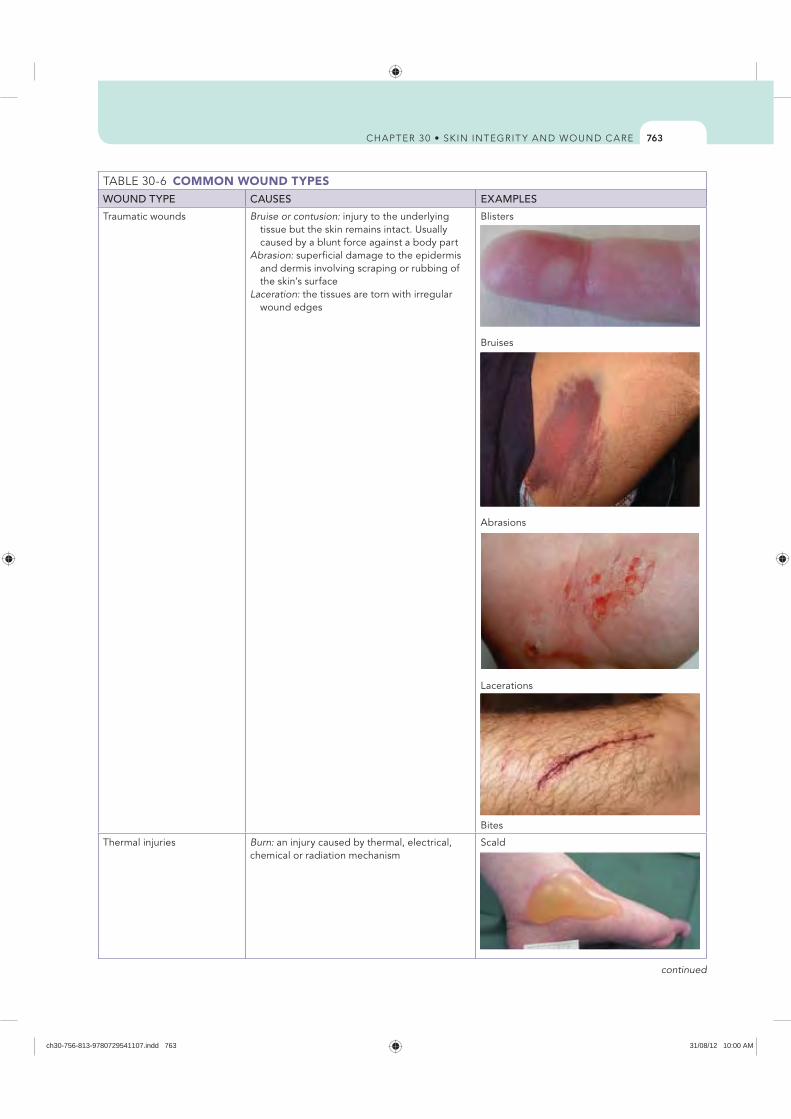
763
COMMON WOUND TYPESWOUND TYPE CAUSES EXAMPLES
Traumatic wounds Bruise or contusion: injury to the underlying tissue but the skin remains intact. Usually caused by a blunt force against a body part
Abrasion: superficial damage to the epidermis and dermis involving scraping or rubbing of the skin’s surface
Laceration: the tissues are torn with irregular wound edges
Blisters
Bruises
Abrasions
Lacerations
Bites
Thermal injuries Burn: an injury caused by thermal, electrical, chemical or radiation mechanism
Scald
continued
ch30-756-813-9780729541107.indd 763 31/08/12 10:00 AM
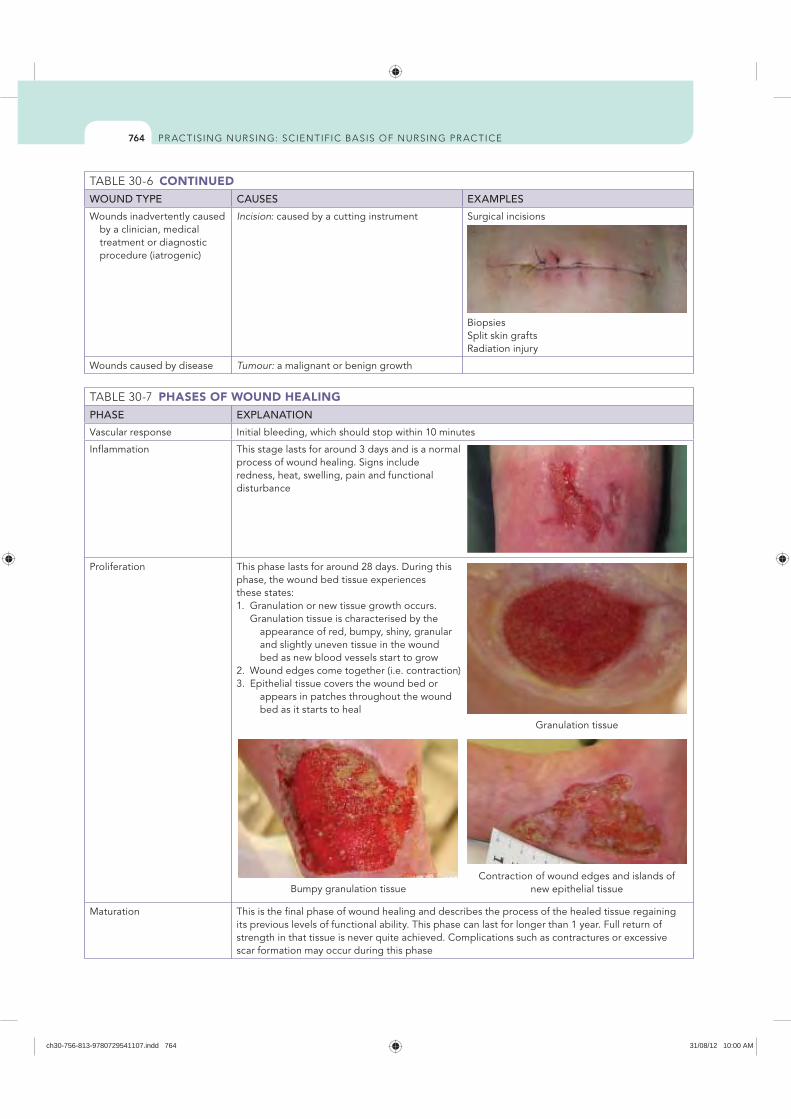
764
CONTINUEDWOUND TYPE CAUSES EXAMPLES
Wounds inadvertently caused by a clinician, medical treatment or diagnostic procedure (iatrogenic)
Incision: caused by a cutting instrument Surgical incisions
Biopsies Split skin grafts Radiation injury
Wounds caused by disease Tumour: a malignant or benign growth
PHASES OF WOUND HEALINGPHASE EXPLANATION
Vascular response Initial bleeding, which should stop within 10 minutes
Inflammation This stage lasts for around 3 days and is a normal process of wound healing. Signs include redness, heat, swelling, pain and functional disturbance
Proliferation This phase lasts for around 28 days. During this phase, the wound bed tissue experiences these states:1. Granulation or new tissue growth occurs.
Granulation tissue is characterised by the appearance of red, bumpy, shiny, granular
and slightly uneven tissue in the wound bed as new blood vessels start to grow
2. Wound edges come together (i.e. contraction)3. Epithelial tissue covers the wound bed or
appears in patches throughout the wound bed as it starts to heal
Maturation This is the final phase of wound healing and describes the process of the healed tissue regaining its previous levels of functional ability. This phase can last for longer than 1 year. Full return of strength in that tissue is never quite achieved. Complications such as contractures or excessive scar formation may occur during this phase
Granulation tissue
Bumpy granulation tissueContraction of wound edges and islands of
new epithelial tissue
ch30-756-813-9780729541107.indd 764 31/08/12 10:00 AM
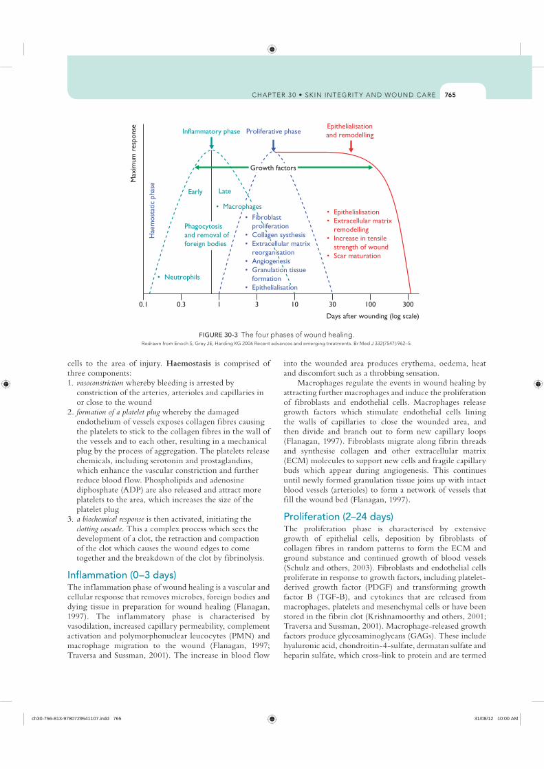
765
into the wounded area produces erythema, oedema, heat and discomfort such as a throbbing sensation.
Macrophages regulate the events in wound healing by attracting further macrophages and induce the proliferation of fibroblasts and endothelial cells. Macrophages release growth factors which stimulate endothelial cells lining the walls of capillaries to close the wounded area, and then divide and branch out to form new capillary loops (Flanagan, 1997). Fibroblasts migrate along fibrin threads and synthesise collagen and other extracellular matrix (ECM) molecules to support new cells and fragile capillary buds which appear during angiogenesis. This continues until newly formed granulation tissue joins up with intact blood vessels (arterioles) to form a network of vessels that fill the wound bed (Flanagan, 1997).
Proliferation (2–24 days)The proliferation phase is characterised by extensive growth of epithelial cells, deposition by fibroblasts of collagen fibres in random patterns to form the ECM and ground substance and continued growth of blood vessels (Schulz and others, 2003). Fibroblasts and endothelial cells proliferate in response to growth factors, including platelet-derived growth factor (PDGF) and transforming growth factor B (TGF-B), and cytokines that are released from macrophages, platelets and mesenchymal cells or have been stored in the fibrin clot (Krishnamoorthy and others, 2001; Traversa and Sussman, 2001). Macrophage-released growth factors produce glycosaminoglycans (GAGs). These include hyaluronic acid, chondroitin-4-sulfate, dermatan sulfate and heparin sulfate, which cross-link to protein and are termed
cells to the area of injury. Haemostasis is comprised of three components: 1. vasoconstriction whereby bleeding is arrested by
constriction of the arteries, arterioles and capillaries in or close to the wound
2. formation of a platelet plug whereby the damaged endothelium of vessels exposes collagen fibres causing the platelets to stick to the collagen fibres in the wall of the vessels and to each other, resulting in a mechanical plug by the process of aggregation. The platelets release chemicals, including serotonin and prostaglandins, which enhance the vascular constriction and further reduce blood f low. Phospholipids and adenosine diphosphate (ADP) are also released and attract more platelets to the area, which increases the size of the platelet plug
3. a biochemical response is then activated, initiating the clotting cascade. This a complex process which sees the development of a clot, the retraction and compaction of the clot which causes the wound edges to come together and the breakdown of the clot by fibrinolysis.
Inflammation (0–3 days)The inf lammation phase of wound healing is a vascular and cellular response that removes microbes, foreign bodies and dying tissue in preparation for wound healing (Flanagan, 1997). The inf lammatory phase is characterised by vasodilation, increased capillary permeability, complement activation and polymorphonuclear leucocytes (PMN) and macrophage migration to the wound (Flanagan, 1997; Traversa and Sussman, 2001). The increase in blood f low
0.1 0.3
Early Late
1 3 10 30 100 300
FIGURE 30-3 The four phases of wound healing.
ch30-756-813-9780729541107.indd 765 31/08/12 10:00 AM
766
after wound closure, but undergoes continual lysis to form a more organised lattice structure that gradually increases tensile strength of scar tissue, fibroblast numbers decrease and blood vessels are restored to normal (Traversa and Sussman, 2001). The tensile strength of scar tissue is never more than 80% of that of non-scar tissue (Flanagan, 1997).
Modes of wound healingWounds can be broadly characterised into two groups: those with and without loss of tissue. A clean surgical incision is an example of a wound with little tissue loss and where the edges of the wound are held in close apposition by sutures, staples or tape. The surgical wound heals by primary intention. However, if a wound is infected or contains foreign bodies, primary wound closure may be delayed for three to five days. This is known as delayed primary intention. A wound that involves extensive loss of tissue, such as a pressure ulcer or severe laceration, heals through a process of granulation, contraction and epithelialisation, and scarring may result. This is known as healing by secondary intention.
Skin grafting is another method to achieve wound healing. A skin graft is a segment of epidermis and dermis that is intentionally separated from one site (donor site) and transplanted to another site (recipient site) (Carville, 2007). Skin grafts depend on the in-growth of capillaries from the recipient site for their survival. A skin graft is selected as a method of wound closure when healing by secondary intention or primary closure is not a suitable option, to speed up the healing process and reduce the risk of infection. There are two types of skin grafts: full-thickness grafts that consist of the epidermis and the full thickness of the dermis, and split-skin grafts consisting of the epidermis and a variable proportion of the dermis. Split-skin grafts are described as thin, intermediate or thick according to the thickness of the graft.
A wound may also be closed by surgical relocation of tissue from one part of the body to another part in order to
proteoglycans. These form an amorphous gel where collagen fibres deposit and aggregate (Traversa and Sussman, 2001). The ground substance secreted by fibroblasts determines the compliance, f lexibility and integrity of the dermis. It provides compressive strength and support and density to tissue, reduces friction between collagen fibres during tissue stress or strain and protects tissue from invasion by microorganisms (McCulloch and others, 1995).
During the proliferation phase, new capillary devel-opment is seen as ruddy, bumpy granulation tissue in the base of a wound, and wound contraction occurs. Epithelial cell migration occurs over the granulated wound bed. Epithelial cells migrate from surrounding wound edges or from hair follicles, sweat or sebaceous glands in the wound and appear as thin, translucent film across the wound bed. At this stage, the epithelial tissue is very fragile and easily removed even by gentle cleansing. Migration of epithelial cells ceases when the wound is covered; then mitosis thickens the epithelium to the four to five layers needed to form the epidermis.
Maturation (24 days to 1 year)The combination of fibronectin and collagen forms the ECM essential for the development of granulation tissue that eventually fills the wound. The endothelial buds increase in vascularity in response to the large metabolic demand of the repair tissue (Traversa and Sussman, 2001). The arrangement of collagen fibres in the wound is random and disorganised, and has a gel-like consistency that gradually matures to form cross-links which provide tensile strength to the wound (Flanagan, 1997).
The remodelling of collagen fibres is regulated by growth factors including TGF-B, PDGF and fibroblast growth factor (FGF), interleukin-1 (IL-1) and interferon-gamma (INF-γ). Depending on the type and severity of the wound, the maturation phase may take up to six months to a year (McCulloch and others, 1995). Collagen synthesis continues
Evidence-based practiceMuch of our understanding about how wounds heal is derived from examination of the wound-healing process in acute wounds. It is widely accepted that acute wounds heal through an orderly process of haemostasis, inflammation, proliferation and maturation. However, chronic wounds such as leg ulcers and diabetic foot ulcers do not follow this trajectory and are often characterised by prolonged inflammation and, even if they do heal, frequently recur even with the highest standard of care.
The concept of wound-bed preparation has emerged as an important paradigm in the management of chronic wounds in order to identify factors that influence wound healing and to provide a framework for clinicians to maximise
the potential for wound for healing. There are four main principles underpinning wound bed preparation—TIME:
The TIME framework is a dynamic concept that can be translated into practical management of different wound types by utilising a standardised framework.
ReferenceSchultz G, Sibbald G, Falanga V and others 2003 Wound bed preparation: a systematic approach to wound management. Wound Repair Regen 11:1–28.
RESEARCH HIGHLIGHT
ch30-756-813-9780729541107.indd 766 31/08/12 10:00 AM
767
shiny surface and sometimes have telangiectasia (commonly known as spider veins) and a pink or purple appearance accompanied by hyperpigmentation. Keloids occur more frequently in darkly pigmented individuals and may be associated with a family history of keloids. While they may form on any part of the body, keloids appear more commonly on the upper chest and shoulders. Keloids may develop up to several years after even minor injuries and may even form spontaneously on the midchest in the absence of any known injury (Gauglitz and others, 2011). They usually persist for long periods of time and do not regress spontaneously.
ContracturesContractures occur when soft tissue such as muscles become shortened and prevent the joint moving through a normal range of movement. Contraction of the wound edges is a normal process of wound healing, but when contracture is excessive it may cause cosmetic and functional deformity. The degree of contraction is inf luenced by the f lexibility and mobility of the surrounding tissue structures.
HaemorrhageHaemorrhage (bleeding) from a wound site is normal during and immediately after the initial trauma. Haemostasis occurs within several minutes unless large blood vessels are involved or the patient has poor clotting function. Haemorrhage occurring after haemostasis indicates a slipped surgical suture, a dislodged clot, an infection, or erosion of a blood vessel by a foreign object (e.g. a drain). Haemorrhage may occur externally or internally. For example, if a surgical suture slips off a blood vessel, bleeding occurs internally within the tissues and there are no visible signs of blood unless a surgical drain, which is inserted into tissues beneath a wound to remove f luid that collects in underlying tissues, is present. Internal bleeding may be detected by looking for distension or swelling of the affected body part, a change in the type and amount of drainage from a surgical drain or signs of hypovolaemic shock.
External haemorrhaging is more obvious. If bleeding is extensive, the dressing soon becomes saturated, and often blood escapes along the sides of the dressing and pools beneath the patient. The risk of haemorrhage is greatest during the first 24–48 hours after surgery.
HaematomaA haematoma is a localised collection of blood underneath the tissues. It appears as a swelling or mass that often takes on a bluish discolouration. A haematoma near a major artery or vein is dangerous because pressure from the expanding haematoma may obstruct blood f low.
EviscerationEvisceration is when there is total separation of wound layers or protrusion of visceral organs through a wound opening. The condition is a medical emergency that requires surgical repair and immediate patient support.
reconstruct a primary defect ( f lap). The relocation of tissue creates a secondary wound that will require skin grafting or primary closure.
Complications of wound healingWhen a wound fails to heal properly, the layers of skin and tissue may separate. This most commonly occurs before collagen formation (3–11 days after injury). Dehiscence is the partial or total separation of wound layers. A patient who is at risk of poor wound healing (e.g. poor nutritional status, infection, obesity) is also at risk of wound dehiscence. However, obese patients have a higher risk because of the constant strain placed on their wounds and the poor healing qualities of fatty tissue. Dehiscence often involves abdominal surgical wounds and occurs after a sudden strain, such as coughing, vomiting or sitting up in bed. Patients often report feeling as though something has given way. An increase in the presence of haemoserous drainage from a wound may indicate wound dehiscence.
HypergranulationHypergranulation is an accumulation of granulation tissue that extends beyond the wound surface and delays epitheliali sation (Carville, 2007). Hypergranulation tissue may bleed easily; it may be seen in surgical incisions or around tubes and devices and can occur in some malignant tumours. Biopsy of the tissue is essential if malignancy is suspected.
Hypertrophic scarsHypertrophic scars are characterised by an overabundant deposition of collagen in healed skin wounds and present as a re-epithelialised, red, raised and firm scar that may be itchy (Carville, 2007). Hypertrophic scars are usually contained within the original boundary of the wound, and are usually linear in appearance following a surgical scar or papular or nodular following inf lammatory and ulcerating injuries (Gauglitz and others, 2011). Hypertrophic scarring may occur if there is excess tension on a healing wound. It can occur within 4–8 weeks following a wound injury such as a burn injury, laceration, abrasion, surgery, piercing or vaccination and usually regresses spontaneously. Scarring grows rapidly for up to 6 months and then gradually regresses over a period of a few years, resulting in a raised, f lat scar with no further symptoms (Gauglitz and others, 2011).
Keloid scarsKeloid scars occur spontaneously to form firm, smooth, fibrous growths that result from abnormal connective tissue in response to trauma, inf lammation, surgery or burns (Carville, 2007). Similar to hypertrophic scars, they are characterised by an overabundant deposition of collagen in healed wounds where the scar extends beyond the boundary of the original wound margin. The wound margins are usually well demarcated but irregular in outline (Gauglitz and others, 2011). They may be tender or painful with a
ch30-756-813-9780729541107.indd 767 31/08/12 10:00 AM
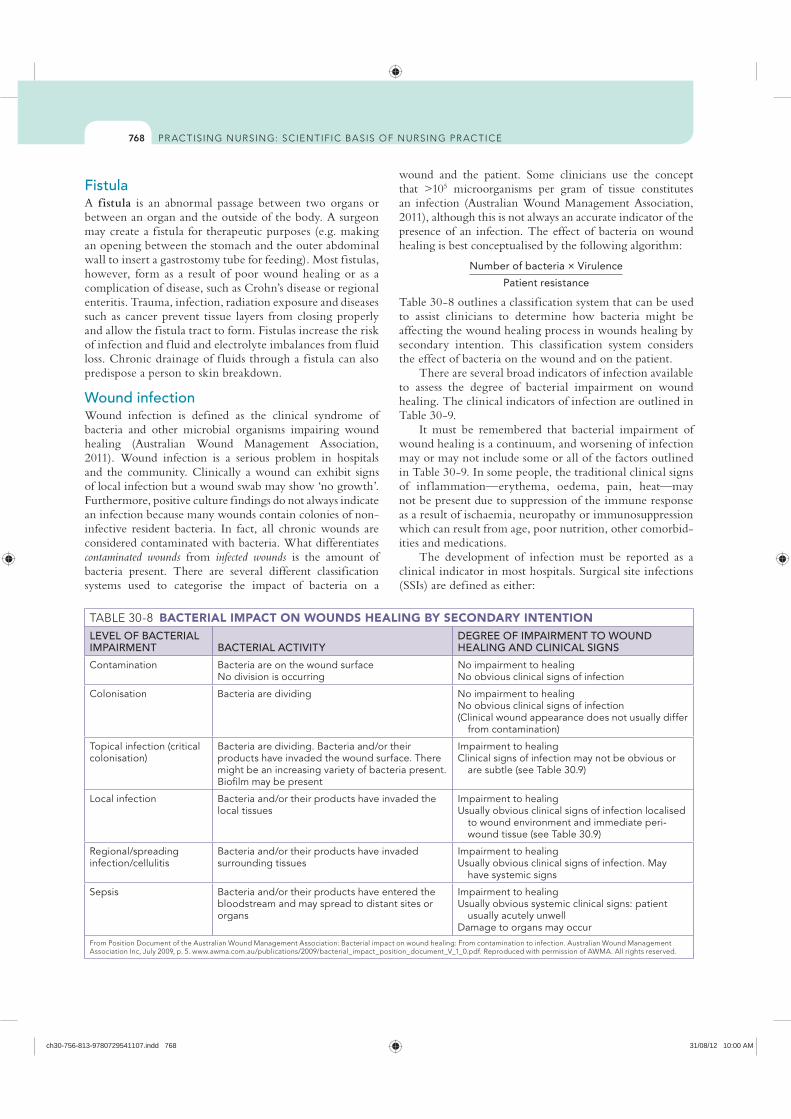
768
wound and the patient. Some clinicians use the concept that >105 microorganisms per gram of tissue constitutes an infection (Australian Wound Management Association, 2011), although this is not always an accurate indicator of the presence of an infection. The effect of bacteria on wound healing is best conceptualised by the following algorithm:
Number of bacteria × Virulence
Patient resistance
Table 30-8 outlines a classification system that can be used to assist clinicians to determine how bacteria might be affecting the wound healing process in wounds healing by secondary intention. This classification system considers the effect of bacteria on the wound and on the patient.
There are several broad indicators of infection available to assess the degree of bacterial impairment on wound healing. The clinical indicators of infection are outlined in Table 30-9.
It must be remembered that bacterial impairment of wound healing is a continuum, and worsening of infection may or may not include some or all of the factors outlined in Table 30-9. In some people, the traditional clinical signs of inf lammation—erythema, oedema, pain, heat—may not be present due to suppression of the immune response as a result of ischaemia, neuropathy or immunosuppression which can result from age, poor nutrition, other comorbid-ities and medications.
The development of infection must be reported as a clinical indicator in most hospitals. Surgical site infections (SSIs) are defined as either:
FistulaA fistula is an abnormal passage between two organs or between an organ and the outside of the body. A surgeon may create a fistula for therapeutic purposes (e.g. making an opening between the stomach and the outer abdominal wall to insert a gastrostomy tube for feeding). Most fistulas, however, form as a result of poor wound healing or as a complication of disease, such as Crohn’s disease or regional enteritis. Trauma, infection, radiation exposure and diseases such as cancer prevent tissue layers from closing properly and allow the fistula tract to form. Fistulas increase the risk of infection and f luid and electrolyte imbalances from f luid loss. Chronic drainage of f luids through a fistula can also predispose a person to skin breakdown.
Wound infectionWound infection is defined as the clinical syndrome of bacteria and other microbial organisms impairing wound healing (Australian Wound Management Association, 2011). Wound infection is a serious problem in hospitals and the community. Clinically a wound can exhibit signs of local infection but a wound swab may show ‘no growth’. Furthermore, positive culture findings do not always indicate an infection because many wounds contain colonies of non-infective resident bacteria. In fact, all chronic wounds are considered contaminated with bacteria. What differentiates contaminated wounds from infected wounds is the amount of bacteria present. There are several different classification systems used to categorise the impact of bacteria on a
BACTERIAL IMPACT ON WOUNDS HEALING BY SECONDARY INTENTIONLEVEL OF BACTERIAL IMPAIRMENT BACTERIAL ACTIVITY
DEGREE OF IMPAIRMENT TO WOUND HEALING AND CLINICAL SIGNS
Contamination Bacteria are on the wound surfaceNo division is occurring
No impairment to healingNo obvious clinical signs of infection
Colonisation Bacteria are dividing No impairment to healingNo obvious clinical signs of infection(Clinical wound appearance does not usually differ
from contamination)
Topical infection (critical colonisation)
Bacteria are dividing. Bacteria and/or their products have invaded the wound surface. There might be an increasing variety of bacteria present. Biofilm may be present
Impairment to healingClinical signs of infection may not be obvious or
are subtle (see Table 30.9)
Local infection Bacteria and/or their products have invaded the local tissues
Impairment to healingUsually obvious clinical signs of infection localised
to wound environment and immediate peri-wound tissue (see Table 30.9)
Regional/spreading infection/cellulitis
Bacteria and/or their products have invaded surrounding tissues
Impairment to healingUsually obvious clinical signs of infection. May
have systemic signs
Sepsis Bacteria and/or their products have entered the bloodstream and may spread to distant sites or organs
Impairment to healingUsually obvious systemic clinical signs: patient
usually acutely unwellDamage to organs may occur
ch30-756-813-9780729541107.indd 768 31/08/12 10:00 AM
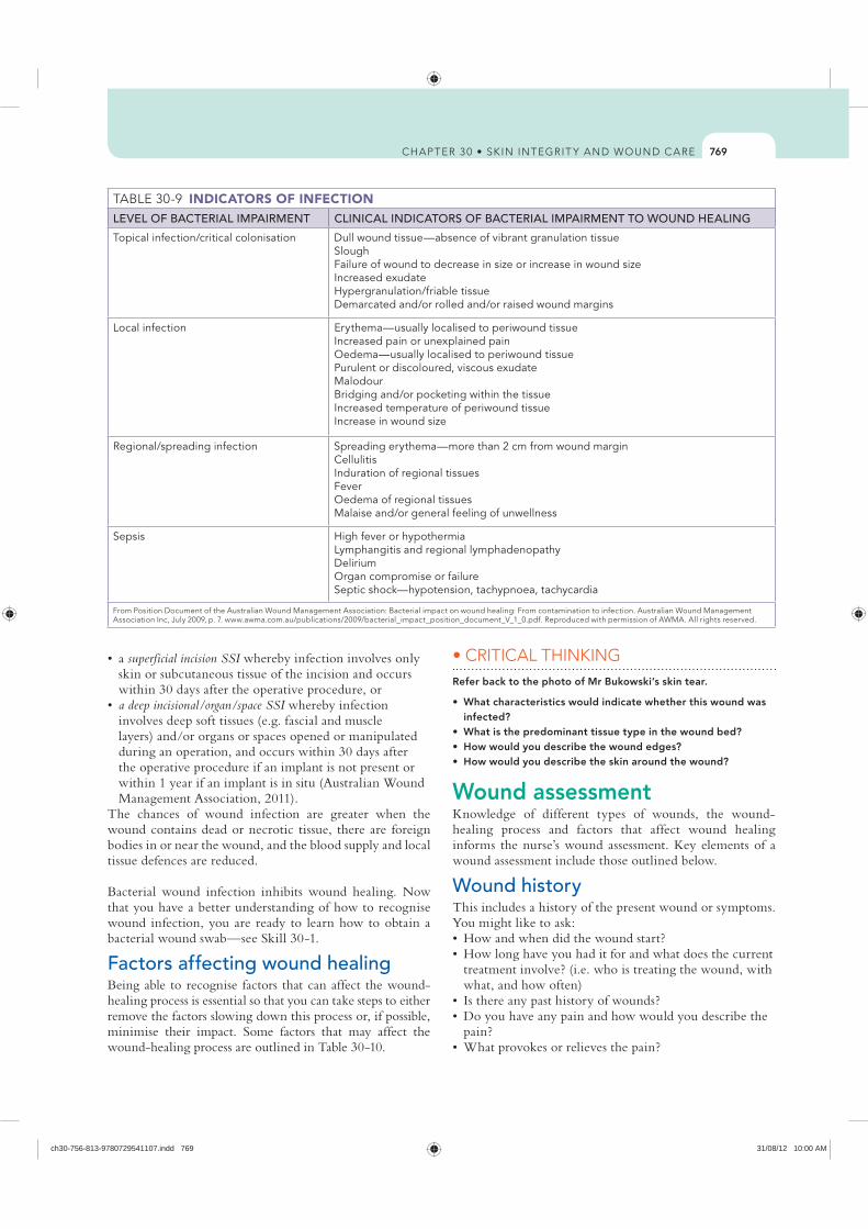
769
Refer back to the photo of Mr Bukowski’s skin tear.
infected?
Wound assessmentKnowledge of different types of wounds, the wound-healing process and factors that affect wound healing informs the nurse’s wound assessment. Key elements of a wound assessment include those outlined below.
Wound historyThis includes a history of the present wound or symptoms. You might like to ask:
what, and how often)
superficial incision SSI whereby infection involves only skin or subcutaneous tissue of the incision and occurs within 30 days after the operative procedure, ora deep incisional/organ/space SSI whereby infection involves deep soft tissues (e.g. fascial and muscle layers) and/or organs or spaces opened or manipulated during an operation, and occurs within 30 days after the operative procedure if an implant is not present or within 1 year if an implant is in situ (Australian Wound Management Association, 2011).
The chances of wound infection are greater when the wound contains dead or necrotic tissue, there are foreign bodies in or near the wound, and the blood supply and local tissue defences are reduced.
Bacterial wound infection inhibits wound healing. Now that you have a better understanding of how to recognise wound infection, you are ready to learn how to obtain a bacterial wound swab—see Skill 30-1.
Factors affecting wound healingBeing able to recognise factors that can affect the wound-healing process is essential so that you can take steps to either remove the factors slowing down this process or, if possible, minimise their impact. Some factors that may affect the wound-healing process are outlined in Table 30-10.
INDICATORS OF INFECTION LEVEL OF BACTERIAL IMPAIRMENT CLINICAL INDICATORS OF BACTERIAL IMPAIRMENT TO WOUND HEALING
Topical infection/critical colonisation Dull wound tissue—absence of vibrant granulation tissueSloughFailure of wound to decrease in size or increase in wound sizeIncreased exudateHypergranulation/friable tissueDemarcated and/or rolled and/or raised wound margins
Local infection Erythema—usually localised to periwound tissueIncreased pain or unexplained painOedema—usually localised to periwound tissuePurulent or discoloured, viscous exudateMalodourBridging and/or pocketing within the tissueIncreased temperature of periwound tissueIncrease in wound size
Regional/spreading infection Spreading erythema—more than 2 cm from wound marginCellulitisInduration of regional tissuesFeverOedema of regional tissuesMalaise and/or general feeling of unwellness
Sepsis High fever or hypothermiaLymphangitis and regional lymphadenopathyDeliriumOrgan compromise or failureSeptic shock—hypotension, tachypnoea, tachycardia
ch30-756-813-9780729541107.indd 769 31/08/12 10:00 AM
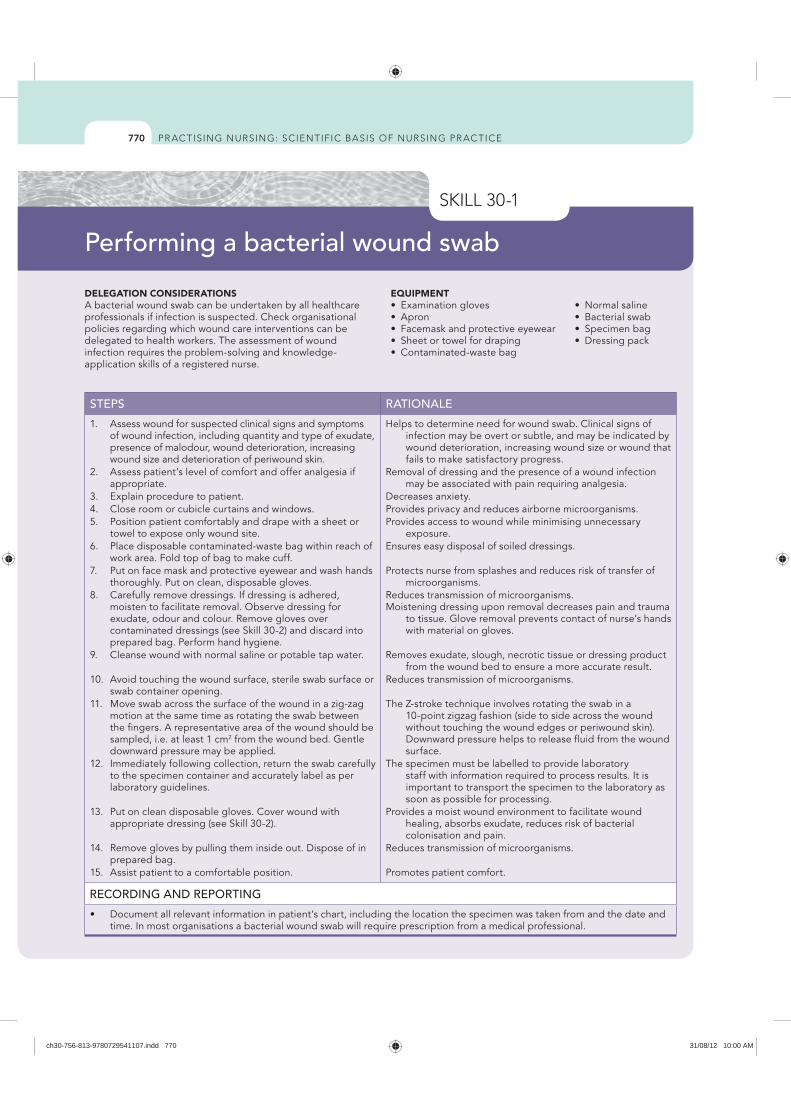
770
DELEGATION CONSIDERATIONSA bacterial wound swab can be undertaken by all healthcare professionals if infection is suspected. Check organisational policies regarding which wound care interventions can be delegated to health workers. The assessment of wound infection requires the problem-solving and knowledge-application skills of a registered nurse.
EQUIPMENT
STEPS RATIONALE
1. Assess wound for suspected clinical signs and symptoms of wound infection, including quantity and type of exudate, presence of malodour, wound deterioration, increasing wound size and deterioration of periwound skin.
Helps to determine need for wound swab. Clinical signs of infection may be overt or subtle, and may be indicated by wound deterioration, increasing wound size or wound that fails to make satisfactory progress.
2. Assess patient’s level of comfort and offer analgesia if appropriate.
Removal of dressing and the presence of a wound infection may be associated with pain requiring analgesia.
3. Explain procedure to patient. Decreases anxiety.4. Close room or cubicle curtains and windows. Provides privacy and reduces airborne microorganisms.5. Position patient comfortably and drape with a sheet or
towel to expose only wound site.Provides access to wound while minimising unnecessary
exposure.6. Place disposable contaminated-waste bag within reach of
work area. Fold top of bag to make cuff.Ensures easy disposal of soiled dressings.
7. Put on face mask and protective eyewear and wash hands thoroughly. Put on clean, disposable gloves.
Protects nurse from splashes and reduces risk of transfer of microorganisms.
8. Carefully remove dressings. If dressing is adhered, moisten to facilitate removal. Observe dressing for exudate, odour and colour. Remove gloves over contaminated dressings (see Skill 30-2) and discard into prepared bag. Perform hand hygiene.
Reduces transmission of microorganisms. Moistening dressing upon removal decreases pain and trauma
to tissue. Glove removal prevents contact of nurse’s hands with material on gloves.
9. Cleanse wound with normal saline or potable tap water. Removes exudate, slough, necrotic tissue or dressing product from the wound bed to ensure a more accurate result.
10. Avoid touching the wound surface, sterile swab surface or swab container opening.
Reduces transmission of microorganisms.
11. Move swab across the surface of the wound in a zig-zag motion at the same time as rotating the swab between the fingers. A representative area of the wound should be sampled, i.e. at least 1 cm2 from the wound bed. Gentle downward pressure may be applied.
The Z-stroke technique involves rotating the swab in a 10-point zigzag fashion (side to side across the wound without touching the wound edges or periwound skin). Downward pressure helps to release fluid from the wound surface.
12. Immediately following collection, return the swab carefully to the specimen container and accurately label as per laboratory guidelines.
The specimen must be labelled to provide laboratory staff with information required to process results. It is important to transport the specimen to the laboratory as soon as possible for processing.
13. Put on clean disposable gloves. Cover wound with appropriate dressing (see Skill 30-2).
Provides a moist wound environment to facilitate wound healing, absorbs exudate, reduces risk of bacterial colonisation and pain.
14. Remove gloves by pulling them inside out. Dispose of in prepared bag.
Reduces transmission of microorganisms.
15. Assist patient to a comfortable position. Promotes patient comfort.
RECORDING AND REPORTING
time. In most organisations a bacterial wound swab will require prescription from a medical professional.
Performing a bacterial wound swab
ch30-756-813-9780729541107.indd 770 31/08/12 10:00 AM
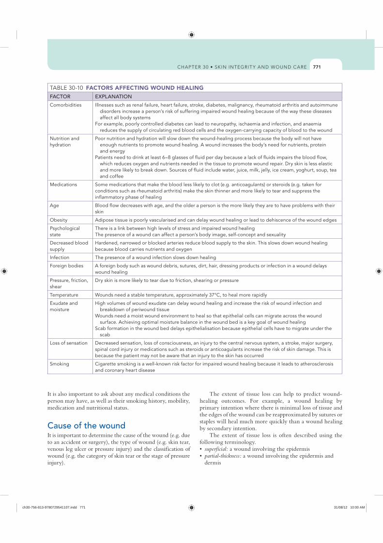
771
FACTORS AFFECTING WOUND HEALINGFACTOR EXPLANATION
Comorbidities Illnesses such as renal failure, heart failure, stroke, diabetes, malignancy, rheumatoid arthritis and autoimmune disorders increase a person’s risk of suffering impaired wound healing because of the way these diseases affect all body systems
For example, poorly controlled diabetes can lead to neuropathy, ischaemia and infection, and anaemia reduces the supply of circulating red blood cells and the oxygen-carrying capacity of blood to the wound
Nutrition and hydration
Poor nutrition and hydration will slow down the wound-healing process because the body will not have enough nutrients to promote wound healing. A wound increases the body’s need for nutrients, protein and energy
Patients need to drink at least 6–8 glasses of fluid per day because a lack of fluids impairs the blood flow, which reduces oxygen and nutrients needed in the tissue to promote wound repair. Dry skin is less elastic and more likely to break down. Sources of fluid include water, juice, milk, jelly, ice cream, yoghurt, soup, tea and coffee
Medications Some medications that make the blood less likely to clot (e.g. anticoagulants) or steroids (e.g. taken for conditions such as rheumatoid arthritis) make the skin thinner and more likely to tear and suppress the inflammatory phase of healing
Age Blood flow decreases with age, and the older a person is the more likely they are to have problems with their skin
Obesity Adipose tissue is poorly vascularised and can delay wound healing or lead to dehiscence of the wound edges
Psychological state
There is a link between high levels of stress and impaired wound healingThe presence of a wound can affect a person’s body image, self-concept and sexuality
Decreased blood supply
Hardened, narrowed or blocked arteries reduce blood supply to the skin. This slows down wound healing because blood carries nutrients and oxygen
Infection The presence of a wound infection slows down healing
Foreign bodies A foreign body such as wound debris, sutures, dirt, hair, dressing products or infection in a wound delays wound healing
Pressure, friction, shear
Dry skin is more likely to tear due to friction, shearing or pressure
Temperature Wounds need a stable temperature, approximately 37°C, to heal more rapidly
Exudate and moisture
High volumes of wound exudate can delay wound healing and increase the risk of wound infection and breakdown of periwound tissue
Wounds need a moist wound environment to heal so that epithelial cells can migrate across the wound surface. Achieving optimal moisture balance in the wound bed is a key goal of wound healing
Scab formation in the wound bed delays epithelialisation because epithelial cells have to migrate under the scab
Loss of sensation Decreased sensation, loss of consciousness, an injury to the central nervous system, a stroke, major surgery, spinal cord injury or medications such as steroids or anticoagulants increase the risk of skin damage. This is because the patient may not be aware that an injury to the skin has occurred
Smoking Cigarette smoking is a well-known risk factor for impaired wound healing because it leads to atherosclerosis and coronary heart disease
It is also important to ask about any medical conditions the person may have, as well as their smoking history, mobility, medication and nutritional status.
Cause of the wound It is important to determine the cause of the wound (e.g. due to an accident or surgery), the type of wound (e.g. skin tear, venous leg ulcer or pressure injury) and the classification of wound (e.g. the category of skin tear or the stage of pressure injury).
The extent of tissue loss can help to predict wound-healing outcomes. For example, a wound healing by primary intention where there is minimal loss of tissue and the edges of the wound can be reapproximated by sutures or staples will heal much more quickly than a wound healing by secondary intention.
The extent of tissue loss is often described using the following terminology.
superficial: a wound involving the epidermispartial-thickness: a wound involving the epidermis and dermis
ch30-756-813-9780729541107.indd 771 31/08/12 10:00 AM
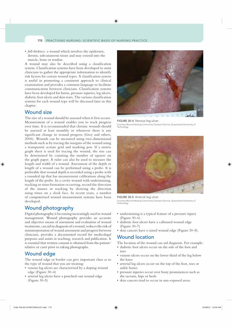
772
(Figure 30-6)
(Figure 30-7)
Wound locationThe location of the wound can aid diagnosis. For example:
toes
the knee
ankle bones
the sacrum, hips or heels
full-thickness: a wound which involves the epidermis, dermis, subcutaneous tissue and may extend into the muscle, bone or tendon.
A wound may also be described using a classification system. Classification systems have been developed to assist clinicians to gather the appropriate information to identify risk factors for certain wound types. A classification system is useful in promoting a consistent approach to clinical examination and provides a common language to facilitate communication between clinicians. Classification systems have been developed for burns, pressure injuries, leg ulcers, diabetic foot ulcers and skin tears. The various classification systems for each wound type will be discussed later in this chapter.
Wound sizeThe size of a wound should be assessed when it f irst occurs. Measurement of a wound enables you to track progress over time. It is recommended that chronic wounds should be assessed at least monthly or whenever there is any significant change in wound progress (Grey and others, 2006). Wounds can be measured using two-dimensional methods such as by tracing the margins of the wound using a transparent acetate grid and marking pen. If a metric graph sheet is used for tracing the wound, the size can be determined by counting the number of squares on the graph paper. A ruler can also be used to measure the length and width of a wound. Assessment of the depth or length of a wound can be performed using a probe. It is preferable that wound depth is recorded using a probe with a rounded tip that has measurement calibrations along the length of the probe. In a cavity wound with undermining, tracking or sinus formation occurring, record the direction of the sinuses or tracking by drawing the direction using times on a clock face. In recent years, a number of computerised wound measurement systems have been developed.
Wound photographyDigital photography is becoming increasingly used in wound management. Wound photography provides an accurate and objective means of assessment and evaluation of wound treatments, can aid in diagnosis of a wound, reduces the risk of misinterpretation of wound assessment and progress between clinicians, provides a documented record for medicolegal purposes and assists in teaching, research and publication. It is essential that written consent is obtained from the patient/relative or carer prior to taking photographs.
Wound edge The wound edge or border can give important clues as to the type of wound that you are treating:
edge (Figure 30-4)
(Figure 30-5)
FIGURE 30-4
Technology.
FIGURE 30-5 Arterial leg ulcer.
Technology.
ch30-756-813-9780729541107.indd 772 31/08/12 10:00 AM
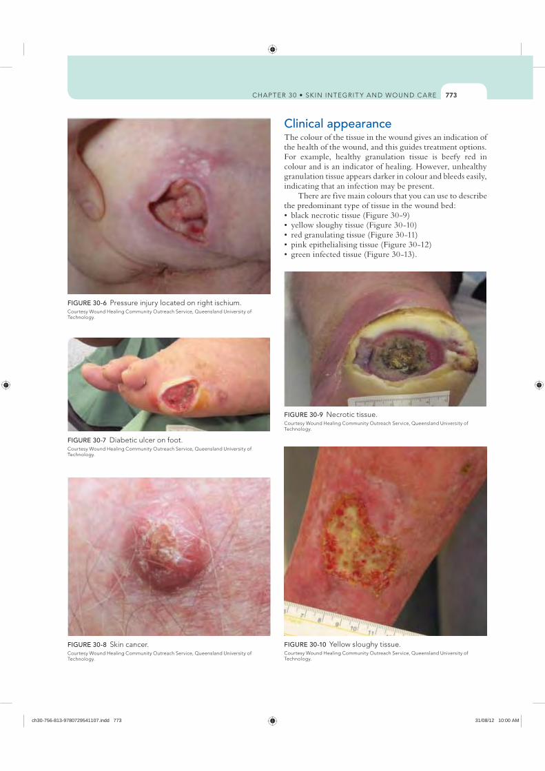
773
Clinical appearanceThe colour of the tissue in the wound gives an indication of the health of the wound, and this guides treatment options. For example, healthy granulation tissue is beefy red in colour and is an indicator of healing. However, unhealthy granulation tissue appears darker in colour and bleeds easily, indicating that an infection may be present.
There are five main colours that you can use to describe the predominant type of tissue in the wound bed:
FIGURE 30-6 Pressure injury located on right ischium.
Technology.
FIGURE 30-7 Diabetic ulcer on foot.
Technology.
FIGURE 30-8 Skin cancer.
Technology.
FIGURE 30-9
Technology.
FIGURE 30-10
Technology.
ch30-756-813-9780729541107.indd 773 31/08/12 10:00 AM
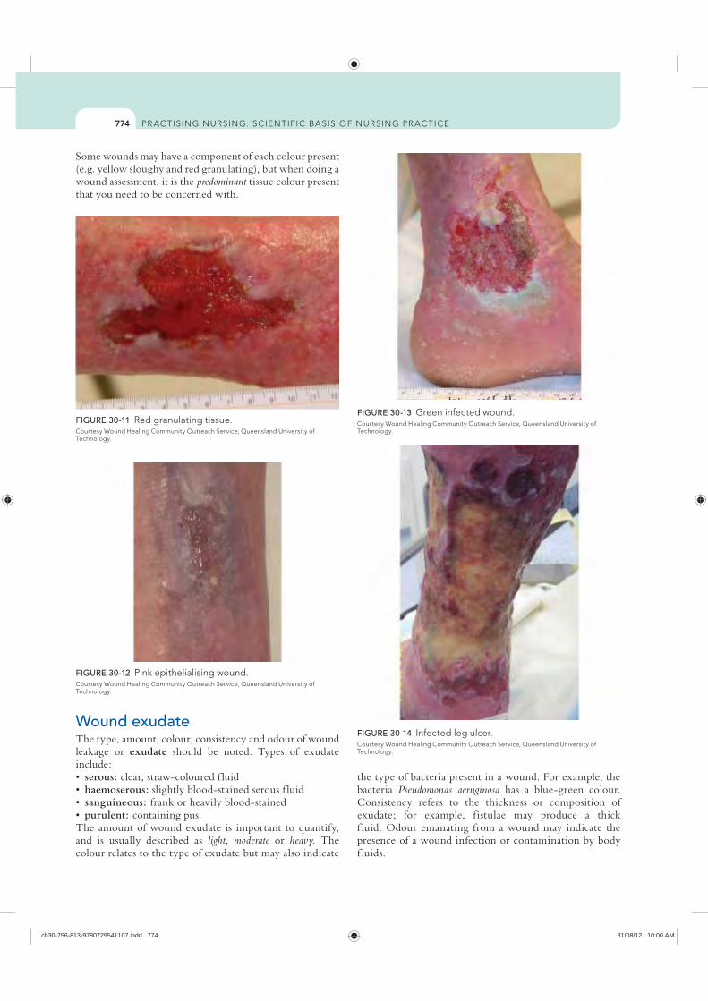
774
the type of bacteria present in a wound. For example, the bacteria Pseudomonas aeruginosa has a blue-green colour. Consistency refers to the thickness or composition of exudate; for example, fistulae may produce a thick f luid. Odour emanating from a wound may indicate the presence of a wound infection or contamination by body f luids.
Some wounds may have a component of each colour present (e.g. yellow sloughy and red granulating), but when doing a wound assessment, it is the predominant tissue colour present that you need to be concerned with.
FIGURE 30-11 Red granulating tissue.
Technology.
FIGURE 30-12 Pink epithelialising wound.
Technology.
FIGURE 30-13
Technology.
Wound exudate The type, amount, colour, consistency and odour of wound leakage or exudate should be noted. Types of exudate include:
serous: clear, straw-coloured f luidhaemoserous: slightly blood-stained serous f luidsanguineous: frank or heavily blood-stainedpurulent: containing pus.
The amount of wound exudate is important to quantify, and is usually described as light, moderate or heavy. The colour relates to the type of exudate but may also indicate
FIGURE 30-14
Technology.
ch30-756-813-9780729541107.indd 774 31/08/12 10:00 AM
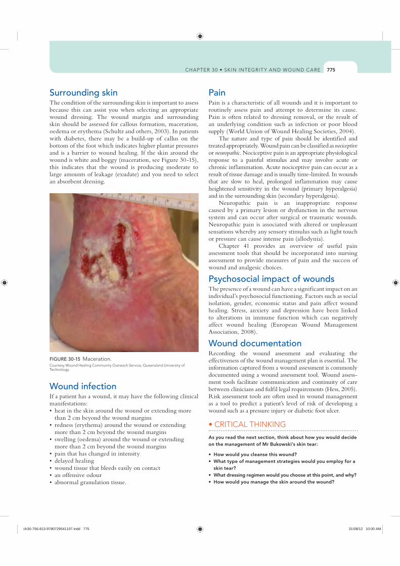
775
PainPain is a characteristic of all wounds and it is important to routinely assess pain and attempt to determine its cause. Pain is often related to dressing removal, or the result of an underlying condition such as infection or poor blood supply (World Union of Wound Healing Societies, 2004).
The nature and type of pain should be identified and treated appropriately. Wound pain can be classified as nociceptive or neuropathic. Nociceptive pain is an appropriate physiological response to a painful stimulus and may involve acute or chronic inf lammation. Acute nociceptive pain can occur as a result of tissue damage and is usually time-limited. In wounds that are slow to heal, prolonged inf lammation may cause heightened sensitivity in the wound (primary hyperalgesia) and in the surrounding skin (secondary hyperalgesia).
Neuropathic pain is an inappropriate response caused by a primary lesion or dysfunction in the nervous system and can occur after surgical or traumatic wounds. Neuropathic pain is associated with altered or unpleasant sensations whereby any sensory stimulus such as light touch or pressure can cause intense pain (allodynia).
Chapter 41 provides an overview of useful pain assessment tools that should be incorporated into nursing assessment to provide measures of pain and the success of wound and analgesic choices.
Psychosocial impact of woundsThe presence of a wound can have a significant impact on an individual’s psychosocial functioning. Factors such as social isolation, gender, economic status and pain affect wound healing. Stress, anxiety and depression have been linked to alterations in immune function which can negatively affect wound healing (European Wound Management Association, 2008).
Wound documentation Recording the wound assessment and evaluating the effectiveness of the wound management plan is essential. The information captured from a wound assessment is commonly documented using a wound assessment tool. Wound assess-ment tools facilitate communication and continuity of care between clinicians and fulfil legal requirements (Hess, 2005). Risk assessment tools are often used in wound management as a tool to predict a patient’s level of risk of developing a wound such as a pressure injury or diabetic foot ulcer.
As you read the next section, think about how you would decide on the management of Mr Bukowski’s skin tear:
skin tear?
Surrounding skinThe condition of the surrounding skin is important to assess because this can assist you when selecting an appropriate wound dressing. The wound margin and surrounding skin should be assessed for callous formation, maceration, oedema or erythema (Schultz and others, 2003). In patients with diabetes, there may be a build-up of callus on the bottom of the foot which indicates higher plantar pressures and is a barrier to wound healing. If the skin around the wound is white and boggy (maceration, see Figure 30-15), this indicates that the wound is producing moderate to large amounts of leakage (exudate) and you need to select an absorbent dressing.
FIGURE 30-15 Maceration.
Technology.
Wound infection If a patient has a wound, it may have the following clinical manifestations:
than 2 cm beyond the wound margins
more than 2 cm beyond the wound margins
more than 2 cm beyond the wound margins
ch30-756-813-9780729541107.indd 775 31/08/12 10:00 AM
776
(Whitney and others, 2006). Cleaning should be undertaken with minimal chemical or mechanical trauma. The ultimate aim of wound cleansing is to remove both organic and inorganic debris while maintaining the optimum local environment to facilitate wound healing (Fernandez and Griffiths, 2012). Before performing wound cleansing, consider the purpose of wound cleansing and whether the advantages of cleansing outweigh the disadvantages; the most appropriate method of wound cleansing; whether the wound requires cleansing at each dressing change; and the type of wound-cleansing product that is most appropriate.
Common reasons for cleansing a wound are to help prevent wound infection and remove excess debris or foreign bodies from the wound; to rehydrate the wound to provide a moist environment; to make wound assessment easier so that the size and extent of the wound can be visualised; before applying a dressing, to minimise trauma when removing adherent dressings; and to promote patient comfort and psychological wellbeing.
The decision to cleanse a wound should be based on the shape, size and location of the wound; condition of the wound bed and stage of healing; availability and effectiveness of different methods of cleansing; availability and effectiveness of different cleansing agents; and the patient’s perceptions and needs.
The decision to use tap-water to cleanse wounds should be based on clinical judgment and take into account the quality of water, the nature of the wound and the patient’s general condition (Fernandez and Griffiths, 2012). It is recommended that tap-water should not be used if it is not suitable for drinking and the tap should be run for 15 seconds prior to use ( Joanna Briggs Institute, 2006). If potable tap-water or normal saline is not available, cooled boiled water is an effective wound-cleansing solution ( Joanna Briggs Institute, 2006). In wounds that are contaminated or infected, irrigation with 1% povidone–iodine is effective at reducing the infection rate, although there is no evidence of the optimal time that povidone–iodine should be left in situ ( Joanna Briggs Institute, 2006).
Gentle irrigation is effective at reducing infection and inf lammation in patients with lacerations and traumatic wounds without causing tissue trauma (Joanna Briggs Institute, 2006). There is no evidence to support or refute swabbing and scrubbing as effective wound-cleansing techniques (Fernandez and Griffiths, 2012). When consider-ing the method of wound cleansing, evidence suggests that showering patients does not affect infection and healing rates of postoperative wounds and may benefit patients with a feeling of wellbeing and health associated with cleanliness ( Joanna Briggs Institute, 2006). Showering for cleansing other wounds should be undertaken with caution.
Wound debridement Wound debridement is an essential component of optimal wound management. Debridement describes any method
Principles of wound management Many factors affect the wound-healing process. Therefore, wound management strategies must be tailored to meet the individual holistic needs of the patient, their wound and their environment.
Assess and correct cause of tissue damage The health status of the patient has a significant impact on the wound-healing process; therefore, a general health history including a medication history is essential in order to identify factors that may affect wound healing. There are many medications that can impair wound healing, including systemic steroids, immunosuppressive drugs and non-steroidal anti-inf lammatories (Schultz and others, 2003). Chronic diseases such as diabetes mellitus and rheumatoid arthritis and autoimmune disorders such as systemic lupus erythematosus, vasculitis and pyoderma gangrenosum also delay wound healing.
Assess wound history and characteristics Ongoing assessment and evaluation of the wound is an essential step in promoting wound healing.
Ensure adequate tissue perfusionIt is essential that the tissue is well perfused, as a well-vascularised wound bed provides nutrients and oxygen to promote granulation tissue formation and to maintain active immunological response to bacteria. Decreased oxygen levels impair the ability of leucocytes to kill bacteria, lower production of collagen and reduce epithelialisation (Schultz and others, 2003).
Wound-bed preparation Wound-bed preparation is the management of the wound to promote wound healing or to facilitate the effectiveness of other therapeutic measures (Schultz and others, 2003). Wound-bed preparation involves four fundamental principles which provide a systematic approach to the management of wounds by focusing on each stage of wound healing: 1. Tissue: removal of non-viable tissue or replacement of
deficient tissue2. Control of infection or inf lammation3. Moisture imbalance: correction of excessive moisture and
prevention of desiccation 4. Revision of the edge of wound to stimulate healing. These principles will now be explored in further detail.
Wound cleansingWounds should be cleaned with a neutral, non-irritating, non-toxic solution such as clean tap-water or normal saline
ch30-756-813-9780729541107.indd 776 31/08/12 10:00 AM
777
risks. Surgical debridement must be used with caution in patients with clotting disorders or on anticoagulant therapy and in patients with diabetes and peripheral vascular disease.Mechanical debridement is the process of physically removing debris from the wound bed. The most commonly used methods of mechanical debridement include wet-to-dry dressings, whirlpool therapy, pulsed lavage and ultrasound. Mechanical debridement is non-selective, time-consuming, frequently painful and associated with an increased risk of infection, damage to granulation tissue, prolonged inf lammation and periwound maceration.Biological debridement involves the application of sterile maggots, specifically the larvae of the green bottle f ly (Lucilia sericata), to the wound bed. Enzymatic debridement is the topical application of an exogenous enzyme to the wound bed to facilitate a host response. Topical enzymatic agents may be plant-, animal- or microbe-derived. They are not commercially available in Australia or New Zealand at present.
When selecting the most appropriate method of debride-ment, the indications and contraindications for the different types of debridement options must be considered. Evidence supports that debridement is an important step in preparing the wound bed for healing (Ayello and Cuddigan, 2004; Kirshen and others, 2006; Moffatt and others, 2008).
Provision of a moist wound environmentA moist wound environment is essential to promote wound healing because cells need moisture to be able to migrate across a wound bed. Extreme wetness or dryness may delay healing. Application of dressings to a wound may help to facilitate wound healing by providing the optimal wound environment. Wound dressings may also be applied to:
Before applying a dressing, it is essential that you know:
There are two different categories of wound dressings: primary dressings—those in direct contact with a wound; and secondary dressings—those not in contact with a wound but covering the primary dressing (Xue, 2008). When selecting a secondary dressing, it is important to ensure that it is compatible with the primary wound-contact layer.
Multiple factors inform the selection of a wound dress-ing: the type or aetiology of the wound, location or position of the wound, extent of tissue damage and size of the wound,
that facilitates the removal of dead or devitalised tissue, metabolic waste, fibrin and foreign material from a wound (Gethin and others, 2010). Removal of necrotic and devitalised tissue can be undertaken through surgical, sharp, mechanical, autolytic, biological or enzymatic debridement techniques (Hopf and others, 2006; World Union of Wound Healing Societies, 2007). If dry gangrene or eschar (leathery brown or black necrotic tissue) is present, however, debridement should not be undertaken until arterial f low has been re-established (Whitney and others, 2006). There is insufficient evidence to conclude that any one debriding agent is more effective than another (Gethin and others, 2010; Williams and others, 2005).
When selecting an appropriate method of debridement a number of factors must be considered, including the patient’s individual wishes and concerns, medical history, factors affecting the wound-healing process, pain, wound characteristics, skill and knowledge of the clinician and available resources (Kirshen and others, 2006). Specific wound characteristics that inf luence debridement decisions include size, depth and amount of exudate, as well as the characteristics of wound tissue, potential for bleeding, and infection. Methods of debridement that are selective are generally preferred because they remove only necrotic tissue.
Autolytic debridement is the most frequently used method of debridement whereby there is breakdown and removal of devitalised tissue by the body’s own cells and enzymes, and this is facilitated through the provision of a moist wound environment (Royal College of Nursing, 1998; Ayello and Cuddigan, 2004). Maceration of periwound tissue may be problematic. For autolytic debridement to be effective the wound requires some level of exudate, and it is a slow method of debridement. It is not recommended for clinically infected wounds and in those with a high potential for anaerobic infection, or if there is ischaemia of the limb or digits (Gethin and others, 2010).
Sharp debridement may be performed at the bedside and involves selective removal of dead or devitalised tissue from within and around the wound to expose healthy tissue using sterile instruments such as scalpel, scissors or curette. This procedure involves similar risk factors to surgical debridement, although it is less aggressive, and must be performed by a skilled and experienced clinician.
Surgical debridement is performed in an operating theatre usually under regional or general anaesthetic. It is indicated if there is extensive necrotic tissue, advancing cellulitis, infected bone or sepsis. This method of debridement is the most rapid and highly selective; however, the number of personnel and degree of expertise required increase cost and limit the availability of the procedure. There is also an increased risk of pain, bleeding, transient bacteraemia, damage to vital structures, tendon sheaths and nerves and anaesthesia
ch30-756-813-9780729541107.indd 777 31/08/12 10:00 AM
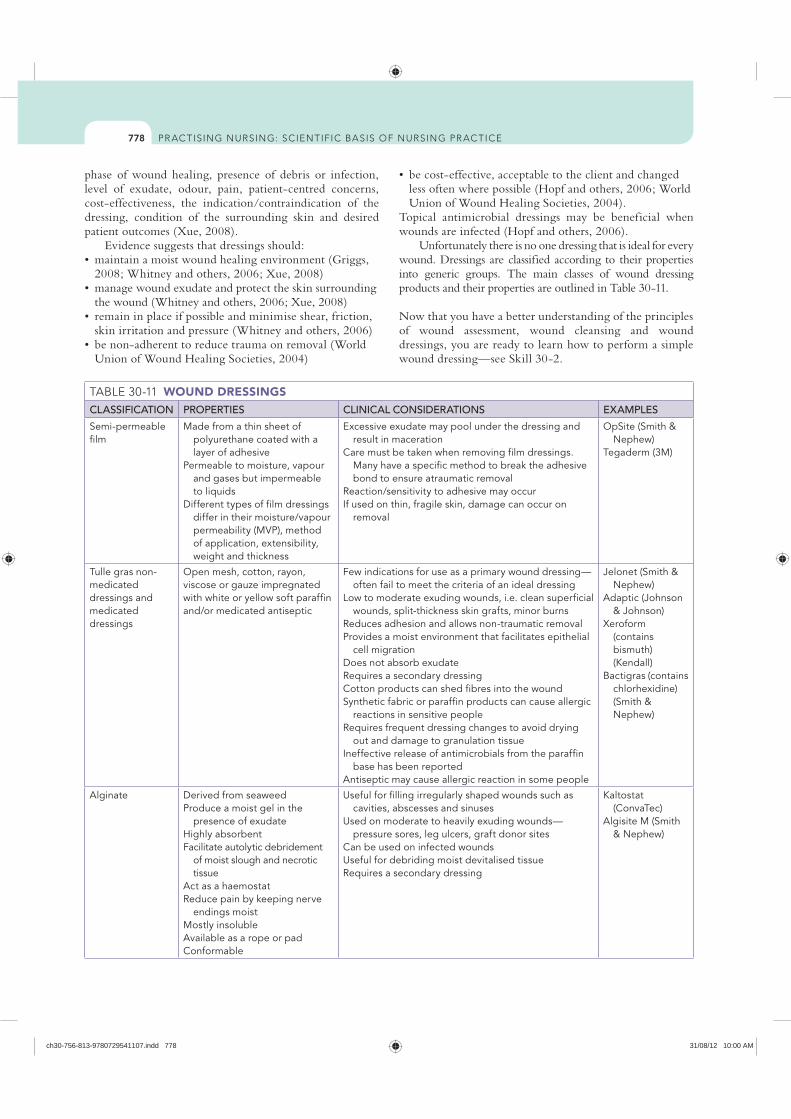
778
less often where possible (Hopf and others, 2006; World Union of Wound Healing Societies, 2004).
Topical antimicrobial dressings may be beneficial when wounds are infected (Hopf and others, 2006).
Unfortunately there is no one dressing that is ideal for every wound. Dressings are classified according to their properties into generic groups. The main classes of wound dressing products and their properties are outlined in Table 30-11.
Now that you have a better understanding of the principles of wound assessment, wound cleansing and wound dressings, you are ready to learn how to perform a simple wound dressing—see Skill 30-2.
phase of wound healing, presence of debris or infection, level of exudate, odour, pain, patient-centred concerns, cost-effectiveness, the indication/contraindication of the dressing, condition of the surrounding skin and desired patient outcomes (Xue, 2008).
Evidence suggests that dressings should:
2008; Whitney and others, 2006; Xue, 2008)
the wound (Whitney and others, 2006; Xue, 2008)
skin irritation and pressure (Whitney and others, 2006)
Union of Wound Healing Societies, 2004)
WOUND DRESSINGSCLASSIFICATION PROPERTIES CLINICAL CONSIDERATIONS EXAMPLES
Semi-permeable film
Made from a thin sheet of polyurethane coated with a layer of adhesive
Permeable to moisture, vapour and gases but impermeable to liquids
Different types of film dressings differ in their moisture/vapour permeability (MVP), method of application, extensibility, weight and thickness
Excessive exudate may pool under the dressing and result in maceration
Care must be taken when removing film dressings. Many have a specific method to break the adhesive bond to ensure atraumatic removal
Reaction/sensitivity to adhesive may occurIf used on thin, fragile skin, damage can occur on
removal
OpSite (Smith & Nephew)
Tegaderm (3M)
Tulle gras non-medicated dressings and medicated dressings
Open mesh, cotton, rayon, viscose or gauze impregnated with white or yellow soft paraffin and/or medicated antiseptic
Few indications for use as a primary wound dressing—often fail to meet the criteria of an ideal dressing
Low to moderate exuding wounds, i.e. clean superficial wounds, split-thickness skin grafts, minor burns
Reduces adhesion and allows non-traumatic removal Provides a moist environment that facilitates epithelial
cell migrationDoes not absorb exudateRequires a secondary dressingCotton products can shed fibres into the woundSynthetic fabric or paraffin products can cause allergic
reactions in sensitive peopleRequires frequent dressing changes to avoid drying
out and damage to granulation tissueIneffective release of antimicrobials from the paraffin
base has been reportedAntiseptic may cause allergic reaction in some people
Jelonet (Smith & Nephew)
Adaptic (Johnson & Johnson)
Xeroform (contains bismuth) (Kendall)
Bactigras (contains chlorhexidine) (Smith & Nephew)
Alginate Derived from seaweedProduce a moist gel in the
presence of exudate Highly absorbent Facilitate autolytic debridement
of moist slough and necrotic tissue
Act as a haemostat Reduce pain by keeping nerve
endings moistMostly insoluble Available as a rope or pad Conformable
Useful for filling irregularly shaped wounds such as cavities, abscesses and sinuses
Used on moderate to heavily exuding wounds—pressure sores, leg ulcers, graft donor sites
Can be used on infected woundsUseful for debriding moist devitalised tissueRequires a secondary dressing
Kaltostat (ConvaTec)
Algisite M (Smith & Nephew)
ch30-756-813-9780729541107.indd 778 31/08/12 10:00 AM
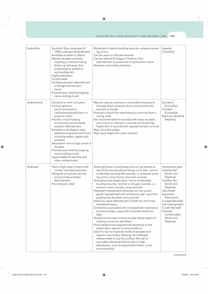
779
Hydrofibre Synthetic fibre composed of 100% carboxymethylcellulose
Available as sheet or ribbon Absorb exudate vertically,
resulting in moisture being drawn up and away thus preventing its spread to surrounding skin
Highly absorbent Conformable Facilitate autolytic debridement
of slough and necrotic tissue
Promote pain relief by keeping nerve endings moist
Moderate to heavily exuding wounds—pressure sores, leg ulcers
Can be used on infected woundsCan be used at all stages of healing, from
debridement to protection of granulation tissueRequires a secondary dressing
Aquacel (ConvaTec)
Hydrocolloid Occlusive or semi-occlusive Contain gelatine,
pectin and sodium carboxymethylcellulose in a polymer matrix
Provide a moist healing environment and facilitate autolytic debridement
Available in all shapes, sizes, adhesive properties and forms including wafers, pastes and powders
Absorbent—low to high levels of exudate
Promote pain relief by keeping nerve endings moist
Impermeable to bacteria and other contaminants
May be used as a primary or secondary dressing to manage select pressure ulcers and wounds with necrosis or slough
Produce a liquid that resembles pus and may have a strong smell
Not recommended for wounds with heavy exudate, sinus tracts or infections, wounds surrounded by fragile skin or wounds with exposed tendon or bone
May curl at the edgesMay injure fragile skin upon removal
Duoderm (ConvaTec)
Comfeel (Coloplast)
Replicare (Smith & Nephew)
Hydrogel Have a high water content and contain insoluble polymers
Designed to hydrate wounds and promote autolytic debridement
Promote pain relief
Sheet gels have a solid shape and can be placed on top of the wound without being cut to size—low to moderately exuding flat wounds, i.e. pressure sores, leg ulcers, minor burns, traumatic wounds
Amorphous (no shape) gels—low to moderately exuding wounds, necrotic or sloughy wounds, i.e. pressure sores, sinuses, cavity wounds
Hydrogel-impregnated dressings are non-woven gauze impregnated with amorphous gel—good for packing low-exudate cavity wounds
Useful for rapid debridement of both dry and moist devitalised tissue
Sometimes associated with increased skin maceration at wound edges, especially if exudate levels are high.
Should not be used in blind sinuses where extent of tracking cannot be identified
Some people have experienced sensitivity to the preservative agents in some products
Used for low to moderate levels of exudate and require a secondary dressing. As hydrogels release water to any dry surface, the use of secondary dressings that are dry or high-absorbency, such as polyurethane foam, is not recommended
Amorphous gels:Intrasite Gel
(Smith and Nephew)
SoloSite Gel (Smith and Nephew)
Gel sheets:Aquaclear
(Hartmann) Curagel (Kendall)Gel-impregnated:Curafil (Kendall)IntraSite
Conformable (Smith and Nephew)
continued
ch30-756-813-9780729541107.indd 779 31/08/12 10:00 AM
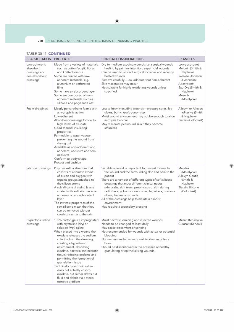
780
CONTINUEDCLASSIFICATION PROPERTIES CLINICAL CONSIDERATIONS EXAMPLES
Low-adherent, absorbent dressings and non-absorbent dressings
Made from a variety of materials such as cotton/acrylic fibres and knitted viscose
Some are coated with low-adherent materials, e.g. aluminium or perforated films
Some have an absorbent layerSome are composed of non-
adherent materials such as silicone and polyamide net
Dry to medium exuding wounds, i.e. surgical wounds healing by primary intention, superficial wounds
Can be used to protect surgical incisions and recently healed wounds
Remove carefully—low-adherent not non-adherent Skin maceration may occurNot suitable for highly exudating wounds unless
specified
Low-absorbent:Melonin (Smith &
Nephew)Release (Johnson
& Johnson)Absorbent:Exu-Dry (Smith &
Nephew) Mesorb
(MöInlycke)
Foam dressings Mostly polyurethane foams with a hydrophilic action
Low-adherent Absorbent dressings for low to
high levels of exudate Good thermal insulating
propertiesPermeable to water vapour,
preventing the wound from drying out
Available as non-adherent and adherent, occlusive and semi-occlusive
Conform to body shapeProtect and cushion
Low to heavily exuding wounds—pressure sores, leg ulcers, burns, graft donor sites
Moist wound environment may not be enough to allow autolysis to occur
May macerate periwound skin if they become saturated
Allevyn or Allevyn adhesive (Smith & Nephew)
Biatain (Coloplast)
Silicone dressings Polymer with a structure that consists of alternate atoms of silicon and oxygen with organic groups attached to the silicon atoms
A soft silicone dressing is one coated with soft silicone as an adhesive or wound-contact layer
The intrinsic properties of the soft silicone mean that they can be removed without causing trauma to the skin
Suitable where it is important to prevent trauma to the wound and the surrounding skin and pain to the patient
There are a number of different types of soft silicone dressings that meet different clinical needs—skin grafts, skin tears, prophylaxis of skin during radiotherapy, burns, donor sites, leg ulcers, pressure ulcers, traumatic wounds
All of the dressings help to maintain a moist environment
May require a secondary dressing
Mepilex (Mölnlycke)
Allevyn Gentle (Smith & Nephew)
Biatain Silicone (Coloplast)
Hypertonic saline dressings
100% cotton gauze impregnated with crystalline (dry) or solution (wet) saline
When placed into a wound the exudate releases the sodium chloride from the dressing, creating a hypertonic environment, absorbing exudate, bacteria and necrotic tissue, reducing oedema and permitting the formation of granulation tissue
Technically hypertonic saline does not actually absorb exudate, but rather draws out fluid and debris via a steep osmotic gradient
Moist necrotic, draining and infected woundsNeeds to be changed at least dailyMay cause discomfort or stinging Not recommended for wounds with actual or potential
bleedingNot recommended on exposed tendon, muscle or
bone Should be discontinued in the presence of healthy
granulating or epithelialising wounds
Mesalt (MöInlycke)Curasalt (Kendall)
ch30-756-813-9780729541107.indd 780 31/08/12 10:00 AM

781
Cadexomer iodine
Made up of starch micro-beads that contain 0.9% iodine
The cadexomer base absorbs exudate, swells and forms a gel
Iodine is released progressively into the wound
Iodine exerts an antimicrobial effect and bacteria are removed from the wound surface by the process of capillary action
Available as a powder, paste or ointment
Low to heavily exuding, sloughy woundsCan be used on infected woundsChanges colour from brown to white when the iodine
has been absorbed, indicating the need for a dressing change
Not for use during pregnancy or with thyroid disease or suspected iodine sensitivities owing to the risk of systemic absorption
Some patients report slight discomfort when dressing first applied
The maximum single application is 50 g and weekly maximum must not exceed 150 g
Treatment duration should not exceed 3 months in a single course of treatment
Will require a secondary dressing
Iodosorb powder, Iodosorb paste, Iodoflex (Smith & Nephew)
Silver dressings All silver-based dressings, whether alginates, foams, films, hydrofibres or other materials, achieve their antimicrobial action by generating and releasing silver into the wound
Silver is a potent antimicrobial; bactericidal mechanisms of action of silver are not fully understood
Silver dressings are active against a variety of micro-organisms including Staphylococcus aureus, methicillin-resistant S. aureus (MRSA), Pseudomonas aeruginosa and vancomycin-resistant enterococci (VRE)
To reduce the risk of wound infection and to treat infected wounds; provide sustained antimicrobial activity
Can dry and adhere if minimal exudate Should not be used with other antimicrobialsDo not dilute with saline (deactivates silver)Should not be used with zinc- or oil-based emollientsVery expensiveMay mask host response to infectionShould be used for short periods of time, i.e.
3–4 weeks, and the effectiveness re-evaluatedContraindicated in patients with known
hypersensitivity to any of the components of the product. If signs of a sensitivity reaction develop during use, treatment should be discontinued
Acticoat (Smith & Nephew)
Aquacel-Ag (ConvaTec)
Biatain Ag (Coloplast)
Actisorb Silver (Johnson & Johnson)
Atraumann-Ag (Hartmann)
Mepilex Ag (Mölnlycke)
Medicated honey Comprised of 82% carbohydrate, enzymes and amino acids
Acidic pH between 3.2 and 4.5; low enough to be inhibitory to many pathogens
Possesses natural anti-inflammatory effects and stimulates granulation tissue
Antimicrobial activity mainly through component of hydrogen peroxide
Reduces odour Provides a moist environment
and produces rapid tissue regeneration and debridement of non-viable tissue
For use on infected or highly contaminated wounds, malodorous wounds
Apply honey liberally to either the surface of the wound or a dressing
Frequency of dressing changes depends on how rapidly the gel is diluted by exudate
Honey is not sterile and there is a perceived risk of wound contamination from the presence of Clostridium botulinum spores and Bacillus spp. in honey
Only honey that has been sterilised/treated by gamma irradiation should be used on wounds
Contraindications: Allergies to bees Hypersensitivity reactions Transient stinging/burning on application
Medihony (Comvita)
Odour-absorbing dressings
Contain activated charcoal to absorb odour, bacteria and exudate
Infected or malodorous wounds Malignant or fungating wounds May need a secondary dressing
Actisorb Plus (Johnson and Johnson)
Carbonet (Smith and Nephew)
CarboFlex (ConvaTec)
ch30-756-813-9780729541107.indd 781 31/08/12 10:00 AM
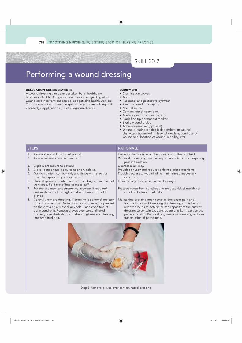
782
DELEGATION CONSIDERATIONSA wound dressing can be undertaken by all healthcare professionals. Check organisational policies regarding which wound care interventions can be delegated to health workers. The assessment of a wound requires the problem-solving and knowledge-application skills of a registered nurse.
EQUIPMENT Examination gloves
characteristics including level of exudate, condition of wound bed, location of wound, mobility, etc)
STEPS RATIONALE
1. Assess size and location of wound. Helps to plan for type and amount of supplies required.2. Assess patient’s level of comfort. Removal of dressing may cause pain and discomfort requiring
pain medication.3. Explain procedure to patient. Decreases anxiety.4. Close room or cubicle curtains and windows. Provides privacy and reduces airborne microorganisms.5. Position patient comfortably and drape with sheet or
towel to expose only wound site.Provides access to wound while minimising unnecessary
exposure.6. Place disposable contaminated-waste bag within reach of
work area. Fold top of bag to make cuff.Ensures easy disposal of soiled dressings.
7. Put on face mask and protective eyewear, if required, and wash hands thoroughly. Put on clean, disposable gloves.
Protects nurse from splashes and reduces risk of transfer of infection between patients.
8. Carefully remove dressing. If dressing is adhered, moisten to facilitate removal. Note the amount of exudate present on the dressing removed, any odour and condition of periwound skin. Remove gloves over contaminated dressing (see illustration) and discard gloves and dressing into prepared bag.
Moistening dressing upon removal decreases pain and trauma to tissue. Observing the dressing as it is being removed helps to determine the capacity of the current dressing to contain exudate, odour and its impact on the periwound skin. Removal of gloves over dressing reduces transmission of pathogens.
Performing a wound dressing
Step 8 Remove gloves over contaminated dressing
ch30-756-813-9780729541107.indd 782 31/08/12 10:00 AM
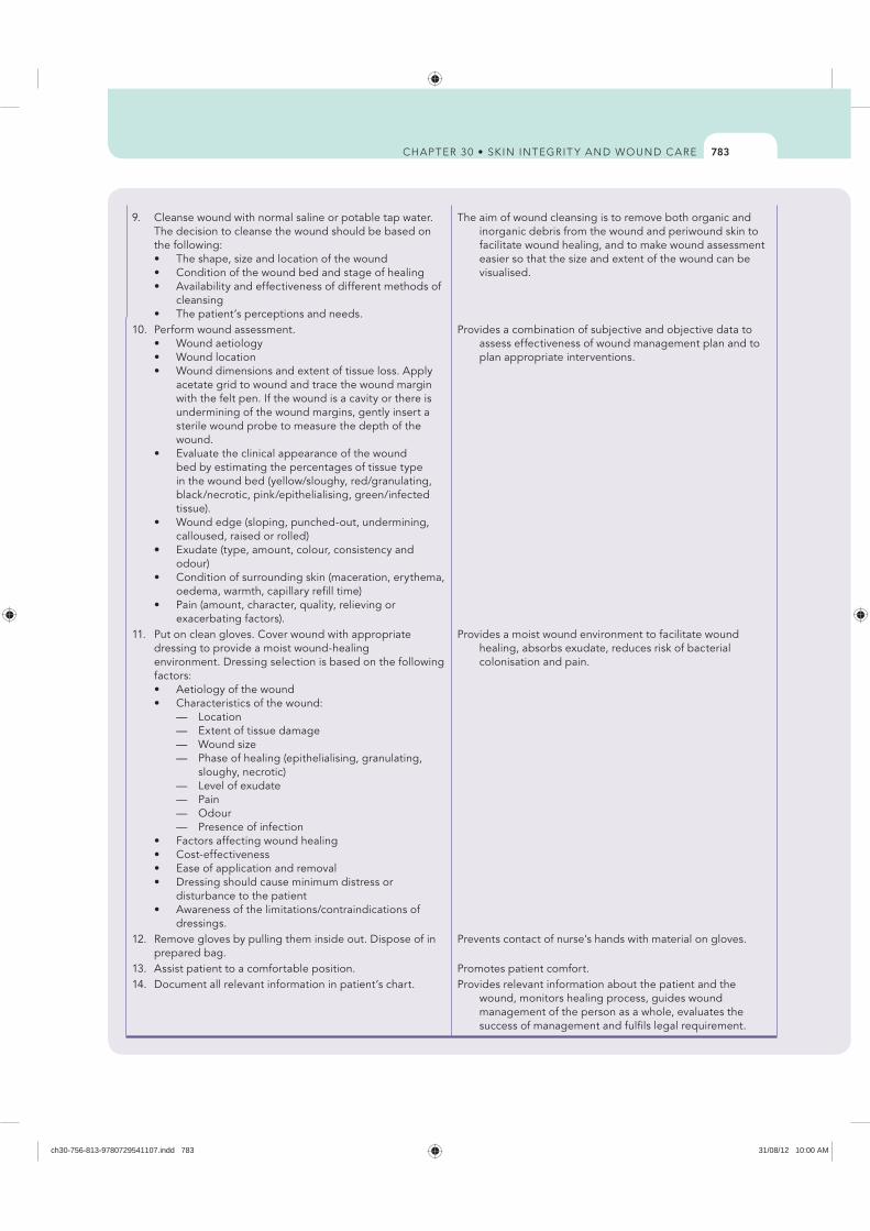
783
9. Cleanse wound with normal saline or potable tap water. The decision to cleanse the wound should be based on the following:
cleansing
The aim of wound cleansing is to remove both organic and inorganic debris from the wound and periwound skin to facilitate wound healing, and to make wound assessment easier so that the size and extent of the wound can be visualised.
10. Perform wound assessment.
acetate grid to wound and trace the wound margin with the felt pen. If the wound is a cavity or there is undermining of the wound margins, gently insert a sterile wound probe to measure the depth of the wound.
bed by estimating the percentages of tissue type in the wound bed (yellow/sloughy, red/granulating, black/necrotic, pink/epithelialising, green/infected tissue).
calloused, raised or rolled) Exudate (type, amount, colour, consistency and odour)
oedema, warmth, capillary refill time)
exacerbating factors).
Provides a combination of subjective and objective data to assess effectiveness of wound management plan and to plan appropriate interventions.
11. Put on clean gloves. Cover wound with appropriate dressing to provide a moist wound-healing environment. Dressing selection is based on the following factors:
— Location — Extent of tissue damage — Wound size — Phase of healing (epithelialising, granulating,
sloughy, necrotic) — Level of exudate — Pain — Odour — Presence of infection
disturbance to the patient
dressings.
Provides a moist wound environment to facilitate wound healing, absorbs exudate, reduces risk of bacterial colonisation and pain.
12. Remove gloves by pulling them inside out. Dispose of in prepared bag.
Prevents contact of nurse’s hands with material on gloves.
13. Assist patient to a comfortable position. Promotes patient comfort.14. Document all relevant information in patient’s chart. Provides relevant information about the patient and the
wound, monitors healing process, guides wound management of the person as a whole, evaluates the success of management and fulfils legal requirement.
ch30-756-813-9780729541107.indd 783 31/08/12 10:00 AM
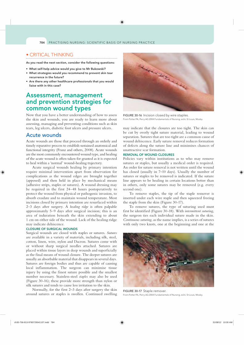
784
may indicate that the closures are too tight. The skin can be cut by overly tight suture material, leading to wound separation. Sutures that are too tight are a common cause of wound dehiscence. Early suture removal reduces formation of defects along the suture line and minimises chances of unattractive scar formation. REMOVAL OF WOUND CLOSURESPolicies vary within institutions as to who may remove sutures or staples, but usually a medical order is required. An order for suture removal is not written until the wound has closed (usually in 7–10 days). Usually the number of sutures or staples to be removed is indicated. If the suture line appears to be healing in certain locations better than in others, only some sutures may be removed (e.g. every second one).
To remove staples, the tip of the staple remover is inserted under each wire staple and then squeezed freeing the staple from the skin (Figure 30-17).
To remove sutures, the type of suturing used must first be identified (Figure 30-18). With intermittent suturing, the surgeon ties each individual suture made in the skin. Continuous suturing, as the name implies, is a series of sutures with only two knots, one at the beginning and one at the
As you read the next section, consider the following questions:
recurrence in the future?
liaise with in this case?
Assessment, management and prevention strategies for common wound typesNow that you have a better understanding of how to assess the skin and wounds, you are ready to learn more about assessing, managing and preventing conditions such as skin tears, leg ulcers, diabetic foot ulcers and pressure ulcers.
Acute woundsAcute wounds are those that proceed through an orderly and timely reparative process to establish sustained anatomical and functional integrity (Franz and others, 2008). Acute wounds are the most commonly encountered wound type, and healing of the acute wound is often taken for granted as it is expected to heal within a ‘normal’ wound-healing trajectory.
Acute surgical wounds healing by primary intention require minimal intervention apart from observation for complications as the wound edges are brought together (apposed) and then held in place by mechanical means (adhesive strips, staples or sutures). A wound dressing may be required in the first 24–48 hours postoperatively to protect the wound from physical or pathogenic invasion, to absorb exudate and to maintain wound temperature. Most incisions closed by primary intention are resurfaced within 2–3 days after surgery. A healing ridge is often palpable approximately 5–9 days after surgical incision; this is an area of induration beneath the skin extending to about 1 cm on either side of the wound. Lack of the healing ridge may indicate dehiscence. CLOSURE OF SURGICAL WOUNDSSurgical wounds are closed with staples or sutures. Sutures are available in a variety of materials, including silk, steel, cotton, linen, wire, nylon and Dacron. Sutures come with or without sharp surgical needles attached. Sutures are placed within tissue layers in deep wounds and superficially as the final means of wound closure. The deeper sutures are usually an absorbable material that disappears in several days. Sutures are foreign bodies and thus are capable of causing local inf lammation. The surgeon can minimise tissue injury by using the finest suture possible and the smallest number necessary. Stainless-steel staples may also be used (Figure 30-16); these provide more strength than nylon or silk sutures and tends to cause less irritation to the skin.
Normally, for the first 2–3 days after surgery the skin around sutures or staples is swollen. Continued swelling
FIGURE 30-16
FIGURE 30-17 Staple remover.
ch30-756-813-9780729541107.indd 784 31/08/12 10:00 AM
785
the tubing as well as around the tubing should be looked for. A sudden decrease in drainage through the tubing may indicate a blocked drain, and the surgeon should be notified. When a drain is connected to suction, the system is assessed to ensure that the pressure ordered is being exerted. Evacuator units such as a Hemovac or Jackson-Pratt exert a constant low pressure as long as the suction device (bladder or bag) is fully compressed. These types of drainage devices are often referred to as self-suction. When the evacuator device is unable to maintain a vacuum on its own, the surgeon is notified, who can then order a secondary vacuum system (such as wall suction). If f luid is allowed to accumulate within the tissues, wound healing will not progress at an optimal rate, and the risk of infection is increased.
When drainage interferes with healing, drainage evacuation can be achieved by using either a drain alone or a drainage tube with continuous suction. Barrier preparations must be applied to protect the skin surrounding drain sites: drainage then f lows on the barrier but not directly on the skin. Drainage evacuators are convenient, portable units that connect to tubular drains lying in a wound bed and exert a safe, constant, low-pressure vacuum to remove and collect drainage. The nurse ensures that suction is exerted and connection points between the evacuator and tubing are intact. The evacuator collects drainage, which is assessed for volume and character every shift and as needed. When the evacuator fills, the output is measured by emptying the contents into a graduated cylinder and immediately resetting the evacuator to apply suction.
Skin graftsIdeally, wounds heal by primary intention. However, large, surgically created wounds and traumatic and chronic wounds can cause extensive tissue destruction, making primary intention healing impossible. Skin grafts may then be necessary to protect underlying structures or to reconstruct areas for cosmetic or functional purposes. Survival of skin grafts relies on revascularisation of the grafted skin (Beldon, 2007).
Skin-graft take is the process by which the donor site is incorporated into a recipient or host bed. Successful graft take requires an unhindered process of restoration of vascular perfusion to the donor skin. A graft will take better if the donor site, the area from where the skin is harvested, is highly vascularised.
For a skin graft to take, it must be closely applied to the recipient bed. Any accumulation of oedema, haematoma or seroma between the skin graft and the recipient bed will prevent the skin graft taking. Movement between the graft and the recipient bed damages the in-growth of capillaries and prevents revascularisation. When skin grafts are applied to movable body parts, splinting is used to immobilise adjacent joints. Bed rest and positioning of the patient to prevent distortion or tension and shearing forces between the graft and the wound bed is essential. Tie-over dressings
end of the suture line. Retention sutures are placed more deeply than skin sutures and may or may not be removed by the nurse, depending on agency policy. The manner in which the suture crosses and penetrates the skin determines the method for removal.
The most important principle in suture removal is never to pull the visible portion of a suture through underlying tissue. Sutures on the skin’s surface harbour microorganisms and debris; the portion of the suture beneath the skin is considered sterile. Pulling the contaminated (external) portion of the suture through tissues may lead to infection. The suture material is cut as close to the skin edge on one side as possible and then the suture is pulled through from the other end.
Every second suture or staple is removed first, and the wound edges are then inspected for healing. The remaining sutures or staples are removed if the wound edges have adhered.
Drains Drains may be inserted into a surgical wound if a large amount of drainage is expected and if keeping wound layers closed is especially important. Some drains are sutured in place. Caution should be exercised when changing the dressing over drains that are not sutured in place to prevent their being accidentally removed. A drain such as a Penrose drain may lie under a dressing, extend through a dressing or be connected to a drainage bag or a suction apparatus. The surgeon often places a pin or clip through the drain to prevent it from slipping further into a wound. It is usually a medical responsibility to pull or advance the drain as drainage decreases to permit healing deep within the drain site.
The nurse assesses the number of drains, drain placement, the character of drainage and the condition of collecting apparatus. The security of the drain and its location with respect to the wound are observed, and the character of the drainage noted. If there is a collecting device, the drainage volume is measured. Because a drainage system must be patent, drainage f low through
FIGURE 30-18 Examples of suturing methods. AB, Continuous. C, Blanket continuous. D, Retention.
ch30-756-813-9780729541107.indd 785 31/08/12 10:00 AM
786
is primarily through re-epithelialisation of the exposed dermis. The sites tend to produce moderate amounts of exudate, and epithelial cells in the hair follicles, sebaceous and sweat glands migrate across the wound bed to form a new layer of epithelium. This process takes approximately 10–21 days. The ideal donor-site dressing should be easy to apply, reduce pain, minimise leakage of exudate, promote rapid wound healing and be inexpensive and acceptable to the patient (Beldon, 2007).
Skin tearsA skin tear is a traumatic injury to the skin that occurs as a result of friction alone, or shearing and friction. It separates the epidermis from the dermis (known as a partial-thickness wound) or separates both the epidermis and the dermis from the underlying structures (known as a full-thickness wound). Most skin tears occur on the arms and hands. Other common sites are the lower legs, feet or the head, although they can occur anywhere on the body. Skin tears are the most common type of wound in older people, so it is important to understand why this is so and to be able to identify those most at risk. Most skin tears occur when the person accidentally bumps into an object such as a wheelchair or furniture, when being transferred, or following a fall.
There are many risk factors for skin tears. These are explained in Table 30-12. CLASSIFICATION OF SKIN TEARS Skin tears vary in size, location and the amount of tissue loss. It is important that you know how to classify the type of skin tear so that you can determine the appropriate management options.
The STAR Skin Tear Classification System (see Figure 30-19) was developed by the Silver Chain Nursing Association and the Curtin University of Technology (2007) to provide an evidence-based consensus on skin tear classification. This means that if you use this system, you are more likely to be able to identify the type of skin tear and be able to select the correct treatment and prevention strategies. Skill 30-3 explains the assessment and management of a skin tear.
SinusA sinus is a cavity or a track that extends from the skin surface to underlying tissue. A sinus may be the result of an infection, ulceration or necrosis of the dermis and underlying tissue, or a surgical wound dehiscence. The goal of management is to gently pack the sinus from the base upwards to eliminate dead space in the wound, prevent abscess formation and facilitate granulation tissue formation ( Joanna Briggs Institute, 2011).
FistulaA fistula is an abnormal passage between two or more structures or spaces. This can involve a communication tract from one body cavity or organ to another organ or to the skin. The goal of management is closure of the fistula, either
that immobilise skin grafts, particularly on the face, may be sufficient to immobilise problematic areas.
Several methods are used to fixate the graft, including the open method, the closed method and topical negative pressure wound therapy. The open method is where the graft is laid onto the recipient site and left open to the air. Initially the graft is pricked every hour with a sterile needle to express any seroma or haematoma that has built up under the graft. As the wound exudate decreases, the procedure is reduced to every few hours. The closed method of skin grafting is where a pressure dressing is applied over the skin to keep it well approximated to the recipient bed (Terrill, 2003). There are many different techniques to achieve this; most involve using a non-adherent dressing on the surface of the graft, such as tulle gras or paraffin-impregnated gauze, and a stack of cotton gauze cut to fit within the defect or a foam sutured/stapled onto the wound margins or a f lexible polyamide net coated with soft silicone against the graft with a secondary absorbent dressing secured with sutures or staples (Terrill, 2003; Young and Fowler, 1998). Sutures or staples are not used if the skin graft is applied in the operating theatre. The fibrin that forms between the skin graft and the recipient bed binds the two surfaces together.
Topical negative-pressure wound therapy is utilised for skin grafts in complex anatomical areas where the base is irregular (Banwell and Teot, 2003). A topical negative-pressure dressing achieves good apposition of the skin graft to the recipient site, absorbs exudate, reduces oedema, stimulates granulation-tissue formation and decreases bacterial colonisation (Banwell and Teot, 2003). Excellent graft take (>95%) has been reported in the literature with this technique for graft fixation (Banwell and Teot, 2003).
With the closed or topical negative-pressure wound therapy techniques, the skin grafts are usually reviewed after 5 days and assessed for graft take. A graft that has taken has a purple-pink hue and has adhered to the wound bed. The graft usually needs protection for at least 2 weeks or 3 months if located on the lower limbs. Any graft that has not taken is dressed using normal moist wound-healing principles. After 4–6 weeks the graft is usually pale pink and the skin depression should be filling in. Further cosmetic improvement can continue with graft maturation over 6–12 months. Cosmesis can be improved using pressure therapy and silicone gel sheeting to soften scars and reduce erythema.
Skin f laps have their own blood supply, and the ability to monitor the status of the f lap perfusion or its viability is essential in the prevention, recognition and treatment of complications. A healthy skin f lap is similar in colour and texture to the donor site. The colour, temperature, capillary refill time, tissue turgor, dermal bleeding and quantity of exudate are techniques used to assess tissue f lap viability.
Donor sites are frequently painful. Factors that affect the healing rate of donor sites are the site, size and depth of tissue excised (Beldon, 2007). Healing of donor sites
ch30-756-813-9780729541107.indd 786 31/08/12 10:00 AM
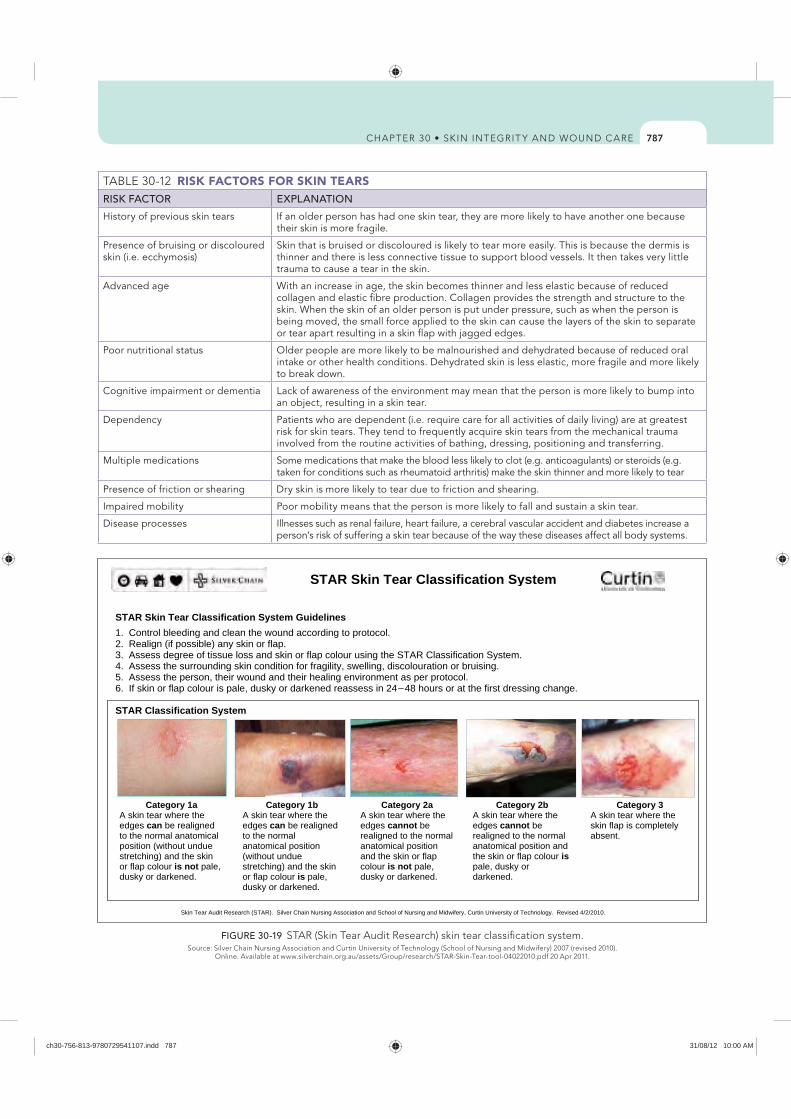
787
RISK FACTORS FOR SKIN TEARSRISK FACTOR EXPLANATION
History of previous skin tears If an older person has had one skin tear, they are more likely to have another one because their skin is more fragile.
Presence of bruising or discoloured skin (i.e. ecchymosis)
Skin that is bruised or discoloured is likely to tear more easily. This is because the dermis is thinner and there is less connective tissue to support blood vessels. It then takes very little trauma to cause a tear in the skin.
Advanced age With an increase in age, the skin becomes thinner and less elastic because of reduced collagen and elastic fibre production. Collagen provides the strength and structure to the skin. When the skin of an older person is put under pressure, such as when the person is being moved, the small force applied to the skin can cause the layers of the skin to separate or tear apart resulting in a skin flap with jagged edges.
Poor nutritional status Older people are more likely to be malnourished and dehydrated because of reduced oral intake or other health conditions. Dehydrated skin is less elastic, more fragile and more likely to break down.
Cognitive impairment or dementia Lack of awareness of the environment may mean that the person is more likely to bump into an object, resulting in a skin tear.
Dependency Patients who are dependent (i.e. require care for all activities of daily living) are at greatest risk for skin tears. They tend to frequently acquire skin tears from the mechanical trauma involved from the routine activities of bathing, dressing, positioning and transferring.
Multiple medications Some medications that make the blood less likely to clot (e.g. anticoagulants) or steroids (e.g. taken for conditions such as rheumatoid arthritis) make the skin thinner and more likely to tear
Presence of friction or shearing Dry skin is more likely to tear due to friction and shearing.
Impaired mobility Poor mobility means that the person is more likely to fall and sustain a skin tear.
Disease processes Illnesses such as renal failure, heart failure, a cerebral vascular accident and diabetes increase a person’s risk of suffering a skin tear because of the way these diseases affect all body systems.
STAR Skin Tear Classification System Guidelines 1. Control bleeding and clean the wound according to protocol. 2. Realign (if possible) any skin or flap. 3. Assess degree of tissue loss and skin or flap colour using the STAR Classification System.
5. Assess the person, their wound and their healing environment as per protocol.
STAR Classification System
Category 1a Category 1b Category 2a Category 2b Category 3 A skin tear where the edges can be realigned to the normal anatomical position (without undue stretching) and the skin or flap colour is not pale, dusky or darkened.
A skin tear where the edges can be realigned to the normal anatomical position (without undue stretching) and the skin or flap colour is pale, dusky or darkened.
A skin tear where the edges cannot be realigned to the normal anatomical position and the skin or flap colour is not pale, dusky or darkened.
A skin tear where the edges cannot be realigned to the normal anatomical position and the skin or flap colour ispale, dusky or darkened.
A skin tear where the skin flap is completely absent.
Skin Tear Audit Research (STAR). Silver Chain Nursing Association and School of Nursing and Midwifery, Curtin University of Technology. Revised 4/2/2010.
STAR Skin Tear Classification System
4. Assess the surrounding skin condition for fragility, swelling, discolouration or bruising.
–6. If skin or flap colour is pale, dusky or darkened reassess in 24 48 hours or at the first dressing change.
FIGURE 30-19
ch30-756-813-9780729541107.indd 787 31/08/12 10:00 AM
788
DELEGATION CONSIDERATIONSThe management and prevention of skin tears can be undertaken by all healthcare professionals. Check organisational policies regarding which wound care interventions can be delegated to health workers. The assessment of a person with or at risk of skin tears requires the problem-solving and knowledge-application skills of a registered nurse.
EQUIPMENT
characteristics including level of exudate, condition of wound bed, location and size of wound, condition of periwound skin, mobility, etc)
STEPS RATIONALE
1. Explain procedure to patient. Decreases anxiety.2. Assess size and location of wound. Helps to plan for type and amount of supplies required.3. Position patient comfortably.4. Place disposable contaminated-waste bag within reach of
work area. Fold top of bag to make cuff.Ensures easy disposal of soiled dressings.
5. Put on face mask and protective eyewear, if required, and wash hands thoroughly.
Protects nurse from splashes and reduces risk of transfer of infection between patients.
6. Control bleeding and clean the wound: a. Stop bleeding by applying firm pressure. Elevate the
limb, if applicable, above heart level. b. Clean the wound by gently irrigating with warm
normal saline. Carefully clean under the skin flap to remove foreign debris or blood clots.
c. Pat dry the surrounding skin.
The aim of wound cleansing is to remove both organic and inorganic debris from the wound and periwound skin to facilitate wound healing, and to make wound assessment easier so that the size and extent of the wound can be visualised.
7. Realign (if possible) any skin or flap. a. Carefully realign by rolling the skin flap with a moist,
sterile cotton bud. The use of a moistened cotton bud assists with approximation of the flap edge.
b. Do not stretch the skin to ‘make it fit’. Otherwise, the skin flap will shrink back on itself or it may tear off completely.
c. If the skin flap cannot be realigned, apply moist wound-dressing principles to promote wound healing.
8. Assess the degree of tissue loss and skin or flap colour: a. Using the STAR Skin Tear Classification System (see
Figure 30-19), select the skin tear category that best describes the type of skin tear you are treating.
b. Note the colour of the skin flap. If the skin or flap colour is pale, dusky or darkened when compared with the person’s normal surrounding skin, this may indicate reduced blood flow (ischaemia) or the presence of a blood clot (haematoma), which may affect the skin or flap viability.
c. If the skin or flap colour is pale, dusky or darkened, reassess in 24–48 hours or during the first dressing change.
Provides a combination of subjective and objective data to assess effectiveness of wound management plan and to plan appropriate interventions.
9. Assess the surrounding skin condition for fragility, swelling, discolouration or bruising. This information will assist you when selecting an appropriate dressing.
Assessment, management and prevention of skin tears
ch30-756-813-9780729541107.indd 788 31/08/12 10:00 AM
789
10. Assess the person, their wound and their healing environment.
A person’s overall medical condition may affect how quickly the wound is likely to heal. This will guide the management and preventive strategies you decide to use.
11. Apply a dressing. Regardless of what type of dressing you select, you should follow these principles:
least 1.5–2 cm.Reduces the risk of trauma on removal of the dressing and
increases the ability of the dressing to absorb any leakage from the wound.
the dressing with a felt pen to indicate the direction of the skin flap and write the due date for removal. When removing the dressing, ensure that you remove it in the direction of the arrow to avoid pulling back the skin flap.
is 75% leakage on the dressing.
leave the dressing intact for 5–7 days.
twice daily with a pH-neutral emollient.12. Protect limbs:
Apply a limb protector or tubular retention bandage to prevent further damage or to hold any dressing in situ.
13. Document the skin tear: a. Document the skin tear category and location in the
patient’s record and wound-care assessment chart. b. Submit an incident report as required.
Provides relevant information about the patient and the wound, monitors healing process, guides wound management of the person as a whole, evaluates the success of management and fulfils legal requirements.
14. Monitor the healing of the wound: a. Check the dressing and surrounding skin regularly for
signs of complications or infection, but leave the dressing intact unless an inspection is clinically indicated.
b. Educate the patient about the signs and symptoms of complications or infection. Advise them to report problems, especially any increase in pain, warmth, odour, redness, purulent exudate or fever.
15. Implement skin-tear prevention strategies, including the following:
People with fragile, thin, bruised or discoloured skin are at a high risk for further skin tears.
possible to reduce the risk of friction or shearing forces on the skin, especially when transferring someone.
had repeated skin tears, encourage them to wear long sleeves and long trousers to protect their skin.
least twice daily. This will help to prevent dry skin.
perfumed lotions as these can dry the skin. Instead, use a pH-neutral cleanser or moisturiser.
risk of tearing the skin when they are removed. It is advisable to use a limb protector instead.
pad bed rails, wheelchair arms and leg supports to reduce the risk of skin injuries.
to reduce risk of people bumping into furniture, doors or equipment.
Many skin tears occur when performing normal daily activities such as bathing, dressing and transferring individuals at risk. Avoid wearing jewellery or a watch.
16. Provide continuing education: Help other staff members, patients and their family members understand the importance of identifying those at risk for skin tears, appropriate treatment and prevention strategies.
17. Document all relevant information in patient’s chart. Provides relevant information about the patient and the wound, monitors healing process, guides wound management of the person as a whole, evaluates the success of management and fulfils legal requirement.
ch30-756-813-9780729541107.indd 789 31/08/12 10:00 AM
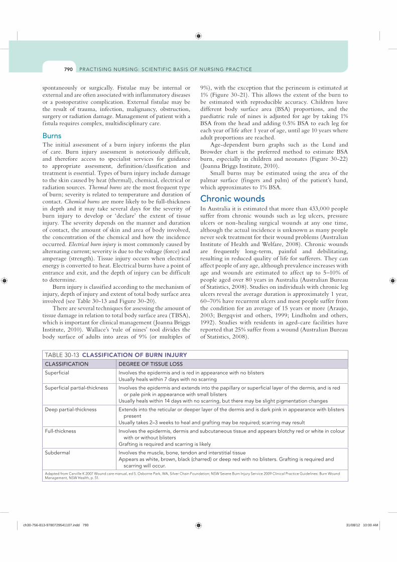
790
9%), with the exception that the perineum is estimated at 1% (Figure 30-21). This allows the extent of the burn to be estimated with reproducible accuracy. Children have different body surface area (BSA) proportions, and the paediatric rule of nines is adjusted for age by taking 1% BSA from the head and adding 0.5% BSA to each leg for each year of life after 1 year of age, until age 10 years where adult proportions are reached.
Age-dependent burn graphs such as the Lund and Browder chart is the preferred method to estimate BSA burn, especially in children and neonates (Figure 30-22) ( Joanna Briggs Institute, 2010).
Small burns may be estimated using the area of the palmar surface (fingers and palm) of the patient’s hand, which approximates to 1% BSA.
Chronic woundsIn Australia it is estimated that more than 433,000 people suffer from chronic wounds such as leg ulcers, pressure ulcers or non-healing surgical wounds at any one time, although the actual incidence is unknown as many people never seek treatment for their wound problems (Australian Institute of Health and Welfare, 2008). Chronic wounds are frequently long-term, painful and debilitating, resulting in reduced quality of life for sufferers. They can affect people of any age, although prevalence increases with age and wounds are estimated to affect up to 5–10% of people aged over 80 years in Australia (Australian Bureau of Statistics, 2008). Studies on individuals with chronic leg ulcers reveal the average duration is approximately 1 year, 60–70% have recurrent ulcers and most people suffer from the condition for an average of 15 years or more (Araujo, 2003; Bergqvist and others, 1999; Lindholm and others, 1992). Studies with residents in aged-care facilities have reported that 25% suffer from a wound (Australian Bureau of Statistics, 2008).
spontaneously or surgically. Fistulae may be internal or external and are often associated with inf lammatory diseases or a postoperative complication. External fistulae may be the result of trauma, infection, malignancy, obstruction, surgery or radiation damage. Management of patient with a fistula requires complex, multidisciplinary care.
Burns The initial assessment of a burn injury informs the plan of care. Burn injury assessment is notoriously difficult, and therefore access to specialist services for guidance to appropriate assessment, definition/classification and treatment is essential. Types of burn injury include damage to the skin caused by heat (thermal), chemical, electrical or radiation sources. Thermal burns are the most frequent type of burn; severity is related to temperature and duration of contact. Chemical burns are more likely to be full-thickness in depth and it may take several days for the severity of burn injury to develop or ‘declare’ the extent of tissue injury. The severity depends on the manner and duration of contact, the amount of skin and area of body involved, the concentration of the chemical and how the incidence occurred. Electrical burn injury is most commonly caused by alternating current; severity is due to the voltage (force) and amperage (strength). Tissue injury occurs when electrical energy is converted to heat. Electrical burns have a point of entrance and exit, and the depth of injury can be difficult to determine.
Burn injury is classified according to the mechanism of injury, depth of injury and extent of total body surface area involved (see Table 30-13 and Figure 30-20).
There are several techniques for assessing the amount of tissue damage in relation to total body surface area (TBSA), which is important for clinical management ( Joanna Briggs Institute, 2010). Wallace’s ‘rule of nines’ tool divides the body surface of adults into areas of 9% (or multiples of
CLASSIFICATION OF BURN INJURYCLASSIFICATION DEGREE OF TISSUE LOSS
Superficial Involves the epidermis and is red in appearance with no blistersUsually heals within 7 days with no scarring
Superficial partial-thickness Involves the epidermis and extends into the papillary or superficial layer of the dermis, and is red or pale pink in appearance with small blisters
Usually heals within 14 days with no scarring, but there may be slight pigmentation changes
Deep partial-thickness Extends into the reticular or deeper layer of the dermis and is dark pink in appearance with blisters present
Usually takes 2–3 weeks to heal and grafting may be required; scarring may result
Full-thickness Involves the epidermis, dermis and subcutaneous tissue and appears blotchy red or white in colour with or without blisters
Grafting is required and scarring is likely
Subdermal Involves the muscle, bone, tendon and interstitial tissue Appears as white, brown, black (charred) or deep red with no blisters. Grafting is required and
scarring will occur.
ch30-756-813-9780729541107.indd 790 31/08/12 10:00 AM
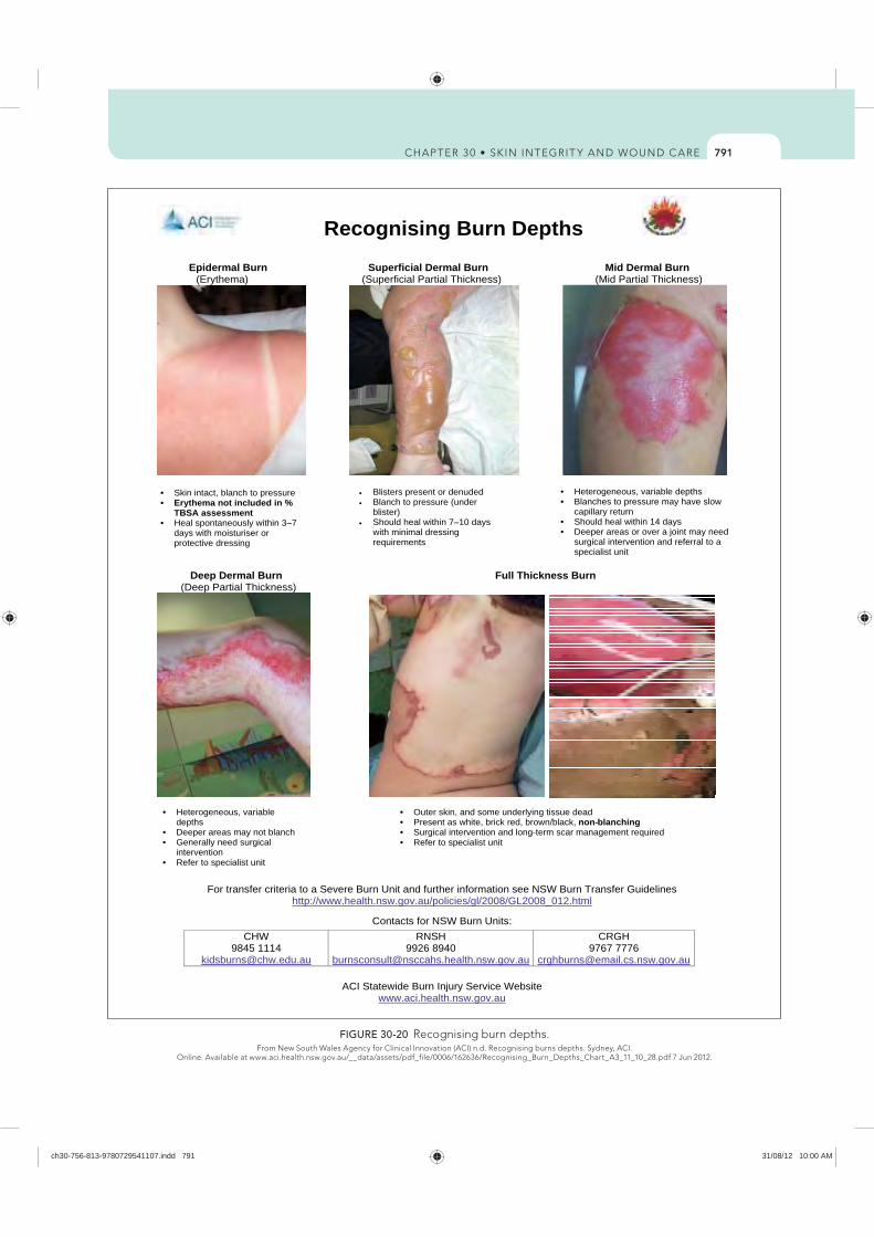
791
ACI Statewide Burn Injury Service Website www.aci.health.nsw.gov.au
Recognising Burn Depths
Epidermal Burn Superficial Dermal Burn Mid Dermal Burn (Erythema) (Superficial Partial Thickness) (Mid Partial Thickness)
Deep Dermal Burn Full Thickness Burn (Deep Partial Thickness)
For transfer criteria to a Severe Burn Unit and further information see NSW Burn Transfer Guidelines http://www.health.nsw.gov.au/policies/gl/2008/GL2008_012.html
Contacts for NSW Burn Units:CHW
9845 1114 [email protected]
RNSH9926 8940
CRGH9767 7776
Skin intact, blanch to pressure Erythema not included in % TBSA assessment
days with moisturiser or protective dressing
Blisters present or denuded Blanch to pressure (under blister)
with minimal dressing requirements
Heterogeneous, variable depths Blanches to pressure may have slow
capillary return Should heal within 14 days Deeper areas or over a joint may need
surgical intervention and referral to a specialist unit
Outer skin, and some underlying tissue dead
Surgical intervention and long-term scar management required Refer to specialist unit
Heterogeneous, variable depths
Deeper areas may not blanch Generally need surgical
intervention Refer to specialist unit
Heal spontaneously within 3– 7 Should heal within 7– 10 days
Present as white, brick red, brown/black, non- blanching
FIGURE 30-20 Recognising burn depths.
ch30-756-813-9780729541107.indd 791 31/08/12 10:00 AM
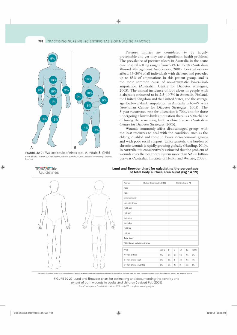
792
FIGURE 30-21 A B, Child.
Elsevier.
Therapeutic Guidelines Limited is an independent not-for-profit organisation dedicated to deriving guidelines for therapy from the latest world literature, interpreted and distilled by Australia’s most eminent and respected experts.
Lund and Browder chart for calculating the percentage of total body surface area burnt (Fig 14.19)
�
��
�
�
��
���� ����
���� ����
��������
� �
� �
�
�
����
����
���� ����
����
���� ����
��������
� �
� �
Region Partial thickness (%) [NB1] Full thickness (%)
head
neck
anterior trunk
posterior trunk
right arm
left arm
buttocks
genitalia
right leg
left leg
Total burn
NB1: Do not include erythema
Area Age 0 1 5 10 15 Adult
A = half of head 9½ 8½ 6½ 5½ 4½ 3½
B = half of one thigh 2¾ 3¼ 4 4½ 4½ 4¾
C = half of one lower leg 2½ 2½ 2¾ 3 3¼ 3½
FIGURE 30-22
Pressure injuries are considered to be largely preventable and yet they are a significant health problem. The prevalence of pressure ulcers in Australia in the acute care hospital setting ranges from 5.4% to 15.6% (Australian Wound Management Association, 2001). Foot ulceration affects 15–20% of all individuals with diabetes and precedes up to 85% of amputations in this patient group, and is the most common cause of non-traumatic lower-limb amputation (Australian Centre for Diabetes Strategies, 2005). The annual incidence of foot ulcers in people with diabetes is estimated to be 2.5–10.7% in Australia, Finland, the United Kingdom and the United States, and the average age for lower-limb amputation in Australia is 65–79 years (Australian Centre for Diabetes Strategies, 2005). The 5-year recurrence rate for ulceration is 70%, and for those undergoing a lower-limb amputation there is a 50% chance of losing the remaining limb within 3 years (Australian Centre for Diabetes Strategies, 2005).
Wounds commonly affect disadvantaged groups with the least resources to deal with the condition, such as the elderly, disabled and those in lower socioeconomic groups and with poor social support. Unfortunately, the burden of chronic wounds is rapidly growing globally (Harding, 2010). In Australia it is conservatively estimated that the problem of wounds costs the healthcare system more than $A2.6 billion per year (Australian Institute of Health and Welfare, 2008).
ch30-756-813-9780729541107.indd 792 31/08/12 10:00 AM
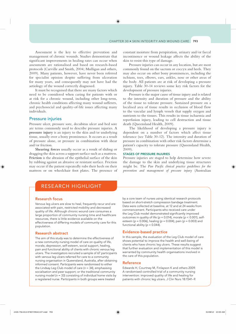
793
constant moisture from perspiration, urinary and/or faecal incontinence or wound leakage affects the ability of the skin to resist this type of damage.
Pressure injuries can occur in any location, but are most commonly found on the sacrum or coccyx and heels. They may also occur on other bony prominences, including the ischium, toes, elbows, ears, ankles, nose or other areas of the body. All patients are at risk of developing a pressure injury. Table 30-14 reviews some key risk factors for the development of pressure injuries.
Pressure is the major cause of tissue injury and is related to the intensity and duration of pressure and the ability of the tissue to tolerate pressure. Sustained pressure on a localised area of tissue results in occlusion of blood f low to the vascular and lymph vessels that supply oxygen and nutrients to the tissues. This results in tissue ischaemia and reperfusion injury, leading to cell destruction and tissue death (Queensland Health, 2009).
The likelihood of developing a pressure injury is dependent on a number of factors which affect tissue tolerance (see Table 30-12). The intensity and duration of pressure in combination with other risk factors determine a patient’s capacity to tolerate pressure (Queensland Health, 2009). STAGES OF PRESSURE INJURIESPressure injuries are staged to help determine how severe the damage to the skin and underlying tissue structures might be. The Pan Pacific clinical practice guidelines for the prevention and management of pressure injury (Australian
Assessment is the key to effective prevention and management of chronic wounds. Studies demonstrate that significant improvements in healing rates can occur when assessments are rationalised and based on research-based protocols (Carville and Smith, 2004; Mulligan and others, 2009). Many patients, however, have never been referred for specialist opinion despite suffering from ulceration for many years, and consequently may not have had the aetiology of the wound correctly diagnosed.
It must be recognised that there are many factors which need to be considered when caring for patients with or at risk for a chronic wound, including other long-term, chronic health conditions affecting many wound sufferers, and psychosocial and quality-of-life issues affecting many individuals.
Pressure injuriesPressure ulcer, pressure sore, decubitus ulcer and bed sore are terms commonly used to describe pressure injuries. A pressure injury is an injury to the skin and/or underlying tissue, usually over a bony prominence. It occurs as a result of pressure alone, or pressure in combination with shear and/or friction.
Shearing forces usually occur as a result of sliding or dragging the skin across a support surface such as a mattress. Friction is the abrasion of the epithelial surface of the skin by rubbing against an abrasive or resistant surface. Friction may occur if the patient repeatedly rubs their heels on their mattress or on wheelchair foot plates. The presence of
Research focus Venous leg ulcers are slow to heal, frequently recur and are associated with pain, restricted mobility and decreased quality of life. Although chronic wound care consumes a large proportion of community nursing time and healthcare resources, there is little evidence available on the effectiveness of differing models of community care for this population.
Research abstractThe aim of this study was to determine the effectiveness of a new community nursing model of care on quality of life, morale, depression, self-esteem, social support, healing, pain and functional ability of clients with chronic venous leg ulcers. The investigators recruited a sample of 67 participants with venous leg ulcers referred for care to a community nursing organisation in Queensland, Australia, after obtaining informed consent. Participants were randomised to either the Lindsay Leg Club model of care (n = 34), emphasising socialisation and peer support; or the traditional community nursing model (n = 33) consisting of individual home visits by a registered nurse. Participants in both groups were treated
by a core team of nurses using identical research protocols based on short-stretch compression bandage treatment. Data were collected at baseline, at 12 and at 24 weeks from commencement. Participants who received care under the Leg Club model demonstrated significantly improved outcomes in quality of life (p = 0.014), morale (p < 0.001), self-esteem (p = 0.006), healing (p = 0.004), pain (p = 0.003) and functional ability (p = 0.044).
Evidence-based practice In this sample, the evaluation of the Leg Club model of care shows potential to improve the health and well-being of clients who have chronic leg ulcers. These results suggest that further evaluation and implementation of this model is warranted by community health organisations involved in the care of this population.
ReferenceEdwards H, Courtney M, Finlayson K and others 2009. A randomised controlled trial of a community nursing intervention: improved quality of life and healing for patients with chronic leg ulcers. J Clin Nurs 18:1541–9.
RESEARCH HIGHLIGHT
ch30-756-813-9780729541107.indd 793 31/08/12 10:00 AM
794
you press on the skin, changes in skin colour are difficult to detect.
STAGE 2 pressure injuries represent partial-thickness loss of the dermis, and present as shallow open ulcers with a red or pink wound bed; they may also present as an intact or open/ruptured f luid-filled blister. If a blister is found with clear f luid present and the epidermis is clearly separated from the dermis, this is recorded as a Stage 2 pressure injury.
Stage 2 pressure injuries present as shiny or dry shallow ulcers without any bruising present, since bruising represents suspected deep-tissue damage.
STAGE 3 pressure injuries represent full-thickness skin loss. Subcutaneous fat may be visible but bone, tendons or muscle are not exposed. Thick yellow tissue (slough) may be present but this does not obscure the depth of tissue loss. There may be undermining and tunnelling of the wound (i.e. the wound is deeper than it appears on the surface of the skin).
The depth of a Stage 3 pressure injury varies depending on where it is located anatomically. A pressure injury on the bridge of the nose, ear, scalp and ankle does not have subcutaneous tissue and Stage 3 pressure injuries can be shallow. In contrast, areas of significant adiposity or fatty tissue can develop extremely deep Stage 3 pressure injuries, although bone and/or tendon is not visible or directly palpable.
STAGE 4 pressure injuries are the most severe, and represent full-thickness tissue loss with exposed bone, tendon or muscle. Thick yellow tissue (slough) or black necrotic tissue may be present on some parts of the wound bed. Often there is undermining and tunnelling of the adjacent tissue.
The depth of a Stage 4 pressure injury varies by anatomical location. The bridge of the nose, ear, scalp and ankles do not have subcutaneous tissue, so ulcers in these locations can be shallow. Stage 4 pressure injuries can
Wound Management Association, 2012) have recently been released and recommend the use of a pressure injury classification system to provide a consistent and accurate means by which the severity of a pressure injury can be communicated and documented. Presently there are four stages of pressure damage. The higher the stage, the deeper the tissue involvement.
STAGE 1 pressure injuries present as areas of persistent, non-blanchable redness when compared with the sur-rounding skin. The skin around the wound may be painful or itchy, firm or boggy, and warmer or cooler to the touch when compared with the surrounding skin.
Stage 1 pressure injury may be difficult to detect in individuals with dark skin tones. In lighter skin-toned individuals, if you press on the skin for a few seconds, normal skin typically goes white in colour (blanching) and then returns to a normal pink colour when the pressure is released. However, patients with more darkly pigmented skin may not have visible blanching and skin colour may differ from the surrounding area. This means that when
RISK FACTORS FOR PRESSURE INJURIESRISK FACTOR EXPLANATION
Alteration in mobility or physical activity
Reduced mobility or physical activity due to contractures, fractures, injury, neurological disease or pain increase the risk of pressure injury developing. This is because the patient is unable to independently reposition themselves to off-load or relieve pressure
Malnutrition and dehydration Malnutrition and dehydration increase the risk of pressure injury. The skin is much drier and less able to withstand the effects of pressure damage
Moisture The presence of constant moisture from wound leakage, urinary or faecal incontinence and perspiration increases the risk of pressure damage. This is because the skin is less able to resist friction or shearing forces
Alteration in sensation and consciousness
Decreased sensation or loss of consciousness because of cognitive impairment such as a central nervous system injury, a cerebrovascular accident, degeneration, major surgery, spinal cord injury or medications which increases the risk of pressure damage. This is because the patient may not be aware that an injury to the skin has occurred
Other health conditions Health conditions such as heart disease, diabetes, circulation disorders and a history of smoking can increase the risk of pressure injury
SKIN ASSESSMENT FOR PRESSURE INJURY
Inspect the skin of all patients on admission and at each repositioning to identify indications of pressure injury including:
erythemablanching responselocalised heatoedemaindurationskin breakdown.
clinical practice guideline for the prevention and management of pressure
ch30-756-813-9780729541107.indd 794 31/08/12 10:00 AM
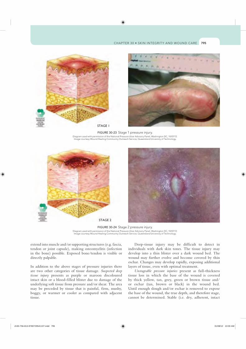
795
Deep-tissue injury may be difficult to detect in individuals with dark skin tones. The tissue injury may develop into a thin blister over a dark wound bed. The wound may further evolve and become covered by thin eschar. Changes may develop rapidly, exposing additional layers of tissue, even with optimal treatment.
Unstageable pressure injuries present as full-thickness tissue loss in which the base of the wound is covered by thick yellow, tan, grey, green or brown tissue and/or eschar (tan, brown or black) in the wound bed. Until enough slough and/or eschar is removed to expose the base of the wound, the true depth, and therefore stage, cannot be determined. Stable (i.e. dry, adherent, intact
extend into muscle and/or supporting structures (e.g. fascia, tendon or joint capsule), making osteomyelitis (infection in the bone) possible. Exposed bone/tendon is visible or directly palpable.
In addition to the above stages of pressure injuries there are two other categories of tissue damage. Suspected deep tissue injury presents as purple or maroon discoloured intact skin or a blood-filled blister due to damage of the underlying soft tissue from pressure and/or shear. The area may be preceded by tissue that is painful, firm, mushy, boggy, or warmer or cooler as compared with adjacent tissue.
STAGE 1
FIGURE 30-23
STAGE 2
FIGURE 30-24
ch30-756-813-9780729541107.indd 795 31/08/12 10:00 AM
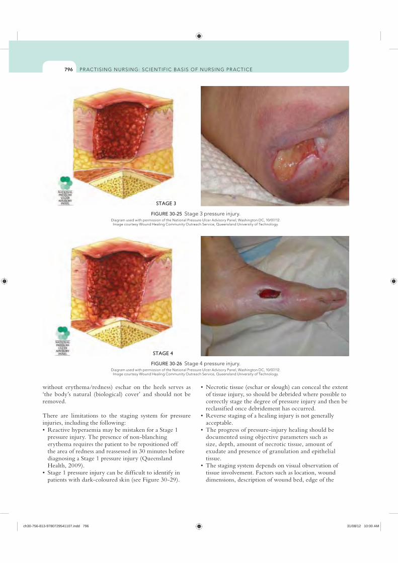
796
of tissue injury, so should be debrided where possible to correctly stage the degree of pressure injury and then be reclassified once debridement has occurred.
acceptable.
documented using objective parameters such as size, depth, amount of necrotic tissue, amount of exudate and presence of granulation and epithelial tissue.
tissue involvement. Factors such as location, wound dimensions, description of wound bed, edge of the
without erythema/redness) eschar on the heels serves as ‘the body’s natural (biological) cover’ and should not be removed.
There are limitations to the staging system for pressure injuries, including the following:
pressure injury. The presence of non-blanching erythema requires the patient to be repositioned off the area of redness and reassessed in 30 minutes before diagnosing a Stage 1 pressure injury (Queensland Health, 2009).
patients with dark-coloured skin (see Figure 30-29).
STAGE 3
FIGURE 30-25 Stage 3 pressure injury.
STAGE 4
FIGURE 30-26
ch30-756-813-9780729541107.indd 796 31/08/12 10:00 AM
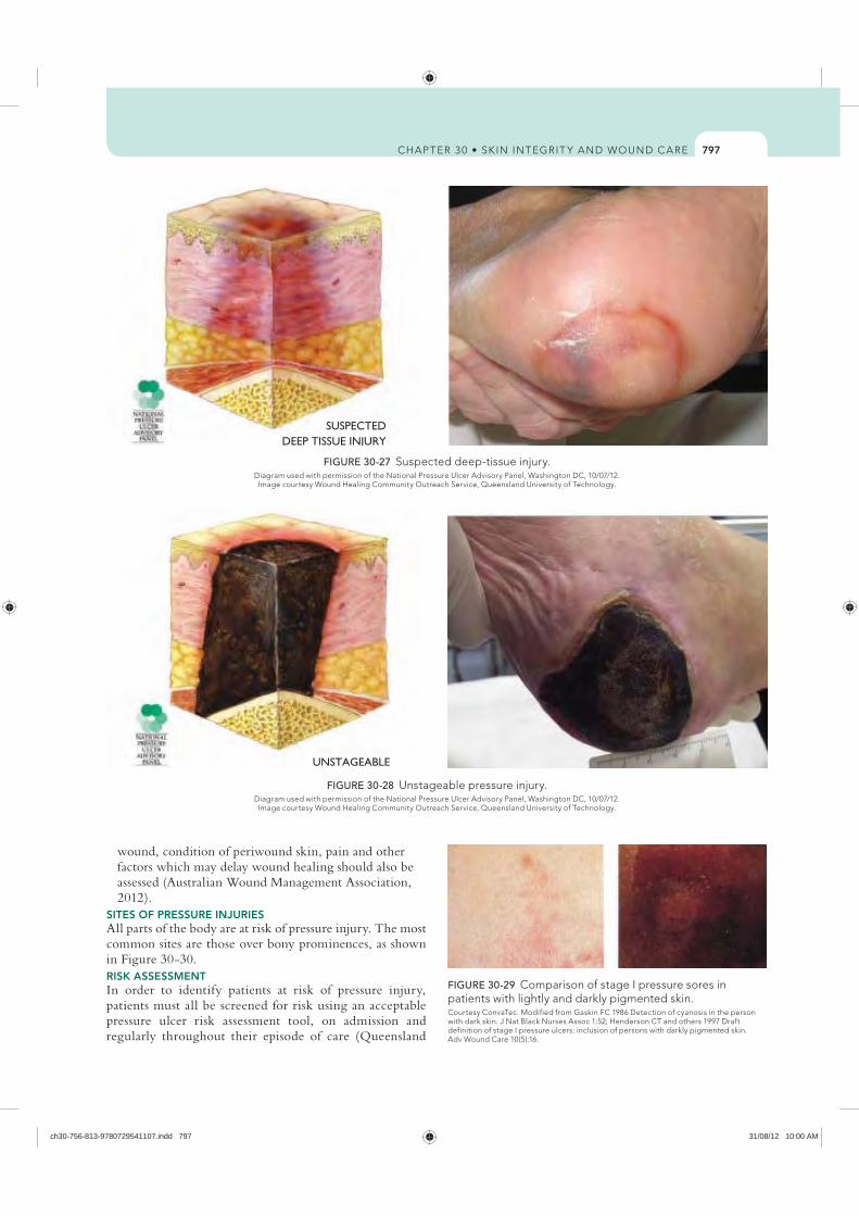
797
wound, condition of periwound skin, pain and other factors which may delay wound healing should also be assessed (Australian Wound Management Association, 2012).
SITES OF PRESSURE INJURIES All parts of the body are at risk of pressure injury. The most common sites are those over bony prominences, as shown in Figure 30-30.RISK ASSESSMENTIn order to identify patients at risk of pressure injury, patients must all be screened for risk using an acceptable pressure ulcer risk assessment tool, on admission and regularly throughout their episode of care (Queensland
SUSPECTEDDEEP TISSUE INJURY
FIGURE 30-27 Suspected deep-tissue injury.
UNSTAGEABLE
FIGURE 30-28
FIGURE 30-29 patients with lightly and darkly pigmented skin.
ch30-756-813-9780729541107.indd 797 31/08/12 10:00 AM
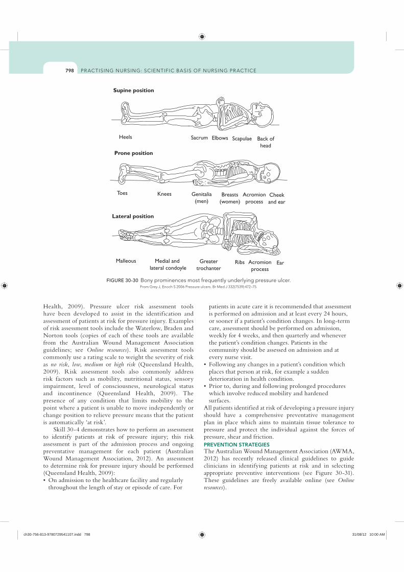
798
patients in acute care it is recommended that assessment is performed on admission and at least every 24 hours, or sooner if a patient’s condition changes. In long-term care, assessment should be performed on admission, weekly for 4 weeks, and then quarterly and whenever the patient’s condition changes. Patients in the community should be assessed on admission and at every nurse visit.
places that person at risk, for example a sudden deterioration in health condition.
which involve reduced mobility and hardened surfaces.
All patients identified at risk of developing a pressure injury should have a comprehensive preventative management plan in place which aims to maintain tissue tolerance to pressure and protect the individual against the forces of pressure, shear and friction. PREVENTION STRATEGIESThe Australian Wound Management Association (AWMA, 2012) has recently released clinical guidelines to guide clinicians in identifying patients at risk and in selecting appropriate preventive interventions (see Figure 30-31). These guidelines are freely available online (see Online resources).
Health, 2009). Pressure ulcer risk assessment tools have been developed to assist in the identif ication and assessment of patients at risk for pressure injury. Examples of risk assessment tools include the Waterlow, Braden and Norton tools (copies of each of these tools are available from the Australian Wound Management Association guidelines; see Online resources). Risk assessment tools commonly use a rating scale to weight the severity of risk as no risk, low, medium or high risk (Queensland Health, 2009). Risk assessment tools also commonly address risk factors such as mobility, nutritional status, sensory impairment, level of consciousness, neurological status and incontinence (Queensland Health, 2009). The presence of any condition that limits mobility to the point where a patient is unable to move independently or change position to relieve pressure means that the patient is automatically ‘at risk’.
Skill 30-4 demonstrates how to perform an assessment to identify patients at risk of pressure injury; this risk assessment is part of the admission process and ongoing preventative management for each patient (Australian Wound Management Association, 2012). An assessment to determine risk for pressure injury should be performed (Queensland Health, 2009):
throughout the length of stay or episode of care. For
Supine position
Heels
Prone position
Sacrum Elbows Scapulae Back ofhead
Toes
Lateral position
Malleous Medial and lateral condoyle
Greatertrochanter
Ribs Acromionprocess
Ear
Knees Genitalia(men)
Breasts(women)
Acromionprocess
Cheekand ear
FIGURE 30-30 Bony prominences most frequently underlying pressure ulcer.
ch30-756-813-9780729541107.indd 798 31/08/12 10:00 AM
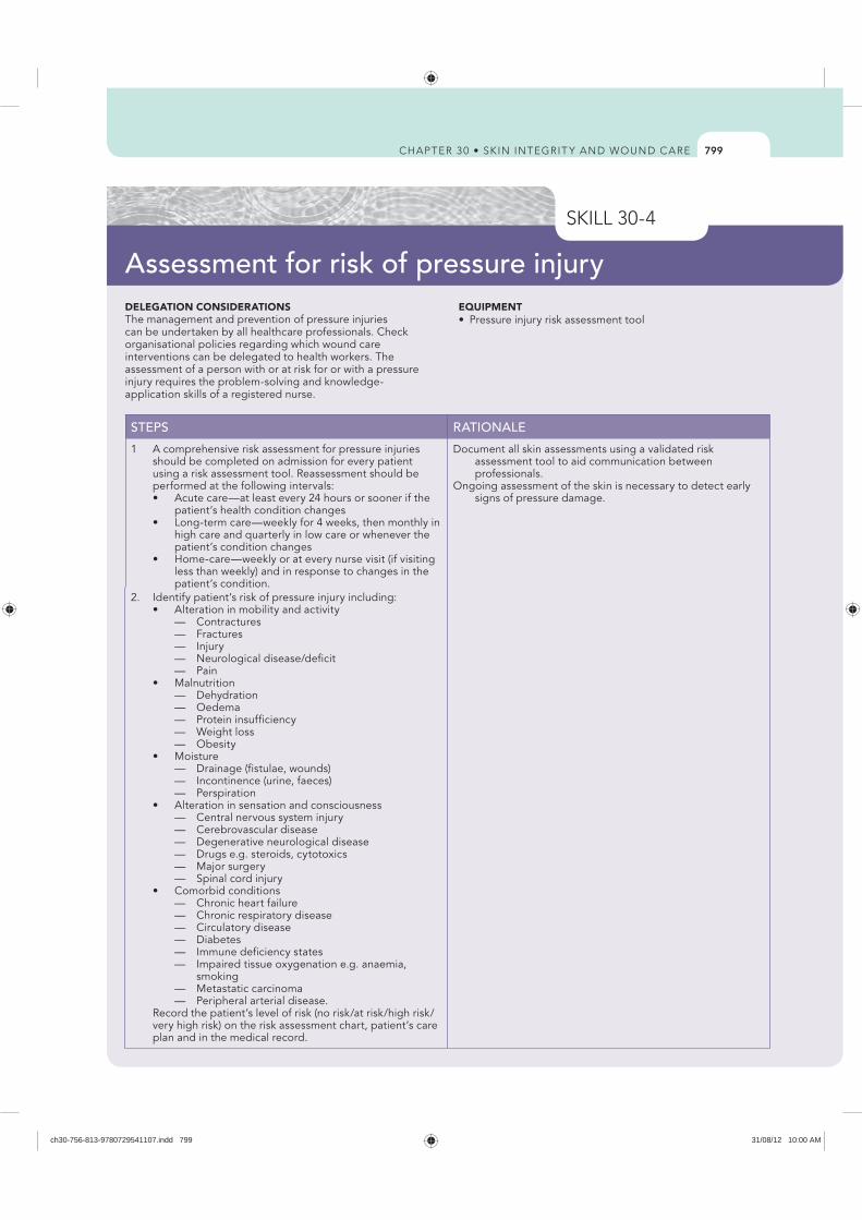
799
DELEGATION CONSIDERATIONSThe management and prevention of pressure injuries can be undertaken by all healthcare professionals. Check organisational policies regarding which wound care interventions can be delegated to health workers. The assessment of a person with or at risk for or with a pressure injury requires the problem-solving and knowledge-application skills of a registered nurse.
EQUIPMENT
STEPS RATIONALE
1 A comprehensive risk assessment for pressure injuries should be completed on admission for every patient using a risk assessment tool. Reassessment should be performed at the following intervals:
patient’s health condition changes
high care and quarterly in low care or whenever the patient’s condition changes
less than weekly) and in response to changes in the patient’s condition.
Document all skin assessments using a validated risk assessment tool to aid communication between professionals.
Ongoing assessment of the skin is necessary to detect early signs of pressure damage.
2. Identify patient’s risk of pressure injury including:
— Contractures — Fractures — Injury — Neurological disease/deficit — Pain
— Dehydration — Oedema — Protein insufficiency — Weight loss — Obesity
— Drainage (fistulae, wounds) — Incontinence (urine, faeces) — Perspiration
— Central nervous system injury — Cerebrovascular disease — Degenerative neurological disease — Drugs e.g. steroids, cytotoxics — Major surgery — Spinal cord injury
— Chronic heart failure — Chronic respiratory disease — Circulatory disease — Diabetes — Immune deficiency states — Impaired tissue oxygenation e.g. anaemia,
smoking — Metastatic carcinoma — Peripheral arterial disease. Record the patient’s level of risk (no risk/at risk/high risk/
very high risk) on the risk assessment chart, patient’s care plan and in the medical record.
Assessment for risk of pressure injury
ch30-756-813-9780729541107.indd 799 31/08/12 10:00 AM
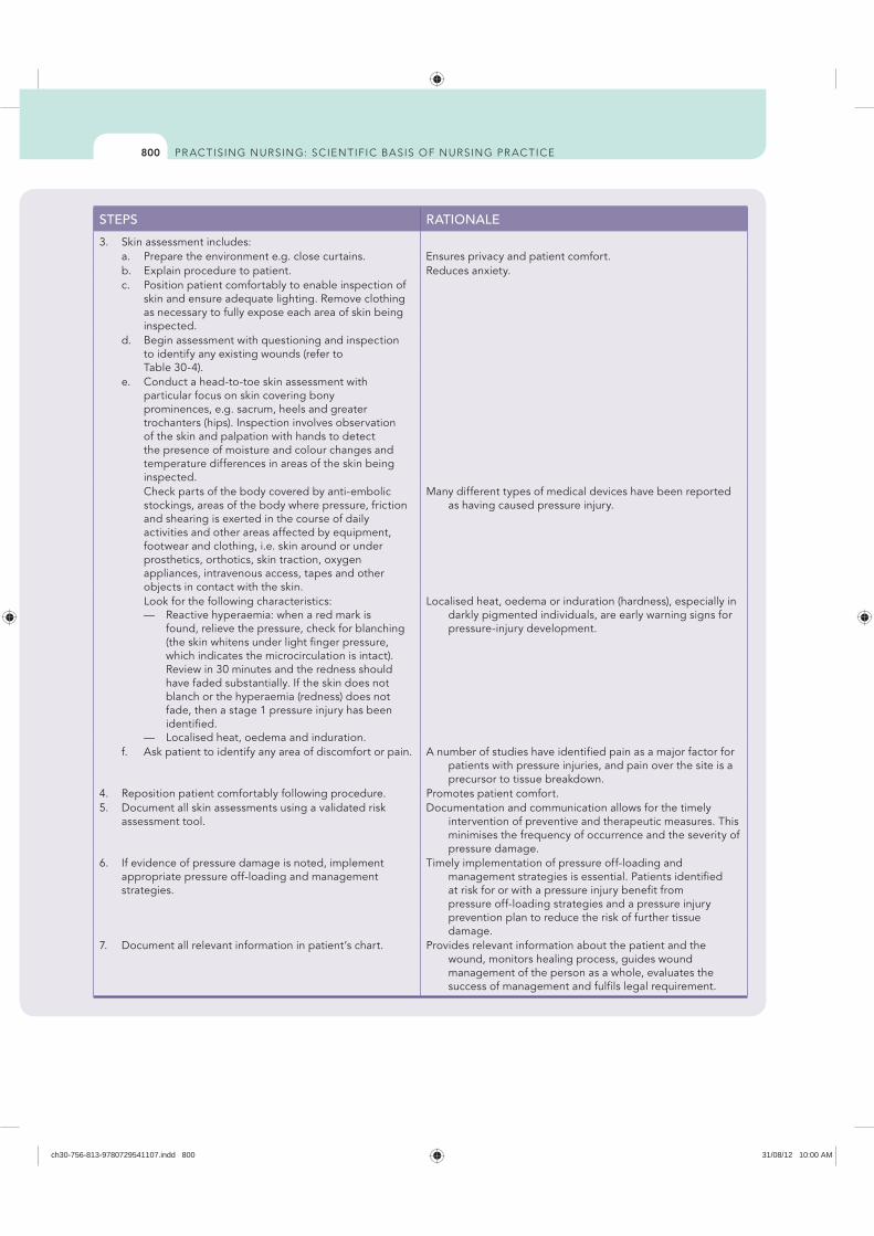
800
STEPS RATIONALE
3. Skin assessment includes: a. Prepare the environment e.g. close curtains. Ensures privacy and patient comfort. b. Explain procedure to patient. Reduces anxiety. c. Position patient comfortably to enable inspection of
skin and ensure adequate lighting. Remove clothing as necessary to fully expose each area of skin being inspected.
d. Begin assessment with questioning and inspection to identify any existing wounds (refer to Table 30-4).
e. Conduct a head-to-toe skin assessment with particular focus on skin covering bony prominences, e.g. sacrum, heels and greater trochanters (hips). Inspection involves observation of the skin and palpation with hands to detect the presence of moisture and colour changes and temperature differences in areas of the skin being inspected.
Check parts of the body covered by anti-embolic stockings, areas of the body where pressure, friction and shearing is exerted in the course of daily activities and other areas affected by equipment, footwear and clothing, i.e. skin around or under prosthetics, orthotics, skin traction, oxygen appliances, intravenous access, tapes and other objects in contact with the skin.
Many different types of medical devices have been reported as having caused pressure injury.
Look for the following characteristics: — Reactive hyperaemia: when a red mark is
found, relieve the pressure, check for blanching (the skin whitens under light finger pressure, which indicates the microcirculation is intact). Review in 30 minutes and the redness should have faded substantially. If the skin does not blanch or the hyperaemia (redness) does not fade, then a stage 1 pressure injury has been identified.
— Localised heat, oedema and induration.
Localised heat, oedema or induration (hardness), especially in darkly pigmented individuals, are early warning signs for pressure-injury development.
f. Ask patient to identify any area of discomfort or pain. A number of studies have identified pain as a major factor for patients with pressure injuries, and pain over the site is a precursor to tissue breakdown.
4. Reposition patient comfortably following procedure. Promotes patient comfort.5. Document all skin assessments using a validated risk
assessment tool.Documentation and communication allows for the timely
intervention of preventive and therapeutic measures. This minimises the frequency of occurrence and the severity of pressure damage.
6. If evidence of pressure damage is noted, implement appropriate pressure off-loading and management strategies.
Timely implementation of pressure off-loading and management strategies is essential. Patients identified at risk for or with a pressure injury benefit from pressure off-loading strategies and a pressure injury prevention plan to reduce the risk of further tissue damage.
7. Document all relevant information in patient’s chart. Provides relevant information about the patient and the wound, monitors healing process, guides wound management of the person as a whole, evaluates the success of management and fulfils legal requirement.
ch30-756-813-9780729541107.indd 800 31/08/12 10:00 AM
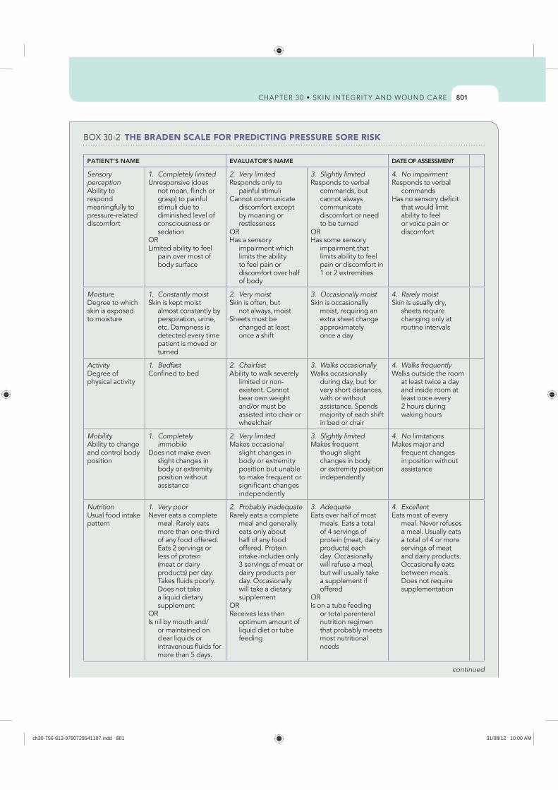
801
THE BRADEN SCALE FOR PREDICTING PRESSURE SORE RISK
PATIENT’S NAME EVALUATOR’S NAME DATE OF ASSESSMENT
Sensory perceptionAbility to respond meaningfully to pressure-related discomfort
1. Completely limitedUnresponsive (does
not moan, flinch or grasp) to painful stimuli due to diminished level of consciousness or sedation
ORLimited ability to feel
pain over most of body surface
2. Very limitedResponds only to
painful stimuliCannot communicate
discomfort except by moaning or restlessness
ORHas a sensory
impairment which limits the ability to feel pain or discomfort over half of body
3. Slightly limitedResponds to verbal
commands, but cannot always communicate discomfort or need to be turned
ORHas some sensory
impairment that limits ability to feel pain or discomfort in 1 or 2 extremities
4. No impairmentResponds to verbal
commandsHas no sensory deficit
that would limit ability to feel or voice pain or discomfort
MoistureDegree to which skin is exposed to moisture
1. Constantly moistSkin is kept moist
almost constantly by perspiration, urine, etc. Dampness is detected every time patient is moved or turned
2. Very moistSkin is often, but
not always, moistSheets must be
changed at least once a shift
3. Occasionally moistSkin is occasionally
moist, requiring an extra sheet change approximately once a day
4. Rarely moistSkin is usually dry,
sheets require changing only at routine intervals
ActivityDegree of physical activity
1. BedfastConfined to bed
2. ChairfastAbility to walk severely
limited or non-existent. Cannot bear own weight and/or must be assisted into chair or wheelchair
3. Walks occasionallyWalks occasionally
during day, but for very short distances, with or without assistance. Spends majority of each shift in bed or chair
4. Walks frequentlyWalks outside the room
at least twice a day and inside room at least once every 2 hours during waking hours
MobilityAbility to change and control body position
1. Completely immobile
Does not make even slight changes in body or extremity position without assistance
2. Very limitedMakes occasional
slight changes in body or extremity position but unable to make frequent or significant changes independently
3. Slightly limitedMakes frequent
though slight changes in body or extremity position independently
4. No limitationsMakes major and
frequent changes in position without assistance
NutritionUsual food intake pattern
1. Very poorNever eats a complete
meal. Rarely eats more than one-third of any food offered. Eats 2 servings or less of protein (meat or dairy products) per day. Takes fluids poorly. Does not take a liquid dietary supplement
ORIs nil by mouth and/
or maintained on clear liquids or intravenous fluids for more than 5 days.
2. Probably inadequateRarely eats a complete
meal and generally eats only about half of any food offered. Protein intake includes only 3 servings of meat or dairy products per day. Occasionally will take a dietary supplement
ORReceives less than
optimum amount of liquid diet or tube feeding
3. AdequateEats over half of most
meals. Eats a total of 4 servings of protein (meat, dairy products) each day. Occasionally will refuse a meal, but will usually take a supplement if offered
ORIs on a tube feeding
or total parenteral nutrition regimen that probably meets most nutritional needs
4. ExcellentEats most of every
meal. Never refuses a meal. Usually eats a total of 4 or more servings of meat and dairy products. Occasionally eats between meals. Does not require supplementation
continued
ch30-756-813-9780729541107.indd 801 31/08/12 10:00 AM
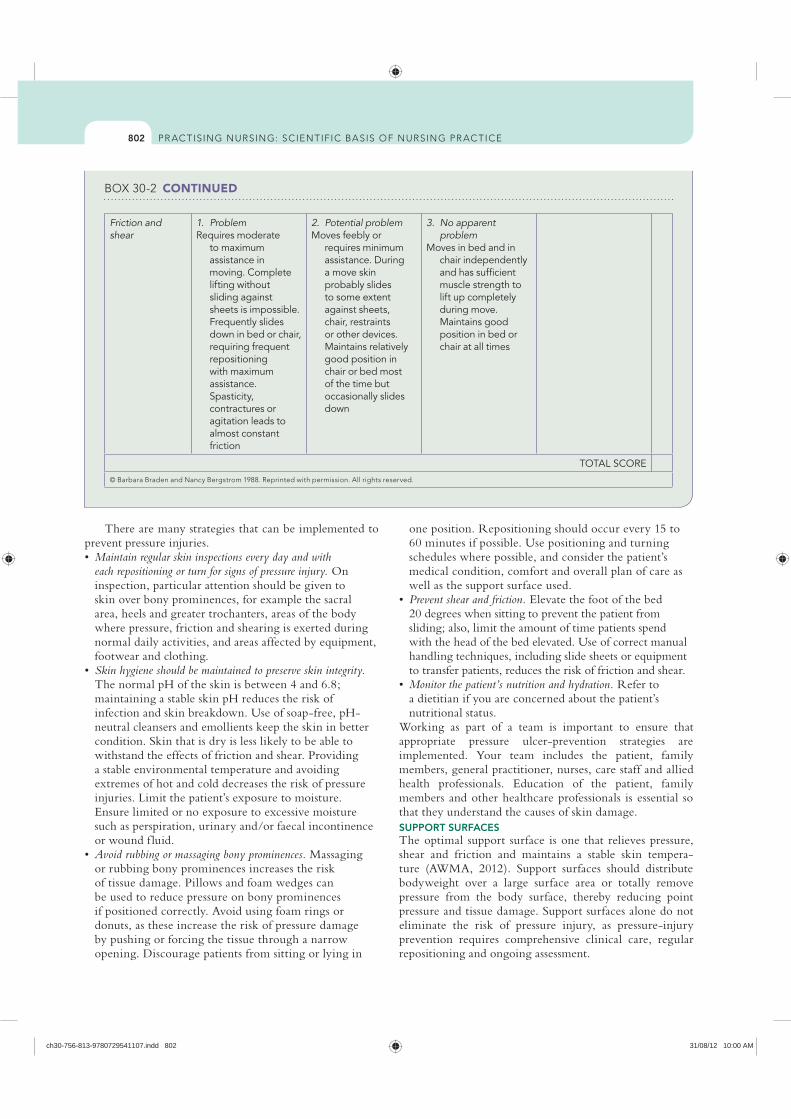
802
one position. Repositioning should occur every 15 to 60 minutes if possible. Use positioning and turning schedules where possible, and consider the patient’s medical condition, comfort and overall plan of care as well as the support surface used. Prevent shear and friction. Elevate the foot of the bed 20 degrees when sitting to prevent the patient from sliding; also, limit the amount of time patients spend with the head of the bed elevated. Use of correct manual handling techniques, including slide sheets or equipment to transfer patients, reduces the risk of friction and shear.Monitor the patient’s nutrition and hydration. Refer to a dietitian if you are concerned about the patient’s nutritional status.
Working as part of a team is important to ensure that appropriate pressure ulcer-prevention strategies are implemented. Your team includes the patient, family members, general practitioner, nurses, care staff and allied health professionals. Education of the patient, family members and other healthcare professionals is essential so that they understand the causes of skin damage. SUPPORT SURFACESThe optimal support surface is one that relieves pressure, shear and friction and maintains a stable skin tempera-ture (AWMA, 2012). Support surfaces should distribute bodyweight over a large surface area or totally remove pressure from the body surface, thereby reducing point pressure and tissue damage. Support surfaces alone do not eliminate the risk of pressure injury, as pressure-injury prevention requires comprehensive clinical care, regular repositioning and ongoing assessment.
There are many strategies that can be implemented to prevent pressure injuries.
Maintain regular skin inspections every day and with each repositioning or turn for signs of pressure injury. On inspection, particular attention should be given to skin over bony prominences, for example the sacral area, heels and greater trochanters, areas of the body where pressure, friction and shearing is exerted during normal daily activities, and areas affected by equipment, footwear and clothing. Skin hygiene should be maintained to preserve skin integrity. The normal pH of the skin is between 4 and 6.8; maintaining a stable skin pH reduces the risk of infection and skin breakdown. Use of soap-free, pH-neutral cleansers and emollients keep the skin in better condition. Skin that is dry is less likely to be able to withstand the effects of friction and shear. Providing a stable environmental temperature and avoiding extremes of hot and cold decreases the risk of pressure injuries. Limit the patient’s exposure to moisture. Ensure limited or no exposure to excessive moisture such as perspiration, urinary and/or faecal incontinence or wound f luid.Avoid rubbing or massaging bony prominences. Massaging or rubbing bony prominences increases the risk of tissue damage. Pillows and foam wedges can be used to reduce pressure on bony prominences if positioned correctly. Avoid using foam rings or donuts, as these increase the risk of pressure damage by pushing or forcing the tissue through a narrow opening. Discourage patients from sitting or lying in
CONTINUED
Friction and shear
1. ProblemRequires moderate
to maximum assistance in moving. Complete lifting without sliding against sheets is impossible. Frequently slides down in bed or chair, requiring frequent repositioning with maximum assistance. Spasticity, contractures or agitation leads to almost constant friction
2. Potential problemMoves feebly or
requires minimum assistance. During a move skin probably slides to some extent against sheets, chair, restraints or other devices. Maintains relatively good position in chair or bed most of the time but occasionally slides down
3. No apparent problem
Moves in bed and in chair independently and has sufficient muscle strength to lift up completely during move. Maintains good position in bed or chair at all times
TOTAL SCORE
ch30-756-813-9780729541107.indd 802 31/08/12 10:00 AM
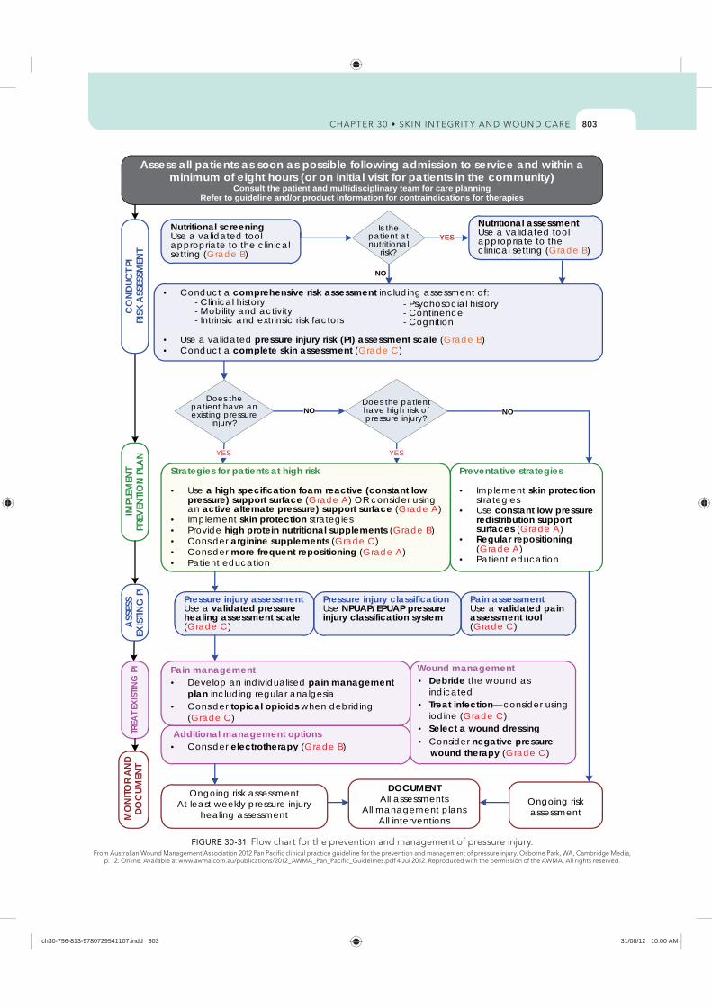
803
Assess all patients as soon as possible following admission to service and within a minimum of eight hours (or on initial visit for patients in the community)
Consult the patient and multidisciplinary team for care planningRefer to guideline and/or product information for contraindications for therapies
Preventative strategies
Implement skin protectionstrategiesUse constant low pressure redistribution support surfaces (Grade A)Regular repositioning(Grade A)Patient education
Wound managementDebride the wound as indicatedTreat infection onsider using iodine (Grade C)Select a wound dressing Consider negative pressure
wound therapy (Grade C)
IMPL
EMEN
TPR
EVEN
TION
PLAN
TREA
T EXI
STIN
G P
I
Additional management optionsConsider electrotherapy (Grade B)
Conduct a comprehensive risk assessment including assessment of: - Clinical history - Mobility and activity - Intrinsic and extrinsic risk factors
Use a validated pressure injury risk (PI) assessment scale (Grade B)Conduct a complete skin assessment (Grade C)
Ongoing risk assessment
MO
NITO
R AN
D DO
CUM
ENT
NO
Strategies for patients at high risk
Use a high specification foam reactive (constant low pressure) support surface (Grade A) OR consider using an active alternate pressure) support surface (Grade A)Implement skin protection strategiesProvide high protein nutritional supplements (Grade B)Consider arginine supplements (Grade C)Consider more frequent repositioning (Grade A)Patient education
Ongoing risk assessmentAt least weekly pressure injury
healing assessment
Pain managementDevelop an individualised pain management plan including regular analgesiaConsider topical opioids when debriding (Grade C)
Pressure injury classificationUse NPUAP/EPUAP pressure injury classification system
NO
YES
Does the patient have an existing pressure
injury?
Does the patient have high risk of pressure injury?
Pressure injury assessmentUse a validated pressure healing assessment scale(Grade C)
Pain assessmentUse a validated pain assessment tool (Grade C)
DOCUMENTAll assessments
All management plansAll interventions
CO
NDUC
T PI
RISK
ASS
ESSM
ENT
ASSE
SSEX
ISTIN
G P
I
Nutritional screeningUse a validated tool appropriate to the clinical setting (Grade B)
Is the patient at nutritional
risk?
Nutritional assessmentUse a validated tool appropriate to the clinical setting (Grade B)
NO
YES
- Psychosocial history- Continence- Cognition
YES
—c
FIGURE 30-31
ch30-756-813-9780729541107.indd 803 31/08/12 10:00 AM
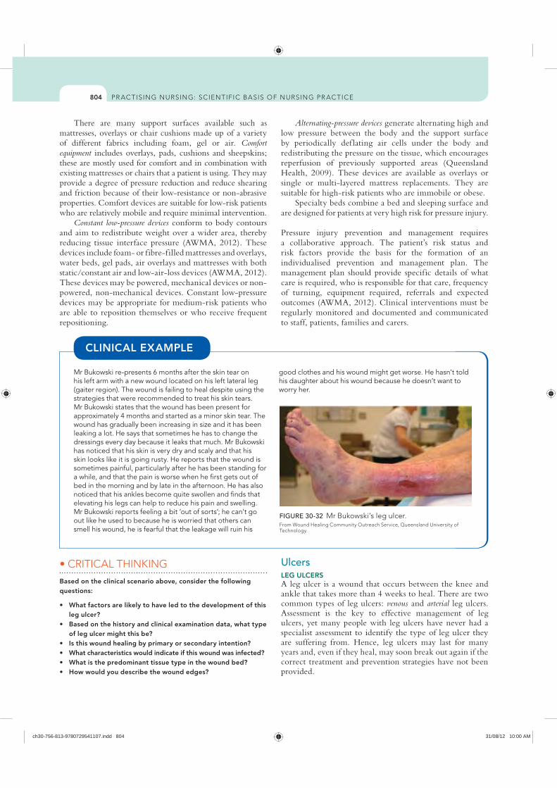
804
Alternating-pressure devices generate alternating high and low pressure between the body and the support surface by periodically def lating air cells under the body and redistributing the pressure on the tissue, which encourages reperfusion of previously supported areas (Queensland Health, 2009). These devices are available as overlays or single or multi-layered mattress replacements. They are suitable for high-risk patients who are immobile or obese.
Specialty beds combine a bed and sleeping surface and are designed for patients at very high risk for pressure injury.
Pressure injury prevention and management requires a collaborative approach. The patient’s risk status and risk factors provide the basis for the formation of an individualised prevention and management plan. The management plan should provide specific details of what care is required, who is responsible for that care, frequency of turning, equipment required, referrals and expected outcomes (AWMA, 2012). Clinical interventions must be regularly monitored and documented and communicated to staff, patients, families and carers.
There are many support surfaces available such as mattresses, overlays or chair cushions made up of a variety of different fabrics including foam, gel or air. Comfort equipment includes overlays, pads, cushions and sheepskins; these are mostly used for comfort and in combination with existing mattresses or chairs that a patient is using. They may provide a degree of pressure reduction and reduce shearing and friction because of their low-resistance or non-abrasive properties. Comfort devices are suitable for low-risk patients who are relatively mobile and require minimal intervention.
Constant low-pressure devices conform to body contours and aim to redistribute weight over a wider area, thereby reducing tissue interface pressure (AWMA, 2012). These devices include foam- or fibre-filled mattresses and overlays, water beds, gel pads, air overlays and mattresses with both static/constant air and low-air-loss devices (AWMA, 2012). These devices may be powered, mechanical devices or non-powered, non-mechanical devices. Constant low-pressure devices may be appropriate for medium-risk patients who are able to reposition themselves or who receive frequent repositioning.
Mr Bukowski re-presents 6 months after the skin tear on his left arm with a new wound located on his left lateral leg (gaiter region). The wound is failing to heal despite using the strategies that were recommended to treat his skin tears. Mr Bukowski states that the wound has been present for approximately 4 months and started as a minor skin tear. The wound has gradually been increasing in size and it has been leaking a lot. He says that sometimes he has to change the dressings every day because it leaks that much. Mr Bukowski has noticed that his skin is very dry and scaly and that his skin looks like it is going rusty. He reports that the wound is sometimes painful, particularly after he has been standing for a while, and that the pain is worse when he first gets out of bed in the morning and by late in the afternoon. He has also noticed that his ankles become quite swollen and finds that elevating his legs can help to reduce his pain and swelling. Mr Bukowski reports feeling a bit ‘out of sorts’; he can’t go out like he used to because he is worried that others can smell his wound, he is fearful that the leakage will ruin his
good clothes and his wound might get worse. He hasn’t told his daughter about his wound because he doesn’t want to worry her.
CLINICAL EXAMPLE
FIGURE 30-32 Mr Bukowski’s leg ulcer.
Technology.
Based on the clinical scenario above, consider the following questions:
leg ulcer?
of leg ulcer might this be?
UlcersLEG ULCERSA leg ulcer is a wound that occurs between the knee and ankle that takes more than 4 weeks to heal. There are two common types of leg ulcers: venous and arterial leg ulcers. Assessment is the key to effective management of leg ulcers, yet many people with leg ulcers have never had a specialist assessment to identify the type of leg ulcer they are suffering from. Hence, leg ulcers may last for many years and, even if they heal, may soon break out again if the correct treatment and prevention strategies have not been provided.
ch30-756-813-9780729541107.indd 804 31/08/12 10:00 AM
805
Apply graduated compression bandaging. The key to healing a venous leg ulcer is to treat the underlying cause (i.e. reduce the high blood pressure in the veins). Graduated compression bandaging works by improving venous return, reducing swelling in the lower limb and improving foot- and calf-muscle pump action. Bandages are applied from the base of the toes to just below the knee. The bandage is said to be graduated because the ankle is smaller than the calf. Therefore, the pressure exerted by the bandage at the ankle is higher than that applied at the calf. You need special training in how to apply graduated compression bandages correctly, because if they are applied incorrectly they can cause significant damage to the patient’s circulation and may even make the ulceration worse.Apply compression stockings. Graduated compression stockings for life after wound healing help to prevent venous leg ulcers from recurring, because they continue to provide compression to treat the underlying cause of the leg ulcers (i.e. poor circulation). A venous leg ulcer is really a symptom of an underlying disease process. Compression stockings can also be used to prevent venous leg ulcers.
Failure to reach an accurate diagnosis and/or to recognise the signs and symptoms of venous disease may result in the unsafe application of the graduated compression bandaging.
ARTERIAL LEG ULCERS are less common than venous leg ulcers and are the result of peripheral arterial disease. Arteries supply blood and oxygen to the lower limbs; arterial leg ulcers result from inadequate blood supply and oxygen in the arteries. If blood supply to the lower legs is poor, the leg is starved of oxygen and nutrients; the skin fails to function normally and an ulcer may develop. A patient with arterial disease may have very cool or cold feet, the skin may be pale, blue (cyanosed) or dusky red in colour and they may complain of pain in the foot or calf
Every leg ulcer has key features or characteristics that help determine the type of leg ulcer the person is suffering from. The key characteristics for the most common types of leg ulcers are outlined in Table 30-18, later in the chapter.
VENOUS LEG ULCERS are the most common type of leg ulcer. They occur because of high blood pressure in the veins in the lower limb (known as venous hypertension). Veins carry blood from the legs back to the heart. Veins in the lower leg have one-way valves to prevent blood running backwards, and when the calf muscles contract, blood is moved along the veins back towards the heart.
However, sometimes the valves in the veins or the calf muscles fail to work properly, for a number of different reasons (e.g. damage from a past clot in the vein), leading to chronic venous insufficiency or venous disease. The constant high pressure in the veins eventually causes f luid to leak out of the veins and into the surrounding tissues and skin. Although the reason for ulcer formation is not fully understood, the combination of high pressure in the veins and particles in the f luid forced into the tissues causes inf lammation. This results in the skin easily breaking down and forming an ulcer. Table 30-15 highlights some of the risk factors for venous leg ulcers.
Once you have identified the signs and symptoms of a venous leg ulcer, you then need to determine the appropriate treatment. Key evidence-based management strategies for venous leg ulcers are summarised here.
Dressings used to treat venous leg ulcers vary depending on the condition of the ulcer and the goals of treatment. Dressings should maintain a moist wound-healing environment, manage wound leakage (exudate), protect the skin around the ulcer, be non-adherent to reduce trauma on removal, and be cost-effective, acceptable to the patient and able to be changed less often where possible.
RISK FACTORS FOR VENOUS LEG ULCERSRISK FACTOR EXPLANATION
Age Blood flow decreases as a person ages, and decreased mobility leads to the calf muscles becoming less effective and not being used as much as they need to
Obesity Excess weight, particularly around the waist and thighs, puts extra pressure on the veins and they have to work harder to move blood flow back towards the heart. Note that multiple pregnancies may also be a cause of excess weight
Varicose veins The one-way valves that stop blood from travelling backwards in the veins stop working and this leads to pooling of blood in the lower legs
Past deep-vein thrombosis or trauma to lower limbs
A history of deep-vein thrombosis (i.e. blood clot) or trauma may result in permanent damage to the veins
Jobs that involve sitting or standing for long periods of time
Occupations that involve sitting or standing for long periods of time may result in less-effective blood flow through the veins and pooling of blood in the lower legs
Family history or congenital valvular incompetence
There is a strong link between family history and leg ulcers. In addition, in rare cases some people are born with problems that affect their veins, placing them at higher risk of venous disease
Health conditions Disorders such as congestive cardiac failure lead to swelling of the lower limbs because the heart is not able to process blood flow effectively
ch30-756-813-9780729541107.indd 805 31/08/12 10:00 AM
806
Recommend a medical review: patients with diabetic foot ulcers may benefit from review by a specialist.Use a team approach to care: care of diabetic foot ulcers should be undertaken by a multidisciplinary team including podiatrists, orthotists, wound-care specialists, the patient’s doctor, an endocrinologist, a dietitian and a diabetic educator.Ensure adequate oxygenation: improving oxygenation of the wound environment promotes healing. Steps to improve oxygen supply to the tissues include avoidance of dehydration, cold, stress and pain.Ensure off-loading of pressure points: it is essential to review the patient’s footwear and ensure that pressure is relieved. Otherwise, the wound will fail to heal. Areas of abnormal pressure distribution on the diabetic foot are usually located on the plantar aspect of the foot under the hallux, f irst and fifth metatarsal heads and under the heel (Grey and others, 2006). Apply suitable dressings: dressings used to treat diabetic foot ulcers vary depending on the condition of the ulcer and goals of treatment. Topical antimicrobial dressings may be helpful at preventing infection.Control risk factors: controlling risk factors known to affect the arteries is essential. For example, try to ensure that blood glucose levels are kept within normal ranges, the blood pressure is controlled, cholesterol is reduced, medications are reviewed, and the patient has a healthy and balanced diet, stops smoking and is encouraged to maintain a healthy weight relative to their height.
ASSESSMENT AND MANAGEMENT Being able to recognise the key signs and symptoms of the different types of ulcers is very important to help obtain an accurate diagnosis. The following steps may assist in clinical examination to determine the aetiology of ulceration. Table 30-17 compares the key characteristics of venous arterial and diabetic ulcers. Assessing these characteristics can assist you to determine the aetiology of ulceration.
muscle. Pain is often worse at night when the lower leg is elevated in bed and pain is only relieved when the legs are lowered. During exercise, the muscles in the lower leg need more oxygen; when a person has arterial disease, the blood supply is decreased and pain occurs in the foot or calf muscle on exercising. This pain is known as intermittent claudication. Table 30-16 outlines the patient risk factors for developing arterial leg ulcers.
Patients with arterial leg ulcers may benefit from a review by a specialist. This is because restoring blood f low by revascularisation is the intervention most likely to heal arterial leg ulcers.
Improving oxygenation of the wound environment promotes healing. Steps to improve oxygen supply to the tissues include avoidance of dehydration, cold, stress and pain. Dressings used to treat arterial leg ulcers vary depending on the condition of the ulcer and goals of the treatment. Topical antimicrobial dressings may be helpful to prevent infection.
Controlling risk factors known to affect the arteries is essential. For example, the healthcare professional should try to ensure that blood glucose levels are kept within normal ranges, blood pressure is controlled, cholesterol levels are reduced, medications are reviewed and the patient has a healthy and balanced diet, stops smoking and is encouraged to maintain a healthy weight relative to their height.DIABETIC FOOT ULCERSA diabetic foot ulcer is an ulcer located on the foot, and is associated with a loss of protective sensation (neuropathy) and/or the presence of ischaemia in a patient with diabetes. Diabetic foot ulcers are more likely to occur if the patient has uncontrolled or prolonged high blood-sugar levels (hyperglycaemia).
Once you have identified the signs and symptoms of a diabetic foot ulcer, you then need to determine the correct treatment. Key evidence-based management strategies for diabetic foot ulcers may include the following:
RISK FACTORS FOR ARTERIAL LEG ULCERSRISK FACTOR EXPLANATION
Atherosclerosis Hardening, narrowing or blockage of the arteries because of fatty deposits (plaques) or cholesterol reduces blood supply to the lower leg
Smoking Cigarette smoking is a well-known risk factor for atherosclerosis and heart disease
Hypertension High blood pressure leads to hardening of the arteries
Diabetes Poorly controlled diabetes leads to high cholesterol levels in the blood and increased risk of hardening of the arteries. There is also an increased risk of strokes, heart disease and ulceration of the lower limbs
High cholesterol High cholesterol is a major risk factor for hardening of the arteries
Stress There is a link between high levels of stress and arterial disease
A history of heart disease, heart attack or stroke
These conditions increase the risk of further problems with the peripheral arteries
Obesity Excess weight, particularly around the waist and thighs, puts extra pressure on the arteries and the heart has to work harder to move blood flow around the body
ch30-756-813-9780729541107.indd 806 31/08/12 10:00 AM
807
BOX 30-3 LEG ULCER PREVENTION STRATEGIES
Follow up and check the skin regularly. Apply a pH-neutral moisturiser to the skin at least twice daily. Ensure shoes are well-fitting, soft and comfortable. Be extremely careful when cutting toenails. Preferably, a podiatrist should do this.
Avoid soaking the feet in hot water, using heating pads and applying harsh topical skin cleansers.
Protect the toes and heels of patients with decreased mobility by using effective pressure-relief devices such as foam or gel pads.
Use leg-protection devices to avoid injury (e.g. limb protectors or long trousers).
Keep the lower legs warm. Passive warming of the lower legs and feet, such as covering the legs with a blanket, improves blood supply (perfusion). However, do not use hot water bottles, electric blankets or foot spas, or position the feet close to heating devices. This is because the person may not be able to feel if damage is occurring, which risks burning of their skin.
Encourage lower-limb exercises, at least every hour. Simple exercises may include drawing figures with the toes, pushing the toes towards the floor and then pointing them up towards the nose, standing on the balls of the feet, tiptoeing up and down, walking or swimming. Any movement of the calf muscle helps to move blood back to the heart. Also discourage crossing of the legs, which reduces arterial blood flow.
Elevate the affected limb at least 3–4 times every day for at least 15 minutes each time, to help reduce high blood pressure in the lower legs and reduce swelling. Elevation means placing the patient’s legs on pillows, a chair or another object so that the feet are above the level of the
knees, and the knees are above the level of the hips. Try to discourage patients from sitting with their legs down and stationary for more than an hour at a time. Patients should put their feet up while performing sit-down tasks. Elevating the legs to the level of the hips doesn’t reduce swelling, but it keeps the swelling from getting worse. Elevating the legs above the level of the heart is needed to reduce swelling.
Smoking cessation interventions are an important part of the treatment plan.
Ensure the patient maintains a healthy weight relative to their height. A well-balanced diet and healthy weight reduces the risk of arterial disease.
Manage any underlying diseases; ensuring that underlying diseases such as diabetes, heart disease or hypertension are well controlled helps to prevent complications such as arterial leg ulcers.
Education of patients, family members and other staff helps to ensure that problems are identified early and strategies are implemented to reduce risk. It is especially important that the patient does not walk barefoot if they have diabetes or arterial disease, as they might tread on something and damage their skin and not be aware they have done so.
For patients with venous leg ulcers, use of graduated compression stockings for life reduces ulcer recurrence rates. Compression stockings should be measured and fitted by a trained practitioner to ensure that they are the correct size for the person. Compression hosiery should be replaced at least every 6 months. Compression stockings must be worn every day except while in bed, bathing or swimming. Stockings should be applied as early in the day as possible to prevent the legs from swelling. Remove hosiery as close to bed-time as possible.
Malignant/fungating woundsMalignant fungating or cancerous wounds are those that affect people with cancer. Fortunately they occur infrequently, but are typically described as a devastating, overwhelming and distressing experience (Alexander, 2010). They are caused by the infiltration of the skin by a local tumour such as a squamous-cell carcinoma or melanoma; haematological malignancy such as cutaneous T-cell lymphoma; or metastatic spread from a primary tumour, for example breast cancer where metastasis occurs along tissue planes, capillaries or lymph vessels (Adderley and Smith, 2007; Naylor, 2002). Lesions are characterised by a process of both ulcerative (crater-like) and proliferative (nodular or caulif lower-shaped) growth that can cause extensive damage to the skin and surrounding structures (Lewellyn and others, 2002).
Although rare, malignant changes may develop in chronic wounds (Marjolin’s ulcer) and are most commonly associated with burn-scar ulcers, pressure or venous ulcers (Naylor, 2002). This type of malignancy is usually aggressive and has a high rate of metastatic spread and local recurrence
PREVENTION STRATEGIESOnce you have learned how to assess and manage venous, arterial and diabetic foot ulcers, there are some important evidence-based strategies that you can put in place to reduce the risk of the ulcer recurring. Box 30-3 highlights some of the key prevention strategies.
Think about how you would decide on the management of Mr
Bukowski’s leg ulcer:
this type of leg ulcer? What dressing regimen would you
skin around the wound?
What strategies would you recommend to prevent leg ulcer recurrence in the future?
liaise with in this case?
ch30-756-813-9780729541107.indd 807 31/08/12 10:00 AM
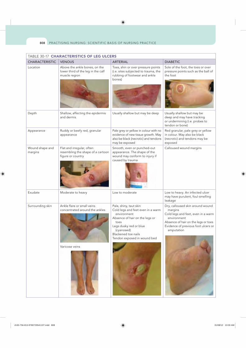
808
CHARACTERISTICS OF LEG ULCERSCHARACTERISTIC VENOUS ARTERIAL DIABETIC
Location Above the ankle bones, on the lower third of the leg in the calf muscle region
Toes, shin or over pressure points (i.e. sites subjected to trauma, the rubbing of footwear and ankle bones)
Sole of the foot, the toes or over pressure points such as the ball of the foot
Depth Shallow, affecting the epidermis and dermis
Usually shallow but may be deep Usually shallow but may be deep and may have tracking or undermining (i.e. probes to tendon or bone)
Appearance Ruddy or beefy red, granular appearance
Pale grey or yellow in colour with no evidence of new tissue growth. May also be black (necrotic) and tendons may be exposed
Red granular, pale grey or yellow in colour. May also be black (necrotic) and tendons may be exposed
Wound shape and margins
Flat and irregular, often resembling the shape of a cartoon figure or country
Smooth, even or punched-out appearance. The shape of the wound may conform to injury if caused by trauma
Calloused wound margins
Exudate Moderate to heavy Low to moderate Low to heavy. An infected ulcer may have purulent, foul-smelling leakage
Surrounding skin Ankle flare or small veins concentrated around the ankles
Varicose veins
Pale, shiny, taut skinCold legs and feet even in a warm
environmentAbsence of hair on the legs or
toesLegs dusky red or blue
(cyanosed) Blackened toe nails Tendon exposed in wound bed
Dry, calloused skin around wound margins
Cold legs and feet, even in a warm environment
Absence of hair on the legs or toesEvidence of previous foot ulcers or
amputation
ch30-756-813-9780729541107.indd 808 31/08/12 10:00 AM
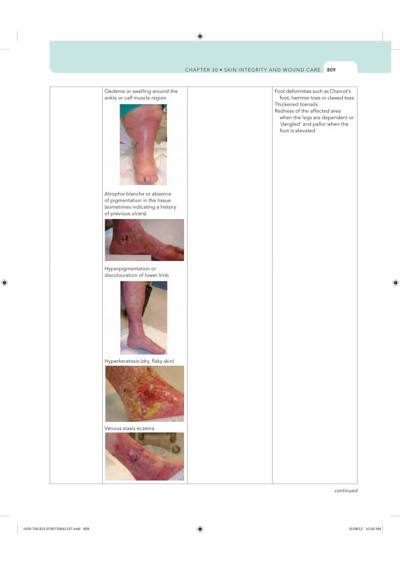
809
Oedema or swelling around the ankle or calf muscle region
Atrophie blanche or absence of pigmentation in the tissue (sometimes indicating a history of previous ulcers)
Foot deformities such as Charcot’s foot, hammer toes or clawed toes
Thickened toenails Redness of the affected area
when the legs are dependent or ‘dangled’ and pallor when the foot is elevated
Hyperpigmentation or discolouration of lower limb
Hyperkeratosis (dry, flaky skin)
Venous stasis eczema
continued
ch30-756-813-9780729541107.indd 809 31/08/12 10:00 AM
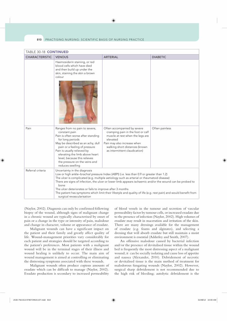
810
CONTINUEDCHARACTERISTIC VENOUS ARTERIAL DIABETIC
Haemosiderin staining, or red blood cells which have died and then build up under the skin, staining the skin a brown colour
Pain Ranges from no pain to severe, constant pain
Pain is often worse after standing for long periods
May be described as an achy, dull pain or a feeling of pressure
Pain is usually relieved by elevating the limb above heart level, because this relieves the pressure on the veins and reduces swelling
Often accompanied by severe cramping pain in the foot or calf muscle at rest when the legs are elevated
Pain may also increase when walking short distances (known as intermittent claudication)
Often painless
Referral criteria Uncertainty in the diagnosis Low or high ankle–brachial pressure Index (ABPI) (i.e. less than 0.9 or greater than 1.2)The ulcer is complicated (e.g. multiple aetiology such as arterial or rheumatoid disease)There are signs of infection, the ulcer or lower limb appears ischaemic and/or the wound can be probed to
boneThe ulcer deteriorates or fails to improve after 3 monthsThe patient has symptoms which limit their lifestyle and quality of life (e.g. rest pain) and would benefit from
surgical revascularisation
(Naylor, 2002). Diagnosis can only be confirmed following biopsy of the wound, although signs of malignant change in a chronic wound are typically characterised by onset of pain or a change in the type or intensity of pain, malodour and change in character, volume or appearance of exudate.
Malignant wounds can have a significant impact on the patient and their family and greatly affect quality of life. Wound-management priorities vary considerably for each patient and strategies should be targeted according to the patient’s preferences. Most patients with a malignant wound will be in the terminal stages of their illness and wound healing is unlikely to occur. The main aim of wound management is aimed at controlling or eliminating the distressing symptoms associated with these wounds.
Malignant wounds often produce copious amounts of exudate which can be difficult to manage (Naylor, 2002). Exudate production is secondary to increased permeability
of blood vessels in the tumour and secretion of vascular permeability factor by tumour cells, or increased exudate due to the presence of infection (Naylor, 2002). High volumes of exudate may result in maceration and irritation of the skin. There are many dressings available for the management of exudate (e.g. foams and alginates), and selecting a dressing that will absorb exudate but still maintain a moist environment is essential (Adderley and Smith, 2007).
An offensive malodour caused by bacterial infection and/or the presence of devitalised tissue within the wound bed is frequently the most distressing aspect of a malignant wound; it can be socially isolating and cause loss of appetite and nausea (Alexander, 2010). Debridement of necrotic or devitalised tissue is the main method of treatment for malodorous fungating wounds (Naylor, 2002). However, surgical sharp debridement is not recommended due to the high risk of bleeding; autolytic debridement is the
ch30-756-813-9780729541107.indd 810 31/08/12 10:00 AM
811
likelihood of developing a reaction depends on a number of factors including the patient’s age, skin integrity, type of radiation and amount of energy used, chemical and skin irritants used by the patient, nutritional status, presence of skin folds and concurrent chemotherapy (Regan, 2007). Skin reactions may be mild such as dry skin or may lead to moist desquamation.
Strategies that can be taken to minimise the effects of radiation include preventing mechanical trauma caused by shaving, friction, pressure, adhesive tapes or constrictive clothing; avoiding the use of chemical trauma from soaps, deodorants, shaving creams, astringents or make-up; avoiding extremes of heat and cold to prevent thermal trauma; avoiding skin products containing metallic substances, including zinc cream and talcum powder; avoiding exposure to the sun; moisturising the skin frequently with a pH-neutral emollient; and ensuring adequate nutrition and hydration (Carville, 2007).
Chemotherapy may be used to reduce the size of a malignant wound; it involves the use of systemic or localised cytotoxic drugs (Regan, 2007). Chemotherapy can cause skin reactions including transient erythema, hyperpigmentation, photosensitivity, nail changes, palmar/plantar syndrome and inf lammatory reactions in tissue that has been previously irradiated resulting in blisters, vesicle formation, exfoliation and ulceration (Regan, 2007). Chemotherapy can decrease the size of a malignant wound by destroying malignant cells, and reduce pressure on nerves and blood or lymph vessels thereby reducing exudate production and potential to bleed (Regan, 2007).
Extravasation of the skin can occur secondary to the use of chemotherapy drugs, resulting in ulceration of the skin, nerve damage, significant pain, disfigurement and potential limb loss (Regan, 2007). Extravasation occurs over a few days with maximum severity over 2–3 weeks. The degree of tissue damage is secondary to the amount of drug extravasated, drug concentration, site, needle insertion device and technique (Regan, 2007). The affected tissue may appear swollen, tender and red and progressively worsen into ulceration, and may involve tendon and nerves. Severe tissue damage may require surgical intervention (Regan, 2007). Prevention of extravasation injuries includes a thorough assessment of the patient, venous access, related risk factors, knowledge of the drug and early recognition of the signs of extravasation injury including burning pain, erythema and oedema at injection site.
Malignant fungating wounds are a significant problem and challenging for both patients and healthcare professionals. The key to effective management is accurate assessment to improve quality of life through symptom control; however, there is insufficient evidence to provide clear direction for practice with regard to improving quality of life or manag ing wound symptoms associated with fungating wounds (Adderley and Smith, 2007; Lo and others, 2008, 2012).
preferred method of debridement (Naylor, 2002). The use of antimicrobials (e.g. metronidazole gel) can help to reduce the amount of bacteria present in the wound, thereby reducing malodour. The use of topical activated-charcoal dressings and occlusive dressings may help to control odour, and environmental agents such as room deodorants and fragrances for control of odour may also assist (Carville, 2007).
Bleeding can occur secondary to erosion of blood vessels by malignant cells and may be exacerbated by decreased platelet function within the tumour (Naylor, 2002). Profuse, spontaneous bleeding can be very distressing and careful management of the wound at dressing changes is essential to minimise the risk of bleeding. Preventative measures to reduce the risk of bleeding, such as the use of non-adherent dressings, maintaining a moist environment, haemostatic dressings (e.g. alginates or haemostatic sponges) and wound cleansing by irrigation, will help to reduce the risk of trauma and bleeding (Naylor, 2002).
Pain is caused by a number of mechanisms, including pressure on nerves and blood vessels and exposure of the dermis (Naylor, 2002). Pain frequently results in loss of sleep, distress and anxiety and may be exacerbated by wound-dressing changes. Pain should be assessed before, during and after wound-dressing changes and analgesia should be prescribed using the World Health Organization (WHO) guidelines (see Chapter 41) for the control of cancer pain and in accordance with local prescribing guidelines (Naylor, 2002). The use of non-adherent and dressings which maintain a moist environment may help to reduce pain associated with wound-dressing changes.
The position of the wound may be a significant source of distress for the patient, particularly if in a highly visible area such as the breast or face. Wound management strategies aimed at devising a cosmetically acceptable dressing may help to promote self-esteem and minimise distress.
The skin around the fungating wound is susceptible to breakdown and must be assessed for signs of bacterial infection, fungal infection, maceration and excoriation. The use of non-traumatic adhesives, skin sealants and creams or ointment barriers can help to provide protection to the periwound skin.
Palliative treatment may include radiotherapy and systemic or topical chemotherapy to reduce tumour size, exudate and malodour (Adderley and Smith, 2007). Radiotherapy in the form of X-rays or radioactive substances is used as a localised cancer treatment or in combination with other treatments such as surgery or chemotherapy, and is used to reduce the size of a tumour, to control the spread of disease or to relieve symptoms of advanced disease (Regan, 2007). Radiotherapy is unable to differentiate between normal and abnormal cancer cells, and the radiation can damage healthy tissue and may result in skin atrophy, soft-tissue fibrosis and microvascular damage or burn injury (Regan, 2007). Skin reactions to radiotherapy typically occur 2 weeks into treatment; the
ch30-756-813-9780729541107.indd 811 31/08/12 10:00 AM
812
REFERENCESAdderley U, Smith R 2007 Topical agents and dressings
for fungating wounds. Cochrane Database Syst Rev (2):CD003948. DOI: 10.1002/14651858. CD003948.pub2.
Alexander S 2010 An intense and unforgettable experience: the lived experience of malignant wounds from the perspectives of patients, caregivers and nurses. Int Wound J 7:456–65.
Araujo T 2003 Managing the patient with venous ulcers. Ann Int Med 138:326.
Australian Bureau of Statistics (ABS) 2008 3222.0 Population projections, Australia, Table B9. Canberra, ABS.
Australian Centre for Diabetes Strategies (Prince of Wales Hospital, Sydney) for the Diabetes Australia Guideline Development Consortium 2005 Detection and prevention of foot problems in type 2 diabetes, part 6. Canberra, National Health and Medical Research Council. Online. Available at www.nhmrc.gov.au/_files_nhmrc/publications/
pdf 8 Jun 2012. Australian Institute of Health and Welfare (AIHW) 2008
Australia’s health 2008, Cat. no. AUS 99. Canberra, AIHW.
Australian Wound Management Association (AWMA) 2001 Clinical practice guidelines for the prediction and prevention of pressure ulcers. Perth, Cambridge Publishing.
Australian Wound Management Association (AWMA) 2011 Bacterial impact on wound healing: from contamination to infection. Perth, AWMA. Online. Available at www.awma.com.au/publications/2011_bacterial_impact_position_1.5.pdf 20 Nov 2011.
Australian Wound Management Association 2012 Pan Pacific clinical practice guideline for the prevention and management of pressure injury. Osborne Park, WA, Cambridge Media, p. 12. Online. Available at www.awma.com.au/publications/2012_AWMA_Pan_Pacific_Guidelines.pdf 4 Jul 2012.
Ayello E, Cuddigan J 2004 Debridement: controlling the necrotic/cellular burden. Wound Repair Regen 17:66–78.
Banwell P, Teot L 2003 Topical negative pressure: the evolution of a novel wound therapy. J Wound Care 12:22–8.
Beldon P 2007 What you need to know about skin grafts and donor site wounds. Wound Essentials 2:149–55.
Bergqvist D, Lindholm C, Nelzen O 1999 Chronic leg ulcers: the impact of venous disease. J Vasc Surg 29:752–5.
Carville K, Smith J 2004 A report on the effectiveness of comprehensive wound assessment and documentation in the community. Primary Intention 12:41–4, 46–8.
Carville K 2007 Wound care manual, ed 5. Osborne Park, WA, Silver Chain Foundation.
Celik S 2007 Surgical wound infections in the intensive care unit: the nurse’s role. J Wound Ostomy Cont Nurs 34:499–504.
European Wound Management Association (EWMA) 2008 Position document: hard-to-heal wounds—a holistic approach. London, MEP Ltd.
Fernandez R, Griffiths R 2012 Water for wound cleansing. Cochrane Database Syst Rev 15(2):CD003861.
Flanagan M 1997 Wound management. New York, Churchill Livingstone.
Franz M, Robson M, Steed D and others 2008 Guidelines to aid healing of acute wounds by decreasing impediments of healing. Wound Repair Regen 16:723–48.
Adjunct wound therapiesHyperbaric oxygen therapy (HBOT) is a treatment designed to increase the supply of oxygen to wounds that are not responding to other measures to treat them. HBOT involves people breathing pure oxygen in a specially designed chamber (such as that used for deep-sea divers suffering pressure problems after resurfacing). Evidence suggests that HBOT seems to reduce the number of major amputations in people with diabetes who have chronic foot ulcers, and may reduce the size of wounds caused by venous disease, but at this stage there is no evidence to confirm or refute any effect on other wounds caused by lack of blood supply due to arterial disease or pressure injury (Goldman, 2009; Kaur and others, 2012; Kranke and others, 2004).
ACKNOWLEDGEMENTSImages in the tables are reproduced courtesy of the Wound Healing Community Outreach Service, Queensland Uni-versity of Technology.
KEY CONCEPTS Having a good understanding of the anatomy and physiology of the skin is essential in order to determine whether a patient is at risk of compromised skin integrity.
Wound healing is achieved through the process of vasodilation, inf lammation, proliferation and maturation.
There are various modes of wound healing, including primary intention or secondary intention.
Wounds are classified as either acute or chronic depending on the duration of injury.
There are many factors that affect the wound-healing process.
Assessment is essential in order to recognise those at risk of skin breakdown and to aid in implementing appropriate wound management and prevention strategies.
Regular evaluation of the effectiveness of the wound management plan is essential.
ONLINE RESOURCESAustralian Wound Management Association; provides a number
of important clinical guidelines such as the Australian and New Zealand clinical practice guideline for prevention and management of venous leg ulcers and The Pan Pacific clinical practice guideline for the prevention and management of pressure injury, www.awma.com.au/publications/publications.php
STAR Skin Tear Classification System, www.silverchain.org.au/assets/files/STAR-Skin-Tear-tool-04022010.pdf
World Union of Wound Healing Societies; consensus documents related to wound care, www.wuwhs.org/general_publications.php
ch30-756-813-9780729541107.indd 812 31/08/12 10:00 AM
813
McCulloch J, Kloth L, Feedar J 1995 Wound healing: alternatives in management, ed 2. Philadelphia, FA Davis.
Moffatt C, Morrison M, Pina E 2008 Wound bed preparation for venous leg ulcers. In European Wound Management Association (EWMA) position document: wound bed preparation in practice. London, Medical Education Partnership, pp. 12–15.
Mulligan S, Scott L, Prentice J and others 2009 WoundsWest wound prevalence survey 2009: state-wide report overview. Perth, Ambulatory Care Services, Department of Health. Online. Available at www.health.wa.gov.au/woundswest/docs/WWWPS_09_state_overview.pdf 8 Jun 2012.
Naylor W 2002 Part 1: symptom control in the management of fungating wounds. World Wide Wounds. Online. Available at www.worldwidewounds.com/2002/march/Naylor/Symptom-Control-Fungating-Wounds.html 20 Apr 2011.
Queensland Health 2009 Pressure ulcer prevention and management resource guidelines 2009. Brisbane, Queensland Health Patient Safety Centre.
Ratliff C 2006 Under pressure: get current on best practices for wound care. Nurs Crit Care 1:37–43.
Regan P 2007 The impact of cancer and its treatment on wound healing. Wounds UK 3:87–95.
Royal College of Nursing (RCN) 1998 Clinical practice guidelines: the nursing management of patients with venous leg ulcers: recommendations. London, Royal College of Nursing.
Schultz G, Sibbald G, Falanga V and others 2003 Wound bed preparation: a systematic approach to wound management. Wound Repair Regen 11:1–28.
Shores J 2007 Skin substitutes and alternatives: a review. Adv Skin Wound Care 20:493–508.
Silver Chain Nursing Association and Curtin University of Technology (School of Nursing and Midwifery) 2007 (revised 2010) STAR (Skin Tear Audit Research) skin tear classification system. Online. Available at www.silverchain.org.au/assets/files/STAR-Skin-Tear-tool-04022010.pdf 20 Apr 2011.
Terrill P 2003 Management of skin grafts and donor sites. Woundcare Network 11:1–5.
Traversa B, Sussman G 2001 The role of growth factors, cytokines and proteases in wound management. Primary Intention 9:161–7.
Whitney A, Phillips L, Aslam R 2006 Guidelines for the treatment of pressure ulcers. Wound Repair Regen 14: 663–79.
Williams D, Enoch S, Miller D and others 2005 Effect of sharp debridement using curette on recalcitrant nonhealing venous leg ulcers: a concurrently controlled, prospective cohort study. Wound Repair Regen 13:131–7.
World Union of Wound Healing Societies (WUWHS) 2004 Principles of best practice: minimising pain at wound-dressing-related procedures. A consensus document. London, Medical Education Partnerships.
World Union of Wound Healing Societies (WUWHS) 2007 Principles of best practice: wound exudate and the role of dressings. A consensus document. London, Medical Education Partnerships.
Xue Y 2008 Evidence summary: wound dressings. JBI COnNECT. Available at: http://www.jbiconnect.org/
Young T, Fowler A 1998 Nursing management of skin grafts and donor sites. Br J Nurs 7:324–34.
Gauglitz G, Korting H, Pavicic T and others 2011 Hypertrophic scarring and keloids: pathomechanisms and current and emerging treatment strategies. Molec Med 17:113–25. Online. Available at http://molmed.org/pdfstore/09_153_Gauglitz.pdf 20 Apr 2011.
Gethin G, Cowman S, Kolbach DN 2010 Debridement for venous leg ulcers. Cochrane Database Syst Rev (7):CD008599. DOI: 10.1002/14651858.CD008599.
Goldman R 2009 Hyperbaric oxygen therapy for wound healing and limb salvage: a systematic review. Phys Med Rehab 1(5):471–89.
Grey J, Enoch S, Harding K 2006 Wound assessment. Br Med J 332:285–8.
Griggs K 2008 Evidence summary: chronic wound management. JBI COnNECT: http://www.jbiconnect.org/connect/docs/jbi/
Harding K 2010 Chronic wounds and their management and prevention is a significant public health issue. Wounds Int 7:125–6.
Harvey C 2005 Wound healing. Orthopaed Nurs 24:143–57.Hess C 2005 The art of skin and wound care documentation.
Adv Skin Wound Care 18:43–53. Hopf H, Ueno C, Aslam R 2006 Guidelines for the treatment of
arterial insufficiency ulcers. Wound Repair Regen 14:693–710.Joanna Briggs Institute ( JBI) 2006 Solutions, techniques and
pressure in wound cleansing. Best practice 10. Adelaide, JBI. Online. Available at http://connect.jbiconnectplus.org/
Joanna Briggs Institute ( JBI) 2010 Burn wound management: primary care facility; evidence summary. Adelaide, JBI.
Joanna Briggs Institute ( JBI) 2011 Cavity wounds: evidence summary. Adelaide, JBI.
Kaur S, Pawar M, Banerjee N and others 2012 Evaluation of the efficacy of hyperbaric oxygen therapy in the management of chronic nonhealing ulcer and role of periwound transcutaneous oximetry as a predictor of wound healing response: a randomised prospective controlled trial. J Anaesth Clin Pharm 28(1):70–5.
Kirshen C, Woo K, Ayello E and others 2006 Debridement: a vital component of wound bed preparation. Adv Skin Wound Care 19:506–17.
Kranke P, Bennett MH, Debus SE and others 2004 Hyperbaric oxygen therapy for chronic wounds. Cochrane Database Syst Rev (1): CD004123. DOI: 10.1002/14651858.CD004123.pub2.
Krishnamoorthy L, Morris H, Harding K 2001 A dynamic regulator: the role of growth factors in tissue repair. J Wound Care 10:99–101.
Lawton S 2007 Addressing the skin-care needs of the older person. Br J Commun Nurs 12:203–10.
Lewellyn M, Jones J, Hopkins A and others 2002 Challenging wounds. In: Harding K, Harker J, editors, Essential wound management for day-to-day practice. London, Medical Education Partnership.
Lindholm C, Bjellerup M, Christensen O 1992 A demographic survey of leg and foot ulcer patients in a defined population. Acta Dermatol Venereol 72:227–30.
Lo S, Hu W, Hayter M and others 2008 Experiences of living with a malignant fungating wound: a qualitative study. J Clin Nurs 17(20):2699–708.
Lo S, Hayter M, Hu W and others 2012 Symptom burden and quality of life in patients with malignant fungating wounds. J Adv Nurs 68(6):1312–21.
ch30-756-813-9780729541107.indd 813 31/08/12 10:00 AM
POTTER & PERRY’S
fundamentals of nursing
ch00-i-xxviii-9780729541107.indd 1 31/08/12 4:19 PM

ch00-i-xxviii-9780729541107.indd 2 31/08/12 4:19 PM
POTTER & PERRY’S
fundamentals of nursing
Australian adaptation edited by
JACKIE CRISP RN, PhD, FCN
Professor of Child and Adolescent NursingSydney Children’s Hospitals Network and
Faculty of Nursing, Midwifery and Health, University of Technology, Sydney, NSW
CATHERINE TAYLOR DipCNE, BAPPSC (Adv Nurs),
MEdPol&Admin, PhD
CLINT DOUGLAS RN, PhD
Lecturer, School of Nursing, Queensland University of Technology, Qld
GERALDINE REBEIRO BAppSc (AdvNsg), BEd Studs,
MEd, RN, Midwife Lecturer in Nursing/Clinical Coordinator (Vic),
Australian Catholic University, Vic
US Editors
Patricia A. Potter, RN, MSN, PhD, FAANDirector of Research, Patient Care Services, Barnes-Jewish Hospital, St. Louis, Missouri
Anne Griffin Perry, RN, EdD, FAANProfessor and Associate Dean, School of Nursing
Southern Illinois University Edwardsville, Edwardsville, Illinois
Patricia A. Stockert, RN, BSN, MS, PhDPresident of the College
Saint Francis Medical Center, College of Nursing Peoria, Illinois
Amy M. Hall, RN, BSN, MS, PhD, CNEChair and White Family Endowed
Professor of Nursing, Dunigan Family Department of
Nursing and Health Sciences University of Evansville, Evansville, Indiana
ch00-i-xxviii-9780729541107.indd 3 31/08/12 4:19 PM

[imprint to come, p. iv, verso]
ch00-i-xxviii-9780729541107.indd 4 31/08/12 4:19 PM

Contributors xviiAustralian and New Zealand reviewers xxiPreface — to the student xxiiText Features xxivAcknowledgements xxviii
Part 1
Evolving nursing: nursing and the healthcare environment
1 Nursing today 2Jill WhiteNursing defined 3The history of modern nursing 4
Florence Nightingale 4Historical perspectives on Australian and
New Zealand nursing 5Social, economic and political inf luences on
nursing 8Health reforms 8Nursing shortage 8Evidence-based practice and nursing research 10Nursing as professional practice 10
Science and art of nursing practice 10Professional responsibilities and roles 11Autonomy and accountability 11Nursing competencies and standards 11Career development 12
Education and its relationship to nursing careers 14Undergraduate education 14Postgraduate education 14Continuing and in-service education 14
Trends in nursing 15Nursing’s impact on politics and health policy 16
2 The healthcare delivery system 19Jill White and Frances HughesA brief history of the Australian healthcare
system 20A brief history of the New Zealand healthcare
system 21A national healthcare system 22Area health boards 22Further reforms 23
Healthcare reform 23Australian health systems and reform strategies 23
New Zealand health system and reform strategies 25
Nurses 27Consumers 28Healthcare services 29
Voluntary agencies 30Common forms of care services 31Rural and remote healthcare 34Allied health services 34
Quality healthcare 36Conclusion 36
3 Nursing models for practice 39Alan PearsonIntroduction 40Nursing’s disciplinary focus 40Theory 40
Components of a theory 41Types of theories 41
Nursing models 42Components of nursing models 42
Historical perspective 43Relationship of theory to the nursing process
and client needs 44Interdisciplinary theories 45
Systems theory 45Basic human needs 46Health and wellness models 46Stress and adaptation 46Developmental theories 47Psychosocial theories 47
Selected nursing theories 47Nightingale 47Peplau’s theory 49Henderson’s theory 49Abdellah’s theory 49Levine’s theory 49Johnson’s theory 49Rogers’ theory 50Orem’s theory 50King’s theory 50Neuman’s theory 50Roy’s theory 51Watson’s theory 51Benner and Wrubel’s theory 51Parse’s theory 51Applying the theories 52
Contents
ch00-i-xxviii-9780729541107.indd 5 31/08/12 4:19 PM
CONTENTSvi
Formulation of the nursing diagnosis 95Nursing diagnosis statement 96Support of the diagnostic statement 98Sources of diagnostic error 98
7 Planning, implementing and evaluating nursing care 100Bronwyn JonesEstablishing priorities 101Critical thinking in establishing goals and
expected outcomes 101Goals of care 101Expected outcomes 103Guidelines for writing goals and expected
outcomes 103Planning nursing care 105
Purpose of care plans 105Care plans in various settings 105Writing the nursing care plan 106Critical (or clinical) pathways 107Protocols and standing orders 107
Critical thinking in designing nursing interventions 111Types of interventions 111Selection of interventions 112
Critical thinking and the implementation process 112Reviewing and revising the existing nursing
care plan 112Organising resources and care delivery 113Implementing nursing interventions 114Achieving a client’s goals of care 115Communicating nursing interventions 116
Critical thinking skills and evaluation of care 116Evaluation of goal achievement 116Care plan revision 118Unmet goals 118
8 Managing client care 121Patricia Mary Davidson and Louise HickmanEvidence to inform nursing practice 122Preparing for complexity 123Chronic care 124Positive practice environments 124The role of the registered nurse 125Models of nursing care 126
Models of nursing care promoting wellness, autonomy and self-care 127
Building a nursing team 127Approaches to delivering nursing care 128Approaches to managing client care 129Communication among the clinical team 131Philosophy and vision for nurses managing
client care 132Leadership skills for nursing students 132Measuring outcomes of nursing care 132Quality improvement processes for nurses 133
Nursing-sensitive indicators 134Skill mix for the student nurse 134
The link between theory and knowledge development in nursing 52
4 Critical inquiry and practice development 55Brendan McCormack and Jackie CrispThree levels of nursing inquiry 56
Inquiry involving critical engagement in everyday practice 56
Inquiry involving collaborative and ongoing evaluation of local practice 58
Inquiry involving nursing research for advancement of nursing knowledge 59
Practice development 63Facilitation of practice development 65Person-centredness and person-centred
practice 66Taking a PEEP 67
Taking a PEEP at people 67Taking a PEEP at practice effects 68Taking a PEEP at impact of environment on
nursing practice 68Taking a PEEP at engagement through praxis 68
The complexity of nursing inquiry 69
Part 2
Framing nursing: critical processes in nursing practice
5 Critical thinking and nursing judgment 74Bronwyn JonesIntroduction 75Critical thinking defined 75
Ref lection 75Intuition 77
Clinical decisions in nursing practice 77Knowledge base 77
Development of critical thinking skills in nursing 79Critical thinking processes 79
Problem solving 79Decision making 80
Clinical judgment model 81Standards for critical thinking 82Critical thinking synthesis 82
6 Nursing assessment and diagnosis 85Bronwyn JonesA critical thinking approach to assessment 86
Organisation of data gathering 87Data collection 88
Types of data 88Sources of data 88Methods of data collection 89
Interview 89Nursing health history 90Physical examination 93
Data documentation 93Analysis and interpretation of data 93
ch00-i-xxviii-9780729541107.indd 6 31/08/12 4:19 PM

CONTENTS vii
Legal relationships in nursing practice 171The law of contract 171The nurse–doctor relationship 172Do no resuscitate orders 173Workload problems 174Floating 174
Legal issues in nursing specialties 175Community health nursing 175Emergency department 175Nursing children 175Medical/surgical nursing and gerontological
nursing 176Critical care nursing 176Perioperative nursing 176Mental health nursing 176Hospital in the home and outreach services 177Remote area nursing 177Professional involvement of nurses 177
11 Legal implications in nursing practice in New Zealand 179Elaine PappsRegulation of nursing in New Zealand 181
Continuing competence and annual practising certificates 181
Regulation of nurses from Australia or other countries 183
Competence notifications and review 183Health notifications 183Complaints about nurses 183Health and Disability Commissioner 184Health Practitioners Disciplinary Tribunal 184
Sources of law 185Legal liability in nursing 185
Treatment injury 185Exemplary damages 185Torts 186Negligence 186Standards of care 186The need for careful documentation 186Confidentiality and privacy 186Obtaining consent 187Use of human tissue and organ donation 188
Legal relationships in relation to employment 188The law of contract 188
Legal issues in nursing specialties 189Nursing children 189Mental health nursing 190
Professional responsibility of nurses 190
12 Communication 193Jane Stein-ParburyCommunication and nursing practice 194The context of nursing practice 194
Why nurses need to communicate 194Healthcare environments and communication 196
Patient-centred communication 196Focusing on solutions 197
Part 3
Positioning nursing: professional responsibilites in nursing practice
9 Ethics and professional practice 138Megan-Jane JohnstoneTerms and concepts 140
Ethics and morality 140Bioethics 140Nursing ethics 141Moral principles 141Moral rules 141Rights 141Moral duties 142
The importance of ethics 142Moral conduct in nursing 143
Moral accountability and responsibility 143Guides to ethical professional conduct 144
Moral theories 144Deontological ethics 144Teleological ethics 145Ethical principlism 145Moral rights theory 146Virtue ethics 147Cross-cultural ethics 147
Nursing codes of ethics 147Moral problems in nursing 148
Nursing point of view 149Distinguishing moral problems from other
kinds of problems 149Identifying and responding effectively to
moral problems in nursing 149Processes of moral decision making 151Bioethical issues in nursing 154
Conclusion 157
10 Legal implications in nursing practice in Australia 160Mary ChiarellaRegulation of nursing 161Legal and professional boundaries of nursing 161
Sources of law 162Legal liability in nursing 162
Torts 163Negligence 163Nursing students 164Standards of care 164The need for careful documentation 166Confidentiality and privacy 166Assault and battery 167The right of the patient to receive
information 168The patient’s right to refuse treatment 169Dying with dignity 169Caring for the dying 171Brain death and organ donation 171
ch00-i-xxviii-9780729541107.indd 7 31/08/12 4:19 PM
CONTENTSviii
Standards 251Types of documentation 251Charting by exception 253Case management and critical pathways 254Common record-keeping forms 254Home healthcare documentation 260Long-term healthcare documentation 261
Computerised documentation 261Reporting 262
Change-of-shift reports 262Telephone reports 264Telephone orders 264Transfer reports 264Incident reports 265
15 Developing a culture of safety and quality 267Geraldine RebeiroScientific knowledge base 268
Environmental safety 268Providing a safe patient environment 269
Nursing knowledge base 273Risks at developmental stages 273Individual risk factors 275Risks in the healthcare agency 275
Critical thinking synthesis 278Safety and the nursing process 278
Assessment 278Nursing diagnosis 279Planning 279Implementation 282Skill 15-1 Applying restraints 290Skill 15-2 Seizure precautions 297Evaluation 298
Part 4
Adapting nursing: nursing across the life span
16 Health and wellness 302Judy Yarwood and Karen BetonyHealth and wellness 303
Social determinants of health 305Wellbeing and wellness 307What determines health and wellbeing? 307
Promoting health and wellness 310Preventive care 310Health promotion at a community and
population level 312Cultural inf luences on health promotion 314
Promoting health in Australia and New Zealand 315
17 Sociocultural considerations and nursing practice 320Leonie Cox and Chris TauaThe context of nursing in Australia and
New Zealand/Aotearoa 321
Effective communication 197Communication and interpersonal relationships 197
Dynamics of interpersonal communication 197Professional nursing relationships 198
Levels of communication 200Intrapersonal communication 200Interpersonal communication 200Small-group communication 200
Forms of communication 201Verbal communication 201Non-verbal communication 201
Developing communication skills 202The need to ‘unlearn’ previous
communication patterns 202Elements of professional communication 204
Courtesy and use of names 204Privacy and confidentiality 204Trustworthiness 204
Communication within the nursing process 204Assessment 204Nursing diagnosis 207Planning 207Implementation 208Evaluation 214
13 Client education 217Trish BurtonPurposes of client education 218
Maintenance and promotion of health and illness prevention 218
Restoration of health 218Coping with impaired functioning 219
Teaching and learning 219Role of the nurse in teaching and learning 220Teaching as communication 220
Domains of learning 222Cognitive learning 222Affective learning 222Psychomotor learning 222
Basic learning principles 223Motivation to learn 223Ability to learn 226Learning environment 228
Integrating the nursing and teaching processes 228Assessment 228Nursing diagnosis 230Planning 231Implementation 235Evaluation 241
Documentation of client teaching 242
14 Documentation 244Pauline CallejaMultidisciplinary communication within the
healthcare team 245Documentation 246
Purposes of records 246Guidelines for high-quality documentation
and reporting 248
ch00-i-xxviii-9780729541107.indd 8 31/08/12 4:19 PM
CONTENTS ix
Moral development theory 370Jean Piaget’s moral development theory 370Lawrence Kohlberg’s moral development
theory 370
20 Conception to adolescence 374Jane Davey, Robyn Galway and Shaun ThompsonGrowth and development 375
Definitions 375Stages of growth and development 375
Critical periods of development 375Major factors inf luencing growth and
development 376Selecting a developmental framework for
nursing 376Conception 376
Intrauterine life 377Transition from intrauterine to extrauterine life 381
Physical changes 381Psychosocial changes 381Other health considerations during
newborn transition 382The newborn 382
Physical changes 382Cognitive changes 385Psychosocial changes 386Other health considerations for newborns 386
The infant 386Physical changes 386Cognitive changes 389Psychosocial changes 389Other health considerations during infancy 390
The toddler 395Physical changes 395Cognitive changes 395Psychosocial changes 395Other health considerations during
toddlerhood 396The preschooler 399
Physical changes 399Cognitive changes 399Psychosocial changes 400
School-age children and adolescents 401Middle childhood 403Preadolescence 409Adolescence 409
21 Young and middle adulthood 421Sue NagyYoung adulthood 423
Physical changes 423Cognitive changes 423Psychosocial changes 423Health risks 426Health concerns 427
Middle adulthood 432Physical changes 432Cognitive changes 434
What is culture? 324The inf luence of whiteness 326The inf luence of class 326Ethnicity—what is it? 327
Worldview and the lifeworld 327What is health? 328Culture shock, culture clash, culture conf lict 330
Culture shock 330Culture clash and culture conf lict 330
Power 332So what has this got to do with nursing? 333
Models of care 334So what is cultural competence? 336
Competence defined 336Ref lecting on self 337Professional nursing regulation and cultural
issues 337Communication skills 338Developing trust 339Negotiating knowledge 340Negotiating outcomes 342
18 Caring for families 346Nicola BrownWhat is a family? 347
Trends in family structure and function in Australia and New Zealand 347
Family theory and models 349Family systems theory 349Family developmental theory 349Family cycle of health and illness model 350Family theory and models: how does this
link to nursing? 350Family-centred care 350Family nursing—what is that? 353
Family as context 353Family as client 353Family as system 353
Family nursing care 354Tools used in family assessment 354
Self-care when working with families 354
19 Developmental theories 358Sue NagyGrowth versus development 359Theories of growth and development 359Biophysical development 360
Gesell’s theory of development 360Genetic theories of ageing 360Non-genetic cellular theories 361Physiological theories of ageing 361
Psychosocial theory 361Sigmund Freud’s psychoanalytical model of
psychosexual development 363Erik Erikson 364Robert Havighurst 368
Cognitive development theory 369Jean Piaget’s theory of cognitive
development 369
ch00-i-xxviii-9780729541107.indd 9 31/08/12 4:19 PM
CONTENTSx
The family’s effect on self-concept development 000
The nurse’s effect on the client’s self-concept 000Altered self-concept 000
Stressors affecting a person’s spirituality 000Spiritual healing 000
Critical thinking synthesis 000Dimensions of self and the nursing process 000
Assessment 000Nursing diagnosis 000Planning 000Implementation 000Evaluation 000
24 Sexual health 000Helen CalabrettoIntroduction 000Sexual development 000
Infancy 000Toddler/preschool period 000School-age years 000Puberty/adolescence 000Adulthood 000Older adulthood 000
Definitions of terms 000Pregnancy 000Abortion 000Current methods of contraception 000
Fertility-awareness based (FAB) methods 000Barrier methods 000Hormonal methods 000Injectable contraception 000Contraceptive implant 000Emergency contraception 000Other contraceptive methods 000Permanent methods of contraception 000
Sexually transmitted infections 000Viruses 000Bacteria 000Parasites 000Prevalence of STIs 000
Circumcision 000Female genital mutilation 000Health promotion activities 000
Testicular cancer 000Prostate cancer 000Cervix cancer (cervical cancer) 000Breast cancer 000Ovarian cancer 000
Talking to clients about sexual issues 000Impact of altered states of health on
sexuality 000Sexual history as part of nursing assessment 000
25 Loss, dying, death and grief 000John RosenbergLoss, grief, bereavement and mourning 000Categories of loss 000
Psychosocial changes 434Health concerns 435
22 Older adulthood 440Susan HuntWorking with older adults—gerontology as
a specialty area 441Older adults as part of our population 442Ageism 443Abuse of the elderly 444Towards an understanding of how we age 445Understanding normal ageing 446
Physiological changes 446Cognitive changes 449Psychosocial changes 450
Assessment of the older adult 450Risks to healthy ageing 450
Risk factors 451Health issues experienced by older people 451
Impaired cognition 451Urinary incontinence 455Constipation and faecal incontinence 455Adverse drug events 456
Successful ageing 456Service provision 457
Home (community care) 458Retirement villages or communities 458Adult day-care 458Respite care 458Subacute care/rehabilitation care 458Residential aged care 458
Health promotion and maintenance: psychosocial health concerns 459Therapeutic communication 459Touch 459Reality orientation 459Validation therapy 460Reminiscence 460Body-image interventions 460
Part 5
Relating nursing: human basis of nursing practice
23 Dimensions of self: pathways to self-identity 000Anthony WelchDimensions of the self 000
Identity 000Body image 000Self-esteem 000Role performance 000Spirituality 000
Development of self-concept 000Stages of development 000Stressors affecting self concept 000Role stressors 000
ch00-i-xxviii-9780729541107.indd 10 31/08/12 4:19 PM
CONTENTS xi
Palpation 000Percussion 000Auscultation 000Olfaction 000
Preparation for examination 000Infection control 000Environment 000Physical preparation of the patient 000Psychological preparation of the patient 000
Assessment of age groups 000Children 000Older adults 000
Organisation of the examination 000General survey 000
General appearance and behaviour 000Vital signs 000Measurement of head and chest
circumference 000Health perception–health management pattern 000
Strengths and problems related to health perception and health management 000
Nutritional–metabolic pattern 000Mouth 000Height and weight 000Skin 000Hair and scalp 000Nails 000Abdomen 000Skill 27-1 Assessment of the abdomen and
gastrointestinal tract 000Activity–exercise pattern 000
Musculoskeletal system 000Skill 27-2 Assessment of the musculoskeletal
system 000Cardiovascular assessment 000Skill 27-3 Assessment of the cardiovascular
and peripheral vascular systems 000Peripheral vascular system 000Respiratory system 000Skill 27-4 Assessment of the respiratory
system 000Cognitive–perceptual pattern 000
Mental and emotional status 000Skill 27-5 Mental state assessment 000Eyes 000Ears 000Sensory function 000Skill 27-6 Assessment of central nervous
system and level of consciousness 000Motor function 000Abnormal findings related to sensory and
motor function 000Sexuality–reproductive pattern 000
Breasts 000External genitalia 000
Value–belief pattern 000Self perception–self concept pattern 000Role–relationships pattern 000
Normal grief patterns for adults 000Grief and gender 000Individual grieving styles 000
Normal grief patterns for children 000Personality 000Social roles 000Personal values 000Perception of the deceased person’s importance 000Complicated or high-risk grief 000Nursing practice and grief 000
Supporting the grieving person 000Assessment and planning 000
The physiological, psychological, existential and social aspects of dying 000
Key approaches to care and support for the dying person 000Settings of care 000What does ‘quality of life’ mean? 000Advance care directives 000
Nursing assessment and implementation of care 000Care of the body following death 000Self-care for nurses providing end-of-life care 000
26 Sensory alterations 000Andrew ScanlonScientific knowledge base 000
Normal sensation 000Sensory alterations 000
Nursing knowledge base 000Factors affecting sensory function 000
Critical thinking synthesis 000Nursing process 000
Assessment 000Nursing diagnosis 000Planning 000Implementation 000Evaluation 000
Part 6
Practising nursing: scientific basis of nursing practice
27 Health assessment 000Helen ForbesHealth assessment and physical examination 000
Frameworks for health assessment 000Gathering a health history: subjective data
collection 000Physical examination: objective data
collection 000Developing problem statements and a
care plan 000Evaluating nursing care 000Integration of physical assessment with
nursing care 000Physical assessment techniques 000
Inspection 000
ch00-i-xxviii-9780729541107.indd 11 31/08/12 4:19 PM

CONTENTSxii
Nursing diagnosis 000Planning 000Implementation 000Skill 29-1 Handwashing 000Skill 29-2 Preparing an aseptic field 000Skill 29-3 Surgical handwashing
(‘scrubbing’): preparing for gowning and gloving 000
Skill 29-4 Open gloving 000Skill 29-5 Donning a sterile gown and
performing closed gloving 000Evaluation 000
30 Skin integrity and wound care 000Michelle GibbScientific knowledge base 000
Normal integument 000Skin changes associated with ageing 000Principles of skin assessment 000Wound classification 000Phases of wound healing 000Modes of wound healing 000Complications of wound healing 000Factors affecting wound healing 000
Wound assessment 000Skill 30-1 Performing a bacterial wound
swab 000Wound history 000Cause of the wound 000Wound size 000Wound photography 000Wound edge 000Wound location 000Clinical appearance 000Wound exudate 000Surrounding skin 000Wound infection 000Pain 000Psychosocial impact of wounds 000Wound documentation 000
Principles of wound management 000Assess and correct cause of tissue damage 000Assess wound history and characteristics 000Ensure adequate tissue perfusion 000Wound-bed preparation 000Skill 30-2 Performing a wound dressing 000
Assessment, management and prevention strategies for common wound types 000Acute wounds 000Skill 30-3 Assessment, management and
prevention of skin tears 000Chronic wounds 000Skill 30-4 Assessment for risk of pressure
injury 000
31 Medication therapy 000Vanessa BrottoQuality use of medications 000
Sleep–rest pattern 000Coping–stress tolerance pattern 000After the examination 000
28 Vital signs 000Helen ForbesGuidelines for assessing vital signs 000Recording vital signs 000Body temperature 000
Physiology and regulation 000Factors affecting body temperature 000Fever 000Hyperthermia 000Hypothermia 000Sites for measuring body temperature 000Skill 28-1 Measuring body temperature 000Nursing interventions 000Clinical decision making: body temperature 000
Pulse 672Physiology and regulation 000Interpretation of pulse 000Skill 28-2 Assessing the radial and apical
pulses 000Clinical decision making: pulse
characteristics 000Respiration 000
Physiology and regulation 000Mechanics of breathing 000Assessment of respirations 000Skill 28-3 Assessing respirations 000Skill 28-4 Measuring oxygen saturation
(pulse oximetry, SpO2) 000
Clinical decision making: respirations 000Blood pressure 000
Physiology of arterial blood pressure 000Factors inf luencing blood pressure 000Blood pressure measurement 000Skill 28-5 Measuring blood pressure (BP) 000Interpreting blood pressure readings 000Hypertension 000Hypotension 000Clinical decision making: blood pressure 000
Vital signs and physical assessment in the acute care setting 000Skill 28-6 Brief body systems assessment
of the hospitalised patient 000Health promotion and vital signs 000
29 Infection control 000Sonya OsborneNature of infection 000
Chain of infection 000The infection process 000
Healthcare-associated infections 000Multi-resistant organisms 000Defences against infection 000
The nursing process in infection control 000Assessment 000
ch00-i-xxviii-9780729541107.indd 12 31/08/12 4:19 PM
CONTENTS xiii
Skill 31-9 Adding medications to intravenous f luid containers 000
Skill 31-10 Administering medications by intravenous bolus 000
Skill 31-11 Administering intravenous medications by piggyback/tandem set-up, intermittent intravenous infusion sets and mini-infusion pumps 000
32 Complementary therapies in nursing practice 000Ysanne Chapman and Melanie BirksCommon terms and their relationships 000
Relationship of terms 000The biomedical model of healthcare 000Inf luences on contemporary healthcare
approaches 000Quantum physics 000Chaos theory 000Human energy fields and centres 000
Principles of complementary therapies 000Tracing the use of complementary therapies
in nursing practice 000Uses of complementary therapies in nursing
practice 000Classifications of complementary therapies 000Examples of complementary therapies 000
Incorporating complementary therapies into nursing practice 000Political issues and implications 000Practice issues and implications 000Educational issues and implications 000Research issues and implications 000Strategies for introducing complementary
therapies 000
Part 7
Focusing nursing: basic human needs
33 Promoting mobility 000Clint DouglasPromoting mobility and preventing immobility 000Scientific knowledge base 000
Overview of body mechanics, exercise and activity 000
Regulation of movement 000Pathological inf luences on mobility 000
Nursing knowledge base 000Complications of immobility 000Systemic effects of immobility 000Psychosocial effects 000Developmental changes 000
Critical thinking synthesis 000Nursing process for impaired mobility 000
Assessment 000Nursing diagnosis 000Planning 000
The medication team 000Scientific knowledge base 000
Application of pharmacology in nursing practice 000
Pharmacokinetics as the basis of medication actions 000
Types of medication action 000Routes of administration 000System of medication measurement 000
Medication administration 000Orders in acute care agencies 000Prescriptions 000Distribution systems 000
Critical thinking in administering medications 000Accountability and responsibility 000Safe medication administration 000
Nursing process and medication administration 000Assessment 000Nursing diagnosis 000Planning 000Implementation 000Evaluation 000
Methods of administration 000Oral administration 000Skill 31-1 Administering oral medications 000Topical medication applications 000Nasal instillation 000Skill 31-2 Administering nasal instillations 000Eye instillation 000Skill 31-3 Administering opthalmic
medications 000Ear instillation 000Vaginal instillation 000Skill 31-4 Administering vaginal
medications 000Rectal instillation 000Skill 31-5 Administering rectal suppositories 000Administering medications by inhalation 000Skill 31-6 Using metered-dose inhalers
(MDIs) 000Administering medication by irrigation 000Parenteral administration of medications 000Skill 31-7 Preparing injections 000
Mixing medications 000Mixing medications from two vials 000Mixing medications from one vial and
one ampoule 000Mixing and preparing insulin 000
Administering injections 000Skill 31-8 Administering injections 000Subcutaneous injections 000Intramuscular injections 000Intradermal injections 000
Safety in administering medications by injection 000Needleless devices 000Needle recapping 000Intravenous administration 000
ch00-i-xxviii-9780729541107.indd 13 31/08/12 4:19 PM
CONTENTSxiv
Implementation 0000Evaluation 0000
36 Nutrition 0000Trish BurtonScientific knowledge base 0000
Nutrients: the biochemical units of nutrition 0000
Anatomy and physiology of the digestive system 0000
Dietary guidelines 0000Nursing knowledge base 0000
Nutrition during human growth and development 0000
Alternative food patterns 0000Alcohol 0000
Critical thinking synthesis 0000Nursing process and nutrition 0000
Assessment 0000Nursing diagnosis 0000Planning 0000Implementation 0000Skill 36-1 Inserting a small-bore
nasoenteric tube for enteral feedings 0000Skill 36-2 Administering enteral feedings
via nasoenteric tubes 0000Skill 36-3 Administering enteral
feedings via gastrostomy or jejunostomy tube 0000
Evaluation 0000
37 Bowel elimination 0000Elizabeth WattScientific knowledge base 0000
Mouth 0000Stomach 0000Small intestine 0000Large intestine 0000Rectum 0000
Nursing knowledge base 0000Factors affecting bowel elimination 0000Common bowel elimination problems 0000
Critical thinking synthesis 0000Nursing process and bowel elimination 0000
Assessment 0000Nursing diagnosis 0000Planning 0000Implementation 0000Skill 37-1 Administering a prepared
enema 0000Skill 37-2 Pouching an ostomy 0000Skill 37-3 Inserting and maintaining a
nasogastric tube (for decompression) 0000Evaluation 0000
38 Urinary elimination 0000Elizabeth WattScientific knowledge base 0000
Implementation 000Skill 33-1 Applying elastic stockings 000Skill 33-2 Positioning patients in bed 000Skill 33-3 Transfer techniques 000Evaluation 000
34 Hygiene 000Trish BurtonScientific knowledge base 000
The skin 000The feet, hands and nails 000The oral cavity 000The hair 000The ears, eyes and nose 000The perineal area 000
Nursing knowledge base 000Social practices 000Personal preferences 000Body image 000Socioeconomic status 000Health beliefs and motivation 000Cultural variables 000Physical condition 000
Critical thinking synthesis 000Nursing process 000
Assessment 000Nursing diagnosis 000Planning 000Implementation 000Skill 34-1 Bathing a patient 000Skill 34-2 Perineal care 000Skill 34-3 Menstrual hygiene 000Skill 34-4 Administering a back rub 000Skill 34-5 Performing nail and foot care 000Skill 34-6 Providing oral hygiene 000Skill 34-7 Performing mouth care for an
unconscious or debilitated patient 000Skill 34-8 Caring for the patient with
contact lenses 000Skill 34-9 Making an occupied bed 000Evaluation 000
35 Sleep 0000Geraldine RebeiroScientific knowledge base 0000
Physiology of sleep 0000Functions of sleep 0000Physical illness 0000Sleep disorders 0000
Nursing knowledge base 0000Sleep and rest 0000Normal sleep requirements and patterns 0000Factors affecting sleep 0000
Critical thinking synthesis 0000Nursing process 0000
Assessment 0000Nursing diagnosis 0000Planning 0000
ch00-i-xxviii-9780729541107.indd 14 31/08/12 4:19 PM

CONTENTS xv
Factors affecting oxygenation 0000Alterations in cardiac functioning 0000Alterations in respiratory functioning 0000
Nursing knowledge base 0000Developmental factors 0000Lifestyle factors 0000Environmental factors 0000
Critical thinking synthesis 0000Nursing process 0000
Assessment 0000Nursing diagnosis 0000Planning 0000Implementation 0000Skill 40-1 Suctioning 0000Skill 40-2 Care of patients with chest
tubes 0000Skill 40-3 Applying a nasal cannula or
oxygen mask 0000Skill 40-4 Using home liqukd oxygen
equiment 0000Skill 40-5 Cardiopulmonary resuscitation—
basic life support 0000Evaluation 0000
41 Pain management 0000Clint Douglas and Anthony SchoenwaldPain management nursing 0000Scientific knowledge base 0000
Defining pain 0000Evolution of pain theories 0000Physiology of pain 0000Psychosocial factors inf luencing pain 0000
Critical thinking synthesis 0000Nursing process 0000
Assessment 0000Skill 41-1 Focused pain assessment 0000Nursing diagnosis 0000Planning 0000Implementation 0000Evaluation 0000
42 Stress and adaptation 0000 Patricia BarkwayScientific knowledge base 0000
Stress and stressors 0000Physiological adaptation 0000Models of stress 0000Factors inf luencing response to stress 0000
Nursing knowledge base 0000Physiological response 0000Psychological response 0000Psychological/emotional issues 0000Developmental factors 0000Intellectual factors 0000Social determinants 0000Spiritual considerations 0000
Critical thinking synthesis 0000Nursing process 0000
Urinary system 0000Pelvic f loor muscles 0000Micturition 0000
Nursing knowledge base 0000Factors affecting urinary elimination 0000Common urinary elimination problems 0000
Critical thinking synthesis 0000Nursing process and urinary elimination 0000
Assessment 0000Skill 38-1 Collecting a midstream
(clean-voided) urine specimen 0000Nursing diagnosis 0000Planning 0000Implementation 0000Skill 38-2 Inserting a straight or
indwelling catheter 0000Skill 38-3 Applying a sheath/condom
drainage device 0000Evaluation 0000
39 Fluid, electrolyte and acid–base balance 0000Karen WottonScientific knowledge base 0000
Application of knowledge of f luid and electrolyte balance to practice 0000
Distribution of body f luids 0000Composition of body f luids 0000Movement of body f luids 0000Regulation of body f luids 0000Regulation of electrolytes 0000Regulation of acid–base balance 0000Disturbances in electrolyte, f luid and
acid–base balances 0000Nursing knowledge base 0000Critical thinking synthesis 0000Nursing process 0000
Assessment 0000Nursing diagnosis 0000Planning 0000Implementation 0000Skill 39-1 Subcutaneous (SC) infusion
(hyperdermoclysis) 0000Skill 39-2 Initiating a peripheral
intravenous (IV) infusion 0000Skill 39-3 Regulating intravenous f low
rate 0000Skill 39-4 Changing intravenous solution
and infusion tubing 0000Skill 39-5 Changing a peripheral
intravenous dressing 0000Evaluation 0000
40 Oxygenation 0000Margaret WheelerScientific knowledge base 0000
Cardiovascular physiology 0000Respiratory physiology 0000
ch00-i-xxviii-9780729541107.indd 15 31/08/12 4:19 PM

CONTENTSxvi
Discharge from the PARU 0000Postoperative rehabilitation 0000
The nursing process in postoperative care 0000Assessment 0000Nursing diagnosis 0000Planning 0000Implementation 0000Evaluation 0000
The client experiencing a medical admission 0000
45 Mental health 0000Anthony O’BrienMental health scope of practice 0000History of mental health nursing 0000
Hildegard Peplau and interpersonal care 0000Recovery and mental health 0000Mental illness 0000
Mental illness and personality disorder 0000Substance use 0000Developmental disability 0000Self-harm and suicide 0000
Psychiatric diagnosis 0000Assessment in mental health nursing 0000
Practice contexts 0000Treatment modalities 0000
Individual psychotherapy 0000Cognitive therapy 0000Dialectical behaviour therapy 0000Group therapy 0000Pharmacological therapy 0000Electroconvulsive therapy 0000
Culture and mental illness 0000Stigma 0000Hearing voices 0000Mental health legislation 0000Clinical supervision in mental health nursing 0000Mental health promotion 0000Professional organisations in mental health
nursing 0000
46 Caring for the cancer survivor 0000Patsy YatesThe effects of cancer on quality of life 0000
Physical wellbeing and symptoms 0000Psychological wellbeing 0000Social wellbeing 0000Spiritual wellbeing 0000
Cancer and families 0000Family distress 0000
Implications for nursing 0000Survivor assessment 0000Client education 0000Providing resources 0000Components of survivorship care 0000Survivorship care plan 0000
Index 0000
Assessment 0000Nursing diagnosis 0000Planning 0000Implementation 0000Evaluation 0000
Part 8
Situtating nursing: contexts of care
43 Community-based nursing focusing on the older person 0000Lynn Chenoweth and Ann McKillopAustralia’s and New Zealand’s health support
for older people 0000Policy contexts 0000
Healthcare for populations as well as individuals 0000Primary healthcare 0000Community and people-focused
healthcare 0000Integrated community health services 0000Strengths-based approach 0000
Supporting the older person with chronic illness 0000Impact of chronic illness 0000Evidence-based chronic illness models 0000
The changing scope of community nursing practice 0000Advanced community nursing 0000Competencies for community nursing 0000Nursing competencies for integrated care 0000
Quality community nursing services for older people 0000Challenges for community nurses 0000Overcoming community nursing
challenges 0000 Summary 0000
44 Acute care 0000Nicole PhillipsAcute care0000The client experiencing surgery 0000Classification of surgery 0000The nursing process in the preoperative
surgical phase 0000Assessment 0000Nursing diagnosis 0000Planning 0000Implementation 0000Skill 44-1 Demonstrating postoperative
exercises 0000Evaluation 0000
Transferring the client to the operating room 0000Intraoperative phase considerations 0000
Postoperative surgical phase 0000Immediate postoperative recovery 0000
ch00-i-xxviii-9780729541107.indd 16 31/08/12 4:19 PM

Lynn Chenoweth, RN, DipRec, BA, GCert Teach/Learn, MA (Hons), MAdEd, PhDProfessor of Aged and Extended Care Nursing, University of Technology Sydney and South Eastern Sydney Local Health District, NSW
Mary Chiarella RN, RM, LLB(Hons), PhDProfessor of Nursing, Sydney Nursing School, The University of Sydney, NSW
Leonie Cox PhD, GCertHEd, RNSenior Lecturer, Queensland University of Technology, Qld
Jackie Crisp RN, PhD, FCNProfessor of Child and Adolescent NursingSydney Children’s Hospitals Network and Faculty of Nursing, Midwifery and Health, University of Technology, Sydney, NSW
Jane Davey RN, RM, BAppSc (Nsg), MN (Nurs Ed), PhDNurse Manager, Professional and Educational Development Service, Sydney Children’s Hospital, Randwick, NSWHonorary Associate (Clinical Fellow), University of Technology, Sydney, NSW
Patricia M. Davidson RN, BA, MEd, PhDProfessor and Director, Centre for Cardiovascular and Chronic Care, Faculty of Health, University of Technology, Sydney, NSW
Clint Douglas RN, PhD Lecturer, School of Nursing, Queensland University of Technology, Qld
Helen Forbes RN, PhD, MedStud, BAppSc (Adv Nsg Ed)Director of Teaching and Learning, School of Nursing and Midwifery, Deakin University, Vic
Australia and New Zealand
Patricia Barkway RN, CMHN, FACMHN, BA, MSc(PHC)Senior Lecturer, Mental Health Nursing, Flinders University, Adelaide, SA
Karen Betony RGN, MSc (Nsg)Nurse Maude Association, Christchurch, New Zealand
Melanie Birks RN, PhD, BN, MEd, FRCNADeputy Dean, CQ University, Qld
Vanessa S.A. Brotto RN, BN, BAppSc (HP), GDipAdvNurs (Crit Care), GCertHEd, MClinNursLecturer, Deakin University, Vic
Nicola Brown RN, MN (Hons), MRCNALecturer, Faculty of Nursing, Midwifery and Health, University of Technology, Sydney, NSW
Trish Burton DipAppSc, BSc, BAppSc, MEd, PhDSenior Lecturer, School of Nursing and Midwifery, Victoria University, Vic
Helen Calabretto RN, RM, DipT (Nsg Ed), BEd (Nsg Stud), MEdStud, PhDManager—Workforce Development and Resources, SHine SAAdjunct Senior Lecturer, School of Nursing and Midwifery, University of South Australia
Pauline Calleja MANP, BNSc, RN, MRCNALecturer, Simulation Coordinator, School of Nursing, Queensland University of Technology, QldVisiting Scholar, Emergency Department, Nurse Practice and Development Unit, Princess Alexandra Hospital, Qld
Ysanne Chapman RN, PhD (Adel), MSc (Hons), BEd (Nsg), GDE, DNE, DRMProfessor and Dean of Nursing and Midwifery, School of Nursing and Midwifery, Central Queensland University, Mackay, Qld
Contributors
ch00-i-xxviii-9780729541107.indd 17 31/08/12 4:19 PM
xviii CONTRIBUTORS
Ann McKillop RN, DNSenior Lecturer, School of Nursing, University of Auckland, New Zealand
Sue Nagy RN, PhD, FCNAdjunct Professor, Faculty of Nursing, Midwifery and Health, University of Technology, Sydney, NSW
Anthony J. O’Brien RN, BA, MPhil (Hons), FANZCMHN Senior Lecturer, School of Nursing, University of Auckland, New ZealandNurse Specialist, Liaison Psychiatry, Auckland District Health Board, Auckland, New Zealand
Sonya Osborne RN, PhD, MACORN, MRCNASenior Lecturer, Queensland University of Technology, Qld
Elaine Papps RN, PhDSenior Lecturer, Faculty of Health Science, Eastern Institute of Technology, Hawke’s Bay, New Zealand
Alan Pearson AM, RN, MSc, PhD, FRCNSA, FAAG, FRCN Executive Director and Professor of Evidence-Based Healthcare in the Joanna Briggs Institute at the University of Adelaide, SACoordinator of the Cochrane Nursing Care Field; Editor-in-Chief of the International Journal of Nursing PracticeMember of the South Australian Health and Medical Research Institute Scientific Advisory Committee
Nicole M. Phillips RN, BN, DipAppSci (Nsg), GDipAdvNsg (Ed), MNS, PhDSenior Lecturer in Nursing, Director of Undergraduate Studies, School of Nursing and Midwifery, Faculty of Health, Deakin University, Vic
Geraldine Rebeiro BAppSc (Adv Nsg), BEdStud, MEd, RN, MidwifeLecturer in Nursing/Clinical Coordinator (Vic), Australian Catholic University, Vic
John Rosenberg RN, PhD, MACNDirector, Calvary Centre for Palliative Care Research, Canberra, ACT
Andrew Scanlon DNP, MNurs (Nurs Pract), MNS, RN, NP, FRCNA Lecturer, La Trobe University, Clinical School of Nursing at Austin Health, School of Nursing and MidwiferyNurse Practitioner—Neurosurgery, Austin Health, Vic
Robyn Galway RN, MN, MEd, GCert Paed, GCertC&FHN, Cert IV TAANurse Educator, Sydney Children’s Hospital Randwick, NSWConjoint Associate Lecturer, University of New South Wales, NSWClinical Fellow, Faculty of Nursing, Midwifery and Health, University of Technology, Sydney, NSW
Michelle Gibb BNsg, MNsgSc (NP), M Wound Care Nurse Practitioner Wound Management, Queensland University of Technology, Qld
Louise Hickman RN, BN, MPH, PhDSenior Lecturer, Faculty of Health, University of Technology Sydney, NSW
Frances Hughes RN, DN, ONZMChief Nursing and Midwifery Officer, Nursing and Midwifery Office, Queensland Health, Qld
Susan Hunt RN, MEd, PhD, FRCNASenior Nurse Advisor, Commonwealth Department of Health and Ageing Adjunct Associate Professor, Australian University of AustraliaAdjunct Associate Professor, University of South Australia, SA
Megan-Jane Johnstone RN, PhDProfessor of Nursing and Director, Centre for Quality and Patient Safety Research (QPS), School of Nursing and Midwifery, Deakin University, Melbourne, Vic
Bronwyn E. Jones RN, BAppSci (Nsg), MAppSci (Health Stud), PhD Adjunct Associate Professor, School of Nursing and Midwifery, Edith Cowan University, WA
Brendan McCormack DPhil, BSc (Hons), PGCEA, RMN, RGNDirector, Institute of Nursing Research and Head of the Person-centred Practice Research Centre, University of Ulster, Northern IrelandAdjunct Professor of Nursing, University of Technology, SydneyAdjunct Professor of Nursing, Faculty of Medicine, Nursing and Health Care, Monash University, MelbourneVisiting Professor, School of Medicine and Dentistry, University of AberdeenProfessor II, Buskerud University College, Drammen, Norway
ch00-i-xxviii-9780729541107.indd 18 31/08/12 4:19 PM

xixCONTRIBUTORS
Patsy Yates PhD, MSocSc, BA, DipAppSc, RN, FRCNAProfessor, School of Nursing and Institute of Health and Biomedical Innovation, Queensland University of Technology, Qld
United States
Paillette M. Archer, RN, EdDProfessorSaint Francis Medical Center, College of Nursing Peoria, Illinois
Marjorie Baier, PhD, RNAssociate Professor School of NursingSouthern Illinois University—Edwardsville, Edwardsville, Illinois
Karen Balakas, PhD, RN, CNEProfessor and Director Clinical Research PartnershipsGoldfarb School of Nursing at Barnes-Jewish College, St. Louis, Missouri
Jeri Burger, PhD, RNAssistant ProfessorUniversity of Southern Indiana, Evansville, Indiana
Linda Cason, MSN, RN-BC, NE-BC, CNRNManager, Employee Education and Development Department Deaconess Hospital, Evansville, Indiana
Janice Colwell, RN, MS, CWOCN, FAANAdvance Practice Nurse, University of Chicago, Chicago, Illinois
Rhonda W. Comrie, PhD, RN, CNE, AE-CAssociate Professor, School of NursingSouthern Illinois University—Edwardsville, Edwardsville, Illinois
Ruth M. Curchoe, RN, MSN, CICDirector, Infection Prevention Unity Health System Rochester, New York
Marinetta DeMoss, RN, MSNManager of Staff DevelopmentSt. Mary’s Medical Center, Evansville, Indiana
Christine R. Durbin, PhD, JD, RNAssistant Professor School of NursingSouthern Illinois University—Edwardsville, Edwardsville, Illinois
Anthony Schoenwald MNS (Nurs Pract), GradDipEd, BNNurse Practitioner, Ipswich Hospital, Qld
Jane Stein-Parbury RN, BSN, MEd, PhD, FCNAProfessor of Mental Health Nursing, University of Technology, Sydney & South East Sydney Local Health District, NSW
Chris Taua RN, BN, MN (Distinction), PGCertMH, CertAdTch, FNZCMHNPrincipal Lecturer, Department of Nursing and Human Services, Christchurch Polytechnic Institute of Technology, Christchurch, New Zealand
Shaun Thompson Clinical Nurse Educator, Sydney Children’s Hospital, NSW
Elizabeth Watt DipN, BAppSc (Adv Nsg), MNS, CertPromCont, RN, RM, FRCNAHead, Clinical School of Nursing at Austin Health, School of Nursing and Midwifery, Faculty of Health Sciences, La Trobe University, Bundoora, Vic
Anthony Welch PhD, MEd, BEd, BN, GradDip (Counselling), DipAppSc (Nurs Ed), RN, ACMHN, MIH&SSRAssociate Professor Mental Health Nursing, Assistant Dean Community Engagement, School of Nursing and Midwifery, CQ University, QldAdjunct Associate Professor, Queensland University of Technology, Qld
Jill White AM, RN, RM, MEd, PhDProfessor of Nursing and Midwifery, Dean Sydney Nursing School, University of Sydney, NSW
Margaret Wheeler RN, RM, BN (Hon), GradDip Adult Ed & TrainingLecturer, School of Nursing, Queensland University of Technology, Qld
Karen Wotton RN, RM, BN, MEMgt, PhDSenior Lecturer, School of Nursing and Midwifery, Chair Simulation Steering Committee, Flinders University, SA
Judy Yarwood RN, MA (Hons), BHlthSc (Nsg), DipTchg (Tert), MNZCN (Aotearoa)Principal Lecturer, Department of Nursing, Christchurch Polytechnic Institute of Technology, Christchurch, New Zealand
ch00-i-xxviii-9780729541107.indd 19 31/08/12 4:19 PM

CONTENTSxx
Frank Lyerla, PhD, RNAssistant Professor School of NursingSouthern Illinois University—Edwardsville, Edwardsville, Illinois
Deborah Marshall, MSNAssistant Professor of NursingDunigan Family Department of Nursing University of Evansville, Evansville, Indiana
Jill Parsons, RN, MSN, PCCNAssistant Professor, MacMurray College, Jacksonville, Illinois
Patsy L. Ruchala, DNSc, RNDirector and Professor, University of Nevada—Reno, Reno, Nevada
Carrie Sona, RN, MSN, CCRN, ACNS, CCNSSurgical Critical Care CNS, Barnes Jewish Hospital, St. Louis, Missouri
Ann B. Tritak, EdD, MA, BSN, RNDean and Professor of NursingSchool of Nursing, Saint Peter ‘s College, Jersey City, New Jersey
Terry L. Wood, PhD, RN, CNEAssistant Clinical Professor School of NursingSouthern Illinois University—Edwardsville, Edwardsville, Illinois
Rita Wunderlich, PhD, RNAssociate ProfessorDirector Baccalaureate Program, Saint Louis University, St. Louis, Missouri
Valerie Yancey, PhD, RNAssociate Professor School of NursingSouthern Illinois University—Edwardsville, Edwardsville, Illinois
Margaret Ecker, RN, MSDirector, Nursing QualityKaiser Permanente Los Angeles Medical Center, Los Angeles, California
Linda Felver, PhD, RNAssociate Professor School of NursingOregon Health & Sciences University, Portland, Oregon
Susan Jane Fetzer, PhD, RN, MBAAssociate ProfessorUniversity of New Hampshire, Durham, New Hampshire
Victoria N. Folse, PhD, APN, PMHCNS-BC, LCPCDirector and Associate ProfessorSchool of Nursing, Illinois Wesleyan University, Bloomington, Illinois
Kay E. Gaehle, PhD, RNAssociate Professor of NursingSchool of Nursing, Southern Illinois University—Edwardsville, Edwardsville, Illinois
Lori Klingman, MSN, RNNursing Faculty and AdvisorOhio Valley General Hospital School of Nursing, McKees Rocks, Pennsylvania
Mary S. Koithan, PhD, RN, CNS-BSAssociate Professor, College of Nursing, University of Arizona, Tucson, Arizona
Karen Korem, RN-BC, MAProfessional Practice SpecialistGeriatric Nurse Clinician, OSF Saint Francis Medical Center, Peoria, Illinois
Jerrilee LaMar, PhD, RN, CNEAssistant Professor of NursingUniversity of Evansville, Evansville, Indiana
Kathy Lever, MSN, WHNP-CAssociate Professor of Nursing University of Evansville, Evansville, Indiana
ch00-i-xxviii-9780729541107.indd 20 31/08/12 4:19 PM
Claire Minton RN, MNLecturer, School of Health and Social Services, Massey University, Palmerston North, New Zealand
Megan Paewai RCpN NZ, BN. PG (Cert) Mental Hlth, PGCert. Adult EdNurse Tutor, Whitireia Polytechnic, Porirua, New Zealand
Marilyn Richardson-Tench PhD, MEdStud, BAppSc (Adv Nsg)Senior Lecturer, School of Nursing and Midwifery, Victoria University, Vic
Natashia Scully BA, BN, PGDipNSc, MPH, RN, MRCNALecturer in Nursing, School of Health, University of New England, NSW
Lacey Smale BNurs, MRCNALecturer in the Disciplines of Nursing and Midwifery, University of Canberra, ACT
Jane Treloggan RN, IC Cert, BHSc (Nsg), MHSc (Nsg)Manager Lions NSW Eye Bank and NSW Bone Bank , NSW Australia
Mandy Williams RN (UK & NZ), MHSc (Hlth Professional Ed), Doctoral CandidateBlendEd Learning Coordinator, Waiariki Institute of Technology, Rotorua, New Zealand
Christine Wilson RN/RM, DipAppSci (Nsg), BH, Midwif Cert, GradDipNsgSci (Intensive Care), GradCertNsgSci (Retrieval), GradCertEd, MNAssociate Lecturer, Flinders University, SAClinical Nurse ICU, Flinders Medical Centre, SA
Kathryn Austin MA (Adult Ed), RNHead Teacher, Health and Community Services, North Sydney TAFE, NSW
Terri Bissell RN, BHSc, MN (A.C.E), PHD candidateNursing Teacher, Queensland University of Technology, Brisbane, Qld
Julie Bradshaw MN (Hons) Lecturer, School of Nursing and Midwifery, CQUniversity, Rockhampton, Qld
Christina Campbell RN, MHN, MCert, BA (Hons), PhD, MACMHNSenior Lecturer Mental Health, Queensland University of Technology, Qld
Paul Coleman RN, GradDipNurs (Spinal Cord Injury)Clinical Support Nurse, Austin Health, Vic
Gihane Endrawes RN, BN, GradCert (Res), CMH, CHlthPromotion, MHSc (Ed), PhD, ANZCMHN, ANTSLecturer, University of Western Sydney, NSW
Ellie Kirov BSc (BiolSc) (Hons), PhDLecturer, School of Natural Sciences, Edith Cowan University, Perth, WALecturer, Health Studies, Perth Institute of Business and Technology, Perth, WA
Sharon Laver RN, GradDip Adult Ed & Training, GradDip Hlth Service Management, MA (Social Ecol)Lecturer, School of Nursing, Midwifery and Indigenous Health, Charles Sturt University
Michelle Maw RN, BHlthSc, MEd, MRCNAAcute Care Lecturer, Sydney Nursing School, The University of Sydney, NSW
Australian and New Zealand reviewers
ch00-i-xxviii-9780729541107.indd 21 31/08/12 4:19 PM
this may seem simple or trivial. They may even wonder why it takes an educated person to do them. We hope that as you work through these chapters, you come to realise why activities such as feeding, bathing, toileting, walking or turning patients are critically important aspects of care, recovery and rehabilitation. The clinical examples and critical thinking questions throughout this text underscore how putting this nursing knowledge and skill into practice can mean the difference between, on the one hand, patient recovery and independence—and, on the other, costly and life-threatening complications, functional decline and disability.
The profound impact of nurse staffing levels, education, workload, skill mix and the nursing work environment on patient outcomes has been well documented in a large and growing body of international research evidence over the past decade (see the box below). These results overwhelmingly support the position that the quality of
To the studentWelcome to the fourth edition of the most successful fundamental text ever to be published for nursing students across Australia and New Zealand. Within this new edition we have maintained the core function of a fundamentals book: that of providing the next generation of nurses with crucial knowledge and skills related to your chosen profession and your practice. However, we have added a goal of supporting your development of a range of critical skills and understandings that will prepare you for the ever-changing and complex world of healthcare.
As editors, we began work on this new edition with the aim of emphasising the importance and complexity of fundamental nursing care. In our experience, many people confuse these complex nursing activities with kindness or niceness. Indeed, to the general public and those new to the profession, many of the topics covered in a textbook like
Preface
BOX 1 Effect of nursing interventions on quality and safety of health care. From Australian Nursing Federation (ANF) 2009 Ensuring quality, safety and positive patient outcomes: why investing in nursing makes $ense. ANF, Melbourne. Online.
Available at http://anf.org.au/documents/reports/Issues_Ensuring_quality.pdf 27 Aug 2012.
ch00-i-xxviii-9780729541107.indd 22 31/08/12 4:19 PM

xxiiiPREFACE
We encourage you to embrace this concept of nursing as knowledge work and engage with the features of this text that aim to cultivate this approach to nursing practice.
The first part of this is to form a critically ref lective approach to self-care and development throughout your nursing career, through supporting your insight into how your own thinking around the information discussed within each chapter is evolving. We are, therefore, seeking to engage your ref lective processes to achieve deep understanding of ‘so what do I think about this now?’, and of the broader ideas around caring for self and others we work with in order to maximise the likelihood of effective workplace cultures and the best outcomes for patients/clients.
The second part is an extension of the above, and seeks to actively engage you in thinking about the content you encounter throughout the book, to facilitate deeper learning and memory and to resist the idea of rote learning. We know that one of the most effective ways of achieving this is to provide examples and stories that are meaningful, and we have taken this approach throughout the book by integrating clinical scenarios or practice examples and critical thinking questions throughout each chapter.
The third part of the approach focuses on ensuring that you are exposed to, and hopefully come to understand, the similarities and differences in patient/client/family experiences and needs, and how these vary across individuals, groups and in relation to environmental and other contextual factors. We have, therefore, moved away from a reliance on highlighting specific cultural issues or age/development stages to a more integrated approach to discussing and dealing with diversity in relation to the content of the specific chapter.
Last, we believe it is crucial that you see the dynamic and evolving nature of evidence for nursing practice—how thinking and knowledge evolve—and understand the need to see ongoing changes in practice as the norm. We also want you to see the need for all clinicians to actively engage in processes associated with their own learning, the learning of others, and the development of practice. We have continued to focus on evidence through the use of research highlights, but once again we have taken a more integrated approach to capture the most up-to-date knowledge/evidence and practices that we can.
Overall, we would like to dedicate this edition to all those students studying to become the best nurses they can be—we wish you well in your endeavours and hope this book provides a solid foundation on which to build the knowledge and expertise required to join one of the most highly regarded, and crucial, professions in the world.
REFERENCEAranda S 2007 Image, identity and voice—nursing in the
public eye. 6th Vivian Bullwinkel Oration. Royal College of Nursing, Australia.
nursing care matters—not because nurses are kind, sweet and self less, but because appropriate nursing care saves lives and improves patient outcomes, as well as patients’ experiences of their care.
As Aranda (2007) argues:
Herein lies the central point of our [nursing’s] image and identity problem—basic nursing care is not understood as skilled practice by nurses themselves or by the public … I point out that while yes we do bath and shower people and engage in work that is sometimes difficult and unpleasant, this work is a door to understanding human experiences of illness. It is through this door that opportunities to make a real difference in the quality of that experience occur.
Nurses themselves contribute to the invisibility and devaluing of nursing work when they sentimentalise and downplay their contribution to patient care. Consider the American journalist and author Suzanne Gordon’s observation that nurses often refer to themselves and each other as ‘ just a nurse’. As part of a nurse-recruitment campaign, Gordon developed the idea of creating a poster that juxtaposed this phrase so that it illuminated the richness and importance of nursing (see the figure below).
FIGURE 1 Poster created by Suzanne Gordon for a nurse-recruitment campaign
Source: http://suzannecgordon.com/just-a-nurse-poster-bookmark
ch00-i-xxviii-9780729541107.indd 23 31/08/12 4:19 PM
TE X T FE ATURESxxiv
Text Features
Working with diversity encourages students to see cultural, gender and age related issues in the broadest possible way.
Caregiver, p. 11Clinical decision maker, p. 11Clinical nurse specialist, p. 12Code of conduct, p. 12Code of ethics, p. 12Continuing education, pp. 14–15Enrolled nurse, p. 14Evidence-based practice, p. 10
Health reforms, p. 8International Council of Nurses
(ICN), p. 3Nurse educator/academic, p. 13Nurse manager, p. 13Nurse practitioner, p. 13Nurse researcher, p. 13Registered nurse, p. 14
Chapter 1
Nursing today
LEARNING OUTCOMES
Mastery of content will enable yo
Define the key terms listed.
Discuss the rich history of profesAustralia and New Zealand.
Describe nursing practice and thundertake.
Discuss educational and career pavailable to registered nurses.
Jill White
KEY TERMS
Key terms are listed (with page numbers) at the beginning of each chapter and defined within the text.
Learning outcomes highlight the key content and what the student will attain from each chapter.
Although the biophysical aspects of sleep architecture and function are universal, sleep patterns, culturally constructed. For example, the tendency to encourage infants and young children to sleefamily members, is a relatively new Western social practice, bringing with it its own problems and childhood development, secure attachment and wellbeing. In contrast, in some Asian groups, clobelieved to be essential as a protective mechanism from a range of physical and spiritual harms. Ifamily homes and smaller houses are the norm, closer sleeping arrangements for infants and childmeans that what is considered normal or problematic sleep habits in infants and young children mfamily.
IMPLICATIONS FOR PRACTICE Assess expectations, beliefs and values, and sleep routines with families. Assess child’s sleep patterns including total hours of nocturnal sleep, patterns of wakefulness, anumbers of naps taken
Assess bedtime rituals and habits Assess sleep issues and plan interventions from the perspective of what is normal for the specif
WORKING WITH DIVERSIT Y
CULTURAL ASPECTS OF CARE
Research focusThis important research project explored variability in the working environment across nursing units (or wards) in New South Wales.
Research abstractThe key finding in this research was that there was no such thing as a ‘typical’ unit or ward. It was found that patient acuity had risen and the diversity of patient conditions on any ward had increased substantially within medical–surgical units over the five years to 2005. The fast turnover of patients and the movement of patients from ward to ward as beds became available increased the nursing workload; this was called ‘churn’. The skill-mix, i.e. the proportion of registered nurses (RNs) to enrolled nurses or assistants in nursing (AIN)s, was very important to the quality of patient care. The research confirmed the work from Magnet hospitals that within this research work environment, elements such as nurses’ autonomy, control over their practice and good leadership were important for both the nurses’ satisfaction
Evidence-based practice
of stay produces extra workloa
previously—higher acuity—thuworkload.
which has changed the skill-mithe number of RNs has a deletoutcomes.
less likely to fall with increased
good leadership increases job
Reference
Glueing it together: nurses, the
RESEARCH HIGHLIGHT
Research highlights provide examples of the most up-to-date
knowledge, evidence and practice.
Researearar hhhchchchchch ffocfocfocfocfocfocfocfo usususususususuThi i h j l d i bili i h
EEEviEviddence-ba
RESEARCH HIGHLIGHTRESEARCH HIGHLIGHT
Although the biophysical aspects of sleep architecture and function arculturally constructed For example the tendency to encourage infant
WWORKING WITH DIVERSIT Y
CULTURAL ASPECTS OF CAREWORKING WITH DIVERSIT Y
CULTURAL ASPECTS OF CARE
ch00-i-xxviii-9780729541107.indd 24 31/08/12 4:19 PM
TE X T FE ATURES xxv
ill, an increase in what is knolevel of nursing care requireda marked increase in day surgon the day of surgery as oppopatients in for tests and prepoutcome of this for nurses is taround more quickly, and are of recuperation. There is, theget to know their patients andtheir recovery—sources of joy
The reorganisation of increased numbers of manahealthcare professional backlittle understanding at senior and its value to the ‘business’nurses were let go in favour oand many directors of nursia job.
In relation to caregiver exthe importance of the need
and refer clients to other healthcare professionals as required. This is an essential final step in the clinical nursing career path, and means that career paths for nurses are available in management, education and clinical practice.
CRITICAL THINKING
Florence Nightingale was a most extraordinary nurse, focused
on public health as much as personal healthcare. Discuss
in a small group the similarities and differences between
Nightingale thinking and the return to a primary healthcare
agenda of the current Western healthcare systems.
Social, economic and political influences on nursing There are many external forces that affect nursing. These include demographic changes such as the ageing of the population; consumer expectations, which have been
I came to work that morning and had two patients in our transplant intensive care unit. One was a 22-year-old man who had received a liver transplant about 48 hours earlier. When I was doing my morning head-to-toe check, I found that he was very sleepy, his eyes were closed, he was jaundiced, and he wouldn’t respond when I talked to him. When he did try to talk to me, he mumbled incomprehensibly.
I knew these symptoms were a problem. As an experienced transplant nurse, I knew that when you give somebody a liver and it works, they’re not jaundiced and they’re alert. They’re perky, eating, talking, and even walking the halls.
This young man was doing none of that. So I checked all his vital signs, his blood pressure, pulse, temperature. Everything was where it should have been at that point in time, two days post transplant. Although his urine output was okay, the urine was a dark amber colour—which was a concern. I did his morning lab work, and everything was fine. But I was still worried. As the shift progressed, he became more lethargic and sleepy. I did another set of blood work on him, and it started to document that life in his liver was deteriorating.
‘Look,’ I told him, ‘I’m sorry, I’mthe surgeon because this is not goand I hung up.
Just as I got off the phone withwalked in. ‘Lou,’ I said, ‘look, this His liver has failed.’
I presented all the data supportexplained that he was going into encephalopathic. He was filling uptransplanted liver was not able to been a transplant nurse for over ethis even without doing any neuro
I was right. Indeed, his new bloliver. The other critical liver lab vafact. So did his urine. The brown dwas bile that the liver was not utiliexcrete your bile in your stool, whbrown). The fact that his labs werewas meaningless because they hacourse of the shift.
‘We have to put him back on th
CLINICAL EXAMPLE
THE OVERLOOKED SYMPTOM
ASSESSMENT*
during his last three visits. During that time James expresses a fear of dying and anger with God: to me? This just can’t be happening!’ The RN attempts to learn more about James’s faith and souJames begins to cry and admits that he feels very alone. ‘I just don’t know what to believe in any so suddenly. It is as though God and everyone else has abandoned me. I am so afraid. Life isn’t mdiscussion, James says has been unable to sleep, has little desire for food, and is having difficultyfriends.
Nursing diagnosis: Spiritual distress related to fear and uncertainty of advanced illness.
PLANNING
SPIRITUAL WELLBEING
GOALS EXPECTED OUTCOMES
Client will express a sense of purpose. Client will discuss how the experiehave a positive influence in life.
Client gains a sense of hope. Client expresses a sense of confidavailable for AIDS.
Client begins to talk of the future.
INTERVENTIONS RATIONALE
Instilling hope
Plan a session to discuss typical course of HIV, emphasising the typical pattern of remissions with drug therapy. Review therapies available for treatment.
Knowledge about disease will helliving with HIV rather than dyingReality of disease course will he
Spiritual support
Encourage client’s expression of loneliness through establishing a caring presence.
Presence reflects being in tune wcaring. It is an effective techniqdiscussion more approachable.
Critical thinking questions provide a focus on the critical thinking and
reflection processes to achieve deeper understanding and meaningful learning.
Clinical examples assist students apply theory to practice by highlighting clinical scenarios they are likely to encounter.
Sample nursing care plans assist students understand the process of assessment,
nursing diagnosis, planning goals, outcomes, identifying interventions and
the process for evaluating care.
CLINICAL EXAMPLE
THEHEETHE OVOVVOVOVOVOVERLRLERLERLERLERLERLERLOOKOKOOKOOKOOKOOKOOKOOKOO EDEDEDEDEDEDEDED SYMSYMSYMSYMSYMSYMSYMSYMPPPPPPPPTTTTTTTTOMOMOMOMOMOMOMOM
CLINICAL EXAMPLE
esses a fear of dying and anger with God: o learn more about James’s faith and sou‘I j t d ’t k h t t b li i
SAMPLE NURSING CARE PLAN
and refer clients to other healthcare professionals as requiThis is an essential final step in the clinical nursing capath, and means that career paths for nurses are availabmanagement, education and clinical practice.
CRITICAL THINKING
Florence Nightingale was a most extraordinary nurse, focuse
on public health as much as personal healthcare. Discuss ed
path, and means that career paths for nurses management, education and clinical practic
CRITICAL THINKING
Florence Nightingale was a most extraordinary n
on public health as much as personal healthcare
ch00-i-xxviii-9780729541107.indd 25 31/08/12 4:19 PM
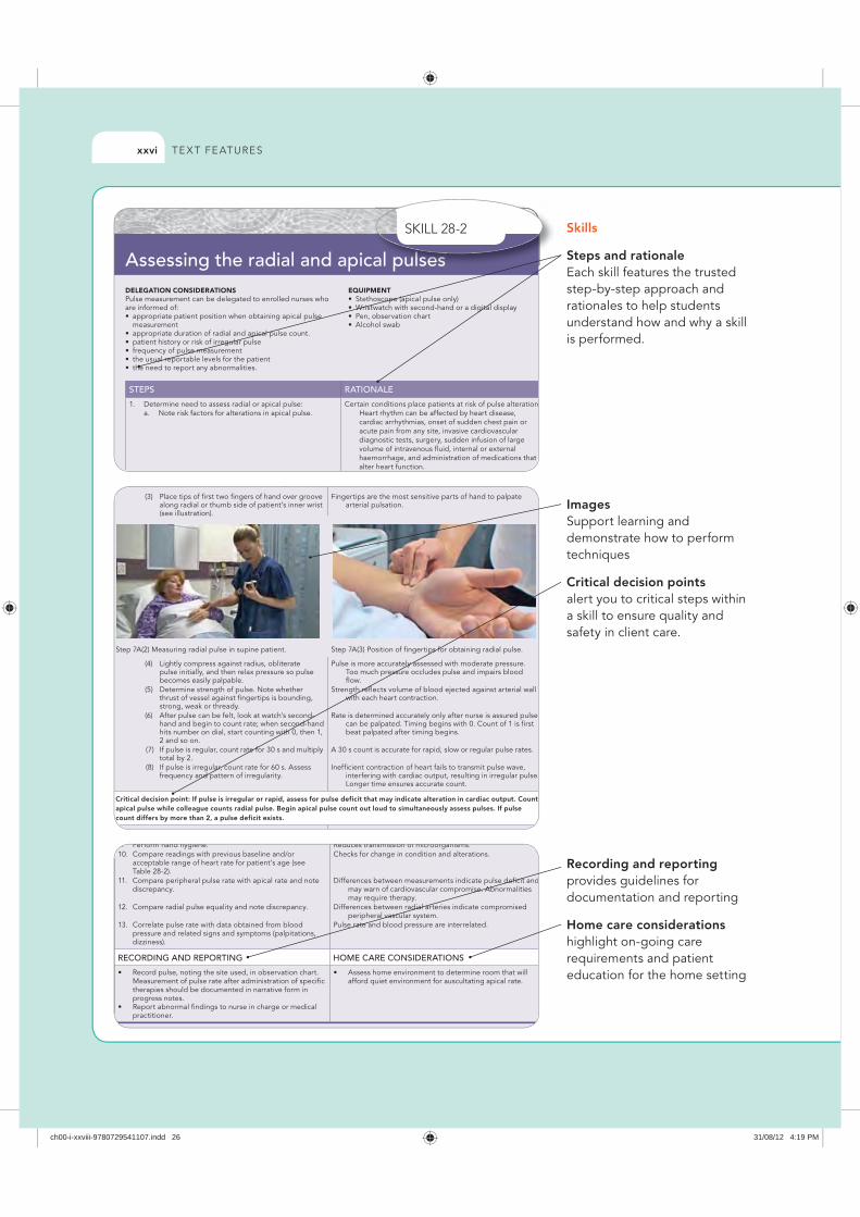
TE X T FE ATURESxxvi
Skills
Steps and rationale Each skill features the trusted step-by-step approach and rationales to help students understand how and why a skill is performed.
Images Support learning and demonstrate how to perform techniques
Critical decision points alert you to critical steps within a skill to ensure quality and safety in client care.
Recording and reporting provides guidelines for documentation and reporting
Home care considerations highlight on-going care requirements and patient education for the home setting
DELEGATION CONSIDERATIONSPulse measurement can be delegated to enrolled nurses who are informed of:
measurement
EQUIPMENT
STEPS RATIONALE
Assessing the radial and apical pulses
Critical decision point: If pulse is irregular or rapid, assess for pulse deficit that may indicate alteration in cardiac output. Count apical pulse while colleague counts radial pulse. Begin apical pulse count out loud to simultaneously assess pulses. If pulse count differs by more than 2, a pulse deficit exists.
B A i l l
RECORDING AND REPORTING HOME CARE CONSIDERATIONS
al puullllllllsssseeeeeesssssssl
ch00-i-xxviii-9780729541107.indd 26 31/08/12 4:19 PM
TE X T FE ATURES xxvii
Nursing process provides a consistent framework and critical thinking model for nursing practice within the clinical skills chapters. These sections are clearly identified within the text with using green headings.
, y puses them when making clinical judgmpatient’s overall nursing care.
NURSING PROCESS ASSESSMENT
Nursing assessment is an ongoing processnot have assessed all body regions beforhygiene; however, the nurse routinely assecondition whenever care is given. For oral care the condition of the teeth andinspected When a patient has had an o
ia, it is common to bathe or shower daily, ther cultural groups it is customary to bathe nce a week (see Working with diversity).
ndition ertain types of physical limitations or lack the physical energy and dexterity to care. A patient in traction or a cast or who us line or other device connected to the ssistance with hygiene. Illnesses that cause he dexterity and range of motion needed n measures. Patients still under the effects ot have the mental clarity or coordination
ONLINE RESOURCESAustralian Nursing and Midwifery Accreditation C
(ANMAC), www.anmc.org.auAustralian Resource Centre for Healthcare Innovati
www.archi.net.auCancer Australia, canceraustralia.gov.auCancer Society of New Zealand, www.cancernz.orgCochrane Collaboration, www.cochrane.orgHealth Research Council of New Zealand,-www.hInternational Council of Nurses, www.icn.chJoanna Briggs Institute, www.joannabriggs.edu.auNational Health and Medical Research Council, ww
gov.auNational Heart Foundation, www.heartfoundation.
www.heartfoundation.org.nz National Institute for Clinical Studies, www.nhmrc
nics Nursing and Midwifery Board of Australia, www.
nursingmidwiferyboard.gov.auNursing Council of New Zealand, www.nursingcouRoyal College of Nursing Australia, www.rcna.org.Sigma Theta Tau International, www.nursingsociet
REFERENCESAustralian Health Practitioner Regulation Agency 2
Nursing and Midwifery Board of Australia. OnlinAvailable at www.nursingmidwiferyboard.gov.au26 Mar 2012.
Banner D, MacLeod MLP and others 2010 Role train rural and remote primary health care nursing: aliterature review Can J Nurs Re 42(4):40–57
stress management are important strategies in promoting your own health and being an effective role model to other staff members and clients.
Also take the time in your early career to critically evaluate the management and clinical styles of your peers. It is often through this ref lective process that we develop and cultivate our own working style. Look for favourable role models and adopt their practices. If possible, it is also optimal to identify a mentor, an individual who can assist and support you in your career development.
KEY CONCEPTS
develop and sustain an effective and efficient nursing workforce.
effective nursing care.
dependent and collaborative nursing roles.
in becoming a registered nurse.
in establishing a philosophy of nursing practice and monitoring nursing standards.
professional practice, recognising the professional, legal regulatory and ethical responsibilities of
and support you in your career development
KEY CONCEPTS
develop and sustain an effective and efficiekf
pp y y
KEY CONCEPTS
Royal College of Nursing Australia, www.rcna.oSigma Theta Tau International, www.nursingsoci
RREFERENCESAustralian Health Practitioner Regulation Agency
Nursing and Midwifery Board of Australia. OnAvailable at www.nursingmidwiferyboard.gov.a
y g g ,Sigma Theta Tau International, www.nurs
REFERENCESAustralian Health Practitioner Regulation
Nursing and Midwifery Board of Austra
Key concepts provide a summary of each chapters key points to reinforce learning.
References provide an up-to-date list of evidence-based sources and journal articles.
OONLINE RESOURCESAustralian Nursing and Midwifery Accreditation
(ANMAC), www.anmc.org.auAustralian Resource Centre for Healthcare Innov
g er
y
ONLINE RESOURCESAustralian Nursing and Midwifery Accred
(ANMAC), www.anmc.org.au
g r
p p g g pTruly individualising hygiene care shows the nurse’s respect for the patient’s needs. As the nurse learns what the patient expects, this information can be incorporated into goal development (see Planning, below).
NURSING DIAGNOSIS
The nurse’s assessment will reveal the condition of the skin, oral cavity and other tissues, as well as the patient’s need for and ability to meet personal hygiene needs. The nurse reviews all data gathered, considers previous patients
g, distance,
n,
re worn
essive eye-nsitivity to
valuable for preventi
Goals and outcThe nurse developsof the patient’s nursplan). The nurse agoals and expectedthe patient’s self-cOutcomes should patient limitations. T
py qfor cleaning and hydrating inf lamed tissues and eliminating sources of irritation will be the focus of nursing care. Box 34-4 summarises possible nursing diagnoses that apply to patients in need of hygiene care.
PLANNING
During planning, the nurse synthesises information from multiple resources (Figure 34-6). Critical thinking ensures that the patient’s plan of care integrates all that the nurse knows about the individual patient and key critical-thinking elements.
offering foot care inmellitus, explain howimpaired and when poses a risk for poorAdapt instruction of anbathing facilities. Not situation that exists iaccessible shower or Use what facilities othat personal care itesafety is ensured, an
the care of a homeless patient may need to be aware of the location of clothing distribution centres for basic hygiene supplies or a shelter where bathing facilities are available. Frequently the nurse will consult with social workers or staff in local churches and schools to be sure patients have the resources they need to maintain hygiene.
IMPLEMENTATION
Providing hygiene is a very basic but important part of a patient’s care. The nurse learns to use caring practices that help to alleviate the patient’s anxiety and promote comfort
interventions. The nuthe patient’s conditioexisting therapies are
Throughout evalucare and gauges whethcritical-thinking apprall factors when evalu
h ’ k l
EVALUATION
Patient care Evaluation of hygiene measures occurs both during and after performance of each particular skill. For example, as the nurse bathes a patient, close inspection of the skin reveals whether drainage or other soiling is effectively removed from the skin’s surface. Once the bath is complete, the nurse asks
h h h ’ f d l h d
patient s overall nursing care.
NNNUNURSING PROCESS S ASSESSMENT
Nursing assessment is an ongoing processt h d ll b d i b f
athe y).
or y to who th
NURSING PROCESS ASSESSMENT or
to
expects, this information can be incorporated intdevelopment (see Planning, below).
NURSING DIAGNOSIS
The nurse’s assessment will reveal the conditionskin, oral cavity and other tissues, as well as the pneed for and ability to meet personal hygiene need
p pdevelopment (see Planning, below).
NURSING DIAGNOSIS
The nurse’s assessment will reveal the skin oral cavity and other tissues as we
EVALUATION
PatPatPP ient care Evaluation of hygiene measures occurs both durinperformance of each particular skill For examp
EVALUATION
Patient care
hat
on en
e klem
staff in local churches and schools to be sure patiethe resources they need to maintain hygiene.
IMPLEMENTATION
Providing hygiene is a very basic but important ppatient’s care. The nurse learns to use caring practhelp to alleviate the patient’s anxiety and promote
the resources they need to maintain hygi
IMPLEMENTATION
Providing hygiene is a very basic but impatient’s care The nurse learns to use ca
Box 34-4 summarises possible nursing diagnosesto patients in need of hygiene care.
PLANNING
During planning, the nurse synthesises informmultiple resources (Figure 34-6). Critical thinkithat the patient’s plan of care integrates all that the n
p gto patients in need of hygiene care.
PLANNING
During planning, the nurse synthesises multiple resources (Figure 34 6) Critica
KNOWLEDGE
EXPERIENCE
ATTITUDES
STANDARDS
ASSESSMENT
ch00-i-xxviii-9780729541107.indd 27 31/08/12 4:19 PM
Special acknowledgmentsTo the all who have worked on this, and past, editions of this book, for their commitment and expertise — Jackie Crisp
To Natalie, who I adore; and my sisters Rachel and Emma — Clint Douglas
To my family, Damian and the Sue(s) for listening — Geraldine Rebeiro
We would like to acknowledge a debt of gratitude to the following people who made the publication of this new edition possible:• Catherine Taylor, for her tireless work on the first three
editions• The Elsevier publishing team: Libby Houston, Elizabeth
Coady and Liz Malcolm • Teresa McIntyre (copy editor) and Sarah Johnson
(permissions editor).The publisher and editors would like to also thank all past and present contributors and reviewers. The book as it stands is the accumulated effort of many dedicated writers and nursing educators, and their invaluable work in all editions of this book is acknowledged with thanks.
Acknowledgments
ch00-i-xxviii-9780729541107.indd 28 31/08/12 4:19 PM