Targeted Magnetic Iron Oxide Nanoparticles for Tumor Imaging

Chapter
21
POTENTIAL USE OF HYBRID IRON OXIDE-GOLD NANOPARTICLES AS DRUG CARRIERS
Anthony D.M. Curtis and Clare Hoskins*
School of Pharmacy, Institute for Science and Technology in Medicine, Keele University, Keele, ST5 5BG, UK *Corresponding author: [email protected]
Chapter 21
Contents 21.1. INTRODUCTION ..................................................................................................................................... 537
21.2. SYNTHESIS OF HNPs ............................................................................................................................ 538
21.3. STABILITY AND BIOCOMPATIBILITY OF HNPs ....................................................................... 539
21.4. USE OF HNPs AS DRUG CARRIERS................................................................................................. 541
21.5. INCORPORATION OF HNPs INTO SUPRAMOLECULAR STRUCTURES ......................... 543
21.6. SUMMARY ................................................................................................................................................. 545
FUTURE OUTLOOK........................................................................................................................................... 545
REFERENCES ...................................................................................................................................................... 546
536
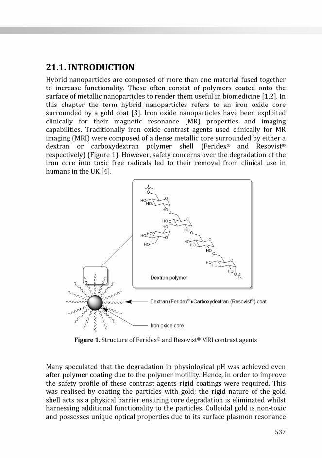
21.1. INTRODUCTION Hybrid nanoparticles are composed of more than one material fused together to increase functionality. These often consist of polymers coated onto the surface of metallic nanoparticles to render them useful in biomedicine [1,2]. In this chapter the term hybrid nanoparticles refers to an iron oxide core surrounded by a gold coat [3]. Iron oxide nanoparticles have been exploited clinically for their magnetic resonance (MR) properties and imaging capabilities. Traditionally iron oxide contrast agents used clinically for MR imaging (MRI) were composed of a dense metallic core surrounded by either a dextran or carboxydextran polymer shell (Feridex® and Resovist® respectively) (Figure 1). However, safety concerns over the degradation of the iron core into toxic free radicals led to their removal from clinical use in humans in the UK [4].
Figure 1. Structure of Feridex® and Resovist® MRI contrast agents
Many speculated that the degradation in physiological pH was achieved even after polymer coating due to the polymer motility. Hence, in order to improve the safety profile of these contrast agents rigid coatings were required. This was realised by coating the particles with gold; the rigid nature of the gold shell acts as a physical barrier ensuring core degradation is eliminated whilst harnessing additional functionality to the particles. Colloidal gold is non-toxic and possesses unique optical properties due to its surface plasmon resonance
537
Chapter 21
(SPR) [5]. SPR is the phenomenon whereby light at a certain wavelength (unique to the particle due to size, shape etc.) can be both scattered and absorbed. This absorption results in a rapid conversion of light into heat and, hence, the nanoparticulates experience a thermal rise. This stimuli responsive nature can be exploited in applications such as tumour ablation and thermo-responsive drug delivery. By gold coating the surface iron oxide nanoparticles a core-shell dual functional structure is observed which is referred to as a hybrid nanoparticle (HNP). The benefit of such particles, other than reduced toxicity is the combined properties conferred by the core and shell components. These particles retain the magnetic character inherent to iron oxide and the optical properties inherent to colloidal gold. Thus, HNPs have potential as theranostic agents. Theranostics are an emerging platform which offer simultaneous diagnosis and therapy, resulting in decreased treatment times. These agents allow for real time imaging after administration, enabling location mapping before initiating drug release. By coupling treatments to diagnostics and controlling drug release a rapid and localised clinical effect can be achieved.
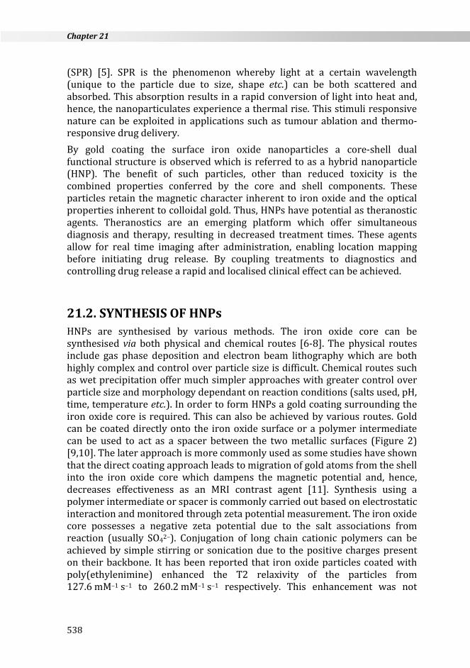
21.2. SYNTHESIS OF HNPs HNPs are synthesised by various methods. The iron oxide core can be synthesised via both physical and chemical routes [6-8]. The physical routes include gas phase deposition and electron beam lithography which are both highly complex and control over particle size is difficult. Chemical routes such as wet precipitation offer much simpler approaches with greater control over particle size and morphology dependant on reaction conditions (salts used, pH, time, temperature etc.). In order to form HNPs a gold coating surrounding the iron oxide core is required. This can also be achieved by various routes. Gold can be coated directly onto the iron oxide surface or a polymer intermediate can be used to act as a spacer between the two metallic surfaces (Figure 2) [9,10]. The later approach is more commonly used as some studies have shown that the direct coating approach leads to migration of gold atoms from the shell into the iron oxide core which dampens the magnetic potential and, hence, decreases effectiveness as an MRI contrast agent [11]. Synthesis using a polymer intermediate or spacer is commonly carried out based on electrostatic interaction and monitored through zeta potential measurement. The iron oxide core possesses a negative zeta potential due to the salt associations from reaction (usually SO42–). Conjugation of long chain cationic polymers can be achieved by simple stirring or sonication due to the positive charges present on their backbone. It has been reported that iron oxide particles coated with poly(ethylenimine) enhanced the T2 relaxivity of the particles from 127.6 mM–1 s–1 to 260.2 mM–1 s–1 respectively. This enhancement was not
538
Potential use of hybrid iron oxide-gold nanoparticles as drug carriers
observed in identical particles coated with poly(allylamine) and poly(L-lysine) [3].
Figure 2. Differing structures of HNPs resultant from synthetic route both A) without
and B) with the use of a polymer intermediate
In order to achieve gold coating, 2 nm gold seeds are conjugated electrostatically onto the cationic polymer intermediate and subsequent iterative reduction of chloroauric acid in solution attaches gold onto the nanoparticle surface and form the complete rigid coating. Coating thickness is controlled via iteration number. Initially a thin coat is formed (<5 nm) which becomes thicker upon increased iteration number. Upon further iterative increase in chloroauric acid reduction steps anchor points appear to initiate morphology change into spikey or star shaped particles. The presence of the gold coat of the HNP does not appear to effect relaxivity with iron oxide core T2 relaxivity being 127.6 mM–1 s–1 before coating and HNP relaxivity being 175.3 mM–1 s–1 respectively in one study [3]. Gold coating thickness not only provides the physical barrier to iron oxide degradation but also possesses SPR for nano-heating. Gold coating thickness is an important parameter in developing HNPs into effective heating sources. Studies have shown that increasing the thickness of the gold coating decreases its ability to absorb and scatter laser irradiation resulting in heating. One study reported a 4 °C decrease in temperature change after 60 seconds of laser irradiation (95 nm). It is proposed that with decreased coating thickness greater surface area is available (internal surface and external surface of the shell) and thus a greater level of SPR is achieved resulting in greater temperature rises [3].
21.3. STABILITY AND BIOCOMPATIBILITY OF HNPs In order for HNPs to be applicable as contrast agents, drug carriers or indeed theranostics they must possess both physical and chemical stability under physiological conditions. Nanoparticles in solution tend to aggregate together
A B
539
Chapter 21
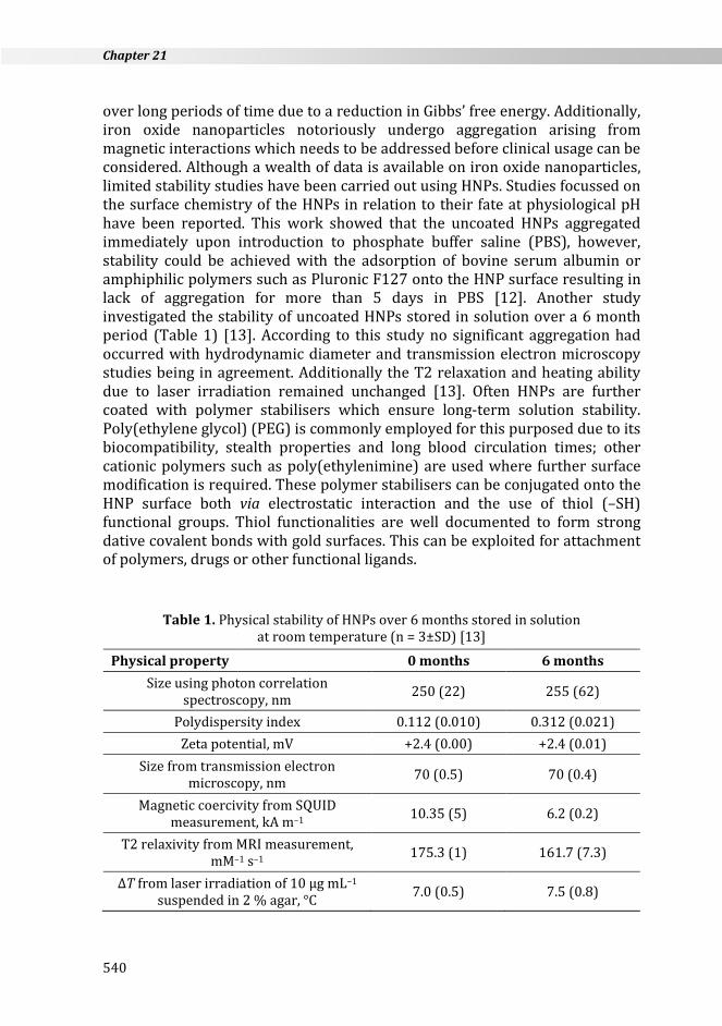
over long periods of time due to a reduction in Gibbs’ free energy. Additionally, iron oxide nanoparticles notoriously undergo aggregation arising from magnetic interactions which needs to be addressed before clinical usage can be considered. Although a wealth of data is available on iron oxide nanoparticles, limited stability studies have been carried out using HNPs. Studies focussed on the surface chemistry of the HNPs in relation to their fate at physiological pH have been reported. This work showed that the uncoated HNPs aggregated immediately upon introduction to phosphate buffer saline (PBS), however, stability could be achieved with the adsorption of bovine serum albumin or amphiphilic polymers such as Pluronic F127 onto the HNP surface resulting in lack of aggregation for more than 5 days in PBS [12]. Another study investigated the stability of uncoated HNPs stored in solution over a 6 month period (Table 1) [13]. According to this study no significant aggregation had occurred with hydrodynamic diameter and transmission electron microscopy studies being in agreement. Additionally the T2 relaxation and heating ability due to laser irradiation remained unchanged [13]. Often HNPs are further coated with polymer stabilisers which ensure long-term solution stability. Poly(ethylene glycol) (PEG) is commonly employed for this purposed due to its biocompatibility, stealth properties and long blood circulation times; other cationic polymers such as poly(ethylenimine) are used where further surface modification is required. These polymer stabilisers can be conjugated onto the HNP surface both via electrostatic interaction and the use of thiol (–SH) functional groups. Thiol functionalities are well documented to form strong dative covalent bonds with gold surfaces. This can be exploited for attachment of polymers, drugs or other functional ligands.
Table 1. Physical stability of HNPs over 6 months stored in solution at room temperature (n = 3±SD) [13]
Physical property 0 months 6 months Size using photon correlation
spectroscopy, nm 250 (22) 255 (62)
Polydispersity index 0.112 (0.010) 0.312 (0.021) Zeta potential, mV +2.4 (0.00) +2.4 (0.01)
Size from transmission electron microscopy, nm 70 (0.5) 70 (0.4)
Magnetic coercivity from SQUID measurement, kA m–1 10.35 (5) 6.2 (0.2)
T2 relaxivity from MRI measurement, mM–1 s–1 175.3 (1) 161.7 (7.3)
∆T from laser irradiation of 10 µg mL–1 suspended in 2 % agar, °C 7.0 (0.5) 7.5 (0.8)
540
Potential use of hybrid iron oxide-gold nanoparticles as drug carriers
Degradation of metallic nanoparticles into toxic free radicals has been well documented especially in metal oxides [14,15]. The withdrawal of Feridex® in the UK has reformed many coating strategies and prompted more stringent toxicity studies. These were lacking previously primarily due to lack of universal understanding and agreement of testing procedures required. Nanosafety committees have formed worldwide to unite the nano research community and advise on best practice for the toxicity evaluation of particles. In HNPs, the toxicity arising from the iron oxide core is theoretically eliminated due to the rigid inert gold coating. However, in order for long term use studies are required to verify such hypotheses. Studies have been carried out in vitro to determine the chemical stability of HNPs (both uncoated and PEGylated) in solutions designed to mimic physiological (pH 7.2) and lysosomal (pH 4.6) conditions [13]. After two weeks levels of Au and Fe were monitored in the media. In uncoated and PEGylated HNPs particles at pH 7.2 & 4.6 negligible levels (< 0.01 %) of both metals were observed indicating that HNPs were resistant to chemical degradation over the duration of study [13]. The safety issues experienced with iron oxide nanoparticles and their degradation in vivo has led to great focus on metallic particle biosafety. Hence, a plethora of in vitro tests have been carried out on HNPs to ensure they are safe for clinical application [14]. HNPs have demonstrated increased levels of biocompatibility compared with polymer coated iron oxide nanoparticles. This further demonstrates that the rigid core protecting the iron does not degrade after cellular internalisation. Cellular response assays including the 3-(4,5-dimethylthiazol-2-yl)-2,5-diphenyltetrazolium bromide) (MTT) assay, trypan blue exclusion, lactate dehydrogenase (LDH) cell membrane integrity, reactive oxygen species generation and lipid peroxidation studies have been reported in various cell lines [13,16]. All studies reviewed to date have been in agreement that HNPs do not cause significant cytotoxicity or trigger and other adverse cellular response. These studies show the potential of HNPs to be used clinically. When compared to the previously used MRI contrast agent Feridex®, HNPs magnetic ability is similar but, their safety profile is enhanced. Cellular uptake studies have also demonstrated that uncoated HNPs are capable of cellular internalisation and polymer coating increases transfection efficiency.
21.4. USE OF HNPs AS DRUG CARRIERS HNPs can be exploited as drug carriers in addition to their imaging capabilities. Drugs can be conjugated both electrostatically via Van der Waals forces or physically via dative covalent bonding. Gold surfaces form permanent covalent bonds with thiol containing molecules. Addition of further polymer coats can also be used to further functionalise the surfaces with drug molecules, functional ligands, dyes etc. Studies have shown the conjugation of anticancer drugs such as cisplatin and 6-thioguanine (6-TG), as well as proteins such as
541
Chapter 21
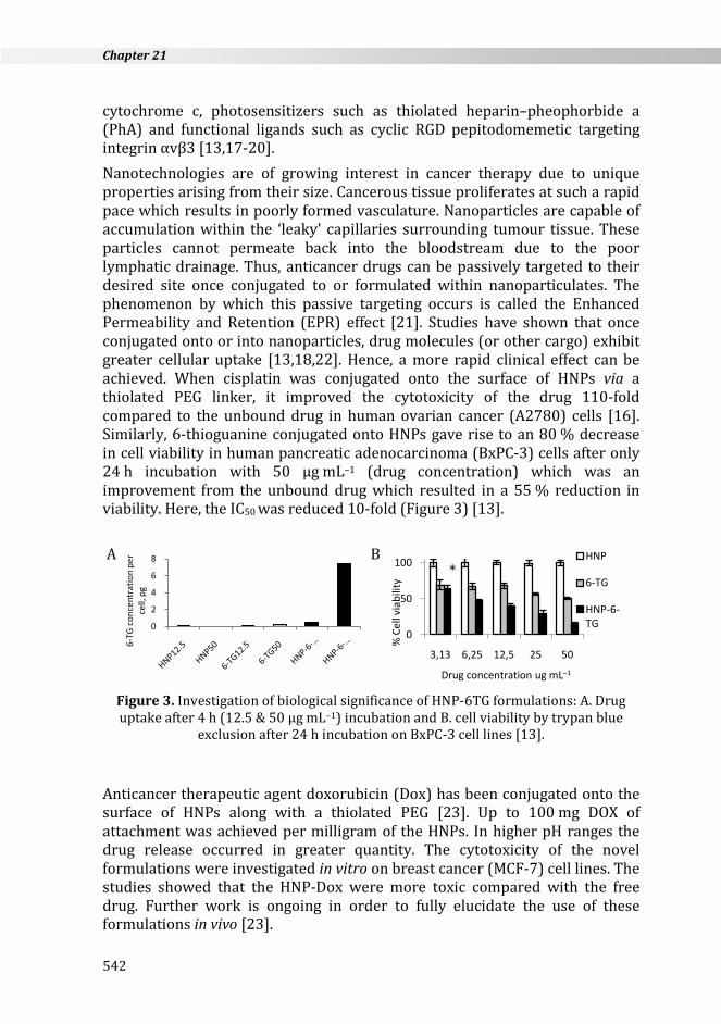
cytochrome c, photosensitizers such as thiolated heparin–pheophorbide a (PhA) and functional ligands such as cyclic RGD pepitodomemetic targeting integrin αvβ3 [13,17-20]. Nanotechnologies are of growing interest in cancer therapy due to unique properties arising from their size. Cancerous tissue proliferates at such a rapid pace which results in poorly formed vasculature. Nanoparticles are capable of accumulation within the ‘leaky’ capillaries surrounding tumour tissue. These particles cannot permeate back into the bloodstream due to the poor lymphatic drainage. Thus, anticancer drugs can be passively targeted to their desired site once conjugated to or formulated within nanoparticulates. The phenomenon by which this passive targeting occurs is called the Enhanced Permeability and Retention (EPR) effect [21]. Studies have shown that once conjugated onto or into nanoparticles, drug molecules (or other cargo) exhibit greater cellular uptake [13,18,22]. Hence, a more rapid clinical effect can be achieved. When cisplatin was conjugated onto the surface of HNPs via a thiolated PEG linker, it improved the cytotoxicity of the drug 110-fold compared to the unbound drug in human ovarian cancer (A2780) cells [16]. Similarly, 6-thioguanine conjugated onto HNPs gave rise to an 80 % decrease in cell viability in human pancreatic adenocarcinoma (BxPC-3) cells after only 24 h incubation with 50 µg mL–1 (drug concentration) which was an improvement from the unbound drug which resulted in a 55 % reduction in viability. Here, the IC50 was reduced 10-fold (Figure 3) [13].
Figure 3. Investigation of biological significance of HNP-6TG formulations: A. Drug uptake after 4 h (12.5 & 50 µg mL–1) incubation and B. cell viability by trypan blue
exclusion after 24 h incubation on BxPC-3 cell lines [13].
Anticancer therapeutic agent doxorubicin (Dox) has been conjugated onto the surface of HNPs along with a thiolated PEG [23]. Up to 100 mg DOX of attachment was achieved per milligram of the HNPs. In higher pH ranges the drug release occurred in greater quantity. The cytotoxicity of the novel formulations were investigated in vitro on breast cancer (MCF-7) cell lines. The studies showed that the HNP-Dox were more toxic compared with the free drug. Further work is ongoing in order to fully elucidate the use of these formulations in vivo [23].
0
2
4
6
8
6-TG
con
cent
ratio
n pe
r ce
ll, p
g
0
50
100
3,13 6,25 12,5 25 50
% C
ell v
iabi
lity
Drug concentration ug mL–1
HNP
6-TG
HNP-6-TG
B *
A
542
Potential use of hybrid iron oxide-gold nanoparticles as drug carriers
These studies show the huge potential of HNPs as drug delivery vehicles. Efforts are now underway worldwide to further exploit this potential in cancer therapy as dual purpose theranostic agents.
21.5. INCORPORATION OF HNPs INTO SUPRAMOLECULAR STRUCTURES
HNPs can be incorporated into larger supramolecular structures which can further enhance their applicability in biomedicine. These include the development of magnetomicelles. Reports have successfully shown conjugation and potential of drug conjugated onto the surface of HNPs [22]. However, insoluble compounds are much more difficult to formulate onto their surface due to the potential to further decrease HNP stability resulting in precipitation. Hydrophobic drugs make up approximately 40 % of all new drugs under development. They pose huge problems for pharmaceutical companies because without careful formulation they are rendered useless. Traditional formulation strategies such as the use of surfactants and co-solvents have their own disadvantages and hence have been improved with the advancement in nanotechnology. Amphiphilic polymers have been used to solubilise hydrophobic compounds with greater stability and efficiency compared with surfactants. Addition of HNPs into the intrinsic structure of amphiphilic polymers provides them with added properties in imaging. A recent study showed incorporation of HNPs into the intrinsic structure of a graft amphiphilic polymer consisting of a poly(allylamine) (PAA) backbone grafted with 5 % molar ratio pendants of oxadiaxole (Ox) [22]. Amphiphilic polymers spontaneously aggregate into core-shell nano-aggregates in aqueous environments due to a reduction in Gibbs’ free energy. Polymer derivatives of PAA have shown huge potential in hydrophobic drug solubilisation and delivery for drugs such as propofol, prednisolone, griseofulvin and novel anticancer drug bis-naphtal-amidopropyl-diamino-octane (BNIPDaoct) [24]. The inclusion of HNPs into these self-assemblies results in greater functionality which can be exploited for image guided or stimuli responsive drug delivery. In this work the HNPs where attached to the polymer backbone via dative covalent bonding between the gold surface and the thiol functional group in the Ox pendant group [22]. The resultant PAA-Ox5 aggregates were exploited as drug carriers using direct conjugation of drug molecules onto the HNP surface (6-TG) and hydrophobic encapsulation (BNIPDaoct). In vitro assays in human pancreatic adenocarcinoma (BxPC-3) cells showed that the polymer-HNP formulations (both conjugated and encapsulated) resulted in increased drug uptake compared with the free drug. MTT assays showed that a significant decrease in the IC50 value of the formulations compared with the free drug was also observed [22].
543
Chapter 21
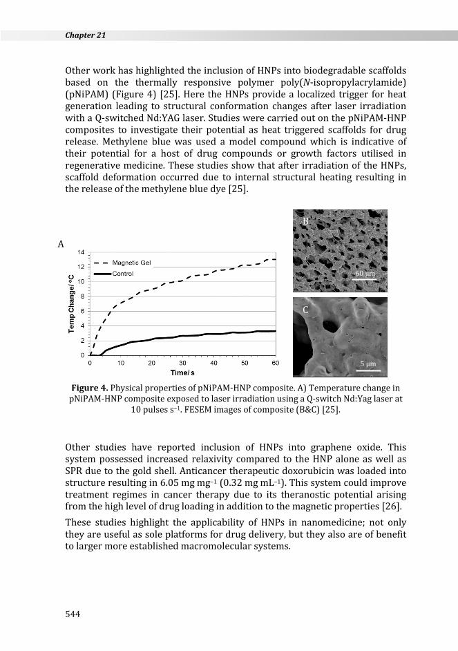
Other work has highlighted the inclusion of HNPs into biodegradable scaffolds based on the thermally responsive polymer poly(N-isopropylacrylamide) (pNiPAM) (Figure 4) [25]. Here the HNPs provide a localized trigger for heat generation leading to structural conformation changes after laser irradiation with a Q-switched Nd:YAG laser. Studies were carried out on the pNiPAM-HNP composites to investigate their potential as heat triggered scaffolds for drug release. Methylene blue was used a model compound which is indicative of their potential for a host of drug compounds or growth factors utilised in regenerative medicine. These studies show that after irradiation of the HNPs, scaffold deformation occurred due to internal structural heating resulting in the release of the methylene blue dye [25].
Figure 4. Physical properties of pNiPAM-HNP composite. A) Temperature change in
pNiPAM-HNP composite exposed to laser irradiation using a Q-switch Nd:Yag laser at 10 pulses s–1. FESEM images of composite (B&C) [25].
Other studies have reported inclusion of HNPs into graphene oxide. This system possessed increased relaxivity compared to the HNP alone as well as SPR due to the gold shell. Anticancer therapeutic doxorubicin was loaded into structure resulting in 6.05 mg mg–1 (0.32 mg mL–1). This system could improve treatment regimes in cancer therapy due to its theranostic potential arising from the high level of drug loading in addition to the magnetic properties [26]. These studies highlight the applicability of HNPs in nanomedicine; not only they are useful as sole platforms for drug delivery, but they also are of benefit to larger more established macromolecular systems.
5 µm
A
B
C
60 µm
544

Potential use of hybrid iron oxide-gold nanoparticles as drug carriers
21.6. SUMMARY The use of HNPs is rapidly emerging in drug delivery. With growing intelligence regarding nanosafety more rigorous tests are being implemented. Although a relatively young area of research the studies to date show a progression in the understanding and development of these particles as suitable carrier vehicles. They have adequately demonstrated good physical and chemical stability properties and biocompatibility in vitro on various cell lines. However, in vivo studies are yet to be realised and will be the next step required before clinical application can be implemented.
FUTURE OUTLOOK The advancement in novel nanomedicines and multifunctional treatment strategies looks set to revolutionise modern healthcare. HNPs and theranostic agents are at the forefront of this exciting era. The development of agents which provide a rapid diagnosis and treatment regime hold great potential for increased patient lifespan and quality of life especially in disease states such as cancer. Additionally, increased time between initial treatment and relapse due to the improved efficiency and penetration ability may occur. There are still hurdles to be crossed and areas where understanding is still lacking and the number of in vivo trials is small, however, interest in this field is growing exponentially and knowledge is rapidly being gained. Current clinical efforts in drug delivery are not swamped by nanomedicines, however, this may be due to their relative age. Many polymeric nanoparticulates are currently undergoing the rigorous clinical trials which are required before marketing. The widely held opinion is that HNPs are expected to follow suit and will advance into clinical trials. A lot of scepticism over nanoparticle toxicities is still at large especially with the use of metallic components, however, as nanosafety is a currently major strategy with funding agencies and charities the growth of nanotoxicity databases will emerge.
545
Chapter 21
REFERENCES 1. R. Namgung, K. Singha, M.K. Yu, S. Jon, Y.S. Kim, Y. Ahn, I.K. Park, W.J. Kim,
Biomaterials 31 (2010) 4204–4213. 2. Y. Sun, X. Ding, Z. Zheng, X. Cheng, X. Hu, Y. Peng, Chem. Commun. (2006) 2765–
2767. 3. C.M. Barnett, M. Gueorguieva, M.R. Lees, D.J. McGarvey, R.J. Darton, C. Hoskins,
J. Nanopart. Res. 14 (2012) 1170. 4. J.P. Bullivant, S. Zhao, B.J. Willenberg, B. Kozissnik, C.D. Batich, J. Dobson, Int. J.
Mol. Sci. 14 (2013) 17501–17510. 5. P. K. Jain, I. H. El-Sayed, M. A. El-Sayed, Nanotoday 2 (2007) 18–29. 6. A.M. Vergés, R. Costo, A.G. Roca, J.P. Marco, G.F. Goya, C.J. Serna, M.P. Morales, J.
Phys. D Appl. Phys. 41 (2008) 134003. 7. C.M. Wang, D.R. Baer, J.E. Amonetter, M.H. Engelhard, Y. Qiang, Y. Antony,
Nanotechnology 18 (2007) 25. 8. D. Ramimoghadam, S. Bagheri, S.B.A. Hamid, J. Magnet. Magnet. Mater. 368
(2014) 207–229. 9. E. Iglesias-Silva, J.L. Vilas-Vilela, M.A. López-Quintela, J. Rivas, M. Rodríguez,
L.M. León, J. Non-Cryst. Solids 356 (2010) 1233–1235. 10. C. Hoskins, Y. Min, M. Gueorguieva, C. McDougall, A. Volovick, P. Prentice, Z.
Wang, A. Melzer, A. Cuschieri, L. Wang, J. Nanobiotechnol. 10 (2012) 27. 11. E.D. Smolensky, M.C. Neary, Y. Zhou, T.S. Berquo, V.C. Pierre, Chem. Comm. 47
(2011) 2149–2151. 12. J.K. Lim, S.A. Majetich, R.D. Tilton, Langmuir 25 (2009) 133384–133393. 13. C.M. Barnett, M. Gueorguieva, M. Lees, D.J. McGarvey, C. Hoskins, J. Nanopart.
Res. 15 (2013) 1706. 14. K. Buyukhatipolu, A.M. Clyne, J. Biomed. Mater. Res. A 96 (2010) 186–195. 15. C. Hoskins, A. Cuschieri, L. Wang, J. Nanobiotechnol. 10 (2012) 15. 16. S. Bandyopadhyay, G. Singh, I. Sandvig, A. Sandvig, R. Mathieu, P.A. Kumar,
W.R. Glomm, Appl. Surf. Sci. 316 (2014) 171–178. 17. A.J. Wagstaff, S.D. Brown, M.R. Holden, G.E. Craig, J.A. Plumb, R.E. Brown, N.
Schreiter, W. Chrzanowski, N.J. Wheate, Inorg. Chim. Acta 393 (2012) 328–333.
18. M. Malekigorji, C. Hoskins, A.D.M. Curtis, G. Varbiro, J. Nanomed. Res. 1 (2014) 00010.
19. L. Li, M. Nurunnabi, M. Nafiujjaman, Y.Y. Jeong, Y.K. Lee, K.M. Huh, J. Mater. Chem. B 2 (2014) 2929–2937.
20. L. Menichetti, L. Manzoni, L. Paduano, A. Flori, C. Kusmic, D. De Marchi, S. Casciaro, F. Conversano, M. Lombardi, V. Positano, D. Arosio, IEEE Sens. J. 13 (2013) 2341–2347.
21. P. Parhi, C. Mohanty, S.K. Sahoo, Drug Discov. Today 17 (2012) 1044–1052. 22. C.M. Barnett, M.R. Lees, A.D.M. Curtis, P.K.T. Lin, W.P. Cheng, C. Hoskins, Pharm.
Nanotechnol. 1 (2013) 224–238. 23. N.S. Elbialy, M.M. Fathy, W.M. Khalil, Phys. Medica 30 (2014) 843–848. 24. C. Hoskins, P.K.T. Lin, X. Qu, L. Tetley, W.P. Cheng, Pharm. Res. 3 (2012) 59–79. 25. P. Roach, D.J. McGarvey, M. Lees, C. Hoskins, Int. J. Mol. Sci. 14 (2013) 8585–
8602. 26. M. Balcioglu, M. Rana, M.V. Yigit, J. Mater. Chem. B 1 (2013) 6187–6193.
546