Post-hemispherectomy Hydrocephalus in … Hydrocephalus in Children: Results of a Comprehensive,...
20
Post-hemispherectomy Hydrocephalus in Children: Results of a Comprehensive, Multi-institutional Review Anne E. Matthews PA-C, Adam Hartman MD, Yong D. Park MD, Anup D. Patel MD, Sean M. Lew MD (on behalf of the Post- Hemispherectomy Hydrocephalus Workgroup)
Transcript of Post-hemispherectomy Hydrocephalus in … Hydrocephalus in Children: Results of a Comprehensive,...

Post-hemispherectomy Hydrocephalus in Children: Results of a Comprehensive,
Multi-institutional Review
Anne E. Matthews PA-C, Adam Hartman MD, Yong D. Park MD, Anup D. Patel MD, Sean M. Lew MD (on behalf of the Post-
Hemispherectomy Hydrocephalus Workgroup)

Introduction
• Hemispherectomy surgery for medically intractable epilepsy causes hydrocephalus in a subset of patients.
• What is the incidence?
• What are the risk factors?
• What is the timing of onset?
• Why the variability in timing?
• Data has been limited by small number of cases at any given center

Year N Surg details HCP Incidence Reference
1993 Davies et all N=17 All AH 3/17 (18%) J Neurosurg 78: 733-740
1996 Carson et al N=52 2AH, 50 HD, 3
periop deaths
16/49 (33%) J Neurosurg 84: 903-911
1996 Peacock et al N=54 27 AH, 27 FH AH 22/27 (81%) FH 3/27 (11%)
Child’s Nervous System 12:376-384
2000 DiRocco et al N=15 (All hemimeg)
11 AH, 2 FH, 2HD AH 3/11 (27%) FH & HD (each 50%)
Pediatric Neurosurg 33:198-207
2004 Cook et al N=115 37AH ,32 FH, 46 PIH
29/37 AH (78%), 3/32 FH (9%), 10/46 (22%)
J Neurosurg (Pediatrics 2) 100:125-146
2005 Gonzalez-
Martinez et al
N=18 4 AH, 13 FH, 3 Mod AH
2/18 (11%) Epilepsia 46(9): 1518-1525
2007 Basheer et al N=22 HD, PIH 2/22 (9%) Epilepsia 48(I): 133-140
2010 Kwan et al N=24 5 HD, 19 PIH 2/23 (9%) Neurosurgery 67:429-436
AH=Anatomic hemispherectomy, HD=Hemidecortication, FH=Functional hemispherectomy, PIH=Periinsular hemispherotomy

Methods
• Fifteen pediatric epilepsy centers participated in this study
• A retrospective chart review was performed on all available patients who had hemispherectomy surgery
• Two-tiered de-identified data collection system

Center
Children’s Hospital-Denver
Children’s Hospital of WI-Milwaukee
Cook Children’s Hospital-Fort Worth
Duke University Med Center-Baltimore
Johns Hopkins Hospital-Baltimore
Medical College of Georgia
National Center for Neurology and Psychiatry-Tokyo
Nationwide Children’s Hospital-Columbus
NYU
Phoenix Children’s Hospital
Sanbo Brain Institute-Beijing
UCLA
University of Alabama-Birmingham
Wayne State-Detroit
Center
Children’s Hospital-Denver
Children’s Hospital of WI-Milwaukee
Cook Children’s Hospital-Fort Worth
Duke University Med Center-Durham
Johns Hopkins Hospital-Baltimore
Medical College of Georgia-Augusta
National Center for Neurology and Psychiatry-Tokyo
Nationwide Children’s Hospital-Columbus
NYU-NYC
Phoenix Children’s Hospital
Sanbo Brain Institute-Beijing
Seattle Children’s Hospital
UCLA
University of Alabama-Birmingham
Wayne State-Detroit

Methods • First tier data collected on all Hemi patients
• Basic demographics
• Etiology of epilepsy
• Surgical technique
–Anatomic, functional, hemicorticectomy
–Any resection of basal ganglia or thalamus tissue?
–Use of hemostatic adjuncts, EVD
• Post-operative infection
• Prior resective brain surgery?
• Pre-existing CSF shunt? Excluded from analysis

Methods
• Second tier data collected on subset of patients who developed HCP requiring shunt placement or ETV (n=1)
• Time to shunt placement
• Presenting symptoms and signs
– Headache, emesis, diminished LOC, cognitive decline, behavioral issue, wound issues
• Presenting imaging changes?
• Confirmatory studies
– ICP monitoring, diagnostic LP, temporary CSF drainage, other invasive diagnostics
• Multivariate logistic regression analysis with a fixed effect controlling for center was performed

Results • Data were collected on 736 patients who had
hemispherectomy surgery between 1986 and 2011.
• Male: Female 367:369 • Age range 0.1 year to 42 years • Follow-up ranged from 21.5 mos to 302 mos, but
F/U data was incomplete on 45 patients • 46 patients had pre-existing shunts-EXCLUDED
• n=690 Functional 435 (63%)
Anatomic 244 (35%)
Hemicorticectomy 11 (2%)
FH
AH
HC

Etiology of epilepsy
Dysplasia 237 (34%)
Rasmussen's 152 (22%)
Stroke 148 (21%)
Sturge Weber 26 (4%)
Trauma 24 (3%)
Idiopathic 16 (2%)
Tumor 9 (1%)
Other 56 (8%)
None listed 22 (3%)

Prior resective surgery
None 568 (82%)
Lobar / multilobarresection 41 (6%)
Lesionectomy /topectomy 13 (2%)
Hemispherectomy 12(2%)
Other 56 (8%)
(12 patients had two prior surgeries and 6 patients had three prior surgeries)
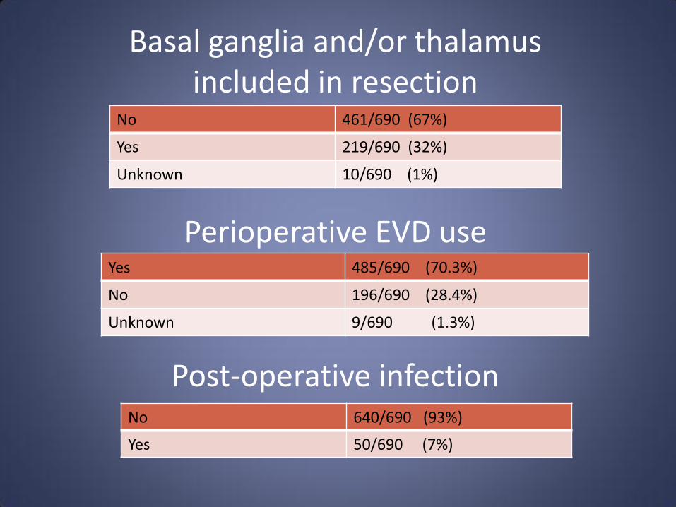
Perioperative EVD use Yes 485/690 (70.3%)
No 196/690 (28.4%)
Unknown 9/690 (1.3%)
Post-operative infection No 640/690 (93%)
Yes 50/690 (7%)
Basal ganglia and/or thalamus included in resection
No 461/690 (67%)
Yes 219/690 (32%)
Unknown 10/690 (1%)
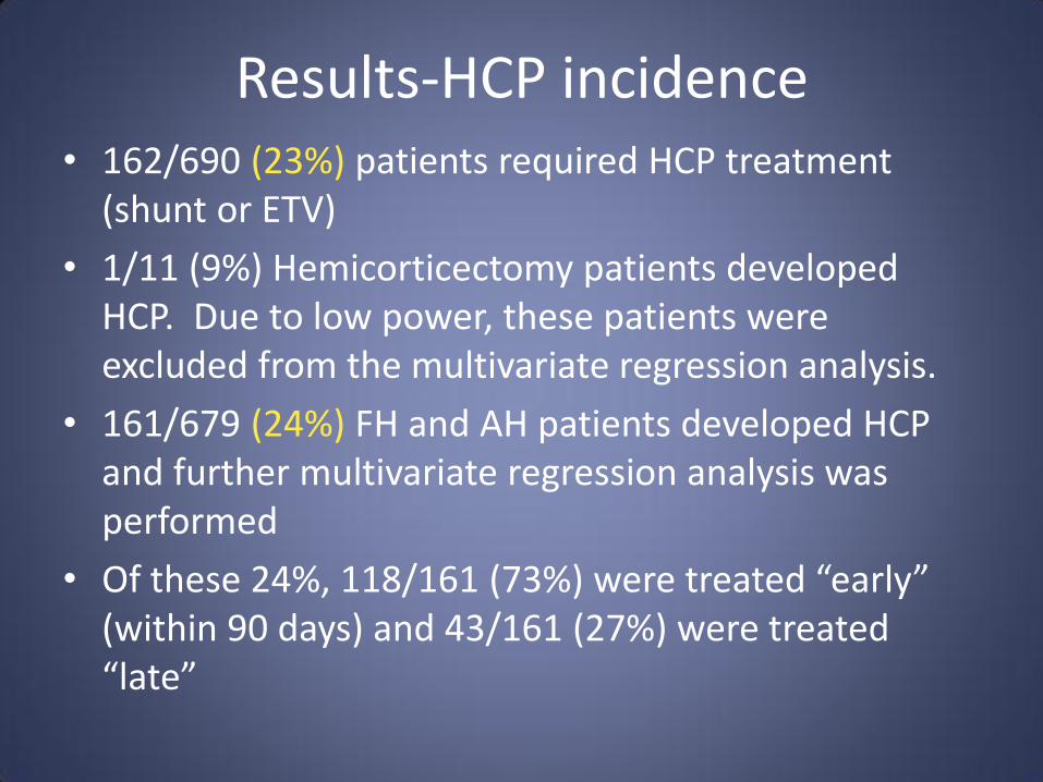
Results-HCP incidence • 162/690 (23%) patients required HCP treatment
(shunt or ETV)
• 1/11 (9%) Hemicorticectomy patients developed HCP. Due to low power, these patients were excluded from the multivariate regression analysis.
• 161/679 (24%) FH and AH patients developed HCP and further multivariate regression analysis was performed
• Of these 24%, 118/161 (73%) were treated “early” (within 90 days) and 43/161 (27%) were treated “late”

0.5
0.6
0.7
0.8
0.9
1.0
0 50 100 150 200 250 300
HCP-freesurvival
censor
Time to develop hydrocephalus
Months post-hemispherectomy
HC
P-f
ree

0.5
0.6
0.7
0.8
0.9
1.0
0 2 4 6 8 10 12 14 16 18
HCP-freesurvival
censor
Time to develop hydrocephalus
Months post-hemispherectomy
HC
P-f
ree

Variable Early Ever
N(%) OR (CI)
p N(%) OR (CI)
p
Anatomic vs Functional 53/244 (22%) vs 65/435 (15%)
6.1 (3.1,12)
<.0001
74/244 (30%) vs 87/435 (20%)
4.2 (2.3,7.6)
<.0001
BGT resection Yes vs No
73/219 (33%) vs 45/471 (10%)
4.1 (1.6, 10)
.0035 75/129 (34%) vs 87/471 (18%)
2.8 (1.2,6.6)
0.0161
Prior resection Yes vs No
21/122 (17%) vs 97/568 (17%)
2.1 (1.1,3.8)
0.0163 32/122 (26%) vs 130/568 (23%)
1.7 (1.1,2.9)
0.0281
Predictive variables – Univariate

Variable Early Ever
N(%) OR p N(%) OR p
Anatomic vs Functional
53/244 (22%) vs 65/435 (15%)
5.4 (2.8,11)
<.0001
74/244 (30%) vs 87/435 (20%)
3.8 (2.1,6.9) <.0001
BGT resection Yes vs No
73/219 (33%) vs 45/471 (10%)
3.0 (1.1,8.1)
0.0344 75/129 (34%) vs 87/471 (18%)
2.2 (0.9,5.2) 0.0801
Prior resection Yes vs No
21/122 (17%) vs 97/568 (17%)
2.0 (1.1,3.8)
0.0215
32/122 (26%) vs 130/568 (23%)
1.7 (1.0,2.8) 0.0377
Predictive variables - Multivariate

Results - Non-predictive variables
Variable Early
Ever
OR P-value OR P-value
Etiology --- 0.3268 --- 0.5012
Hemostatic adjunct --- 0.9537 --- 0.3535
Post-op EVD 0.72 0.6417 0.84 0.7513
Post-op infection 1.61 0.3303 1.54 0.2526

Results-HCP presentation and work-up
Presenting signs or sx Incidence
Failure to wean drain 48%
Change in imaging findings 41%
Headache 23%
Emesis 23%
Diminished LOC 22%
Other neurologic symptoms 12%
Cognitive decline 8%
Behavioral changes 8%
Wound issues 8%
Evaluation Incidence
Temporary CSF drainage
14%
Diagnostic LP 10%
ICP monitoring 4%
Other invasive test 1%
No significant differences between “early” and “late” patients

Conclusions
• Hydrocephalus is a common sequela of hemispherectomy surgery
• Surgical technique and prior brain surgery influence the occurrence of post-hemispherectomy hydrocephalus
• A significant portion of patients develop hydrocephalus on a delayed basis, indicating the need for long-term surveillance.

Acknowledgements
Data analysis provided by Daniel Eastwood, Biostatistician with the Medical College of Wisconsin, supported IN PART by grant 1UL1RR031973 from the Clinical and Translational Science Award (CTSA) Program of the National Center for Research Resources, National Institutes of Health.