Possible Homologies in the Proventriculi of Dicondylia
16
Zool. Anz. 237 (1998): 43-58 © by Gustav Fischer Verlag ZOOLOGiSCHER ANZEIGER Possible Homologies in the Proventriculi of Dicondylia (Hexapoda) and Malacostraca (Crustacea) Klaus-Dieter KLASS Ludwig-Maximilians-Universitat, Zoologisches Institut, Miinchen, Germany Abstract. Striking similarities were found between the armarial part of the proventriculus of Lepismatidae (Hexapoda: Dicondylia: Zygentoma) and the cardia of Decapoda (Crustacea: Malacostraca: Eucarida). These in- clude a similar shape and arrangement of the sclerites and denticles, a similar pattern of apodemes, and similar sym- metry relations. The armarium of Dicondylia and the cardia of Malacostraca may, therefore, be homologous, and the similarities between Lepismatidae and Decapoda may belong to the ground-plan of Mandibulata. On the other hand, the distribution of the structural features of these organs within Tracheata and within Crustacea is inconsistent with this assumption and may indicate that this similar morphology was developed independently in Dicondylia and in Malacostraca. The homology question cannot yet be definitely resolved. Key words. Zygentoma, Decapoda, Mandibulata, gizzard, stomach, armarium, cardia, phylogeny, homoplasy, ground-plan, symmetry. 1. INTRODUCTION Many Hexapoda Dicondylia (Zygentoma + Pterygota) have the posterior part of the ectodermal, cuticle-lined foregut differentiated into a strongly muscular proven- triculus (gizzard). Its morphology and function are very diverse in the individual orders. The proventricular wall usually has longitudinal folds (plicae), which often form sclerotised structures in the anterior part (armar- ium). Based on a comparison of Lepismatidae (Zygen- toma), Blattidae (Dictyoptera), and nymphs of Corduli- idae (Odonata), the ground-plan of the proventriculus of Dicondylia was reconstructed (KLASS 1998). This ground-plan is very similar to the proventricular mor- phology of Lepismatidae. In the armarium, six large sclerites, each with a strong denticle, are present in an essentially hexaradial arrangement. Hexaradial symme- try is overlain by a distinct bilateral symmetry estab- lished by individual differentiation of the sclerites and denticles: A dorsal and a ventral sclerite/denticle lying in the plane of bilateral symmetry are single (unpaired elements) and have an intrinsic bilateral symmetry. Two dorsolateral sclerites/denticles are a pair of mirror- images, and the same is true for two ventrolateral scle- rites/denticles; these paired elements have an intrinsic asymmetry. The primary function of this armature is certainly mastication. Many Crustacea Malacostraca also have the posterior part of the foregut differentiated into a masticatory- and filtering organ called stomach or proventriculus. It has a bilateral symmetry, and it is composed of an anterior car- dia and a posterior pylorus, both of which may have a complicated cuticular morphology with many sclerites. Some cardia sclerites bear denticles. As in Dicondylia, the morphology of this organ varies strongly within the Malacostraca. Morphological descriptions are numer- ous. SIEWING (1952,1954, 1956) gives a comparative ac- count throughout the Malacostraca; PATWARDHAN (1934, 1935a-e) and SCHAEFER (1970) survey the Decapoda; HAFFER (1965) surveys mainly the Peracarida. So far, no detailed comparison was made between the proventriculi of Dicondylia and Malacostraca, and, to my knowledge, no homology relations have been sus- pected. The organs are generally regarded as having de- veloped independently in Dicondylia and Malacostraca (e.g. SIEWING 1956; GRUNER et al. 1993).
Transcript of Possible Homologies in the Proventriculi of Dicondylia

Zool. Anz. 237 (1998): 43-58 © by Gustav Fischer Verlag
ZOOLOGiSCHER ANZEIGER
Possible Homologies in the Proventriculi of Dicondylia (Hexapoda) and Malacostraca (Crustacea)
Klaus-Dieter KLASS
Ludwig-Maximilians-Universitat, Zoologisches Institut, Miinchen, Germany
Abstract. Striking similarities were found between the armarial part of the proventriculus of Lepismatidae (Hexapoda: Dicondylia: Zygentoma) and the cardia of Decapoda (Crustacea: Malacostraca: Eucarida). These include a similar shape and arrangement of the sclerites and denticles, a similar pattern of apodemes, and similar symmetry relations. The armarium of Dicondylia and the cardia of Malacostraca may, therefore, be homologous, and the similarities between Lepismatidae and Decapoda may belong to the ground-plan of Mandibulata. On the other hand, the distribution of the structural features of these organs within Tracheata and within Crustacea is inconsistent with this assumption and may indicate that this similar morphology was developed independently in Dicondylia and in Malacostraca. The homology question cannot yet be definitely resolved.
Key words. Zygentoma, Decapoda, Mandibulata, gizzard, stomach, armarium, cardia, phylogeny, homoplasy, ground-plan, symmetry.
1. INTRODUCTION
Many Hexapoda Dicondylia (Zygentoma + Pterygota) have the posterior part of the ectodermal, cuticle-lined foregut differentiated into a strongly muscular proventriculus (gizzard). Its morphology and function are very diverse in the individual orders. The proventricular wall usually has longitudinal folds (plicae), which often form sclerotised structures in the anterior part (armarium). Based on a comparison of Lepismatidae (Zygentoma), Blattidae (Dictyoptera), and nymphs of Corduli-idae (Odonata), the ground-plan of the proventriculus of Dicondylia was reconstructed (KLASS 1998). This ground-plan is very similar to the proventricular morphology of Lepismatidae. In the armarium, six large sclerites, each with a strong denticle, are present in an essentially hexaradial arrangement. Hexaradial symmetry is overlain by a distinct bilateral symmetry established by individual differentiation of the sclerites and denticles: A dorsal and a ventral sclerite/denticle lying in the plane of bilateral symmetry are single (unpaired elements) and have an intrinsic bilateral symmetry. Two dorsolateral sclerites/denticles are a pair of mirror-
images, and the same is true for two ventrolateral sclerites/denticles; these paired elements have an intrinsic asymmetry. The primary function of this armature is certainly mastication. Many Crustacea Malacostraca also have the posterior part of the foregut differentiated into a masticatory- and filtering organ called stomach or proventriculus. It has a bilateral symmetry, and it is composed of an anterior cardia and a posterior pylorus, both of which may have a complicated cuticular morphology with many sclerites. Some cardia sclerites bear denticles. As in Dicondylia, the morphology of this organ varies strongly within the Malacostraca. Morphological descriptions are numerous. SIEWING (1952,1954, 1956) gives a comparative account throughout the Malacostraca; PATWARDHAN (1934, 1935a-e) and SCHAEFER (1970) survey the Decapoda; HAFFER (1965) surveys mainly the Peracarida. So far, no detailed comparison was made between the proventriculi of Dicondylia and Malacostraca, and, to my knowledge, no homology relations have been suspected. The organs are generally regarded as having developed independently in Dicondylia and Malacostraca (e.g. SIEWING 1956; GRUNER et al. 1993).

44 K.-D. KLASS
2. MATERIAL AND METHODS
2.1. Species investigated and preparation The cuticular elements of the armarium of Ctenolepisma li-neata (Fabricius, 1775) (Hexapoda: Zygentoma: Lepismati-dae) and those of the cardia of Carcinus maenas (Linnaeus, 1758) (Crustacea: Decapoda: Brachyura: Portunidae) were investigated. Hereafter, these species are referred to by the generic name alone. The specimens were stored in 4% formaldehyde. The soft tissues were removed by treatment with 10% KOH, and the remaining cuticle was washed in distilled water and stored in 70% isopropanol. When data from previous studies are referred to, the names of the respective taxa, mostly species, are specified as completely as in the original papers (specification often incomplete).
2.2. Abbreviations (Single small letters: compare in chapter 3)
acp anterolateral cardiac plate ap apodeme on primary armarial sclerite (numbered 1-6) at tooth/denticle bearing "lateral accessory teeth" ca apodeme on posterolateral cardiac plate cl lobe formed by posterior part of cardiopyloric valve cpv cardiopyloric valve D position of dorsal side of whole animal da apodeme anterior to denticle mt dcp dorsal cardiac plate dp primary armarial denticle (numbered 1-6) ea apodeme along anterior margin of exopyloric ossicle epo exopyloric ossicle fp primary armarial plica (numbered 1-6) ia plate-like posterior part of apodeme on inferolateral
cardiac ossicle ico inferolateral cardiac ossicle ip primary interplicium = interspace between two neigh
boring primary plicae fp: ip2-3 between fp2 and fp3; ip5-6 between fp5 and fp6
la anterior apodeme of prepectineal ossicle It lateral tooth/denticle mco mesocardiac ossicle mt median tooth/denticle pa posterior apodeme of prepectineal ossicle, extending
onto pectineal ossicle pco pterocardiac ossicle pep posterolateral cardiac plate peo pectineal ossicle poo postpectineal ossicle ppo prepyloric ossicle pro prepectineal ossicle pyo pyloric ossicle sdo subdentary ossicle sp primary armarial sclerite (numbered 1-6) ta apodeme along ventral and posterior margins of pecti
neal ossicle ua U-shaped apodeme on zygocardiac ossicle uco urocardiac ossicle V position of ventral side of whole animal va median apodeme of cardiopyloric valve
za apodeme along dorsomedian margin of zygocardiac ossicle
zco zygocardiac ossicle
3. CARDIAC/ARMARIAL MORPHOLOGY OF CARCINUS AND CTENOLEPISMA
3.1. Carcinus maenas
The cuticular morphology and the muscles of the proventriculus are described in COCHRAN (1935) for the closely related Blue Crab, Callinectes sapidus Rathbun (Decapoda: Brachyura: Portunidae), and in PATWARD-
HAN (1934) for the freshwater crab Paratelphusa guerini Milne-Edwards (Decapoda: Brachyura: Telphu-sidae). The cuticular morphology of the crab Cyclo-grapsus punctatus Milne-Edwards (Decapoda: Brachyura: Grapsidae) is described by SCHAEFER (1970). The cardiac morphology of Carcinus, Callinectes, Paratelphusa, and Cyclograpsus is very similar. The terminology of COCHRAN (1935) and PATWARDHAN
(1934, 1935a-e) is largely adopted here; additionally, abbreviations are used. As a general difference from Ctenolepisma, most cardiac sclerites are encrusted with calcite. COCHRAN and PATWARDHAN refer to them as "ossicles" or "plates". Some sclerites form bulges projecting into the cardiac lumen. COCHRAN and PATWARD
HAN call these bulges "teeth"; I call them, as in Cten-lepisma, "denticles". Many sclerites form apodemes, which are in most cases low, massive ridges along the sclerite's epidermal face, and which are usually seen as shallow grooves on the opposite face of the cuticle. These apodemes are not named in COCHRAN or in PAT
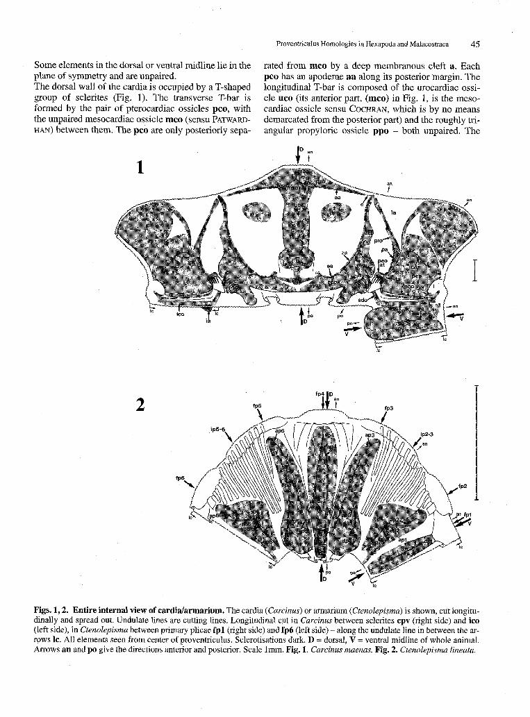
WARDHAN. The abbreviations used here for the sclerites comprise three lower case letters; those for the denticles comprise two lower case letters, with "t" in the second position; those for the apodemes comprise two lower case letters, with "a" in the second position. (Only some apodemes are named; in Fig. 1 the line along which the apodeme infolds, i.e. the apodeme base, is labeled with the abbreviation of the apodeme.) Some further conspicuous elements are designated by single lower case letters. Two elements forming a pair (see below) have the same name. Setae, very important for the function of the cardia but not for the scope of this comparison, are omitted from the figures although some are mentioned in the description. The heaviness of sclerotisa-tion or calcification is, with some exceptions, not represented in the figures. The following description proceeds mainly from dorsal to ventral. Cardiac symmetry is purely bilateral (Fig. 1). The plane of symmetry coincides with that of the whole animal
• (dorsal side = D, ventral side = V in Fig. 1). Most elements are paired, i.e. represented by two mirror-image counterparts in the left and the right half of the cardia.
Proventriculus Homologies in Hexapoda and Malacostraca 45
Some elements in the dorsal or ventral midline lie in the plane of symmetry and are unpaired. The dorsal wall of the cardia is occupied by a T-shaped group of sclerites (Fig. 1). The transverse T-bar is formed by the pair of pterocardiac ossicles pco, with the unpaired mesocardiac ossicle mco (sensu PATWARD-
HAN) between them. The pco are only posteriorly sepa-
*fe
t
rated from mco by a deep membranous cleft a. Each pco has an apodeme aa along its posterior margin. The longitudinal T-bar is composed of the urocardiac ossicle uco (its anterior part, (mco) in Fig. 1, is the mesocardiac ossicle sensu COCHRAN, which is by no means demarcated from the posterior part) and the roughly triangular prepyloric ossicle ppo - both unpaired. The
J
2C0
\ Id
C '
^ ' ua \ peo ' ' / ' uc° m ej , zco \ Kit .' , / '
py° k ^ -x*\J' 7 rpoo / '
.- h ' . J . . ^ r ^ H - -
/
i
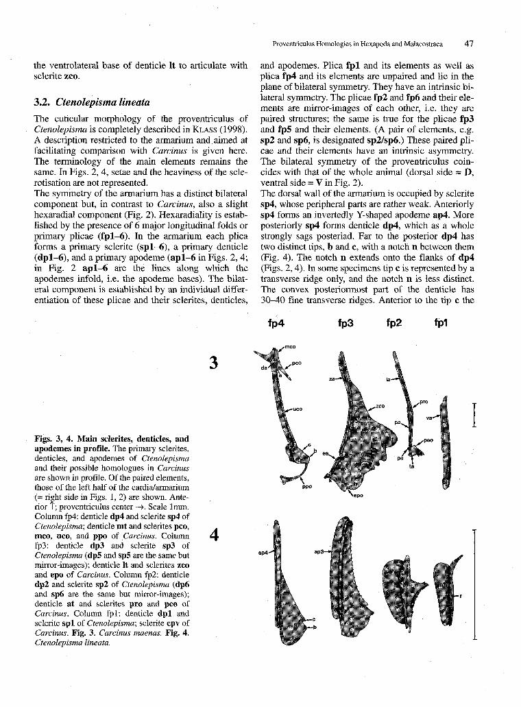
Figs. 1,2. Entire internal view of cardia/armarium. The cardia (Carcinus) or armarium (Ctenolepisma) is shown, cut longitudinally and spread out. Undulate lines are cutting lines. Longitudinal cut in Carcinus between sclerites cpv (right side) and ico (left side), in Ctenolepisma between primary plicae fpl (right side) and fp6 (left side) - along the undulate line in between the arrows 1c. All elements seen from center of proventriculus. Sclerotisations dark. D = dorsal, V = ventral midline of whole animal. Arrows an and po give the directions anterior and posterior. Scale 1mm. Fig. 1. Carcinus maenas. Fig. 2. Ctenolepisma lineata.

46 K.-D. KLASS
uco is firmly connected with the mco anterior to it; the border is the invertedly Y-shaped apodeme da. The ppo has a forked anterior part, and the two tips support the uco from the posterior and articulate with it. The articulations are concealed beneath the posterior margin of uco. The cuticle between the posterior margin of uco and the ppo-branches is strongly thickened. The ppo has apodemal ridges all along its margins (not represented in Figs. 1,3). The sclerotisations uco and ppo lie upon a massive denticle, the median tooth mt, which as a whole sags posteriad. The mt has its main tip b far posteriorly (Figs. 1, 3fp4) and a transverse ridge c immediately anterior to it. The posteriormost part of mt is strongly convex in profile (Fig. 3fp4). Anterior to ridge c, mt has a broad, shallow median groove d. Lateral to mt there is, on each side, a wide and mostly membranous area (Fig. 1). Its central part has a weak sclerotisation named here the dorsal cardiac plate dcp (not mentioned in COCHRAN or in PATWARDHAN). The dorsolateral cardiac wall is occupied by a sclerite group comprising the large zygocardiac ossicle zco, the exopyloric ossicle epo, and the pyloric ossicle pyo - all paired elements. The anterior end of zco is connected with the adjacent lateral end of sclerite pco by a strip of thickened cuticle e. The posterior half of zco lies upon a large, broadly shovel-shaped denticle, the lateral tooth It. The crest of It is highly differentiated: 4 distinct tips lie one behind the other; the anteriormost and largest tip f is slightly inclined dorsomedially, towards denticle mt. On its median surface near the 3 smaller tips, It has a series of transverse ridges g. The posteriormost part of the denticle is a weaker sclerotised, bristled lobe h. Along the entire dorsomedian margin of zco runs an apodemal ridge za. Another apodeme ua, with the shape of a broad U, is present centrally on zco. The small sclerite epo is only partly separated from zco by a short membranous cleft i, and the mobility between the two sclerites is low. The epo has an apodeme ea, which begins at the posterior margin of the cleft i and extends along the anterior margin of epo - like a posteromedian continuation of the apodeme za on zco. The plate-like sclerite pyo is almost completely separated from epo by a long membranous cleft k. Its anterior end articulates with the lateral end of sclerite ppo. The median ends of the two pyo are close to each other. The prepectineal ossicle pro in the lateral wall of the cardia is an L-shaped, rod-like sclerite. It has two ridgelike apodemes: pa runs along the posterior arm of the L, la runs along the anterior arm. The anterodorsal end of the L either approaches or reaches (both conditions found) the anterior end of sclerite zco. The posterior end of the L articulates with the pectineal ossicle peo. This peo lies on a small bulge-shaped denticle at. 5 or 6 spines (the lateral accessory teeth sensu COCHRAN)
originate from the crest of this denticle. The spine bases are arranged in a dorsoventral row whose ventral section curves slightly posteriad (Fig. 7). The spines become gradually shorter from dorsal to ventral. The apodemal ridge pa of sclerite pro bridges the articulation between pro and peo, bends dorsad, and continues along the anterior margin of peo (Figs. 1, 3fp2, 7). (At the articulation, pa consists of strongly thickened but not sclerotised cuticle.) The ventral and posterior margins of peo are strengthened by another apodemal ridge ta, which begins at the articulation between pro and peo but is distinctly separated from apodeme pa. The dorsal and central parts of peo, where the spines originate, are weakly sclerotised, and the spines are mobile at their bases. The postpectineal ossicle poo supports sclerite peo from the posterior and articulates with it (further data on poo below). The ventrolateral cardiac wall is occupied by two large sclerite plates, the anterolateral cardiac plate acp and the posterolateral cardiac plate pep. Their sclerotisation is mostly weak, and they have rather indistinct borders. Some parts are stronger, mainly the center of pep along the apodemal ridge ca. Anteroventral to plate pep there is a densely setose membranous cushion 1; in the undamaged proventriculus it is close to the opening of the oesophagus into the cardia. The ventral cardiac wall forms the unpaired cardiopy-loric valve cpv, a longitudinal plate whose median part may be calcified (various degrees of calcification were observed). The anterior part of cpv has an apodemal ridge va along its median line. Apodeme va is also visible as a shallow groove along the internal face. Posteriorly cpv forms a broad lobe cl overlying the entrance to the pylorus. The ventral margin of plate pep folds over the dorsolateral margin of cpv. The narrow area beneath this fold (the paired ventral groove sensu PATWARDHAN; it is unfolded in Fig. 1) has a complex morphology: The ventral margin m of pep bears a comb of setae, which are directed ventromesad and cover the lateral parts of cpv. Beneath these setae the rod-like sclerite poo extends anteroposteriad. The posterior part of poo bends dorsad and, as mentioned above, articulates with sclerite peo. The anterior part of poo also bends dorsad and then forks, with the branches embracing the base of cushion 1. Ventromedian to poo, and parallel to it, there is another fold o armed with a comb of setae similar to that on the pep-margin m. Ventromedian to fold o extends the inferolateral cardiac ossicle ico; both its dorsal and ventral margins form an apodemal ridge (not represented in Fig. 1). The ventral apodeme becomes excessively deep in its posteriormost part to form a plate-like apodeme ia (Fig. 1, left side). The posterior end of sclerite ico articulates with another stout rodlike sclerite, the subdentary ossicle sdo, which runs to
Proventriculus Homologies in Hexapoda and Malacostraca 4 7
the ventrolateral base of denticle It to articulate with sclerite zco.
3.2. Ctenolepisma lineata The cuticular morphology of the proventriculus of Ctenolepisma is completely described in KLASS (1998). A description restricted to the armarium and aimed at facilitating comparison with Carcinus is given here. The terminology of the main elements remains the same. In Figs. 2, 4, setae and the heaviness of the scle-rotisation are not represented. The symmetry of the armarium has a distinct bilateral component but, in contrast to Carcinus, also a slight hexaradial component (Fig. 2). Hexaradiality is established by the presence of 6 major longitudinal folds or primary plicae (fpl-6). In the armarium each plica forms a primary sclerite (spl-6), a primary denticle (dpl-6), and a primary apodeme (apl-6 in Figs. 2, 4; in Fig. 2 ap l -6 are the lines along which the apodemes infold, i.e. the apodeme bases). The bilateral component is established by an individual differentiation of these plicae and their sclerites, denticles,
and apodemes. Plica fpl and its elements as well as plica fp4 and its elements are unpaired and lie in the plane of bilateral symmetry. They have an intrinsic bilateral symmetry. The plicae fp2 and fp6 and their elements are mirror-images of each other, i.e. they are paired structures; the same is true for the plicae fp3 and fp5 and their elements. (A pair of elements, e.g. sp2 and sp6, is designated sp2/sp6.) These paired plicae and their elements have an intrinsic asymmetry. The bilateral symmetry of the proventriculus coincides with that of the whole animal (dorsal side = D, ventral side = V in Fig. 2). The dorsal wall of the armarium is occupied by sclerite sp4, whose peripheral parts are rather weak. Anteriorly sp4 forms an invertedly Y-shaped apodeme ap4. More posteriorly sp4 forms denticle dp4, which as a whole strongly sags posteriad. Far to the posterior dp4 has two distinct tips, b and c, with a notch n between them (Fig. 4). The notch n extends onto the flanks of dp4 (Figs. 2, 4). In some specimens tip c is represented by a transverse ridge only, and the notch n is less distinct. The convex posteriormost part of the denticle has 30-40 fine transverse ridges. Anterior to the tip c the
fp4 fp3 fp2 fp1
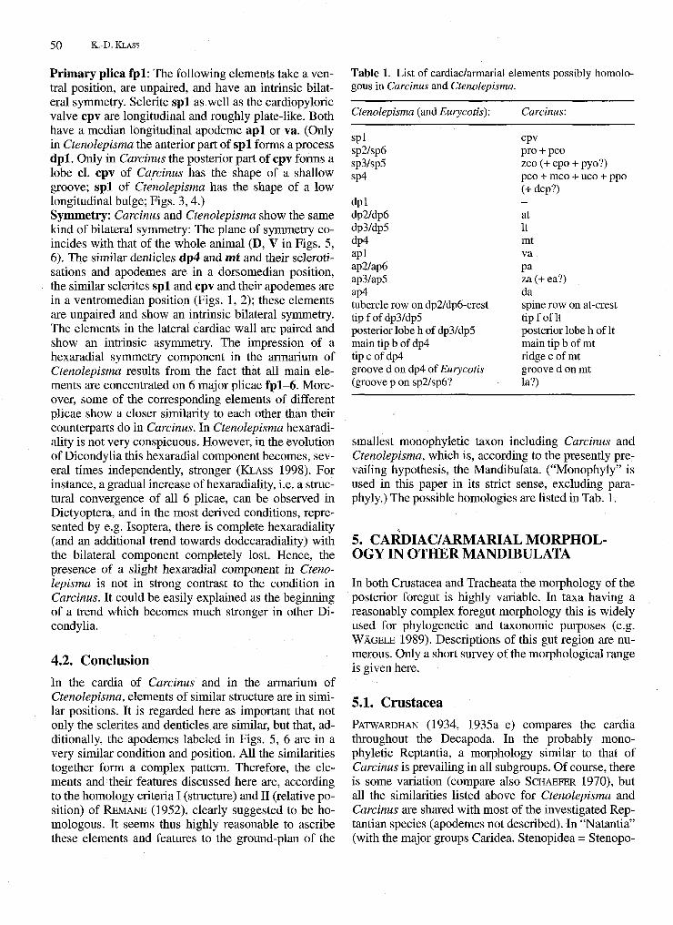
Figs. 3, 4. Main sclerites, denticles, and apodemes in profile. The primary sclerites, denticles, and apodemes of Ctenolepisma and their possible homologues in Carcinus ait shown in profile. Of the paired elements, those of the left half of the cardia/armarium (= right side in Figs. 1, 2) are shown. Anterior T; proventriculus center —». Scale 1mm. Column fp4: denticle dp4 and sclerite sp4 of Ctenolepisma; denticle nit and sclerites pco, mco, uco, and ppo of Carcinus. Column fp3: denticle dp3 and sclerite sp3 of Ctenolepisma (dp5 and sp5 are the same but mirror-images); denticle It and sclerites zco and epo of Carcinus. Column fp2: denticle dp2 and sclerite sp2 of Ctenolepisma (dp6 and sp6 are the same but mirror-images); denticle at and sclerites pro and peo of Carcinus. Column fpl: denticle dpi and sclerite spl of Ctenolepisma; sclerite cpv of Carcinus. Fig. 3. Carcinus maenas. Fig. 4. Ctenolepisma lineata.
A ap4-*\ I
v a - " I
ap3-~l
•h
ap2 I P " • '3P1 J1
J L /

4 8 K.-D. KLASS
crest of dp4 is even; Eurycotis floridana (Walker, 1868) (Blattaria: Blattidae), whose dp4 is very similar to that of Ctenolepisma, has a shallow median longitudinal groove along the anterior crest of this denticle (KLASS 1998, fig. 4). The dorsolateral armarial walls are occupied by the scle-rite pair sp3/sp5. The major posterior part of sp3/sp5 lies upon a large, broadly shovel-shaped denticle dp3/ dp5, which has two distinct tips (the anterior one is labeled f) strongly inclined towards denticle dp4. The posteriormost part h of dp3/dp5 is lobe-like and less strongly sclerotised. sp3/sp5 has a longitudinal apodeme ap3/ ap5 which folds in along most of the sclerite's dor-somedian margin. Ventral to sp3/sp5, in the area designated ip2-3/ip5-6, the anterior part of the armarial wall has a cluster of membranous longitudinal folds. The ventrolateral armarial walls are occupied by the sclerite pair sp2/sp6. sp2/sp6 has a weak dorsal part expanded towards sp3/sp5 and traversed by a shallow groove p (groove indistinct or absent in many specimens). The ventral part of sp2/sp6 forms a denticle dp2/dp6 whose anterior part is most prominent. The arch-shaped, uniformly serrate or tuberculate crest of this denticle has a dorsoventral orientation anteriorly but curves, becoming lower, far posteriad in its ventral part (Figs. 2, 8). Far posteriorly sp2/sp6 has an area q with a polished appearance. sp2/sp6 forms a longitudinal plate-like apodeme ap2/ap6, which infolds along the dorsal flank of dp2/dp6 but extends anteriorly beyond the denticle base.
The ventral armarial wall is occupied by sclerite spl. Most anteriorly spl forms a spatulate, anteriad-directed process dpi. (This dpi is the anterior, strongly projecting part of denticle dpi as defined in KLASS 1998.) Posterior to dpi, spl forms a longitudinal apodeme plate apl, which folds inwards along the sclerite's median line. On each side spl has a distinctly bordered area r with a polished appearance.
4. POSSIBLE HOMOLOGIES BETWEEN CARCINUS AND CTENOLEPISMA
In this chapter the similarities between Carcinus and Ctenolepisma are analysed. The cardiac morphology of Carcinus is more complicated than the armarial morphology of Ctenolepisma, mainly in the higher number of separate sclerites and apodemes. However, nearly each armarial element of Ctenolepisma - sclerites, denticles, and apodemes - has in the cardia of Carcinus a structurally similar counterpart in the same position. Figs. 5, 6 give a survey of these similarities. (In Fig. 5 the cardia of Carcinus is labelled according to the Ctenolepisma terminology; compare Fig. 1 for the original Carcinus terminology.)
4.1. Analysis of similarities
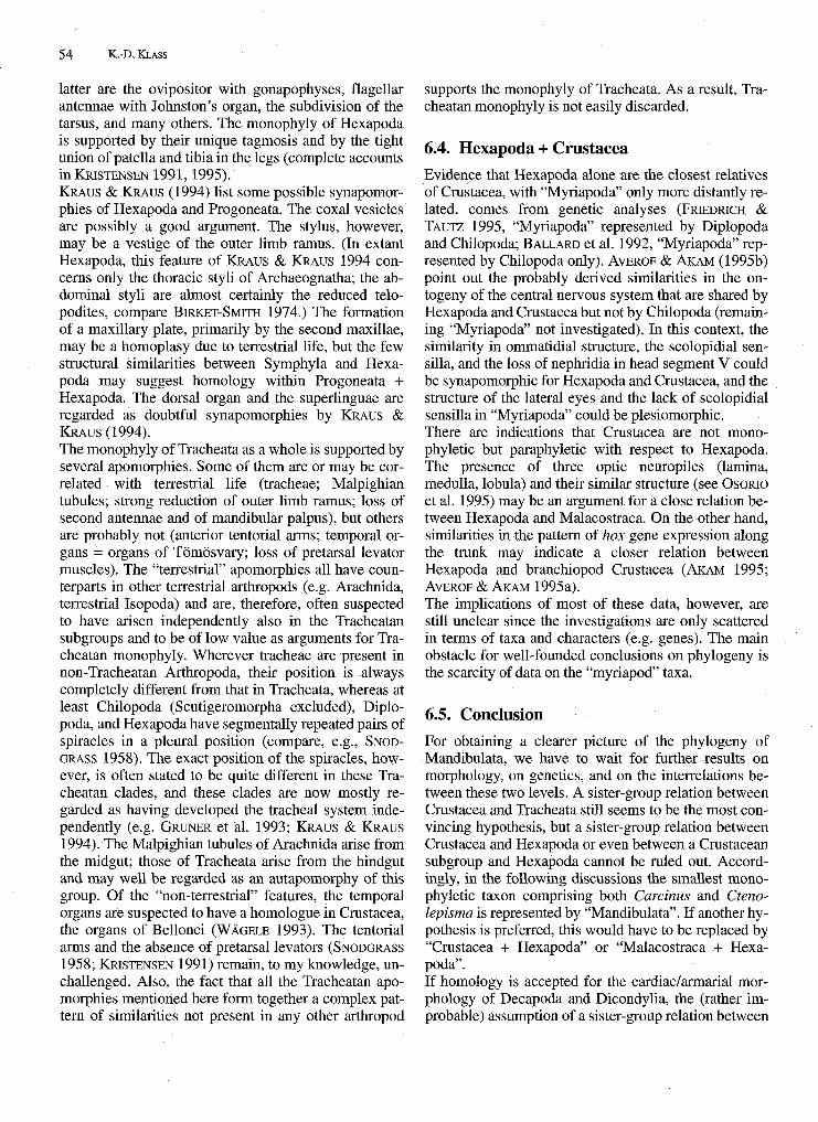
Primary plica fp4: The following elements take a dorsal position and are mostly unpaired (single) with an intrinsic bilateral symmetry. Denticle dp4 and denticle mt have a similar shape: The anterior part of the denticle ascends straightly and gradually (Figs. 3, 4); in Carcinus and Eurycotis, but not in Ctenolepisma, this part has a shallow median groove d. The main tip b has a far posterior position. Just anterior to b there is another tip or ridge c, which is more elaborate in Ctenolepisma. The part posterior to tip b is short and convex in profile. The apodemes ap4 and da are similar in their invertedly Y-like shape and in their position anterolateral to the base of denticle dp4 or mt. (In Ctenolepisma the arms of ap4 extend farther to the posterior.) Regarding the sclerotisations occupying these denticles and apodemes, sp4 corresponds to the sclerites pco + mco + uco, all firmly connected, and ppo; the additional inclusion of dcp is conceivable. (Only Carcinus has the posterior sclerotisation ppo of denticle mt articulated with the anterior sclerotisation.) Primary plicae fp3/fp5: The following elements take a dorsolateral position, are paired (two mirror-image counterparts in the left and the right half of the cardia/armarium), and have an intrinsic asymmetry. Denticle dp3/dp5 and denticle It arise from the posterior part of their sclerites (sp3/sp5 or zco) and are, as a whole, broad and somewhat shovel-shaped. They have a strong anterior tip f and one (Ctenolepisma) or more (Carcinus) smaller tips behind it. The posteriormost part of the denticle is a weakly sclerotised lobe h. The sclerites sp3/sp5 and zco (possibly including the ex-opyloric ossicle epo and the pyloric ossicle pyo) have a rather similar shape. The apodemes ap3/ap5 and za (possibly including ea) take the same position along the dorsomedian margin of their sclerites. Primary plicae fp2/fp6: The following elements take a ventrolateral (Ctenolepisma) or lateral (Carcinus) position, are paired, and have an intrinsic asymmetry. Denticle dp2/dp6 as well as denticle at are bulges with a row of short tubercles (Ctenolepisma) or long spines (Carcinus) along the crest. The tubercle/spine bases are in an arch-like arrangement (Figs. 7, 8), with the arch spanning from anterodorsally to posteroventrally. In Ctenolepisma, the curvature of the arch to the posterior is stronger, the tubercles are shorter and stouter than the spines of Carcinus, and the number of tubercles/spines is much higher. The crab Cyclograpsus punctatus (SCHAE-FER 1970, Figs. 4, 5), however, closely resembles Ctenolepisma in these features: The curvature of the arch is stronger than in Carcinus, and the spines are stouter and numerous. The anterodorsal part of sp2/sp6 and the prepectineal ossicle pro are in a similar position and extend towards sclerite sp3/sp5 or the zygocardiac ossicle

Proventriculus Homologies in Hexapoda and Malacostraca 4 9
zco. (This sclerotisation is much narrower and separated from the denticle sclerotisation in Carcinus.) Apodeme ap2/ap6 of Ctenolepisma is entirely straight and infolds anterior and dorsal to denticle dp2/dp6. Apodeme pa of Carcinus resembles it in lying anterior to the denticle and extending posteriorly towards the dorsal denticle flank (Figs. 7, 8); however, its sclerotisation - but not the ridgelike thickening of the cuticle - has a gap at the articulation between peo and pro, and behind this articulation
apodeme pa sharply bends dorsad. The groove p on the weak dorsal part of sp2/sp6 of Ctenolepisma and apodeme la of Carcinus are similar in their transverse course directed towards sp3/sp5 or the zygocardiac ossicle zco. Since groove p is mostly indistinct, the indication of homology with la is weak. When the proventriculus is constricted, the main body of denticle dp2/dp6 or at (the anterior part in Ctenolepisma) rests upon the crest of denticle dp3/dp5 or It (upon the part shortly anterior to tip f).
6
Figs. 5,6. Possible homology relations between cardia and armarium. Cardia and armarium shown as in Figs. 1, 2. Sclero-tisations present in both species differently patterned according to possible homology relations. Terminology of Ctenolepisma used for Carcinus, too. Only suspectedly homologous elements labeled. Fig. 5. Carcinus maenas. Fig. 6. Ctenolepisma lineata.
50 K.-D. KLASS
Primary plica fpl: The following elements take a ventral position, are unpaired, and have an intrinsic bilateral symmetry. Sclerite sp l as well as the cardiopyloric valve cpv are longitudinal and roughly plate-like. Both have a median longitudinal apodeme a p l or va. (Only in Ctenolepisma the anterior part of spl forms a process dpi. Only in Carcinus the posterior part of cpv forms a lobe cl. cpv of Carcinus has the shape of a shallow groove; sp l of Ctenolepisma has the shape of a low longitudinal bulge; Figs. 3,4.) Symmetry: Carcinus and Ctenolepisma show the same kind of bilateral symmetry: The plane of symmetry coincides with that of the whole animal (D, V in Figs. 5, 6). The similar denticles dp4 and mt and their scleroti-sations and apodemes are in a dorsomedian position, the similar sclerites spl and cpv and their apodemes are in a ventromedian position (Figs. 1,2); these elements are unpaired and show an intrinsic bilateral symmetry. The elements in the lateral cardiac wall are paired and show an intrinsic asymmetry. The impression of a hexaradial symmetry component in the armarium of Ctenolepisma results from the fact that all main elements are concentrated on 6 major plicae fpl-6. Moreover, some of the corresponding elements of different plicae show a closer similarity to each other than their counterparts do in Carcinus. In Ctenolepisma hexaradi-ality is not very conspicuous. However, in the evolution of Dicondylia this hexaradial component becomes, several times independently, stronger (KLASS 1998). For instance, a gradual increase of hexaradiality, i.e. a structural convergence of all 6 plicae, can be observed in Dictyoptera, and in the most derived conditions, represented by e.g. Isoptera, there is complete hexaradiality (and an additional trend towards dodecaradiality) with the bilateral component completely lost. Hence, the presence of a slight hexaradial component in Ctenolepisma is not in strong contrast to the condition in Carcinus. It could be easily explained as the beginning of a trend which becomes much stronger in other Dicondylia.
4.2. Conclusion In the cardia of Carcinus and in the armarium of Ctenolepisma, elements of similar structure are in similar positions. It is regarded here as important that not only the sclerites and denticles are similar, but that, additionally, the apodemes labeled in Figs. 5, 6 are in a very similar condition and position. All the similarities together form a complex pattern. Therefore, the elements and their features discussed here are, according to the homology criteria I (structure) and II (relative position) of REMANE (1952), clearly suggested to be homologous. It seems thus highly reasonable to ascribe these elements and features to the ground-plan of the
Table 1. List of cardiac/armarial elements possibly homologous in Carcinus and Ctenolepisma.
Ctenolepisma (and Eurycotis):
spl sp2/sp6 sp3/sp5 sp4
dpi dp2/dp6 dp3/dp5 dp4 apl ap2/ap6 ap3/ap5 ap4 tubercle row on dp2/dp6-crest tipf ofdp3/dp5 posterior lobe h of dp3/dp5 main tip b of dp4 t ipcofdp4 groove d on dp4 of Eurycotis (groove p on sp2/sp6?
Carcinus:
cpv pro + peo zco (+ epo + pyo?) pco + mco + uco + ppo (+ dcp?) -at It mt va pa za (+ ea?) da spine row on at-crest tipf of It posterior lobe h of It main tip b of mt ridge c of mt groove d on mt la?)
smallest monophyletic taxon including Carcinus and Ctenolepisma, which is, according to the presently prevailing hypothesis, the Mandibulata. ("Monophyly" is used in this paper in its strict sense, excluding para-phyly.) The possible homologies are listed in Tab. 1.
5. CARDIAC/ARMARIAL MORPHOLOGY IN OTHER MANDIBULATA
In both Crustacea and Tracheata the morphology of the posterior foregut is highly variable. In taxa having a reasonably complex foregut morphology this is widely used for phylogenetic and taxonomic purposes (e.g. WAGELE 1989). Descriptions of this gut region are numerous. Only a short survey of the morphological range is given here.
5.1. Crustacea PATWARDHAN (1934, 1935a-e) compares the cardia throughout the Decapoda. In the probably monophyletic Reptantia, a morphology similar to that of Carcinus is prevailing in all subgroups. Of course, there is some variation (compare also SCHAEFER 1970), but all the similarities listed above for Ctenolepisma and Carcinus are shared with most of the investigated Rep-tantian species (apodemes not described). In "Natantia" (with the major groups Caridea, Stenopidea = Stenopo-

Proventriculus Homologies in Hexapoda and Malacostraca 51
Figs. 7, 8. Denticles dp6 and at and associated structures. Scheme of denticle at, sclerites peo and pro, and apodemes pa and la of Careinus, and of denticle dp6, sclerite sp6, apodeme ap6, and groove p of Ctenolepisma. The elements of the right pro ventricular wall (left side in Figs. 1, 2) are shown as seen from the pro ventricular center. Black dots are bases of spines or tubercles. Anterior t , dorsal —>. No scale. Fig. 7. Careinus maenas. Fig. 8. Ctenolepisma lineata.
didea, and Penaeidea = Dendrobranchiata), regarded as a paraphyletic assemblage of more primitive Decapoda (e.g. GRUNER et al. 1993), the cardia is more diverse: In all Caridea, PATWARDHAN found a rather simple morphology. The denticles are represented by folds that are hardly sclerotised or calcified and hardly show any further differentiation. Only some species have distinct median and lateral denticles with sclerotised tips (mt and It; best developed in Caridina brachydactyla de Mann; Atyidae), but most lack these. The Dendrobranchiata and Stenopodidea have a cardia resembling that of Reptantia. According to PATWARDHAN'S descriptions, the dorsal sclerites (pco, mco, uco, ppo, and zco) and denticles (mt and It) are in a similar condition to the Reptantia. The posterior spine-row of the infero-lat-eral cardiac plate (ile of PATWARDHAN 1935e, fig. 7) of Stenopus spinosus Risso. (Stenopodidae) is, regarding its similar shape and position, probably homologous with the spine-row on the pectineal ossicle peo of Careinus. Regarding the ventral elements, PATWARD
HAN'S descriptions are not very detailed, and a comparison with Reptantia is not possible. Since the Dendrobranchiata are probably the sister-group of the remaining Decapoda (= Pleocyemata) (e.g. GRUNER et al. 1993), it is reasonable to assume that at least the dorsal sclerites and denticles (and possibly also the ventral ones) are, in the condition found in Dendrobranchiata and Reptantia, ground-plan elements of Decapoda, and that the reduction of this cardiac armature in Caridea is a secondary trait. PATWARDHAN (1935e) also comes to this conclusion.
Decapoda, Euphausiacea, and Amphionidacea together form the Eucarida. The superorders Eucarida, Peracarida, Pancarida, Syncarida, Hoplocarida, and Phyllo-carida constitute the Malacostraca, which are generally accepted as a monophyletic taxon. SIEWING (1952, 1954, 1956) surveys the proventricular morphology throughout the Malacostraca and gives a comprehensive hypothesis on the evolution of the organ. His descriptions focus on the geometry of the proventricular folds (the denticles are part of them) and the distribution of setae. Hardly any sclerotisations and no articulations or apodemes are mentioned. Hence, most of the elements discussed above are not considered in S E WING'S descriptions, or they are largely absent in the species he has studied. HAFFER (1965) gives a survey on the proventriculus of Peracarida and Eucarida - including the muscles - and a synonymy list of the terminologies used previously in these taxa. HAFFER describes the overall shape, the position, and the setae or spines of the sclerites and denticles but, unfortunately, not the exact structure of the sclerotisations or any articulations or apodemes. Similar inadequacies in the morphological descriptions are true for most other available contributions (compare also WAGELE 1989, p. 25). A comparison between Decapoda and other Malacostracan taxa by means of the previous literature is thus only partly possible, and some of the homologies assumed below are tentative. HAFFER (1965) points out the similarity between the proventriculi of Decapoda, Euphausiacea (Eucarida), and Mysidacea (Peracarida): "Dekapodentyp" of proventriculus. The cardiac elements of Neomysis vulgaris (Mysidacea) are, in the frame of HAFFER'S data, mostly similar to those of Dendrobranchiata and Reptantia (HAFFER'S abbreviations in brackets): sclerite cpv (= CV), setal comb on fold o (= BG on CLi), sclerites acp and pep (= CLa), sclerite peo and denticle at with their spines (= CLp), sclerite zco and denticle It (= CD1; CL according to HAFFER, p. 203), and denticle mt with a shallow groove d (= CD with concavity KV). For some Euphausiacea, HAFFER-reports a similar condition to that in Neomysis vulgaris. ZIMMER (1912), however, says that in Euphausia superba Dana (Euphausiacea) parts of the cardiac wall epithelium are thickened and bulge into the lumen (denticles; ZIMMER'S "Stiicke"), but the cuticle has roughly the same thickness in all parts of the cardia. Distinct sclerotisations or calcifications, comparable with the ossicles of Decapoda, are not mentioned. Whether crucial elements such as the apodemes described above are present in Mysidacea and Euphausiacea cannot be decided from these descriptions. The proventriculi of Isopoda and Tanaidacea (both Peracarida) are rather different from those of Eucarida and Mysidacea (HAFFER 1965; LAUTERBACH 1970; SCHELOSKE 1976; WAGELE 1989). WAGELE

52 K.-D. KLASS
(1989) describes, for various Isopoda, chitinous ridges ("Chitinleisten = Sklerite") in the anterior part of the proventriculus; these are, regarding their positions, partly comparable to the apodemal ridges of Carcinus (Ski = aa; Sk2 = za+ua; Sk4 = la+pa). Strong scleroti-sations in similar positions are also present in Tanai-dacea (LAUTERBACH 1970). However, for a detailed comparison of the apodemes the descriptions are too scarce, and there are probably also some stronger differences between these taxa. As a result, these data on Eucarida and Peracarida altogether show that a morphology at least similar to that of Carcinus was already present in the common ground-plan of Eucarida and Peracarida. If Pancarida are actually the sister-group of Peracarida (e.g. GRUNER et al. 1993) the cardiac morphology of this taxon (no detailed morphological data available) must also be derived from this ground-plan. Syncarida and Phyllocarida have a much simpler cardiac morphology than Dendrobranchiata and Reptantia (SIEWING 1956; CLAUS 1888). The cardia of Hoplo-carida is also quite simple in some respects, but it also shows some peculiar derivations (SIEWING 1956; REDDY 1935).
Hence, no other Malacostracan taxa seem to have a cardiac morphology as similar to that of Ctenolepisma as the Reptantia. However, at least some other Eucarida and some Peracarida also show many of the respective similarities, and it is unclear whether the remaining elements and features are actually absent or whether they are just not considered in the descriptions. For completeness, it may be mentioned here that various Malacostraca share similarities with Ctenolepisma that are absent in Carcinus: (1) Many Reptantia (PAT-WARDHAN 1935b,c), Lophogastrida, and Cumacea (SIEWING 1956) have the lateral parts of the cardiopy-loric valve cpv differentiated as longitudinal bulges distinctly set off from the main median part. These bulges have the same position as the lateral areas r of spl of Ctenolepisma. (2) At least in some Hoplocarida and Syncarida (Squilla mantis and Anaspides tasmaniae in SIEWING 1956) the anterior part of cpv forms an ante-riad-directed process in the same position as dpi of Ctenolepisma. (3) In some Peracarida, Euphausiacea, and Caridea (HAFFER 1965), sclerite cpv does not have the shape of a shallow longitudinal groove as in Carcinus (Fig. 3 fpl) but bulges into the proventricular lumen like spl of Ctenolepisma (Fig. 4fpl). (4) In Neomysis vulgaris (HAFFER 1965, fig. 6) denticle It (= CD1) has two strong tips strongly inclined towards denticle mt, like denticle dp3/dp5 of Ctenolepisma. Of course, the assumption of homology would be highly speculative in these cases.
Most Malacostracan superorders include subgroups in which simple cardiac morphology is undoubtedly due to secondary reduction. Within Peracarida, such a re
duction trend is present in e.g. some Mysidacea (comparison of Mysis stenolepis Smith and Spelaeomysis longipes in FRIESEN et al. 1986) and Tanaidacea (SIEWING 1954). In Eucarida, as mentioned above, at least Caridea and, according to ZIMMER'S (1912) descriptions, some Euphausiacea show strong secondary reductions. In Syncarida, strong reduction is certainly true for Bathynellacea (GRUNER et al. 1993) and has been suspected also for Anaspidacea (SIEWING 1956, p. 112). In Phyllocarida, Nebaliopsis typica has lost most cardiac elaborations (GRUNER et al. 1993). Certain elements of these reduction trends are similar in different taxa (e.g. SIEWING 1956, p. 112). As regards the remaining Crustacea, sclerotised differentiations of the posterior foregut are, to my knowledge, only reported for some Ostracoda (SIEWING 1956) and Acrothoracica (GRUNER et al. 1993). These structures are regarded as completely different from and evolved independently of the proventriculus of Malacostraca. Many non-Malacostracan Crustacea have a set of 4 simple longitudinal folds running along the foregut (1 dorsal, 1 ventral, 2 lateral; SIEWING 1956). SIEWING (1956) regards this latter condition as the starting point of proventricular anagenesis in Malacostraca. Some simple subdivisions of the 4 folds lead to the ground-plan of Malacostraca, which includes a still indistinct separation between cardia and pylorus and, in the cardia, a subdivision of the lateral fold pair into lat-eralia superiores (roughly denticle It) and lateralia infe-riores (roughly area of plates acp and pep and denticle at). According to this hypothesis, the condition in Mysidacea, Dendrobranchiata, and especially Reptantia has tabe regarded as highly apomorphic within Malacostraca. Within the range of SIEWING'S comparison, which is restricted to Crustacea, this view is certainly conclusive.
5.2. Tracheata
The armarium of Ctenolepisma (Zygentoma: Lepis-matidae) completely, or at least nearly so, shows the ground-plan condition of Dicondylia (cf. descriptions in KLASS 1998). Of the Dicondylia so far investigated, only further Lepismatidae, some Blattaria (mainly Blat-tidae), and the nymphs of some Odonatan families (Corduliidae, Libellulidae) have an armarium which is in many respects close to that of Ctenolepisma. The remaining Dicondylia have the armarium either strongly modified or strongly reduced. Modifications and reductions also occur within Blattaria and Odonata, and the anagenetic trends are partly similar in these two taxa (details in KLASS 1998). For Diplopoda, Pauropoda, and Symphyla (together the Progoneata), and for the Hexapoda Parainsecta (= El-lipura = Protura + Collembola), Diplura, Archaeog-

Proventriculus Homologies in Hexapoda and Malacostraca 53
natha, and Lepidotrichidae (Zygentoma; the most primitive taxon of Dicondylia, possibly the sister-group of all other Dicondylia; KRISTENSEN 1995) no cuticular differentiations of the posterior foregut are reported which could be compared with the armarium of Ctenolepisma. Many Chilopoda have, according to the survey in LEWIS (1981), longitudinal folds variously armed with small sclerites, spines, or setae in the posterior foregut; these structures, however, bear no resemblance to the armarium of Ctenolepisma.
5.3. Other Arthropoda
Of the Chelicerata, I have checked Limulus polyphemus (Linnaeus, 1758) (Xiphosura: Limulidae) regarding its foregut morphology. It has a muscular proventriculus with ca. 14 longitudinal folds that are weakly sclero-tised and transversely corrugated. No further similarity with the proventriculi of Carcinus or Ctenolepisma was detected. For the Onychophora, no foregut structures bearing any resemblance to those of Carcinus or Ctenolepisma are reported.
5.4. Conclusion
Regarding the complex similarities between the armarium of Ctenolepisma and the cardia of Carcinus, it seems reasonable to assume homology for the elements and features listed above and to ascribe them to the ground-plan of Mandibulata. In this case, the anagenesis of the Malacostracan proventriculus would be the reverse of SIEWING'S hypothesis. On the other hand, the distribution of these elements and features over the Mandibulatan subgroups contradicts this assumption: In Crustacea, a proventriculus is found only in Malacostraca, and a morphology closely similar to Carcinus is known only from Decapoda (but may be present in other Eucarida and Peracarida, then being plesiomorphic for these taxa). In Tracheata, an armarium similar to that of Ctenolepisma is found only within Dicondylia. Many parallel reductions or losses of the cardia/armarium within the more basal offshoots of Crustacea and Tracheata have to be assumed in the case of homology, and only a highly derived clade of both Crustacea (Decapoda, or possibly Eucarida + Peracarida + Pancarida) and Tracheata (Dicondylia) would have retained the Mandibulatan ground-plan condition. In this respect, the assumption of homology seems very unparsimonious, and it seems more reasonable to assume that the cardiac morphology as found in Carcinus is a development within Malacostraca, and that the ar-marial morphology as found in Ctenolepisma is an au-tapomorphy of Dicondylia (or of Lepismatidae + Ptery-gota if Zygentoma are paraphyletic with respect to Pterygota as suspected by KRISTENSEN 1995).
6. HYPOTHESES ON THE PHYLOGENY OF MANDIBULATA
To obtain an impression on the extent of non-parsimony inherent in the assumption of cardiac/armarial homology, the present knowledge on the phylogeny of Mandibulata has to be briefly surveyed - focused on the estimation of the phylogenetic distance between Malacostraca and Dicondylia. For detailed information see the cited papers. STYS & ZRZAVY (1994) give a more comprehensive survey on current hypotheses and recent literature.
6.1. Mandibulata
The traditional view, advocated by e.g. WAGELE (1993), assumes monophyly for Mandibulata. (These probably include Pentastomida, which are not considered here.) The Uniramia hypothesis sensu MANTON (e.g. 1977) is regarded here as discarded because of its incorrect argumentation (compare WEYGOLDT 1986; STYS & ZRZAVY 1994) and because of recent evidence on several organs (compare AVEROF & AKAM 1995b; OSORIO et al. 1995). Mostly, Crustacea and Tracheata are regarded as the two basal sister-groups within Mandibulata (e.g. WEYGOLDT 1986).
6.2. Crustacea
Malacostraca are generally accepted as a monophyletic unit, with their peculiar tagmosis and the characteristic position of the genital openings being their most convincing autapomorphies. The Phyllocarida are regarded as the sister-group of the remaining Malacostraca; the relations between the other superorders are, in part, still subject to debate. The relations between Malacostraca and the other major Crustacean groups are uncertain. Crustacea as a whole are mostly regarded as monophyletic, but the support is weak. WAGELE (1993) lists three possible autapomorphies: (1) The nephridia of head-segment V (segment of first maxillae) have been lost (like in Hexapoda). (2) A nauplius eye is present, which, however, is merely a closer grouping of the medial group of the frontal eyes of the Mandibulatan ground-plan. (3) The sinus gland, or some of its properties, may be suspected to be autapomorphic. The implications of the carapace and the stalked eyes are highly uncertain, and there are cases of secondary reduction.
6.3. Tracheata
Dicondylia as well as Insecta (= Ectognatha) are certainly monophyletic taxa. Autapomorphies of the former are, for instance, a derived morphology of the go-nangulum and of the mandible. Autapomorphies of the
5 4 K.-D. KLASS
latter are the ovipositor with gonapophyses, flagellar antennae with Johnston's organ, the subdivision of the tarsus, and many others. The monophyly of Hexapoda is supported by their unique tagmosis and by the tight union of patella and tibia in the legs (complete accounts in KRISTENSEN 1991,1995). KRAUS & KRAUS (1994) list some possible synapomor-phies of Hexapoda and Progoneata. The coxal vesicles are possibly a good argument. The stylus, however, may be a vestige of the outer limb ramus. (In extant Hexapoda, this feature of KRAUS & KRAUS 1994 concerns only the thoracic styli of Archaeognatha; the abdominal styli are almost certainly the reduced telo-podites, compare BIRKET-SMITH 1974.) The formation of a maxillary plate, primarily by the second maxillae, may be a homoplasy due to terrestrial life, but the few structural similarities between Symphyla and Hexapoda may suggest homology within Progoneata + Hexapoda. The dorsal organ and the superlinguae are regarded as doubtful synapomorphies by KRAUS & KRAUS (1994). The monophyly of Tracheata as a whole is supported by several apomorphies. Some of them are or may be correlated with terrestrial life (tracheae; Malpighian tubules; strong reduction of outer limb ramus; loss of second antennae and of mandibular palpus), but others are probably not (anterior tentorial arms; temporal organs = organs of Tomosvary; loss of pretarsal levator muscles). The "terrestrial" apomorphies all have counterparts in other terrestrial arthropods (e.g. Arachnida, terrestrial Isopoda) and are, therefore, often suspected to have arisen independently also in the Tracheatan subgroups and to be of low value as arguments for Tracheatan monophyly. Wherever tracheae are present in non-Tracheatan Arthropoda, their position is always completely different from that in Tracheata, whereas at least Chilopoda (Scutigeromorpha excluded), Diplo-poda, and Hexapoda have segmentally repeated pairs of spiracles in a pleural position (compare, e.g., SNOD-GRASS 1958). The exact position of the spiracles, however, is often stated to be quite different in these Tracheatan clades, and these clades are now mostly regarded as having developed the tracheal system independently (e.g. GRUNER et al. 1993; KRAUS & KRAUS 1994). The Malpighian tubules of Arachnida arise from the midgut; those of Tracheata arise from the hindgut and may well be regarded as an autapomorphy of this group. Of the "non-terrestrial" features, the temporal organs are suspected to have a homologue in Crustacea, the organs of Bellonci (WAGELE 1993). The tentorial arms and the absence of pretarsal levators (SNODGRASS 1958; KRISTENSEN 1991) remain, to my knowledge, unchallenged. Also, the fact that all the Tracheatan apomorphies mentioned here form together a complex pattern of similarities not present in any other arthropod
supports the monophyly of Tracheata. As a result, Tracheatan monophyly is not easily discarded.
6.4. Hexapoda + Crustacea Evidence that Hexapoda alone are the closest relatives of Crustacea, with "Myriapoda" only more distantly related, comes from genetic analyses (FRIEDRICH & TAUTZ 1995, "Myriapoda" represented by Diplopoda and Chilopoda; BALLARD et al. 1992, "Myriapoda" represented by Chilopoda only). AVEROF & AKAM (1995b) point out the probably derived similarities in the ontogeny of the central nervous system that are shared by Hexapoda and Crustacea but not by Chilopoda (remaining "Myriapoda" not investigated). In this context, the similarity in ommatidial structure, the scolopidial sen-silla, and the loss of nephridia in head segment V could be synapomorphic for Hexapoda and Crustacea, and the structure of the lateral eyes and the lack of scolopidial sensilla in "Myriapoda" could be plesiomorphic. There are indications that Crustacea are not mono-phyletic but paraphyletic with respect to Hexapoda. The presence of three optic neuropiles (lamina, medulla, lobula) and their similar structure (see OSORIO et al. 1995) may be an argument for a close relation between Hexapoda and Malacostraca. On the other hand, similarities in the pattern of hox gene expression along the trunk may indicate a closer relation between Hexapoda and branchiopod Crustacea (AKAM 1995; AVEROF & AKAM 1995a).
The implications of most of these data, however, are still unclear since the investigations are only scattered in terms of taxa and characters (e.g. genes). The main obstacle for well-founded conclusions on phylogeny is the scarcity of data on the "myriapod" taxa.
6.5. Conclusion For obtaining a clearer picture of the phylogeny of Mandibulata, we have to wait for further results on morphology, on genetics, and on the interrelations between these two levels. A sister-group relation between Crustacea and Tracheata still seems to be the most convincing hypothesis, but a sister-group relation between Crustacea and Hexapoda or even between a Crustacean subgroup and Hexapoda cannot be ruled out. Accordingly, in the following discussions the smallest mono-phyletic taxon comprising both Carcinus and Cteno-lepisma is represented by "Mandibulata". If another hypothesis is preferred, this would have to be replaced by "Crustacea + Hexapoda" or "Malacostraca + Hexapoda". If homology is accepted for the cardiac/armarial morphology of Decapoda and Dicondylia, the (rather improbable) assumption of a sister-group relation between

Proventriculus Homologies in Hexapoda and Malacostraca 55
Malacostraca and Hexapoda would be most parsimonious in terms of independent losses or reductions of this morphology, but some losses or reductions in "basal offshoots" have still to be assumed: in several Malacostracan subgroups, and in Entognatha (or in Parainsecta and Diplura), Archaeognatha, and Lepi-dotrichidae among the Hexapoda. Therefore, these similarities between Decapoda and Dicondylia certainly yield no argument for close Malacostracan-Hexapodan relationships.
7. HOMOLOGY OR HOMOPLASY OF CARDIAC/ARMARIAL MORPHOLOGY?
7.1. The basic problem
As a result from the foregoing discussions, there is one major problem in the homologisation of the cardiac/ar-marial structures of Lepismatidae and Decapoda: The homology criteria I (structure) and II (relative position) strongly support homology, but the distribution of the features over the phylogenetic tree contradicts it. The question is, which of the two following hypotheses is more parsimonious: (1) Complete homoplasy: The complex pattern of sclerites, denticles, and apodemes has developed twice independently - in Decapoda, or possibly Eucarida + Peracarida + Pancarida, and in Dicondylia - from a condition where all these structures were absent. (2) Complete homology: This complex pattern was present in the ground-plan of Mandibulata and has been simplified or lost many times independently - also in taxa regarded as "basal" such as Phyllo-carida, Diplura, or Archaeognatha. Both hypotheses are, in different ways, very unparsi-monious. Any hypothesis intermediate between (1) and (2), assuming homology for part of the similarities and homoplasy for the remainder, would not be more parsimonious than (1) or (2) (see below). The parsimony problem cannot be decided by a mathematical approach since there are no objective parameters one could insert in a calculation.
7.2. The possibility of complete homoplasy
It could be hypothesised that Decapoda and Dicondylia had, due to a change in nutrition, a similar functional requirement (mastication in foregut), and that the morphological solution was, due to a similar constructional and genetic background, rather similar ("Kanalisie-rung" sensu SUDHAUS & REHFELD 1992). Such a hypothesis could explain the use of the same material (scle-rotised cuticle) and constructional principles (apodemes, denticles) and a similar arrangement and shape of elements which have to fulfil a similar function.
A classical example of a homoplasy of this type are the grasping forelegs of Mantodea (Hexapoda: Dicty-optera) and Mantispidae (Hexapoda: Planipennia). UL-RICH (1965), who compares the forelegs of Mantis reli-giosa (Linnaeus) (Mantodea) and Mantispa styriaca (Poda) (Mantispidae), finds a similar shape and function of the main parts of the legs and the prothorax, and similar constructional elements are used (spines, articulations as such). Distinct differences are present in the finer structures, e.g. in the details of the articulations or in the distribution of spines. Some points are of importance: (1) The peculiar leg morphology is not so different from an already complex common basic pattern, which is the plesiomorphic Neopteran leg. (2) Some of the elements having the same function are clearly nonhomologous (claw-like tarsus of Mantispidae versus tibial claw of Mantodea), i.e. these elements do not fit into the pattern of similarities. (3) All similarities peculiar to these two taxa but absent from the plesiomorphic Neopteran leg are correlated with function. Hence, the pattern of similarities has originated from a common basic pattern, is not so comprehensive, and depends entirely on function. These circumstances render the apomorphic peculiarities easily explainable as homo-plasies.
Comparing the proventriculi of Carcinus and Cteno-lepisma, however, the situation is completely different: (1) The similar morphology of the two taxa is, if SIEWING'S (1956) hypothesis is followed and homoplasy is assumed, far remote from the presumptive common starting point, which is a foregut without any cuticular elaborations. (2) All elements present in both species fit into the pattern of similarities, i.e. this pattern is very comprehensive. (3) Some elements are similar in details of their structure but not in their shape and certainly also not in their function. This last point is best demonstrated by the denticles dp2/dp6 of Ctenolepisma and at of Carcinus (Figs. 1-4, 7, 8): dp2/dp6 and at have some structural details in common (compare in chapter 4). In Ctenolepisma, when the proventriculus is constricted, the flanks of dp2/dp6 are in occlusion with the neighboring flanks of spl and dp3/dp5. The tubercles on the crest of dp2/dp6 probably grind upon the lateral area r of scle-rite spl and upon that flank of denticle dp3/dp5 facing dp2/dp6. Such a grinding action can hardly be expected from denticle at of Carcinus, whose shape is obviously not adapted to such a function and whose ventral flank is, as seen in a complete proventriculus, not able to reach sclerite cpv and to grind upon its lateral area. Also, the setal combs on the folds m and o would be damaged by such a grinding action. SCHAE-
FER (1970, p. 324) assumes, for Cyclograpsus and other Reptantia, that the "lateral accessory teeth" at "assist in preventing food escaping anteriorly". Be-
56 K.-D. KLASS
cause of these functional differences, the structural similarities between dp2/dp6 and at and between some other elements cannot be explained by "Kanal-isierung". It seems thus unlikely that the complex pattern of similarities described in chapter 4 has developed twice by pure homoplasy. One could assume that there was, like in the forelegs of Mantodea/Mantispidae, a simpler basic pattern of car-diac/armarial morphology already present in the ground-plan of Mandibulata which then became, independently in Malacostraca and Dicondylia but in a very similar way, better adapted to the same function. (This would be a hypothesis intermediate between complete homoplasy and complete homology, see above.) This hypothesis, however, is still unlikely since in some elements it is not the shape and the function that are similar but the structural details. Also, the assumption of multiple secondary loss of this basic pattern would be inherent in this hypothesis.
7.3. The possibility of complete homology and multiple secondary reduction or loss
In Malacostraca, frequent secondary reduction of cardiac morphology has to be assumed - also in the frame of SEWING'S (1956) hypothesis on proventricular anagenesis (compare in chapter 5). In the various taxa affected, these reduction trends are often quite similar. Hence, the view that (1) the relatively simple (as compared to e.g. Carcinus) morphologies representing the most plesiomorphic condition in Hoplocarida and especially Phyllocarida and Syncarida are already the result of partial reduction and not close to the Malacostracan ground-plan, and that (2) the morphology of Reptantia, which may be the plesiomorphic condition of Eucarida + Peracarida + Pancarida, is close to this ground-plan does not seem completely impossible. Note that Hoplocarida have a peculiarly specialised proventriculus, and that for the Syncarida Anaspidacea reduction was also regarded as possible by SIEWING (1956). In Odonata, the Corduliidae and Libellulidae, the subgroups whose nymphs resemble Lepismatidae and the Dicondylian ground-plan most closely in armarial morphology (KLASS 1998), are members of a rather subordinate odonatan clade (PFAU 1991). The derivations found in the other Odonata - mainly simplifications but also some positive developments such as the intercalation of subordinate plicae - are, though developed several times independently, often rather similar. Hence, the pattern of the distribution of character states over the taxa in Odonata is quite similar to that speculated above for Malacostraca. A still more extreme situation is also found in Dicondylia as a whole: In nearly all "orders" the ground-plan morphology of the armarium has been strongly modified, simplified, or lost.
In view of the frequent independent reductions and modifications that have certainly occurred, the assumption of complete homology and of multiple independent reduction in Crustacea and Tracheata does not seem too improbable.
7.4. Alternative explanations
There are some concepts that can be "applied" in the explanation of such problematical cases of similarity which are inconsistent with the "knowledge" on the phylogenetic tree. Homology of the similarities is assumed to various extents. (1) Reactivation of previously lost features ("Reak-tivierung aus dem Kryptotypus" according to SUDHAUS & REHFELD 1992): The cardiac/armarial morphology as described above was present in the ground-plan of Mandibulata, was lost, but reappeared in Decapoda and Dicondylia - due to a reactivation of its genetic background, which had been conserved. All similarities except the reactivation itself are homologies. Such a course is hardly probable since the morphology concerned is very complex and since this concept could hardly explain a gradual reappearance as it would be the case within Malacostraca (with a condition like in Carcinus representing the final step). Moreover, such an assumption would not be very parsimonious since reappearance would have to be assumed both within Malacostraca and Dicondylia. (2) Underlying synapomorphy (SAETHER 1979): The preconditions in the genetic background to form the cardiac/armarial morphology were almost completed in the ground-plan of Mandibulata. The last few steps leading to the phenetical appearance of this morphology were performed independently in Decapoda and Dicondylia. The larger portion of each similarity is homologous. Such a procedure can be suspected if closely related taxa are concerned and if the structure completed independently is not very complex. Mainly the latter restriction renders this concept inappropriate for the explanation of the cardiac/armarial similarities. (3) Parallel anagenesis due to similar selective pressure, starting from a similar basic pattern (a kind of "Kanalisierung"). Only the basic pattern is homologous. This possibility has already been discussed above and regarded as unlikely.
7.5. Conclusion
Complete homology is, in my opinion, the most probable assumption, but it is certainly better to regard the matter as still undecided. Further investigations are necessary.
Proventriculus Homologies in Hexapoda and Malacostraca 57
8. FUTURE PROSPECTS AND APPROACHES TOWARDS A SOLUTION
An important future task to solve this homology problem would be to search in Malacostraca and Tracheata for similar morphologies as present in Carcinus and Cteno-lepisma - focused on the elements and features discussed in chapter 4. In this search, not only taxa regarded as "primitive" but also taxa regarded as derived and also juvenile stages should be included. The situation in Odonata demonstrates this necessity. An additional wide-ranged comparison of the proventricular musculature and innervation, already rather advanced in Malacostraca (e.g. HAFFER 1965; SCHELOSKE 1976) but not in Dicondylia, could also be helpful. In this way we might move from the present incomplete picture to a comprehensive view of the anagenesis of the posterior foregut in Mandibulata. This may contribute to a better understanding of phylogeny, especially in Malacostraca. Another possible way towards a solution of this homology question is to compare the underlying genetic information and developmental mechanisms. AVEROF & AKAM (1995b) suggest, in a more general discussion, to examine the pattern of gene expression in the various gut sections and to compare them in the various Arthro-podan clades - hoping to find conserved molecular markers for specific gut regions. Such a comparison could be focused on the prospective cardiac/armarial area of Dicondylia and Malacostraca and on the un- or less-differentiated posterior foregut of other Crustacea and Tracheata. A comparison of the gene expression patterns could permit conclusions on the (non-)homol-ogy of cardia and armarium. Genes responsible for the formation of the peculiar cardiac/armarial pattern could be helpful in this way - but not genes of a more general responsibility such as the in- or evagination of the body surface or the sclerotisation of the cuticle (to form apodemes, denticles, or sclerites). If this approach should be successful, and if the result should be the homology of the Malacostracan cardia and the Dicondylian armarium, some evidence on the phylogeny of Mandibulata may also come out of such an analysis: The distinction of primary absence and secondary loss is often difficult in the morphological approach. In the genetic approach, under favorable circumstances, such a distinction might be easier. The basic question would be whether the absence or simple state of foregut differentiation in the various respective Crustacea and Tracheata is, as compared to Decapoda and Dicondylia, due either to the same lacks in gene expression or to different lacks. Roughly speaking, the former would suggest primary absence in the respective taxa - with the lack-of-expression set representing the plesiomorphic condition of Mandibulata - whereas the latter would suggest secondary reduction in at least
most of these taxa - with the various lack-of-expression sets being different apomorphic conditions, but with one set possibly representing the plesiomorphic condition of Mandibulata. The presence of a complex cardiac/armarial morphology or of the respective gene expression pattern could then be used as a synapomorphy. This prospect, however, is certainly very optimistic. The anagenesis of the cardia/armarium is an interesting topic for evolutionary biologists. If the similarities between Decapoda and Lepismatidae are homologies, i.e. if the respective structures belong to the ground-plan of Mandibulata, it would be interesting why so many taxa have independently simplified or lost the complex morphology of the cardia/armarium. If these similarities are not homologies, the cardiac/armarial morphology gives the opportunity for a case study on how extremely similar complex patterns can originate by pure homoplasy in rather closely related taxa such as Malacostraca and Dicondylia. PATWARDHAN (1935e) demonstrates a correlation between cardiac and mandibular morphology: The better the mandibles are adapted to mastication, the less is the proventriculus, and the less is its structural differentiation. The findings of SCHAEFER (1970) and others indicate a correlation between cardiac morphology and diet: The finer the ingested food particles are, the less are the cardiac adaptations to mastication, the weaker are the cardiac sclerotisations, and the finer are the setae, spines, denticle tips, and other projecting structures. Such anagenetic correlations between armarial morphology, mandibular morphology, and the adaptation to certain food niches, however, are not obvious (or not yet found?) in e.g. Blattaria. Such correlations should certainly be considered in a comprehensive evolutionary reconstruction, but they may be of differing importance in different taxa.
Acknowledgements. My thanks go to H. Bonn from the Zoological Institute of the Ludwig-Maximilians-Universitat Munich and to two unknown referees for reviewing the manuscript and for many helpful suggestions. This study was supported by Deutsche Forschungsgemeinschaft (DFG).
REFERENCES
AKAM, M. (1995): Hox genes and the evolution of diverse body plans. Phil. Trans. R. Soc. Lond. B 349: 313-319.
AVEROF, M. & AKAM, M. (1995a): Hox genes and the diversification of insect and crustacean body plans. Nature 376: 420-423.
AVEROF, M. & AKAM, M. (1995b): Insect-crustacean relationships: insights from comparative developmental and molecular studies. Phil. Trans. R. Soc. Lond. B 347: 293-303.
BALLARD, J. W. O., OLSEN, G. J., FAITH, D. P., ODGERS, W. A., ROWELL, D. M. & ATKINSON, P. W. (1992): Evidence

58 K.-D. KLASS
from 12S ribosomal RNA sequences that onychophorans are modified arthropods. Science 258: 1345-1348.
BIRKET-SMITH, J. (1974): On the abdominal morphology of Thysanura (Archaeognatha and Thysanura s.str.). Ent. scand. Suppl. 6: 1-67.
CLAUS, C. (1888): Uber den Organismus der Nebaliden und die systematische Stellung der Leptostraken. Arb. Zool. Inst. Wien 8: 1-148 + plates.
COCHRAN, D. M: (1935): The skeletal musculature of the Blue Crab, Callinectes sapidus Rathbun. Smithson. misc. Collns. 92: 1-76.
FRIEDRICH, M. & TAUTZ, D. (1995): Ribosomal DNA phylogeny of the major extant arthropod classes and the evolution of myriapods. Nature 376: 165-167.
FRIESEN, J. A., MANN, K. H. & WILLISON, J. H. M. (1986):
Gross anatomy and fine structure of the gut of the marine mysid shrimp Mysis stenolepis Smith. Can. J. Zool. 64: 431-441.
GRUNER, H.-E., MORITZ, M. & DUNGER, W. (1993): Arthropoda (ohne Insecta). 1279 pp. in: GRUNER, H.-E. (ed.) Lehrbuch der Speziellen Zoologie 1/4,4th ed., G. Fischer, Jena etc.
HAFFER, K. (1965): Zur Morphologie der Malacostraca: Der Kaumagen der Mysidacea im Vergleich zu dem ver-schiedener Peracarida und Eucarida. Helgol. wiss. Meeres-unters. 12: 156-206.
KLASS, K.-D. (1998): The proventriculus of the Dicondylia, with comments on evolution and phylogeny in Dictyoptera and Odonata (Insecta). Zool. Anz. 237: 15^-2.
KRAUS, O. & KRAUS, M. (1994): Phylogenetic system of Tra-cheata (Mandibulata): on "Myriapoda" - Insecta interrelationships, phylogenetic age and primary ecological niches. Verh. naturwiss. Ver. Hamburg NF 34: 5-31.
KRISTENSEN, N. P. (1991): Phylogeny of extant hexapods. Pp. 125-140 in: Csiro (ed.) The Insects of Australia, 2nd edn., Melbourne.
KRISTENSEN, N. P. (1995): Forty years' insect phylogenetic systematics. Zool. Beitr. NF 36: 83-124.
LAUTERBACH, K.-E. (1970): Der Cephalothorax von Tanais cavolinii Milne Edwards (Crustacea - Malacostraca). Zool. Jb. Anat. 87: 94-204.
LEWIS, J. G. E. (1981): The biology of centipedes, 476 pp., Cambridge University Press, Cambridge etc.
MANTON, S. M. (1977): The Arthropoda: Habits, functional morphology, and evolution, xx+527 pp., Clarendon Press, Oxford.
OSORIO, D., AVEROF, M. & BACON, J. P. (1995): Arthropod evolution: great brains, beautiful bodies. Trends Ecol. Evol. 10: 449^-55.
PATWARDHAN, M. S. (1934): On the structure and mechanism of the gastric mill in Decapoda I. The structure of the gastric mill in Paratelphusa guerini (M. Edw.). Proc. Ind. Acad. Sci. 1: 183-196 + plates.
PATWARDHAN, M. S. (1935a): On the structure and mechanism of the gastric mill in Decapoda II. A comparative account of the gastric mill in Brachyura. Proc. Ind. Acad. Sci. 1:359-375.
PATWARDHAN, M. S. (1935b): On the structure and mechanism of the gastric mill in Decapoda III. Structure of the gastric mill in Anomura. Proc. Ind. Acad. Sci. 1: 405^-13.
PATWARDHAN, M. S. (1935c): On the structure and mechanism of the gastric mill in Decapoda IV. Structure of the gastric mill in reptantous Macrura. Proc. Ind. Acad. Sci. 1:414-422.
PATWARDHAN, M. S. (1935d): On the structure and mechanism of the gastric mill in Decapoda V. The structure of the gastric mill in natantous Macrura - Caridea. Proc. Ind. Acad. Sci. 1: 693-704.
PATWARDHAN, M. S. (1935e): On the structure and mechanism of the gastric mill in Decapoda VI. The structure of the gastric mill in natantous Macrura - Penaeidea and Stenopidea; Conclusion. Proc. Ind. Acad. Sci. 2: 155-174.
PFAU, H. K. (1991): Contributions of functional morphology to the phylogenetic systematics of Odonata. Adv. Odona-tol. 5: 109-141.
REDDY, A. R. (1935): The structure, mechanism, and development of the gastric armature in Stomatopoda with a discussion as to its evolution in Decapoda. Proc. Ind. Acad. Sci. 1: 650-675.
REMANE, A. (1952): Die Grundlagen des naturlichen Systems, der vergleichenden Anatomie und der Phylogenetik; Theo-retische Morphologie und Systematik I. 400 pp., Geest & Portig, Leipzig.
SAETHER, O. A. (1979): Underlying synapomorphies and ana-genetic analysis. Zool. Scripta8: 305-312.
SCHAEFER, N. (1970): The functional morphology of the fore-gut of three species of decapod Crustacea: Cyclograpsus punctatus Milne-Edwards, Diogenes brevirostris Stimpson, and Upogebia africana (Ortmann). Zool. afr. 5: 309-326.
SCHELOSKE, H.-W. (1976): Vergleichend-morphologische und funktionelle Untersuchungen am Magen von Asellus aquati-cus (L.) (Asellidae, Isopoda). Zool. Jb. Anat. 95: 519-573.
SKEWING, R. (1952): Morphologische Untersuchungen an Cu-maceen (Cumopsis goodsiri v. Beneden). Zool. Jb. Anat. 72: 522-559.
SIEWEMG, R. (1954): Morphologische Untersuchungen an Tanai-daceen und Lophogastriden. Z. wiss. Zool. 157: 333-426.
SIEWING, R. (1956): Untersuchungen zur Morphologie der Malacostraca (Crustacea). Zool. Jb. Anat. 75: 39-176.
SNODGRASS, R. E. (1958): Evolution of arthropod mechanisms. Smithson. misc. Collns 138: 1-77.
STYS, P. & ZRZAVY, J. (1994): Phylogeny and classification of extant Arthropoda: Review of hypotheses and nomenclature. Eur. J. Entomol. 91: 257-275.
SUDHAUS, W. & REHFELD, K. (1992): Einfiihrung in die Phylogenetik und Systematik. xi+241 pp., G. Fischer, Stuttgart etc.
ULRICH, H. (1965): Der Fang- und Greifapparat von Mantispa -ein Vergleich mit Mantis. Natur und Museum 95: 499-508.
WAGELE, J.-W. (1989): Evolution und phylogenetisches System der Isopoda. Zoologica 140: 1-262.
WAGELE, J.-W. (1993): Rejection of the "Uniramia" hypothesis and implications of the Mandibulata concept. Zool. Jb. Syst. 120: 253-288.
WEYGOLDT, P. (1986): Arthropod interrelationships - the phylogenetic-systematic approach. Z. zool. Syst. Evolut-forsch. 24: 19-35.
ZIMMER, C. (1912): Untersuchungen iiber den inneren Bau von Euphausia superba Dana. Zoologica 67: 65-128 + plates.
Author's address: Dr. Klaus-Dieter KLASS, Ludwig-Maxi-milians-Universitat, Zoologisches Institut, Karlstr. 23, D -80333 Miinchen, Germany
Received: 14. 04. 1997 Accepted: 02.12.1997 Corresponding Editor: M. SCHMITT