Possible Biomarkers for Assessing Deliberate Self …suicidology-online.com/pdf/SOL-2016-7-9.pdf ·...
9
Suicidology Online 2016; 7:9. ISSN 2078-5488 1 Original research Possible Biomarkers for Assessing Deliberate Self-Injury Risk A Study in Dermatoglyphics* Israel Oron (Ostre) 1, 1 Ph.D. Psychologist, Private practice, Jerusalem, Israel Submitted to SOL: December 12 th , 2015; accepted: October 17 th , 2016; published online: November 21 st , 2016 Abstract: Background: The term dermatoglyphics literally means skin carving. It is the scientific study of the skin configurations on the volar side of fingers, palms, toes and soles, and a branch of physical anthropology, medicine, genetics and psychobiology. Since the 1920s, numerous studies have reported that unusual and even anomalous dermatoglyphics are associated with specific medical and psychiatric disorders, but to the best of my knowledge no dermatoglyphic analysis has been undertaken in individuals who deliberately injure themselves. Since skin configurations are hereditary, and since several studies suggest that self-injurious behavior is influenced by genetic factors, dermatoglyphics may help provide clues to its detection before the very first self-harmful act is inflicted. Methods: Dermatoglyphic prints of fingers and palms were obtained from a sample of women who injure themselves, and compared with the dermatoglyphics of a control group of healthy women. Results: The results show that the research group is characterized by a distinctive set of sixteen uncommon dermatoglyphic features, and thus suggest that dermatoglyphics can contribute to the detection of women at risk for self-injury. Keywords: dermatoglyphics, biomarkers, deliberate self-harm, non-suicidal self-injury Copyrights belong to the Author(s). Suicidology Online (SOL) is a peer-reviewed open-access journal publishing under the Creative Commons Licence 3.0. The psychological category "Suicidal Behavior" includes three kinds of behavior: suicide, attempted suicide and deliberate self- injury. The third sub-category includes people who intentionally injure their body, seeking immediate (and/or prolonged) tissue damage, but with no intent to die (Babiker & Arnold, 1997; Simeon & Favazza, 2001; Klonsky, 2007; Oron, 2008, 2012). It is well known that non-suicidal self-injurious behavior is manifested in a variety of forms which may lead to serious medical complications, and is * A preliminary version of this paper was presented at the 10 th Annual Conference of the International Society for the Study of Self-Injury, Heidelberg, June 2015 Israel Oron (Ostre) Ph.D., Psychologist, Private practice, Jerusalem, Israel. Mail: [email protected] associated with increased risk of suicide and suicide attempts (Hawton et al., 2003; Cooper et al., 2005). It is known as well that the key feature of self-injury is the inability to resist or delay the impulse to hurt oneself, once the decision to self- injure has been made. Therefore, professionals search for measures to prevent these harmful acts. At present, such measures mainly aim at preventing or alleviating relapses or sequelae to self-injury that has already occurred, since virtually nothing is known concerning effective ways of preventing the onset of this practice (i.e. primary prevention). Since dermatoglyphic configurations are hereditary (Cummins and Midlo, 1961; Mulvihill and Smith, 1969), and since self injurious behavior is under contributory genetic factors (Maciejewski et al., 2014), I have designed this exploratory study to
Transcript of Possible Biomarkers for Assessing Deliberate Self …suicidology-online.com/pdf/SOL-2016-7-9.pdf ·...
Suicidology Online 2016; 7:9.
ISSN 2078-5488
1
Original research
Possible Biomarkers for Assessing Deliberate Self-Injury Risk A Study in Dermatoglyphics*
Israel Oron (Ostre)1,
1 Ph.D. Psychologist, Private practice, Jerusalem, Israel
Submitted to SOL: December 12 th, 2015; accepted: October 17th, 2016; published online: November 21st, 2016
Abstract: Background: The term dermatoglyphics literally means skin carving. It is the scientific study of the skin configurations on the volar side of fingers, palms, toes and soles, and a branch of physical anthropology, medicine, genetics and psychobiology. Since the 1920s, numerous studies have reported that unusual and even anomalous dermatoglyphics are associated with specific medical and psychiatric disorders, but to the best of my knowledge no dermatoglyphic analysis has been undertaken in individuals who deliberately injure themselves. Since skin configurations are hereditary, and since several studies suggest that self-injurious behavior is influenced by genetic factors, dermatoglyphics may help provide clues to its detection before the very first self-harmful act is inflicted. Methods: Dermatoglyphic prints of fingers and palms were obtained from a sample of women who injure themselves, and compared with the dermatoglyphics of a control group of healthy women. Results: The results show that the research group is characterized by a distinctive set of sixteen uncommon dermatoglyphic features, and thus suggest that dermatoglyphics can contribute to the detection of women at risk for self-injury. Keywords: dermatoglyphics, biomarkers, deliberate self-harm, non-suicidal self-injury Copyrights belong to the Author(s). Suicidology Online (SOL) is a peer-reviewed open-access journal publishing under the Creative Commons Licence 3.0.
The psychological category "Suicidal Behavior" includes three kinds of behavior: suicide, attempted suicide and deliberate self-injury. The third sub-category includes people who intentionally injure their body, seeking immediate (and/or prolonged) tissue damage, but with no intent to die (Babiker & Arnold, 1997; Simeon & Favazza, 2001; Klonsky, 2007; Oron, 2008, 2012). It is well known that non-suicidal self-injurious behavior is manifested in a variety of forms which may lead to serious medical complications, and is
* A preliminary version of this paper was presented at the 10th
Annual Conference of the International Society for the Study of Self-Injury, Heidelberg, June 2015
Israel Oron (Ostre) Ph.D., Psychologist, Private practice,
Jerusalem, Israel. Mail: [email protected]
associated with increased risk of suicide and suicide attempts (Hawton et al., 2003; Cooper et al., 2005). It is known as well that the key feature of self-injury is the inability to resist or delay the impulse to hurt oneself, once the decision to self-injure has been made. Therefore, professionals search for measures to prevent these harmful acts. At present, such measures mainly aim at preventing or alleviating relapses or sequelae to self-injury that has already occurred, since virtually nothing is known concerning effective ways of preventing the onset of this practice (i.e. primary prevention). Since dermatoglyphic configurations are hereditary (Cummins and Midlo, 1961; Mulvihill and Smith, 1969), and since self injurious behavior is under contributory genetic factors (Maciejewski et al., 2014), I have designed this exploratory study to

Suicidology Online 2016; 7:9.
ISSN 2078-5488
2
examine whether individuals who injure themselves are characterized by distinctive skin configurations on digits and palms, and thus show that dermatoglyphics can contribute to the detection of such individuals before the very first self-harming act is inflicted. What is the rationale for this approach? The interest in the complex configurations of ridges and creases on digits and palms probably dates to the dawn of humanity. The oldest fingerprints were found as carvings on the walls of a Neolithic burial passage on the Isle of Gavrinis in France (Cummins and Midlo, 1961). The earliest known scientific publications on fingerprints date back to the seventeenth century (conducted by anatomists in Britain, Holland and Italy), and their utility in personal identification was first published 200 years later (by a Scottish physician and by a British officer in India). In addition, since Galton (1892), a large number of studies have confirmed that ridge configurations and creases are determined by heredity. Anthropological studies indicate that there are no population-specific dermatoglyphic configurations, but rather that population differences exist only in statistical measures of central tendency. However, since the 1920s, numerous studies have reported that unusual dermatoglyphics are associated with specific medical disorders such as chromosomal aberrations (Stocker et al., 2001), diabetes (Ziegler et al., 1993) and childhood leukemia (Purvis-Smith et al., 1973), and with psychiatric problems, including psychoses (Rosa et al., 2000), mental retardation (Vashist et al., 2010), hypertension (Floris et al., 1998) and infantile autism (Tarca and Barabolski, 2003; Stosljevic and Adamovic, 2013; Oron, 2014). An updated medical review reports on dermatoglyphic malformations in relation to seven diseases which are purely genetic disorders, and to seventeen diseases which have some genetic background (Bhant et al., 2014). With regard to psychopathology, or psychiatric conditions, quite a few papers and reviews have been published in recent years concerning the association of dermatoglyphic malformations with schizophrenia (Galembo-Smith et al., 2012), bipolar disorder (Shrivastava et al., 2016), hypertension (Wijerathne et al., 2015) and panic disorder (Sabanciogullari et al., 2010). In other words, epidermal ridges and patterns, which are fully developed by the end of the second trimester of fetal development and remain unchanged throughout the person's life-time, are considered as markers of prenatal disturbances (Rife, 1990).
Since, to the best of my knowledge, no study has been published yet concerning the association between dermatoglyphics and self-injurious behavior, the aim of this study was to conduct an exploratory search for preliminary evidence supporting such an association. With regard to the broad category of suicidal behavior it is relevant to mention that except in Russia, no study has been published yet concerning the association between dermatoglyphics and complete suicide. Zoroastrov and colleagues (2009) found that a few foot skin patterns do characterize cadavers of male suicide victims, which differed from patterns found in the control group. (Dermatoglyphic study encompasses plantar features as well). On the other hand Ivanenko and his colleagues (2011) showed that the correlation between selected dermatoglyphic features (on fingers, palms, toes, and soles) and the predisposition to suicide is either insignificant or moderately significant.
Aim of the study The purpose of the study was to identify specific dermatoglyphic features on fingers and palms of women who injure themselves, and compare them to healthy controls. (The study focused exclusively on women since the number of male volunteers was very small and practically useless).This study thus provides the first insight into the issue, and was intended to test the viability and feasibility of the proposition that there is an association between self-inflicted injury and specific fingerprint and handprint characteristics.
Method Participants The deliberate self-harm (DSH) research group was made up of sixteen females. Participants provided 160 fingerprints and 32 palmprints in total, compared to sixteen healthy control group participants, with no current or previously known medical or psychological disorders. All DSH participants were neither inpatients nor outpatients (in the sense of being discharged for continued follow-up care), and it was unnecessary to hospitalize them after the harmful acts. The first self-harm act was done at the age of 13–16 and all the participants have been continuing the behavior ever since. The most frequent acts were cutting and scratching, and the main diagnosis for all women was borderline personality.
Measures and Procedure Participation was voluntary. The participants were recruited through announcements in popular social networking websites and made first contact

Suicidology Online 2016; 7:9.
ISSN 2078-5488
3
with the author by email, leaving their phone numbers for subsequent direct communication. Each participant received an explanation regarding the purpose and procedure of the study and signed an informed consent. The research was carried out at an independent private practice setting and in full compliance with the local ethical regulations of the Psychology Licensing Law. Dermatoglyphic prints of fingers and palms of all the participants were obtained and analyzed by the author. Qualitative analysis of the prints employed the classifications in Henry (1900, Part I), Cummins and Midlo (1961, Chaps. 4,5), Johnson and Opitz (1973), FBI (1993, Ch. II) and Bali (1994, Ch. 9). Quantitative analysis employed the method explained by Cummins and Midlo (1961).
Statistical analysis 1- Statistical tests of significance. The chi square test was used to compare the frequencies of the discrete data, and the T-test to compare the means of the quantitative features. Three levels of significance were predefined: p ≤ 0.1, 0.05 and 0.01. (The p ≤ 0.1 level was included since an exploratory study merits a more liberal attitude regarding the decision criteria for concluding that the results can be ascribed to factors other than chance). 2- Effect size measurements. Statistical tests of significance tell us the likelihood that research results differ from chance expectations, but even a statistically significant difference may not be practically important, and vice versa. Effect-size measurements tell us the relative magnitude of the difference between the research and control groups, which is an interpretable description of the size of an effect. The effect size of the discrete data was calculated by the odds ratio measurement, and that of the continuing features by Cohen's d.
Results Controlling for age: The research group was made up of sixteen females aged 19 to 37 years, mean 28.75 Std 5.31, compared to sixteen healthy control group participants in the 18-44 age range, mean 27.06 Std 8.39. No statistical age difference was found between the two groups. Based on the analysis of the total number of 160 × 2 digits and 32 × 2 palms, sixteen dermatoglyphic characteristics were found which differentiate between the deliberate self-harm (DSH) participants and the healthy ones - six on finger tips and ten on palms. Twelve of these characteristics are more frequent in the research group than in the control group, and four are less frequent.
The common and the uncommon dermatoglyphic characteristics are described below in detail (especially for those readers who are not familiar with the study of dermatoglyphics) and numbered consecutively. Fingers (n=160)
Figure 1 shows the three basic (or frequent) patterns located on the tips of the fingers (Source: Cummins and Midlo, 1961, p. 56). The ridges on the skin form these patterns. The pattern of the whorl, for example, is made up of circular ridges. In addition the whorl pattern possesses two deltas, one on the lower left side of the pattern and one on the lower right side of the pattern. A delta (triradius) indicates a meeting point of three opposing ridge systems that form a Y shape. The loop pattern possesses only one delta, and the arch none.
Figure 1 The three basic patterns on fingers
(1-3) The distribution of these three common patterns in the research group is opposite to the one in the control group: more whorl pattern and less loops and arches. Three uncommon patterns were more frequent in the research group (source of figures: Henry, 1900, pp. 23,35, 39): (4) Tented arch An arch pattern in which horizontal ridges rise up high in the mid-axis of the transversely coursing ridges, creating a tent-like pattern. ( See Figure 2).
Figure 2 Tented arch
delta

Suicidology Online 2016; 7:9.
ISSN 2078-5488
4
(5) Central pocket loop An intermediate pattern between a true whorl and a pure loop, which is essentially a whorl of diminutive size (see Figure 3, the light blue outline) with only one delta (circled) lying in the interior of the "head" of a loop (the yellow outline).
Figure 3 Central pocket loop
(6) Lateral-pocket loop Formed when the ridges constituting the loop bend sharply downwards before recurving (Figure 4, in orange), thereby forming an interspace ("pocket"), which is filled by another loop (in green).
Figure 4 Lateral-pocket loop
Table 1 presents the percentage frequency of the six (qualitative) characteristics, compared with the control group. (Patterns 5 and 6 are combined as "composite", Cummins and Midlo, 1961).
Digital characteristics
Characteristic DSH group
n=160 Control group
n=160 Significance
Level
OR
1. Whorl 33.8% 24.4% p ≤ 0.03
1.58
2. Loop 36.9% 48.8% p ≤ 0.02
1.63
3. Arch 0.6% 11.9% p ≤ 0.0001
21.4
4. Tented arch 3.8% 0.6% p ≤ 0.03
6.2
5-6. Composite 15.6% 8.2% p ≤ 0.02
2.1
Table 1 Comparison of percentage frequency of digital characteristics
Palms (n=32)
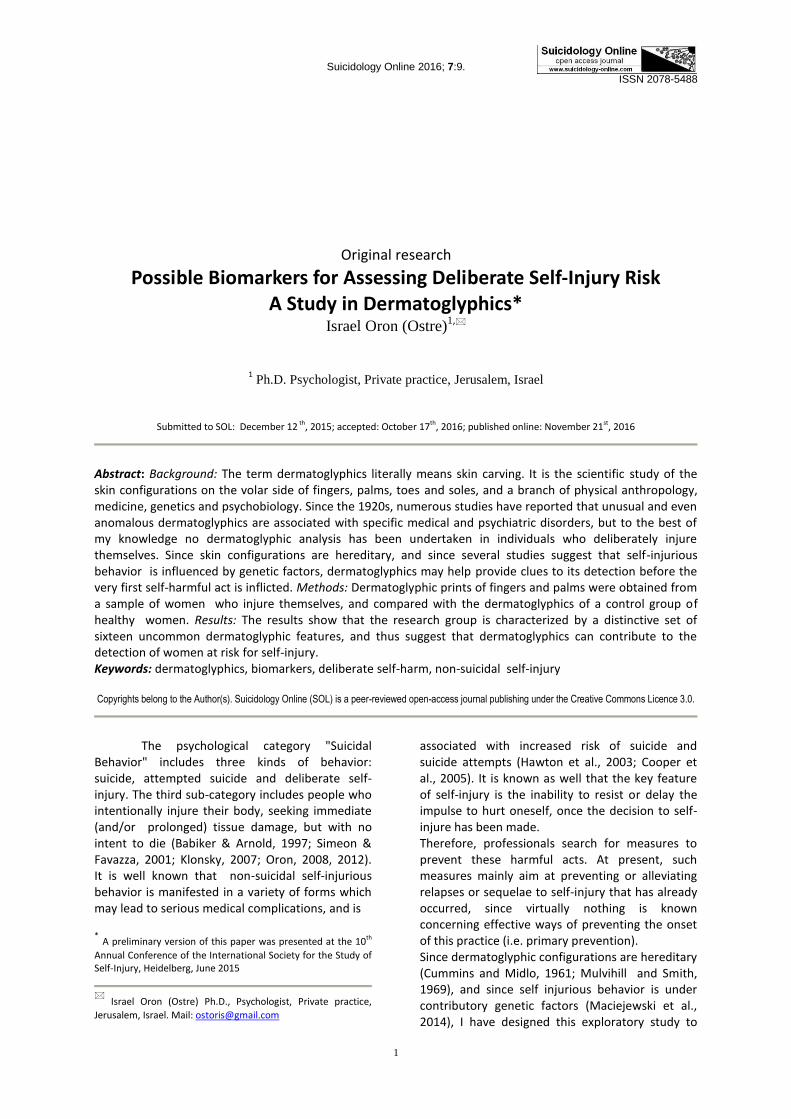
The human palm is presented in Figure 5 (Source: Cummins and Midlo, 1961, p. 88). Usually on the palm there are five deltas (triradii): four digital
deltas (marked: A, B, C and D) and an axial delta (marked by the letter:T). As was mentioned in regard to the finger patterns, a delta indicates a meeting point of three opposing
Suicidology Online 2016; 7:9.
ISSN 2078-5488
5
ridge systems that form a Y shape (the bold shape above each letter and beneath the letter T).
Figure 5 The human palm
On the palms, ten dermatoglyphic characteristics were observed which differentiate between the DSH participants and the healthy ones. (7) Unsuccessful delta C This anomalous condition takes two forms. In the first, delta C (Figure 5, embracing the fourth digit) is missing.The second is an aberrant form in which the delta is present, but its proximal radiant is not oriented toward the interior of the palm, as it is normally in all of the digital deltas (Figure 5, the bold lines beneath each letter), but instead stops running altogether. (8) Accessory digital delta An infrequent condition in which an extra delta is present near one or more of the digital deltas. (9) Distal axial delta Compared to its regular position (Figure 5, the letter T) here the axial delta is located in a distal position, at and beyond an imaginary line extending from the upper meeting point of the thumb with the palm, straight to the opposite side of the palm. (10-11) Interdigital configurations Ridges form configurations. A configuration is defined as a clear arrangement of ridges which separate it from the surrounding ridges (for an example see Figure 5, between the third and the fourth digits). (A pattern is a more definite form of configuration which is composed of sharply recurved ridges, e.g. a whorl).
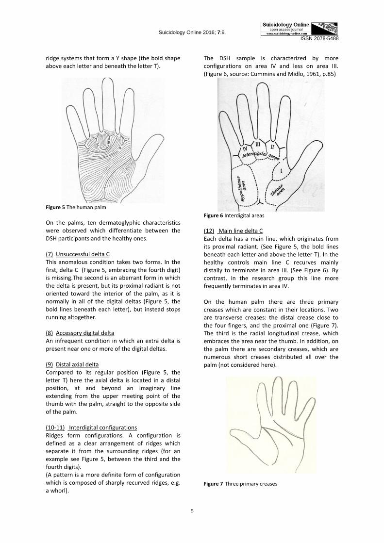
The DSH sample is characterized by more configurations on area IV and less on area III. (Figure 6, source: Cummins and Midlo, 1961, p.85)
Figure 6 Interdigital areas
(12) Main line delta C Each delta has a main line, which originates from its proximal radiant. (See Figure 5, the bold lines beneath each letter and above the letter T). In the healthy controls main line C recurves mainly distally to terminate in area III. (See Figure 6). By contrast, in the research group this line more frequently terminates in area IV. On the human palm there are three primary creases which are constant in their locations. Two are transverse creases: the distal crease close to the four fingers, and the proximal one (Figure 7). The third is the radial longitudinal crease, which embraces the area near the thumb. In addition, on the palm there are secondary creases, which are numerous short creases distributed all over the palm (not considered here).
Figure 7 Three primary creases
Suicidology Online 2016; 7:9.
ISSN 2078-5488
6
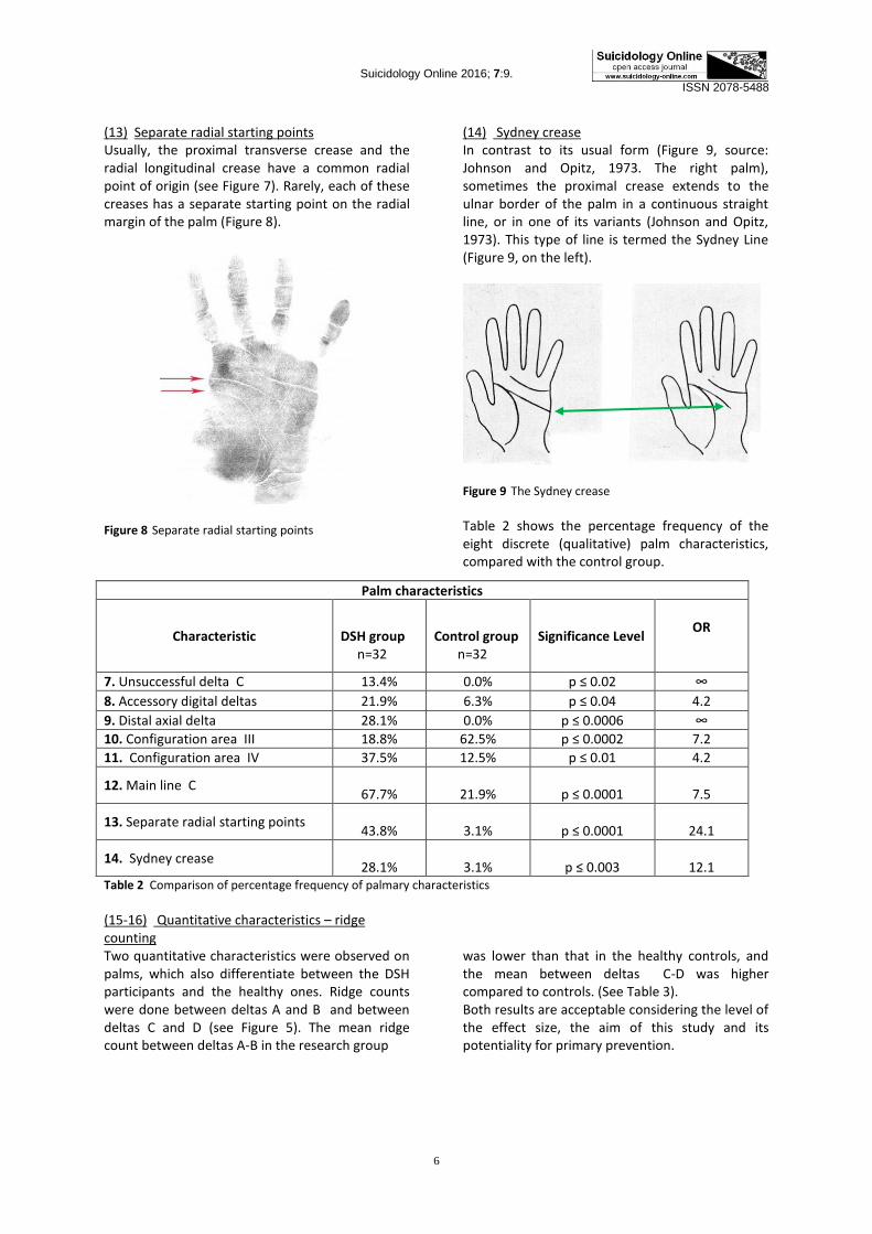
(13) Separate radial starting points Usually, the proximal transverse crease and the radial longitudinal crease have a common radial point of origin (see Figure 7). Rarely, each of these creases has a separate starting point on the radial margin of the palm (Figure 8).
Figure 8 Separate radial starting points
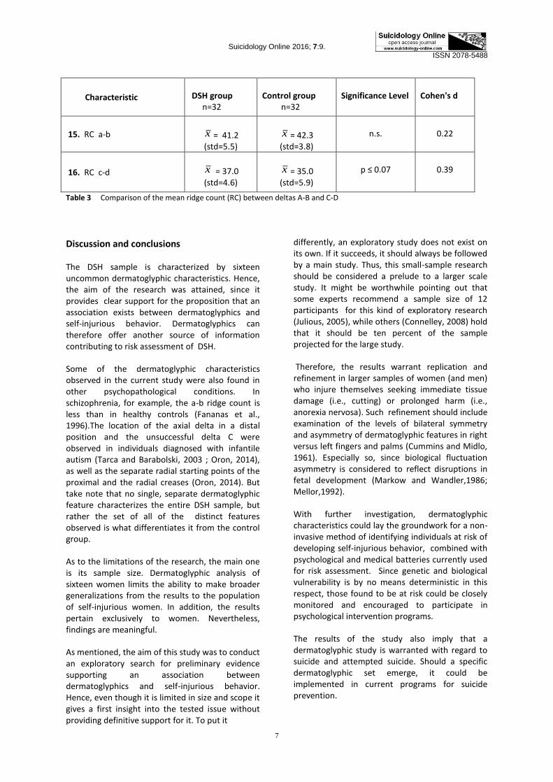
(14) Sydney crease In contrast to its usual form (Figure 9, source: Johnson and Opitz, 1973. The right palm), sometimes the proximal crease extends to the ulnar border of the palm in a continuous straight line, or in one of its variants (Johnson and Opitz, 1973). This type of line is termed the Sydney Line (Figure 9, on the left).
Figure 9 The Sydney crease
Table 2 shows the percentage frequency of the eight discrete (qualitative) palm characteristics, compared with the control group.
Palm characteristics
Characteristic DSH group n=32
Control group n=32
Significance Level
OR
7. Unsuccessful delta C 13.4% 0.0% p ≤ 0.02 ∞
8. Accessory digital deltas 21.9% 6.3% p ≤ 0.04 4.2
9. Distal axial delta 28.1% 0.0% p ≤ 0.0006 ∞
10. Configuration area III 18.8% 62.5% p ≤ 0.0002 7.2
11. Configuration area IV 37.5% 12.5% p ≤ 0.01 4.2
12. Main line C
67.7%
21.9%
p ≤ 0.0001
7.5
13. Separate radial starting points
43.8%
3.1%
p ≤ 0.0001
24.1
14. Sydney crease
28.1%
3.1%
p ≤ 0.003
12.1 Table 2 Comparison of percentage frequency of palmary characteristics
(15-16) Quantitative characteristics – ridge counting Two quantitative characteristics were observed on palms, which also differentiate between the DSH participants and the healthy ones. Ridge counts were done between deltas A and B and between deltas C and D (see Figure 5). The mean ridge count between deltas A-B in the research group
was lower than that in the healthy controls, and the mean between deltas C-D was higher compared to controls. (See Table 3). Both results are acceptable considering the level of the effect size, the aim of this study and its potentiality for primary prevention.
Suicidology Online 2016; 7:9.
ISSN 2078-5488
7
Characteristic
DSH group n=32
Control group n=32
Significance Level
Cohen's d
15. RC a-b
x = 41.2 (std=5.5)
x = 42.3
(std=3.8)
n.s.
0.22
16. RC c-d
x = 37.0 (std=4.6)
x = 35.0
(std=5.9)
p ≤ 0.07
0.39
Table 3 Comparison of the mean ridge count (RC) between deltas A-B and C-D
Discussion and conclusions
The DSH sample is characterized by sixteen uncommon dermatoglyphic characteristics. Hence, the aim of the research was attained, since it provides clear support for the proposition that an association exists between dermatoglyphics and self-injurious behavior. Dermatoglyphics can therefore offer another source of information contributing to risk assessment of DSH.
Some of the dermatoglyphic characteristics observed in the current study were also found in other psychopathological conditions. In schizophrenia, for example, the a-b ridge count is less than in healthy controls (Fananas et al., 1996).The location of the axial delta in a distal position and the unsuccessful delta C were observed in individuals diagnosed with infantile autism (Tarca and Barabolski, 2003 ; Oron, 2014), as well as the separate radial starting points of the proximal and the radial creases (Oron, 2014). But take note that no single, separate dermatoglyphic feature characterizes the entire DSH sample, but rather the set of all of the distinct features observed is what differentiates it from the control group.
As to the limitations of the research, the main one is its sample size. Dermatoglyphic analysis of sixteen women limits the ability to make broader generalizations from the results to the population of self-injurious women. In addition, the results pertain exclusively to women. Nevertheless, findings are meaningful.
As mentioned, the aim of this study was to conduct an exploratory search for preliminary evidence supporting an association between dermatoglyphics and self-injurious behavior. Hence, even though it is limited in size and scope it gives a first insight into the tested issue without providing definitive support for it. To put it
differently, an exploratory study does not exist on its own. If it succeeds, it should always be followed by a main study. Thus, this small-sample research should be considered a prelude to a larger scale study. It might be worthwhile pointing out that some experts recommend a sample size of 12 participants for this kind of exploratory research (Julious, 2005), while others (Connelley, 2008) hold that it should be ten percent of the sample projected for the large study.
Therefore, the results warrant replication and refinement in larger samples of women (and men) who injure themselves seeking immediate tissue damage (i.e., cutting) or prolonged harm (i.e., anorexia nervosa). Such refinement should include examination of the levels of bilateral symmetry and asymmetry of dermatoglyphic features in right versus left fingers and palms (Cummins and Midlo, 1961). Especially so, since biological fluctuation asymmetry is considered to reflect disruptions in fetal development (Markow and Wandler,1986; Mellor,1992).
With further investigation, dermatoglyphic characteristics could lay the groundwork for a non-invasive method of identifying individuals at risk of developing self-injurious behavior, combined with psychological and medical batteries currently used for risk assessment. Since genetic and biological vulnerability is by no means deterministic in this respect, those found to be at risk could be closely monitored and encouraged to participate in psychological intervention programs.
The results of the study also imply that a dermatoglyphic study is warranted with regard to suicide and attempted suicide. Should a specific dermatoglyphic set emerge, it could be implemented in current programs for suicide prevention.

Suicidology Online 2016; 7:9.
ISSN 2078-5488
8
Acknowledgements
I wish to express my sincere appreciation to all the participants for their cooperation.
References Babiker, G., & Arnold, L. (1997), The language of
injury: Comprehending self-mutilation, British Psychological Society. Ch.1
Bali, R.S. (1994), Anthropology of crease morphogenesis: A scientific analysis, Ashok Kumar Mittal Pub., New-Delhi. Ch. 9
Bhat, G.M., M. Mukhdoomi, A.M., Shah, B.A., & Ittoo, M.S. (2014), Dermatoglyphics: in health and disease - a review, International Journal of Research in Medical Sciences, 2(1), pp. 31-37
Connelley, L.M. (2008), Pilot studies, Journal of the Academy of Medical-Surgical Nurses, 17(6), pp. 411-412
Cooper, J., Kapur, N., Webb, R., Lawlor, M., Guthrie, E., Mackway-Jones, K., & Appleby, L. (2005), Suicide after deliberate self-harm: A 4-year cohort study, American Journal of Psychiatry, 162, pp. 297–303
Cummins, H., & Midlo, C. (1961), Finger prints, palms and soles: An introduction to dermatoglyphics, Dover Pub. Inc., New-York
Fananas, L., van Os J., Hoyos, C., McGrath, J., Mellor, C.S., & Murray R (1996), Dermatoglyphic a-b ridge count as a possible marker for developmental disturbance in schizophrenia: replication in two samples. Schizophrenia Research, 5, pp. 307-14
Federal Bureau of Investigation (1993), The science of fingerprints: Classification and uses, Diane Pub. Co. Ch. II
Floris, G., & Marini, E. (1998), The analysis of digital-palmar dermatoglyphics in a sample of individuals affected by essential hypertension. International Journal of Anthropology, 13(1), pp. 1-10
Galton, F. (1892), Finger prints, Macmillan & Co., London
Golembo-Smith, S., Walder, D.J., Daly, M.P., Mittal, V.A., Kline, E., Reeves, G., & Schiffman, J. (2012),The presentation of dermatoglyphic abnormalities in schizophrenia: a meta-analytic review, Schizophrenia Research;142(1-3), pp. 1-11
Hawton, K., Zahl, D., & Weatherall, R. (2003), Suicide following deliberate self-harm: long-term follow-up of patients who presented to a general hospital, The British Journal of Psychiatry, 182, pp. 537-542
Henry, E.R. (1900), Classification and uses of finger prints, George Routledge & Sons, London
Ivanenko, S.A., Bozhchenko, A.P., & Tolmachev, I.A. (2011), Dermatoglyphic features in suicidents: characteristic and implications for the solution of forensic medical problems, [Article in Russian], Sudebno-Meditsinskaia Ekspertiza, 54(5), pp. 26-29
Johnson, C.F. & Opitz, E. (1973), Unusual palm creases and unusual children, Clinical Pediatrics, 12(2), pp. 101-111
Julious, S.A. (2005), Sample size of 12 per group rule of thumb for a pilot study, Pharmaceutical Statistics, 4(4), pp. 287 - 291
Klonsky, E.D. (2007), The functions of deliberate self-injury: A review of the evidence, Clinical Psychology Review, 27, pp. 226–239
Maciejewski, D.F., Creemers, H.E., Lynskey, M.T., Madden, P.A., Heath, A.C., Stathan, D.J., Martin, N.G., & Verweij, K.J. (2014), Overlapping genetic and environmental influences on nonsuicidal self-injury and suicidal ideation: different outcomes, same etiology? Journal of the American Medical Association, 71(6), pp. 699-705
Markow, T. A., & Wandler, K. (1986). Fluctuating dermatoglyphic asymmetry and the genetics of liability to schizophrenia. Psychiatry Research, 19(4), pp. 323-328
Mellor, C. S. (1992). Dermatoglyphic evidence of fluctuating asymmetry in schizophrenia. British Journal of Psychiatry, 160, 467-472
Mulvihill, J.J., Smith DW (1969), The genesis of dermatoglyphics, Journal of Pediatrics, 75, pp. 579-589
Oron (Ostre), I. (2008), "Goodbye to the living! I preferred death" : Farewell letters written by Israelis who committed suicide, Ach Pub. [Hebrew]. Ch. 14
Oron (Ostre), I. (2012), How many people commit suicide after surviving a suicide attempt?, Psycho-Actuality, Journal of the Israel Psychological Association [Hebrew], October 2012 Issue, pp. 46-49
Suicidology Online 2016; 7:9.
ISSN 2078-5488
9
Oron (Ostre), I. (2014), Possible biomarkers for earlier detection of infantile autism: A study in dermatoglyphics, New Statistical Projects and Publications in Israel, 159(70), Central Bureau of Statistics, Israel
Purvis-Smith, S.G., & Menser, MA (1973), Dermatoglyphics in children with acute leukemia, British Medical Journal, 4, pp. 646-648
Rife, D.C. (1990), Dermatoglyphics as genetic marker. In: Durham, N.M., & Plato, C.C. Trends in dermatoglyphic research, Kluwer Academic Pub, pp. 10-15
Rosa, A., Fananas, L., Bracha, H.S., Torrey, E.F., & van Os, J. (2000), Congenital dermatoglyphic malformations and psychosis: A twin study. American Journal of Psychiatry, 157(9), pp. 1511-1513
Sabanciogullari , V., Ersan, E.E., Doğan, O., & Sabanciogullari, S. (2010), Dermatoglyphic characteristics in panic disorder, HealthMED Journal, 4(2), pp.366-372
Shrivastava, M., Mathur, R.K., Dhaneria, V., & Goyal, S. (2016), Dermatoglyphic study in bipolar disorder, Indian Journal of Clinical Anatomy and Physiology, 3(2), pp. 243-247
Simeon, D., & Favazza, A.R. (2001), Self-injurious behaviors: Phenomenology and assessment, in: Simeon, D. & Hollander, E. (Eds), Self-injurious behaviors: Assessment and treatment, American Psychiatric Publishing, Inc. Pp. 1-28
Stocker, J.T., & Dehmer, L.P. (2001), Pediatric Pathology, Lippincott Williams & Wilkins. Vol 1, 2nd edition, Ch. 4
Stosljevic, M, & Adamovic, M. (2013), Dermatoglyphic characteristics of digito-palmar complex in autistic boys in Serbia, Military-Medical and Pharmaceutical Review [Vojnosanit Pregl] , 70(4), pp. 386–390
Tarca, A., & Barabolski, S. (2003), Pathology of dermatoglyphics in infantile autism, The Journal of Preventive Medicine, 11 (1), 11-17
Vashist, M., Yadav, R., & Kumar, A. (2010), Axial triradius as a preliminary diagnostic tool in patients of mental retardation. The Internet Journal of Biological Anthropology, Vol. 4(1)
Wijerathne, B.T.B., Meier, R.J., Agampodi, T.C., & Agampodi, S.B. (2015), Dermatoglyphics in hypertension: a review, Journal of Physiological Anthropology, 34(1), 29
Ziegler, A.G., Mathies, R., Ziegelmayer, G., Baumgartl, H.J., Rodewald, A., Chopra, V., & Standl, E. (1993), Dermatoglyphics in type 1 diabetes mellitus, Diabetic Medicine, 10(8), pp. 720-724
Zoroastrov, O.M., Chistikina, T.A., Zoroastrov, M.O., & Bevza, A.L. (2009), Forensic-medical aspects of the examination of dermatoglyphic patterns as markers of predisposition to drug addiction and related suicides, [Article in Russian], Sudebno-Meditsinskaia Ekspertiza, 52(4), pp. 41-43