Positive transcriptional regulation of an iron-regulated virulence gene in Vibrio cholerae
5
Proc. Nail. Acad. Sci. USA Vol. 88, pp. 1125-1129, February 1991 Medical Sciences Positive transcriptional regulation of an iron-regulated virulence gene in Vibrio cholerae (transcriptional activation/LysR family/Fur/IrgA/IrgB) MARCIA B. GOLDBERG*, STEPHANIE A. BOYKO*, AND STEPHEN B. CALDERWOOD*t* *Infectious Disease Unit, Massachusetts General Hospital, Boston, MA 02114; and tDepartment of Microbiology and Molecular Genetics, Harvard Medical School, Boston, MA 02215 Communicated by A. M. Pappenheimer, Jr., November 19, 1990 ABSTRACT We have previously described a virulence gene in Vibrio cholerae (irgA) that is more than 850-fold regulated in response to iron. Negative regulation of irgA by iron occurred at the transcriptional level, and there was a dyad symmetric nucleotide sequence in the vicinity of the uAg promoter homologous to the Fur binding site in Escherchia coUl. When irgA was cloned into E. coi, we showed that transcription of irgA required 900 base pairs of DNA upstream of the irgA promoter that contained an open reading frame in inverse orientation to irgA. In the present study, we show that this upstream region of DNA encodes a gene in inverse orien- tation to irgA (named irgB) that is also negatively regulated by iron. Insertional inactivation of irgB on the V. cholrae chro- mosome leads to loss of expression of a chromosomal irgA'- 'phoA fusion (in which the primes indicate truncated genes), which is restored to normal by provision of irgB on a plasmid in trans. DNA sequencing of irgB shows that the protein product (IrgB) is homologous to the LysR family of positive transcriptional activators, and secondary structure analysis of IrgB predicts a helix-turn-helix DNA binding motif. The pro- moters of irgB and irgA are divergent but overlap each other and the previously defined Fur-binding site. We propose a model for iron regulation of irgA expression in V. choerae. In the presence of sufficient iron, transcription of both irgA and irgB is negatively regulated by a Fur-like protein. In low iron conditions, negative regulation of transcription is removed, and production of IrgB leads to positive transcriptional acti- vation of irgA. It seems likely that the high induction ratio of irgA expression under low- and high-iron conditions (850-fold) relates to the fact that its cognate positive transcriptional activator (irgB) is itself negatively regulated by iron. Vibrio cholerae infection in humans may cause a severe dehydrating diarrhea. Illness occurs following ingestion of the organism in contaminated fresh or salt water. The bac- teria must pass through the acid barrier of the stomach to reach the small intestine, where they penetrate the mucus gel and adhere to the brush border of intestinal epithelial cells by specific adhesins, including toxin coregulated pilus (TcpA) and other accessory colonization factors. At the intestinal brush border, V. cholerae produces a number of virulence factors, including cholera toxin, neuraminidase, hemolysin, and other extracellular secreted proteins. The genetic regu- lation of cholera toxin and TcpA in response to environmen- tal stimuli has been described (1, 2). The genetic regulation of other virulence determinants in response to environmental signals is less well understood. Many bacterial virulence determinants are regulated by the environmental concentration of free iron, with increased expression occurring under low-iron conditions. Examples of iron-regulated virulence determinants include diphtheria toxin of Corynebacterium diphtheriae (3), Shiga toxin of Shigella dysenteriae 1 (4), Shiga-like toxin I of enterohem- orrhagic Escherichia coli (5), exotoxin A of Pseudomonas aeruginosa (6, 7), and several outer membrane proteins of Vibrio anguillarum (8) and Yersinia species (9). We have described an iron-regulated virulence determi- nant in V. cholerae (10). Strain MBG40 of V. cholerae, which contains a TnphoA insertion mutation in the iron-regulated gene irgA, has reduced virulence in a newborn mouse model and has lost the 77-kDa major iron-regulated outer membrane protein (10). The deduced amino acid sequence of the amino terminus of IrgA is homologous to FepA, the ferrienteroche- lin outer membrane receptor of E. coli, suggesting that IrgA could be the iron-vibriobactin outer membrane receptor of V. cholerae (11). Expression of the irgA'-'phoA gene fusion (in which the primes indicate truncated genes), as measured by alkaline phosphatase activity, increases 850-fold in low- versus high-iron media (10). Negative regulation of irgA by iron occurs at the transcriptional level, and its promoter region, defined by primer extension, contains a dyad sym- metric sequence homologous to dyad elements in E. coli that bind the Fur protein, an iron-responsive repressor of tran- scription (11). When the irgA'-'phoA gene fusion was cloned from V. cholerae onto a plasmid in E. coli, expression of alkaline phosphatase activity and transcription of the gene fusion required an additional 900 base pairs (bp) of DNA upstream of the irgA promoter (11); this region of DNA, which we call irgB,§ contains an open reading frame in inverse orientation to irgA (Fig. 1). We wished to test the model in which this open reading frame encodes a positive activator for irgA transcription. MATERIALS AND METHODS Bacterial Strains. Bacterial strains and plasmids used in this study are shown in Table 1. V. cholerae strain MBG260 and plasmids pSBC45 and pMBG111 were constructed as de- scribed below. Media. Luria-Bertoni (LB) medium with or without the addition of the iron chelator 2,2-dipyridyl (final concentra- tion, 0.2 mM) was used to evaluate the effect of iron con- centration on gene expression as described (10). Construction of plasmids. Strain MBG40 contains a chro- mosomal gene fusion between irgA and phoA, constructed by TnphoA mutagenesis (10). Plasmid pMBG59 contains a subclone of the intact irgA::TnphoA gene fusion from MBG40 into plasmid pBR322. Plasmid pMBG59 also con- tains all of the open reading frame upstream of irgA (subse- quently named irgB) (see Results) (Fig. 1). tTo whom reprint requests should be addressed. §The sequence reported in this paper has been deposited in the GenBank data base (accession no. M55988). 1125 The publication costs of this article were defrayed in part by page charge payment. This article must therefore be hereby marked "advertisement" in accordance with 18 U.S.C. §1734 solely to indicate this fact.
Transcript of Positive transcriptional regulation of an iron-regulated virulence gene in Vibrio cholerae
Proc. Nail. Acad. Sci. USAVol. 88, pp. 1125-1129, February 1991Medical Sciences
Positive transcriptional regulation of an iron-regulated virulencegene in Vibrio cholerae
(transcriptional activation/LysR family/Fur/IrgA/IrgB)
MARCIA B. GOLDBERG*, STEPHANIE A. BOYKO*, AND STEPHEN B. CALDERWOOD*t**Infectious Disease Unit, Massachusetts General Hospital, Boston, MA 02114; and tDepartment of Microbiology and Molecular Genetics, Harvard MedicalSchool, Boston, MA 02215
Communicated by A. M. Pappenheimer, Jr., November 19, 1990
ABSTRACT We have previously described a virulencegene in Vibrio cholerae (irgA) that is more than 850-foldregulated in response to iron. Negative regulation of irgA byiron occurred at the transcriptional level, and there was a dyadsymmetric nucleotide sequence in the vicinity of the uAgpromoter homologous to the Fur binding site in EscherchiacoUl. When irgA was cloned into E. coi, we showed thattranscription of irgA required 900 base pairs of DNA upstreamof the irgA promoter that contained an open reading frame ininverse orientation to irgA. In the present study, we show thatthis upstream region of DNA encodes a gene in inverse orien-tation to irgA (named irgB) that is also negatively regulated byiron. Insertional inactivation of irgB on the V. cholrae chro-mosome leads to loss of expression of a chromosomal irgA'-'phoA fusion (in which the primes indicate truncated genes),which is restored to normal by provision of irgB on a plasmidin trans. DNA sequencing of irgB shows that the proteinproduct (IrgB) is homologous to the LysR family of positivetranscriptional activators, and secondary structure analysis ofIrgB predicts a helix-turn-helix DNA binding motif. The pro-moters of irgB and irgA are divergent but overlap each otherand the previously defined Fur-binding site. We propose amodel for iron regulation of irgA expression in V. choerae. Inthe presence of sufficient iron, transcription of both irgA andirgB is negatively regulated by a Fur-like protein. In low ironconditions, negative regulation of transcription is removed,and production of IrgB leads to positive transcriptional acti-vation of irgA. It seems likely that the high induction ratio ofirgA expression under low- and high-iron conditions (850-fold)relates to the fact that its cognate positive transcriptionalactivator (irgB) is itself negatively regulated by iron.
Vibrio cholerae infection in humans may cause a severedehydrating diarrhea. Illness occurs following ingestion ofthe organism in contaminated fresh or salt water. The bac-teria must pass through the acid barrier of the stomach toreach the small intestine, where they penetrate the mucus geland adhere to the brush border of intestinal epithelial cells byspecific adhesins, including toxin coregulated pilus (TcpA)and other accessory colonization factors. At the intestinalbrush border, V. cholerae produces a number of virulencefactors, including cholera toxin, neuraminidase, hemolysin,and other extracellular secreted proteins. The genetic regu-lation of cholera toxin and TcpA in response to environmen-tal stimuli has been described (1, 2). The genetic regulation ofother virulence determinants in response to environmentalsignals is less well understood.Many bacterial virulence determinants are regulated by the
environmental concentration of free iron, with increasedexpression occurring under low-iron conditions. Examples ofiron-regulated virulence determinants include diphtheria
toxin of Corynebacterium diphtheriae (3), Shiga toxin ofShigella dysenteriae 1 (4), Shiga-like toxin I of enterohem-orrhagic Escherichia coli (5), exotoxin A of Pseudomonasaeruginosa (6, 7), and several outer membrane proteins ofVibrio anguillarum (8) and Yersinia species (9).We have described an iron-regulated virulence determi-
nant in V. cholerae (10). Strain MBG40 of V. cholerae, whichcontains a TnphoA insertion mutation in the iron-regulatedgene irgA, has reduced virulence in a newborn mouse modeland has lost the 77-kDa major iron-regulated outer membraneprotein (10). The deduced amino acid sequence of the aminoterminus of IrgA is homologous to FepA, the ferrienteroche-lin outer membrane receptor of E. coli, suggesting that IrgAcould be the iron-vibriobactin outer membrane receptor of V.cholerae (11). Expression of the irgA'-'phoA gene fusion (inwhich the primes indicate truncated genes), as measured byalkaline phosphatase activity, increases 850-fold in low-versus high-iron media (10). Negative regulation of irgA byiron occurs at the transcriptional level, and its promoterregion, defined by primer extension, contains a dyad sym-metric sequence homologous to dyad elements in E. coli thatbind the Fur protein, an iron-responsive repressor of tran-scription (11).When the irgA'-'phoA gene fusion was cloned from V.
cholerae onto a plasmid in E. coli, expression of alkalinephosphatase activity and transcription of the gene fusionrequired an additional 900 base pairs (bp) ofDNA upstreamof the irgA promoter (11); this region ofDNA, which we callirgB,§ contains an open reading frame in inverse orientationto irgA (Fig. 1). We wished to test the model in which thisopen reading frame encodes a positive activator for irgAtranscription.
MATERIALS AND METHODSBacterial Strains. Bacterial strains and plasmids used in this
study are shown in Table 1. V. cholerae strain MBG260 andplasmids pSBC45 and pMBG111 were constructed as de-scribed below.Media. Luria-Bertoni (LB) medium with or without the
addition of the iron chelator 2,2-dipyridyl (final concentra-tion, 0.2 mM) was used to evaluate the effect of iron con-centration on gene expression as described (10).
Construction of plasmids. Strain MBG40 contains a chro-mosomal gene fusion between irgA and phoA, constructedby TnphoA mutagenesis (10). Plasmid pMBG59 contains asubclone of the intact irgA::TnphoA gene fusion fromMBG40 into plasmid pBR322. Plasmid pMBG59 also con-tains all of the open reading frame upstream of irgA (subse-quently named irgB) (see Results) (Fig. 1).
tTo whom reprint requests should be addressed.§The sequence reported in this paper has been deposited in theGenBank data base (accession no. M55988).
1125
The publication costs of this article were defrayed in part by page chargepayment. This article must therefore be hereby marked "advertisement"in accordance with 18 U.S.C. §1734 solely to indicate this fact.

1126 Medical Sciences: Goldberg et al.
500 bp 0
Hind III
,, Bg/I[NruI CIal HincII SmaI
pMBG59
pMBG111
pSBC45
t- ' I' E \\\\\,1 ~~~~~~~~~phoA
'SirgB
I1 ~~~~~~~~~irgA_
I ~ ~~~~~~~~~~~~
l-
-I I.
Plasmids pSBC45 and pSBC46 are derivatives of pA-CYC184, a plasmid encoding chloramphenicol resistance.Plasmids pSBC45 and pSBC46 were constructed by isolatingthe Nru I-Sma I fragment of plasmid pMBG59 by electro-elution from a gel and ligation into Nru I-digested pA-CYC184; the two plasmids differ only in the orientation of theinserted fragment, which contains the intact gene irgB (Fig.1; see Results).Plasmid pMBG111 was derived from pGP704, a broad-
host-range plasmid containing the ampicillin-resistance genefrom pBR322, the mobilization domain of plasmid RP4 (14),the origin of replication from plasmid R6K (15), and apolylinker from phage M13 tgl31 (Amersham). PlasmidpGP704 was a gift of Gregory D. N. Pearson and is itselfderived from plasmid pJM703.1 (13). Plasmid pGP704 and itsderivatives are able to replicate only in strains containing thepir gene, which encodes the 7r protein necessary for thefunction of the R6K origin (15). To construct pMBG111, a676-bp HincII-Bgl II fragment of pMBG59 internal to irgB(Fig. 1) was ligated into the EcoRV and Bgl II sites of thepGP704 polylinker.
Genetic Methods. V. cholerae strain MBG260, which con-tains an insertion mutation in irgB, was constructed fromstrain MBG40 in the following manner. Plasmid pMBG111was transferred from strain SY327 A pir into SM10 A pir bytransformation. SM10 A pir contains a chromosomally-integrated RP4-2 (Tc::Mu), which encodes trans-acting fac-
FIG. 1. Partial restriction map of thechromosomal DNA upstream of theTnphoA fusion junction in V. choleraestrain MBG40, subcloned as described(11) to make plasmid pMBG59. Loca-tions of relevant restriction enzyme sitesare indicated, as well as fragments usedto construct plasmids pMBG111 andpSBC45. The solid bar representsMBG40 chromosomal DNA. The diago-nally cross-hatched bar representsTnphoA DNA. The stippled bar indicatesthe position and direction oftranscriptionof irgA upstream of the fusion withphoA. The striped bar indicates the po-sition and direction of transcription ofirgB.
tors necessary to mobilize pGP704 derivatives into a broadrange of recipients without RP4 itself being transferred (13).SM10 A pir containing pMBG111 was conjugated withMBG40, with double selection for ampicillin resistance (en-coded by pMBG111) and streptomycin resistance (encodedby MBG40). Because MBG40 does not contain the pir gene,pMBG111 is unable to replicate in this strain, so that doublyresistant colonies arise by homologous recombination be-tween the internal fragment of irgB on pMBG111 and thecorresponding chromosomal gene on the recipient, causinginsertional inactivation ofirgB. To confirm that chromosomalintegration occurred within irgB on the recipient, we per-formed Southern hybridization ofchromosomal DNA digestswith EcoRV, an enzyme that does not cut within either irgBor pMBG111. The blot was probed with the HincII-Bgl IIfragment of irgB, radioactively labeled by random primerextension with a commercial kit (Prime Time, InternationalBiotechnologies).Plasmids were transformed into E. coli strains by standard
techniques (16). Plasmids pACYC184 and pSBC45 wereintroduced into V. cholerae strains by electroporation byusing the protocol of the manufacturer (Gene Pulser, Bio-Rad), with the exception of substitution of 2 mM CaCl2 as thebuffer for resuspending cells during preparation, rather thanwater or Hepes buffer. Plasmid content of the electroporantswas confirmed by restriction enzyme digestion of plasmid
Table 1. Bacterial strains and plasmids used in this studyStrain or plasmid Relevant genotype or phenotype Ref. or source
V. cholerae strains0395 Smr 27MBG40 irgA::TnphoA, Smr Kmr 10MBG260 irgA::TnphoA irgB::pMBG111, This study
E. coli strainsCC118 A(ara leu)7697 A(lac)X74 araD139 phoAA20 galE galK thi 12
rpsE rpoB argE(Am) recAlSY327 A pir A(lac pro) nalA recA56 araD argE(Am) A pirR6K 13SM10 A pir thi thr leu tonA lac Y supE recA::RP4-2-Tc::Mu A pirR6K, Kmr 13
PlasmidspMBG59 pBR322 with 4.6-kb irgB irgA::TnphoA insert from MBG40 11pJM703.1 oriR6K mobRP4, Apr 13pGP704 pJM703.1 with 1.5-kb Sph I-Pvu II deletion and 75-bp insert G. D. N. Pearson
of M13 tgl31 polylinkerpMBG111 pGP704 with 676-bp HincII-Bgl II insert of pMBG59 This studypSBC45 pACYC184 with 1.6-kb Nru I-Sma I insert of pMBG59 This studyApr, ampicillin resistance; Kmr, kanamycin resistance; Smr, streptomycin resistance.
Proc. Natl. Acad Sci. USA 88 (1991)
Proc. Natl. Acad. Sci. USA 88 (1991) 1127
minipreps, analyzed by agarose gel electrophoresis as de-scribed (11).
Assays. The enzymatic activity of alkaline phosphataseencoded on TnphoA permitted the comparison offusion geneexpression when strains were grown in low- versus high-ironmedia. Strains were grown overnight in LB medium with or
without added 2,2-dipyridyl. Alkaline phosphatase activitywas determined as described (10).DNA and RNA Analysis. Analysis of DNA and RNA,
including DNA and RNA preparation, restriction mapping,DNA sequencing, RNA (Northern) blot analysis, and primerextension were performed as described (11). For Northernblot analysis, an equivalent quantity of RNA, as calculatedfrom OD260, was loaded into each lane.
Synthetic oligonucleotides used as probes for Northernblot analysis and as primers for DNA sequencing and primerextension were the gift of Brian Seed (Massachusetts GeneralHospital).
Protein Analysis and Protein Data Base Searches. Proteinanalysis and protein data base searches were performed byusing IBI-Pustell sequence analysis software (InternationalBiotechnologies). The hydropathicity index profile of IrgBwas calculated by the formula of Kyte-Doolittle (17). Theprotein secondary structure prediction ofIrgB was calculatedby the algorithm of Chou-Fasman (18). Data base searchesand protein alignments were performed by searching theProtein Identification Resource, National Biomedical Re-search Foundation data base (release 19) using the FASTPalgorithm for protein homology (19).
RESULTS
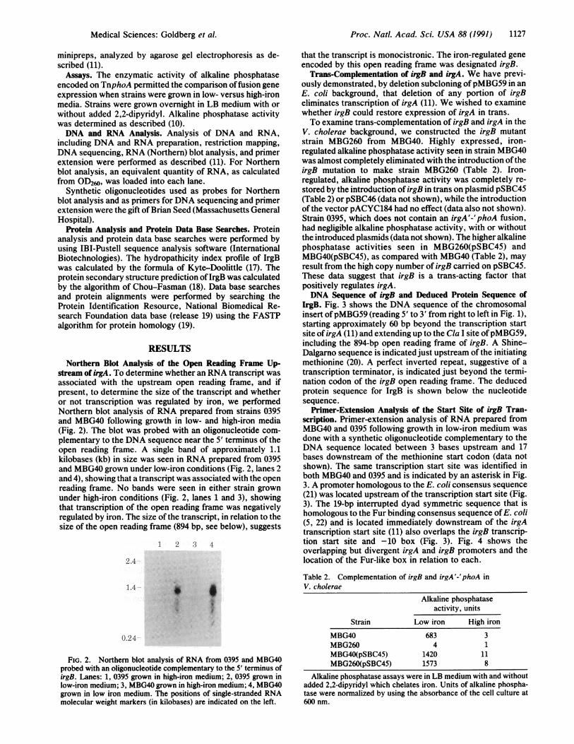
Northern Blot Analysis of the Open Reading Frame Up-stream of irgA. To determine whether an RNA transcript wasassociated with the upstream open reading frame, and ifpresent, to determine the size of the transcript and whetheror not transcription was regulated by iron, we performedNorthern blot analysis of RNA prepared from strains 0395and MBG40 following growth in low- and high-iron media(Fig. 2). The blot was probed with an oligonucleotide com-
plementary to the DNA sequence near the 5' terminus of theopen reading frame. A single band of approximately 1.1kilobases (kb) in size was seen in RNA prepared from 0395and MBG40 grown under low-iron conditions (Fig. 2, lanes 2and 4), showing that a transcript was associated with the openreading frame. No bands were seen in either strain grownunder high-iron conditions (Fig. 2, lanes 1 and 3), showingthat transcription of the open reading frame was negativelyregulated by iron. The size of the transcript, in relation to thesize of the open reading frame (894 bp, see below), suggests
1 2 3 4
2.4
1.4
I
0.24
FIG. 2. Northern blot analysis of RNA from 0395 and MBG40probed with an oligonucleotide complementary to the 5' terminus ofirgB. Lanes: 1, 0395 grown in high-iron medium; 2, 0395 grown inlow-iron medium; 3, MBG40 grown in high-iron medium; 4, MBG40grown in low iron medium. The positions of single-stranded RNAmolecular weight markers (in kilobases) are indicated on the left.
that the transcript is monocistronic. The iron-regulated geneencoded by this open reading frame was designated irgB.Trans-Complementation of irgB and irgA. We have previ-
ously demonstrated, by deletion subcloning ofpMBG59 in anE. coli background, that deletion of any portion of irgBeliminates transcription of irgA (11). We wished to examinewhether irgB could restore expression of irgA in trans.To examine trans-complementation of irgB and irgA in the
V. cholerae background, we constructed the irgB mutantstrain MBG260 from MBG40. Highly expressed, iron-regulated alkaline phosphatase activity seen in strain MBG40was almost completely eliminated with the introduction oftheirgB mutation to make strain MBG260 (Table 2). Iron-regulated, alkaline phosphatase activity was completely re-
stored by the introduction of irgB in trans on plasmid pSBC45(Table 2) or pSBC46 (data not shown), while the introductionof the vector pACYC184 had no effect (data also not shown).Strain 0395, which does not contain an irgA'-'phoA fusion,had negligible alkaline phosphatase activity, with or withoutthe introduced plasmids (data not shown). The higher alkalinephosphatase activities seen in MBG260(pSBC45) andMBG40(pSBC45), as compared with MBG40 (Table 2), mayresult from the high copy number of irgB carried on pSBC45.These data suggest that irgB is a trans-acting factor thatpositively regulates irgA.DNA Sequence of irgB and Deduced Protein Sequence of
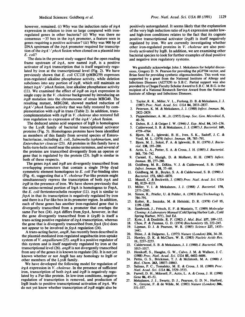
IrgB. Fig. 3 shows the DNA sequence of the chromosomalinsert ofpMBG59 (reading 5' to 3' from right to left in Fig. 1),starting approximately 60 bp beyond the transcription startsite of irgA (11) and extending up to the Cla I site ofpMBG59,including the 894-bp open reading frame of irgB. A Shine-Dalgarno sequence is indicated just upstream of the initiatingmethionine (20). A perfect inverted repeat, suggestive of a
transcription terminator, is indicated just beyond the termi-nation codon of the irgB open reading frame. The deducedprotein sequence for IrgB is shown below the nucleotidesequence.
Primer-Extension Analysis of the Start Site of irgB Tran-scription. Primer-extension analysis of RNA prepared fromMBG40 and 0395 following growth in low-iron medium was
done with a synthetic oligonucleotide complementary to theDNA sequence located between 3 bases upstream and 17bases downstream of the methionine start codon (data notshown). The same transcription start site was identified inboth MBG40 and 0395 and is indicated by an asterisk in Fig.3. A promoter homologous to the E. coli consensus sequence(21) was located upstream of the transcription start site (Fig.3). The 19-bp interrupted dyad symmetric sequence that ishomologous to the Fur binding consensus sequence ofE. coli(5, 22) and is located immediately downstream of the irgAtranscription start site (11) also overlaps the irgB transcrip-tion start site and -10 box (Fig. 3). Fig. 4 shows theoverlapping but divergent irgA and irgB promoters and thelocation of the Fur-like box in relation to each.
Table 2. Complementation of irgB and irgA'-'phoA inV. cholerae
Alkaline phosphataseactivity, units
Strain Low iron High iron
MBG40 683 3MBG260 4 1MBG40(pSBC45) 1420 11MBG260(pSBC45) 1573 8
Alkaline phosphatase assays were in LB medium with and withoutadded 2,2-dipyridyl which chelates iron. Units of alkaline phospha-tase were normalized by using the absorbance of the cell culture at600 nm.
Medical Sciences: Goldberg et al.

1128 Medical Sciences: Goldberg et al.
10 20 30 40 50 * 60 70 80
-35-10
90 100 110 120 130 140 150 160
SD
170 =I 190 Hin=I 200 210 220ATMC M CAA GAT CmC AGC CTC GTA AAA OCT TIC CAT 00 cCT M OAT AM MC
Mat Gln Asp la Ser Ala Val Lys Ala n. His Ala Lau Cys Gln His Lys Ser
230 240 250 260 270 280CmG ACT GC OCr GOC AMA GOG CrT GMA CAG Ocr AMA TCC AGO CMAGr 0CC GOT TM G00Lou Thr Ala Ala Ala Iys Ala Izu Glu Gln Pro Iys Ser Thr Iu Ser Arg Arg Lr Ala
290 300 Hi 320 330 340CMA CIT GM GAM GMT OGA CM MC Tm TG AM CM GM 0CC TM AM cMGln lu Glu Glu Asp ILu Gly Gln Ser Iau Lou MNt Arg Gln Gly Ann Arq TAUhr TI
350 360 370 380 390 __L 400ACG AM GCA Gh A GM TGm GCGG0TT TM TOG GAG CM CrA CT G cm GCC AM AAAThr Lys Ala Gly Glu Val The Ala Val Tyr Ser Glu Gln lou Ieu Glu Imu Ala Asn Lys
410 420 430 440 450 460AMr CAG GMA 0CG TTG CMA GMA TmG AAC AAT CMA GTG AC GOC GA CMC AG Cm GTM GTASer Gln Glu Ala Lau Gln Glu Iu Amn AMn Gln Val Thr Gly Glu Lou Thr Leu Val Val
470 480 490 500 510 520CMC CaC MT TTG ATC 0CC 0GC TOG CC AC CM GMT TG GAT GAG mATThGaM M CATHis Pro Asn Lu Ile Arg Gly Trp Lou Ser Gln Val Leu Asp Glu The Met Gln Gln His
530 AwI 550 560 570 580TMG ACA TM AAG AMCOT CIA CC AMC CG TmT CMA CAC AM GAT GAG GTG TT GAG COCSer Tbr Lau LIs Ile Arg LIeu Lu Ser Gln The Gln His Ser Asp Glu Val RTe Glu Pro
590 600 610 NoLI 630 640GAT TTG ATC A 1rT& ATT GAA CAC GOC0CC O AMG GOT TAT 0OC AAM GMA CC TTA G0CAsp Lou Ile Ile Trp Ile Glu His Ala Ala Pro Mat Gly Tyr Arg IYs Glu Arg Ieu Gly
650 660 670 ..I 690 700TAT TOG 0CC TAT GOC AM TAC C MrT CC AMA TAT ITG GOC CAT GA GOAT AAA COG ACTyr Trp Arg Tyr Ala Thr Tyr Ala Ser Pro Lys Tyr Ieu Ala His Arg Asp Ly's Pro Tr
710 720 NOD. 740 750 760CAT CO COT GAG CTG ATI CAT CAC CA TMG AT GATrm ATrGMT TlOT OCG 0CC 0 GAGHis Pro Arg Glu Leu Ile His His Pro Tsp Ile Asp ghe Ile Ala Cy Arq Arg Ala Glu
770 780 790 800 810 820CIT GMA C CMGCAT OCA GAG TTC GC MAT TAT TCA Cm CCR GCA CIA GAG AM CT TTALea Glu Ta_ His His Pro Glu RP Gly Ser Tyr Ser Leu Pro Ala I Glu Ser Arg L=
830 840 850 860 870 880CA AMC GAO AAT Cll GCC AMG CAA GCC GAT G0G MT OCT AM GOTGO GOT AMT GOT TMGin Ser Asp Amn le Ala Met Gln Ala Asp Ala Ile Ala Iys Gly Arg Gly Ile Gly LIa
890 900 910 920 930 940Crr 0CO AC l&G TT GOC AAT GOT Tm GAA AMG G0G CT CCG 0CC MC CV AM GOT I&CLeu pro Thr Trp The Ala Asn Gly 1hu Glu lTr Ala His Pro Gly Ser LeuIle Pro Cys
HincI 950 960 970 980 990 1000GTC AAC GGA TOG CMA TCA CAG CA ACA GAA ATC AAC TC TmC TAT CCG CIc GOT Gr CMVal Asn Gly Trp Gln Ser Gln Pro Thr Glu Ile Asn Cys The [yr Pro IBU Gly Arg His
1010 1020 1030 1040 1050 1060CCGA CIT CG crGm 0c CIA TT MT OAT GOG 0CGC CMAOGA AGG OC AT GMA lG CAPro LeuAArg Lou Arg loa Phe Ile Asp Ala Loa Arg Gln Ala Arg Pro Asp Glu Trp Gln
1070 1080 1090 ClaITAA
FIG. 3. Nucleotide sequence of the chromosomal DNA inpMBG59 (reading 5' to 3' from right to left in Fig. 1), startingdownstream of the start site of irgA transcription and extending upto the Cla I restriction site in pMBG59. The locations of restrictionenzyme sites are noted. The deduced amino acid sequence of IrgB isshown in three-letter code. The approximate start site of transcrip-tion (*), the -10 and -35 boxes of the promoter, and the putativeShine-Dalgarno sequence (SD) are indicated. A 19-bp interrupteddyad symmetric element homologous to the Fur box of E. coli isindicated by heavy horizontal arrows below the sequence in thevicinity of the promoter. The termination codon of IrgB is indicated(.. .), followed by a probable transcription terminator (light hori-zontal arrows at end of sequence).
IrgB Protein Analysis. The predicted amino acid sequenceof IrgB is shown below the nucleotide sequence in Fig. 3.
Proc. Natl. Acad. Sci. USA 88 (1991)-35 -10 *
5' GGG GGA TGG ATT GAA TCT GGA CAT TTACCACTCCTTTAAA ATAATTATTCTTAATTTC A3' CCC CCT ACC TAA CTT AGA CCT GTA AATGGTGAGGAAATTT TAT
Pro Ser Pro Asn Phe Arg Ser Met SD *-4---IrgA
GAAGCGGATTATTCATATATTCTGTACTGGAGATAGATCGACACATGGAATAGTTCGGTCCAAATATGGCTTCGCCTAATAAGTATATAAGACATGACCTCTATCTAGCTGTGTACCTTATCAAGCCAGGTTTATACC
-10
IrgB _-SD Met Gln Asp Leu Ser Ala Val
AACGTCTGTGGTAATTATTCTTTAAGGGTCAAATACCT ATG CAA GAT CTC AGC GCC GTA 3'TTGCAGACACCATTAATAAGAAATTCCCAGTTTATGGA TAC GTT CTA GAG TCG CGG CAT 5'
FIG. 4. Detail of the overlapping, divergent promoters of irgAand irgB. The promoter of irgB (-35, -10), start site of transcription(*), Shine-Dalgarno sequence (SD), and open reading frame areindicated on the upper strand, while the corresponding features ofirgA are noted on the bottom strand. Note that irgA does not havea consensus -35 box. The dyad symmetric element homologous tothe E. coli Fur-binding site is enclosed within a box.
Hydropathicity profile. The hydropathicity profile of IrgBshowed no stretches of hydrophobic residues that would besuggestive of either a signal sequence or a transmembranedomain (data not shown).Homology of IrgB to the LysRfamily ofpositive transcrip-
tional activators. Comparison of IrgB to the Protein Identi-fication Resource, National Biomedical Research Founda-tion (release 19) data base demonstrated significant homologybetween IrgB and the LysR family of positive transcriptionalactivator proteins in bacteria (23). The best match in thisfamily was to the E. coli positive activator protein IlvY; theoptimized score between IrgB and IlvY was 217. The regionsof highest homology between IrgB and the LysR family werenear the amino terminus and were at roughly the same
positions in each protein. The homology near the aminoterminus of IrgB with several members of the LysR family isshown in Fig. 5. All of the proteins shown are of approxi-mately the same size (IrgB is 298 amino acids long; IlvY,LysR, AmpR, NodD, and CysB are 297, 300, 291, 300, and301 amino acids long, respectively).
Prediction of the secondary structure of IrgB. The sec-
ondary structure of IrgB was predicted by using the Chou-Fasman algorithm (18). A helix-turn-helix motif was seen inthe same region of IrgB as in the other members of the LysRfamily (Fig. 5) (23).
DISCUSSIONThe expression of many bacterial virulence determinants isregulated by the concentration of iron in the environment,with increased expression occurring under low-iron condi-tions. We have previously described such an iron-regulatedvirulence determinant in V. cholerae named irgA (10). V.cholerae strain MBG40, which contains a TnphoA genefusion with the amino terminus and promoter of irgA, showsseveral-hundred-fold regulation of alkaline phosphatase ac-
tivity in response to iron (10). We demonstrated that tran-scription of irgA was negatively regulated by iron and thatthere was a 19-bp dyad symmetric sequence homologous toE. coli Fur-binding sites present in the promoter region ofirgA (11). Questions about the regulation of irgA expression,
helix- turn-helix~~~~~~-0SA HA CQH P RLAC LEEDLGQ L
EFQGNR1
LRD1.D ESRFR HVS PTSTSEQIQ LEEDLO 1 F DNR L E E
IEE MTASL LHTSQPTS EELAR EV I F VRGR] 4 1R4 NSW t LS HIE4VTHSA HVK L NC( VSRGT IE14
GI NLLVAL TE AAARKINLSQPAII IA4SYFRD TRGRE pG
(64)
(63) FIG. 5. Homology between the amino terminus(66) of IrgB and several members of the LysR family of
positive transcriptional activators. Amino acids are(68) identified in single letter code and identical residues(68) are enclosed in boxes. The conserved helix-turn-
helix domain of these proteins is indicated above(64) the sequences.
IrgB
IlvY
LysR
AmpR
NodD
CysB
(1)
(1)
(3)
(5)
(5)
(3) LQZYI -V-EWNHNLNVSS3gTSQPG QVR DEIQIFSRSGKH0VTPAGQ
Proc. Natl. Acad. Sci. USA 88 (1991) 1129
however, remained. (i) Why was the induction ratio of irgAexpression in relation to iron so large compared with iron-regulated genes in other bacteria? (ii) Why was there noconsensus -35 box in the irgA promoter, a feature seen ingenes requiring a positive activator? (iii) Why was 900 bp ofDNA upstream of the irgA promoter required for transcrip-tion of the irgA'-'phoA fusion when cloned on a plasmid intoE. coli?The data in the present study suggest that the open reading
frame upstream of irgA, now named irgB, is a positiveactivator of irgA transcription that is itself negatively regu-
lated by iron at the transcriptional level (Fig. 2). We havepreviously shown that E. coli CC118 (pMBG59) expresses
iron-regulated alkaline phosphatase activity, while deletionsubclones into any portion of irgB, which still maintain an
intact irgA'-'phoA fusion, lose alkaline phosphatase activity(11). We examined the effect of irgB on irgA expression insingle copy in the V. cholerae background by introducing an
irgB mutation into the chromosome of strain MBG40. Theresulting mutant, MBG260, showed marked reduction ofirgA'-'phoA fusion activity that was fully restored by com-
plementation with irgB in trans (Table 2). In addition, trans-complementation with irgB in V. cholerae also restored fulliron regulation to expression of the irgA'-'phoA fusion.The deduced amino acid sequence of IrgB is homologous
to the LysR family of bacterial transcriptional activatorproteins (Fig. 5). Homologous proteins have been identifiedas members of this family from several species of Entero-bacteriacae, including E. coli, Salmonella typhimurium, andEnterobacter cloacae (23). All proteins in this family have a
helix-turn-helix motifnear the amino terminus, and several ofthe proteins are transcribed divergently from an operon or
gene that is regulated by the protein (23). IrgB is similar inboth of these respects.The genes irgA and irgB are divergently transcribed from
overlapping promoters that contain a single 19-bp dyadsymmetric element homologous to E. coli Fur-binding sites(Fig. 4), suggesting that a V. cholerae Fur-like protein mightsimultaneously repress the transcription of both irgA andirgB in the presence of iron. We have shown previously thatthe amino-terminal portion of IrgA is homologous to FepA,the E. coli ferrienterochelin receptor (11). irgA is similar tofepA in that its transcription is negatively regulated by ironand there is a Fur-like box in its promoter region. In addition,each of these genes has another iron-regulated gene that isdivergently transcribed from a promoter that overlaps thesame Fur box (24). irgA differs from fepA, however, in thatthe gene divergently transcribed from it (irgB) is itself a
trans-acting positive regulator of irgA transcription, whereasthe gene that is divergently transcribed from fepA (fes) doesnot appear to be involved in fepA regulation (24).A trans-acting factor, angR, has recently been described in
the plasmid-mediated iron-regulated anguibactin iron-uptakesystem of V. anguillarum (25). angR is a positive regulator ofthis system and is itself negatively regulated by iron at thetranscriptional level (26). angR is not divergently transcribedfrom any of the genes it is known to regulate (26). It is not yetknown whether or not AngR has any homology to IrgB or
other members of the LysR family.We have developed the following model for regulation of
irgA expression in V. cholerae. In the presence of sufficientiron, transcription of both irgA and irgB is negatively regu-
lated by a Fur-like protein. In low-iron conditions, negativeregulation of transcription is removed, and production ofIrgB leads to positive transcriptional activation of irgA. Wedo not yet know whether transcription of irgB might also be
positively autoregulated. It seems likely that the explanationof the very high induction ratio of irgA expression under low-and high-iron conditions relates to the fact that its cognatepositive transcriptional activator (irgB) is itself negativelyregulated by iron. We are currently investigating whetherother iron-regulated proteins in V. cholerae are also posi-tively activated by IrgB. In addition, we are examining otherbacterial species to look for further examples of dual positiveand negative iron regulatory systems.
We gratefully acknowledge John J. Mekalanos for helpful discus-sions, Gregory D. N. Pearson for providing the pGP704 vector, andBrian Seed for providing synthetic oligonucleotides. This work wassupported by a grant from the National Institute of Allergy andInfectious Diseases (AI27329) to S.B.C. Partial support was alsoprovided by aChugai Faculty Scholar Award to S.B.C. M.B.G. is therecipient of a National Research Service Award from the NationalInstitute of Allergy and Infectious Diseases.
1. Taylor, R. K., Miller, V. L., Furlong, D. B. & Mekalanos, J. J.(1987) Proc. Natl. Acad. Sci. USA 84, 2833-2837.
2. Peterson, K. M. & Mekalanos, J. J. (1988) Infect. Immun. 56,2822-2829.
3. Pappenheimer, A. M., Jr. (1955) Symp. Soc. Gen. Microbiol. 5,40-56.
4. Dubos, R. J. & Geiger J. W. (1946) J. Exp. Med. 84, 143-156.5. Calderwood, S. B. & Mekalanos, J. J. (1987) J. Bacteriol. 169,
4759-4764.6. Bjorn, M. J., Iglewski, B. H., Ives, S. K., Sadoff, J. C. &
Vasil, M. L. (1978) Infect. Immun. 19, 785-791.7. Bjorn, M. J., Sokol, P. A. & Iglewski, B. H. (1979) J. Bacte-
riol. 138, 193-200.8. Actis, L. A., Potter, S. A. & Crosa, J. H. (1985) J. Bacteriol.
161, 736-742.9. Carniel, E., Mazigh, D. & Mollaret, H. H. (1987) Infect.
Immun. 55, 277-280.10. Goldberg, M. B., DiRita, V. J. & Calderwood, S. B. (1990)
Infect. Immun. 58, 55-60.11. Goldberg, M. B., Boyko, S. A. & Calderwood, S. B. (1990) J.
Bacteriol. 172, 6863-6870.12. Manoil, C. & Beckwith, J. (1985) Proc. Natl. Acad. Sci. USA
82, 8129-8133.13. Miller, V. I. & Mekalanos, J. J. (1988) J. Bacteriol. 170,
2575-2583.14. Simon, R., Priefer, U. & Puhler, A. (1983) BiolTechnology 1,
784-791.15. Kolter, R., Inuzuka, M. & Helsinki, D. R. (1978) Cell 15,
1199-1208.16. Sambrook, J., Fritsch, E. F. & Maniatis, T. (1989) Molecular
Cloning:A Laboratory Manual (Cold Spring Harbor Lab., ColdSpring Harbor, NY), 2nd Ed.
17. Kyte, J. & Doolittle, R. F. (1982) J. Mol. Biol. 157, 109-132.18. Chou, P. & Fasman, G. D. (1974) Biochemistry 13, 222-245.19. Lipman, D. J. & Pearson, W. R. (1985) Science 227, 1435-
1441.20. Shine, J. & Dalgarno, L. (1975) Nature (London) 254, 34-38.21. Hawley, D. K. & McClure, W. R. (1983) Nucleic Acids Res.
11, 2237-2255.22. Calderwood, S. B. & Mekalanos, J. J. (1988) J. Bacteriol. 170,
1015-1017.23. Henikoff, S., Haughn, G. W., Calvo, J. M. & Wallace, J. C.
(1988) Proc. Natl. Acad. Sci. USA 85, 6602-6606.24. Pettis, G. S., Brickman, T. J. & McIntosh, M. A. (1988) J.
Biol. Chem. 263, 18857-18863.25. Salinas, P. C., Tomalsky, M. E. & Crosa, J. H. (1989) Proc.
Natl. Acad. Sci. USA 86, 3529-3533.26. Farrell, D. H., Mikesell, P., Actis, L. A. & Crosa, J. H. (1990)
Gene 86, 45-51.27. Mekalanos, J. J., Swartz, D. J., Pearson, G. D. N., Harford,
N., Groyne, F. & de Wilde, M. (1983) Nature (London) 306,551-557.
Medical Sciences: Goldberg et al.