Portal Hypertension and Variceal Hemorrhage - University of
24
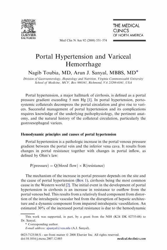
Portal Hypertension and Variceal Hemorrhage Nagib Toubia, MD, Arun J. Sanyal, MBBS, MD * Division of Gastroenterology, Hepatology and Nutrition, Virginia Commonwealth University School of Medicine, MCV, Box 980341, Richmond, VA 23298-0341, USA Portal hypertension, a major hallmark of cirrhosis, is defined as a portal pressure gradient exceeding 5 mm Hg [1]. In portal hypertension, porto- systemic collaterals decompress the portal circulation and give rise to vari- ces. Successful management of portal hypertension and its complications requires knowledge of the underlying pathophysiology, the pertinent anat- omy, and the natural history of the collateral circulation, particularly the gastroesophageal varices. Hemodynamic principles and causes of portal hypertension Portal hypertension is a pathologic increase in the portal venous pressure gradient between the portal vein and the inferior vena cava. It results from changes in portal resistance together with changes in portal inflow, as defined by Ohm’s law: PðpressureÞ¼ Qðblood flowÞ RðresistanceÞ The mechanism of the increase in portal pressure depends on the site and the cause of portal hypertension (Box 1), cirrhosis being the most common cause in the Western world [2]. The initial event in the development of portal hypertension in cirrhosis is an increase in resistance to outflow from the portal venous bed. This results from a relatively fixed component from distor- tion of the intrahepatic vascular bed from the disruption of hepatic architec- ture and a dynamic component from impaired intrahepatic vasodilation. An estimated 30% of the increased portal resistance is due to the hemodynamic This work was supported, in part, by a grant from the NIH (K24 DK 02755-08) to Dr. Sanyal. * Corresponding author. E-mail address: [email protected] (A.J. Sanyal). 0025-7125/08/$ - see front matter Ó 2008 Elsevier Inc. All rights reserved. doi:10.1016/j.mcna.2007.12.003 medical.theclinics.com Med Clin N Am 92 (2008) 551–574
Transcript of Portal Hypertension and Variceal Hemorrhage - University of
Portal Hypertension and VaricealHemorrhage
Nagib Toubia, MD, Arun J. Sanyal, MBBS, MD*Division of Gastroenterology, Hepatology and Nutrition, Virginia Commonwealth University
School of Medicine, MCV, Box 980341, Richmond, VA 23298-0341, USA
Portal hypertension, a major hallmark of cirrhosis, is defined as a portalpressure gradient exceeding 5 mm Hg [1]. In portal hypertension, porto-systemic collaterals decompress the portal circulation and give rise to vari-ces. Successful management of portal hypertension and its complicationsrequires knowledge of the underlying pathophysiology, the pertinent anat-omy, and the natural history of the collateral circulation, particularly thegastroesophageal varices.
Med Clin N Am 92 (2008) 551–574
Hemodynamic principles and causes of portal hypertension
Portal hypertension is a pathologic increase in the portal venous pressuregradient between the portal vein and the inferior vena cava. It results fromchanges in portal resistance together with changes in portal inflow, asdefined by Ohm’s law:
PðpressureÞ ¼ Qðblood flowÞ �RðresistanceÞ
The mechanism of the increase in portal pressure depends on the site andthe cause of portal hypertension (Box 1), cirrhosis being the most commoncause in the Western world [2]. The initial event in the development of portalhypertension in cirrhosis is an increase in resistance to outflow from theportal venous bed. This results from a relatively fixed component from distor-tion of the intrahepatic vascular bed from the disruption of hepatic architec-ture and a dynamic component from impaired intrahepatic vasodilation. Anestimated 30% of the increased portal resistance is due to the hemodynamic
This work was supported, in part, by a grant from the NIH (K24 DK 02755-08) to
Dr. Sanyal.
* Corresponding author.
E-mail address: [email protected] (A.J. Sanyal).
0025-7125/08/$ - see front matter � 2008 Elsevier Inc. All rights reserved.
doi:10.1016/j.mcna.2007.12.003 medical.theclinics.com
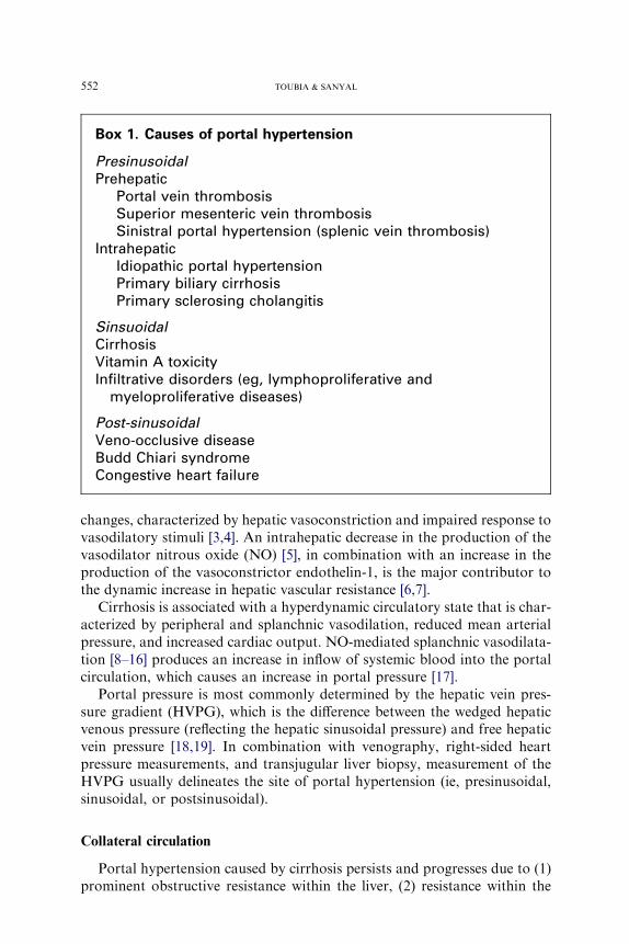
Box 1. Causes of portal hypertension
PresinusoidalPrehepatic
Portal vein thrombosisSuperior mesenteric vein thrombosisSinistral portal hypertension (splenic vein thrombosis)
IntrahepaticIdiopathic portal hypertensionPrimary biliary cirrhosisPrimary sclerosing cholangitis
SinsuoidalCirrhosisVitamin A toxicityInfiltrative disorders (eg, lymphoproliferative and
myeloproliferative diseases)
Post-sinusoidalVeno-occlusive diseaseBudd Chiari syndromeCongestive heart failure
552 TOUBIA & SANYAL
changes, characterized by hepatic vasoconstriction and impaired response tovasodilatory stimuli [3,4]. An intrahepatic decrease in the production of thevasodilator nitrous oxide (NO) [5], in combination with an increase in theproduction of the vasoconstrictor endothelin-1, is the major contributor tothe dynamic increase in hepatic vascular resistance [6,7].
Cirrhosis is associated with a hyperdynamic circulatory state that is char-acterized by peripheral and splanchnic vasodilation, reduced mean arterialpressure, and increased cardiac output. NO-mediated splanchnic vasodilata-tion [8–16] produces an increase in inflow of systemic blood into the portalcirculation, which causes an increase in portal pressure [17].
Portal pressure is most commonly determined by the hepatic vein pres-sure gradient (HVPG), which is the difference between the wedged hepaticvenous pressure (reflecting the hepatic sinusoidal pressure) and free hepaticvein pressure [18,19]. In combination with venography, right-sided heartpressure measurements, and transjugular liver biopsy, measurement of theHVPG usually delineates the site of portal hypertension (ie, presinusoidal,sinusoidal, or postsinusoidal).
Collateral circulation
Portal hypertension caused by cirrhosis persists and progresses due to (1)prominent obstructive resistance within the liver, (2) resistance within the
553VARICEAL HEMORRHAGE
collaterals, and (3) continued increase in portal vein inflow. This hyperten-sion leads to the formation of collaterals that decompress the portal circu-lation by returning blood to the heart via the systemic venous circulation.The major sites of collaterals are:
Rectum, where the systemic inferior mesenteric vein connects with theportal pudendal vein and results in rectal varices
Umbilicus, where the vestigial umbilical vein communicates with the leftportal vein and gives rise to prominent collaterals around the umbilicus(caput medusa)
Retroperitoneum, where collaterals, especially in women, communicatebetween ovarian vessels and iliac veins
Distal esophagus and proximal stomach, where gastroesophageal varicesform major collaterals between the portal venous system and thesystemic venous system
The following four zones of venous drainage are involved in the forma-tion of gastroesophageal varices [20]:
The gastric zone is 2 to 3 cm below the gastroesophageal junction, wherethe veins meet at the upper end of the cardia of the stomach, drain intoshort gastric and left gastric veins, and then drain into the splenic andportal veins, respectively.
The palisade zone is 2 to 3 cm proximal to the gastric zone into the loweresophagus, where the veins communicate with extrinsic (periesopha-geal) veins in the distal esophagus. This zone forms the dominantwatershed area between the portal and the systemic circulations.
More proximal to the palisade zone in the esophagus is the perforatingzone, where a network of submucosal veins in the esophagus connectsto the periesophageal veins, which drain into the azygous system andsubsequently into the systemic circulation.
The truncal zone is approximately 10 cm in length and is located proxi-mally to the perforating zone in the esophagus. It typically has fourlongitudinal veins in the lamina propria.
Most patients who have intrahepatic causes of portal hypertension havegastroesophageal varices because this provides the largest collateral flow viathe short and left gastric veins.
Varices form only when the HVPG exceeds 10 mm Hg and bleed onlywhen the HVPG exceeds 12 mm Hg [21,22]. Not all patients who havea HVPG greater than 12 mm Hg bleed. Other local factors that increase var-iceal wall tension are required [23]. The wall tension is defined by Frank’smodification of Laplace’s law [24]: T ¼ (P varices – P esophageal lumen) �(radius of varix)/wall thickness. The varix ruptures when the toleratedwall tension is exceeded because the variceal wall thins and the varixincreases in diameter and has an increased pressure. Larger varices at sitesof limited soft tissue support, notably the gastroesophageal junction, are
554 TOUBIA & SANYAL
at greater risk for variceal rupture and bleeding in patients who have portalhypertension.
Diagnosis of varices
Upper gastrointestinal endoscopy is the most common method to diag-nose varices. Various criteria have been used to standardize the descriptionof esophageal varices. The Japanese Research Society for Portal Hyperten-sion described varices in terms of red color signs, color of the varix, form(size) of the varix, and location of the varix [25]. The Northern ItalianEndoscopy Club simplified this scheme by classifying varices as F1, F2, orF3 (corresponding to small, medium, or large) with or without red signs.The clinically important decision is whether varices warrant therapeuticintervention. It is therefore useful to evaluate varices in terms of thosethat require treatment. It is recommended that varices be classified as small,which do not always warrant intervention, or large, which include those thatwere previously called large [26]. This provides a relatively simple and easilyreproducible classification.
Gastric varices are classified by location, which correlates with their riskof hemorrhage. Varices in direct continuity with the esophagus along thelesser and greater curves of the stomach are called gastroesophageal varices(GOV) types 1 and 2, respectively. Isolated gastric varices in the fundus(IGV1) occur less frequently than GOVs (10% versus 90%) but are themost likely to bleed. They may be caused by splenic vein thrombosis orspontaneous splenorenal collaterals.
Endoscopic ultrasound (EUS) has been used to study esophageal varicesand to identify a high risk of bleeding by assessment of the cross-sectionalarea of varices [24]; the size of and flow in the left gastric vein, azygousvein, and paraesophageal collaterals; the changes after endoscopic therapy;and the recurrence of esophageal varices after variceal ligation (collateralsO5 mm are at high risk for recurrent varices) [27]. It is unclear if EUS issuperior to standard endoscopy.
Esophageal capsule endoscopy is a promising modality to assess varices.It may provide an accurate, less invasive alternative to EGD for the detec-tion of esophageal varices or portal hypertensive gastropathy [28,29]. Alarge recent trial, reported only in abstract form, found excellent concor-dance with endoscopy. The role of capsule endoscopy in the managementof varices is still evolving.
Natural history of gastroesophageal varices
De novo varices develop in 5% to 15% of patients who have cirrhosis perannumand enlarge by 4% to 10%per annum.Most patients who have cirrho-sis develop varices, but only one third of them experiences variceal bleeding.Only 40% to 50% of actively bleeding varices spontaneously stop bleeding.
555VARICEAL HEMORRHAGE
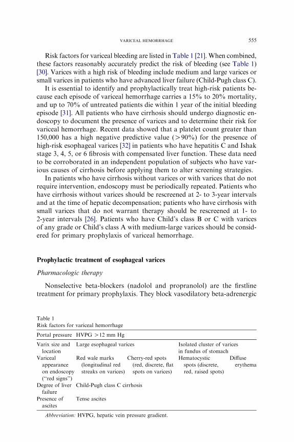
Risk factors for variceal bleeding are listed in Table 1 [21]. When combined,these factors reasonably accurately predict the risk of bleeding (see Table 1)[30]. Varices with a high risk of bleeding include medium and large varices orsmall varices in patients who have advanced liver failure (Child-Pugh class C).
It is essential to identify and prophylactically treat high-risk patients be-cause each episode of variceal hemorrhage carries a 15% to 20% mortality,and up to 70% of untreated patients die within 1 year of the initial bleedingepisode [31]. All patients who have cirrhosis should undergo diagnostic en-doscopy to document the presence of varices and to determine their risk forvariceal hemorrhage. Recent data showed that a platelet count greater than150,000 has a high negative predictive value (O90%) for the presence ofhigh-risk esophageal varices [32] in patients who have hepatitis C and Ishakstage 3, 4, 5, or 6 fibrosis with compensated liver function. These data needto be corroborated in an independent population of subjects who have var-ious causes of cirrhosis before applying them to alter screening strategies.
In patients who have cirrhosis without varices or with varices that do notrequire intervention, endoscopy must be periodically repeated. Patients whohave cirrhosis without varices should be rescreened at 2- to 3-year intervalsand at the time of hepatic decompensation; patients who have cirrhosis withsmall varices that do not warrant therapy should be rescreened at 1- to2-year intervals [26]. Patients who have Child’s class B or C with varicesof any grade or Child’s class A with medium-large varices should be consid-ered for primary prophylaxis of variceal hemorrhage.
Prophylactic treatment of esophageal varices
Pharmacologic therapy
Nonselective beta-blockers (nadolol and propranolol) are the firstlinetreatment for primary prophylaxis. They block vasodilatory beta-adrenergic
Table 1
Risk factors for variceal hemorrhage
Portal pressure HVPG O12 mm Hg
Varix size and
location
Large esophageal varices Isolated cluster of varices
in fundus of stomach
Variceal
appearance
on endoscopy
(‘‘red signs’’)
Red wale marks
(longitudinal red
streaks on varices)
Cherry-red spots
(red, discrete, flat
spots on varices)
Hematocystic
spots (discrete,
red, raised spots)
Diffuse
erythema
Degree of liver
failure
Child-Pugh class C cirrhosis
Presence of
ascites
Tense ascites
Abbreviation: HVPG, hepatic vein pressure gradient.
556 TOUBIA & SANYAL
receptors, permitting unopposed alpha-adrenergic vasoconstriction in themesenteric arterioles, thereby reducing portal venous inflow and pressure.They also decrease cardiac output, which further decreases the portal inflow.Meta-analysis of clinical trials shows that the risk of bleeding is reduced bybeta-blocker therapy versus placebo from 25% to 15% [33]. The effec-tiveness of beta-blockers is most accurately assessed by the HVPG. Thebest predictor of success is a sustained decrease in the HPVG to less than12 mm Hg, whereas patients who have a sustained 20% decrease inHPVG to greater than 12 mm Hg have a risk of bleeding of less than10% [34,35]. This approach is not widely applied to clinical practice. Theefficacy of beta-blockers is clinically monitored by a decrease in the restingheart rate greater than 25% but not to a rate less than 55 beats/min. Only20% to 30% of subjects achieve these endpoints, and 15% to 20% of sub-jects cannot tolerate and require discontinuation of this therapy.
Short-acting (nitroglycerin) or long-acting (isosorbide mononitrates)nitrates cause venodilatation, rather than arterial dilatation, and decreaseportal pressure predominantly by decreasing portal venous blood flow.The effect on intrahepatic resistance is not impressive, and nitrates are nolonger recommended for primary prophylaxis due to discrepant results ofclinical trials.
Other agents that may decrease intrahepatic resistance includea1-adrenergic blockers and angiotensin II receptor antagonists. Prazosin,an a1-adrenergic blocker, caused worsening of the systemic hyperdynamiccirculation and was associated with portal hypertension and consequent so-dium retention and ascites [36]. Losartan, an angiotensin II receptor antag-onist, caused a reduction in portal pressure without significant effects on thesystemic circulation [37]. It did not significantly reduce portal pressure inrandomized controlled trials, but it worsened the renal function [38,39].Endothelin receptor blockers and liver-selective NO donors that targetintrahepatic vascular resistance are promising investigational therapies [40].
Endoscopic sclerotherapy
Prophylactic endoscopic sclerotherapy (EST) was used in the 1980s. Al-though controlled trials initially reported that it significantly reduced therisk of a first variceal bleed and improved survival [41–43], subsequent trialsdid not show a survival benefit. EST may provoke bleeding that is difficultto control and may increase the mortality [44,45]. A meta-analysis showeda marked reduction in the risk for a first episode of bleeding, but the mor-tality was higher in certain studies [46]. Consequently, EST is not recom-mended for prophylaxis of esophageal varices.
Endoscopic variceal ligation
Endoscopic variceal ligation (EVL) is associated with fewer procedure-related complications than EST. EVL significantly reduces the risk of the

557VARICEAL HEMORRHAGE
first bleeding compared with no treatment [47–49] or compared with pro-pranolol [50–52], with a relative risk reduction of almost 40% (Fig. 1).No survival benefit was seen compared with propranolol. One study didnot reveal any benefit of EVL and propranolol versus EVL alone; however,given the low risk of bleeding after variceal eradication and the use of EVLwhen varices recurred, the investigators likely missed any chance of demon-strating a benefit of combined therapy because beta-blockers would be ex-pected to prevent varices as their main effect.
In summary, nonselective beta-blockers or EVL are recommended first-line treatments for primary prophylaxis of variceal hemorrhage. EVL maybe used in subjects who cannot tolerate beta-blockers. The authors useEVL in patients who are likely not to tolerate beta-blockers (eg, patientswho have low blood pressure or asthma) and who have medium-largevarices, whereas they preferentially use beta-blockers when the varices aresmall and technically difficult to band. This practice is based on evidencethat up to 6% of subjects undergoing EVL may experience potentiallylife-threatening iatrogenic hemorrhage.
Management of acute variceal bleed
The management of acute variceal bleeding includes hemodynamic resus-citation, general treatments, prevention of complications, and achievementof hemostasis. Intravenous access must be promptly secured (Box 2). Air-way intubation is indicated in patients who are bleeding severely or whohave mental status changes that preclude their ability to protect their air-way. Intravascular volume loss is estimated and replaced with crystalloids
Fig. 1. A meta-analysis of trials of endoscopic variceal ligation versus beta-blockers for the pri-
mary prophylaxis of variceal hemorrhage. Each data point reflects the odds ratio for benefit of
one treatment versus another for a given study. Horizontal bars represent the confidence limits
for the data. Although most of the trials show a modest benefit for EVL, the confidence limits
intersect the unit line indicating that these data did not reach significance for individual trials.
(FromTriantos CK, Burroughs AK. Prevention of the development of varices and first portal hy-
pertensive bleeding episode. Best Pract Res Clin Gastroenterol 2007;21:31–42; with permission.)
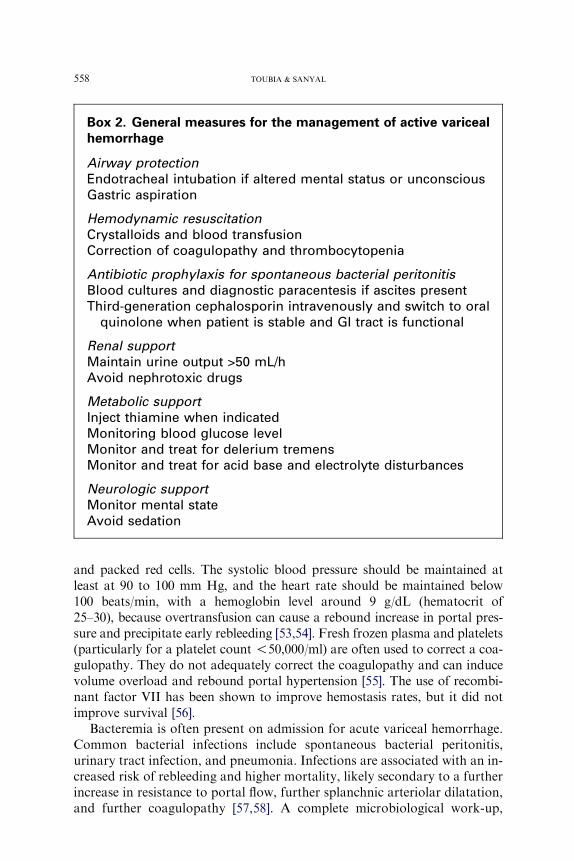
Box 2. General measures for the management of active varicealhemorrhage
Airway protectionEndotracheal intubation if altered mental status or unconsciousGastric aspiration
Hemodynamic resuscitationCrystalloids and blood transfusionCorrection of coagulopathy and thrombocytopenia
Antibiotic prophylaxis for spontaneous bacterial peritonitisBlood cultures and diagnostic paracentesis if ascites presentThird-generation cephalosporin intravenously and switch to oral
quinolone when patient is stable and GI tract is functional
Renal supportMaintain urine output >50 mL/hAvoid nephrotoxic drugs
Metabolic supportInject thiamine when indicatedMonitoring blood glucose levelMonitor and treat for delerium tremensMonitor and treat for acid base and electrolyte disturbances
Neurologic supportMonitor mental stateAvoid sedation
558 TOUBIA & SANYAL
and packed red cells. The systolic blood pressure should be maintained atleast at 90 to 100 mm Hg, and the heart rate should be maintained below100 beats/min, with a hemoglobin level around 9 g/dL (hematocrit of25–30), because overtransfusion can cause a rebound increase in portal pres-sure and precipitate early rebleeding [53,54]. Fresh frozen plasma and platelets(particularly for a platelet count !50,000/ml) are often used to correct a coa-gulopathy. They do not adequately correct the coagulopathy and can inducevolume overload and rebound portal hypertension [55]. The use of recombi-nant factor VII has been shown to improve hemostasis rates, but it did notimprove survival [56].
Bacteremia is often present on admission for acute variceal hemorrhage.Common bacterial infections include spontaneous bacterial peritonitis,urinary tract infection, and pneumonia. Infections are associated with an in-creased risk of rebleeding and higher mortality, likely secondary to a furtherincrease in resistance to portal flow, further splanchnic arteriolar dilatation,and further coagulopathy [57,58]. A complete microbiological work-up,
559VARICEAL HEMORRHAGE
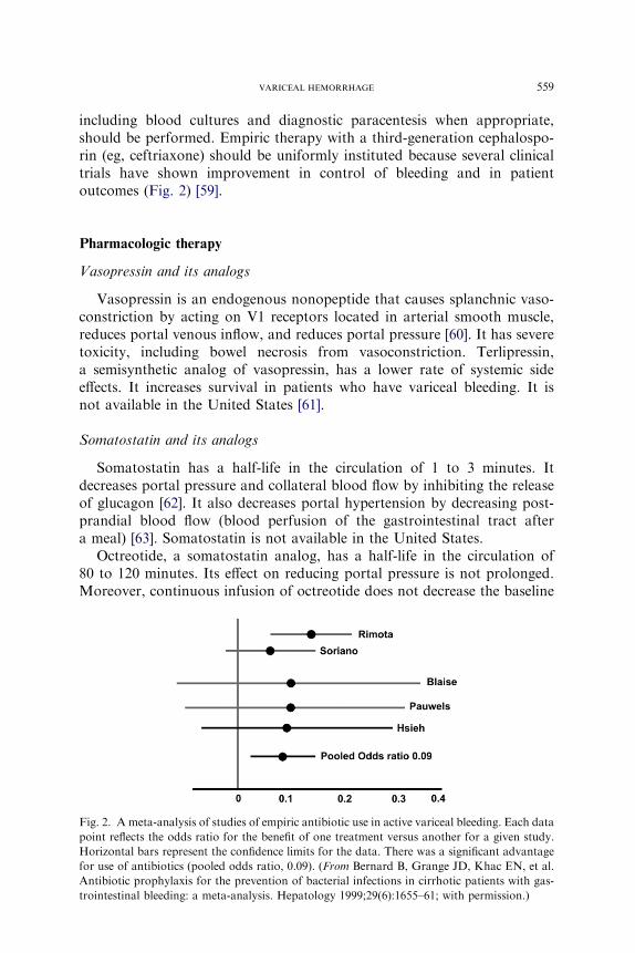
including blood cultures and diagnostic paracentesis when appropriate,should be performed. Empiric therapy with a third-generation cephalospo-rin (eg, ceftriaxone) should be uniformly instituted because several clinicaltrials have shown improvement in control of bleeding and in patientoutcomes (Fig. 2) [59].
Pharmacologic therapy
Vasopressin and its analogs
Vasopressin is an endogenous nonopeptide that causes splanchnic vaso-constriction by acting on V1 receptors located in arterial smooth muscle,reduces portal venous inflow, and reduces portal pressure [60]. It has severetoxicity, including bowel necrosis from vasoconstriction. Terlipressin,a semisynthetic analog of vasopressin, has a lower rate of systemic sideeffects. It increases survival in patients who have variceal bleeding. It isnot available in the United States [61].
Somatostatin and its analogs
Somatostatin has a half-life in the circulation of 1 to 3 minutes. Itdecreases portal pressure and collateral blood flow by inhibiting the releaseof glucagon [62]. It also decreases portal hypertension by decreasing post-prandial blood flow (blood perfusion of the gastrointestinal tract aftera meal) [63]. Somatostatin is not available in the United States.
Octreotide, a somatostatin analog, has a half-life in the circulation of80 to 120 minutes. Its effect on reducing portal pressure is not prolonged.Moreover, continuous infusion of octreotide does not decrease the baseline
Fig. 2. A meta-analysis of studies of empiric antibiotic use in active variceal bleeding. Each data
point reflects the odds ratio for the benefit of one treatment versus another for a given study.
Horizontal bars represent the confidence limits for the data. There was a significant advantage
for use of antibiotics (pooled odds ratio, 0.09). (From Bernard B, Grange JD, Khac EN, et al.
Antibiotic prophylaxis for the prevention of bacterial infections in cirrhotic patients with gas-
trointestinal bleeding: a meta-analysis. Hepatology 1999;29(6):1655–61; with permission.)

560 TOUBIA & SANYAL
portal pressure despite decreasing the postprandial increase in portal pres-sure [64]. Although some studies failed to prove the superiority of somato-statin or its analogs compared with placebo in the control of acute varicealbleeding [65], other studies showed efficacy [66]. Early administration ofvapreotide may be associated with improved control of bleeding but withouta significant reduction in mortality [67].
Endoscopic therapy
Endoscopic sclerotherapy
EST has largely been supplanted by EVL, except when poor visualizationprecludes effective band ligation of bleeding varices. Current evidence doesnot support emergency EST as first-line treatment of variceal bleeding [68].The technique involves injection of a sclerosant into (intravariceal) oradjacent to (paravariceal) a varix. Complications of EST occurring duringor after the procedure include chest discomfort, ulcers (and ulcer-relatedbleeding), strictures, and perforation. The risk of ulcers can be reduced byprescribing sucralfate after EST [69].
Endoscopic variceal ligation
EVL is the preferred endoscopic modality for control of acute esophagealvariceal bleeding and for prevention of rebleeding. Varices at the gastro-esophageal junction are banded initially, and then more proximal varicesare banded in a spiral manner at intervals of approximately every 2 cm.Varices in the middle or proximal esophagus do not need to be banded.EVL is associated with similar but fewer complications [70] than EST andrequires fewer sessions to achieve variceal obliteration.
In summary, the first-line treatment for active esophageal variceal hemor-rhage is a combination of pharmacologic treatment (ie, octerotide) andendoscopic treatment (EVL or EST) (Fig. 3) [71]. About 80% to 90% of pa-tients achieve hemostasis with first-line therapy; the remaining patients failto achieve hemostasis or experience early rebleeding [72].
Within the first 6 hours, failure to control bleeding is recognized by [73](1) transfusion requirement of more than four units of packed red cells and(2) the inability tomaintain the systolic blood pressure greater than 70mmHgor to raise it by 20mmHg or to reduce the resting pulse to less than 100/min orto decrease it by 20 beats/min.
After 6 hours, early rebleeding is defined by hematemesis together with(1) reduction in systolic blood pressure by 20 mm Hg from the level at6 hours, (2) increase in pulse rate by 20/min from the rate at 6 hours ontwo consecutive readings 1 hour apart, or(3) the need to transfuse two ormore units of packed red cells to increase the hematocrit to more than27% or the hemoglobin to more than 9 g/dL.
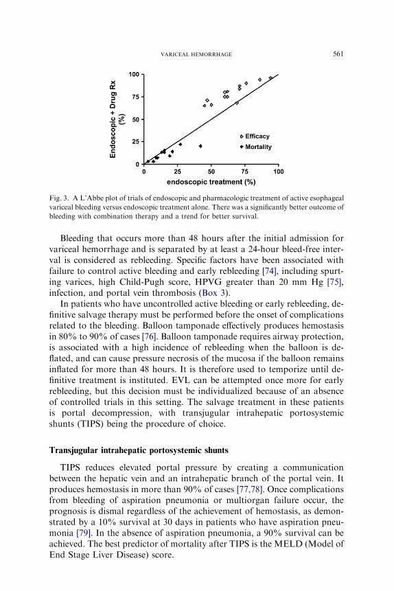
Fig. 3. A L’Abbe plot of trials of endoscopic and pharmacologic treatment of active esophageal
variceal bleeding versus endoscopic treatment alone. There was a significantly better outcome of
bleeding with combination therapy and a trend for better survival.
561VARICEAL HEMORRHAGE
Bleeding that occurs more than 48 hours after the initial admission forvariceal hemorrhage and is separated by at least a 24-hour bleed-free inter-val is considered as rebleeding. Specific factors have been associated withfailure to control active bleeding and early rebleeding [74], including spurt-ing varices, high Child-Pugh score, HPVG greater than 20 mm Hg [75],infection, and portal vein thrombosis (Box 3).
In patients who have uncontrolled active bleeding or early rebleeding, de-finitive salvage therapy must be performed before the onset of complicationsrelated to the bleeding. Balloon tamponade effectively produces hemostasisin 80% to 90% of cases [76]. Balloon tamponade requires airway protection,is associated with a high incidence of rebleeding when the balloon is de-flated, and can cause pressure necrosis of the mucosa if the balloon remainsinflated for more than 48 hours. It is therefore used to temporize until de-finitive treatment is instituted. EVL can be attempted once more for earlyrebleeding, but this decision must be individualized because of an absenceof controlled trials in this setting. The salvage treatment in these patientsis portal decompression, with transjugular intrahepatic portosystemicshunts (TIPS) being the procedure of choice.
Transjugular intrahepatic portosystemic shunts
TIPS reduces elevated portal pressure by creating a communicationbetween the hepatic vein and an intrahepatic branch of the portal vein. Itproduces hemostasis in more than 90% of cases [77,78]. Once complicationsfrom bleeding of aspiration pneumonia or multiorgan failure occur, theprognosis is dismal regardless of the achievement of hemostasis, as demon-strated by a 10% survival at 30 days in patients who have aspiration pneu-monia [79]. In the absence of aspiration pneumonia, a 90% survival can beachieved. The best predictor of mortality after TIPS is the MELD (Model ofEnd Stage Liver Disease) score.

Box 3. Factors affecting risk of continued bleeding or recurrentbleeding
Factors associated with failure to control acute hemorrhageSpurting varicesHigh Child-Pugh scoreHigh hepatic venous pressure gradientInfectionPortal vein thrombosis
Factors associated with early rebleedingSevere initial bleedingOverly aggressive volume resuscitationInfectionHigh hepatic venous pressure gradientComplications of endoscopic therapyRenal failure
Factors associated with late rebleedingHigh Child-Pugh scoreLarge variceal sizeContinued alcohol useHepatocellular carcinoma
Based on data from pooled sources.
562 TOUBIA & SANYAL
Contraindications to TIPS include severe congestive heart failure, severepulmonary hypertension, severe hepatic failure, portal vein thrombosis withcavernomatous transformation, and polycystic liver disease. In a decision inan individual patient, the risk of exsanguination must be weighed against thetype of contraindication. Although hemostasis may be achieved with TIPS,patients who have multiorgan failure have a dismal prognosis in the authors’experience. After consultation with the next of kin, provision of only palli-ative care may often be appropriate in such patients.
Surgical decompression of the portal system via portosystemic shunts isanother salvage modality. The use of surgical shunts has declined markedlydue to the increasing availability of TIPS, the high morbidity of surgery, andthe decline in the number of surgeons trained to perform these procedures.
Secondary prophylaxis
Once acute variceal bleeding is controlled, prevention of recurrent bleed-ing should be emphasized. After an index bleed, 70% of patients experiencerecurrent variceal hemorrhage within 1 year [80], and these patients have
563VARICEAL HEMORRHAGE
a 70% 1-year mortality. The risk of rebleeding is greatest within the first6 weeks, with more than 50% of rebleeding occurring within 3 to 4 days.Risk factors for rebleeding include severe initial bleeding as defined by a he-moglobin level less than 8 mg/dL, gastric variceal bleeding, active bleedingat endoscopy, and a high HPVG [81,82]. Age greater than 60 years, largeesophageal varices, severe liver disease, continued alcoholism, renal failure,and the presence of a hepatoma also increase the risk of rebleeding [81,83]. Itis important to prevent recurrent hemorrhage, preserve liver function, main-tain a normal renal function, prevent ascites, and avoid alcohol consump-tion to prolong survival.
Orthotopic liver transplant is the only treatment that achieves most ofthese objectives andprolongs long-term survival. Somepatients are unsuitablecandidates for liver transplantation, and, even if orthotopic liver transplant isbeing considered, patients often have to wait several months before a donorliver becomes available.During this time, they are at risk for recurrent varicealhemorrhage and therefore require treatment to prevent this complication.
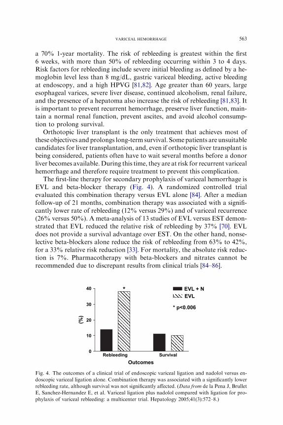
The first-line therapy for secondary prophylaxis of variceal hemorrhage isEVL and beta-blocker therapy (Fig. 4). A randomized controlled trialevaluated this combination therapy versus EVL alone [84]. After a medianfollow-up of 21 months, combination therapy was associated with a signifi-cantly lower rate of rebleeding (12% versus 29%) and of variceal recurrence(26% versus 50%). A meta-analysis of 13 studies of EVL versus EST demon-strated that EVL reduced the relative risk of rebleeding by 37% [70]. EVLdoes not provide a survival advantage over EST. On the other hand, nonse-lective beta-blockers alone reduce the risk of rebleeding from 63% to 42%,for a 33% relative risk reduction [33]. For mortality, the absolute risk reduc-tion is 7%. Pharmacotherapy with beta-blockers and nitrates cannot berecommended due to discrepant results from clinical trials [84–86].
40
30
10
20
*
0
Rebleeding Survival
(%
)
Outcomes
* p<0.006
EVL + N
EVL
Fig. 4. The outcomes of a clinical trial of endoscopic variceal ligation and nadolol versus en-
doscopic variceal ligation alone. Combination therapy was associated with a significantly lower
rebleeding rate, although survival was not significantly affected. (Data from de la Pena J, Brullet
E, Sanchez-Hernandez E, et al. Variceal ligation plus nadolol compared with ligation for pro-
phylaxis of variceal rebleeding: a multicenter trial. Hepatology 2005;41(3):572–8.)
564 TOUBIA & SANYAL
A meta-analysis of 12 clinical trials of TIPS versus endoscopic treatmentindicates that TIPS is superior to endoscopic treatment for the prevention ofrebleeding (19% versus 47%) [87], but this advantage is offset by its failure toimprove survival, its higher morbidity from the development of liver failureand encephalopathy (34% versus 19%), and its lack of a cost benefit [88].Based on these considerations, TIPS is primarily used as a salvage treatmentfor patients who experience recurrent bleeding despite adequate endoscopicand pharmacologic treatment. In one recent study, subjects who had bleed-ing esophageal varices underwent early HVPG measurement after initialstabilization with band ligation. Patients who had a HVPG greater than20 mm Hg were randomized to TIPS versus band ligation. In this high-risksubgroup, early, elective TIPS dramatically improved outcomes comparedwith EVL. These results await corroboration from other prospective trials.
In summary, the following regimen is recommended for secondaryprophylaxis of esophageal variceal hemorrhage:
1. Eradication of esophageal varices by EVL (every 7–14 days until varicesare eradicated) with concomitant use of nonselective beta-blockers (pro-pranolol or nadolol)
2. Long-term endoscopic control and banding of recurrent varices every 3to 6 months
3. If EVL is unavailable or contraindicated, nonselective beta-blockers canbe used alone.
4. TIPS is considered if pharmacologic and endoscopic therapy faild(recurrence of variceal hemorrhage despite at least two sessions of endo-scopic treatment performed not more than 2 weeks apart).
Always consider liver transplantation if the patient is Child-Pugh B or C.
Gastric varices
Gastric varices most commonly are caused by portal hypertension,usually in patients who have cirrhosis. Patients who have splenic veinthrombosis or spontaneous splenorenal collaterals can develop isolated gas-tric varices, particularly IGV1. GOV1 disappears in approximately 58% and70% after EST and EVL of esophageal varices, respectively [89,90]. Theobliteration of varices at the gastroesophageal junction blocks the shuntingveins in the palisade zone, leading to dilatation and the formation of new orsecondary gastric varices [91]. These secondary varices occur at a rate of9.7% to 15.3% [92–94] and have a higher frequency of bleeding comparedwith primary gastric varices. On the other hand, bleeding from primary gas-tric varices after endoscopic treatment of esophageal varices is uncommon.
The prevalence of gastric varices is 5% to 33% in patients who haveportal hypertension, with an overall incidence of bleeding ranging from3% to 30% [95,96]. Mortality associated with gastric variceal hemorrhageis 30% to 53%, with a 30% rebleeding rate.
565VARICEAL HEMORRHAGE
Because GOV1 constitutes an extension of esophageal varices along thelesser curvature of the stomach, it is managed like esophageal varices.IGV1 secondary to splenic vein thrombosis is treated by splenectomy. Thereis no consensus on primary prophylaxis of bleeding GOV2 or IGV due tolimited data.
EST controls active bleeding in 40% to 100% of cases of GOV1 [97–103].The primary drawback is the high risk of recurrent bleeding. EST is fre-quently associated with ulceration, which bleeds in approximately 50% ofcases [97,102,103]. Rebleeding rates have been reported to be 5.5% inGOV1, 19% in GOV2, and as high as 53% in IGV1 [102].
In prospective uncontrolled studies, EVL has been shown to achieve he-mostasis rates of up to 89%, with a rebleeding rate of 18.5% [104]. Thesestudies included all types of gastric varices. The major concern after gastricEVL is the potential of partial ligation of large gastric varices, which mayproduce bleeding [105,106]. EVL is generally not recommended for IGV1.
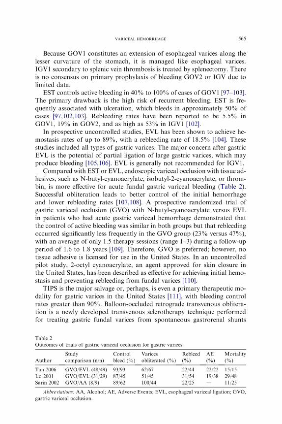
Compared with EST or EVL, endoscopic variceal occlusion with tissue ad-hesives, such as N-butyl-cyanoacrylate, isobutyl-2-cyanoacrylate, or throm-bin, is more effective for acute fundal gastric variceal bleeding (Table 2).Successful obliteration leads to better control of the initial hemorrhageand lower rebleeding rates [107,108]. A prospective randomized trial ofgastric variceal occlusion (GVO) with N-butyl-cyanoacrylate versus EVLin patients who had acute gastric variceal hemorrhage demonstrated thatthe control of active bleeding was similar in both groups but that rebleedingoccurred significantly less frequently in the GVO group (23% versus 47%),with an average of only 1.5 therapy sessions (range 1–3) during a follow-upperiod of 1.6 to 1.8 years [109]. Therefore, GVO is preferred; however, notissue adhesive is licensed for use in the United States. In an uncontrolledpilot study, 2-octyl cyanoacrylate, an agent approved for skin closure inthe United States, has been described as effective for achieving initial hemo-stasis and preventing rebleeding from fundal varices [110].
TIPS is the major salvage or, perhaps, is even a primary therapeutic mo-dality for gastric varices in the United States [111], with bleeding controlrates greater than 90%. Balloon-occluded retrograde transvenous oblitera-tion is a newly developed transvenous sclerotherapy technique performedfor treating gastric fundal varices from spontaneous gastrorenal shunts
Table 2
Outcomes of trials of gastric variceal occlusion for gastric varices
Author
Study
comparison (n/n)
Control
bleed (%)
Varices
obliterated (%)
Rebleed
(%)
AE
(%)
Mortality
(%)
Tan 2006 GVO/EVL (48/49) 93/93 62/67 22/44 22/22 15/15
Lo 2001 GVO/EVL (31/29) 87/45 51/45 31/54 19/38 29/48
Sarin 2002 GVO/AA (8/9) 89/62 100/44 22/25 d 11/25
Abbreviations: AA, Alcohol; AE, Adverse Events; EVL, esophageal variceal ligation; GVO,
gastric variceal occlusion.

566 TOUBIA & SANYAL
[112,113]. Shiba and colleagues [114] recently showed that balloon-occludedinjection sclerotherapy is safe and effective even in patients who do not havegastrorenal shunts. Surgical shunts also control bleeding gastric varices andprevent recurrent bleeding.
Ectopic varices
Ectopic varices are defined as portosystemic shunts, resulting from portalhypertension, that occur at any site in the gut or abdomen except in thegastroesophageal region. These sites include the duodenum, jejunum, ileum,colon, rectum, biliary tree, and ostomy sites [115,116]. It is important to dif-ferentiate anal varices from hemorrhoids: Anal varices collapse with digitalpressure, whereas hemorrhoids do not [115].
Ectopic varices account for 1% to 5% of all variceal bleeding [117]. Pa-tients who have ectopic variceal hemorrhage typically present with sudden,profuse melena or hematochezia. Because brisk upper gastrointestinal(UGI) bleeding can result in hematochezia, all subjects who have suspectedvariceal hemorrhage should initially undergo emergency upper endoscopy.If the upper endoscopy fails to reveal a source of UGI hemorrhage, colono-scopy is performed after a rapid colonic purge. If the colon looks normaland bleeding continues, angiography may be useful to identify varices orto localize a nonvariceal source of hemorrhage.
Several studies have reported successful therapy of duodenal varices withsclerosant injection [118] or with an occluding agent [119]. There are no dataregarding band ligation of ectopic varices. The next treatment option isembolization. Unlike arterial embolization for gastrointestinal hemorrhage,the goal in this setting is to occlude the feeding vein (on the portal venousside) to the ectopic varices rather than to occlude the bleeding site. Bal-loon-occluded retrograde transvenous obliteration has been successfullyused in several reported cases. If the patient continues to bleed despiteembolization, options include TIPS or surgery. At our center, TIPS is thepreferred approach for the treatment of bleeding ectopic varices.
Gastric antral vascular ectasia
Gastric antral vascular ectasia (GAVE), or watermelon stomach, de-scribes a vascular lesion of the gastric antrum that consists of ectatic andsacculated antral mucosal vessels radiating toward from the pylorus(Fig. 5). Its cause is unknown, but it has been proposed that gastric peristal-sis causes prolapse of the loose antral mucosa into the duodenum withconsequent elongation and ectasia of the mucosal vessels [120,121]. Micro-scopic features include dilated capillaries with focal thrombosis, dilated andtortuous submucosal venous channels, and fibromuscular hyperplasia of themuscularis mucosa. Most cases are idiopathic, but it has been associated
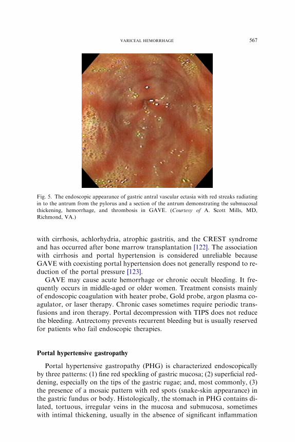
Fig. 5. The endoscopic appearance of gastric antral vascular ectasia with red streaks radiating
in to the antrum from the pylorus and a section of the antrum demonstrating the submucosal
thickening, hemorrhage, and thrombosis in GAVE. (Courtesy of A. Scott Mills, MD,
Richmond, VA.)
567VARICEAL HEMORRHAGE
with cirrhosis, achlorhydria, atrophic gastritis, and the CREST syndromeand has occurred after bone marrow transplantation [122]. The associationwith cirrhosis and portal hypertension is considered unreliable becauseGAVE with coexisting portal hypertension does not generally respond to re-duction of the portal pressure [123].
GAVE may cause acute hemorrhage or chronic occult bleeding. It fre-quently occurs in middle-aged or older women. Treatment consists mainlyof endoscopic coagulation with heater probe, Gold probe, argon plasma co-agulator, or laser therapy. Chronic cases sometimes require periodic trans-fusions and iron therapy. Portal decompression with TIPS does not reducethe bleeding. Antrectomy prevents recurrent bleeding but is usually reservedfor patients who fail endoscopic therapies.
Portal hypertensive gastropathy
Portal hypertensive gastropathy (PHG) is characterized endoscopicallyby three patterns: (1) fine red speckling of gastric mucosa; (2) superficial red-dening, especially on the tips of the gastric rugae; and, most commonly, (3)the presence of a mosaic pattern with red spots (snake-skin appearance) inthe gastric fundus or body. Histologically, the stomach in PHG contains di-lated, tortuous, irregular veins in the mucosa and submucosa, sometimeswith intimal thickening, usually in the absence of significant inflammation
568 TOUBIA & SANYAL
[124]. PHG is correlated with the severity of liver disease [125]. It is diag-nosed by endoscopy. It is an uncommon cause of significant UGI bleedingin patients who have portal hypertension. In one study [126], acute bleedingfrom PHG was observed in only 2.5% of patients.
Treatment is directed at decreasing portal pressure. Propranolol has beenshown to significantly reduce the rate of recurrent bleeding compared withplacebo (35% versus 62% at 1 year) [127]. Vasopressin, terlipressin, somato-statin, and octerotide have not been studied for this indication. TIPS is thenext therapy. It is associated with significant improvement in the endoscopicfindings and a decrease in the transfusion requirements. If bleeding con-tinues, surgical portal decompression is performed. Liver transplantation isindicated for decompensated liver disease. Endoscopic thermal coagulationis not effective for controlling or preventing this diffuse form of bleeding.
Downhill varices
Esophageal veins form a plexus on the outer surface of the esophagus.The lower part drains into the short and left gastric veins of the portal sys-tem, whereas the upper part drains into the azygous, thyroid, and internalmammary veins and then into the superior vena cava. ‘‘Downhill’’ esopha-geal varices (DEV) form in the upper third of the esophagus as collateralbranches directing blood flow ‘‘downward’’ to bypass superior vena cava(SVC) obstruction via the azygous vein or to drain the systemic superior ve-nous system via the portal vein when the SVC and the azygous vein areobstructed.
DEV are mostly due to SVC syndrome secondary to mass effects(external compression of the SVC) from lung cancer, intrathoracic goiter,mediastinal lymphoma, thyroid carcinoma, thymoma, or mediastinallymphadenopathy secondary to head and neck cancers. DEV usually disap-pear after treatment of the underlying condition. Several cases have beenassociated with gastrointestinal hemorrhage, which can be life threatening[128]. Due to its rarity, neither controlled trials nor a general consensus ex-ists on the best therapeutic approach. Isolated case reports have suggestedsuccess with EST, EBL, or balloon tamponade.
References
[1] Bosch J. The sixthCarlos E.RubioMemorial Lecture. Prevention and treatment of variceal
hemorrhage. P R Health Sci J 2000;19:57–67.
[2] Garcia-Tsao G. Portal hypertension. Curr Opin Gastroenterol 2006;22:254–62.
[3] Gupta PS, TorunerM,ChungM, et al. Endothelial dysfunction anddecreased production of
nitricoxide in the intrahepaticmicrocirculationof cirrhotic rats.Hepatology1998;41:926–31.
[4] Shah V, Garcia-Cardena G, SessaW, et al. The hepatic circulation in health and in disease:
report of a single-topic symposium. Hepatology 1998;27:279–88.
569VARICEAL HEMORRHAGE
[5] Sarin S, Groszmann RT,Mosca P. Propranolol ameliorates the development of portal-sys-
temic shunting in a chronic murine schistosomiasis model of portal hypertension. J Clin In-
vest 1991;87:1032–6.
[6] Pinzani M, Milani S, De Franco R, et al. Endothelin 1 is overexpressed in human cirrhotic
liver and exerts multiple effects on activated hepatic stellate cells. Gastroenterology 1996;
110:534–48.
[7] Rockey DC, Weisiger RA. Endothelin induced contractility of stellate cells from normal
and cirrhotic rat liver: implications for regulation of portal pressure and resistance. Hepa-
tology 1996;59:233–40.
[8] Atucha NM, Shah V, Garcia-Cardena G, et al. Role of endothelium in the abnormal
response of mesenteric vessels in rats with portal hypertension and liver cirrhosis. Gastro-
enterology 1996;111:1627–32.
[9] Cahill PA, Foster C, Redmond EM, et al. Enhanced nitric oxide synthase activity in portal
hypertensive rabbits. Hepatology 1995;22:598–606.
[10] Cahill PA, Redmond EM, Hodges R, et al. Increased endothelial nitric oxide synthase
activity in the hyperemic vessels of portal hypertensive rats. J Hepatol 1996;25:370–8.
[11] Garcia-Pagan JC, Fernandez M, Bernadich C, et al. Effects of continued NO inhibition
on portal hypertensive syndrome after portal vein stenosis in rat. Am J Phys 1994;267:
G984–90.
[12] NiederbergerM,Gines P,Martin PY, et al. Comparison of vascular nitric oxide production
and systemic hemodynamics in cirrhosis versus prehepatic portal hypertension in rats.
Hepatology 1996;24:947–51.
[13] Sieber CC, Groszmann RJ. In vitro hyporeactivity to methoxamine in portal hypertensive
rats: reversal by nitric oxide blockade. Am J Phys 1992;262:G996–1001.
[14] Sieber CC, Groszmann RJ. Nitric oxide mediates hyporeactivity to vasopressors in mesen-
teric vessels of portal hypertensive rats. Gastroenterology 1992;103:235–9.
[15] Sieber CC, Lopez-Talavera JC, Groszmann RJ. Role of nitric oxide in the in vitro splanch-
nic vascular hyporeactivity in ascitic cirrhotic rats. Gastroenterology 1993;104:1750–4.
[16] Sogni P, Sabry S,MoreauR, et al. Hyporeactivity of mesenteric resistance arteries in portal
hypertensive rats. J Hepatol 1996;24:487–90.
[17] Vorobioff J, Bredfeldt JE, Groszmann RJ. Increased blood flow through the portal system
in cirrhotic rats. Gastroenterology 1984;87:1120–6.
[18] Garcia-Pagan JC, Groszmann RJ, Bosch J. Measurement of portal pressure. Clin Gastro-
enterol Hepatol 2005;981.
[19] GroszmannRJ, GlickmanM, Blei AT.Wedged and free hepatic venous pressure measured
with a balloon catheter. Gastroenterology 1979;76:253–8.
[20] ViannaA,Hayes PC,MoscosoG, et al. Normal venous circulation of the gastroesophageal
junction: a route to understanding varices. Gastroenterology 1987;93:876–89.
[21] Garcia-TsaoG, Groszmann RJ, Fisher RL, et al. Portal pressure, presence of gastroesoph-
ageal varices and variceal bleeding. Hepatology 1985;5:419–24.
[22] Groszmann RJ, Garcia-Tsao G, Bosch J, et al. Beta-blockers to prevent gastroesophageal
varices in patients with cirrhosis. N Engl J Med 2005;353:2254–61.
[23] Escorsell A, Gines A, Llach J, et al. Increasing intra-abdominal pressure increases pressure,
volume, and wall tension in esophageal varices. Hepatology 2002;36:936–40.
[24] Rigau J, Bosch J, Bordas JM, et al. Endoscopic measurement of variceal pressure in
cirrhosis: correlation with portal pressure and variceal hemorrhage. Gastroenterology
1989;96:873–80.
[25] Beppu K, Inokuchi K, Koyanagi N, et al. Prediction of variceal hemorrhage by esophageal
endoscopy. Gastrointest Endosc 1981;27:213–8.
[26] Garcia-Tsao G, Sanyal AJ, Grace ND, et al, and the Practice Guidelines Committee of the
Americal Association for the Study of Liver Diseases, the Practice Parameters Committee
of theAmericanCollege ofGastroenterology. Prevention andmanagement of gastroesoph-
ageal varices and variceal hemorrhage in cirrhosis. Hepatology 2007;46:922–38.
570 TOUBIA & SANYAL
[27] KonishiY,NakamuraT,KidaH, et al. CatheterUSprobeEUS evaluation of gastric cardia
and perigastric vascular structures to predict esophageal variceal recurrence. Gastrointest
Endosc 2002;55:197–203.
[28] Eisen GM, Eliakim R, Zaman A. The accuracy of PillCam ESO capsule endoscopy versus
conventional upper endoscopy for the diagnosis of esophageal varices: a prospective three-
center pilot study. Endoscopy 2006;38:31–5.
[29] Lapalus MG, Dumortier J, Fumex F, et al. Esophageal capsule endoscopy versus esopha-
gogastroduodenoscopy for evaluating portal hypertension: a prospective comparative
study of performance and tolerance. Endoscopy 2006;38:36–41.
[30] Prediction of the first variceal hemorrhage in patients with cirrhosis of the liver and esoph-
ageal varices: a prospective multicenter study. The North Italian Endoscopic Club for the
Study and Treatment of Esophageal Varices. N Engl J Med 1988;319:983–9.
[31] Smith JL, Graham DY. Variceal hemorrhage: a critical evaluation of survival analysis.
Gastroenterology 1982;82:968–73.
[32] Sanyal AJ, Fontana R, Di Bisceglie AM. The prevalence and risk factors associated with
esophageal varices in subjects with hepatitis C and advanced fibrosis. Gastrointest Endosc
2006;64:855–64.
[33] D’Amico G, Pagliaro L, Bosch J. Pharmacological treatment of portal hypertension: an
evidence-based approach. Semin Liver Dis 1999;19:475–505.
[34] Feu F, Garcia-Pagan JC, Bosch J, et al. Relation between portal pressure response to phar-
macotherapy and risk of recurrent variceal haemorrhage in patients with cirrhosis. Lancet
1995;346:1056–9.
[35] Groszmann RJ, Bosch J, Grace ND, et al. Hemodynamic events in a prospective random-
ized trial of propranolol versus placebo in the prevention of a first variceal hemorrhage.
Gastroenterology 1990;99:1401–7.
[36] Albillos A, Lledo JL, Rossi I, et al. Continuous prazosin administration in cirrhotic
patients: effects on portal hemodynamics and on liver and renal function. Gastroenterology
1995;109:1257–65.
[37] SchneiderAW,Kalk JF,KleinCP. Effect of losartan, an angiotensin II receptor antagonist,
on portal pressure in cirrhosis. Hepatology 1999;29:334–9.
[38] Gonzalez-Abraldes J, Albillos A, Banares R, et al. Randomized comparison of long-term
losartan versus propranolol in lowering portal pressure in cirrhosis.Gastroenterology 2001;
121:382–8.
[39] SchepkeM,Werner E, Biecker E, et al. Hemodynamic effects of the angiotensin II receptor
antagonist irbesartan in patients with cirrhosis and portal hypertension. Gastroenterology
2001;121:389–95.
[40] Fiorucci S, Antonelli E,Morelli O, et al. NCX-1000, aNO-releasing derivative of ursodeox-
ycholic acid, selectively delivers NO to the liver and protects against development of portal
hypertension. Proc Natl Acad Sci U S A 2001;98:8897–902.
[41] Paquet KJ. Prophylactic endoscopic sclerosing treatment of the esophageal wall in varices:
a prospective controlled randomized trial. Endoscopy 1982;14:4–5.
[42] Piai G, Cipolletta L, Claar M, et al. Prophylactic sclerotherapy of high-risk eso-
phageal varices: results of a multicentric prospective controlled trial. Hepatology
1988;8:1495–500.
[43] Witzel L, Wolbergs E, Merki H. Prophylactic endoscopic sclerotherapy of oesophageal
varices: a prospective controlled study. Lancet 1985;1:773–5.
[44] Prophylactic sclerotherapy for esophageal varices in men with alcoholic liver disease: a ran-
domized, single-blind, multicenter clinical trial. The Veterans Affairs Cooperative Variceal
Sclerotherapy Group. N Engl J Med 1991;324:1779–84.
[45] SantangeloWC,DuenoMI, Estes BL, et al. Prophylactic sclerotherapy of large esophageal
varices. N Engl J Med 1988;13:814–8.
[46] Fardy JM, Laupacis A. A meta-analysis of prophylactic endoscopic sclerotherapy for
esophageal varices. Am J Gastroenterol 1994;89:1938–48.

571VARICEAL HEMORRHAGE
[47] Lay CS, Tsai YT, Teg CY, et al. Endoscopic variceal ligation in prophylaxis of first variceal
bleeding in cirrhotic patients with high-risk esophageal varices. Hepatology 1997;25:
1346–50.
[48] Lo GH, Lai KH, Cheng JS, et al. Prophylactic banding ligation of high-risk esophageal
varices in patients with cirrhosis: a prospective, randomized trial. J Hepatol 1999;31:451–6.
[49] Sarin SK,GuptanRK, Jain AK, et al. A randomized controlled trial of endoscopic variceal
band ligation for primary prophylaxis of variceal bleeding. Eur J Gastroenterol Hepatol
1996;8:337–42.
[50] De BK, Ghoshal UC, Das T, et al. Endoscopic variceal ligation for primary prophylaxis of
oesophageal variceal bleed: preliminary report of a randomized controlled trial. J Gastro-
enterol Hepatol 1999;14:220–4.
[51] Imperiale TF, Chalasani N. A meta-analysis of endoscopic variceal ligation for primary
prophylaxis of esophageal variceal bleeding. Hepatology 2001;33:802–7.
[52] Sarin SK, LambaGS,KumarM, et al. Comparison of endoscopic ligation and propranolol
for the primary prevention of variceal bleeding. N Engl J Med 1999;340:988–93.
[53] KravetzD, Bosch J, ArderiuM, et al. Hemodynamic effects of blood volume restitution fol-
lowing a hemorrhage in rats with portal hypertension due to cirrhosis of the liver: influence
of the extent of portal-systemic shunting. Hepatology 1989;9:808–14.
[54] KravetzD, Sikuler E,GroszmannRJ. Splanchnic and systemic hemodynamics in portal hy-
pertensive rats during hemorrhage and blood volume restitution. Gastroenterology 1986;
90:1232–40.
[55] Youssef WI, Salazar F, Dasarathy S, et al. Role of fresh frozen plasma infusion in correc-
tion of coagulopathy of chronic liver disease: a dual phase study. Am JGastroenterol 2003;
98:1391–4.
[56] Bosch J, Thabut D, Bendtsen F, et al. Recombinant factor VIIa for upper gastrointestinal
bleeding in patients with cirrhosis: a randomized, double-blind trial. Gastroenterology
2004;127:1123–30.
[57] Bernard B, Cardanel JF, Valla. Prognostic significance of bacterial infection in bleeding cir-
rhotic patients: a prospective study. Gastroenterology 1995;108:1828–34.
[58] Goulis J, Armonis A, Patch D, et al. Bacterial infection is independently associated with
failure to control bleeding in cirrhotic patients with gastrointestinal hemorrhage. Hepatol-
ogy 1998;27:1207–12.
[59] Bernard B, Grange JD, Khac EN, et al. Antibiotic prophylaxis for the prevention of
bacterial infections in cirrhotic patients with ascites: a meta-analysis. Digestion 1998;
59(Suppl 2):54–7.
[60] Reichen J. Liver function and pharmacological considerations in pathogenesis and treat-
ment of portal hypertension. Hepatology 1990;11:1066–78.
[61] Escorsell A, Ruiz del Arbol L, Planas R, et al. Multicenter randomized controlled trial of
terlipressin versus sclerotherapy in the treatment of acute variceal bleeding: the TEST
study. Hepatology 2000;32:471–6.
[62] Bosch J, Kravetz D, Rodes J. Effects of somatostatin on hepatic and systemic hemodynam-
ics in patients with cirrhosis of the liver: comparison with vasopressin. Gastroenterology
1981;80:518–25.
[63] Villanueva C, Ortiz J, Minana J, et al. Somatostatin treatment and risk stratification by
continuous portal pressure monitoring during acute variceal bleeding. Gastroenterology
2001;121:110–7.
[64] Buonamico P, Sabba C, Garcia-Tsao G, et al. Octreotide blunts postprandial splanchnic
hyperemia in cirrhotic patients: a double-blind randomized echo-Doppler study. Hepatol-
ogy 1995;21:134–9.
[65] Abraldes JG, Bosch J. Somatostatin and analogues in portal hypertension. Hepatology
2002;35:1305–12.
[66] Jenkins SA, Baxter JN, Corbett W, et al. Efficacy of somatostatin and vasopressin in the
control of acute variceal hemorrhage. Hepatology 1985;5:344–5.

572 TOUBIA & SANYAL
[67] Cales P, Masliah C, Bernard B, et al. Early administration of vapreotide for variceal bleed-
ing in patients with cirrhosis. N Engl J Med 2001;344:23–8.
[68] D’Amico G, Pietrosi G, Tarantino I, et al. Emergency sclerotherapy versus vasoactive
drugs for variceal bleeding in cirrhosis: a Cochrane meta-analysis. Gastroenterology
2003;124:1277–91.
[69] Kahn D, Jones B, Bornman PC. Incidence and management of complications of injection
sclerotherapy: a ten-year prospective evaluation. Surgery 1989;105:160–5.
[70] LaineL, CookD. Endoscopic ligation comparedwith sclerotherapy for treatment of esoph-
ageal variceal bleeding: a meta-analysis. Ann Intern Med 1995;123:280–7.
[71] Banares R, Albillos A, RinconD, et al. Endoscopic treatment versus endoscopic plus phar-
macologic treatment for acute variceal bleeding: a meta-analysis. Hepatology 2002;35:
609–15.
[72] D’Amico G, Pagliaro L, Bosch J. The treatment of portal hypertension: a meta-analytic
review. Hepatology 1995;22:332–54.
[73] de Franchis R. Updating consensus in portal hypertension: report of the Baveno III Con-
sensus Workshop on definitions, methodology and therapeutic strategies in portal hyper-
tension. J Hepatol 2000;33:846–52.
[74] D’Amico G, De Franchis R. Upper digestive bleeding in cirrhosis: post-therapeutic
outcome and prognostic indicators. Hepatology 2003;38:599–612.
[75] Moitinho E, Escorsell A, Bandi JC, et al. Prognostic value of early measurements of portal
pressure in acute variceal bleeding. Gastroenterology 1999;117:626–31.
[76] CookD, Laine L. Indications, technique, and complications of balloon tamponade for var-
iceal gastrointestinal bleeding. J Intensive Care Med 1992;7:212–8.
[77] Azoulay D, Castaing D, Majno P, et al. Salvage transjugular intrahepatic portosystemic
shunt for uncontrolled variceal bleeding in patients with decompensated cirrhosis. J Hep-
atol 2001;35:590–7.
[78] Sanyal AJ, Freedman AM, Luketic VA, et al. Transjugular intrahepatic portosystemic
shunts for patients with active variceal hemorrhage unresponsive to sclerotherapy. Gastro-
enterology 1996;111:138–46.
[79] Malinchoc M, Kamath PS, Gordon FD, et al. A model to predict poor survival in patients
undergoing transjugular intrahepatic portosystemic shunts. Hepatology 2000;31:864–71.
[80] GrahamDY, Smith JL. The course of patients after variceal hemorrhage.Gastroenterology
1981;80:800–9.
[81] de Franchis R, Primignani M. Why do varices bleed? Gastroenterol Clin North Am 1992;
21:85–101.
[82] McCormick PA, Jenkins SA,McIntyre N, et al. Why portal hypertensive varices bleed and
bleed: a hypothesis. Gut 1995;36:100–3.
[83] de Dombal FT, Clarke JR, Clamp SE, et al. Prognostic factors in upper G.I. bleeding.
Endoscopy 1986;18(Suppl 2):6–10.
[84] Lo GH, Lai KH, Cheng JS, et al. Endoscopic variceal ligation plus nadolol and sucralfate
compared with ligation alone for the prevention of variceal rebleeding: a prospective,
randomized trial. Hepatology 2000;32:461–5.
[85] Patch D, Sabin CA, Goulis J. A randomized, controlled trial of medical therapy versus en-
doscopic ligation for the prevention of variceal re-bleeding in patients with cirrhosis. Gas-
troenterology 2002;123:1013–9.
[86] Villanueva C, Minana J, Ortiz J, et al. Endoscopic ligation compared with combined treat-
ment with nadolol and isosorbide mononitrate to prevent recurrent variceal bleeding.
N Engl J Med 2001;345:647–55.
[87] LucaA,D’AmicoG, LaGallaR, et al. TIPS for prevention of recurrent bleeding in patients
with cirrhosis: meta-analysis of randomized clinical trials. Radiology 1999;212:411–21.
[88] Meddi P, Merli M, Lionetti R, et al. Cost analysis for the prevention of variceal rebleeding:
a comparison between transjugular intrahepatic portosystemic shunt and endoscopic
sclerotherapy in a selected group of Italian cirrhotic patients. Hepatology 1999;29:1074–7.
573VARICEAL HEMORRHAGE
[89] KageM,Korula J, HaradaA, et al. Effects of sodium tetradecyl sulfate endoscopic variceal
sclerotherapy on the esophagus: a prospective clinical and histopathologic study. J Clin
Gastroenterol 1987;9:635–43.
[90] Sarin SK, SachdevG,NandaR. Follow-up of patients after variceal eradication: a compar-
ison of patients with cirrhosis, noncirrhotic portal fibrosis, and extrahepatic obstruction.
Ann Surg 1986;204:78–82.
[91] Korula J, Ralls P. The effects of chronic endoscopic variceal sclerotherapy on portal
pressure in cirrhotics. Gastroenterology 1991;101:800–5.
[92] Hashizume M, Kitano S, Yamago H. Endoscopic classification of gastric varices. Gastro-
intest Endosc 1990;36:276–80.
[93] Hosking SW, Johnson AG. Gastric varices: a proposed classification leading to manage-
ment. Br J Surg 1988;75:195–6.
[94] Korula J, Chin K, Ko Y, et al. Demonstration of two distinct subsets of gastric varices: ob-
servations during a seven-year study of endoscopic sclerotherapy. Dig Dis Sci 1991;36:
303–9.
[95] Ryan BM, Stockbrugger RW, Ryan JM. A pathophysiologic, gastroenterologic, and
radiologic approach to the management of gastric varices. Gastroenterology 2004;126:
1175–89.
[96] Sarin SK, Lahoti D, Saxena SP, et al. Prevalence, classification and natural history of
gastric varices: a long-term follow-up study in 568 portal hypertension patients. Hepatol-
ogy 1992;16:1343–9.
[97] Chang KY, Wu CS, Chen PC. Prospective, randomized trial of hypertonic glucose water
and sodium tetradecyl sulfate for gastric variceal bleeding in patients with advanced liver
cirrhosis. Endoscopy 1996;28:481–6.
[98] Ciuh KW, Changchien CS, Chuah SK. Endoscopic imjection sclerotherapy with 1.5%
sotradecol for bleeding gastric varices. J Clin Gastroenterol 1997;24:161–4.
[99] Gimson AE, Westaby D, Williams R. Endoscopic sclerotherapy in the management of
gastric variceal haemorrhage. J Hepatol 1991;13:274–8.
[100] OgawaK, Ishikawa S, Naritaka Y, et al. Clinical evaluation of endoscopic injection sclero-
therapy using n-butyl-2-cyanoacrylate for gastric variceal bleeding. J Gastroenterol Hepa-
tol 1999;14:245–50.
[101] OhoK, IwaoT, SuminoM, et al. Ethanolamine oleate versus butyl cyanoacrylate for bleed-
ing gastric varices: a nonrandomized study. Endoscopy 1995;27:349–54.
[102] Sarin SK. Long-term follow-up of gastric variceal sclerotherapy: an eleven-year experience.
Gastrointest Endosc 1997;46:8–14.
[103] TrudeauW, Prindiville T. Endoscopic injection sclerosis in bleeding gastric varices.Gastro-
intest Endosc 1986;32:264–8.
[104] ShihaG, El-Sayed SS.Gastric variceal ligation: a new technique.Gastrointest Endosc 1999;
49:437–41.
[105] TakeuchiM,Nakai Y, SyuA. Endoscopic ligation of gastric varices. Lancet 1996;348:1038.
[106] Vitte RL, Eugene C, Fingerhut A. Fatal outcome following endoscopic fundal variceal li-
gation. Gastrointest Endosc 1996;43:82.
[107] Lo GH, Lai KH, Cheng JS, et al. A prospective, randomized trial of butyl cyanoacrylate
injection versus band ligation in the management of bleeding gastric varices. Hepatology
2001;33:1060–4.
[108] Sarin SK, Jain AK, Jain M, et al. A randomized controlled trial of cyanoacrylate versus
alcohol injection in patients with isolated fundic varices. Am J Gastroenterol 2002;97:
1010–5.
[109] Tan PC, Hou MC, Lin HC, et al. A randomized trial of endoscopic treatment of acute
gastric variceal hemorrhage: N-butyl-2-cyanoacrylate injection versus band ligation.
Hepatology 2006;43:690–7.
[110] Rengstorff DS, Binmoeller KF. A pilot study of 2-octyl cyanoacrylate injection for treat-
ment of gastric fundal varices in humans. Gastrointest Endosc 2004;59:553–8.
574 TOUBIA & SANYAL
[111] Boyer TD. Transjugular intrahepatic portosystemic shunt: current status. Gastroenterol-
ogy 2003;124:1700–10.
[112] Chikamori F, Kuniyoshi N, Shibuya S. Eight years of experience with transjugular retro-
grade obliteration for gastric varices with gastrorenal shunts. Surgery 2001;129:414–20.
[113] Kanagawa H, Mima S, Kouyama H, et al. Treatment of gastric fundal varices by balloon-
occluded retrograde transvenous obliteration. J Gastroenterol Hepatol 1996;11:51–8.
[114] Shiba M, Higuchi K, Nakamura K, et al. Efficacy and safety of balloon-occluded
endoscopic injection sclerotherapy as a prophylactic treatment for high-risk gastric fundal
varices: a prospective, randomized, comparative clinical trial. Gastrointest Endosc 2002;
56:522–8.
[115] Hosking SW, Smart HL, Johnson AG, et al. Anorectal varices, haemorrhoids, and portal
hypertension. Lancet 1989;1:349–52.
[116] Weisner RH, LaRusso NF, Dozois RR. Peristomal varices after proctocolectomy in
patients with primary sclerosing cholangitis. Gastroenterology 1986;90:316–22.
[117] KinkhabwalaM. Bleeding ileal varicosity demonstrated by transhepatic portography. Am
J Roentgenol 1977;129:514–6.
[118] Barbish AW, Ehrinpreis MN. Successful endoscopic injection sclerotherapy of a bleeding
duodenal varix. Am J Gastroenterol 1993;88:90–2.
[119] Bhasin DK, Sharma BC, Sriram PV, et al. Endoscopic management of bleeding ectopic
varices with histoacryl. HPB Surg 1999;11:171–3.
[120] Jabbari M, Cherry R, Lough JO. Gastric antral vascular ectasia: the watermelon stomach.
Gastroenterology 1984;87:1167–70.
[121] ToyotaM, Hinoda Y, NakagawaN. Gastric antral vascular ectasia causing severe anemia.
J Gastroenterol 1996;31:710–3.
[122] Fisher NC. Gastric antral vascular ectasia and its relation with portal hypertension. Gut
2000;46:441–2.
[123] Spahr L, Villeneuve JP, Dufresne MP, et al. Gastric antral vascular ectasia in cirrhotic
patients: absence of relation with portal hypertension. Gut 1999;44:739–42.
[124] Iwao T, Toyonaga A, Sumino M, et al. Portal hypertensive gastropathy in patients with
cirrhosis. Gastroenterology 1992;102:2060–5.
[125] PrimignaniM, Carpinelli L, Preatoni P, et al. Natural history of portal hypertensive gastro-
pathy in patients with liver cirrhosis. The New Italian Endoscopic Club for the study and
treatment of esophageal varices (NIEC). Gastroenterology 2000;119:181–7.
[126] Perez-AyusoRM, Pique JM, Bosch J, et al. Propranolol in prevention of recurrent bleeding
from severe portal hypertensive gastropathy in cirrhosis. Lancet 1991;337:1431–4.
[127] Trevino HH, Brady CE 3rd, Schenker S. Portal hypertensive gastropathy. Dig Dis 1996;
14:258–70.
[128] Fleig W. Upper gastrointestinal hemorrhage from ‘‘downhill’’ esophageal varices. Dig Dis
Sci 1982;27:23–7.


![Acute Gastrointestinal Hemorrhage: Radiologic Diagnosis ... · bleeding are peptic ulcer disease, variceal bleeding, Mallory-Weisstear,vascularlesions,andneoplasms(Table1) [2]. Lower](https://static.fdocuments.us/doc/165x107/6021c6749b53ea1a471bc940/acute-gastrointestinal-hemorrhage-radiologic-diagnosis-bleeding-are-peptic.jpg)