Polypyrrole-incorporated conductive hyaluronic acid hydrogels ...
7
RESEARCH ARTICLE Open Access Polypyrrole-incorporated conductive hyaluronic acid hydrogels Jongcheol Yang † , Goeun Choe † , Sumi Yang, Hyerim Jo and Jae Young Lee * Abstract Background: Hydrogels that possess hydrophilic and soft characteristics have been widely used in various biomedical applications, such as tissue engineering scaffolds and drug delivery. Conventional hydrogels are not electrically conductive and thus their electrical communication with biological systems is limited. Method: To create electrically conductive hydrogels, we fabricated composite hydrogels of hyaluronic acid and polypyrrole. In particular, we synthesized and used pyrrole-hyaluronic acid-conjugates and further chemically polymerized polypyrrole with the conjugates for the production of conductive hydrogels that can display suitable mechanical and structural properties. Results: Various characterization methods, using a rheometer, a scanning electron microscope, and an electrochemical analyzer, revealed that the PPy/HA hydrogels were soft and conductive with ~ 3 kPa Young’s modulus and ~ 7.3 mS/cm conductivity. Our preliminary in vitro culture studies showed that fibroblasts were well attached and grew on the conductive hydrogels. Conclusion: These new conductive hydrogels will be greatly beneficial in fields of biomaterials in which electrical properties are important such as tissue engineering scaffolds and prosthetic devices. Keywords: Polypyrrole, Hyaluronic Acid, Hydrogel, Conductive, Biomaterials Background Various types of hydrogels have gained attention as ef- fective biomaterials for a last few decades. Hydrogels are three dimensional insoluble networks of hydrophilic polymer chains and swell in aqueous solutions. They can absorb a lot of water within their matrices. Hydrogels usually exhibit great biocompatibility, porosity, soft mechanical properties and ease in modification. There- fore, they have been extensively employed for various applications, such as tissue engineering scaffolds, tissue augments, and drug delivery vehicles. Although hydro- gels have such good characteristics, hydrogels do not generally possess electrical conductivity [1]. Since elec- trical signals are involved in various biological events, such as tissue regeneration, muscle movement, cell com- munications, biomaterials that have electrical conduct- ance have been fabricated to modulate cell/tissue responses for various applications, including tissue en- gineering scaffolds and bio-electrodes [2]. Recently, conductive polymers, such as polypyrrole (PPy), polyaniline, poly(3,4-ethylenedioxythiophene (PEDOT)), polythiophene, have been used as components for biomate- rials and their applications where electrical signaling is important [3, 4] because they have good electrical cha- racteristics and softer mechanical properties than metals [5–7]. Electrical signals can be efficiently transferred at the interfaces between cells and conductive substrates. For ex- ample, lower potentials can lead to more effective cellular modulation on conductive substrates compared to on non- conductive substrates allowing uses of lower electrical potentials. To take advantage of hydrogels and electrically conductive polymers for uses as biomaterials, electrically conductive hydrogel can be a promising platform. Conduct- ive hydrogels typically consist of polymeric co-networks of structural polymers and electrically conductive polymers [3, 8–12]. The conductive hydrogel scaffolds have potentials to achieve electrical communications between cells and stimu- late cellular activity such as differentiation [13]. * Correspondence: [email protected] † Equal contributors School of Materials Science and Engineering, Gwangju Institute of Science and Engineering (GIST), Gwangju 500-712, Republic of Korea © 2016 The Author(s). Open Access This article is distributed under the terms of the Creative Commons Attribution 4.0 International License (http://creativecommons.org/licenses/by/4.0/), which permits unrestricted use, distribution, and reproduction in any medium, provided you give appropriate credit to the original author(s) and the source, provide a link to the Creative Commons license, and indicate if changes were made. The Creative Commons Public Domain Dedication waiver (http://creativecommons.org/publicdomain/zero/1.0/) applies to the data made available in this article, unless otherwise stated. Yang et al. Biomaterials Research (2016) 20:31 DOI 10.1186/s40824-016-0078-y
Transcript of Polypyrrole-incorporated conductive hyaluronic acid hydrogels ...
RESEARCH ARTICLE Open Access
Polypyrrole-incorporated conductivehyaluronic acid hydrogelsJongcheol Yang†, Goeun Choe†, Sumi Yang, Hyerim Jo and Jae Young Lee*
Abstract
Background: Hydrogels that possess hydrophilic and soft characteristics have been widely used in variousbiomedical applications, such as tissue engineering scaffolds and drug delivery. Conventional hydrogels are notelectrically conductive and thus their electrical communication with biological systems is limited.
Method: To create electrically conductive hydrogels, we fabricated composite hydrogels of hyaluronic acid andpolypyrrole. In particular, we synthesized and used pyrrole-hyaluronic acid-conjugates and further chemicallypolymerized polypyrrole with the conjugates for the production of conductive hydrogels that can display suitablemechanical and structural properties.
Results: Various characterization methods, using a rheometer, a scanning electron microscope, and anelectrochemical analyzer, revealed that the PPy/HA hydrogels were soft and conductive with ~ 3 kPa Young’smodulus and ~ 7.3 mS/cm conductivity. Our preliminary in vitro culture studies showed that fibroblasts were wellattached and grew on the conductive hydrogels.
Conclusion: These new conductive hydrogels will be greatly beneficial in fields of biomaterials in which electricalproperties are important such as tissue engineering scaffolds and prosthetic devices.
Keywords: Polypyrrole, Hyaluronic Acid, Hydrogel, Conductive, Biomaterials
BackgroundVarious types of hydrogels have gained attention as ef-fective biomaterials for a last few decades. Hydrogels arethree dimensional insoluble networks of hydrophilicpolymer chains and swell in aqueous solutions. They canabsorb a lot of water within their matrices. Hydrogelsusually exhibit great biocompatibility, porosity, softmechanical properties and ease in modification. There-fore, they have been extensively employed for variousapplications, such as tissue engineering scaffolds, tissueaugments, and drug delivery vehicles. Although hydro-gels have such good characteristics, hydrogels do notgenerally possess electrical conductivity [1]. Since elec-trical signals are involved in various biological events,such as tissue regeneration, muscle movement, cell com-munications, biomaterials that have electrical conduct-ance have been fabricated to modulate cell/tissue
responses for various applications, including tissue en-gineering scaffolds and bio-electrodes [2].Recently, conductive polymers, such as polypyrrole (PPy),
polyaniline, poly(3,4-ethylenedioxythiophene (PEDOT)),polythiophene, have been used as components for biomate-rials and their applications where electrical signaling isimportant [3, 4] because they have good electrical cha-racteristics and softer mechanical properties than metals[5–7]. Electrical signals can be efficiently transferred at theinterfaces between cells and conductive substrates. For ex-ample, lower potentials can lead to more effective cellularmodulation on conductive substrates compared to on non-conductive substrates allowing uses of lower electricalpotentials. To take advantage of hydrogels and electricallyconductive polymers for uses as biomaterials, electricallyconductive hydrogel can be a promising platform. Conduct-ive hydrogels typically consist of polymeric co-networks ofstructural polymers and electrically conductive polymers [3,8–12]. The conductive hydrogel scaffolds have potentials toachieve electrical communications between cells and stimu-late cellular activity such as differentiation [13].
* Correspondence: [email protected]†Equal contributorsSchool of Materials Science and Engineering, Gwangju Institute of Scienceand Engineering (GIST), Gwangju 500-712, Republic of Korea
© 2016 The Author(s). Open Access This article is distributed under the terms of the Creative Commons Attribution 4.0International License (http://creativecommons.org/licenses/by/4.0/), which permits unrestricted use, distribution, andreproduction in any medium, provided you give appropriate credit to the original author(s) and the source, provide a link tothe Creative Commons license, and indicate if changes were made. The Creative Commons Public Domain Dedication waiver(http://creativecommons.org/publicdomain/zero/1.0/) applies to the data made available in this article, unless otherwise stated.
Yang et al. Biomaterials Research (2016) 20:31 DOI 10.1186/s40824-016-0078-y
In our studies, we synthesized novel conductive hydro-gels which are composed of pyrrole incorporated hyalur-onic acid (HA) and PPy. HA is a non-sulfatedglycosaminoglycan which is a major component of extra-cellular matrix. HA has been extensively utilized for anumber of biomaterial applications due to its many ad-vantages, such as biodegradability, biocompatibility,bioresorption, easy modification with many functionalgroups. It is also known as interaction with CD44+ cellssuch as normal stem cells (e.g., mesenchymal stem cells,neural stem cells, and hematopoietic stem cells) andcancer stem cells [14–22]. PPy is an organic conductivepolymer and can be easily synthesized electrochemicallyor chemically. PPy displays inherent good conductivity,long-term stability, and biocompatibility [23], whichhave made PPy useful in numerous application such asbiosensor, drug delivery system and other biomaterials[24–27]. In this study, covalent bond formation betweenHA and pyrrole were designed to enhance structural sta-bility and uniformity of hydrogel. HA-pyrrole conjugateswere first synthesized and polymerized together withpyrrole monomers to elongate PPy chains inside thecomposite hydrogels and also to form crosslinks betweenHA and PPy chains. Pyrrole monomer and oxidant con-centrations were varied to produce different conductivehydrogels (i.e., PyHA-PPy). Additionally, fibroblasts werecultured on the produced PyHA-PPy hydrogels and itsadhesion and growth were examined.
MethodsMaterials1-(2-cyanoethyl) pyrrole, lithium aluminum hydride, N-(3-dimethylaminopropyl)-N’-ethylcarbodiimide hydro-chloride (EDC), N-Hydroxysuccinimide (NHS), Ammo-nium persulfate (APS), and diethyl ether were providedfrom Sigma-Aldrich (St. Louis, MO, USA). Hyaluronicacid (1 × 10⁶ Da) was kindly provided from the LG LifeScience Ltd (South Korea). Dulbecco’s modified Eagle’smedium, fetal bovine serum (FBS), and Dulbecco’s phos-phate buffered saline (DPBS) were produced fromHyclone. Penicillin/Streptomycin and trypsin/EDTAwere provided from Gibco (Gaithersburg, MD, USA).LIVE/DEAD viability/cytotoxicity kit and CMFDA celltracker kit were purchased from Life ScienceTechnology.
Synthesis of N-(3-aminopropyl)pyrroleN-(3-aminopropyl) pyrrole was synthesized as previouslydescribed in the literature [27]. In brief, 0.02 mol 1-2(2-cyanoethyl)pyrrole was dissolved in anhydrous ethylether (15 mL). The 1-2(2-cyanoethyl)pyrrole solutionwas added into a LiAlH4 solution (0.05 mol in anhyd-rous ethyl ether, 150 mL). Then, the mixture wasrefluxed for 12 h. After cooling, the excess hydride was
precipitated to a solid form by the addition of the solu-tions in sequence of water (1.7 mL), 15 % (w/v) NaOH(1.7 mL), and water (5.1 mL). The precipitations werefiltered and the remained solvent was completely eva-porated. 1H NMR (CDCl3) was obtained with this ma-terial was obtained. 1.9 (m, 2H, CH2-2), 2.75 (t, 2H,CH2-3), 4.0 (t, 2H, CH2-1), 6.1 (d, 2H, CH-β), 6.65 (d,2H, CH-α).
Preparation of pyrrole-hyaluronic acid conjugate (PyHA)0.1 % (w/v) hyaluronic acid sodium salt (HA, 1 × 10⁶ Da,medical use) solution was prepared by dissolving HApowder in deionized (DI) water. EDC (1 mmol) andNHS (1 mmol) were added in to the HA solution. Syn-thesized N-(3-aminopropyl)pyrrole 1 mmol was thenadded into the solution. After perfect dissolution pH ofthe solution pH was adjusted to 5.5 to enhance the reac-tion yield. After 20 h reaction in room temperature, thesolution was dialyzed using (3.5 kDa MWCO, Spectrumlaboratories) in DI water at room temperature for 6 days.The water was exchanged every 12 h for three days. Thesolution was freeze-dried after filtered with 0.22 μm Bot-tom Top filter (Corning) and stored at −20 °C until use.PyHA was characterized using 1H NMR (D2O): 1.95 (s,3H, C(=O)CH3), 6.2 (d, 2H, CH-α-pyrrole), 6.7 (d, 2H,CH-β-carbon). Degree of pyrrole subunit substitution iscalculated via 1H NMR from the ratio of the relativepeak integrations of the pyrrole protons and HA methylprotons as ~20 %.
Fabrication of the PyHA-PPy hydrogelsPolypyrrole/HA composite (PyHA-PPy) hydrogels werefabricated by polymerizing pyrrole within the pre-prepared PyHA hydrogels. To this end, oxidizing agent(i.e., APS) was added to induce PPy polymerization andcrosslink the pyrrole moieties attached onto PyHA back-bone. The previously synthesized PyHA was dissolved inDI water to have the final concentration (1.0 w/v%).Concentrations of pyrrole solutions (in DI water) werevaried to be 0 mM, 10 mM, 25 mM, 50 mM and100 mM, respectively. Then, the APS solution was pre-pared in the ranges from 50 mM to 250 mM of finalconcentrations. PyHA solution and pyrrole solution wasmixed together and placed on ice to reach the solutiontemperature to 0 °C. The APS solution is added into thesolution containing PyHA and pyrrole. Then, the mixedsolution is vigorously stirred for 30 s and placed between2 mm gap for 2 h in room temperature. After a hydrogelwas formed, the hydrogel sheet moved into the DPBSand incubated for 3 days by changing the DPBS for every6 h to remove unreacted residual APS and pyrrolemonomers inside the hydrogel.
Yang et al. Biomaterials Research (2016) 20:31 Page 2 of 7
Mechanical property measurementThe mechanical property of the fabricated hydrogel wasmeasured using a rheometer (KINEXUS). The hydrogelsheet was punched with 6 mm diameter matching withthe geometry. The rheological measurement was takenwith frequency sweep measurement from 0.1 Hz to10 Hz with 0.04 strain. The Young’s modulus was calcu-lated from the obtained shear modulus at 1 Hz using theequation according to the literature.
Electrical property measurementThe electrical property of the hydrogel was measuredusing the 4-point probe system with Versastat. Beforemeasurement, the hydrogels were washed with DPBSand dried in the air overnight. The dried hydrogels wereswollen in DI water. Linear sweep voltammetry was ap-plied and a bulk resistivity of the hydrogel was calculatedas shown below.
p ¼ 4:53� t� VI
where ρ is the bulk resistivity and t is the thickness ofthe substrate. The bulk resistivity could be calculatedwith the equation above. Next, the conductivity (σ) wasobtained from 1/ρ.
In vitro fibroblast cultureNIH 3 T3 fibroblasts were maintained in DMEM with10 % FBS, 1 % anti-anti with a 5 % CO2 at 37 °C humidi-fied incubator. The medium was changed every 3 daysto fresh medium. They were subculture when their con-fluency reached to 80 %. Subculture was performed with0.05 % trypsine-0.53 mM EDTA solution treatment for5 min and cells were collected by centrifugation at1200 rpm, 5 min. Cell numbers were counted using ahemocytometer. NIH-3 T3 was seeded as 5 × 10⁴ cells/cm2.For the studies of cell growth on the PyHA-PPy hydro-
gels, the hydrogels were first washed for a week andpunched with 8 mm diameter. And then washed with70 % of ethanol solution for 30 min and extensivelywashed with DPBS for 3 days, changing the DPBS everyday. The NIH 3 T3 were seeded onto the hydrogels at acell density of 50,000 cells/cm2
. The culture medium wasadded after 3 h in order to make the cells adhere ontothe hydrogels. The medium was changed every 3 day.Cell viability was measured using the Live/dead viability/cytotoxicity kit according to the protocol provided bythe manufacturer. In brief, 5 μL of 2 mM calcein AMand 20 μL of 4 mM EthD-1 per 10 mL solution wereused. After 10–15 min staining, the individual sampleswere washed with DPBS twice. Fixing was performedwith 3.74 % paraformaldehyde. Fluorescence imageswere acquired using a fluorescence microscope (Leica
DMI3000B). Live and dead cells were counted as greenand red colors, respectively. Live cell numbers werecounted from at least 5 randomly taken images.
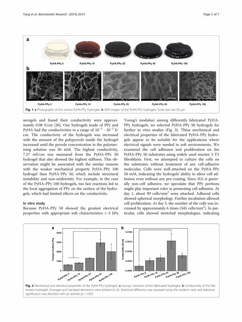
Results and discussionPyHA-PPy hydrogels fabricationThe various PyHA-PPy hydrogels were fabricated withthe different pyrrole concentrations (i.e., 0 mM, 10 mM,25 mM, 50 mM, and 100 mM) as shown in Table 1. Thefabricated PyHA-PPy hydrogels were clean and not brit-tle. First, PyHA conjugates were chemically synthesized(Scheme 1). N-(3-aminopropyl) pyrrole was conjugatedonto HA backbone using EDC/NHS chemistry. Hydro-gel formation was expected to result from the oxidativecoupling of the pyrrole moieties between HA chainsand/or the coupling between the polymerized PPy chainsand the conjugated pyrrole moieties presented on HA.The fabricated hydrogel in this manner could formstable covalent bonds between HA chains and the PPychains, allowing for its structural stability. The fact thatthe hydrogel could be formed even without any add-itional pyrrole monomers in the presence of the APSsuggests that the pyrrole moieties on PyHA were associ-ated to form covalent bonds. Furthermore, with an in-crease in a pyrrole monomer concentration, PPycontents in the PyHA-PPy hydrogels appeared to in-crease, which could consequently increase stiffness andelectrical conductivity. In our studies, as the pyrrolemonomer and oxidant concentrations increased, the re-sultant hydrogels exhibited darker color, which indicatesthat the added pyrrole monomers were oxidized intoPPy with the PyHA hydrogels. As mentioned above, sim-ple mixing of the PyHA solutions and APS without anyadditional pyrrole monomers could lead to hydrogel for-mation (Fig. 1a). It should be noted that the sizes ofhydrogels decreased after the PPy polymerization withoxidants. These size decreases of the hydrogels weremore distinct for the samples synthesized at higherpyrrole monomer concentrations (higher PPy con-tents). These results may result from the high en-tanglement degrees due to more chain units and/ordecreases in hydrophilicity due to increases in lesshydrophilic PPy portions.
PyHA-PPy hydrogel morphologiesInternal structures of the hydrogels were examined bySEM. All fabricated hydrogels showed the microporousstructures inside the hydrogel (Fig. 1b). The pore sizesappeared to be in the ranges of 10 μm. Interestingly,web-like structures with globular shape with size lessthan 100 nm were observed when the PPy portions werehigh in the hydrogels. These PPy structures were prom-inently observed from PyHA-PPy100 hydrogels andPyHA-PPy10 hydrogels. The conventional PPy was
Yang et al. Biomaterials Research (2016) 20:31 Page 3 of 7

reported to have sphere-like structures or web-likeglobular shape structures when polymerized chemicaloxidants. Observed web-like PPy morphologies implythat PPy chains grew inside the hydrogels.
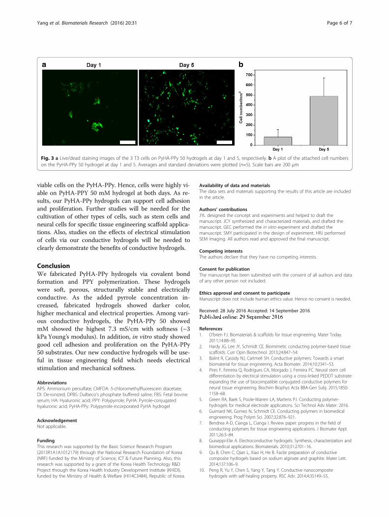
Characterization of PyHA-PPy hydrogelsThe modulus was measured using an oscillatory rheom-eter in a frequency sweep mode. The moduli of thehydrogels increased with increases in the added pyrrolemonomer concentrations by 50 mM pyrrole. Results in-dicate that PPy in the hydrogel might contribute the en-hancement of the modulus of the hydrogels. TheYoung’s modulus was in a range from 0.6 kPa to 3 kPa.However, the modulus decreased when the pyrrole con-centration was above 50 mM (Fig. 2a). PyHA-PPy 100hydrogels did not follow the general trend that the in-creases in the PPy portions inside the hydrogels result inthe increases of both the stiffness and electrical proper-ties. It may be due to heterogeneous composite
formation by heterogeneous PPy incorporation. Toohigh pyrrole concentrations and oxidants might lead totoo fast reaction rate inside the hydrogel or in the poly-merizing solution (outside the hydrogel). Since free pyr-role monomers can be oxidized more readily than thepyrrole moieties attached on PyHA, PPy formed in thesolution not in the hydrogels and deposited on the sur-faces of the hydrogels. Also, the pyrrole groups on thePyHA might not be sufficiently associated with PPypolymerization in PyHA-PPy 100 hydrogel, resulting ininsufficient covalent bond formation in PyHA-PPy andpoor stability of mechanical and electrical properties.The electrical conductivity of the hydrogels was mea-
sured (Fig. 2b). There were few reports about the meas-urement of conductivity of conductive hydrogels andtherefore it is difficult to directly compare the results.Hur et al. reported that the PPy agarose based hydrogelshowed the conductivity in the order of 10−1 S/cm [28].Shi et al. fabricated the cellulose/polypyrrole composite
Table 1 The names of various PyHA-PPy hydrogels and their synthetic conditions
Sample Names PyHA-PPy 0 PyHA-PPy 10 PyHA-PPy 25 PyHA-PPy 50 PyHA-PPy 100
PyHA solution 1.0 % 1.0 % 1.0 % 1.0 % 1.0 %
Pyrrole 0 mM 10 mM 25 mM 50 mM 100 mM
APS 50 mM 75 mM 100 mM 125 mM 100 mM
All concentrations were described as the final concentrations after mixing all components
Scheme 1 (a) Scheme of PyHA-PPy hydrogels synthesis. (b) Chemistry of N-(3-aminopropyl)pyrrole synthesis and pyrrole-HA conjugate synthesis(c) H1 NMR spectra of N-(3-aminopropyl)pyrrole (top) and PyHA conjugate (bottom)
Yang et al. Biomaterials Research (2016) 20:31 Page 4 of 7
aerogels and found their conductivity were approxi-mately 0.08 S/cm [26]. Our hydrogels made of PPy andPyHA had the conductivities in a range of 10−3 - 10−2 S/cm. The conductivity of the hydrogels was increasedwith the amount of the polypyrrole inside the hydrogelincreased until the pyrrole concentration in the polymer-izing solution was 50 mM. The highest conductivity,7.27 mS/cm was measured from the PyHA-PPy 50hydrogel that also showed the highest stiffness. This ob-servation might be associated with the similar reasonswith the weaker mechanical property PyHA-PPy 100hydrogel than PyHA-PPy 50, which include structuralinstability and non-uniformity. For example, in the caseof the PyHA-PPy 100 hydrogels, too fast reactions led tothe local aggregation of PPy on the surface of the hydro-gels, which had limited effects on the conductivity.
In vitro studyBecause PyHA-PPy 50 showed the greatest electricalproperties with appropriate soft characteristics (~3 kPa
Young’s modulus) among differently fabricated PyHA-PPy hydrogels, we selected PyHA-PPy 50 hydrogels forfurther in vitro studies (Fig. 3). These mechanical andelectrical properties of the fabricated PyHA-PPy hydro-gels appear to be suitable for the applications whereelectrical signals were needed in soft environments. Weexamined the cell adhesion and proliferation on thePyHA-PPy 50 substrates using widely used murine 3 T3fibroblasts. First, we attempted to culture the cells onthe substrates without treatment of any cell-adhesivemolecules. Cells were well-attached on the PyHA-PPy50 mM, indicating the hydrogels’ ability to allow cell ad-hesion even without any pre-coating. Since HA is gener-ally non-cell adhesive, we speculate that PPy portionsmight play important roles in promoting cell adhesion. Atday 1, about 90 cells/mm2 were attached. Adhered cellsshowed spherical morphology. Further incubation allowedcell proliferation. At day 5, the number of the cells was in-creased by approximately 6 times (545 cells/mm2). In par-ticular, cells showed stretched morphologies, indicating
Fig. 1 a Photographs of the various PyHA-PPy hydrogels. b SEM images of the PyHA-PPy hydrogels. Scale bars are 50 μm
Fig. 2 Mechanical and electrical properties of the PyHA-PPy hydrogels. a Young’s modulus of the fabricated hydrogels. b Conductivity of the fab-ricated hydrogels. Averages and standard deviations were plotted (n=3). Statistical difference was assessed using the student t-test and statisticalsignificance was denoted with an asterisk (p < 0.05)
Yang et al. Biomaterials Research (2016) 20:31 Page 5 of 7
viable cells on the PyHA-PPy. Hence, cells were highly vi-able on PyHA-PPY 50 mM hydrogel at both days. As re-sults, our PyHA-PPy hydrogels can support cell adhesionand proliferation. Further studies will be needed for thecultivation of other types of cells, such as stem cells andneural cells for specific tissue engineering scaffold applica-tions. Also, studies on the effects of electrical stimulationof cells via our conductive hydrogels will be needed toclearly demonstrate the benefits of conductive hydrogels.
ConclusionWe fabricated PyHA-PPy hydrogels via covalent bondformation and PPY polymerization. These hydrogelswere soft, porous, structurally stable and electricallyconductive. As the added pyrrole concentration in-creased, fabricated hydrogels showed darker color,higher mechanical and electrical properties. Among vari-ous conductive hydrogels, the PyHA-PPy 50 showedmM showed the highest 7.3 mS/cm with softness (~3kPa Young’s modulus). In addition, in vitro study showedgood cell adhesion and proliferation on the PyHA-PPy50 substrates. Our new conductive hydrogels will be use-ful in tissue engineering field which needs electricalstimulation and mechanical softness.
AbbreviationsAPS: Ammonium persulfate; CMFDA: 5-chloromethylfluorescein diacetate;DI: De-ionized; DPBS: Dulbeco’s phosphate buffered saline; FBS: Fetal bovineserum; HA: Hyaluronic acid; PPY: Polypyrrole; PyHA: Pyrrole-conjugatedhyaluronic acid; PyHA-PPy: Polypyrrole-incorporated PyHA hydrogel
AcknowledgementNot applicable.
FundingThis research was supported by the Basic Science Research Program(2013R1A1A1012179) through the National Research Foundation of Korea(NRF) funded by the Ministry of Science, ICT & Future Planning. Also, thisresearch was supported by a grant of the Korea Health Technology R&DProject through the Korea Health Industry Development Institute (KHIDI),funded by the Ministry of Health & Welfare (HI14C3484), Republic of Korea.
Availability of data and materialsThe data sets and materials supporting the results of this article are includedin the article.
Authors’ contributionsJYL designed the concept and experiments and helped to draft themanuscript. JCY synthesized and characterized materials, and drafted themanuscript. GEC performed the in vitro experiment and drafted themanuscript. SMY participated in the design of experiment. HRJ performedSEM imaging. All authors read and approved the final manuscript.
Competing interestsThe authors declare that they have no competing interests.
Consent for publicationThe manuscript has been submitted with the consent of all authors and dataof any other person not included.
Ethics approval and consent to participateManuscript does not include human ethics value. Hence no consent is needed.
Received: 28 July 2016 Accepted: 14 September 2016
References1. O’brien FJ. Biomaterials & scaffolds for tissue engineering. Mater Today.
2011;14:88–95.2. Hardy JG, Lee JY, Schmidt CE. Biomimetic conducting polymer-based tissue
scaffolds. Curr Opin Biotechnol. 2013;24:847–54.3. Balint R, Cassidy NJ, Cartmell SH. Conductive polymers: Towards a smart
biomaterial for tissue engineering. Acta Biomater. 2014;10:2341–53.4. Pires F, Ferreira Q, Rodrigues CA, Morgado J, Ferreira FC. Neural stem cell
differentiation by electrical stimulation using a cross-linked PEDOT substrate:expanding the use of biocompatible conjugated conductive polymers forneural tissue engineering. Biochim Biophys Acta BBA-Gen Subj. 2015;1850:1158–68.
5. Green RA, Baek S, Poole-Warren LA, Martens PJ. Conducting polymer-hydrogels for medical electrode applications. Sci Technol Adv Mater. 2016.
6. Guimard NK, Gomez N, Schmidt CE. Conducting polymers in biomedicalengineering. Prog Polym Sci. 2007;32:876–921.
7. Bendrea A-D, Cianga L, Cianga I. Review paper: progress in the field ofconducting polymers for tissue engineering applications. J Biomater Appl.2011;26:3–84.
8. Guiseppi-Elie A. Electroconductive hydrogels: Synthesis, characterization andbiomedical applications. Biomaterials. 2010;31:2701–16.
9. Qu B, Chen C, Qian L, Xiao H, He B. Facile preparation of conductivecomposite hydrogels based on sodium alginate and graphite. Mater Lett.2014;137:106–9.
10. Peng R, Yu Y, Chen S, Yang Y, Tang Y. Conductive nanocompositehydrogels with self-healing property. RSC Adv. 2014;4:35149–55.
Fig. 3 a Live/dead staining images of the 3 T3 cells on PyHA-PPy 50 hydrogels at day 1 and 5, respectively. b A plot of the attached cell numberson the PyHA-PPy 50 hydrogel at day 1 and 5. Averages and standard deviations were plotted (n=5). Scale bars are 200 μm
Yang et al. Biomaterials Research (2016) 20:31 Page 6 of 7

11. Guarino V, Alvarez‐Perez MA, Borriello A, Napolitano T, Ambrosio L.Conductive PANi/PEGDA macroporous hydrogels for nerve regeneration.Adv Healthc Mater. 2013;2:218–27.
12. Kim D, Abidian M, Martin DC. Conducting polymers grown in hydrogelscaffolds coated on neural prosthetic devices. J Biomed Mater Res A. 2004;71:577–85.
13. Thrivikraman G, Madras G, Basu B. Intermittent electrical stimuli for guidanceof human mesenchymal stem cell lineage commitment towards neural-likecells on electroconductive substrates. Biomaterials. 2014;35:6219–35.
14. Collins MN, Birkinshaw C. Hyaluronic acid based scaffolds for tissueengineering—A review. Carbohydr Polym. 2013;92:1262–79.
15. Prestwich GD. Hyaluronic acid-based clinical biomaterials derived for celland molecule delivery in regenerative medicine. J Controlled Release. 2011;155:193–9.
16. Segura T, Anderson BC, Chung PH, Webber RE, Shull KR, Shea LD.Crosslinked hyaluronic acid hydrogels: a strategy to functionalize andpattern. Biomaterials. 2005;26:359–71.
17. Kogan G, Šoltés L, Stern R, Gemeiner P. Hyaluronic acid: a naturalbiopolymer with a broad range of biomedical and industrial applications.Biotechnol Lett. 2007;29:17–25.
18. Yoo HS, Lee EA, Yoon JJ, Park TG. Hyaluronic acid modified biodegradablescaffolds for cartilage tissue engineering. Biomaterials. 2005;26:1925–33.
19. Solis MA, Chen Y-H, Wong TY, Bittencourt VZ, Lin Y-C, Huang LL.Hyaluronan regulates cell behavior: a potential niche matrix for stem cells.Biochem Res Int. 2012;2012.
20. Zhu H, Mitsuhashi N, Klein A, Barsky LW, Weinberg K, Barr ML, et al. The roleof the hyaluronan receptor CD44 in mesenchymal stem cell migration inthe extracellular matrix. Stem Cells. 2006;24:928–35.
21. Miyake K, Underhill CB, Lesley J, Kincade PW. Hyaluronate can function as acell adhesion molecule and CD44 participates in hyaluronate recognition. JExp Med. 1990;172:69–75.
22. Lei Y, Gojgini S, Lam J, Segura T. The spreading, migration and proliferationof mouse mesenchymal stem cells cultured inside hyaluronic acidhydrogels. Biomaterials. 2011;32:39–47.
23. Ateh D, Navsaria H, Vadgama P. Polypyrrole-based conducting polymersand interactions with biological tissues. J R Soc Interface. 2006;3:741–52.
24. Stewart E, Kobayashi NR, Higgins MJ, Quigley AF, Jamali S, Moulton SE, et al.Electrical stimulation using conductive polymer polypyrrole promotesdifferentiation of human neural stem cells: a biocompatible platform fortranslational neural tissue engineering. Tissue Eng Part C Methods. 2014;21:385–93.
25. Ahuja T, Mir IA, Kumar D. Biomolecular immobilization on conductingpolymers for biosensing applications. Biomaterials. 2007;28:791–805.
26. Shi Z, Gao H, Feng J, Ding B, Cao X, Kuga S, et al. In situ synthesis of robustconductive cellulose/polypyrrole composite aerogels and their potentialapplication in nerve regeneration. Angew Chem Int Ed. 2014;53:5380–4.
27. Abu-Rabeah K, Polyak B, Ionescu RE, Cosnier S, Marks RS. Synthesis andcharacterization of a pyrrole-alginate conjugate and its application in abiosensor construction. Biomacromolecules. 2005;6:3313–8.
28. Hur J, Im K, Kim SW, Kim J, Chung D-Y, Kim T-H, et al. Polypyrrole/agarose-based electronically conductive and reversibly restorable hydrogel. ACSNano. 2014;8:10066–76.
• We accept pre-submission inquiries
• Our selector tool helps you to find the most relevant journal
• We provide round the clock customer support
• Convenient online submission
• Thorough peer review
• Inclusion in PubMed and all major indexing services
• Maximum visibility for your research
Submit your manuscript atwww.biomedcentral.com/submit
Submit your next manuscript to BioMed Central and we will help you at every step:
Yang et al. Biomaterials Research (2016) 20:31 Page 7 of 7