Polymerase Chain Reaction (PCR) - anna onofriannaonofri.net/files/pcr.pdf · Polymerase Chain...
31
Polymerase Chain Reaction (PCR) • PCR is a means to amplify a particular piece of DNA • Amplify= making numerous copies of a segment of DNA • PCR can make billions of copies of a target sequence of DNA in a few hours • PCR was invented in the 1984 as a way to make numerous copies of DNA fragments in the laboratory • Its applications are vast and PCR is now an integral part of Molecular Biology
Transcript of Polymerase Chain Reaction (PCR) - anna onofriannaonofri.net/files/pcr.pdf · Polymerase Chain...
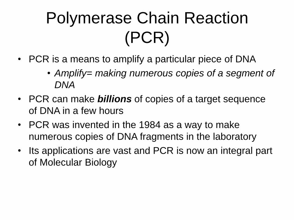
Polymerase Chain Reaction
(PCR)
• PCR is a means to amplify a particular piece of DNA
• Amplify= making numerous copies of a segment of
DNA
• PCR can make billions of copies of a target sequence
of DNA in a few hours
• PCR was invented in the 1984 as a way to make
numerous copies of DNA fragments in the laboratory
• Its applications are vast and PCR is now an integral part
of Molecular Biology

DNA Replication vs. PCR
• PCR is a laboratory version of DNA
Replication in cells• The laboratory version is commonly called “in vitro”
since it occurs in a test tube while “in vivo” signifies
occurring in a living cell.
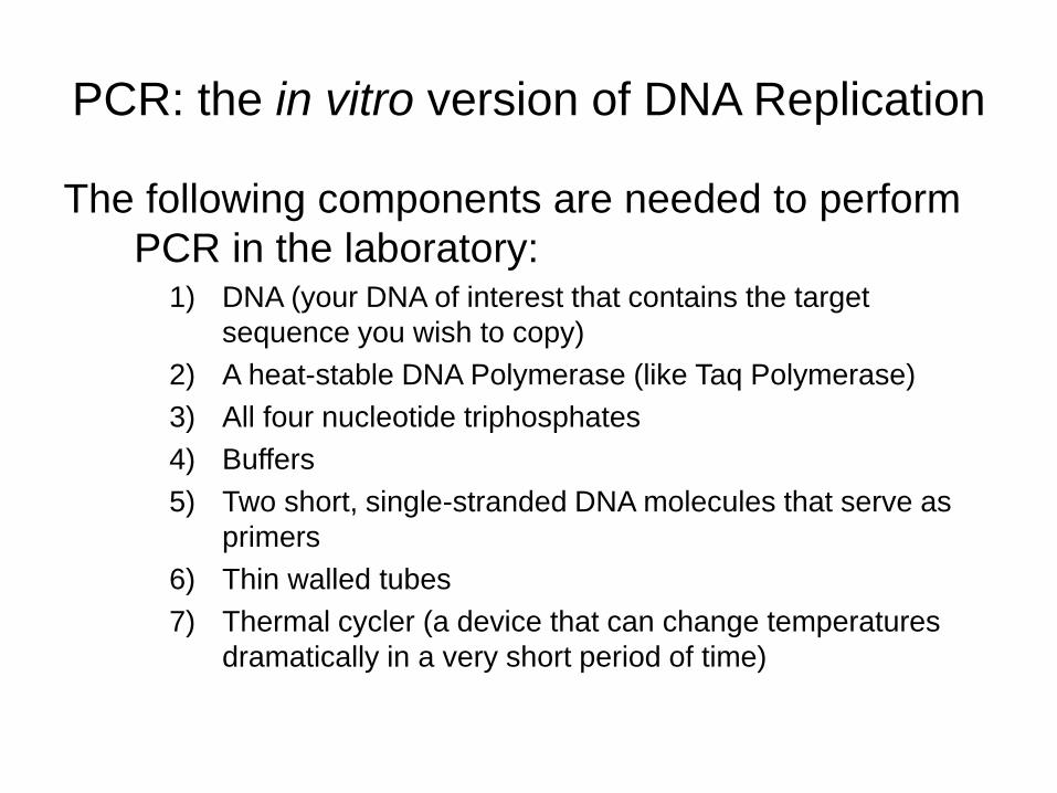
PCR: the in vitro version of DNA Replication
The following components are needed to perform
PCR in the laboratory:1) DNA (your DNA of interest that contains the target
sequence you wish to copy)
2) A heat-stable DNA Polymerase (like Taq Polymerase)
3) All four nucleotide triphosphates
4) Buffers
5) Two short, single-stranded DNA molecules that serve as
primers
6) Thin walled tubes
7) Thermal cycler (a device that can change temperatures
dramatically in a very short period of time)
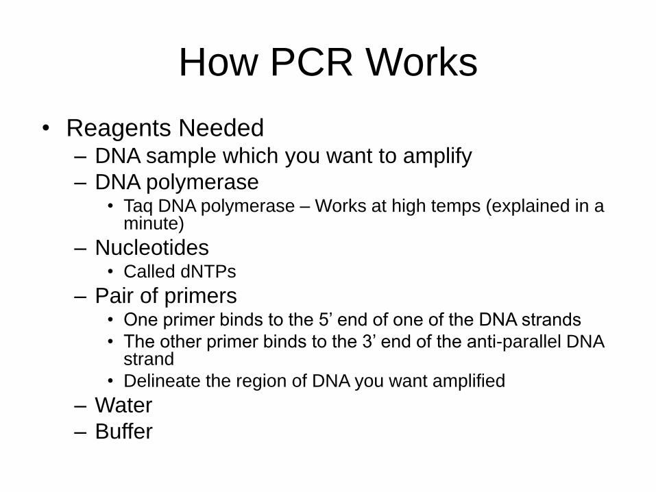
How PCR Works
• Reagents Needed– DNA sample which you want to amplify
– DNA polymerase• Taq DNA polymerase – Works at high temps (explained in a
minute)
– Nucleotides• Called dNTPs
– Pair of primers• One primer binds to the 5’ end of one of the DNA strands
• The other primer binds to the 3’ end of the anti-parallel DNA strand
• Delineate the region of DNA you want amplified
– Water
– Buffer

How PCR Works
• Protocol
– Put all reagents into a PCR tube
– Break the DNA ladder down the middle to create two strands, a 5’ to 3’ strand and a 3’ to 5’ strand
• Melting or heat denaturation
– Bind each primer to its appropriate strand• 5’ primer to the 5’ to 3’ strand
• 3’ primer to the 3’ to 5’ strand
– Annealing
– Copy each strand• DNA polymerase
– Extending

How PCR Works
• Temperature Protocol
– Initial Melt: 94ºC for 2 minutes
– Melt: 94ºC for 30 seconds
– Anneal: 55ºC for 30 seconds
– Extend: 72ºC for 1 minute
– Final Extension: 72ºC for 6 minutes
– Hold: 4ºC
30-35 cycles
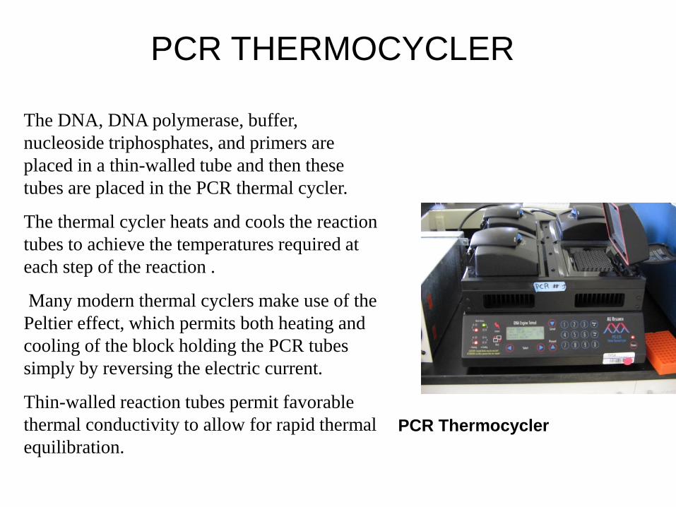
PCR THERMOCYCLER
The DNA, DNA polymerase, buffer,
nucleoside triphosphates, and primers are
placed in a thin-walled tube and then these
tubes are placed in the PCR thermal cycler.
The thermal cycler heats and cools the reaction
tubes to achieve the temperatures required at
each step of the reaction .
Many modern thermal cyclers make use of the
Peltier effect, which permits both heating and
cooling of the block holding the PCR tubes
simply by reversing the electric current.
Thin-walled reaction tubes permit favorable
thermal conductivity to allow for rapid thermal
equilibration.
PCR Thermocycler

The three main steps of PCR• The basis of PCR is temperature changes and the effect that these
temperature changes have on the DNA.
• In a PCR reaction, the following series of steps is repeated 20-40 times
(note: 25 cycles usually takes about 2 hours and amplifies the DNA fragment of interest 100,000 fold)
Step 1: Denature DNA
At 95C, the DNA is denatured (i.e. the two strands are separated)
Step 2: Primers Anneal
At 40C- 65C, the primers anneal (or bind to) their complementary sequences on the single strands of DNA
Step 3: DNA polymerase Extends the DNA chain
At 72C, DNA Polymerase extends the DNA chain by adding nucleotides to the 3’ ends of the primers.

Heat-stable DNA Polymerase
• Given that PCR involves very high temperatures,
it is imperative that a heat-stable DNA
polymerase be used in the reaction. • Most DNA polymerases would denature (and thus not
function properly) at the high temperatures of PCR.
• Taq DNA polymerase was purified from the hot
springs bacterium Thermus aquaticus in 1976
• Taq has maximal enzymatic activity at 75 C to
80 C, and substantially reduced activities at
lower temperatures.
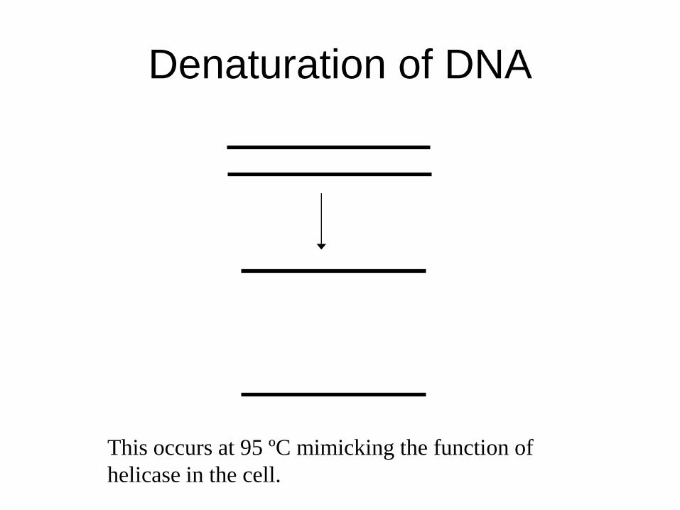
Denaturation of DNA
This occurs at 95 ºC mimicking the function of
helicase in the cell.
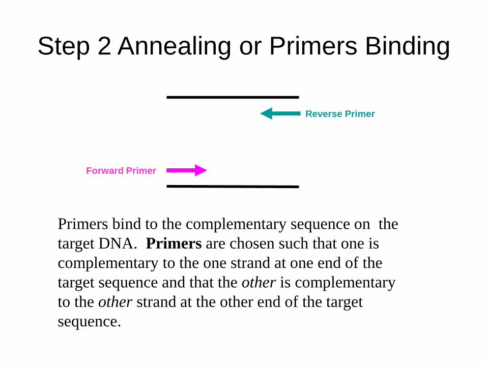
Step 2 Annealing or Primers Binding
Primers bind to the complementary sequence on the
target DNA. Primers are chosen such that one is
complementary to the one strand at one end of the
target sequence and that the other is complementary
to the other strand at the other end of the target
sequence.
Forward Primer
Reverse Primer

Step 3 Extension or Primer Extension
DNA polymerase catalyzes the extension of the
strand in the 5-3 direction, starting at the
primers, attaching the appropriate nucleotide
(A-T, C-G)
extension
extension

• The next cycle will begin by denaturing
the new DNA strands formed in the
previous cycle

Theoretical amplification rate: 2n

The Size of the DNA Fragment Produced
in PCR is Dependent on the Primers
• The PCR reaction will amplify the DNA section between the two
primers.
• If the DNA sequence is known, primers can be developed to amplify
any piece of an organism’s DNA.
Forward primer
Reverse primer
Size of fragment that is amplified

The DNA of interest is amplified by
a power of 2 for each PCR cycle
For example, if you subject your DNA of interest to 5 cycles of
PCR, you will end up with 25 (or 64) copies of DNA.
Similarly, if you subject your DNA of interest to 40 cycles of
PCR, you will end up with 240 (or 1,099511612 ) copies of
DNA!

• To check whether the PCR generated the
anticipated DNA fragment (also
sometimes referred to as the amplimer or
amplicon), agarose gel electrophoresis
is employed for size separation of the
PCR products.
• The size(s) of PCR products is
determined by comparison with a DNA
ladder (a molecular weight marker),
which contains DNA fragments of known
size, run on the gel alongside the PCR
products
Gel Electrophoresis of DNA

Gel Electrophoresis of DNA
• Gel electrophoresis detects the presence of DNA
in a sample
• Gel electrophoresis detects the number of
nucleotides in a fragment of DNA
– e.g., the number of nucleotides in a DNA region which
was amplified by PCR
– Is a rough estimate, is not exact, need more
sophisticated sequencing techniques to get an exact
number of nucleotides
– Can be used to tentatively identify a gene because
we know the number of nucleotides in many genes

How Gel Electrophoresis of DNA Works
• A sample which contains fragments of DNA is forced by an electrical
current through a firm gel which is really a sieve with small holes of
a fixed size
– Phosphate group in DNA is negatively charged so it is moved towards a
positive electrode by the current
– Longer fragments have more nucleotides
• So have a larger molecular weight
• So are bigger in size
• So aren’t able to pass through the small holes in the gel and get hung up at
the beginning of the gel
– Shorter fragments are able to pass through and move farther along the
gel
– Fragments of intermediate length travel to about the middle of the gel
• DNA fragments are then visualized in the gel with a special dye
• The number of nucleotides are then estimated by comparing it to a
known sample of DNA fragments which is run through the gel at the
same time
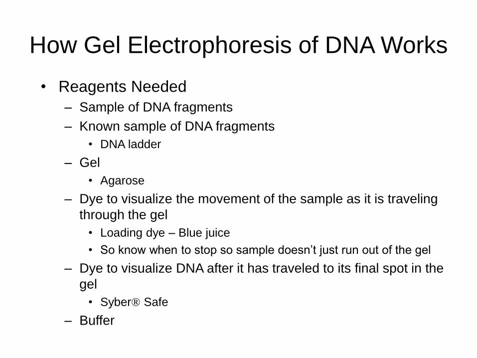
How Gel Electrophoresis of DNA Works
• Reagents Needed
– Sample of DNA fragments
– Known sample of DNA fragments
• DNA ladder
– Gel
• Agarose
– Dye to visualize the movement of the sample as it is traveling
through the gel
• Loading dye – Blue juice
• So know when to stop so sample doesn’t just run out of the gel
– Dye to visualize DNA after it has traveled to its final spot in the
gel
• Syber® Safe
– Buffer
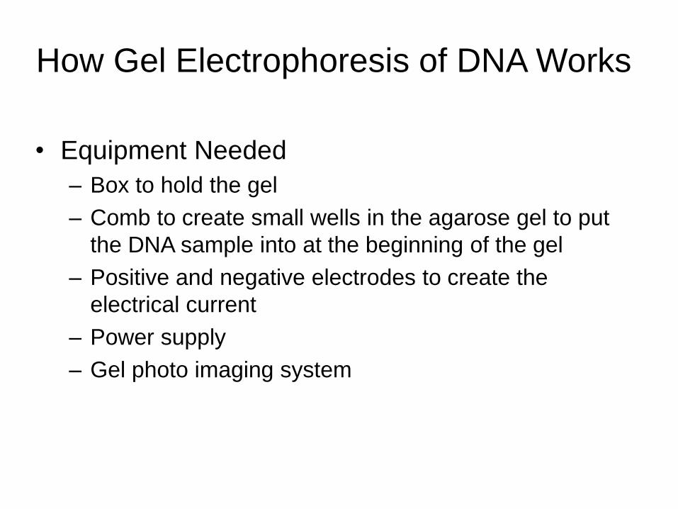
How Gel Electrophoresis of DNA Works
• Equipment Needed
– Box to hold the gel
– Comb to create small wells in the agarose gel to put
the DNA sample into at the beginning of the gel
– Positive and negative electrodes to create the
electrical current
– Power supply
– Gel photo imaging system

How Gel Electrophoresis of DNA Works

PCR has become a very powerful
tool in molecular biology
• One can start with a single sperm cell or stand of
hair and amplify the DNA sufficiently to allow for
DNA analysis and a distinctive band on an
agarose gel.
• One can amplify fragments of interest in an
organism’s DNA by choosing the right primers.
• One can use the selectivity of the primers to
identify the likelihood of an individual carrying a
particular allele of a gene.
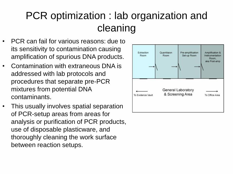
PCR optimization : lab organization and
cleaning• PCR can fail for various reasons: due to
its sensitivity to contamination causing
amplification of spurious DNA products.
• Contamination with extraneous DNA is
addressed with lab protocols and
procedures that separate pre-PCR
mixtures from potential DNA
contaminants.
• This usually involves spatial separation
of PCR-setup areas from areas for
analysis or purification of PCR products,
use of disposable plasticware, and
thoroughly cleaning the work surface
between reaction setups.
PCR optimization : Primers
• Primer-design techniques are important in improving PCR product yield and in avoiding the formation of spurious products, and the usage of alternate buffer components or polymerase enzymes can help with amplification of long or otherwise problematic regions of DNA
• PCR primers are short, single stranded DNA molecules (15-40 bp)
• They are manufactured commercially and can be ordered to match any DNA sequence
• Primers are sequence specific, they will bind to a particular sequence in a genome
• As you design primers with a longer length (15 → 40 bp), the primers become more selective.
• DNA polymerase requires primers to initiate replication

Selectivity of Primers
• Primers bind to their complementary sequence
on the target DNA
– A primer composed of only 3 letter, ACC, for example,
would be very likely to encounter its complement in a
genome.
– As the size of the primer is increased, the likelihood
of, for example, a primer sequence of 35 base letters
repeatedly encountering a perfect complementary
section on the target DNA become remote.
PCR and Disease
• Primers can be created that will only bind and amplify certain alleles of genes or mutations of genes
• This is the basis of genetic counseling and PCR is used as part of the diagnostic tests for genetic diseases.
• Some diseases that can be diagnosed with the help of PCR:
• Huntington's disease
• cystic fibrosis
• Human immunodeficiency virus

Huntington’s Disease (HD)
• HD is a genetic disorder characterized by abnormal body
movements and reduced mental abilities
• HD is caused by a mutation in the Huntingtin (HD) gene
• In individuals with HD, the HD gene is “expanded”
– In non-HD individuals, the HD gene has a pattern called trinucleotide
repeats with “CAG” occurring in repetition less than 30 times.
– IN HD individuals, the “CAG” trinucleotide repeat occurs more that 36
times in the HD gene
• PCR can be performed on an individual’s DNA to determine whether
the individual has HD.
– The DNA is amplified via PCR and sequenced (a technique by which
the exact nucleotide sequence is determined) and the number of
trinucleotide repeats is then counted.

Cystic Fibrosis (CF)
• CF is a genetic disease characterized by severe breathing
difficulties and a predisposition to infections.
• CF is caused by mutations in the cystic fibrosis transmembrane
conductance regulator (CTFR) gene.
• In non-CF individuals, the CTFR gene codes for a protein that is a
chloride ion channel and is involved in the production of sweat,
digestive juices and mucus.
• In CF individuals, mutations in the CTFR gene lead to thick mucous
secretions in the lungs and subsequent persistent bacterial
infections.
• The presence of CTFR mutations in a individual can be detected by
performing PCR and sequencing on that individual’s DNA.
Human Immunodeficiency Virus (HIV)
• HIV is a retrovirus that attacks the immune system.
• HIV tests rely on PCR with primers that will only amplify
a section of the viral DNA found in an infected
individual’s bodily fluids.
Therefore if there is a PCR product, the person is likely to be
HIV positive. If there is no PCR product the person is likely to
be HIV negative.
• Protein detection based tests are available as well but all US blood
is tested by PCR.

PCR and Forensic Science
• Forensic science is the application of a broad spectrum of sciences to answer questions of interest to the legal system. This may be in relation to a crime or to a civil action.
• It is often of interest in forensic science to identify individuals genetically. In these cases, one is interested in looking at variable regions of the genome as opposed to highly-conserved genes.
• PCR can be used to amplify highly variable regions of the human genome. These regions contain runs of short, repeated sequences (known as variable number of tandem repeat (VNTR) sequences) . The number of repeats can vary from 4-40 in different individuals.
• Primers are chosen that will amplify these repeated areas and the genomic fragments generated give us a unique “genetic fingerprint” that can be used to identify an individual.