Polymer sieving matrices in microanalytical...
20
Polymer sieving matrices in microanalytical electrophoresis Minsub Chung,† a Dohyun Kim† b and Amy E. Herr * cd Microfluidic design has advanced existing protein separation capabilities and supported novel assays. Key metrics for successful protein separations include fast, robust, and sensitive analysis of complex mixtures of bio-macromolecules. Attaining high separation resolution is a chief concern. Here we review recent advances in polymer-based electrophoresis sieving materials that are impacting microfluidic bioanalytical applications. Looking forward, we comment on unmet needs for advanced separation media in micro- to-nanoscale devices. 1. Introduction Polyacrylamide slab-gel electrophoresis has been a workhorse protein analysis tool for over four decades. In polyacrylamide gel electrophoresis (PAGE), the hydrogel acts as a molecular sieving matrix underpinning size-based separation of proteins, thus enabling more straightforward protein identication than conferred by determining the charge-to-mass ratio. 1 To accu- rately determine protein molecular mass, proteins are treated with the surfactant sodium dodecyl sulphate (SDS), 2,3 making slab-gel SDS-PAGE a dominant protein analysis tool. 4 Poly- acrylamide gel is also an anti-convective medium yielding sharp separated protein zones with minimal convective dispersion. 1 However, slab-gel electrophoresis is far from ideal; the tool suffers from: appreciable sample consumption, signicant heat generation, poor reproducibility, semi-quantitative results due to run-to-run variability, laborious manual handling, and cumbersome post-separation analysis relying on gel staining and washing. 1 To overcome the limitations of slab-gel electrophoresis, capillary gel electrophoresis was introduced. 5,6 Electrophoresis in small bore glass capillaries (diameter: 10–300 mm) offers performance metrics that can be superior to slab-gels including: small sample consumption, fast separation speeds enabled by high applied electric elds, 7 UV- 7 or LIF (laser-induced uores- cence)-based on-line detection, 1 a capacity for quantitative analysis, efficient cooling by effective heat dissipation, 7 and automated operation. 1 While slab-gels separate multiple protein samples per gel, capillaries can be arrayed for the parallel protein analysis. 7 Introduction of capillary-lling gels (polyacrylamide, agarose) yielded a hybrid separation technique offering fast size determination with low peak dispersion. 5,6 Crosslinked gels were supplanted by “replaceable” non- crosslinked water-soluble polymers, such as linear poly- acrylamide (LPA). 8 Replaceable gels gained popularity, as capillaries lled with crosslinked gels cannot be readily replenished by pressure-driven ushing of the channel. Refreshing the sieving media allows reuse of a channel, even if the separation matrix has deteriorated. Specically, complex biological samples oen clog the sieving matrix and sometimes bubbles form because of Joule heating in the channel. Polymer solutions, although not crosslinked, are “entangled” to form the dynamic polymer network acting as a molecular sieve. 7 A consideration of non-crosslinked polymer separation media is the need for wall surface coatings to minimize EOF (electroos- motic ow) and analyte adsorption; both are sources of dispersion. Electrophoretic separation conducted in microdevices has been a dominant trend in modern analytical chemistry owing to the possibility of mass fabrication by exploiting the mature microfabrication techniques derived from the semiconductor industry. Further interest stems from the capability of inte- grating pre- and post-separation analytical steps for streamlined protein analysis (e.g., the lab-on-a-chip concept) and the possi- bility of massive parallelization. 9–11 Analytical instrumentation has been adopted from capillary electrophoresis (CE) and adapted to microuidic electrophoresis (e.g., high-voltage power supply, LIF detection, and crosslinked or non-cross- linked sieving media). For microuidic gel electrophoresis, linear polymers have been used extensively due to the benets of replaceable gels, while crosslinked gels have only recently drawn attention from the analytical community. In particular, in situ polymerized crosslinked polyacrylamide gels are a Department of Chemical Engineering, Hongik University, Mapo-gu, Seoul, 121-791, Republic of Korea b Department of Mechanical Engineering, Myongji University, Cheoin-gu, Yongin, 449- 728, Republic of Korea c Department of Bioengineering, University of California, Berkeley, CA 94706, USA. E-mail: [email protected] d The University of California, Berkeley – University of California, San Francisco Graduate Program in Bioengineering, Berkeley, CA 94706, USA † M. Chung and D. Kim equally contributed to this work. Cite this: Analyst, 2014, 139, 5635 Received 1st July 2014 Accepted 7th August 2014 DOI: 10.1039/c4an01179a www.rsc.org/analyst This journal is © The Royal Society of Chemistry 2014 Analyst, 2014, 139, 5635–5654 | 5635 Analyst CRITICAL REVIEW Published on 08 August 2014. Downloaded by Myongji University on 15/10/2014 11:13:57. View Article Online View Journal | View Issue
Transcript of Polymer sieving matrices in microanalytical...
Analyst
CRITICAL REVIEW
Publ
ishe
d on
08
Aug
ust 2
014.
Dow
nloa
ded
by M
yong
ji U
nive
rsity
on
15/1
0/20
14 1
1:13
:57.
View Article OnlineView Journal | View Issue
Polymer sieving
aDepartment of Chemical Engineering, Hon
Republic of KoreabDepartment of Mechanical Engineering, My
728, Republic of KoreacDepartment of Bioengineering, University
E-mail: [email protected] University of California, Berkeley –
Graduate Program in Bioengineering, Berke
† M. Chung and D. Kim equally contribut
Cite this: Analyst, 2014, 139, 5635
Received 1st July 2014Accepted 7th August 2014
DOI: 10.1039/c4an01179a
www.rsc.org/analyst
This journal is © The Royal Society of C
matrices in microanalyticalelectrophoresis
Minsub Chung,†a Dohyun Kim†b and Amy E. Herr*cd
Microfluidic design has advanced existing protein separation capabilities and supported novel assays. Key
metrics for successful protein separations include fast, robust, and sensitive analysis of complex mixtures
of bio-macromolecules. Attaining high separation resolution is a chief concern. Here we review recent
advances in polymer-based electrophoresis sieving materials that are impacting microfluidic bioanalytical
applications. Looking forward, we comment on unmet needs for advanced separation media in micro-
to-nanoscale devices.
1. Introduction
Polyacrylamide slab-gel electrophoresis has been a workhorseprotein analysis tool for over four decades. In polyacrylamidegel electrophoresis (PAGE), the hydrogel acts as a molecularsieving matrix underpinning size-based separation of proteins,thus enabling more straightforward protein identication thanconferred by determining the charge-to-mass ratio.1 To accu-rately determine protein molecular mass, proteins are treatedwith the surfactant sodium dodecyl sulphate (SDS),2,3 makingslab-gel SDS-PAGE a dominant protein analysis tool.4 Poly-acrylamide gel is also an anti-convective medium yielding sharpseparated protein zones with minimal convective dispersion.1
However, slab-gel electrophoresis is far from ideal; the toolsuffers from: appreciable sample consumption, signicant heatgeneration, poor reproducibility, semi-quantitative results dueto run-to-run variability, laborious manual handling, andcumbersome post-separation analysis relying on gel stainingand washing.1
To overcome the limitations of slab-gel electrophoresis,capillary gel electrophoresis was introduced.5,6 Electrophoresisin small bore glass capillaries (diameter: 10–300 mm) offersperformancemetrics that can be superior to slab-gels including:small sample consumption, fast separation speeds enabled byhigh applied electric elds,7 UV-7 or LIF (laser-induced uores-cence)-based on-line detection,1 a capacity for quantitativeanalysis, efficient cooling by effective heat dissipation,7 and
gik University, Mapo-gu, Seoul, 121-791,
ongji University, Cheoin-gu, Yongin, 449-
of California, Berkeley, CA 94706, USA.
University of California, San Francisco
ley, CA 94706, USA
ed to this work.
hemistry 2014
automated operation.1 While slab-gels separate multipleprotein samples per gel, capillaries can be arrayed for theparallel protein analysis.7 Introduction of capillary-lling gels(polyacrylamide, agarose) yielded a hybrid separation techniqueoffering fast size determination with low peak dispersion.5,6
Crosslinked gels were supplanted by “replaceable” non-crosslinked water-soluble polymers, such as linear poly-acrylamide (LPA).8 Replaceable gels gained popularity, ascapillaries lled with crosslinked gels cannot be readilyreplenished by pressure-driven ushing of the channel.Refreshing the sieving media allows reuse of a channel, even ifthe separation matrix has deteriorated. Specically, complexbiological samples oen clog the sieving matrix and sometimesbubbles form because of Joule heating in the channel. Polymersolutions, although not crosslinked, are “entangled” to form thedynamic polymer network acting as a molecular sieve.7 Aconsideration of non-crosslinked polymer separation media isthe need for wall surface coatings to minimize EOF (electroos-motic ow) and analyte adsorption; both are sources ofdispersion.
Electrophoretic separation conducted in microdevices hasbeen a dominant trend in modern analytical chemistry owing tothe possibility of mass fabrication by exploiting the maturemicrofabrication techniques derived from the semiconductorindustry. Further interest stems from the capability of inte-grating pre- and post-separation analytical steps for streamlinedprotein analysis (e.g., the lab-on-a-chip concept) and the possi-bility of massive parallelization.9–11 Analytical instrumentationhas been adopted from capillary electrophoresis (CE) andadapted to microuidic electrophoresis (e.g., high-voltagepower supply, LIF detection, and crosslinked or non-cross-linked sieving media). For microuidic gel electrophoresis,linear polymers have been used extensively due to the benetsof replaceable gels, while crosslinked gels have only recentlydrawn attention from the analytical community. In particular,in situ polymerized crosslinked polyacrylamide gels are
Analyst, 2014, 139, 5635–5654 | 5635
Analyst Critical Review
Publ
ishe
d on
08
Aug
ust 2
014.
Dow
nloa
ded
by M
yong
ji U
nive
rsity
on
15/1
0/20
14 1
1:13
:57.
View Article Online
compatible with photolithographic fabrication and provideunique advantages including the possibilities of: integratingmultiple functionalities into a single chip and extremely shortseparation distances which support large-scale integration ofelectrophoresis in a single microdevice.
Ideally, sieving matrices for electrophoresis separationswould offer: (1) a uniform pore distribution for reproducibleseparation, (2) no adsorption to analytes and low-to-no back-ground signal, (3) compatibility to an appropriate detectionmethod [e.g., uorescence detection, mass spectrometry orsurface enhanced Raman spectroscopy (SERS)], and (4) robustoperation (e.g., resistance to high electric elds, and chemicaldecomposition). In addition to these essentials are propertiesspecic to each application or format.
In this review, we focus on polymer sieving matricesprepared inside channels or microuidic devices for advancedproteomic applications. We have intentionally omitted devel-opments in separation media developed primarily for off-chipanalytical operations. Instead of considering historical rarelyused separation media, we emphasize recent novel approachesand materials. We summarize separation matrix properties,common or particularly noteworthy separation conditions, andinteresting applications using these functional materials.
2. Microanalytical separationmechanisms in crosslinked gels andentangled polymer solutions
The native conformation of protein species spans a staggeringrange of molecular masses, geometric shapes, and surfacecharges. Electrophoresis separations of native proteins arisefrom a differential mobility, m, between species of interest. Theelectrophoretic mobility is a function of the charge-to-massratio of each protein species. The mobility difference can beenhanced using a molecular sieving matrix (as compared to freesolution), so that protein bands migrate as sharp zones,resolved from neighbouring species.1 Denatured proteins canunfold and adopt a rod-like shape.12 When proteins complexwith a detergent like SDS, all proteins acquire the similarcharge-to-mass ratio and, thus, the near identical electropho-retic mobility in background buffer.13 Thus, in free solution, thespecies would not resolve from each other during electropho-resis. In contrast, during electromigrating through a sievingmatrix, the SDS-treated proteins interact with the molecularsieve yielding different electrophoretic mobilities for thedifferent species. The mobility in this case depends on molec-ular mass, which affords size-based protein separations andultimately allows molecular-mass determination. Because ofthese advantages, electrophoresis of proteins is commonlyperformed using water-soluble molecular-sieving polymernetworks.
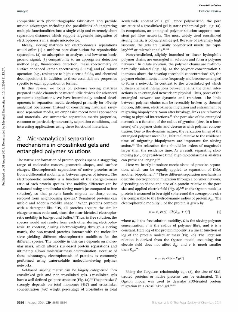
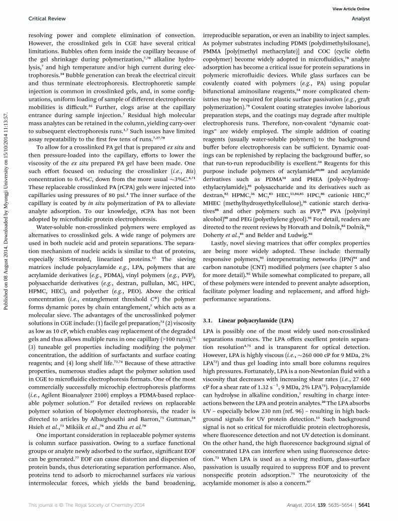
Gel-based sieving matrix can be largely categorized intocrosslinked gels and non-crosslinked gels. Crosslinked gelshave a well-dened gel pore structure (Fig. 1a).12 The pore size xstrongly depends on total monomer (%T) and crosslinkerconcentration (%C, weight percentage of crosslinker in total
5636 | Analyst, 2014, 139, 5635–5654
acrylamide content of a gel). Once polymerized, the porestructure of a crosslinked gel is static (“chemical gel”, Fig. 1a).In comparison, an entangled polymer solution supports tran-sient gel bre networks. The most widely used crosslinkedsieving matrix is polyacrylamide gel. Because of extremely highviscosity, the gels are usually polymerized inside the capil-lary5,6,14 or microchannels.15–24
Non-crosslinked, slightly branched or linear hydrophilicpolymer chains are entangled in solution and form a polymernetwork.1 In dilute solution, the polymer chains are hydrody-namically isolated (Fig. 1b). As the polymer concentration Cincreases above the “overlap threshold concentration” C*, thepolymer chains interact more frequently and become entangledto form a network. In contrast to the crosslinked gel whichutilizes chemical interactions between chains, the chain inter-actions in an entangled network are physical. Thus, pores of theentangled network are dynamic and transient. The linksbetween polymer chains can be reversibly broken by thermalmotion, diffusion, electrokinetic migration and entrainment bymigrating biopolymers. Soon aer breakage, links are reformedowing to physical interactions.25 The pore size of the entanglednetwork is a function of the radius of gyration (size, in a loosesense) of a polymer chain and decreases with polymer concen-tration. Due to the dynamic nature, the relaxation times of theentangled polymer mesh (i.e., lifetime) relative to the residencetime of migrating biopolymers are important for sievingaction.26 The relaxation time should be orders of magnitudelarger than the residence time. As a result, separating slow-moving (i.e., long residence time) high-molecular-mass analytescan prove challenging.26
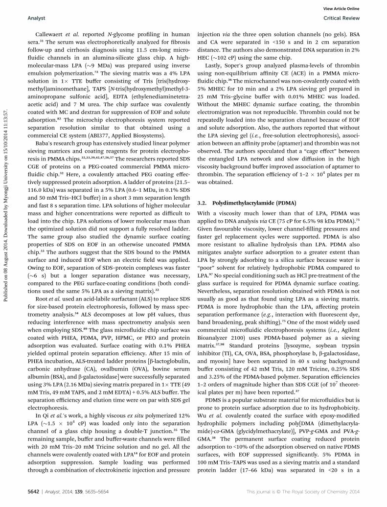
Here we briey introduce mechanisms of proteins separa-tion, which can be equally applied to separation of DNA,another biopolymer.7,12 Three different separation mechanismsdescribe electrophoretic migration through a polymer network,depending on shape and size of a protein relative to the poresize and applied electric eld (Fig. 2).1,12 In the Ogston model, aprotein is assumed to be a rigid sphere and the average pore sizex is comparable to the hydrodynamic radius of protein Rgp. Theelectrophoretic mobility m of the protein is given by:
m ¼ m0 exp[�Cb(Rgp + r)2] (1)
where m0 is the free-solution mobility, C is the sieving-polymerconcentration, r is the radius of polymer bre, and b is aconstant. Here log of the protein mobility is a linear function oflog of the protein molecular mass (Fig. 2b). The Fergusonrelation is derived from the Ogston model, assuming thatelectric eld does not affect Rgp and r is much smallerthan Rgp:28
m ¼ m0 exp[�KRC] (2)
Using the Ferguson relationship eqn (2), the size of SDS-treated proteins or native proteins can be estimated. TheOgston model was used to describe SDS-treated proteinmigration in a crosslinked gel.28,29
This journal is © The Royal Society of Chemistry 2014
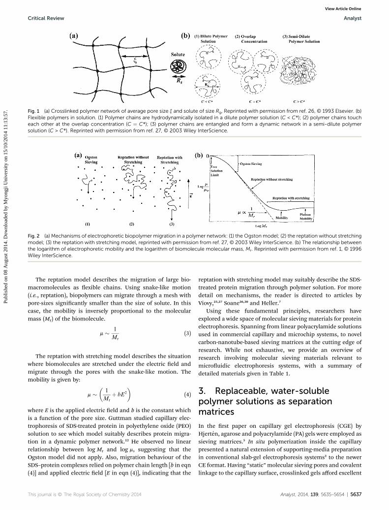
Fig. 1 (a) Crosslinked polymer network of average pore size x and solute of size Rg, Reprinted with permission from ref. 26, © 1993 Elsevier. (b)Flexible polymers in solution. (1) Polymer chains are hydrodynamically isolated in a dilute polymer solution (C < C*); (2) polymer chains toucheach other at the overlap concentration (C ¼ C*); (3) polymer chains are entangled and form a dynamic network in a semi-dilute polymersolution (C > C*). Reprinted with permission from ref. 27, © 2003 Wiley InterScience.
Fig. 2 (a) Mechanisms of electrophoretic biopolymer migration in a polymer network: (1) the Ogstonmodel; (2) the reptation without stretchingmodel; (3) the reptation with stretching model, reprinted with permission from ref. 27, © 2003 Wiley InterScience. (b) The relationship betweenthe logarithm of electrophoretic mobility and the logarithm of biomolecule molecular mass, Mr. Reprinted with permission from ref. 1, © 1996Wiley InterScience.
Critical Review Analyst
Publ
ishe
d on
08
Aug
ust 2
014.
Dow
nloa
ded
by M
yong
ji U
nive
rsity
on
15/1
0/20
14 1
1:13
:57.
View Article Online
The reptation model describes the migration of large bio-macromolecules as exible chains. Using snake-like motion(i.e., reptation), biopolymers can migrate through a mesh withpore-sizes signicantly smaller than the size of solute. In thiscase, the mobility is inversely proportional to the molecularmass (Mr) of the biomolecule.
m � 1
Mr
(3)
The reptation with stretching model describes the situationwhere biomolecules are stretched under the electric eld andmigrate through the pores with the snake-like motion. Themobility is given by:
m ��
1
Mr
þ bE2
�(4)
where E is the applied electric eld and b is the constant whichis a function of the pore size. Guttman studied capillary elec-trophoresis of SDS-treated protein in polyethylene oxide (PEO)solution to see which model suitably describes protein migra-tion in a dynamic polymer network.12 He observed no linearrelationship between log Mr and log m, suggesting that theOgston model did not apply. Also, migration behaviour of theSDS–protein complexes relied on polymer chain length [b in eqn(4)] and applied electric eld [E in eqn (4)], indicating that the
This journal is © The Royal Society of Chemistry 2014
reptation with stretching model may suitably describe the SDS-treated protein migration through polymer solution. For moredetail on mechanisms, the reader is directed to articles byViovy,25,27 Soane26,30 and Heller.7
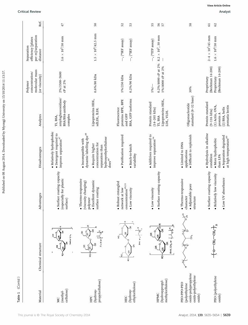
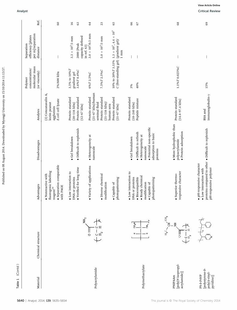
Using these fundamental principles, researchers haveexplored a wide space of molecular sieving materials for proteinelectrophoresis. Spanning from linear polyacrylamide solutionsused in commercial capillary and microchip systems, to novelcarbon-nanotube-based sieving matrices at the cutting edge ofresearch. While not exhaustive, we provide an overview ofresearch involving molecular sieving materials relevant tomicrouidic electrophoresis systems, with a summary ofdetailed materials given in Table 1.
3. Replaceable, water-solublepolymer solutions as separationmatrices
In the rst paper on capillary gel electrophoresis (CGE) byHjerten, agarose and polyacrylamide (PA) gels were employed assieving matrices.5 In situ polymerization inside the capillarypresented a natural extension of supporting-media preparationin conventional slab-gel electrophoresis systems2 to the newerCE format. Having “static”molecular sieving pores and covalentlinkage to the capillary surface, crosslinked gels afford excellent
Analyst, 2014, 139, 5635–5654 | 5637

Tab
le1
Molecu
larsievingmaterialsforprotein
electrophoresis
Material
Chem
ical
structure
Adv
antages
Disad
vantages
Analytes
Polymer
concentration/
molecular
mass
(orviscosity)
Sepa
ration
efficien
cy[plates
perm]/sepa
ration
distan
ceRef.
LPA
(linear
polyacryl-
amide)
�Mostwidelyus
ed�H
ighviscosity
Serum
glycop
rotein,
N-glycans
4%/9
MDa
—31
�Excellentsepa
ration
resolution
�Hyd
rolysisin
alka
linecondition
Proteinstan
dard
(21.5–11
6.0kD
a)5%
/0.6–1
MDa
0.5–
5.3�
105/5
mm
32
�Optically
tran
sparen
t�S
tron
gUV
absorban
ceProteinstan
dard
(21.5–11
6.0kD
a)5%
/0.6–1
MDa
9–23
�10
5/5
mm
33
�Fluorescence
backgrou
ndwhen
concentrated
Proteinstan
dard
(18–11
6kD
a)3%
/2.16MDa
—34
�Nosu
rface
coatingcapa
city
CA,B
SA12
%/1.5
�10
4
cPat
12%
—35
�Neu
rotoxicity
Thrombin/
thrombin–aptam
ercomplex
2%/insitu
polymerized
1–2�
104/25mm
36
PDMA
(polyd
imethyl-
acrylamide)
�Low
viscosity
�Lessresolution
than
LPA
Proteinstan
dard
(14–20
5kD
a)3.25
%/proprietary
�107/14mm
37
�Resistance
toalka
line
hyd
rolysis
�Morehyd
roph
obic
than
LPA
Proteinstan
dard
(17–66
kDa)
5%/200
kDa
9�
105/30mm
38
�Surface
coatingcapa
city
PVP
(polyvinyl-
pyrrolidon
e)
�Low
viscosity
�Lessresolution
than
LPA
CAisoforms
1.8%
/0.36–1.3MDa
—39
�Surface
coatingcapa
city
�Morehyd
roph
obic
than
LPA
Dextran
�Low
viscosity
�Weaksu
rface
coatingcapa
city
40
Proteinstan
dard
(14.4–29
.0kD
a)6%
/6cP
4.4–
4.6�
105/30mm
42
�Low
UVab
sorban
ce�H
eatrequ
ired
toim
provesepa
ration
41
a-Lactalbum
in,b
-lactog
lobu
lin,B
SA10
%/425
–575
kDa
1.8�
105/3
mm
43
�Sep
arationresolution
ashighas
LPA
Pullu
lan
�Low
viscosity
�Weaksu
rface
coatingcapa
city
(add
itives
requ
ired
)44,45
Proteinstan
dard
(6proteins,
not
specied
)15
%/—
—46
�Low
UVab
sorban
ce�S
eparationresolution
compa
rableto
PAGE
5638 | Analyst, 2014, 139, 5635–5654 This journal is © The Royal Society of Chemistry 2014
Analyst Critical Review
Publ
ishe
d on
08
Aug
ust 2
014.
Dow
nloa
ded
by M
yong
ji U
nive
rsity
on
15/1
0/20
14 1
1:13
:57.
View Article Online
Tab
le1
(Contd.)
Material
Chem
ical
structure
Adv
antages
Disad
vantages
Analytes
Polymer
concentration/
molecular
mass
(orviscosity)
Sepa
ration
efficien
cy[plates
perm]/sepa
ration
distan
ceRef.
MC
(methyl-
cellu
lose)
�Surface
coatingcapa
city
(esp
ecially
forplastic
surface)
�Relativelyhyd
roph
obic
TI,BSA
,am
ylog
luocosidase/
BSA
/BSA
-antibo
dycomplex
2%/350
0–56
00cP
at2%
3.6�
105/30mm
47
�Detergentrequ
ired
toim
provesepa
ration
47
HPC
(hyd
roxy-
prop
ylcellu
lose)
�Thermo-resp
onsive
(viscosity
chan
ging)
polymer
�Incompa
tiblewith
dynam
iclabe
llingdy
e48
Lipo
proteinsHDL,
sdLD
L,lLDL
0.6%
/80kD
a1.5�
106/42.5mm
50�E
xcellentdy
nam
icsu
rfacecoating
�Req
uire
higher
concentrationfor
sepa
ration
than
hyd
roxyethylcellu
lose
does
49
HEC
(hyd
roxy-
ethylcellu
lose)
�Rob
usten
tangled
networkat
low
concentration49,51
�Purication
requ
ired
Fluo
rescen
ceproteinsRPE
,BPE
andGFP
1%/250
kDa
—,(*IEFassay)
52
�Low
viscosity
�Batch
-to-ba
tch
variab
ility
BSA
,GFP
isoforms
0.2%
/90kD
a—,(*IEFassay)
53
HPM
C(hyd
roxyprop
yl-
methylcellu
lose)
�Low
viscosity
�Add
itives
requ
ired
toim
provesepa
ration
54
Proteinstan
dard
(14–20
5kD
a)1%
/——,(*ITPassay)
55
�Surface
coatingcapa
city
TI,BSA
0.2%
/400
0cP
at2%
8.2�
105,3
0mm
56Lipo
proteinsHDL,
LDL,
VLD
L1%
/400
0cP
at2%
—57
PEO-PPO
-PEO
(polyethylen
eoxide-po
lyprop
ylen
eoxide-po
lyethylen
eoxide)
�Thermo-resp
onsive
character
�Lim
ited
toDNA
applications
Oligo
nuc
leotide
stan
dard
(8–3
2ba
se)
30%
58�A
djus
tablepo
restructure
�Diffi
cultto
replen
ish
PEO
(polyethylen
eoxide)
�Surface
coatingcapa
city
�Hyd
rolysisin
alka
line
condition
Proteinstan
dard
(9–116
kDa)
Prop
rietary
(Beckm
an14
-200
)2–
4�
105/45mm
61
�Relativelylowviscosity
�Morehyd
roph
obic
than
LPA
(1)Actin,O
VA,
proteinA
Prop
rietary
(Beckm
an14
-200
)1.6�
106/30mm
62
�Low
UVab
sorban
ce�S
eparationde
grad
edat
hightempe
rature
60
(2)BSA
,helix
pomatia
lectin
This journal is © The Royal Society of Chemistry 2014 Analyst, 2014, 139, 5635–5654 | 5639
Critical Review Analyst
Publ
ishe
d on
08
Aug
ust 2
014.
Dow
nloa
ded
by M
yong
ji U
nive
rsity
on
15/1
0/20
14 1
1:13
:57.
View Article Online
Tab
le1
(Contd.)
Material
Chem
ical
structure
Adv
antages
Disad
vantages
Analytes
Polymer
concentration/
molecular
mass
(orviscosity)
Sepa
ration
efficien
cy[plates
perm]/sepa
ration
distan
ceRef.
�Non
reactive
with
uo
rogenic
labe
lling
reag
ent59
(3)Con
canavalin
A,
lectin
pean
utag
glutinin
�Sep
arationcompa
rable
withPA
GE
E.colicelllysate
2%/600
kDa
—60
Polyacrylamide
�Low
interactionto
DNAor
proteins
�Gel
breakd
own
Proteinstan
dard
(20–11
6kD
a)3.5%
to10
%T
grad
ientgel
3.3�
102/3
mm
16
�Veried
forlongtime
�Diffi
cultto
replen
ish
Proteinstan
dard
(12–67
kDa)
3.6%
T0.4%
C28
80(Peak
capa
city
dened
inref.63
)
63
�Variety
ofap
plications
�Heterog
eneity
atnan
oscale
Proteinstan
dard
(21–67
kDa)/hum
anseminal
uid
6%T2.5%
C3.0�
103/0.5
mm
64
�Diverse
chem
ical
mod
ication
Proteinstan
dard
(21–11
6kD
a)/
hum
ansera
7.5%
T2.5%
C5.0�
103/3
mm
23
�Cap
able
ofph
otop
atterning
Proteinstan
dard
(21–67
kDa)
10%
to20
%T3.33
%C(free-stan
dinggel)
1.3�
103,4
.4�
103
(gradien
tgel)/
1mm
65
Polymethacrylate
�Low
interactionto
DNAor
proteins
�Gel
breakd
own
Proteinstan
dard
(18–24
0kD
a)3%
—66
�Biocompa
tible
�Diffi
cultto
refresh
Peptidemixture
40%
—67
�Ready
chem
ical
mod
ication
�Heterog
eneity
atnan
oscale
�Cap
able
ofph
otop
atterning
�Poten
tial
non
-spe
cic
adsorption
toba
sic
proteins
PNIPAAm
[poly(N-is
opropy
l-acrylamide)]
�Sup
eriorthermo-
resp
onsive
character
�Morehyd
roph
obic
than
polyacrylamide
�Protein
adsorption
Proteinstan
dard
(14.4–97
.4kD
a)1.1%
T0.02
5%C
—68
PS-b-P4V
P[polystyrene-b-
poly(4-vinyl-
pyridine)]
�pH-respo
nsive
character
�Diffi
cultto
replen
ish
BSA
and
immun
oglobu
lin-g
15%
—69
�Low
interactionwith
proteinscompa
redto
other
pH-respo
nsive
polymer
5640 | Analyst, 2014, 139, 5635–5654 This journal is © The Royal Society of Chemistry 2014
Analyst Critical Review
Publ
ishe
d on
08
Aug
ust 2
014.
Dow
nloa
ded
by M
yong
ji U
nive
rsity
on
15/1
0/20
14 1
1:13
:57.
View Article Online
Critical Review Analyst
Publ
ishe
d on
08
Aug
ust 2
014.
Dow
nloa
ded
by M
yong
ji U
nive
rsity
on
15/1
0/20
14 1
1:13
:57.
View Article Online
resolving power and complete elimination of convection.However, the crosslinked gels in CGE have several criticallimitations. Bubbles oen form inside the capillary because ofthe gel shrinkage during polymerization,7,70 alkaline hydro-lysis,7 and high temperature and/or high current during elec-trophoresis.54 Bubble generation can break the electrical circuitand thus terminate electrophoresis. Electrophoretic sampleinjection is common in crosslinked gels, and, in some cong-urations, uniform loading of sample of different electrophoreticmobilities is difficult.35 Further, clogs arise at the capillaryentrance during sample injection.7 Residual high molecularmass analytes can be retained in the column, yielding carry-overto subsequent electrophoresis runs.1,7 Such issues have limitedassay repeatability to the rst few tens of runs.7,27,70
To allow for a crosslinked PA gel that is prepared ex situ andthen pressure-loaded into the capillary, efforts to lower theviscosity of the ex situ prepared PA gel have been made. Onesuch effort focused on reducing the crosslinker (i.e., Bis)concentration to 0.4%C, down from the more usual �3%C.4,71
These replaceable crosslinked PA (rCPA) gels were injected intocapillaries using pressures of 80 psi.4 The inner surface of thecapillary is coated by in situ polymerization of PA to alleviateanalyte adsorption. To our knowledge, rCPA has not beenadopted by microuidic protein electrophoresis.
Water-soluble non-crosslinked polymers were employed asalternatives to crosslinked gels. A wide range of polymers areused in both nucleic acid and protein separations. The separa-tion mechanism of nucleic acids is similar to that of proteins,especially SDS-treated, linearized proteins.12 The sievingmatrices include polyacrylamide e.g., LPA, polymers that areacrylamide derivatives (e.g., PDMA), vinyl polymers (e.g., PVP),polysaccharide derivatives (e.g., dextran, pullulan, MC, HPC,HPMC, HEC), and polyether (e.g., PEO). Above the criticalconcentration (i.e., entanglement threshold C*) the polymerforms dynamic pores by chain entanglement,7 which acts as amolecular sieve. The advantages of the uncrosslinked polymersolutions in CGE include: (1) facile gel preparation;72 (2) viscosityas low as 10 cP, which enables easy replacement of the degradedgels and thus allows multiple runs in one capillary (>100 runs);73
(3) tuneable gel properties including modifying the polymerconcentration, the addition of surfactants and surface coatingreagents; and (4) long shelf life.72,74 Because of these attractiveproperties, numerous studies adapt the polymer solution usedin CGE to microuidic electrophoresis formats. One of the mostcommercially successfully microchip electrophoresis platforms(i.e., Agilent Bioanalyzer 2100) employs a PDMA-based replace-able polymer solution.37 For detailed reviews on replaceablepolymer solution of biopolymer electrophoresis, the reader isdirected to articles by Albarghouthi and Barron,75 Guttman,54
Hsieh et al.,73 Miksık et al.,76 and Zhu et al.70
One important consideration in replaceable polymer systemsis column surface passivation. Owing to a surface functionalgroups or analyte newly adsorbed to the surface, signicant EOFcan be generated.77 EOF can cause distortion and dispersion ofprotein bands, thus deteriorating separation performance. Also,proteins tend to adsorb to microchannel surfaces via variousintermolecular forces, which yields the band broadening,
This journal is © The Royal Society of Chemistry 2014
irreproducible separation, or even an inability to inject samples.As polymer substrates including PDMS (polydimethylsiloxane),PMMA [poly(methyl methacrylate)] and COC (cyclic olencopolymer) become widely adopted in microuidics,78 analyteadsorption has become a critical issue for protein separations inpolymeric microuidic devices. While glass surfaces can becovalently coated with polymers (e.g., PA) using popularbifunctional aminosilane reagents,54 more complicated chem-istries may be required for plastic surface passivation (e.g., grapolymerization).79 Covalent coating strategies involve laboriouspreparation steps, and the coatings may degrade aer multipleelectrophoresis runs. Therefore, non-covalent “dynamic coat-ings” are widely employed. The simple addition of coatingreagents (usually water-soluble polymers) to the backgroundbuffer before electrophoresis can be sufficient. Dynamic coat-ings can be replenished by replacing the background buffer, sothat run-to-run reproducibility is excellent.54 Reagents for thispurpose include polymers of acrylamide60,80 and acrylamidederivatives such as PDMA34 and PHEA (poly-N-hydroxy-ethylacrylamide),81 polysaccharide and its derivatives such asdextran,82 HPMC,56 MC,83 HEC,53,84,85 HPC,86 cationic HEC,87
MHEC (methylhydroxyethylcellulose),36 cationic starch deriva-tives88 and other polymers such as PVP,89 PVA (polyvinylalcohol)90 and PEG (polyethylene glycol).91 For detail, readers aredirected to the recent reviews by Horvath and Dolnık,83 Dolnık,91
Doherty et al.,81 and Belder and Ludwig.92
Lastly, novel sieving matrices that offer complex propertiesare being more widely adopted. These include: thermallyresponsive polymers,93 interpenetrating networks (IPN)94 andcarbon nanotube (CNT) modied polymers (see chapter 5 alsofor more detail).95 While somewhat complicated to prepare, allof these polymers were intended to prevent analyte adsorption,facilitate polymer loading and replacement, and afford high-performance separations.
3.1. Linear polyacrylamide (LPA)
LPA is possibly one of the most widely used non-crosslinkedseparations matrices. The LPA offers excellent protein separa-tion resolution4,75 and is transparent for optical detection.However, LPA is highly viscous (i.e., �260 000 cP for 9 MDa, 2%LPA72) and thus gel loading into small bore columns requireshigh pressures. Fortunately, LPA is a non-Newtonian uid with aviscosity that decreases with increasing shear rates (i.e., 27 600cP for a shear rate of 1.32 s�1, 9 MDa, 2% LPA72). Polyacrylamidecan hydrolyse in alkaline condition,7 resulting in charge inter-actions between the LPA and protein analytes.20 The LPA absorbsUV – especially below 230 nm (ref. 96) – resulting in high back-ground signals for UV protein detection.13 Such backgroundsignal is not so critical for microuidic protein electrophoresis,where uorescence detection and not UV detection is dominant.On the other hand, the high uorescence background signal ofconcentrated LPA can interfere when using uorescence detec-tion.73 When LPA is used as a sieving medium, glass-surfacepassivation is usually required to suppress EOF and to preventnonspecic protein adsorption.75 The neurotoxicity of theacrylamide monomer is also a concern.97
Analyst, 2014, 139, 5635–5654 | 5641
Analyst Critical Review
Publ
ishe
d on
08
Aug
ust 2
014.
Dow
nloa
ded
by M
yong
ji U
nive
rsity
on
15/1
0/20
14 1
1:13
:57.
View Article Online
Callewaert et al. reported N-glycome proling in humansera.31 The serum was electrophoretically analyzed for brosisfollow-up and cirrhosis diagnosis using 11.5 cm-long micro-uidic channels in an alumina-silicate glass chip. A high-molecular-mass LPA (�9 MDa) was prepared using inverseemulsion polymerization.72 The sieving matrix was a 4% LPAsolution in 1� TTE buffer consisting of Tris [tris(hydroxy-methyl)aminomethane], TAPS [N-tris(hydroxymethyl)methyl-3-aminopropane sulfonic acid], EDTA (ethylenediaminetetra-acetic acid) and 7 M urea. The chip surface was covalentlycoated with MC and dextran for suppression of EOF and soluteadsorption.82 The microchip electrophoresis system reportedseparation resolution similar to that obtained using acommercial CE system (ABI377, Applied Biosystems).
Baba's research group has extensively studied linear polymersieving matrices and coating reagents for protein electropho-resis in PMMA chips.32,33,39,42,47,56,57 The researchers reported SDSCGE of proteins on a PEG-coated commercial PMMA micro-uidic chip.32 Here, a covalently attached PEG coating effec-tively suppressed protein adsorption. A ladder of proteins (21.5–116.0 kDa) was separated in a 5% LPA (0.6–1 MDa, in 0.1% SDSand 50 mM Tris–HCl buffer) in a short 3 mm separation lengthand fast 8 s separation time. LPA solutions of higher molecularmass and higher concentrations were reported as difficult toload into the chip. LPA solutions of lower molecular mass thanthe optimized solution did not support a fully resolved ladder.The same group also studied the dynamic surface coatingproperties of SDS on EOF in an otherwise uncoated PMMAchip.33 The authors suggest that the SDS bound to the PMMAsurface and induced EOF when an electric eld was applied.Owing to EOF, separation of SDS–protein complexes was faster(�6 s) but a longer separation distance was necessary,compared to the PEG surface-coating conditions (both condi-tions used the same 5% LPA as a sieving matrix).32
Root et al. used an acid-labile surfactant (ALS) to replace SDSfor size-based protein electrophoresis, followed by mass spec-trometry analysis.34 ALS decomposes at low pH values, thusreducing interference with mass spectrometry analysis seenwhen employing SDS.89 The glass microuidic chip surface wascoated with PHEA, PDMA, PVP, HPMC, or PEO and proteinadsorption was evaluated. Surface coating with 0.1% PHEAyielded optimal protein separation efficiency. Aer 15 min ofPHEA incubation, ALS-treated ladder proteins [b-lactoglobulin,carbonic anhydrase (CA), ovalbumin (OVA), bovine serumalbumin (BSA), and b-galactosidase] were successfully separatedusing 3% LPA (2.16 MDa) sieving matrix prepared in 1� TTE (49mM Tris, 49 mM TAPS, and 2 mM EDTA) + 0.5% ALS buffer. Theseparation efficiency and elution time were on par with SDS gelelectrophoresis.
In Qi et al.'s work, a highly viscous ex situ polymerized 12%LPA (�1.5 � 104 cP) was loaded only into the separationchannel of a glass chip housing a double-T junction.35 Theremaining sample, buffer and buffer-waste channels were lledwith 20 mM Tris–20 mM Tricine solution and no gel. All thechannels were covalently coated with LPA14 for EOF and proteinadsorption suppression. Sample loading was performedthrough a combination of electrokinetic injection and pressure
5642 | Analyst, 2014, 139, 5635–5654
injection via the three open solution channels (no gels). BSAand CA were separated in <150 s and in 2 cm separationdistance. The authors also demonstrated DNA separation in 2%HEC (�102 cP) using the same chip.
Lastly, Soper's group analyzed plasma-levels of thrombinusing non-equilibrium affinity CE (ACE) in a PMMA micro-uidic chip.36 Themicrochannel was non-covalently coated with5% MHEC for 10 min and a 2% LPA sieving gel prepared in25 mM Tris–glycine buffer with 0.01% MHEC was loaded.Without the MHEC dynamic surface coating, the thrombinelectromigration was not reproducible. Thrombin could not berepeatedly loaded into the separation channel because of EOFand solute adsorption. Also, the authors reported that withoutthe LPA sieving gel (i.e., free-solution electrophoresis), associ-ation between an affinity probe (aptamer) and thrombin was notobserved. The authors speculated that a “cage effect” betweenthe entangled LPA network and slow diffusion in the highviscosity background buffer improved association of aptamer tothrombin. The separation efficiency of 1–2 � 104 plates per mwas obtained.
3.2. Polydimethylacrylamide (PDMA)
With a viscosity much lower than that of LPA, PDMA wasapplied to DNA analysis via CE (75 cP for 6.5% 98 kDa PDMA).75
Given favourable viscosity, lower channel-lling pressures andfaster gel replacement cycles were supported. PDMA is alsomore resistant to alkaline hydrolysis than LPA. PDMA alsomitigates analyte surface adsorption to a greater extent thanLPA by strongly adsorbing to a silica surface because water is“poor” solvent for relatively hydrophobic PDMA compared toLPA.97 No special conditioning such as HCl pre-treatment of theglass surface is required for PDMA dynamic surface coating.Nevertheless, separation resolution obtained with PDMA is notusually as good as that found using LPA as a sieving matrix.PDMA is more hydrophobic than the LPA, affecting proteinseparation performance (e.g., interaction with uorescent dye,band broadening, peak shiing).75 One of the most widely usedcommercial microuidic electrophoresis systems (i.e., AgilentBioanalyzer 2100) uses PDMA-based polymer as a sievingmatrix.37,98 Standard proteins [lysozyme, soybean trypsininhibitor (TI), CA, OVA, BSA, phosphorylase b, b-galactosidase,and myosin] have been separated in 40 s using backgroundbuffer consisting of 42 mM Tris, 120 mM Tricine, 0.25% SDSand 3.25% of the PDMA-based polymer. Separation efficiencies1–2 orders of magnitude higher than SDS CGE (of 107 theoret-ical plates per m) have been reported.37
PDMS is a popular substrate material for microuidics but isprone to protein surface adsorption due to its hydrophobicity.Wu et al. covalently coated the surface with epoxy-modiedhydrophilic polymers including poly[DMA (dimethylacryla-mide)-co-GMA (glycidylmethacrylate)], PVP-g-GMA and PVA-g-GMA.38 The permanent surface coating reduced proteinadsorption to <10% of the adsorption observed on native PDMSsurfaces, with EOF suppressed signicantly. 5% PDMA in100 mM Tris–TAPS was used as a sieving matrix and a standardprotein ladder (17–66 kDa) was separated in <20 s in a
This journal is © The Royal Society of Chemistry 2014
Critical Review Analyst
Publ
ishe
d on
08
Aug
ust 2
014.
Dow
nloa
ded
by M
yong
ji U
nive
rsity
on
15/1
0/20
14 1
1:13
:57.
View Article Online
separation distance of 3 cm. The authors postulated that thePDMA surface coating was more stable than non-covalentprotein (e.g., IgG, neutravidin) or dextran coatings. Columnefficiencies of 9 � 105 plate per m were observed.
3.3. Polyvinylpyrrolidone (PVP)
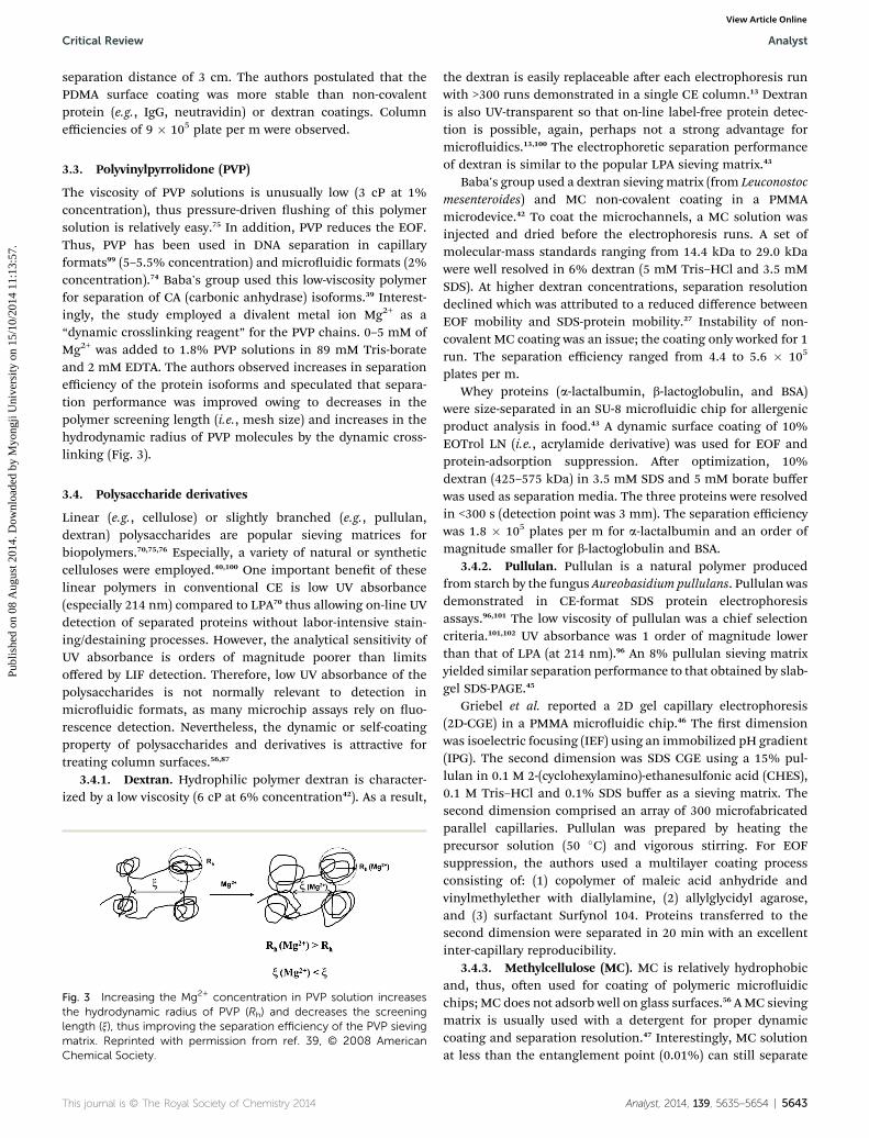
The viscosity of PVP solutions is unusually low (3 cP at 1%concentration), thus pressure-driven ushing of this polymersolution is relatively easy.75 In addition, PVP reduces the EOF.Thus, PVP has been used in DNA separation in capillaryformats99 (5–5.5% concentration) and microuidic formats (2%concentration).74 Baba's group used this low-viscosity polymerfor separation of CA (carbonic anhydrase) isoforms.39 Interest-ingly, the study employed a divalent metal ion Mg2+ as a“dynamic crosslinking reagent” for the PVP chains. 0–5 mM ofMg2+ was added to 1.8% PVP solutions in 89 mM Tris-borateand 2 mM EDTA. The authors observed increases in separationefficiency of the protein isoforms and speculated that separa-tion performance was improved owing to decreases in thepolymer screening length (i.e., mesh size) and increases in thehydrodynamic radius of PVP molecules by the dynamic cross-linking (Fig. 3).
3.4. Polysaccharide derivatives
Linear (e.g., cellulose) or slightly branched (e.g., pullulan,dextran) polysaccharides are popular sieving matrices forbiopolymers.70,75,76 Especially, a variety of natural or syntheticcelluloses were employed.40,100 One important benet of theselinear polymers in conventional CE is low UV absorbance(especially 214 nm) compared to LPA70 thus allowing on-line UVdetection of separated proteins without labor-intensive stain-ing/destaining processes. However, the analytical sensitivity ofUV absorbance is orders of magnitude poorer than limitsoffered by LIF detection. Therefore, low UV absorbance of thepolysaccharides is not normally relevant to detection inmicrouidic formats, as many microchip assays rely on uo-rescence detection. Nevertheless, the dynamic or self-coatingproperty of polysaccharides and derivatives is attractive fortreating column surfaces.56,87
3.4.1. Dextran. Hydrophilic polymer dextran is character-ized by a low viscosity (6 cP at 6% concentration42). As a result,
Fig. 3 Increasing the Mg2+ concentration in PVP solution increasesthe hydrodynamic radius of PVP (Rh) and decreases the screeninglength (x), thus improving the separation efficiency of the PVP sievingmatrix. Reprinted with permission from ref. 39, © 2008 AmericanChemical Society.
This journal is © The Royal Society of Chemistry 2014
the dextran is easily replaceable aer each electrophoresis runwith >300 runs demonstrated in a single CE column.13 Dextranis also UV-transparent so that on-line label-free protein detec-tion is possible, again, perhaps not a strong advantage formicrouidics.13,100 The electrophoretic separation performanceof dextran is similar to the popular LPA sieving matrix.43
Baba's group used a dextran sievingmatrix (from Leuconostocmesenteroides) and MC non-covalent coating in a PMMAmicrodevice.42 To coat the microchannels, a MC solution wasinjected and dried before the electrophoresis runs. A set ofmolecular-mass standards ranging from 14.4 kDa to 29.0 kDawere well resolved in 6% dextran (5 mM Tris–HCl and 3.5 mMSDS). At higher dextran concentrations, separation resolutiondeclined which was attributed to a reduced difference betweenEOF mobility and SDS-protein mobility.27 Instability of non-covalent MC coating was an issue; the coating only worked for 1run. The separation efficiency ranged from 4.4 to 5.6 � 105
plates per m.Whey proteins (a-lactalbumin, b-lactoglobulin, and BSA)
were size-separated in an SU-8 microuidic chip for allergenicproduct analysis in food.43 A dynamic surface coating of 10%EOTrol LN (i.e., acrylamide derivative) was used for EOF andprotein-adsorption suppression. Aer optimization, 10%dextran (425–575 kDa) in 3.5 mM SDS and 5 mM borate bufferwas used as separation media. The three proteins were resolvedin <300 s (detection point was 3 mm). The separation efficiencywas 1.8 � 105 plates per m for a-lactalbumin and an order ofmagnitude smaller for b-lactoglobulin and BSA.
3.4.2. Pullulan. Pullulan is a natural polymer producedfrom starch by the fungus Aureobasidium pullulans. Pullulan wasdemonstrated in CE-format SDS protein electrophoresisassays.96,101 The low viscosity of pullulan was a chief selectioncriteria.101,102 UV absorbance was 1 order of magnitude lowerthan that of LPA (at 214 nm).96 An 8% pullulan sieving matrixyielded similar separation performance to that obtained by slab-gel SDS-PAGE.45
Griebel et al. reported a 2D gel capillary electrophoresis(2D-CGE) in a PMMA microuidic chip.46 The rst dimensionwas isoelectric focusing (IEF) using an immobilized pH gradient(IPG). The second dimension was SDS CGE using a 15% pul-lulan in 0.1 M 2-(cyclohexylamino)-ethanesulfonic acid (CHES),0.1 M Tris–HCl and 0.1% SDS buffer as a sieving matrix. Thesecond dimension comprised an array of 300 microfabricatedparallel capillaries. Pullulan was prepared by heating theprecursor solution (50 �C) and vigorous stirring. For EOFsuppression, the authors used a multilayer coating processconsisting of: (1) copolymer of maleic acid anhydride andvinylmethylether with diallylamine, (2) allylglycidyl agarose,and (3) surfactant Surfynol 104. Proteins transferred to thesecond dimension were separated in 20 min with an excellentinter-capillary reproducibility.
3.4.3. Methylcellulose (MC). MC is relatively hydrophobicand, thus, oen used for coating of polymeric microuidicchips; MC does not adsorb well on glass surfaces.56 AMC sievingmatrix is usually used with a detergent for proper dynamiccoating and separation resolution.47 Interestingly, MC solutionat less than the entanglement point (0.01%) can still separate
Analyst, 2014, 139, 5635–5654 | 5643
Analyst Critical Review
Publ
ishe
d on
08
Aug
ust 2
014.
Dow
nloa
ded
by M
yong
ji U
nive
rsity
on
15/1
0/20
14 1
1:13
:57.
View Article Online
proteins as hydrogen bonding yields entanglement of sparselylocated cellulose bers.103 Baba's group used a combination ofMC and the nonionic detergent polysorbate (Tween 20) as self-coating reagent and sieving matrix for native protein separa-tions in a PMMA chip.47 TI (20.1 kDa), BSA (66.3 kDa), andamyloglucosidase (100 kDa) were separated in <100 s in a 20mM Tris–HCl buffer containing 2% MC and 0.02% Tween 20.The separation efficiency was 3.6 � 105 plates per m for TI andan order of magnitude lower for the two other proteins. Theaddition of Tween 20 was observed to play an important role inpreventing protein adsorption on the hydrophobic PMMAsurface, thus supporting polymer entanglement (for proteinseparation)47 and reducing the injection pressure required forpressure loading of a sieving matrix of concentrated MC solu-tion (>1.5%) into microuidic channels.
3.4.4. Hydroxypropylcellulose (HPC). HPC has beendemonstrated as a sieving matrix in CE of proteins100 and,recently, in microuidic DNA93 and protein separations.50 HPCis a thermo-responsive polymer, with viscosity dependent ontemperature. The gel form may be heated for easy loading(lowering viscosity) and cooled for electrophoresis (increasingviscosity).93 HPC is oen used as a dynamic-coating additive toprevent protein adsorption and to reduce EOF.86,104
Wang et al. analyzed lipoproteins using gel electrophoresisin a PDMS/glass microuidic chip.50 HPC (80 kDa) andn-dodecyl-b-D-maltoside (DDM) in 50 mM MOPS (3-morpholi-nopropanesulfonic acid) buffer was used as a sieving matrix.Pre-coating the PDMS surface with a mild nonionic detergent(DDM) for 10 min before electrophoresis successfully sup-pressed EOF and protein adsorption. The optimal HPCconcentration was 0.6%. At higher concentrations, proteinmigration was retarded and injection of the polymer solutionwas difficult due to viscosity. The lipoproteins HDL (high-density lipoprotein), sdLDL (small, dense low-density lipopro-tein), and lLDL (large buoyant LDL) were separated in <3 minwith acceptable assay reproducibility. A separation efficiency of1.5 � 106 plates per m was obtained for HDL.
3.4.5. Hydroxyethylcellulose (HEC). HEC acquires a stiffand extended conformation in solution, resulting in a robustentangled polymer network even at a low concentration.49,51
Separation resolution has been reported as comparable to thatobtained with LPA.75 The viscosity of HEC is relatively low(150 cP at 5%).53 Disadvantages of this cellulose derivativeinclude the need for purication and batch-to-batch variabilityin composition.75 Although HEC was frequently used in DNA75
and protein analyses in CE formats,105 microuidic CGE hasseen limited use of the material.48 HEC has been employed as adynamic surface coating reagent in microuidic isoelectricfocusing.52,53,84
Landers' group compared common sieving polymersincluding PEO (200 kDa), HPC (100 kDa), dextran (2000 kDa)and HEC (250 kDa) for separation of eight standard ladderproteins using microchip electrophoresis.48 For dextran, addi-tional covalent surface coating was required for EOF suppres-sion. The viscosity of PEO made pressure injection into themicrochannel difficult. The authors reported that HEC and PEOshowed the best resolving power and background uorescence
5644 | Analyst, 2014, 139, 5635–5654
levels. The same group also used HEC to separate proteins in amicrouidic IEF assay.52 Microchannels were incubated with2.5% HEC for 10 min for coating. Then 1% HEC in the carrierampholyte solution (pH 3–10) was used to separate the naturallyuorescent proteins R-phycoerythrin (RPE), B-phycoerythrin(BPE), and green uorescent protein (GFP).
Fan's group published a series of studies regarding IEF inCOC chips using HEC dynamic surface coatings.53,84,85 COC is ahydrophobic plastic substrate and, thus, protein adsorption offocused proteins is of great concern.53 In their rst two papers, amixture of 0.73% HEC (90 kDa) and 1.83% HPC was used as asieving matrix.84,85 Later, they optimized the separation mediaso that lower levels of HEC (0.2%) were effective at reducing EOFand protein adsorption.53 BSA and GFP isoforms were success-fully separated in a solution of 2% carrier ampholytes (pH 3–10)and 0.2% HEC. A 0.2 pI point difference was baseline resolvedin <3 min.
3.4.6. Hydroxypropylmethylcellulose (HPMC). Anothercellulose derivative used for CGE of DNA (dsDNA) isHPMC.7,75,106,107 HPMC is also low in viscosity (50 cP for 2% inwater106) and easily replaceable for repetitive electrophoresis runs.HPMC is also frequently used to suppress analyte adsorption andEOF.57,81 Additives such as mannitol or glycerol are added toHPMC solution in order to enhance separation performance.54
Lin et al., reported microchip CGE coupled with ITP (iso-tachophoresis) for sample stacking.55 A solution of 1%HPMC in1� TBE buffer was used as a sieving matrix in a PMMA chip.Protein markers (TI, CA, alcohol dehydrogenase, BSA, b-galac-tosidase, myosin) were enriched 21–40 times by ITP before theelectrophoresis separation.
Baba's group studied the dynamic surface coating propertiesof MC, HPMC, PVA, and PVP on PMMA microchannel surfacesand the inuence on electrophoresis of non-denaturedproteins.56 These researchers found HPMC reduced protein-adsorption most effectively, among the four water-solublepolymers considered. HPMC was also observed to be effective atattenuating EOF. Although an HPMC concentration below theentanglement point (e.g., 0.2%) was used in the backgroundelectrolyte, this amphiphilic polymer interacted with proteins,causing differential reduction of protein mobility. Such differ-ential mobility change may have facilitated protein separationeven in a dilute polymer solution. A separation efficiency of8.2 � 105 plates per m was obtained for 0.2% HPMC. The samegroup used a similar PMMA microchip to analyze lipoproteinsHDL, low-density lipoprotein (LDL), and very low-density lipo-protein (VLDL), biomarkers important in assessing cardiovas-cular system health.57 As lipoproteins strongly adsorb to thehydrophobic PMMA surface, a dynamic coating of HPMC (up to0.5%) alone did not suppress protein surface adsorption tosatisfaction. Thus, SDS was added to coat the protein andPMMA surface with negative charges, reducing lipoproteinadsorption. HPMC (>0.05%) effectively suppressed EOF.Depending on polymer concentration, the mode of separationwas different; at low HPMC concentration, lipoproteins wereseparated by zone electrophoresis (�0.05%). At a higher HPMCconcentration (1%), three lipoproteins were separated bymolecular sieving.
This journal is © The Royal Society of Chemistry 2014
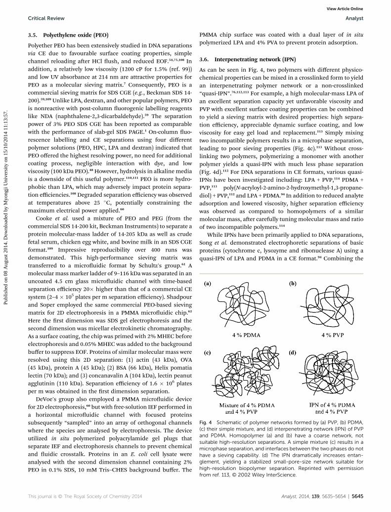
Fig. 4 Schematic of polymer networks formed by (a) PVP, (b) PDMA,(c) their simple mixture, and (d) interpenetrating network (IPN) of PVPand PDMA. Homopolymer (a) and (b) have a coarse network, notsuitable high-resolution separations. A simple mixture (c) results in amicrophase separation, and interfaces between the two phases do nothave a sieving capability. (d) The IPN dramatically increases entan-glement, yielding a stabilized small-pore-size network suitable forhigh-resolution biopolymer separation. Reprinted with permission
Critical Review Analyst
Publ
ishe
d on
08
Aug
ust 2
014.
Dow
nloa
ded
by M
yong
ji U
nive
rsity
on
15/1
0/20
14 1
1:13
:57.
View Article Online
3.5. Polyethylene oxide (PEO)
Polyether PEO has been extensively studied in DNA separationsvia CE due to favourable surface coating properties, simplechannel reloading aer HCl ush, and reduced EOF.54,75,108 Inaddition, a relatively low viscosity (1200 cP for 1.5% (ref. 99))and low UV absorbance at 214 nm are attractive properties forPEO as a molecular sieving matrix.7 Consequently, PEO is acommercial sieving matrix for SDS CGE (e.g., Beckman SDS 14-200).59,109 Unlike LPA, dextran, and other popular polymers, PEOis nonreactive with post-column uorogenic labelling reagentslike NDA (naphthalene-2,3-dicarbaldehyde).59 The separationpower of 3% PEO SDS CGE has been reported as comparablewith the performance of slab-gel SDS PAGE.1 On-column uo-rescence labelling and CE separations using four differentpolymer solutions (PEO, HPC, LPA and dextran) indicated thatPEO offered the highest resolving power, no need for additionalcoating process, negligible interaction with dye, and lowviscosity (100 kDa PEO).48 However, hydrolysis in alkaline mediais a downside of this useful polymer.110,111 PEO is more hydro-phobic than LPA, which may adversely impact protein separa-tion efficiencies.108 Degraded separation efficiency was observedat temperatures above 25 �C, potentially constraining themaximum electrical power applied.60
Cooke et al. used a mixture of PEO and PEG (from thecommercial SDS 14-200 kit, Beckman Instruments) to separate aprotein molecular-mass ladder of 14-205 kDa as well as crudefetal serum, chicken egg white, and bovine milk in an SDS CGEformat.109 Impressive reproducibility over 400 runs wasdemonstrated. This high-performance sieving matrix wastransferred to a microuidic format by Schultz's group.61 Amolecular massmarker ladder of 9–116 kDa was separated in anuncoated 4.5 cm glass microuidic channel with time-basedseparation efficiency 20� higher than that of a commercial CEsystem (2–4 � 105 plates per m separation efficiency). Shadpourand Soper employed the same commercial PEO-based sievingmatrix for 2D electrophoresis in a PMMA microuidic chip.62
Here the rst dimension was SDS gel electrophoresis and thesecond dimension was micellar electrokinetic chromatography.As a surface coating, the chip was primed with 2%MHEC beforeelectrophoresis and 0.05%MHEC was added to the backgroundbuffer to suppress EOF. Proteins of similar molecular mass wereresolved using this 2D separation: (1) actin (43 kDa), OVA(45 kDa), protein A (45 kDa); (2) BSA (66 kDa), Helix pomatialectin (70 kDa); and (3) concanavalin A (104 kDa), lectin peanutagglutinin (110 kDa). Separation efficiency of 1.6 � 106 platesper m was obtained in the rst dimension separation.
DeVoe's group also employed a PMMA microuidic devicefor 2D electrophoresis,60 but with free-solution IEF performed ina horizontal microuidic channel with focused proteinssubsequently “sampled” into an array of orthogonal channelswhere the species are analysed by electrophoresis. The deviceutilized in situ polymerized polyacrylamide gel plugs thatseparate IEF and electrophoresis channels to prevent chemicaland uidic crosstalk. Proteins in an E. coli cell lysate wereanalysed with the second dimension channel containing 2%PEO in 0.1% SDS, 10 mM Tris–CHES background buffer. The
This journal is © The Royal Society of Chemistry 2014
PMMA chip surface was coated with a dual layer of in situpolymerized LPA and 4% PVA to prevent protein adsorption.
3.6. Interpenetrating network (IPN)
As can be seen in Fig. 4, two polymers with different physico-chemical properties can be mixed in a crosslinked form to yieldan interpenetrating polymer network or a non-crosslinked“quasi-IPN”.76,112,113 For example, a high molecular-mass LPA ofan excellent separation capacity yet unfavorable viscosity andPVP with excellent surface coating properties can be combinedto yield a sieving matrix with desired properties: high separa-tion efficiency, appreciable dynamic surface coating, and lowviscosity for easy gel load and replacement.113 Simply mixingtwo incompatible polymers results in a microphase separation,leading to poor sieving properties (Fig. 4c).113 Without cross-linking two polymers, polymerizing a monomer with anotherpolymer yields a quasi-IPN with much less phase separation(Fig. 4d).112 For DNA separations in CE formats, various quasi-IPNs have been investigated including: LPA + PVP,114 PDMA +PVP,113 poly(N-acryloyl-2-amino-2-hydroxymethyl-1,3-propane-diol) + PVP,112 and LPA + PDMA.94 In addition to reduced analyteadsorption and lowered viscosity, higher separation efficiencywas observed as compared to homopolymers of a similarmolecular mass, aer carefully tuningmolecular mass and ratioof two incompatible polymers.114
While IPNs have been primarily applied to DNA separations,Song et al. demonstrated electrophoretic separations of basicproteins (cytochrome c, lysozyme and ribonuclease A) using aquasi-IPN of LPA and PDMA in a CE format.94 Combining the
from ref. 113, © 2002 Wiley InterScience.
Analyst, 2014, 139, 5635–5654 | 5645

Analyst Critical Review
Publ
ishe
d on
08
Aug
ust 2
014.
Dow
nloa
ded
by M
yong
ji U
nive
rsity
on
15/1
0/20
14 1
1:13
:57.
View Article Online
high sieving capacity of LPA and the dynamic surface coatingproperties of PDMA, separation efficiencies ranged from 3.02 to4.29 � 105 plates per m. The separation efficiency of the IPNcontaining a low molecular mass LPA (<1 MDa) was higher thanthat of a homopolymer LPA of much higher molecular mass(>3 MDa). The IPN matrix was loaded with ease (20 psi for10 min) and EOF was well suppressed compared to a bare fusedsilica capillary. IPN appears suitable to adaptation to microchipprotein electrophoresis.
4. Crosslinked polymers as separationmatrices
Regardless of many aforementioned advantages of thereplaceable polymer solutions, pressure-injection of linearpolymer into narrow columns is difficult, because of viscositylimits.115 Owing to higher uidic resistances, the loading chal-lenge is even more pronounced in micro-to-nanouidic deviceswith complex geometries. The low viscosity of the polymerprecursor solution (monomer and porogen) affords easychannel lling with the desired polymer gel subsequentlyformed via chemical- or photo-initiation.116 Fabrication ofcomplex gel structures63 or even regionally patterned micro-chambers20–22 is feasible with crosslinked polymers. The poresize and density of the crosslinked monolith can be readilycontrolled in each specic zone designated for sample pre-concentration, separation, or immobilization.117 Moreover,stiffer networks appear to enhance separations,51,75 but aredifficult to achieve with linear polymers as viscosity increaseswith stiffness. A major drawback of crosslinked polymermonoliths is difficulty in removing the material from micro-channels, which makes it difficult to recycle chips. Piranhaetching is generally used to chemically decompose the polymermatrix in glass or borosilicate chips.17 The heterogeneity of thepore structure under certain conditions can pose potentialproblems, as only the average pore size of a monolith can bewell-controlled.118
Fig. 5 Design and operation of the microfluidic western blot. (a) Aglass microfluidic device with microchannels linking two fluid reser-voirs (dye added for clarity). (Scale bar: 2 mm.) (b) The 80 min five-stage immunoprobing assay is completed in a single microchannel. (c)Schematic of microchannel cross-section depicting principle of theprotein immobilization: analytes are electrophoresed through thereactive nanoporous hydrogel, exposed to UV, and covalentlyimmobilized. (d) Schematic of reaction between polypeptide back-bone and benzophenone groups. Ph denotes phenyl group. (e, left)SDS-PAGE of fluorescently labeled six protein ladder (black), completein 60 s (4� magnification; band weights are 155, 98, 63, 40, 32, and21 kDa). (e, right) Multiplexed immunoblot readout (red) in 40 min totalassay times using primary antibodies for (i) OVA (ovalbumin), and (ii)b-gal (b-galactosidase), OVA, and TI (trypsin inhibitor); all at 1 mM.Fluorescence micrographs is shown for red fluorescent primary anti-bodies (Ab*). Reprinted with permission from ref. 23 and 24, © 2012National Academy of Science.
4.1. Polyacrylamide
Polyacrylamide (PA) was used for separation of serum albuminsin the 1960s,119 and has become ubiquitous as a separationmedia for analysis of proteins. Two formats are most widelyused: PAGE and SDS-PAGE. With applicability to a diverse arrayof measurement challenges and a long history, PA-based sepa-ration media have been applied to numerous formats. Theminiaturization of bench-top PAGE,63 SDS-PAGE,21,23 IEF,24,60
and immunoblotting17,64,117,120 have all been reported usingconventional crosslinked PA. Adaptation of PA to microuidicdevices preserves the separation mechanism of slab-gel systemsthat have been extensively investigated. Thus miniaturization ofPAGE-based separations benets from the deep existingunderstanding of separation performance.
Hughes et al. developed an integrated microuidic systemfor western blotting following SDS-PAGE23 and IEF.24 Theresearchers integrated a discontinuous PA gel supportingtransient ITP and subsequent SDS-PAGE by photopatterning the
5646 | Analyst, 2014, 139, 5635–5654
crosslinked polymer in a straight microchannel (Fig. 5).Benzophenone-functionalized methacrylamide co-monomerwas incorporated into the polyacrylamide gels, thus allowingcovalent UV-initiated immobilization of separated proteins.Aer brief UV exposure, the immobilized proteins were probedby antibodies, comprising a western blotting assay. Themicrouidic approach overcomes several limitations of slab-gelsystem by greatly reducing antibody consumption (from 1 mg to1 ng), human intervention and assay time (from days to hours),owing to miniaturization and automation of the otherwiselaborious workow.
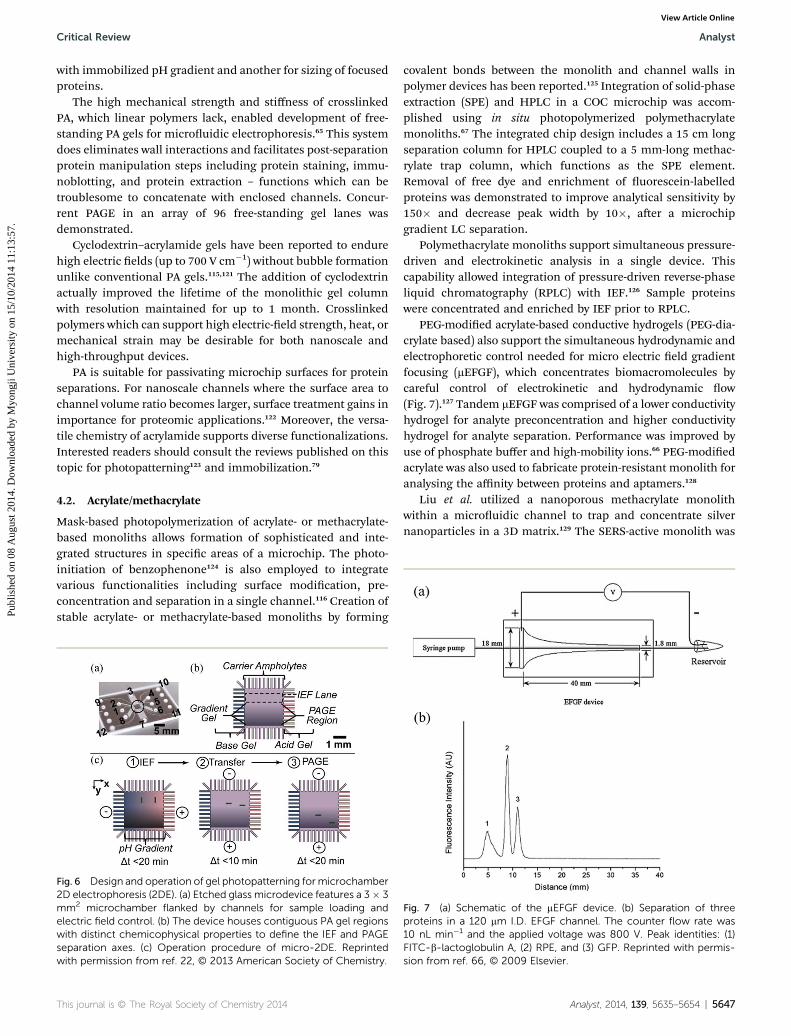
A two-dimensional microuidic system is reported forprotein separations combining IEF and SDS-PAGE employing insitu photopolymerized PA gels.60,63 In an alternate approach tothe microchannel array used in that work, a microchambersupported 2-D protein separations (Fig. 6).22 Tentori et al.formed spatially distinct PA gels by photopatterning; one for IEF
This journal is © The Royal Society of Chemistry 2014
Critical Review Analyst
Publ
ishe
d on
08
Aug
ust 2
014.
Dow
nloa
ded
by M
yong
ji U
nive
rsity
on
15/1
0/20
14 1
1:13
:57.
View Article Online
with immobilized pH gradient and another for sizing of focusedproteins.
The high mechanical strength and stiffness of crosslinkedPA, which linear polymers lack, enabled development of free-standing PA gels for microuidic electrophoresis.65 This systemdoes eliminates wall interactions and facilitates post-separationprotein manipulation steps including protein staining, immu-noblotting, and protein extraction – functions which can betroublesome to concatenate with enclosed channels. Concur-rent PAGE in an array of 96 free-standing gel lanes wasdemonstrated.
Cyclodextrin–acrylamide gels have been reported to endurehigh electric elds (up to 700 V cm�1) without bubble formationunlike conventional PA gels.115,121 The addition of cyclodextrinactually improved the lifetime of the monolithic gel columnwith resolution maintained for up to 1 month. Crosslinkedpolymers which can support high electric-eld strength, heat, ormechanical strain may be desirable for both nanoscale andhigh-throughput devices.
PA is suitable for passivating microchip surfaces for proteinseparations. For nanoscale channels where the surface area tochannel volume ratio becomes larger, surface treatment gains inimportance for proteomic applications.122 Moreover, the versa-tile chemistry of acrylamide supports diverse functionalizations.Interested readers should consult the reviews published on thistopic for photopatterning123 and immobilization.79
4.2. Acrylate/methacrylate
Mask-based photopolymerization of acrylate- or methacrylate-based monoliths allows formation of sophisticated and inte-grated structures in specic areas of a microchip. The photo-initiation of benzophenone124 is also employed to integratevarious functionalities including surface modication, pre-concentration and separation in a single channel.116 Creation ofstable acrylate- or methacrylate-based monoliths by forming
Fig. 6 Design and operation of gel photopatterning formicrochamber2D electrophoresis (2DE). (a) Etched glass microdevice features a 3� 3mm2 microchamber flanked by channels for sample loading andelectric field control. (b) The device houses contiguous PA gel regionswith distinct chemicophysical properties to define the IEF and PAGEseparation axes. (c) Operation procedure of micro-2DE. Reprintedwith permission from ref. 22, © 2013 American Society of Chemistry.
This journal is © The Royal Society of Chemistry 2014
covalent bonds between the monolith and channel walls inpolymer devices has been reported.125 Integration of solid-phaseextraction (SPE) and HPLC in a COC microchip was accom-plished using in situ photopolymerized polymethacrylatemonoliths.67 The integrated chip design includes a 15 cm longseparation column for HPLC coupled to a 5 mm-long methac-rylate trap column, which functions as the SPE element.Removal of free dye and enrichment of uorescein-labelledproteins was demonstrated to improve analytical sensitivity by150� and decrease peak width by 10�, aer a microchipgradient LC separation.
Polymethacrylate monoliths support simultaneous pressure-driven and electrokinetic analysis in a single device. Thiscapability allowed integration of pressure-driven reverse-phaseliquid chromatography (RPLC) with IEF.126 Sample proteinswere concentrated and enriched by IEF prior to RPLC.
PEG-modied acrylate-based conductive hydrogels (PEG-dia-crylate based) also support the simultaneous hydrodynamic andelectrophoretic control needed for micro electric eld gradientfocusing (mEFGF), which concentrates biomacromolecules bycareful control of electrokinetic and hydrodynamic ow(Fig. 7).127 Tandem mEFGF was comprised of a lower conductivityhydrogel for analyte preconcentration and higher conductivityhydrogel for analyte separation. Performance was improved byuse of phosphate buffer and high-mobility ions.66 PEG-modiedacrylate was also used to fabricate protein-resistant monolith foranalysing the affinity between proteins and aptamers.128
Liu et al. utilized a nanoporous methacrylate monolithwithin a microuidic channel to trap and concentrate silvernanoparticles in a 3D matrix.129 The SERS-active monolith was
Fig. 7 (a) Schematic of the mEFGF device. (b) Separation of threeproteins in a 120 mm I.D. EFGF channel. The counter flow rate was10 nL min�1 and the applied voltage was 800 V. Peak identities: (1)FITC-b-lactoglobulin A, (2) RPE, and (3) GFP. Reprinted with permis-sion from ref. 66, © 2009 Elsevier.
Analyst, 2014, 139, 5635–5654 | 5647
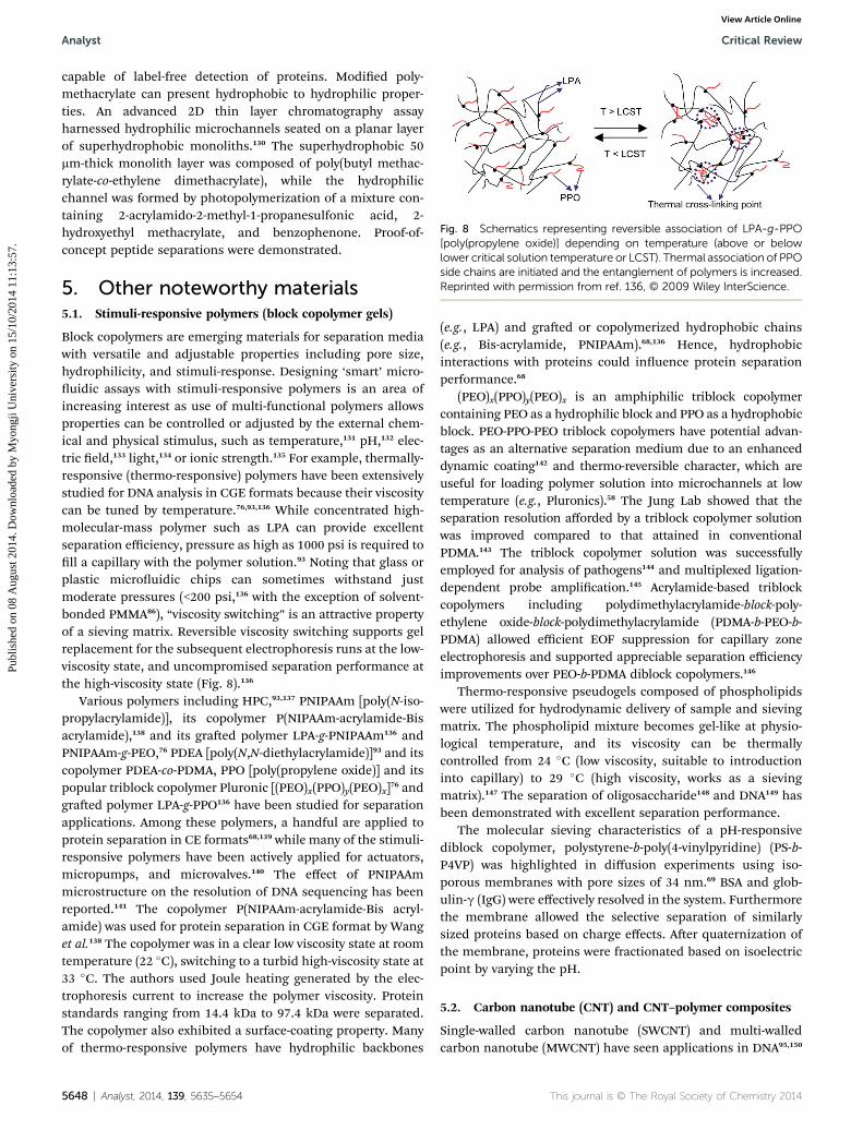
Fig. 8 Schematics representing reversible association of LPA-g-PPO[poly(propylene oxide)] depending on temperature (above or belowlower critical solution temperature or LCST). Thermal association of PPOside chains are initiated and the entanglement of polymers is increased.Reprinted with permission from ref. 136, © 2009 Wiley InterScience.
Analyst Critical Review
Publ
ishe
d on
08
Aug
ust 2
014.
Dow
nloa
ded
by M
yong
ji U
nive
rsity
on
15/1
0/20
14 1
1:13
:57.
View Article Online
capable of label-free detection of proteins. Modied poly-methacrylate can present hydrophobic to hydrophilic proper-ties. An advanced 2D thin layer chromatography assayharnessed hydrophilic microchannels seated on a planar layerof superhydrophobic monoliths.130 The superhydrophobic 50mm-thick monolith layer was composed of poly(butyl methac-rylate-co-ethylene dimethacrylate), while the hydrophilicchannel was formed by photopolymerization of a mixture con-taining 2-acrylamido-2-methyl-1-propanesulfonic acid, 2-hydroxyethyl methacrylate, and benzophenone. Proof-of-concept peptide separations were demonstrated.
5. Other noteworthy materials5.1. Stimuli-responsive polymers (block copolymer gels)
Block copolymers are emerging materials for separation mediawith versatile and adjustable properties including pore size,hydrophilicity, and stimuli-response. Designing ‘smart’ micro-uidic assays with stimuli-responsive polymers is an area ofincreasing interest as use of multi-functional polymers allowsproperties can be controlled or adjusted by the external chem-ical and physical stimulus, such as temperature,131 pH,132 elec-tric eld,133 light,134 or ionic strength.135 For example, thermally-responsive (thermo-responsive) polymers have been extensivelystudied for DNA analysis in CGE formats because their viscositycan be tuned by temperature.76,93,136 While concentrated high-molecular-mass polymer such as LPA can provide excellentseparation efficiency, pressure as high as 1000 psi is required toll a capillary with the polymer solution.93 Noting that glass orplastic microuidic chips can sometimes withstand justmoderate pressures (<200 psi,136 with the exception of solvent-bonded PMMA86), “viscosity switching” is an attractive propertyof a sieving matrix. Reversible viscosity switching supports gelreplacement for the subsequent electrophoresis runs at the low-viscosity state, and uncompromised separation performance atthe high-viscosity state (Fig. 8).136
Various polymers including HPC,93,137 PNIPAAm [poly(N-iso-propylacrylamide)], its copolymer P(NIPAAm-acrylamide-Bisacrylamide),138 and its graed polymer LPA-g-PNIPAAm136 andPNIPAAm-g-PEO,76 PDEA [poly(N,N-diethylacrylamide)]93 and itscopolymer PDEA-co-PDMA, PPO [poly(propylene oxide)] and itspopular triblock copolymer Pluronic [(PEO)x(PPO)y(PEO)x]76 andgraed polymer LPA-g-PPO136 have been studied for separationapplications. Among these polymers, a handful are applied toprotein separation in CE formats68,139 while many of the stimuli-responsive polymers have been actively applied for actuators,micropumps, and microvalves.140 The effect of PNIPAAmmicrostructure on the resolution of DNA sequencing has beenreported.141 The copolymer P(NIPAAm-acrylamide-Bis acryl-amide) was used for protein separation in CGE format by Wanget al.138 The copolymer was in a clear low viscosity state at roomtemperature (22 �C), switching to a turbid high-viscosity state at33 �C. The authors used Joule heating generated by the elec-trophoresis current to increase the polymer viscosity. Proteinstandards ranging from 14.4 kDa to 97.4 kDa were separated.The copolymer also exhibited a surface-coating property. Manyof thermo-responsive polymers have hydrophilic backbones
5648 | Analyst, 2014, 139, 5635–5654
(e.g., LPA) and graed or copolymerized hydrophobic chains(e.g., Bis-acrylamide, PNIPAAm).68,136 Hence, hydrophobicinteractions with proteins could inuence protein separationperformance.68
(PEO)x(PPO)y(PEO)x is an amphiphilic triblock copolymercontaining PEO as a hydrophilic block and PPO as a hydrophobicblock. PEO-PPO-PEO triblock copolymers have potential advan-tages as an alternative separation medium due to an enhanceddynamic coating142 and thermo-reversible character, which areuseful for loading polymer solution into microchannels at lowtemperature (e.g., Pluronics).58 The Jung Lab showed that theseparation resolution afforded by a triblock copolymer solutionwas improved compared to that attained in conventionalPDMA.143 The triblock copolymer solution was successfullyemployed for analysis of pathogens144 and multiplexed ligation-dependent probe amplication.145 Acrylamide-based triblockcopolymers including polydimethylacrylamide-block-poly-ethylene oxide-block-polydimethylacrylamide (PDMA-b-PEO-b-PDMA) allowed efficient EOF suppression for capillary zoneelectrophoresis and supported appreciable separation efficiencyimprovements over PEO-b-PDMA diblock copolymers.146
Thermo-responsive pseudogels composed of phospholipidswere utilized for hydrodynamic delivery of sample and sievingmatrix. The phospholipid mixture becomes gel-like at physio-logical temperature, and its viscosity can be thermallycontrolled from 24 �C (low viscosity, suitable to introductioninto capillary) to 29 �C (high viscosity, works as a sievingmatrix).147 The separation of oligosaccharide148 and DNA149 hasbeen demonstrated with excellent separation performance.
The molecular sieving characteristics of a pH-responsivediblock copolymer, polystyrene-b-poly(4-vinylpyridine) (PS-b-P4VP) was highlighted in diffusion experiments using iso-porous membranes with pore sizes of 34 nm.69 BSA and glob-ulin-g (IgG) were effectively resolved in the system. Furthermorethe membrane allowed the selective separation of similarlysized proteins based on charge effects. Aer quaternization ofthe membrane, proteins were fractionated based on isoelectricpoint by varying the pH.
5.2. Carbon nanotube (CNT) and CNT–polymer composites
Single-walled carbon nanotube (SWCNT) and multi-walledcarbon nanotube (MWCNT) have seen applications in DNA95,150
This journal is © The Royal Society of Chemistry 2014
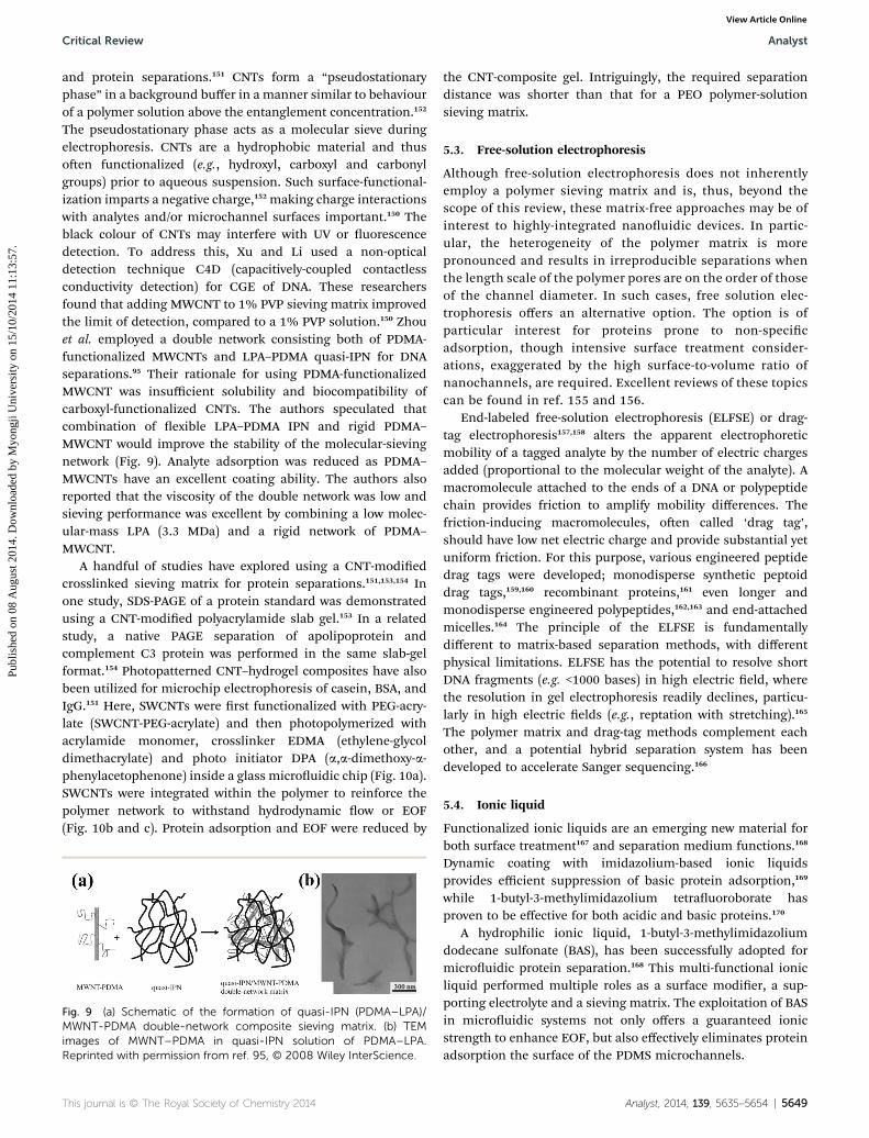
Critical Review Analyst
Publ
ishe
d on
08
Aug
ust 2
014.
Dow
nloa
ded
by M
yong
ji U
nive
rsity
on
15/1
0/20
14 1
1:13
:57.
View Article Online
and protein separations.151 CNTs form a “pseudostationaryphase” in a background buffer in a manner similar to behaviourof a polymer solution above the entanglement concentration.152
The pseudostationary phase acts as a molecular sieve duringelectrophoresis. CNTs are a hydrophobic material and thusoen functionalized (e.g., hydroxyl, carboxyl and carbonylgroups) prior to aqueous suspension. Such surface-functional-ization imparts a negative charge,152 making charge interactionswith analytes and/or microchannel surfaces important.150 Theblack colour of CNTs may interfere with UV or uorescencedetection. To address this, Xu and Li used a non-opticaldetection technique C4D (capacitively-coupled contactlessconductivity detection) for CGE of DNA. These researchersfound that adding MWCNT to 1% PVP sieving matrix improvedthe limit of detection, compared to a 1% PVP solution.150 Zhouet al. employed a double network consisting both of PDMA-functionalized MWCNTs and LPA–PDMA quasi-IPN for DNAseparations.95 Their rationale for using PDMA-functionalizedMWCNT was insufficient solubility and biocompatibility ofcarboxyl-functionalized CNTs. The authors speculated thatcombination of exible LPA–PDMA IPN and rigid PDMA–MWCNT would improve the stability of the molecular-sievingnetwork (Fig. 9). Analyte adsorption was reduced as PDMA–MWCNTs have an excellent coating ability. The authors alsoreported that the viscosity of the double network was low andsieving performance was excellent by combining a low molec-ular-mass LPA (3.3 MDa) and a rigid network of PDMA–MWCNT.
A handful of studies have explored using a CNT-modiedcrosslinked sieving matrix for protein separations.151,153,154 Inone study, SDS-PAGE of a protein standard was demonstratedusing a CNT-modied polyacrylamide slab gel.153 In a relatedstudy, a native PAGE separation of apolipoprotein andcomplement C3 protein was performed in the same slab-gelformat.154 Photopatterned CNT–hydrogel composites have alsobeen utilized for microchip electrophoresis of casein, BSA, andIgG.151 Here, SWCNTs were rst functionalized with PEG-acry-late (SWCNT-PEG-acrylate) and then photopolymerized withacrylamide monomer, crosslinker EDMA (ethylene-glycoldimethacrylate) and photo initiator DPA (a,a-dimethoxy-a-phenylacetophenone) inside a glass microuidic chip (Fig. 10a).SWCNTs were integrated within the polymer to reinforce thepolymer network to withstand hydrodynamic ow or EOF(Fig. 10b and c). Protein adsorption and EOF were reduced by
Fig. 9 (a) Schematic of the formation of quasi-IPN (PDMA–LPA)/MWNT-PDMA double-network composite sieving matrix. (b) TEMimages of MWNT–PDMA in quasi-IPN solution of PDMA–LPA.Reprinted with permission from ref. 95, © 2008 Wiley InterScience.
This journal is © The Royal Society of Chemistry 2014
the CNT-composite gel. Intriguingly, the required separationdistance was shorter than that for a PEO polymer-solutionsieving matrix.
5.3. Free-solution electrophoresis
Although free-solution electrophoresis does not inherentlyemploy a polymer sieving matrix and is, thus, beyond thescope of this review, these matrix-free approaches may be ofinterest to highly-integrated nanouidic devices. In partic-ular, the heterogeneity of the polymer matrix is morepronounced and results in irreproducible separations whenthe length scale of the polymer pores are on the order of thoseof the channel diameter. In such cases, free solution elec-trophoresis offers an alternative option. The option is ofparticular interest for proteins prone to non-specicadsorption, though intensive surface treatment consider-ations, exaggerated by the high surface-to-volume ratio ofnanochannels, are required. Excellent reviews of these topicscan be found in ref. 155 and 156.
End-labeled free-solution electrophoresis (ELFSE) or drag-tag electrophoresis157,158 alters the apparent electrophoreticmobility of a tagged analyte by the number of electric chargesadded (proportional to the molecular weight of the analyte). Amacromolecule attached to the ends of a DNA or polypeptidechain provides friction to amplify mobility differences. Thefriction-inducing macromolecules, oen called ‘drag tag’,should have low net electric charge and provide substantial yetuniform friction. For this purpose, various engineered peptidedrag tags were developed; monodisperse synthetic peptoiddrag tags,159,160 recombinant proteins,161 even longer andmonodisperse engineered polypeptides,162,163 and end-attachedmicelles.164 The principle of the ELFSE is fundamentallydifferent to matrix-based separation methods, with differentphysical limitations. ELFSE has the potential to resolve shortDNA fragments (e.g. <1000 bases) in high electric eld, wherethe resolution in gel electrophoresis readily declines, particu-larly in high electric elds (e.g., reptation with stretching).165
The polymer matrix and drag-tag methods complement eachother, and a potential hybrid separation system has beendeveloped to accelerate Sanger sequencing.166
5.4. Ionic liquid
Functionalized ionic liquids are an emerging new material forboth surface treatment167 and separation medium functions.168
Dynamic coating with imidazolium-based ionic liquidsprovides efficient suppression of basic protein adsorption,169
while 1-butyl-3-methylimidazolium tetrauoroborate hasproven to be effective for both acidic and basic proteins.170
A hydrophilic ionic liquid, 1-butyl-3-methylimidazoliumdodecane sulfonate (BAS), has been successfully adopted formicrouidic protein separation.168 This multi-functional ionicliquid performed multiple roles as a surface modier, a sup-porting electrolyte and a sieving matrix. The exploitation of BASin microuidic systems not only offers a guaranteed ionicstrength to enhance EOF, but also effectively eliminates proteinadsorption the surface of the PDMS microchannels.
Analyst, 2014, 139, 5635–5654 | 5649
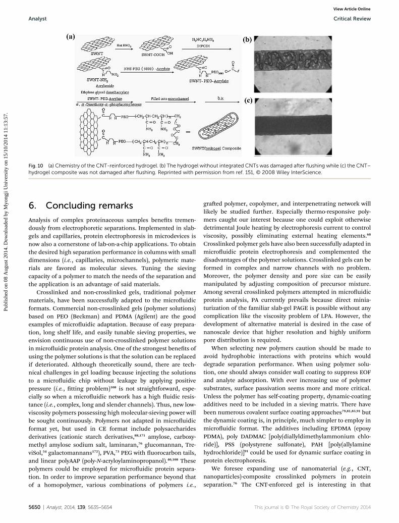
Fig. 10 (a) Chemistry of the CNT-reinforced hydrogel. (b) The hydrogel without integrated CNTs was damaged after flushing while (c) the CNT–hydrogel composite was not damaged after flushing. Reprinted with permission from ref. 151, © 2008 Wiley InterScience.
Analyst Critical Review
Publ
ishe
d on
08
Aug
ust 2
014.
Dow
nloa
ded
by M
yong
ji U
nive
rsity
on
15/1
0/20
14 1
1:13
:57.
View Article Online
6. Concluding remarks
Analysis of complex proteinaceous samples benets tremen-dously from electrophoretic separations. Implemented in slab-gels and capillaries, protein electrophoresis in microdevices isnow also a cornerstone of lab-on-a-chip applications. To obtainthe desired high separation performance in columns with smalldimensions (i.e., capillaries, microchannels), polymeric mate-rials are favored as molecular sieves. Tuning the sievingcapacity of a polymer to match the needs of the separation andthe application is an advantage of said materials.
Crosslinked and non-crosslinked gels, traditional polymermaterials, have been successfully adapted to the microuidicformats. Commercial non-crosslinked gels (polymer solutions)based on PEO (Beckman) and PDMA (Agilent) are the goodexamples of microuidic adaptation. Because of easy prepara-tion, long shelf life, and easily tunable sieving properties, weenvision continuous use of non-crosslinked polymer solutionsin microuidic protein analysis. One of the strongest benets ofusing the polymer solutions is that the solution can be replacedif deteriorated. Although theoretically sound, there are tech-nical challenges in gel loading because injecting the solutionsto a microuidic chip without leakage by applying positivepressure (i.e., tting problem)108 is not straightforward, espe-cially so when a microuidic network has a high uidic resis-tance (i.e., complex, long and slender channels). Thus, new low-viscosity polymers possessing highmolecular-sieving power willbe sought continuously. Polymers not adapted in microuidicformat yet, but used in CE format include polysaccharidesderivatives (cationic starch derivatives,88,171 amylose, carboxy-methyl amylose sodium salt, laminaran,76 glucomannan, Tre-viSol,54 galactomannans172), PVA,73 PEG with uorocarbon tails,and linear polyAAP (poly-N-acryloylaminopropanol).40,108 Thesepolymers could be employed for microuidic protein separa-tion. In order to improve separation performance beyond thatof a homopolymer, various combinations of polymers i.e.,
5650 | Analyst, 2014, 139, 5635–5654
graed polymer, copolymer, and interpenetrating network willlikely be studied further. Especially thermo-responsive poly-mers caught our interest because one could exploit otherwisedetrimental Joule heating by electrophoresis current to controlviscosity, possibly eliminating external heating elements.68
Crosslinked polymer gels have also been successfully adapted inmicrouidic protein electrophoresis and complemented thedisadvantages of the polymer solutions. Crosslinked gels can beformed in complex and narrow channels with no problem.Moreover, the polymer density and pore size can be easilymanipulated by adjusting composition of precursor mixture.Among several crosslinked polymers attempted in microuidicprotein analysis, PA currently prevails because direct minia-turization of the familiar slab-gel PAGE is possible without anycomplication like the viscosity problem of LPA. However, thedevelopment of alternative material is desired in the case ofnanoscale device that higher resolution and highly uniformpore distribution is required.
When selecting new polymers caution should be made toavoid hydrophobic interactions with proteins which woulddegrade separation performance. When using polymer solu-tion, one should always consider wall coating to suppress EOFand analyte adsorption. With ever increasing use of polymersubstrates, surface passivation seems more and more critical.Unless the polymer has self-coating property, dynamic-coatingadditives need to be included in a sieving matrix. There havebeen numerous covalent surface coating approaches79,81,83,91 butthe dynamic coating is, in principle, much simpler to employ inmicrouidic format. The additives including EPDMA (epoxyPDMA), poly DADMAC [poly(diallyldimethylammonium chlo-ride)], PSS (polystyrene sulfonate), PAH [poly(allylaminehydrochloride)]91 could be used for dynamic surface coating inprotein electrophoresis.
We foresee expanding use of nanomaterial (e.g., CNT,nanoparticles)-composite crosslinked polymers in proteinseparation.76 The CNT-enforced gel is interesting in that
This journal is © The Royal Society of Chemistry 2014
Critical Review Analyst
Publ
ishe
d on
08
Aug
ust 2
014.
Dow
nloa
ded
by M
yong
ji U
nive
rsity
on
15/1
0/20
14 1
1:13
:57.
View Article Online
hydrodynamic-electrokinetic hybrid uidic control is possibleowing to high mechanical strength. This may enable multi-stepmicrouidic protein assay such as western or eastern blottingbecause blotting, staining and destaining reagents could beloaded quickly by hydrodynamic injection over the gel whereproteins were separated. One downside is the black colour ofCNT, thus transparent polymer nanowires173 could replace theCNT for optical protein detection. Beyond the said advantage,photopatterning of crosslinked polymers are attractive becauseit allows integration of multiple assay functionality in a singledevice. However, bubble formation by gel breakage/shrinkage,especially gel of a low monomer concentration, is problematic.Thus one could replace the dilute crosslinked gel (low %T) withpolymer solution and use it in conjunction with the high-monomer-content gels (high %T) to render the multi-stepprotein assay more robust. As such, by combining benets ofcrosslinked gel and non-crosslinked gel, gel-electrophoresis-based microuidic protein assays could be improved.
Microsystems not only augment conventional separations,but for multiplexing and integrated assays, such systems openup new possibilities. In the next decades, the advances ofseparation media will be required in reproducibility, separationresolution, detection sensitivity and ease of fabrication,together with the integration of different functional operations.
Acknowledgements
M. Chung was supported by the Hongik University new facultyresearch support fund. D. Kim was supported by MyongjiUniversity ‘Assistant Professor Support Program’. D. Kim isgrateful to School of Engineering at Myongji University for thetechnical support and management.
References
1 A. Guttman, Electrophoresis, 1996, 17, 1333–1341.2 A. L. Shapiro, E. Vinuela and J. V. Maizel Jr., Biochem.Biophys. Res. Commun., 1967, 28, 815–820.
3 K. Weber and M. Osborn, J. Biol. Chem., 1969, 244, 4406–4412.
4 J. J. Lu, S. Liu and Q. Pu, J. Proteome Res., 2005, 4, 1012–1016.
5 S. Hjerten, J. Chromatogr., A, 1983, 270, 1–6.6 A. S. Cohen and B. L. Karger, J. Chromatogr., A, 1987, 397,409–417.
7 C. Heller, J. Chromatogr., A, 1995, 698, 19–31.8 A. Widhalm, C. Schwer, D. Blaas and E. Kenndler, J.Chromatogr., A, 1991, 549, 446–451.
9 J. H. Aborn, S. A. El-Difrawy, M. Novotny, E. A. Gismondi,R. Lam, P. Matsudaira, B. K. McKenna, T. O'Neil,P. Streechon and D. J. Ehrlich, Lab Chip, 2005, 5, 669–674.
10 B. M. Paegel, C. A. Emrich, G. J. Wedemayer, J. R. Schererand R. A. Mathies, Proc. Natl. Acad. Sci. U. S. A., 2002, 99,574–579.
11 P. C. Simpson, D. Roach, A. T. Woolley, T. Thorsen,R. Johnston, G. F. Sensabaugh and R. A. Mathies, Proc.Natl. Acad. Sci. U. S. A., 1998, 95, 2256–2261.
This journal is © The Royal Society of Chemistry 2014
12 A. Guttman, Electrophoresis, 1995, 16, 611–616.13 K. Ganzler, K. S. Greve, A. S. Cohen, B. L. Karger,
A. Guttman and N. C. Cooke, Anal. Chem., 1992, 64, 2665–2671.
14 S. Hjerten, J. Chromatogr., A, 1985, 347, 191–198.15 A. E. Herr, D. J. Throckmorton, A. A. Davenport and
A. K. Singh, Anal. Chem., 2005, 77, 585–590.16 C. T. Lo, D. J. Throckmorton, A. K. Singh and A. E. Herr, Lab
Chip, 2008, 8, 1273–1279.17 M. He and A. E. Herr, Nat. Protoc., 2010, 5, 1844–1856.18 S. Q. Tia, M. He, D. Kim and A. E. Herr, Anal. Chem., 2011,
83, 3581–3588.19 K. Karns and A. E. Herr, Anal. Chem., 2011, 83, 8115–8122.20 D. Kim, K. Karns, S. Q. Tia, M. He and A. E. Herr, Anal.
Chem., 2012, 84, 2533–2540.21 M. Chung, D. Kim and A. E. Herr, Anal. Chem., 2013, 85,
7753–7761.22 A. M. Tentori, A. J. Hughes and A. E. Herr, Anal. Chem.,
2013, 85, 4538–4545.23 A. J. Hughes and A. E. Herr, Proc. Natl. Acad. Sci. U. S. A.,
2012, 109, 21450–21455.24 A. J. Hughes, R. K. C. Lin, D. M. Peehl and A. E. Herr, Proc.
Natl. Acad. Sci. U. S. A., 2012, 109, 5972–5977.25 J.-L. Viovy, Rev. Mod. Phys., 2000, 72, 813–872.26 Y. C. Bae and D. Soane, J. Chromatogr., A, 1993, 652,
17–22.27 A. Sartori, V. Barbier and J.-L. Viovy, Electrophoresis, 2003,
24, 421–440.28 W. E. Werner, Cereal Chem., 1995, 72, 248–251.29 A. Chrambach and D. Rodbard, Science, 1971, 172, 440–451.30 P. D. Grossman and D. S. Soane, Biopolymers, 1991, 31,
1221–1228.31 N. Callewaert, R. Contreras, L. Mitnik-Gankin, L. Carey,
P. Matsudaira and D. Ehrlich, Electrophoresis, 2004, 25,3128–3131.
32 H. Nagata, M. Tabuchi, K. Hirano and Y. Baba,Electrophoresis, 2005, 26, 2687–2691.
33 H. Nagata, M. Tabuchi, K. Hirano and Y. Baba,Electrophoresis, 2005, 26, 2247–2253.
34 B. E. Root, B. Zhang and A. E. Barron, Electrophoresis, 2009,30, 2117–2122.
35 L.-Y. Qi, X.-F. Yin, L. Zhang and M. Wang, Lab Chip, 2008, 8,1137–1144.
36 A. Obubuafo, S. Balamurugan, H. Shadpour, D. Spivak,R. L. McCarley and S. A. Soper, Electrophoresis, 2008, 29,3436–3445.
37 L. Bousse, S. Mouradian, A. Minalla, H. Yee, K. Williamsand R. Dubrow, Anal. Chem., 2001, 73, 1207–1212.
38 D. Wu, B. Zhao, Z. Dai, J. Qin and B. Lin, Lab Chip, 2006, 6,942–947.
39 M. R. Mohamadi, N. Kaji, M. Tokeshi and Y. Baba, Anal.Chem., 2008, 80, 312–316.
40 C. Heller, Electrophoresis, 1998, 19, 1691–1698.41 A. Guttman, J. Horvath and N. Cooke, Anal. Chem., 1993, 65,
199–203.42 H. Okada, N. Kaji, M. Tokeshi and Y. Baba, J. Chromatogr.,
A, 2008, 1192, 289–293.
Analyst, 2014, 139, 5635–5654 | 5651
Analyst Critical Review
Publ
ishe
d on
08
Aug
ust 2
014.
Dow
nloa
ded
by M
yong
ji U
nive
rsity
on
15/1
0/20
14 1
1:13
:57.
View Article Online
43 B.-R. M. Del Mar, A. Crevillen and J. Diez-Masa, J. Sep. Sci.,2013, 36, 2530–2537.
44 M. Nakatani, A. Shibukawa and T. Nakagawa,Electrophoresis, 1996, 17, 1210–1213.
45 S. Hu, L. Zhang, L. M. Cook and N. J. Dovichi,Electrophoresis, 2001, 22, 3677–3682.
46 A. Griebel, S. Rund, F. Schonfeld, W. Dorner, R. Konrad andS. Hardt, Lab Chip, 2004, 4, 18–23.
47 M. R. Mohamadi, L. Mahmoudian, N. Kaji, M. Tokeshi andY. Baba, Electrophoresis, 2007, 28, 830–836.
48 B. C. Giordano, L. Jin, A. J. Couch, J. P. Ferrance andJ. P. Landers, Anal. Chem., 2004, 76, 4705–4714.
49 A. E. Barron, W. M. Sunada and H. W. Blanch,Electrophoresis, 1996, 17, 744–757.
50 H. Wang, D. Wang, J. Wang, H. Wang, J. Gu, C. Han, Q. Jin,B. Xu, C. He, L. Cao, Y. Wang and J. Zhao, J. Chromatogr., A,2009, 1216, 6343–6347.
51 M. A. Quesada, Curr. Opin. Biotechnol., 1997, 8, 82–93.52 J. C. Sanders, Z. Huang and J. P. Landers, Lab Chip, 2001, 1,
167–172.53 J. Zhang, C. Das and Z. H. Fan,Microuid. Nanouid., 2008,
5, 327–335.54 A. Guttman, J. Chromatogr. Sci., 2003, 41, 449–459.55 C. -C. Lin, B. -K. Hsu and S. -H. Chen, Electrophoresis, 2008,
29, 1228–1236.56 T. Yasui, M. R. Mohamadi, N. Kaji, Y. Okamoto, M. Tokeshi
and Y. Baba, Biomicrouidics, 2011, 5, 044114.57 G. Ping, B. Zhu, M. Jabasini, F. Xu, H. Oka, H. Sugihara and
Y. Baba, Anal. Chem., 2005, 77, 7282–7287.58 J. Zhang, M. Gassmann, W. He, F. Wan and B. Chu, Lab
Chip, 2006, 6, 526–533.59 T. Kaneta, D. Yamamoto and T. Imasaka, Electrophoresis,
2009, 30, 3780–3785.60 J. Liu, S. Yang, C. S. Lee and D. L. DeVoe, Electrophoresis,
2008, 29, 2241–2250.61 S. Yao, D. S. Anex, W. B. Caldwell, D. W. Arnold, K. B. Smith
and P. G. Schultz, Proc. Natl. Acad. Sci. U. S. A., 1999, 96,5372–5377.
62 H. Shadpour and S. A. Soper, Anal. Chem., 2006, 78, 3519–3527.
63 S. Yang, J. Liu, C. S. Lee and D. L. DeVoe, Lab Chip, 2009, 9,592–599.
64 M. He and A. E. Herr, J. Am. Chem. Soc., 2010, 132, 2512–2513.
65 T. A. Duncombe and A. E. Herr, Lab Chip, 2013, 13, 2115–2123.
66 X. Sun, P. B. Farnsworth, H. D. Tolley, K. F. Warnick,A. T. Woolley and M. L. Lee, J. Chromatogr., A, 2009, 1216,159–164.
67 J. Liu, C. -F. Chen, C. -W. Tsao, C. -C. Chang, C. -C. Chu andD. L. DeVoe, Anal. Chem., 2009, 81, 2545–2554.
68 C. Yang, C. Chang, J. Wang, Q. Qu, X. Hu and Y. Wang, Anal.Methods, 2011, 3, 2717–2720.
69 X. Qiu, H. Yu, M. Karunakaran, N. Pradeep, S. P. Nunes andK. -V. Peinemann, ACS Nano, 2013, 7, 768–776.
70 Z. Zhu, J. J. Lu and S. Liu, Anal. Chim. Acta, 2012, 709,21–31.
5652 | Analyst, 2014, 139, 5635–5654
71 J. J. Lu, Z. Zhu, W. Wang and S. Liu, Anal. Chem., 2011, 83,1784–1790.
72 W. Goetzinger, L. Kotler, E. Carrilho, M. C. Ruiz-Martinez,O. Salas-Solano and B. L. Karger, Electrophoresis, 1998, 19,242–248.
73 M.-M. Hsieh, T.-C. Chiu, W.-L. Tseng and H.-T. Chang,Curr. Anal. Chem., 2006, 2, 17–33.
74 Z. Ronai, C. Barta, M. Sasvari-Szekely and A. Guttman,Electrophoresis, 2001, 22, 294–299.
75 M. N. Albarghouthi and A. E. Barron, Electrophoresis, 2000,21, 4096–4111.
76 I. Miksık, P. Sedlakova, K. Mikulıkova, A. Eckhardt,T. Cserhati and T. Horvath, Biomed. Chromatogr., 2006,20, 458–465.
77 T. D. Boone, Z. H. Fan, H. H. Hooper, A. J. Ricco, H. Tan andS. J. Williams, Anal. Chem., 2002, 74, 78A–86A.
78 A. De Mello, Lab Chip, 2002, 2, 31N–36N.79 D. Kim and A. E. Herr, Biomicrouidics, 2013, 7, 041501.80 C. Hou and A. E. Herr, Analyst, 2013, 138, 158–163.81 E. A. S. Doherty, R. J. Meagher, M. N. Albarghouthi and
A. E. Barron, Electrophoresis, 2003, 24, 34–54.82 S. Hjerten and K. Kubo, Electrophoresis, 1993, 14, 390–395.83 J. Horvath and V. Dolnık, Electrophoresis, 2001, 22, 644–655.84 C. Das and Z. H. Fan, Electrophoresis, 2006, 27, 3619–3626.85 C. Das, C. K. Fredrickson, Z. Xia and Z. H. Fan, Sens.
Actuators, A, 2007, 134, 271–277.86 R. T. Kelly, T. Pan and A. T. Woolley, Anal. Chem., 2005, 77,
3536–3541.87 R. Yang, R. Shi, S. Peng, D. Zhou, H. Liu and Y. Wang,
Electrophoresis, 2008, 29, 1460–1466.88 M. Kato, Y. Gyoten, K. Sakai-Kato, T. Nakajima and
T. Toyo'oka, Anal. Chem., 2004, 76, 6792–6796.89 S. Konig, O. Schmidt, K. Rose, S. Thanos, M. Besselmann
and M. Zeller, Electrophoresis, 2003, 24, 751–756.90 M. Gilges, M. H. Kleemiss and G. Schomburg, Anal. Chem.,
1994, 66, 2038–2046.91 V. Dolnık, Electrophoresis, 2004, 25, 3589–3601.92 D. Belder and M. Ludwig, Electrophoresis, 2003, 24, 3595–
3606.93 B. A. Buchholz, E. A. S. Doherty, M. N. Albarghouthi,
F. M. Bogdan, J. M. Zahn and A. E. Barron, Anal. Chem.,2001, 73, 157–164.
94 W. Song, D. Zhou, S. Peng and Y. Wang, J. Sep. Sci., 2009,32, 671–680.
95 D. Zhou, L. Yang, R. Yang, W. Song, S. Peng and Y. Wang,Electrophoresis, 2008, 29, 4637–4645.
96 M. Nakatani, A. Shibukawa and T. Nakagawa, J.Chromatogr., A, 1994, 672, 213–218.
97 R. S. Mandabhushi, Electrophoresis, 1998, 19, 224–230.98 V. Dolnık and S. Liu, J. Sep. Sci., 2005, 28, 1994–2009.99 Q. Gao and E. S. Yeung, Anal.Chem., 1998, 70, 1382–1388.100 S. Hu, Z. Zhang, L. M. Cook, E. J. Carpenter and
N. J. Dovichi, J. Chromatogr., A, 2000, 894, 291–296.101 S. Hu, L. Zhang, L. M. Cook and N. J. Dovichi,
Electrophoresis, 2001, 22, 3677–3682.102 M. Nakatani, A. Shibukawa and T. Nakagawa,
Electrophoresis, 1996, 17, 1584–1586.
This journal is © The Royal Society of Chemistry 2014
Critical Review Analyst
Publ
ishe
d on
08
Aug
ust 2
014.
Dow
nloa
ded
by M
yong
ji U
nive
rsity
on
15/1
0/20
14 1
1:13
:57.
View Article Online
103 Y. Kim and M. D. Morris, Anal. Chem., 1994, 66, 3081–3085.104 X. Sun, W. Yang, T. Pan and A. T. Woolley, Anal. Chem.,
2008, 80, 5126–5130.105 Y. Yamaguchi, K. Hashino, M. Ito, K. Ikawa, T. Nishioka
and K. Matsumoto, Anal. Sci., 2009, 25, 327–332.106 D. Wu, Y. Luo, X. Zhou, Z. Dai and B. Lin, Electrophoresis,
2005, 26, 211–218.107 M. Gilges, H. Husmann, M. -H. Kleemiß, S. R. Motsch and
G. Schomburg, J. High Resolut. Chromatogr., 1992, 15, 452–457.
108 M. N. Albarghouthi, B. A. Buchholz, P. J. Huiberts,T. M. Stein and A. E. Barron, Electrophoresis, 2002, 23,1429–1440.
109 P. C. H. Shieh, D. Hoang, A. Guttman and N. Cooke, J.Chromatogr., A, 1994, 676, 219–226.
110 J. Preisler and E. S. Yeung, Anal. Chem., 1996, 68, 2885–2889.
111 H.-S. Chen and H.-T. Chang, Anal. Chem., 1999, 71, 2033–2036.
112 Q. Wang, X. Xu and L. Dai, Electrophoresis, 2006, 27, 1749–1757.
113 Y. Wang, D. Liang, J. Hao, D. Fang and B. Chu,Electrophoresis, 2002, 23, 1460–1466.
114 L. Song, T. Liu, D. Liang, D. Fang and B. Chu,Electrophoresis, 2001, 22, 3688–3698.
115 A. Vegvari and S. Hjerten, Electrophoresis, 2002, 23, 3479–3486.
116 D. S. Peterson, T. Rohr, F. Svec and J. M. J. Frechet, Anal.Chem., 2003, 75, 5328–5335.
117 A. E. Herr, A. V. Hatch, D. J. Throckmorton, H. M. Tran,J. S. Brennan, W. V. Giannobile and A. K. Singh, Proc.Natl. Acad. Sci. U. S. A., 2007, 104, 5268–5273.
118 D. S. Peterson, Lab Chip, 2005, 5, 132–139.119 B. J. Davis, Ann. N. Y. Acad. Sci., 1964, 121, 404–427.120 M. He and A. E. Herr, Anal. Chem., 2009, 81, 8177–8184.121 A. Vegvari, A. Foldesi, C. Hetenyi, O. Kocnegarova,
M. G. Schmid, V. Kudirkaite and S. Hjerten,Electrophoresis, 2000, 21, 3116–3125.
122 N. T. Tran, I. Ayed, A. Pallandre and M. Taverna,Electrophoresis, 2010, 31, 147–173.
123 A. M. Tentori and A. E. Herr, J. Micromech. Microeng., 2011,21, 054001.
124 T. Rohr, C. Yu, M. H. Davey, F. Svec and J. M. J. Frechet,Electrophoresis, 2001, 22, 3959–3967.
125 T. B. Stachowiak, T. Rohr, E. F. Hilder, D. S. Peterson, M. Yi,F. Svec and J. M. J. Frechet, Electrophoresis, 2003, 24, 3689–3693.
126 J. Liu, C.-F. Chen, S. Yang, C.-C. Chang and D. L. DeVoe, LabChip, 2010, 10, 2122–2129.
127 J. Liu, X. Sun, P. B. Farnsworth and M. L. Lee, Anal. Chem.,2006, 78, 4654–4662.
128 C. Gao, X. Sun and A. T. Woolley, J. Chromatogr., A, 2013,1291, 92–96.
129 J. Liu, I. White and D. L. DeVoe, Anal. Chem., 2011, 83,2119–2124.
130 Y. Han, P. Levkin, I. Abarientos, H. Liu, F. Svec andJ. M. J. Frechet, Anal. Chem., 2010, 82, 2520–2528.
This journal is © The Royal Society of Chemistry 2014
131 Y. S. Park, Y. Ito and Y. Imanishi, Langmuir, 1998, 14, 910–914.
132 D. Lee, A. J. Nolte, A. L. Kunz, M. F. Rubner and R. E. Cohen,J. Am. Chem. Soc., 2006, 128, 8521–8529.
133 G. Brisson, M. Britten and Y. Pouliot, J. Membr. Sci., 2007,297, 206–216.
134 Y. S. Park, Y. Ito and Y. Imanishi,Macromolecules, 1998, 31,2606–2610.
135 Y. Ito, M. Inaba, D. J. Chung and Y. Imanishi,Macromolecules, 1992, 25, 7313–7316.
136 F. Wan, W. He, J. Zhang and B. Chu, Electrophoresis, 2009,30, 2488–2498.
137 B. P. Adrados, I. Y. Galaev, K. Nilsson and B. Mattiasson, J.Chromatogr., A, 2001, 930, 73–78.
138 S. Wang, M. L. Circu, H. Zhou, D. Figeys, T. Y. Aw andJ. Feng, J. Chromatogr., A, 2011, 1218, 6756–6762.
139 I. Miksık, A. Eckhardt, E. Forgacs, T. Cserhati and Z. Deyl,Electrophoresis, 2002, 23, 1882–1886.
140 S. Argentiere, G. Gigli, M. Mortato, I. Gerges and L. Blasi, inAdvances in Microuidics, ed. R. T. Kelly, InTech, 2012.
141 J. Sudor, V. Barbier, S. Thirot, D. Godfrin, D. Hourdet,M. Millequant, J. Blanchard and J.-L. Viovy,Electrophoresis, 2001, 22, 720–728.
142 C. Wu, T. Liu and B. Chu, Electrophoresis, 1998, 19, 231–241.
143 S. J. Kim, G. W. Shin, S. J. Choi, H. S. Hwang, G. Y. Jung andT. S. Seo, Electrophoresis, 2010, 31, 1108–1115.
144 G. W. Shin, H. S. Hwang, S. W. Seo, M.-H. Oh, C. Y. Ryu,C. J. Salvo, S. Feldman, J. Doh and G. Y. Jung, J. Sep. Sci.,2010, 33, 1639–1643.
145 G. W. Shin, Y. T. Kim, H. Y. Heo, B. Chung, T. S. Seo andG. Y. Jung, Electrophoresis, 2012, 33, 3574–3578.
146 J. Xu, L. Yang, Z. Luo and Y. Wang, Electrophoresis, 2010, 31,1713–1720.
147 T. J. Pappas and L. A. Holland, Sens. Actuators, B, 2008, 128,427–434.
148 R. Luo, S. A. Archer-Hartmann and L. A. Holland, Anal.Chem., 2010, 82, 1228–1233.
149 B. C. Durney, J. A. Lounsbury, B. L. Poe, J. P. Landers andL. A. Holland, Anal. Chem., 2013, 85, 6617–6625.
150 Y. Xu and S. F. Y. Li, Electrophoresis, 2006, 27, 4025–4028.151 H. Makamba, J.-W. Huang, H. H. Chen and S.-H. Chen,
Electrophoresis, 2008, 29, 2458–2465.152 Z. Wang, G. Luo, J. Chen, S. Xiao and Y. Wang,
Electrophoresis, 2003, 24, 4181–4188.153 M. Parthasarathy, J. Debgupta, B. Kakade, A. A. Ansary,
M. Islam Khan and V. K. Pillai, Anal. Biochem., 2011, 409,230–235.
154 G. Huang, Y. Zhang, J. Ouyang, W. R. G. Baeyens andJ. R. Delanghe, Anal. Chim. Acta, 2006, 557, 137–145.
155 T. B. L. Kist and M. Mandaji, Electrophoresis, 2004, 25,3492–3497.
156 X. Wang, L. Liu, G. Guo, W. Wang, S. Liu, Q. Pu andP. K. Dasgupta, Trends Anal. Chem., 2012, 35, 122–134.
157 J. Noolandi, Electrophoresis, 1993, 14, 680–681.158 A. R. Volkel and J. Noolandi, Electrophoresis, 1995, 16, 2086–
2093.
Analyst, 2014, 139, 5635–5654 | 5653
Analyst Critical Review
Publ
ishe
d on
08
Aug
ust 2
014.
Dow
nloa
ded
by M
yong
ji U
nive
rsity
on
15/1
0/20
14 1
1:13
:57.
View Article Online
159 R. D. Haynes, R. J. Meagher, J.-I. Won, F. M. Bogdan andA. E. Barron, Bioconjugate Chem., 2005, 16, 929–938.
160 R. D. Haynes, R. J. Meagher and A. E. Barron, J. Pept. Sci.,2011, 96, 702–707.
161 R. J. Meagher, J.-I. Won, J. A. Coyne, J. Lin and A. E. Barron,Anal. Chem., 2008, 80, 2842–2848.
162 J. S. Lin, J. C. Albrecht, R. J. Meagher, X. Wang andA. E. Barron, Biomacromolecules, 2011, 12, 2275–2284.
163 J. C. Albrecht, J. S. Lin and A. E. Barron, Anal. Chem., 2011,83, 509–515.
164 S. T. Grosser, J. M. Savard and J. W. Schneider, Anal. Chem.,2007, 79, 9513–9519.
165 J.C.Albrecht,M.B.Kerby,T.P.Niedringhaus, J. S. Lin,X.Wangand A. E. Barron, Electrophoresis, 2011, 32, 1201–1208.
166 C. P. Fredlake, D. G. Hert, C.-W. Kan, T. N. Chiesl,B. E. Root, R. E. Forster and A. E. Barron, Proc. Natl. Acad.Sci. U. S. A., 2008, 105, 476–481.
5654 | Analyst, 2014, 139, 5635–5654
167 Y. Xu, H. Jiang and E. Wang, Electrophoresis, 2007, 28,4597–4605.
168 Y. Xu, J. Li and E. Wang, J. Chromatogr., A, 2008, 1207, 175–180.
169 D. Corradini, I. Nicoletti and G. K. Bonn, Electrophoresis,2009, 30, 1869–1876.
170 X. Wu, W. Wei, Q. Su, L. Xu and G. Chen, Electrophoresis,2008, 29, 2356–2362.
171 K. Sakai-Kato, M. Kato, T. Nakajima, T. Toyo'oka, K. Imaiand N. Utsunomiya-Tate, J. Chromatogr., A, 2006, 1111,127–132.
172 V. Dolnık, W. Gurske and A. Padua, Electrophoresis, 2001,22, 707–719.
173 W. E. Teo and S. Ramakrishna, Nanotechnology, 2006, 17,R89.
This journal is © The Royal Society of Chemistry 2014