Polyethylene and pipecleaner models of biological polymers
2
Several methods for construction of molecular models have been proposed in recent years.'-a Fieser's plastic Dreiding models have gone into mass production. Relatively low costs for hoth Fieser's models and others have made student ownership and construction possible. However, most of these systems are not practical for construction of large biological polymers. An inexpensive, relatively accurate model- building method for high order polymers such as poly- saccharides, nucleic acids, and proteins is described here. Harvey Bruce Pollard Department of Biochem~stry IJniversity of Chicago Chicago, lllinois Pipecleaners, polyethylene tubing, and straight pins constitute the materials needed for construction of the model. The pipecleaner is marked at intervals corresponding to interatomic hond distances. The proportion used was 1 A = 0.5 cm. The cleaner is then threaded into a sleeve of polyethylene tubing of appropriate diameter. A razor blade is used to make nicks at places where bends, corresponding to bond angles, are to be made. Each corner then represents an atom. Polyethylene and Pipetleaner Models of Biological Polymers polypeptide (E'ig. la). The side groups shown in Figures l a and h are tyrosine and asparagine residues. Several types of polymer models have been construc- ted. These are polysaccharides, derivative nucleic acids, and proteins. There are a few tricks involved in making these stiuctures hoth as rigid and as flexible as needed, and these will be discussed in the next sec- tions. Because the models are made of polyethylene and pipecleaners, me have called them "PEP" models. Proteins The protein model can he constructed by creating a continuous alpha helix to which individual side chains are attached. One assumes that the protein can he approximated by a sequence of planar peptide groups, and one can let each peptide group be represented by a single segment of tubing and pipecleauer. Since each planar peptide group forms a triangle, C-C(=O)- -N, one can make the length of polyethylene-pipe- cleaner segment be equal to the base of the ttiangle. The length is then 1.4 cm, corresponding to 2.8 A. There are other useful bits of information concerniue - the construction of protein models. Since common pipecleaners are 15 cm long, it is best to start a new pipecleaner in the middle of a pept,ide bond. Stapling two pipecleaners where they join will make the union stronger. Disulfide bonds are made by uniting two sulfur groups with a polyethylene sleeve. This is illustrated in Figure lb. Different reactive groups can ,b. mm,6". (01 lbl Figwe 1. lo1 A photograph of o polypeptide of order twelve ir shown. Two cyrteine residues ore joined through diwlflde bonds to form one cyrtine re3idue. The alpha helix in the upper port of the model is dir- rvpted by the diwltlde bond. The planar peptide is represented by a short tubular segment of 1.2 cm. (b) A line-drowing of o polypeptide of order twelve. A cysteine reridue and 0 tyrorine and orporogine residue ore drawn in. A side group, made of pipecleaner with tubing added, may he attached to the main part of the model by threading the sleeve down a pin previously introduced through the uncut part of a corner in the main model. The procedure is illustrated in the phot,ograph of the LARSON, G. OLAF, J. CREM. EDUC., 42,274 (1965). BRUMLIK. G. C.. BARRETT. E. J.. AND BBUMGARTEN. R. L.. J. CHEM. ED&., 41,'221(1964j. FIESER, L. F., J. CHEM. EDUC., 40,457 (1963). Figure 2. A model of hemoglobin A. There ore four subunits in tetra- hedral orroy in this molecule. For rimplicily, ride groups h m e been re- moved. A color code has been used to represent the various classer of amino ocidr. The heme groups have also been removed. be represented by different hond lengths, and by using a different color pipecleaner for the different atoms. Segments can he dipped into colored ink. Our color scheme was the usual red = oxygen, black = nitrogen, yellow = sulfur, etc. Relatively accurate models can be made by assuming Volume 43, Number 6, June 1966 / 327
-
Upload
harvey-bruce -
Category
Documents
-
view
214 -
download
2
Transcript of Polyethylene and pipecleaner models of biological polymers

Several methods for construction of molecular models have been proposed in recent years.'-a Fieser's plastic Dreiding models have gone into mass production. Relatively low costs for hoth Fieser's models and others have made student ownership and construction possible. However, most of these systems are not practical for construction of large biological polymers. An inexpensive, relatively accurate model- building method for high order polymers such as poly- saccharides, nucleic acids, and proteins is described here.
Harvey Bruce Pollard Department of Biochem~stry
IJniversity of Chicago Chicago, lllinois
Pipecleaners, polyethylene tubing, and straight pins constitute the materials needed for construction of the model. The pipecleaner is marked a t intervals corresponding to interatomic hond distances. The proportion used was 1 A = 0.5 cm. The cleaner is then threaded into a sleeve of polyethylene tubing of appropriate diameter. A razor blade is used to make nicks at places where bends, corresponding to bond angles, are to be made. Each corner then represents an atom.
Polyethylene and Pipetleaner Models of Biological Polymers
polypeptide (E'ig. la). The side groups shown in Figures l a and h are tyrosine and asparagine residues.
Several types of polymer models have been construc- ted. These are polysaccharides, derivative nucleic acids, and proteins. There are a few tricks involved in making these stiuctures hoth as rigid and as flexible as needed, and these will be discussed in the next sec- tions. Because the models are made of polyethylene and pipecleaners, me have called them "PEP" models.
Proteins
The protein model can he constructed by creating a continuous alpha helix to which individual side chains are attached. One assumes that the protein can he approximated by a sequence of planar peptide groups, and one can let each peptide group be represented by a single segment of tubing and pipecleauer. Since each planar peptide group forms a triangle, C-C(=O)- -N, one can make the length of polyethylene-pipe- cleaner segment be equal to the base of the ttiangle. The length is then 1.4 cm, corresponding to 2.8 A.
There are other useful bits of information concerniue - the construction of protein models. Since common pipecleaners are 15 cm long, it is best to start a new pipecleaner in the middle of a pept,ide bond. Stapling two pipecleaners where they join will make the union stronger. Disulfide bonds are made by uniting two sulfur groups with a polyethylene sleeve. This is illustrated in Figure l b . Different reactive groups can
,b. mm,6".
(01 lb l
Figwe 1. lo1 A photograph of o polypeptide of order twelve ir shown. Two cyrteine residues ore joined through diwlflde bonds to form one cyrtine re3idue. The alpha helix in the upper port of the model is dir- rvpted by the diwltlde bond. The planar peptide is represented by a short tubular segment of 1.2 cm. (b) A line-drowing of o polypeptide of order twelve. A cysteine reridue and 0 tyrorine and orporogine residue ore drawn in.
A side group, made of pipecleaner with tubing added, may he attached to the main part of the model by threading the sleeve down a pin previously introduced through the uncut part of a corner in the main model. The procedure is illustrated in the phot,ograph of the
LARSON, G. OLAF, J. CREM. EDUC., 42,274 (1965). BRUMLIK. G. C.. BARRETT. E. J.. AND BBUMGARTEN. R. L..
J. CHEM. ED&., 41,'221(1964j. FIESER, L. F., J. CHEM. EDUC., 40,457 (1963).
Figure 2. A model of hemoglobin A. There ore four subunits in tetra- hedral orroy in this molecule. For rimplicily, ride groups h m e been re- moved. A color code has been used to represent the various classer of amino ocidr. The heme groups have also been removed.
be represented by different hond lengths, and by using a different color pipecleaner for the different atoms. Segments can he dipped into colored ink. Our color scheme was the usual red = oxygen, black = nitrogen, yellow = sulfur, etc.
Relatively accurate models can be made by assuming
Volume 43, Number 6, June 1966 / 327
that proteins are variants of the alpha helix structure. They are more than adequate for teaching, and for some theoretical purposes. Models of several short peptides like oxytosin, ACTH, and insulin have been construc- ted, and Dr. J. Feitelson has made a hemoglobin mole cule of some theoretical interest. See Figure 2. Side chains were not added in this model; instead, a color code was used to denote whether a peptide bond held an acidic, basic, or neutral group.
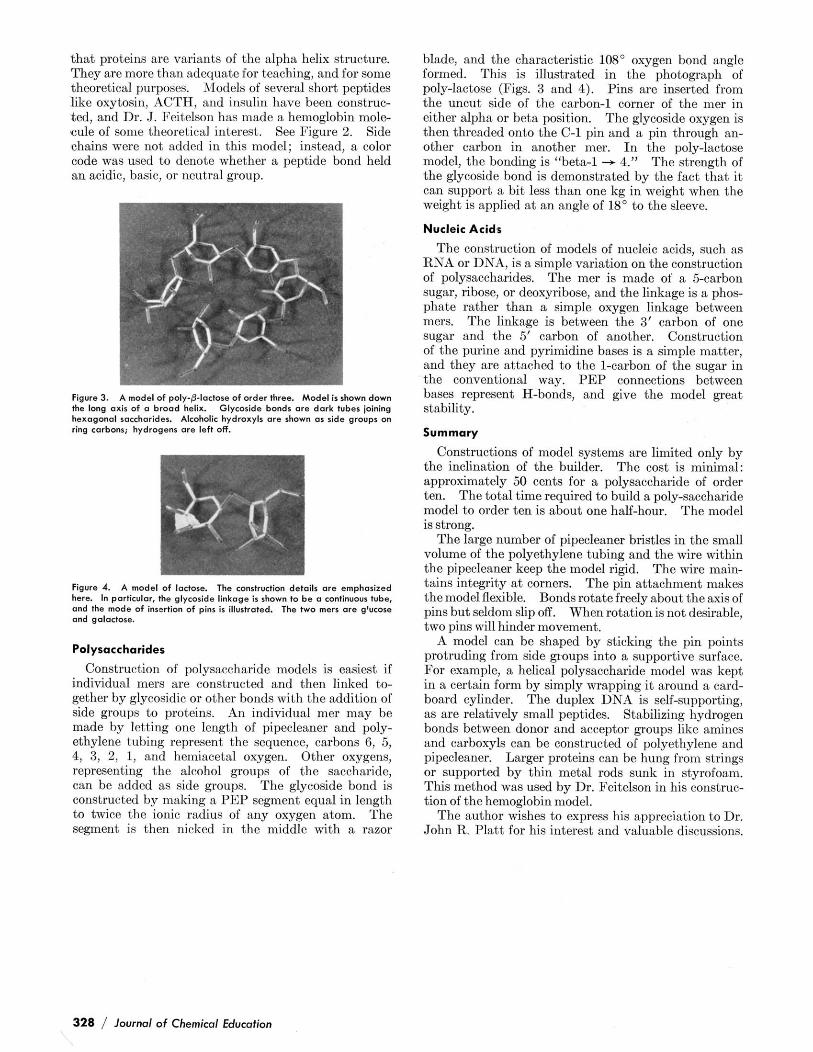
Figure 3. A model of poly-0-lactose of order three. Model is shown down the long axis of o brood helix. Glyceride bonds are dark tuber ioining hexoganol saccharides. Alcohdic hydroxylr ore shown as ride groups on ring carbons; hydrogens ore left off.
Figure 4. A model of lactose. The conrtruction details ore emphorired here. In particular, the glyceride linkoge is shown to be a continuour tube, and the mode of insertion of pin. ir illurtrated. The two merr ore glucose and galoctore.
Polysacchorides
Construction of polysaccharide models is easiest if individual mers are constructed and then linked to- gether by glycosidic or other bonds with the addition of side groups to proteins. An individual mer may be made by letting one length of pipecleaner and poly- ethylene tubing represent the sequence, carbons 6, 5, 4, 3, 2, 1, and hemiacetal oxygen. Other oxygens, representing the alcohol groups of the saccharide, can be added as side groups. The glycoside bond is constructed by making a PEP segment equal in length to twice the ionic radius of any oxygen atom. The segment is then nicked in the middle with a razor
blade, and the characteristic 108" oxygen bond angle formed. This is illustrated in the photograph of poly-lactose (Figs. 3 and 4). Pins are inserted from the uncut side of the carbon-1 corner of the mer in either alpha or beta position. The glycoside oxygen is then threaded onto the C-1 pin and a pin through an- other carbon in anotber mer. In the poly-lactose model, the bonding is "beta-1 - 4." The strength of the glycoside bond is demonstrated by the fact that it can support a bit less than one kg in weight when the weight is applied a t an angle of 18" to the sleeve.
Nucleic Acids
The construction of models of nncleic acids, such as RNA or DNA, is a simple variation on the construction of polysaccharides. The mer is made of a 5-carbon sugar, ribose, or deoxyribose, and the linkage is a phos- phate rather than a simple oxygen linkage between mers. The linkage is between the 3' carbon of one sugar and the 5' carbon of another. Construction of the purine and pyrimidine bases is a simple matter, and they are attached to the l-carbon of the sugar in the conventional way. PEP connections between bases represent H-bonds, and give the model great stability.
Summary
Constructions of model systems are limited only by the inclination of the builder. The cost is minimal: approximately 50 cents for a polysaccharide of order ten. The total time required to build a poly-saccharide model to order ten is about one balf-hour. The model is strong.
The large number of pipecleaner bristles in the small volume of the polyethylene tubing and the wire within the vivecleaner keeo the model rieid. The wire main- tains htegrity a t ccorners. The p& attachment makes the model flexible. Bonds rotate freely about the axis of pins but seldom slip off. When rotation is not desirable, two pins will hinder movement.
A model can be shaped by sticking the pin points protruding from side groups into a supportive surface. For example, a helical polysaccharide model was kept in a certain form by simply wrapping it around a card- board cylinder. The duplex DNA is self-supporting, as are relatively small peptides. Stabilizing hydrogen bonds between donor and acceptor groups like amines and carboxyls can be constructed of polyethylene and pipecleaner. Larger proteins can be hung from strings or supported by thin metal rods sunk in styrofoam. This method was used by Dr. Feitelson in his constrnc- tion of the hemoglobin model.
The author wishes to express his appreciation to Dr. John R. Platt for his interest and valuable discussions.
328 / lournol of Chemiml Education