Polyelectrolyte-mediated non-micellar synthesis of monodispersed ‘aggregates’ of gold...
8
Colloids and Surfaces A: Physicochem. Eng. Aspects 330 (2008) 143–150 Contents lists available at ScienceDirect Colloids and Surfaces A: Physicochemical and Engineering Aspects journal homepage: www.elsevier.com/locate/colsurfa Polyelectrolyte-mediated non-micellar synthesis of monodispersed ‘aggregates’ of gold nanoparticles using a microwave approach Subrata Kundu ∗ , Hong Liang Materials Science and Mechanical Engineering, Texas A&M University, College Station, TX 77843-3123, USA article info Article history: Received 27 February 2008 Accepted 20 July 2008 Available online 31 July 2008 Keywords: Gold nanoparticle Microwave Polyelectrolyte ‘Aggregates’ Synthesis abstract ‘Aggregates’ of monodispersed gold nanoparticles were synthesized for the first time in large quantities. Those particles were made in presence or in absence of gold seed in a polyelectrolyte solution using microwave heating for about 30–60s. The average diameters of the particles calculated from TEM and SEM analysis were 22 ± 2nm. Our analysis found that the polyelectrolyte acted as a reducing as well as a stabilizing agent. It controlled the growth of particles by aligning them on the polymeric chain and the nanoparticles were subsequently self-assembled to form an ‘aggregate’ structure. The synthetic procedure is very fast and the particles are stable for at least 6 months under ambient conditions. © 2008 Elsevier B.V. All rights reserved. 1. Introduction Over the last few years, significant effort has been made to synthesize nanoparticles (NPs) of noble metals [1,2]. Due to their extreme size and the surface to volume ratio, those NPs have poten- tial applications in the emerging field of catalysis, optoelectronics, drug-delivery, among others in science and technology [3,4]. Of the different noble metals studied to date, gold is of particular inter- est because of its enhanced properties and potential applications in non-linear optics [5], electronics [6], catalysis [7], biology [8], and clinical medicine [9,10]. Among different noble metals, the absorption of light by gold NPs is due to the oscillations of the con- duction band electrons induced by the interacting electromagnetic field [11]. Attempts have been made to develop simple and versa- tile method for the synthesis of size and shape selective gold NPs [12,13]. A variety of methods have been developed to assemble the gold NPs into one dimensional nanostructure because the synthesis methods can greatly influence their new properties and applica- tions. Recent reports of gold NPs synthesis include three steps: firstly, by dissolving the gold salt in appropriate solvent; secondly, reduction of the gold salt in presence of reducing agent; and finally, the stabilization of the synthesized gold NPs in presence of appro- priate stabilizing/capping agents. So far, many methods have been used for the preparation of gold NPs from the liquid phase. Choosing appropriate solvents plays an important role in the process of reac- ∗ Corresponding author. Tel.: +1 979 862 2578; fax: +1 979 845 3081. E-mail address: [email protected] (S. Kundu). tion. Recently, an alcohol reduction method has been developed to prepare metal NPs [14]. In that process, solution of the metal ions is refluxed at a particular temperature under an inert atmo- sphere. Various alcohols as well as polyols and organic solvents have been attempted for the preparation of gold NPs [14–16] using the reflux technique or oil bath heating. Other methods include laser irradiation [17], sonochemical deposition [18], photochemical reduction [19], electrochemical method [20] and chemical reduc- tion [21] of metal salts for the synthesis of gold NPs. In most of those cases, the surface passivation reagents like surfactants play a major role in the prevention of the aggregation of NPs. Polyvinyl pyrrolidone (PVP) has been widely used as a stabilizing agent in the synthesis of metallic NPs [15,16]. The synthesis of stable metallic NPs in polymer matrix needs the combination of a low concentra- tion of solute, as well as adhereness of polymeric monolayer on the growing surface. Both of the factors, low concentration and polymeric monolayer, would affect the diffusion process in the growth stage and resulting in the formation of uniformly sized NPs. Polyelectrolyte-mediated nanocomposites have been found much attention due to their unique thermal, mechanical, elec- trical, and optical properties [22,23]. Silver and gold NPs in a polymer matrix have shown unique electrical property called coulomb blockade [24,25], which is potentially used for elec- tronic device application [24]. Polyelectrolyte-mediated NPs have been commonly prepared via a two-step method where gold salt was initially mixed with polyelectrolyte followed by the addi- tion of a reducing agent under rapid stirring. Recently, Sun et al., have developed a simple heat-treatment-based method for syn- 0927-7757/$ – see front matter © 2008 Elsevier B.V. All rights reserved. doi:10.1016/j.colsurfa.2008.07.043
-
Upload
subrata-kundu -
Category
Documents
-
view
215 -
download
1
Transcript of Polyelectrolyte-mediated non-micellar synthesis of monodispersed ‘aggregates’ of gold...

Colloids and Surfaces A: Physicochem. Eng. Aspects 330 (2008) 143–150
Contents lists available at ScienceDirect
Colloids and Surfaces A: Physicochemical andEngineering Aspects
journa l homepage: www.e lsev ier .com/ locate /co lsur fa
Polyelectrolyte-mediated non-micellar synthesis of monodispersed ‘aggregates’of gold nanoparticles using a microwave approach
Subrata Kundu ∗, Hong LiangMaterials Science and Mechanical Engineering, Texas A&M University, College Station, TX 77843-3123, USA
a r t i c l e i n f o
Article history:Received 27 February 2008Accepted 20 July 2008Available online 31 July 2008
a b s t r a c t
‘Aggregates’ of monodispersed gold nanoparticles were synthesized for the first time in large quantities.Those particles were made in presence or in absence of gold seed in a polyelectrolyte solution usingmicrowave heating for about 30–60 s. The average diameters of the particles calculated from TEM andSEM analysis were 22 ± 2 nm. Our analysis found that the polyelectrolyte acted as a reducing as well as astabilizing agent. It controlled the growth of particles by aligning them on the polymeric chain and the
Keywords:Gold nanoparticleMicrowavePolyelectrolyte‘S
nanoparticles were subsequently self-assembled to form an ‘aggregate’ structure. The synthetic procedureis very fast and the particles are stable for at least 6 months under ambient conditions.
© 2008 Elsevier B.V. All rights reserved.
1
setddeiaadfit[gmtfirtpua
ttishtlrttapsNttpgN
m
0d
Aggregates’ynthesis
. Introduction
Over the last few years, significant effort has been made toynthesize nanoparticles (NPs) of noble metals [1,2]. Due to theirxtreme size and the surface to volume ratio, those NPs have poten-ial applications in the emerging field of catalysis, optoelectronics,rug-delivery, among others in science and technology [3,4]. Of theifferent noble metals studied to date, gold is of particular inter-st because of its enhanced properties and potential applicationsn non-linear optics [5], electronics [6], catalysis [7], biology [8],nd clinical medicine [9,10]. Among different noble metals, thebsorption of light by gold NPs is due to the oscillations of the con-uction band electrons induced by the interacting electromagneticeld [11]. Attempts have been made to develop simple and versa-ile method for the synthesis of size and shape selective gold NPs12,13]. A variety of methods have been developed to assemble theold NPs into one dimensional nanostructure because the synthesisethods can greatly influence their new properties and applica-
ions. Recent reports of gold NPs synthesis include three steps:rstly, by dissolving the gold salt in appropriate solvent; secondly,eduction of the gold salt in presence of reducing agent; and finally,
he stabilization of the synthesized gold NPs in presence of appro-riate stabilizing/capping agents. So far, many methods have beensed for the preparation of gold NPs from the liquid phase. Choosingppropriate solvents plays an important role in the process of reac-∗ Corresponding author. Tel.: +1 979 862 2578; fax: +1 979 845 3081.E-mail address: [email protected] (S. Kundu).
tpctbwth
927-7757/$ – see front matter © 2008 Elsevier B.V. All rights reserved.oi:10.1016/j.colsurfa.2008.07.043
ion. Recently, an alcohol reduction method has been developedo prepare metal NPs [14]. In that process, solution of the metalons is refluxed at a particular temperature under an inert atmo-phere. Various alcohols as well as polyols and organic solventsave been attempted for the preparation of gold NPs [14–16] usinghe reflux technique or oil bath heating. Other methods includeaser irradiation [17], sonochemical deposition [18], photochemicaleduction [19], electrochemical method [20] and chemical reduc-ion [21] of metal salts for the synthesis of gold NPs. In most ofhose cases, the surface passivation reagents like surfactants playmajor role in the prevention of the aggregation of NPs. Polyvinylyrrolidone (PVP) has been widely used as a stabilizing agent in theynthesis of metallic NPs [15,16]. The synthesis of stable metallicPs in polymer matrix needs the combination of a low concentra-
ion of solute, as well as adhereness of polymeric monolayer onhe growing surface. Both of the factors, low concentration andolymeric monolayer, would affect the diffusion process in therowth stage and resulting in the formation of uniformly sizedPs.
Polyelectrolyte-mediated nanocomposites have been founduch attention due to their unique thermal, mechanical, elec-
rical, and optical properties [22,23]. Silver and gold NPs in aolymer matrix have shown unique electrical property calledoulomb blockade [24,25], which is potentially used for elec-
ronic device application [24]. Polyelectrolyte-mediated NPs haveeen commonly prepared via a two-step method where gold saltas initially mixed with polyelectrolyte followed by the addi-ion of a reducing agent under rapid stirring. Recently, Sun et al.,ave developed a simple heat-treatment-based method for syn-

1 : Phys
tscafitc
icaeiwolintsrawffcTtu
m‘tpoaltptms
2
2
soPtacde
2
4ce
atss�eo(i
2
20tpt2tparticle size measured from a transmission electron micrographwas 4 ± 0.7 nm (Fig. 1B). The citrate serves only as cappingagent since it cannot reduce gold salt at room temperature(25 ◦C).
44 S. Kundu, H. Liang / Colloids and Surfaces A
hesis of polyelectrolyte-capped gold NPs in the presence of goldalt solution and an amine-containing polyelectrolyte [26]. Thisonventional heating method produces both larger, polydispersend uncontrollable particles size. Other recent reports have beenound on the preparation of gold NPs in aqueous solution involv-ng the use of poly(ethylene oxide) [27], amine functionalizedhird-generation poly(propyleneimine) dendrimers [28] and block-o-polymer micelles [29].
Recently, the microwave (MW) approach has made a greatmpact and is a promising technique for the preparation of size-ontrolled metallic nanostructures due to its rapid heating capacitynd high penetration power. The main reason is that the quantumnergy of microwave (10–5 eV) is much lower than that of chem-cal bonds in the molecules so that microwave will not break or
eaken the bonds within the molecules. In addition, the intensityf the electric field of microwave cannot cause the shift of any equi-ibrium chemical reactions. The greatest advantage of MW heatings that it can heat a substance uniformly leading to a more homoge-eous nucleation and shorter crystallization time compared withhose for conventional heating. Several researchers have recentlyynthesized gold NPs exploiting microwave approach [30-32]. Mosteported methods, however, yield mixture of different shapes, suchs triangular, square, hexagonal, and spherical NPs. Huo and co-orkers [33] have synthesized gold–polylysine hybrid materials to
orm a ‘necklace’ arrangement of gold NPs. Sun et al. [34], haveound that optical limiting properties of these ‘necklace’ parti-les are enhanced in comparison to the monocarboxylated Au NPs.o our best knowledge, it has not been achieved using the MWechnique to make monodispersed ‘aggregates’ of gold NPs withniformed size.
In this report, we demonstrate a one-step and non-micellaricrowave strategy to generate well-defined monodispersed
aggregates’ of gold nanoparticles. These particles were syn-hesized in aqueous solution of chloroauric acid (HAuCl4) andoly-allylamine hydrochloride (PAH) in presence or in absencef gold seed particles. Our proposed method does not need anydditional reducing or capping agent like most other methods initerature. This one-step process needs only 20–30 s to completehe reaction in presence and 45–60 s in absence of the Au seedarticles. The key discovery was “guiding” the synthesis of the par-icles with a polyelectrolyte to form ‘aggregates’ of gold NPs. This
ethod is straightforward, less time consuming, cost-effective, andcalable.
. Experimental
.1. Reagents
Poly-allylamine hydrochloride (PAH, 0.1% by weight) and poly-tyrene sulfonate (PSS, 0.1% by weight) of average molecular weightf 7 × 10⁴ Dalton were purchased from Aldrich and Scientificolymer Products Inc., respectively. Hydrogen tetrachloroaurateri-hydrate (HAuCl4·3H2O, 99.9%), sodium borohydride (NaBH4),nd tri-sodium citrate dihydrate (Na3C6H5O7·2H2O) were pur-hased from Sigma–Aldrich and used as received. Ultrapure-istilled (UPD) water was used for the whole synthesis duringxperiment.
.2. Instruments
UV–vis absorption spectra are recorded in a Hitachi (model U-100) UV–vis–NIR spectrophotometer equipped with 1 cm quartzuvette holder for liquid samples. A high resolution-transmissionlectron microscope (HR-TEM) (ZEOL ZEM 2010) was used at an
Ftss
icochem. Eng. Aspects 330 (2008) 143–150
ccelerating voltage of 200 kV. The energy dispersive X-ray spec-rum (EDS) was recorded with the Oxford Instruments INCA energyystem connected with the TEM. The XRD analysis was done with acanning rate 0.020 S−1 in the 2� range 40–100◦ using a Rigeku dmax
A X-ray diffractometer with Cu K� radiation (� = 0.154178). Fieldmission scanning electron microscopy (FE-SEM) images werebtained using a Hitachi S-4700. A domestic microwave (MW) ovenGold star company, EM-Z200S, 1000 W, 60 Hz) was used for MWrradiation for the entire synthesis.
.3. Synthesis of gold seed particles
A 20-mL aqueous solution containing 2.5 × 10⁻⁴ M HAuCl4 and.5 × 10−4 M tri-sodium citrate was prepared in a flask. Next,.6 mL of ice cold 0.1 M NaBH4 solution was added to the solu-ion all at once with continuous stirring. The solution turnedink immediately after adding NaBH4, indicating particle forma-ion. The particles in this solution were used as seeds within–5 h after preparation. The UV–vis spectrum showed an absorp-ion band maximum at ∼502 nm shown in Fig. 1A. The average
ig. 1. (A) The UV–vis absorption spectrum of the citrate stabilized gold seed par-icles having surface plasmon absorption band maxima at ∼502 nm. Inset of Fig. 1Ahows the corresponding pink color gold seed solution. (B) TEM image of the corre-ponding gold seed particles having average diameter was 4 ± 0.7 nm.

S. Kundu, H. Liang / Colloids and Surfaces A: Physicochem. Eng. Aspects 330 (2008) 143–150 145
regat
2o
UcpmmisiatarsIpetti
2
nbsi7ToigN
3
agat
c‘A‘
c[foIt[at[oabsorption band below 240 nm due to the absence of any aromaticmoiety (Fig. 2, curve A). The addition of HAuCl4 with PAH leadsto the instantaneous formation of the PAH–Au(III) complex. Thatwas observed as 7% increase of absorbance value (Fig. 2, curve B) of
Scheme 1. Schematic presentation for the microwave synthesis of ‘agg
.4. General route for the synthesis of monodispersed ‘aggregates’f Au nanoparticles on PAH using MW heating
PAH solution (0.1% by weight) was prepared by dissolving inPD water and stirring for about 10 h. A stock solution of 10–2 Mhloroauric acid (HAuCl4·3H2O) was made and kept under dark forrotection against light. In a typical synthesis 5 mL of PAH (0.1%)ixed with 200 �L of (10–2 M) chloroauric acid solution and theixture was stirred for 2–3 min using magnetic stirrer and allowed
t to equilibrate for ∼2 min. The equilibrium was determined by thetability of the surface plasmon resonance (SPR) band. The result-ng solution was heated in MW for 30–60 s with intermittent pausefter every 10 s to cool the reaction vessel. The gold particle forma-ion started just after 30 s of MW heating as observed from colors well as UV–vis spectrum. In presence of gold seed particles, theeaction mixture (containing PAH, gold salt solution, and gold seedolution) produces Au NPs and requires minimum irradiation time.n this case, the formation of particles started after 10 s and com-leted after 30 s of MW irradiation. The formation of gold NPs wasvident by appearance of the reddish pink coloration of the solu-ion. The pink color of the synthesized gold NPs remains stable morehan 6 months of storage in ambient environment without changen optical properties.
.5. Preparation of samples for HR-TEM and FE-SEM analysis
The samples for TEM are prepared by placing a drop of freshanoparticle solution on a carbon-film-coated cupper grid followedy slow evaporation of solvent at ambient condition. The FE-SEMamples are prepared on a Si-chip with native oxide layer. The chips cleaned thoroughly with ethanol and piranha (30% H2O2 and0% H2SO4) followed by final cleaning with ethanol and acetone.he chip is then air dried and dipped in 0.1% solution of PSS forvernight, followed by a vigorous wash to remove excess PSS leav-ng a monolayer. The sample was then blow-air dried and placed onold NPs solution for about 30 min to deposit ‘aggregates’ of goldPs.
. Results and discussion
‘Aggregates’ of monodispersed gold NPs were synthesized in1000 W microwave (MW) oven in presence or in absence of
old seed particles using poly(allylamine) hydrochloride (PAH) asstabilizing agent (Scheme 1). The key discovery was ‘guiding’
he synthesis of the particles with a polyelectrolyte rather than
FAPgg
es’ of gold NPs in presence or in absence of gold seed particles in PAH.
onventional low molecular weight amphiphiles that do not yieldaggregates’-type gold NPs. PAH helps to support the growth of theu particles in chain-like morphology leading to the formation of
aggregated’ gold NPs.The UV–vis spectrum of the citrate stabilized gold seed parti-
les exhibit a surface plasmon absorption band maxima at ∼502 nm35], as shown in Fig. 1A. The size of the seed particles measuredrom TEM analysis was 4 ± 0.7 nm, as shown in Fig. 1B. The insetf Fig. 1A shows the image of deep pink color gold seed solution.t is known that on heating or irradiation of aqueous HAuCl4 solu-ion in sunlight or UV light, gold micro-particles could be produced36]. However, the particles immediately got precipitated due tobsence of any stabilizer and the rate of photo-reduction was slowhat was found to be increased in the presence of organic ligands36]. Fig. 2 compares the UV–vis spectrum of the solution at vari-us stages of the process. Aqueous solution of PAH exhibits an UV
ig. 2. UV–vis spectrum at various stages of ‘aggregates’ gold NPs synthesis. (A)bsorption spectra of PAH (0.1% by weight) in water; (B) spectrum of the mixture ofAH, gold(III) solution before MW irradiation; (C) surface plasmon band for ‘aggre-ates’ gold particles with absorption maxima at ∼528 nm. Inset shows the pink colorold solution synthesized after 60 s of MWI.

1 : Phys
PctopsfidopcM
ioaccMa
Fe2os
46 S. Kundu, H. Liang / Colloids and Surfaces A
AH having similarity to the formation of other biomolecular–metalomplex [37]. On MW irradiation in the absence of gold seed par-icles for 60 s, the solution mixture turns pink with the appearancef an additional absorption band at ∼528 nm due to the surfacelasmon resonance (SPR) mode of gold particles [38,39]. The sharpurface plasmon band of Au NPs observed in Fig. 2C clearly signi-es the formation of Au NPs of size 10–40 nm range [38–40]. The
ecrease and slight red shift in absorption intensity peak for PAHn MW irradiation attributed to MW-influenced aggregation of theolymer chains having similarity with the UV-influenced super-oiling of DNA strands due to trimeric complex formation [41].onitoring the surface plasmon peak at 528 nm as a function ofio
im
ig. 3. TEM images and corresponding histograms for the size distribution of ‘aggregatexposure in absence of Au seed particles, shows average length of the chains are 4–5 m2 ± 2 nm. (C) shows the self-assemble ‘aggregates’-type structure of the PAH–Au NPs havf the PAH–Au NPs in much higher magnification. Inset shows the selected area electronhow low and high magnified images of the PAH–Au NPs synthesized in presence of Au see
icochem. Eng. Aspects 330 (2008) 143–150
rradiation time indicates that no gold formation occurs before 30 sf exposure in absence of seed particles. Furthermore, the plasmonbsorption peak ceases to increase beyond 60 s exposure indicatesompletion of the reaction. The reaction time decreases signifi-antly in presence of Au seed particles and completed in just 30 s ofW exposure, as shown in the Scheme 1. This is due to fast nucle-
tion in the presence of negatively charged Au seed particles. The
nset of Fig. 2 shows the image of pink color ‘aggregates’ of gold NPsbtained after 60 s of MW irradiation.Fig. 3, illustrates the transmission electron microscopy (TEM)mages of PAH–Au ‘aggregates’ of NPs prepared with the above
ethod (given in experimental section) in the absence and in
s’ PAH–Au NPs under different conditions. (A) is image of gold NPs after 60 s MWicron. (B) is higher magnified image of the same sample having average diametering particles of diameter 22 ± 2.1 nm. (D) shows the perfect ‘aggregate’ morphologydiffraction (SAED) pattern indicating crystalline nature of the particles. (E) and (F)d particles for 30 s MW heating. Average diameters of the particles are 22 ± 1.1 nm.

S. Kundu, H. Liang / Colloids and Surfaces A: Physicochem. Eng. Aspects 330 (2008) 143–150 147
(Conti
tmgtfiM‘iiadof‘e
cPsT(sooifw
Fig. 3.
he presence of gold seed particles. As stated before the solutionixture irradiated in MW for 60 s in absence and 30 s in presence of
old seed particles. The TEM samples were prepared by depositinghe PAH–Au NPs on a carbon-coated Cu grid by solution castingollowed by air drying. Fig. 3A shows the low magnified TEMmages of the PAH–Au NPs in the absence of gold seed after 60 s
W heating. The image shows that the gold NPs are formed inaggregate’-like structure and the average length of a long chains approximately 4–5 micron. Fig. 3B shows the higher magnifiedmage from the same sample. The particles are monodisperse andlmost equal size. The average diameter and standard deviations
etermined from the histogram are 22 ± 0.3 nm, after calculatingver 100 particles. Fig. 3C, shows the higher magnified imagesrom the other part of the sample. The images clearly show theaggregates’-type structure of the PAH–Au NPs. The average diam-ters of the particles are the same like Fig. 3B and calculated fromoaiei
nued ).
orresponding histogram are 22 ±2 nm. The particles grow in theAH chain and self-assemble to ‘aggregates’-type structure. Fig. 3Dhows the perfect ‘aggregate’ morphology of the PAH–Au NPs.he inset of Fig. 3D, shows the selected area electron diffractionSAED) pattern and suggested that the particles are single crystaltructure. Fig. 3E shows the PAH–Au NPs synthesized in presencef gold seed particles for 30 s of MW irradiation. The size and shapef the particles are almost the same with the particles synthesizedn absence of gold seed particles. The average diameter calculatedrom corresponding histogram is 22 ± 1.1 nm having similaritiesith Fig. 3A–D. The linear part of the ‘aggregates’/chain structure
f the PAH–Au NPs is shown in Fig. 3F. All particles shown abovere uniform and have almost the same size indicating homogeneityn the growth process. The synthesized gold particles do not touchach other as clearly observed from the highly magnified TEMmages in Fig. 3F. Otherwise it was obvious to find a SPR band at

148 S. Kundu, H. Liang / Colloids and Surfaces A: Physicochem. Eng. Aspects 330 (2008) 143–150
Ftli
liciit
acosstss
sata
F
tartos
Xpcnc
ftowJXmpFpeaks and (2 2 0)–(1 1 1) peaks are slightly higher than the literaturevalue. This indicates that our ‘aggregated’ Au NPs were abundant in{1 0 0} and {1 1 0} planes respectively and tended to be preferen-tially oriented parallel to the surface of the gold particles. Accordingto Wang et al. [45], the shape of an fcc nanocrystal was mainly deter-
ig. 4. (A) and (B) are the TEM images of the PAH–Au NPs synthesized having Au(III)o PAH molar ratio’s are 1:20 and 1:10, respectively. Here particles are aggregated andarger in diameter than the particles in Fig. 3A–F. Average diameter of the particless ∼28 ± 5 nm.
onger wavelengths region in the UV–vis spectrum. But the TEMmages and the SPR band do not signify that in our study. Wealculated that the interparticles spacing between two particless around 1.5 nm. It implies that the gold seed particles do notnterfere the growth of the ‘aggregates’ particles. Instead, they helphe nucleation and enhance the reaction in an effective way.
Fig. 4A and B shows the TEM images of PAH–Au NPs synthesizedt a high concentration of metal ion having Au(III) to PAH molaroncentration ratios of 1:20 and 1:10, respectively. The average sizef the particles increases due to high concentration of Au(III) in theolution leading to particle–particle aggregation to minimize theurface energy of the gold particles. In Fig. 4A and B, particles showo aggregate in some parts also authenticated from the slight red-hift and broadened absorption spectra in UV–vis spectrum (nothown here).
It is to be noted that this ‘aggregates’-type arrangement occurstrongly in solution although metal particles have tendency torrange or self-assemble in TEM grids during drying. UV–vis spec-ra confirms the self-assemblance of the gold particles with a broadbsorbance maxima at ∼530–560 nm (not shown here). Although,
ig. 5. Energy dispersive X-ray spectrum (EDS) of the ‘aggregates’ PAH–Au NPs.
he expected absorbance maxima for 20–25 nm gold nanoparticlesre ∼515–530 nm (Fig. 1) as reported in literature, but in our caseed-shifting of absorption maxima occurred that might be due tohis ‘aggregates’ of the monodispersed gold particles. Similar typesf spontaneous arrangements were also observed with metal andemiconductor nanomaterials too [42,43].
Fig. 5 presents the results obtained from the energy dispersive-ray spectroscopy (EDS) analysis to determine the chemical com-osition. The spectrum consisted different peaks for gold, copper,arbon, and nitrogen. The gold peak was from the gold NPs and theitrogen peak was from the polymer matrix. The Cu and C peaksame from the carbon-coated cupper grid.
The X-ray diffraction pattern (XRD) of the gold NPs recordedrom the sample displayed in Fig. 6 and the peaks were assignedo diffract from the (1 1 1), (2 0 0), (2 2 0), (3 1 1), and (2 2 2) planesf fcc gold. The lattice constant of the fcc Au was 0.405 nm whichas within the error reported value (with a = 0.4078 nm given by
CPDS file No. 4-0784). The size of the gold NPs was measured by-ray diffraction peak line width broadening using the Debye for-ula for small crystalline spheres [44]. The mean diameters of the
articles are consistent with the results of TEM measurements inig. 3. The ratios between the intensities of (2 0 0)–(1 1 1) diffraction
Fig. 6. Powder X-ray diffraction pattern of the ‘aggregates’ PAH–Au NPs.

S. Kundu, H. Liang / Colloids and Surfaces A: Physicochem. Eng. Aspects 330 (2008) 143–150 149
F s’ PAHi zes of
mdefd
FacAe2gmittiriss
eAttffcn
vftps
tsithiswisso
aTsfmbl
ig. 7. (A) and (B) are the low and high magnified FE-SEM images of the ‘aggregatemages of gold NPs at Au(III) to PAH molar ratio’s 1:20 and 1:10, respectively. The si
ined by the ratio between growth rates along 〈1 0 0〉 and 〈1 1 1〉irections. As PAH used as a stabilizer, we believed that the prefer-ntial interaction between the polyelectrolyte and different crystalacets of the gold NPs reduced the growth rate along the 〈1 0 0〉irection and enhanced the growth rate along the 〈1 1 1〉 direction.
Field emission scanning electron micrographs are shown inig. 7. Fig. 7A shows the ‘aggregates’ of gold particles formed whichre similar with the TEM images in Fig. 3A–D. Fig. 7B shows theorresponding high-magnified image of the gold ‘aggregated’ NPs.lthough in some parts the particles aggregated more due to dryingffect in the substrate. The average diameters of the particles are2 ± 2 nm, similar to the TEM images. Fig. 7C shows the aggregatedold NPs at higher metal ion concentration where the Au(III) to PAHolar concentration ratio’s are 1:20. At a high concentration, the
nterparticle distance decreases due to weaker capping capacity ofhe polymer which turns to increase the aggregation tendency ofhe particles. Fig. 7D shows increasingly aggregated NPs. Here thencreased ion concentration (Au(III) to PAH molar concentrationatio 1:10) increases the aggregation tendency. This was observedn the TEM images in Fig. 4 and with the red shifting of the UV–vispectra (not shown here). The size of the individual particles waseen to increase to ∼28 ± 5 nm ranges.
To check the suitability of NPs, we varied the reaction param-ters such as concentrations of polyelectrolyte, concentration ofu(III), MW heating time, etc. It was observed that the ‘aggregate’-ype nanostructures formed only in certain concentrations. When
he PAH concentration was increased from 0.1% to 1% or decreasedrom 0.1% to 0.001%, no particles having ‘aggregate’ structure wereormed within 60 s of MW irradiation. The ‘aggregate’ gold parti-les were formed only at the Au(III) concentration of 0.01 M andone otherwise (0.1 M nor 0.001 M). The MW irradiation time wastpfet
–Au NPs. The average diameter of the particles is 22 ± 2 nm. (C) and (D) show thethe individual particles are ∼28 ± 5 nm.
aried and it was found that a 60 s irradiation was sufficient for theormation of ‘aggregates’ structures and completion of the reac-ion. We tried the same reaction by other conventional heatingrocess instead of MW heating but that results aggregated biggerize particles as observed from TEM analysis (not shown here).
The roles of MW heating and the concentration of PAH are cen-ral to the synthesis of ‘aggregates’ gold NPs. Upon heating HAuCl4alt solution using a resistor heater or a MW oven, in the presence orn the absence of Au seed particles, the heat causes thermal reduc-ion of Au3+ to Au0. The MW is effective for rapid and uniformedeating. Without the addition of polyelectrolye (PAH), the precip-
tates of clusters of micro-particles did not form any recognizablehape (Fig. 8). In the presence of PAH, with or without the gold seed,ell-defined ‘aggregates’ NPs were formed. We note that in pass-
ng the use of negatively charged polyelectrolyte (i.e. polystyreneulfonate, PSS) or negatively charged surfactants like sodium laurylulfate (SDS), Au NPs did not form due to lack of needed chemistryf complex with AuCl4−.
The spontaneous formation of ‘aggregates’ gold NPs can bettributed to the direct redox reaction between PAH and HAuCl4.his is supported by the lack of other reducing agent in the mixedolution. The AuCl4− was complexed with the PAH as evidencedrom the absorption spectra in Fig. 2. The amino group in the poly-
er acts as a reducing site for the reaction. The amino group haseen reported to be oxidized to imine in the aqueous phase as alky-
amine complex with metal ions [46]. It has also been reported that
he metal ion induced oxidation of amine to nitrile [47]. In theresent study PAH functions as both a reductant and a stabilizeror gold NPs formation. The absorption of PAH to AuCl4− facilitateslectron transfer from Au3+ to the amine group in the polyelec-rolyte. The high temperature resulted from MW irradiation can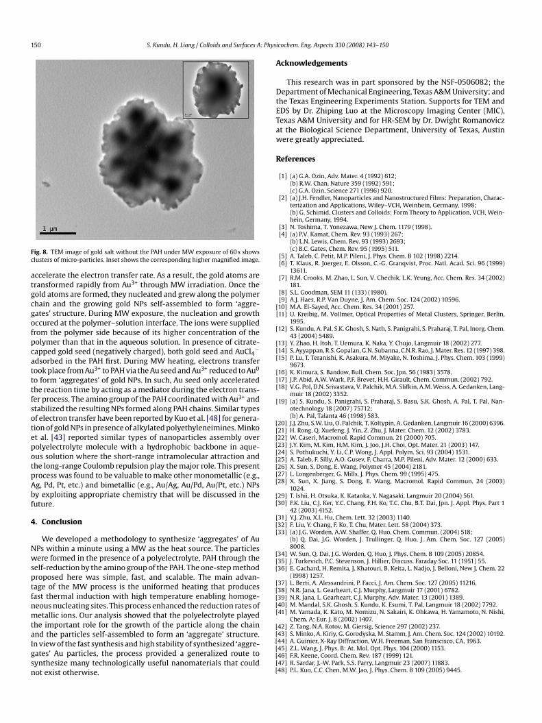
150 S. Kundu, H. Liang / Colloids and Surfaces A: Phys
Fc
atgcgofpcatttfsotepotpAbf
4
NwsptfnmtaIgsn
A
DtETaw
R
[[
[
[[[
[[[
[
[[[[[[[[[
[[
[[[
[[[
[[[[[
[
ig. 8. TEM image of gold salt without the PAH under MW exposure of 60 s showslusters of micro-particles. Inset shows the corresponding higher magnified image.
ccelerate the electron transfer rate. As a result, the gold atoms areransformed rapidly from Au3+ through MW irradiation. Once theold atoms are formed, they nucleated and grew along the polymerhain and the growing gold NPs self-assembled to form ‘aggre-ates’ structure. During MW exposure, the nucleation and growthccured at the polymer–solution interface. The ions were suppliedrom the polymer side because of its higher concentration of theolymer than that in the aqueous solution. In presence of citrate-apped gold seed (negatively charged), both gold seed and AuCl4−
dsorbed in the PAH first. During MW heating, electrons transferook place from Au3+ to PAH via the Au seed and Au3+ reduced to Au0
o form ‘aggregates’ of gold NPs. In such, Au seed only acceleratedhe reaction time by acting as a mediator during the electron trans-er process. The amino group of the PAH coordinated with Au3+ andtabilized the resulting NPs formed along PAH chains. Similar typesf electron transfer have been reported by Kuo et al. [48] for genera-ion of gold NPs in presence of alkylated polyethyleneimines. Minkot al. [43] reported similar types of nanoparticles assembly overolyelectrolyte molecule with a hydrophobic backbone in aque-us solution where the short-range intramolecular attraction andhe long-range Coulomb repulsion play the major role. This presentrocess was found to be valuable to make other monometallic (e.g.,g, Pd, Pt, etc.) and bimetallic (e.g., Au/Ag, Au/Pd, Au/Pt, etc.) NPsy exploiting appropriate chemistry that will be discussed in theuture.
. Conclusion
We developed a methodology to synthesize ‘aggregates’ of AuPs within a minute using a MW as the heat source. The particlesere formed in the presence of a polyelectrolyte, PAH through the
elf-reduction by the amino group of the PAH. The one-step methodroposed here was simple, fast, and scalable. The main advan-age of the MW process is the uniformed heating that producesast thermal induction with high temperature enabling homoge-eous nucleating sites. This process enhanced the reduction rates ofetallic ions. Our analysis showed that the polyelectrolyte played
he important role for the growth of the particle along the chain
nd the particles self-assembled to form an ‘aggregate’ structure.n view of the fast synthesis and high stability of synthesized ‘aggre-ates’ Au particles, the process provided a generalized route toynthesize many technologically useful nanomaterials that couldot exist otherwise.[[[[[[
icochem. Eng. Aspects 330 (2008) 143–150
cknowledgements
This research was in part sponsored by the NSF-0506082; theepartment of Mechanical Engineering, Texas A&M University; and
he Texas Engineering Experiments Station. Supports for TEM andDS by Dr. Zhiping Luo at the Microscopy Imaging Center (MIC),exas A&M University and for HR-SEM by Dr. Dwight Romanoviczt the Biological Science Department, University of Texas, Austinere greatly appreciated.
eferences
[1] (a) G.A. Ozin, Adv. Mater. 4 (1992) 612;(b) R.W. Chan, Nature 359 (1992) 591;(c) G.A. Ozin, Science 271 (1996) 920.
[2] (a) J.H. Fendler, Nanoparticles and Nanostructured Films: Preparation, Charac-terization and Applications, Wiley–VCH, Weinhein, Germany, 1998;(b) G. Schimid, Clusters and Colloids: Form Theory to Application, VCH, Wein-hein, Germany, 1994.
[3] N. Toshima, T. Yonezawa, New J. Chem. 1179 (1998).[4] (a) P.V. Kamat, Chem. Rev. 93 (1993) 267;
(b) L.N. Lewis, Chem. Rev. 93 (1993) 2693;(c) B.C. Gates, Chem. Rev. 95 (1995) 511.
[5] A. Taleb, C. Petit, M.P. Pileni, J. Phys. Chem. B 102 (1998) 2214.[6] T. Klaus, R. Joerger, E. Olsson, C.-G. Granqvist, Proc. Natl. Acad. Sci. 96 (1999)
13611.[7] R.M. Crooks, M. Zhao, L. Sun, V. Chechik, L.K. Yeung, Acc. Chem. Res. 34 (2002)
181.[8] S.L. Goodman, SEM 11 (133) (1980).[9] A.J. Haes, R.P. Van Duyne, J. Am. Chem. Soc. 124 (2002) 10596.10] M.A. El-Sayed, Acc. Chem. Res. 34 (2001) 257.11] U. Kreibig, M. Vollmer, Optical Properties of Metal Clusters, Springer, Berlin,
1995.12] S. Kundu, A. Pal, S.K. Ghosh, S. Nath, S. Panigrahi, S. Praharaj, T. Pal, Inorg. Chem.
43 (2004) 5489.13] Y. Zhao, H. Itoh, T. Uemura, K. Naka, Y. Chujo, Langmuir 18 (2002) 277.14] S. Ayyappan, R.S. Gopalan, G.N. Subanna, C.N.R. Rao, J. Mater. Res. 12 (1997) 398.15] P. Lu, T. Teranishi, K. Asakura, M. Miyake, N. Toshima, J. Phys. Chem. 103 (1999)
9673.16] K. Kimura, S. Bandow, Bull. Chem. Soc. Jpn. 56 (1983) 3578.17] J.P. Abid, A.W. Wark, P.F. Brevet, H.H. Girault, Chem. Commun. (2002) 792.18] V.G. Pol, D.N. Srivastava, V. Palchik, M.A. Slifkin, A.M. Weiss, A. Gedanken, Lang-
muir 18 (2002) 3352.19] (a) S. Kundu, S. Panigrahi, S. Praharaj, S. Basu, S.K. Ghosh, A. Pal, T. Pal, Nan-
otechnology 18 (2007) 75712;(b) A. Pal, Talanta 46 (1998) 583.
20] J.J. Zhu, S.W. Liu, O. Palchik, T. Koltypin, A. Gedanken, Langmuir 16 (2000) 6396.21] H. Rong, Q. Xuefeng, J. Yin, Z. Zhu, J. Mater. Chem. 12 (2002) 3783.22] W. Caseri, Macromol. Rapid Commun. 21 (2000) 705.23] J.Y. Kim, M. Kim, H.M. Kim, J. Joo, J.H. Choi, Opt. Mater. 21 (2003) 147.24] S. Pothukuchi, Y. Li, C.P. Wong, J. Appl. Polym. Sci. 93 (2004) 1531.25] A. Taleb, F. Silly, A.O. Gusev, F. Charra, M.P. Pileni, Adv. Mater. 12 (2000) 633.26] X. Sun, S. Dong, E. Wang, Polymer 45 (2004) 2181.27] L. Longenberger, G. Mills, J. Phys. Chem. 99 (1995) 475.28] X. Sun, X. Jiang, S. Dong, E. Wang, Macromol. Rapid Commun. 24 (2003)
1024.29] T. Ishii, H. Otsuka, K. Kataoka, Y. Nagasaki, Langmuir 20 (2004) 561.30] F.K. Liu, C.J. Ker, Y.C. Chang, F.H. Ko, T.C. Chu, B.T. Dai, Jpn. J. Appl. Phys. Part 1
42 (2003) 4152.31] Y.J. Zhu, X.L. Hu, Chem. Lett. 32 (2003) 1140.32] F. Liu, Y. Chang, F. Ko, T. Chu, Mater. Lett. 58 (2004) 373.33] (a) J.G. Worden, A.W. Shaffer, Q. Huo, Chem. Commun. (2004) 518;
(b) Q. Dai, J.G. Worden, J. Trullinger, Q. Huo, J. Am. Chem. Soc. 127 (2005)8008.
34] W. Sun, Q. Dai, J.G. Worden, Q. Huo, J. Phys. Chem. B 109 (2005) 20854.35] J. Turkevich, P.C. Stevenson, J. Hillier, Discuss. Faraday Soc. 11 (1951) 55.36] E. Gachard, H. Remita, J. Khatouri, B. Keita, L. Nadjo, J. Belloni, New J. Chem. 22
(1998) 1257.37] L. Berti, A. Alessandrini, P. Facci, J. Am. Chem. Soc. 127 (2005) 11216.38] N.R. Jana, L. Gearheart, C.J. Murphy, Langmuir 17 (2001) 6782.39] N.R. Jana, L. Gearheart, C.J. Murphy, Adv. Mater. 13 (2001) 1389.40] M. Mandal, S.K. Ghosh, S. Kundu, K. Esumi, T. Pal, Langmuir 18 (2002) 7792.41] M. Yamada, K. Kato, M. Nomizu, N. Sakairi, K. Ohkawa, H. Yamamoto, N. Nishi,
Chem. A: Eur. J. 8 (2002) 1407.42] Z. Tang, N.A. Kotov, M. Giersig, Science 297 (2002) 237.
43] S. Minko, A. Kiriy, G. Gorodyska, M. Stamm, J. Am. Chem. Soc. 124 (2002) 10192.44] A. Guinier, X-Ray Diffraction, W.H. Freeman, San Franscisco, CA, 1963.45] Z.L. Wang, J. Phys. B: At. Mol. Opt. Phys. 104 (2000) 1153.46] F.R. Keene, Coord. Chem. Rev. 187 (1999) 121.47] R. Sardar, J.-W. Park, S.S. Parry, Langmuir 23 (2007) 11883.48] P.L. Kuo, C.C. Chen, M.W. Jao, J. Phys. Chem. B 109 (2005) 9445.