Pocket Guides for Gynaecologists mind the following principle: Attention: Thehysteroscope is...
161
Transcript of Pocket Guides for Gynaecologists mind the following principle: Attention: Thehysteroscope is...


Pocket Guides for Gynaecologists
Editors: Thomas Romer, Andreas D. Ebert


Thomas Romer
Diagnostic HysteroscopyA practical guide
2nd Edition
DE GRUYTER

Professor Dr. med. Thomas RomerEvangelisches KrankenhausKoln-Weyertal gGmbHWeyertal 7650931 [email protected]
Translated by Dr. Christina Romer, Cologne.
This book has 134 figures and 6 tables.
ISBN 978-3-11-022497-9
Library of Congress Cataloging-in-Publication Data
Romer, T. (Thomas)[Hysteroskopischer Wegweiser fur Gynakologen. English]Diagnostic hysteroscopy : a practical guide / Thomas Roemer. - - 2nd ed.
p. ; cm. - - (Pocket guides for gynaecologists)ISBN 978-3-11-022497-9 (alk. paper)
1. Hysterocopy- -Handbooks, manuals, etc. I. Title. II. Series: Pocket guides forgynaecologists.
[DNLM: 1. Infertility, Female- -diagnosis- -Handbooks. 2. Hysteroscopy- -Handbooks. 3. Menstruation Disturbances- -diagnosis- -Handbooks. WP 39R763h 2010a]
RG304.5.H97R6613 2010618.10407545- -dc22
2010003282
Bibliographic information published by the Deutsche Nationalbibliothek
The Deutsche Nationalbibliothek lists this publication in the Deutsche Natio-nalbibliografie; detailed bibliographic data are available in the Internet athttp://dnb.d-nb.de.
# 2010 Walter de Gruyter GmbH & Co. KG, Berlin/New York. The publisher,together with the authors and editors, has taken great pains to ensure that allinformation presented in this work (programs, applications, amounts, dosages,etc.) reflects the standard of knowledge at the time of publication. Despite care-ful manuscript preparation and proof correction, errors can nevertheless occur.Authors, editors and publisher disclaim all responsibility and for any errors oromissions or liability for the results obtained from use of the information, or partsthereof, contained in this work.The citation of registered names, trade names, trade marks, etc. in this workdoes not imply, even in the absence of a specific statement, that such namesare exempt from laws and regulations protecting trade marks etc. and thereforefree for general use. Printed in Germany.Typesetting, printing and binding: Druckhaus “Thomas Muntzer”, Bad Langensalza.
Preface to the 2nd edition
The hysteroscopic diagnostics and therapy have become a mainfocus in the clinical and research activities at the Department ofObstetrics and Gynaecology at the University of Greifswald at thebeginning of the 1990s, and hundreds of gynaecologists havebeen trained in hysteroscopy during the traditional Days of Hyste-roscopy in Greifswald. As a consequence we decided to pass onthis extensive experience and published a Hysteroscopic Guidefor Gynaecologists together with Professor Straube in 1996. Withthis book the idea of Gynaecological Pocket Guides, which shallrepresent in a short, concise and pictorial way the main areas ofour speciality, was born.After more than ten years hysteroscopy has further developed,which led to this 2nd edition containing the latest aspects of diag-nostic hysteroscopy and its practical applications.The 2nd edition shall contribute to the further enhancement of di-agnostic hysteroscopy in the practices and in hospitals.I would like to thank everyone who supported me in completingthe book. I thank Ms. Timm for typing the manuscript, and Ms.Dr. Kowalski and Ms. Dobler from Walter de Gruyter publishinghouse for their excellent advice and for having responded to allmy comments and requests.
Cologne, February 2010 Prof. Dr. med. Thomas Romer


Contents
1. Introduction . . . . . . . . . . . . . . . . . . . . . . . . . . . . . . . . . . . . . . 12. History . . . . . . . . . . . . . . . . . . . . . . . . . . . . . . . . . . . . . . . . . . 23. Indications for diagnostic hysteroscopy . . . . . . . . . . . . . . . 34. Instrumentation and distending medium . . . . . . . . . . . . . . 45. Examination procedure and techniques . . . . . . . . . . . . . . . 226. Distinctive features of hysteroscopy in the gynaecologist’s
practice . . . . . . . . . . . . . . . . . . . . . . . . . . . . . . . . . . . . . . . . . . 347. Hysteroscopy in the diagnostics of sterility and infertility 368. Hysteroscopy with bleeding disorders . . . . . . . . . . . . . . . . 669. Hysteroscopy with sonographically suspect endometrial
findings . . . . . . . . . . . . . . . . . . . . . . . . . . . . . . . . . . . . . . . . . 11810. Hysteroscopy and lost IUD/IUS. . . . . . . . . . . . . . . . . . . . . . 13311. Special cases . . . . . . . . . . . . . . . . . . . . . . . . . . . . . . . . . . . . . 14012. Complications . . . . . . . . . . . . . . . . . . . . . . . . . . . . . . . . . . . . 14513. Summary . . . . . . . . . . . . . . . . . . . . . . . . . . . . . . . . . . . . . . . . 14914. List of abbreviations . . . . . . . . . . . . . . . . . . . . . . . . . . . . . . . 150

1. Introduction
In the last two decades hysteroscopy has been established as amethod for the diagnostics and therapy of intrauterine diseases.The scope of indications has permanently increased so that todaythis method belongs to the standard practices in gynaecology.With the development of thin lenses hysteroscopy is not only fea-sible in hospitals but for many indications also in the gynaecolo-gical practice without anaesthesia.For that reason aspects of the diagnostic hysteroscopy for outpati-ent treatment are especially considered.For the diagnostics of sterility and bleeding disorders hysteroscopyconstitutes only one form of treatment. Therefore in the case stu-dies of this 2nd edition of the Hysteroscopic Guide this method isplaced in line with anamnesis, sonography, histology and therapy.The present guide sets out to offer to the gynaecologist a compa-nion for the practical use of hysteroscopy.

2. History
The first hysteroscopy was reported by PANTALEONI in the Eng-lish journal The Medical Press in 1869. The Frankfurt physicianBOZZINI, who in 1804 developed the so-called light conductor,already then talked of the possibility of hysteroscopy.In the next century there were many attempts to establish hystero-scopy as a method for gynaecological diagnostics.Its decisive impetus hysteroscopy owes to LINDEMANN, whosucceeded in the 1970s in improving CO2-hysteroscopy as amethod.With the possibility of therapeutic hysteroscopies and as a resultof numerous technical improvements this method has now foundits well-deserved application.Over the last decades the scope of applications of hysteroscopy,especially for the diagnostics of bleeding disorders, has increasedby the use of fluid distending media.Thin lenses and sophisticated optical systems facilitate a high pic-ture quality. The development of compact systems for the use inthe practices (Telepack) is going to further enhance this method.

3. Indications for diagnostic hysteroscopy
1. bleeding disorders2. diagnostics and staging of endometrial cancer3. diagnostic assessment of sonographically suspect endometrial
findings4. sterility/infertility5. control after intrauterine operations (intrauterine adhesiolyses,
septum dissections, curettages following an abortion, curet-tages post partum or in childbed)
6. control after medical therapy of endometrial hyperplasias7. lost IUD/IUS

4. Instrumentation and distending medium
1. Compulsory–– hysteroscope (30$ lenses), when indicated with continuous
flow sheath–– distending medium–– light source–– (self-holding) specula
2. Optional–– video documentation–– grasping forceps–– probe/Hegar’s dilatators–– small curette for endometrial biopsy
Attention: Diagnostic hysteroscopy can be best performed with30$ lenses.

Small curette for target curettage or endometrial biopsy for outpatientdiagnostic hysteroscopy. Self-holding specula (available in varioussizes).
Attention: Extraction of histological material is possible withthis curette without further cervix dilatation.
Attention: Self-holding specula are especially recommendedfor outpatient hysteroscopy because a fixation of the cervixwith grasping forceps may be dispensed with in most of thecases.
Instrumentation and distending medium 5

Diagnostic hysteroscope (2 mm-30$-lenses), with a 2.8-mm-diagnos-tic sheath and a 3.6-mm-flow sheath with the possibility of continu-ous flow.
Attention: The continuous flow sheath is especially suited forfresh bleeding ex utero or coagula in utero for the clearingirrigation of the cavum uteri.
Attention: A flushing effect may also be reached when the cer-vix is dilated further (Hegar 8), so that the outflow may bereached via the dilated cervical canal.
Instrumentation and distending medium6

Bettocchi-hysteroscope with working sheath for semi-rigid instru-ments (biopsy forceps, grasping forceps, microscissors) and continu-ous flow sheath, lenses 2 mm, outer diameter: 4.2 mm.
Attention: The small-size instruments are only suited for thebiopsy of focal lesions, cutting off of small polyps, IUD-extrac-tion and cutting of intrauterine adhesions grade 1 and 2.
Instrumentation and distending medium 7

Semi-rigid instruments for the Bettocchi-hysteroscope
1. biopsy- and grasping forceps2. biopsy spoon forceps3. punch4. blunt scissors5. sharp scissors6. myoma-fixation instrument
Attention: For the insertion of the working sheath the non-anaes-thetized patient may be given a local anaesthetic if necessary.
Attention: The tissue gained from biopsy may often be verysmall, so that a small curette may be used.
Instrumentation and distending medium8

Diagnostic standard hysteroscope, 4 mm-30$-lenses and 5.1 mm out-er diameter (without continuous flow sheath).
Attention: With patients suffering from cervical stenosis mini-hysteroscopy is primarily used.
Instrumentation and distending medium 9

Xenon light source with up to 300 W.
Attention: A high-performance light sources enhances diagnos-tic reliability.
Instrumentation and distending medium10

HAMOU- microhysteroflator for CO2-hysteroscopyLeft: Digital CO2-pressure indication (mmHg)Right: Digital CO2-flow indication (ml/min).
Attention: The pressure in CO2-insufflators is limited to200 mmHg.
Attention: CO2-insufflators for laparoscopy (Laparomat) mustnot be used for hysteroscopy.
Instrumentation and distending medium 11

Pressure-cuff for diagnostic hysteroscopy with a fluid distending med-ium for 1-l-fluid (usually isotonic saline solution). Pressure on the cuffis in most of the cases adjusted to 150 mmHg (up to at most200 mmHg).
Attention: With a more difficult passage through the cervicalcanal a short-term increase in pressure is recommended be-cause it facilitates the opening of the cervical canal and thefollowing passage.
Instrumentation and distending medium12

Videocamera Image 1 (digital 3-chip-camera) with pendular headand Image 1 (standard head) with control gear.
Advantage: The pendular camera remains centred even whenmoved, which facilitates orientation.
Instrumentation and distending medium 13

Telepack-system with connected camera head (Telekam) and lightcable.
Advantages of the Telepack-system:
–– mobile use–– space-saving–– multi-functional (light source, screen, camera, documentation
– all in one device)–– allows for video and photo documentation–– cost-effective (low costs)
Instrumentation and distending medium14

Attention: Especially for the diagnostics of bleeding disordershysteroscopy with a fluid distending medium should be pre-ferred.
Attention: One advantage of the hysteroscopy with a fluid dis-tending medium is that a follow-up control of the cavity aftermechanical removal of polyps or dilatation/curettage is possi-ble.
Table 1: Comparison of CO2-hysteroscopy and hysteroscopy with afluid distending medium
CO2-hysteroscopy hysteroscopywith fluidmedium
1. picture very clear clear2. technical complexity higher low3. usage some experience
necessaryeasy
4. risk of dissemination(infection, tumour cells)
very low slightly higher
5. diagnostics of bleedingdisorders
limited (withcurrent bleeding)
very good
6. diagnostics of sterility very good good
Instrumentation and distending medium 15

Hysteroscopic finding: Passage of the cervical canal with CO2-hys-teroscopy. Further forward movement towards the gas bubbles.
Hysteroscopic finding16

Hysteroscopic finding: Passage of the cervical canal with hystero-scopy with a fluid medium. Clear view of the cervical canal withfurther forward movement of the hysteroscope towards the cavity.
Hysteroscopic finding 17

Hysteroscopic finding after the setting up of the hysteroscope on theexternal os of the cervical canal.
Attention: Through the supply of the distending medium thecervical canal unfolds and the passage with the hysteroscopebecomes possible.
Hysteroscopic finding18

Hysteroscopic finding (CO2 as distending medium): Regular cavitywith atrophic endometrium.Panoramic hysteroscopy: Fundus, posterior wall and tubal cornuaclearly visible on both sides.Disadvantage: Gas bubbles obstruct the assessment of the poster-ior wall of the cavity.
Hysteroscopic finding 19

52-year-old patient
1. Clinical diagnosis cervical stenosis2. Anamnesis cervical stenosis, cytological swab not
possible3. Sonography endometrium thickness: 6 mm
4. Hysteroscopy regular cavity after dilatation of the cervix
5. Therapy dilatation of the cervix, dilatation/curettage
6. Histology atrophic endometrium, cervix withoutfindings
Regular cavity with cervical stenosis20

Hysteroscopic finding: Regular cavity with normal endometrium.Hysteroscopy with a fluid distending medium makes assessment diffi-cult by air bubbles on the anterior wall.
Attention: Air bubbles in the cavity can be avoided by payingattention to an empty input tube for the fluid distending med-ium during hysteroscopy.
Hysteroscopic finding 21

5. Examination procedure and techniques
Examination procedure for the diagnostic hysteroscopy
–– palpation/sonography–– vaginal disinfection–– adjustment of specula–– (grasping forceps)–– setting up of hysteroscope (Attention: There must be no air in
the input tube!)–– monitored passage through the cervical canal–– panoramic hysteroscopy–– assessment of the fundus and the tubal ostia–– assessment of the cavity walls–– assessment of the cervical canal when removing the hystero-
scope–– biopsy (eye-directed/target biopsy) or dilatation/curettage
Attention: For hysteroscopy the tubal ostia are the major pointsof orientation (landmarks) in the uterine cavity.
Technique
Lesions of the endometrium are to be avoided. Therefore mindthe following principle:
Attention: The hysteroscope is always the first instrument in thecervical canal.
Attention: Probing of the uterine cavity and dilatation of thecervical canal with Hegar’s dilators should, if possible, only beperformed after a hysteroscopic inspection of the original cavity.Exception: cervical stenosis

Problematic situations in diagnostic hysteroscopy
1. nullipara2. craurosis fornicis3. state after conisation
solution:–– use of a thinner hysteroscope (2-mm-hysteroscope)–– local application of prostaglandinsdisadvantages of the local application of prostaglandins:–– side effects (gastrointestinal)–– bleedings may obstruct vision–– danger of via falsa because of softening-up of the whole
cervix–– additional costs
Attention: The local application of prostaglandins is only ne-cessary in rare cases. With a cervical stenosis the use of mini-hysteroscopes is the prime choice.
Examination procedure and techniques 23

Hysteroscopic finding: With further forward movement of the hystero-scope a cervical stenosis becomes visible. In this case passage isonly possible after dilatation up to Hegar 5.
Attention: Cervical stenosis (mostly on the ostium cervicis in-ternum) can be hysteroscopically exactly verified and thus bedirectly dilated.
Examination procedure and techniques24

Regular cavity (minihysteroscopy, cervical stenosis) 25

52-year-old patient
1. Clinical diagnosis cervical stenosis, lower abdominal pain
2. Anamnesis cervical stenosis, cytological swab notpossible, occasional abdominal pain
3. Sonography endometrium thickness: 3 mm(secretory congestion, mucous cervix)
4. Hysteroscopy mucous cervixcervical stenosisregular cavity, ostia free
5. Therapy dilatation of the cervix, dilatation/curettage
6. Histology atrophic endometrium, cervix withoutfindings
Optimal time for examination
1. in sterile and infertile patients – immediately post menstruatio-nem
2. in perimenopausal patients – with bleeding disorders possiblypost menstruationem
3. in postmenopausal patients – as soon as possible after thebleeding
Local anaesthesia
Indication1. cervical stenosis2. craurosis fornicis3. nullipara4. necessity of use of a 7-mm-hysteroscope with working sheath
Examination procedure and techniques26

Local anaesthesia
Paracervical block
depot in the subvaginal epithelium for grasping forceps
5 ml of a local anaesthetic paracervically
Attention: Before injection always aspiration to avoid intravas-cular injection.
Attention: Wait for the effect of local anaesthesia (3–5 min-utes) before beginning with hysteroscopy or further manipula-tions.
Attention: With the development of thin hysteroscopes localanaesthesia with outpatient hysteroscopies has become neces-sary only in rare cases.(Patients may suffer from more pain from the injection thanfrom the passage of the cervical canal with the minihystero-scope.)
Examination procedure and techniques 27

Permitted movements of the hysteroscope with a patient withoutanaesthesia
1. forward and backward movement2. rotation with use of 30$-angle lenses
Examination procedure and techniques28

Attention: With the help of these two movements 95% of alluterine cavities can be completely assessed.
Examination procedure and techniques 29

Attention: Avoid horizontal and vertical movements of the hys-teroscope because that may be painful for the patient withoutanaesthesia.
correct
forbidden
Examination procedure and techniques30

Description of a hysteroscopic finding:
1. cervix: width, state of the mucous membrane, pathology(e.g. cervical polyp)
2. corpus: size (length, width, symmetry)( endometrium thickness (test with sheath of hysteroscope)
endometrium sliding test( vascularisation ( free-running vessels)( local change (polyp, hyperplasia)( myomas (submucous/intramural) – grade scale( ostia open/obstructed (landmarks of hysteroscopy)
Attention: Especially with pathological changes hysteroscopicfindings should be precisely described.
Attention: Myomas, uterine malformations and intrauterine ad-hesions should be classified according to the standard gradesof ESGE.
Documentation of the findings 31

Regulary cavity – documentation32

36-year-old patient
1. Clinical diagnosis recurrent menorrhagias anddysmenorrheas
2. Anamnesis for two years increasing menorrhagias anddysmenorrheas, for one year patient haswanted child/ren
3. Sonography endometrium thickness (post menstruatio-nem): 8 mm
4. Hysteroscopy regular cavity and cervix, ostia free
5. Therapy endometrium biopsy, laparoscopy:resection of the endometriosisbilateral chromopertubation: positive
6. Histology proliferative endometrium
Attention: The photo documentation should consist of at leastthree pictures (both ostia, panoramic view of the cavity). Withpathological findings a systematic documentation and corre-sponding description of the findings are necessary.
Regular cavity – documentation 33

6. Distinctive features of hysteroscopyin the gynaecologist’s practice
The performance of hysteroscopy without anaesthesia in a gynae-cologist’s practice has to fulfil certain requirements with regardto:
( the patient( the examiner( the equipment
The examination procedure is similar to the one described onpage 22.
Distinctive features
1. use of self-holding specula2. no grasping with the forceps3. use of as thin as possible hysteroscopes4. mostly use of endometrium biopsy5. patient can watch the findings on the monitor screen
The outpatient hysteroscopy has many advantages:
1. For the patient( no anaesthesia( outpatient treatment (in the practice)( direct information about medical findings
2. For the gynaecologist( direct treatment of the patient( additional offer( relatively low costs

Requirements for the outpatient hysteroscopy withoutanaesthesia
1. Patient–– no cervical stenosis–– no extensive intracavitary findings–– no extreme anteflexion or retroflexion of the uterus–– co-operative
2. Examiner
–– sufficient experience in hysteroscopy–– sonographic and clinical check of the indication–– trained staff for the assistance
3. Equipment–– Telepack system–– thin lenses (2 to 3.6 mm)–– vaginal sonography available
Attention: With sonographically verified intracavitary findings(polyp, myoma) minihysteroscopy should be used only aftercareful consideration to avoid double interventions.
Distinctive features of hysteroscopy in the gynaecologist’s 35

7. Hysteroscopy in the diagnosticsof sterility and infertility
Indications for diagnostic hysteroscopy in the diagnosticsof sterility
1. primary sterility2. secondary sterility3. infertility (habitual abortions)4. post-abortion-hysteroscopy
Diagnostic hysteroscopy in patients with desire of pregnancyabout 8 to 12 weeks after abortion curettage for the early di-agnostics of intrauterine causes of abortions and adhesions
5. control hysteroscopy after septum dissections or intrauterineadhesiolyses
Attention: For the diagnostics of any sterility and infertility hys-teroscopy is a standard method and therefore essential.

HSG versus hysteroscopy in the diagnostics of sterility
Table 2: HSG versus hysteroscopy
HSG– findings hysteroscopy
( round defect of contrast medium ( polyp( myoma( air bubbles
(hysteroscopicallynormal findings)
( median, clean defect ofcontrast medium
( uterus septus–– uterus bicornis
( blurred defect of contrast medium ( intrauterine adhesions
Attention: Intrauterine adhesions of smaller grades of extentcannot be safely detected by hysterosalpingography.
Attention: Hysterosalpingography lost its importance by the in-creasing use of endoscopic diagnostic methods (hysteroscopy,laparoscopy).
Hysteroscopy in the diagnostics of sterility and infertility 37

Hysterosalpingography with patient suffering from habitual abortionsDiagnosis: uterus septus/bicornis – diagnostic assessment by hystero-scopy and laparoscopy.Final diagnosis: uterus subseptus.
Sonography versus hysteroscopy in the diagnostics of sterility
( Sonographically, intrauterine adhesions can be presumed onlyin a third of the cases. Sonographically diagnosed endometrialdefects are a sign of more severe adhesions (ESGE grade ex-tent III and IV) (see page 58).
( Slight uterus malformations (septum smaller than 2 cm) are of-ten missed by sonography (in about 30% of the cases).
Attention: Sonography for the detection of uterus malforma-tions should be performed immediately before menstruation(thicker endometrium). Then in most of the cases two endome-trial areas can be visualized.
Hysteroscopy in the diagnostics of sterility and infertility38

Sonographic picture of a uterus septus in the 2nd half of the menstru-al cycle (2 endometrial areas).
Sonographic suspicion of endometrial defects with intrauterine adhe-sions grade extent 4.
Hysteroscopy in the diagnostics of sterility and infertility 39

Regular cavity with primary sterility40

41-year-old patient
1. Clinical diagnosis primary sterility
2. Anamnesis for 3 years desire to have child/ren,ovarian cyst on the left
3. Sonography endometrium thickness: 6 mm (7th dayof menstrual cycle)
4. Hysteroscopy regular cavity without pathologicalchanges, tubal ostia free bilaterally
5. Therapy laparoscopy (extirpation of the ovariancysts), chromopertubation (bilaterallypositive)
6. Histology none
Regular cavity with primary sterility 41

40-year-old patient
1. Clinical diagnosis secondary sterility for 5 years
2. Anamnesis one delivery 12 years ago, now new part-ner, for 4 years desire to have a child(spermiogram and hormonal status with-out pathological findings)
3. Sonography endometrium thickness: 6 mm (6th dayof menstrual cycle)
4. Hysteroscopy regular cavity, tubal ostia bilaterallyfree
5. Therapy laparoscopic resection of the endometrio-sis, chromopertubation (bilaterally posi-tive)
6. Histology none
Regular cavity with secondary sterility42

39-year-old patient
1. Clinical diagnosis desire to have a child, myoma on the pos-terior wall
2. Anamnesis known isthmic myoma on the posteriorwall, growing, for 2 years desire to have achild
3. Sonography 4-cm isthmic, subserous-intramural myomaon the posterior wall, endometrium thick-ness: 8 mm (8th day of menstrual cycle)
4. Hysteroscopy small corpus polyp in the left tubalcornua, otherwise regular cavity,tubal ostia free bilaterally
5. Therapy target curettage with removal of thepolyp, laparoscopic myoma enucleationand resection of the endometriosis, chro-mopertubation (bilaterally positive)
6. Histology glandular corpus polyp
Corpus polyp with a patient wanting a child 43

32-year-old patient
1. Clinical diagnosis submucous- intramural myoma gradeextent 2
2. Anamnesis desire to have a child for 2 years, fundalmyoma that continues to grow, bleedingdisorders
3. Sonography 3.5-cm submucous-intramural myoma,endometrium thickness: 6 mm
4. Hysteroscopy submucous-intramural myoma in the leftfundal area, left tubal ostium not visible
5. Therapy transcervical myoma resection
6. Histology parts of a leiomyoma (40 g)
Attention: Submucous myomas are rarely the cause of sterility (ob-struction of the tubal ostium), but a frequent cause of infertility (high-er incidence of abortions by nidation problems and lack of space).
Classification of myomas (see table 4, p. 74)
Submucous myoma grade extent 244

29-year-old patient
1. Clinical diagnosis uterus malformation
2. Anamnesis the uterus malformation was discoveredby an externally performed laparoscopybut not clearly specified. Now again diag-nostic assessment before planned IVF(tubal factor)
3. Sonography 2 endometrium areas
4. Hysteroscopy uterus septus extending up to the internalos of the uterus (5 cm)
5. Therapy ( laparoscopy: fundal area of the uterussmooth and wide
( transcervical septum dissection andIUD insertion
Attention: Before any intervention of assisted reproduction(especially IVF/ICSI) a hysteroscopic examination of the uterinecavity should be performed.Otherwise, uterus malformations and submucous myomas cancause an abortion after successful embryo transfer.
Uterus septus 45

18-year-old patient
1. Clinical diagnosis uterus septus
2. Anamnesis recurrent hypermenorrheas and dysmenor-rheas, suspected endometriosis
3. Sonography 2 endometrium areas, suspected uterusmalformation
4. Hysteroscopy uterus septus (small septum of a lengthof 5 cm extending up to the internal osof the uterus)
5. Therapy ( laparoscopy: fundal area of the uterussmooth and wide; resection of the en-dometriosis Douglas
( transcervical septum dissection andIUD insertion
Attention: For the differential diagnosis of uterus malformationsa laparoscopy is compulsory.
Uterus septus46

18-year-old patient
1. Clinical diagnosis residual septum after septum dissection
2. Anamnesis 3 months ago septum dissection withcomplete septum and IUD insertion
3. Sonography cavity without findings, IUD in place
4. Hysteroscopy 1.5 cm residual septum, median
5. Therapy extraction of the IUD, transcervicaldissection of the septum residuals
Attention: With complete septa a control hysteroscopy for thediagnostics and therapy of possible septum residuals or intrau-terine adhesions is recommended.
Residual septum after septum dissection 47

Uterus subseptus48

28-year-old patient
1. Clinical diagnosis habitual abortions with uterus subseptus
2. Anamnesis 3 abortions (8th/10th/11th week of preg-nancy)
3. Sonography 2 endometrial areas
4. Hysteroscopy broad-based complete septum (3 cm)
5. Therapy ( hysteroscopic septum dissection andIUD insertion
( laparoscopy: wide uterine fundus with-out raphe
6. Histology none
Attention: With habitual abortions the search for uterus malfor-mations (mostly uterus subseptus) is part of the standard diag-nostic procedure.
Uterus subseptus 49

35-year-old patient
1. Clinical diagnosis uterus subseptus
2. Anamnesis abortion curettage 8 weeks ago, medianresistance noticeablerecommendation: diagnostic assessment bymeans of hysteroscopy and laparoscopydysmenorrhea (suspected endometriosis)
3. Sonography 2- cave-phenomenon (2 endometrial areas)
4. Hysteroscopy complete septum (3 cm)
5. Therapy laparoscopy (uterine fundus smooth), resec-tion of the endometriosis and of the myoma,transcervical septum dissection
Attention: There is a high coincidence between uterus malfor-mations and endometriosis (about 60%).
Uterus subseptus50

Uterus septus 51

29-year-old patient
1. Clinical diagnosis uterus septus
2. Anamnesis sonographic suspicion of uterus malforma-tion with dysmenorrhea and desire for achild
3. Sonography 2 endometrial areas
4. Hysteroscopy complete septum extending up to theinternal os of the uterus (4.5 cm)
5. Therapy ( laparoscopy (uterine fundus smooth andwide), resection of the endometriosis
( transcervical septum dissection andIUD insertion
Attention: Uterus malformations are frequently associated withdysmenorrhea.
Uterus septus52

Uterus unicornis 53

29-year-old patient
1. Clinical diagnosis uterus unicornis
2. Anamnesis desire for a child for 2 years, during child-hood nephrectomy on the right
3. Sonography uterus displaced to the left side, endome-trium thickness: 6 mm (post menstruatio-nem)
4. Hysteroscopy small cavity, narrowed to the left, only onetubal ostium, circular structure of the cav-ity
5. Therapy laparoscopy: uterus unicornis on the leftwithout rudimentary cornual horn on theright, resection of the endometriosis Dou-glas, chromopertubation on the left posi-tive
Attention: The uterus unicornis is a very rare malformation andoften combined with a malformation of the urinary tract.
Uterus unicornis54

Uterus unicornis 55

65-year-old patient
1. Clinical diagnosis uterus unicornis
2. Anamnesis serometra gaining in size, cystic ovariantumour on the right, 3 regular sponta-neous deliveries
3. Sonography serometra 10 mm, endometrium 2 mm
4. Hysteroscopy small cavity with only one ostium, circularstructures, suspicion of uterus unicornis
5. Therapy dilatation/curettage after dilatation of thecervixlaparoscopy: uterus unicornis on the rightwithout rudimentary cornua on the left! bilateral adnexectomy and resection ofthe left rudimentary cornua
6. Histology ( atrophic endometrium
( rudimentary uterine cornua without re-mains of endometrium
( serous adenocyst in the right ovary
Attention: Even an obstetric anamnesis without pathologicalfindings does not exclude the existence of uterus malforma-tions.
Uterus unicornis56

57-year-old patient
1. Clinical diagnosis Pap III, bleeding disorders
2. Anamnesis Pap III (twice), histological examination ofthe cavity is recommended
3. Sonography endometrium thickness: 6 mm
4. Hysteroscopy uterus arcuatus, regular cervix, otherwiseregular cavity
5. Therapy dilatation/curettage
6. Histology ( atrophic endometrium( regular cervical mucosa
Attention: The uterus arcuatus is a physiological variation withinthe normal range without relevance for sterility and infertility.
Uterus arcuatus 57

Table 3: Intrauterine adhesions – classification of the European Socie-ty of Gynaecological Endoscopy (ESGE)
grade I: thin, filmy adhesions( easily ruptured by sheath of the hysteroscope( regular cornual areas
grade II: singular firm adhesions( in different areas of the cavity( connect uterine walls, but both tubal ostia are visible( cannot be ruptured by sheath of the hysteroscope
grade II A: occluding adhesions only in the area of the internalcervical os, upper uterine cavity is regular
grade III: multiple firm adhesions( in several areas( unilateral obliteration of the cornual area
grade III A: extensive scarring of the uterine cavity with amenor-rhea or pronounced hypomenorrhea
grade III B: combination of III and III A
grade IV: extensive firm adhesions with agglutination of the ute-rine walls – both tubal ostia are occluded
Attention: A classification according to the grade extent is pri-marily necessary because of its therapeutic and prognostic con-sequences.
ESGE classification of intrauterine adhesions58

Intrauterine adhesions grade 1 59

48-year-old patient
1. Clinical diagnosis recurrent hypermenorrhea
2. Anamnesis several operations because of endometrio-sis, 3 years ago hysteroscopy and dilata-tion/curettage with regular findings
3. Sonography endometrium thickness: 10 mm
4. Hysteroscopy intrauterine adhesions grade 1(ruptured by sheath of hysteroscope)
5. Therapy dilatation/curettage
6. Histology proliferative endometrium
Attention: Intrauterine adhesions of grade 1 are mostly inciden-tal findings without clinical importance.
Intrauterine adhesions grade 160

29-year-old patient
1. Clinical diagnosis intrauterine adhesions grade 2
2. Anamnesis 3 months ago hysteroscopic dissection ofthe septum because of primary sterilitywith IUD-insertion for prevention of adhe-sions
3. Sonography IUD in –situ, otherwise: regular
4. Hysteroscopy intrauterine adhesions grade 2(located median)
5. Therapy intrauterine adhesiolysis
Attention: After extensive intrauterine operations there is an in-creased incidence of adhesions.
Intrauterine adhesions grade 2 (after septum dissection) 61

39-year-old patient
1. Clinical diagnosis intrauterine adhesions grade 2
2. Anamnesis hysteroscopic myoma resection (external)2.5 years ago, then secondary amenorrheawith regular hormonal status; 12 monthsago intrauterine adhesiolysis with adhe-sions of grade 4 with simultaneous IUD-in-sertion, control hysteroscopy – normal.Now again secondary amenorrhea
3. Sonography endometrium very small
4. Hysteroscopy adhesions IUA grade 2 in the right fundalarea, left tubal cornual area IUA grade 1,proliferative endometrium on the poster-ior wall
5. Therapy repeated intrauterine adhesiolysis
Attention: Due to large defects of the endometrium extensivehysteroscopic myoma resections can also result in intrauterineadhesions. There is an increased risk with multiple (especiallyoppositely located) myomas.
Intrauterine adhesions grade 262

29-year-old patient
1. Clinical diagnosis intrauterine adhesions grade 3
2. Anamnesis hysteroscopic myoma resection (per-formed externally) 3 years ago, nowplanned IVF because of tubal and andro-logic causes of sterility, hypomenorrhea
3. Sonography endometrium only partly visible
4. Hysteroscopy whole left half of the cavity obliteratedby adhesions, right ostium visible (in-trauterine adhesions grade 3)
5. Therapy operative hysteroscopy, intrauterine elec-trosurgical adhesiolysis and IUD-insertion
Attention: Secondary amenorrhea or hypomenorrheas after in-trauterine interventions are an important indicator of possibleintrauterine adhesions.
Intrauterine adhesions grade 3 63

37-year-old patient
1. Clinical diagnosis secondary amenorrhea with intrauterineadhesions
2 Anamnesis 3 years ago postoperative curettage be-cause of placental residuals, after that sec-ondary amenorrhea, regular hormonal sta-tus
3. Sonography endometrium only partly visible
4. Hysteroscopy median-located solid adhesion (IUA grade 3)
5. Therapy intrauterine adhesiolysis, IUD-insertion andestrogen medication
Attention: Curettages post-partum and in childbed very oftenresult in intrauterine adhesions.
Intrauterine adhesions grade 364

34-year-old patient
1. Clinical diagnosis intrauterine adhesions grade 3
2. Anamnesis 1 year ago intrauterine adhesiolysis withIUD-insertion and estrogen medicationbecause of intrauterine adhesions grade 4,afterwards regular bleeding, IUD-extrac-tion 6 months ago
3. Sonography endometrium only partly visible
4. Hysteroscopy median adhesion obliterates right tubalostium, left tubal ostium is visible
5. Therapy intrauterine adhesiolysis
6. Recommendation repeated estrogen medication for 3 months,then pregnancy should be considered assoon as possible
Attention: With intrauterine adhesions grade 3 and 4 there is ahigh risk of relapses and often several surgical interventions arenecessary.
Check-up after intrauterine adhesiolysis 65

8. Hysteroscopy with bleeding disorders
Indications for diagnostic hysteroscopy with bleeding disorders
1. hypermenorrhea, menorrhagia2. metrorrhagia3. recurrent additional bleedings4. postmenopausal bleedings5. bleeding disorders under oral contraception6. bleeding disorders under hormone replacement therapy7. bleeding disorders under Tamoxifen8. bleeding disorders with inserted IUD/IUS

Hysteroscopic finding: With bleeding disorders the typical first hys-teroscopic view which gets clearer after influx of the distending med-ium. If not, check or increase the influx of the distending medium.
Attention: With more serious bleeding a continuous flow hys-teroscopic sheath should be used or the cervix should befurther dilated.
Potential sources of unclear view:
1. lack of flow2. lack of pressure on the cuff3. bend in the inflow sheath4. closed flow-tap on hysteroscope5. instruments are not correctly assembled6. empty infusion7. lenses are blurred by coagula8. defective lenses or light source
Hysteroscopy with bleeding disorders 67

Sonography versus hysteroscopy in the diagnosticsof bleeding disorders
A differential diagnosis of polyps and myomas is sonographicallynot possible with certainty.A localization of the myoma (intramural or submucous) is notpossible by sonography with precision.Hyperplastic endometrial structures diagnosed by sonography inpostmenopausal women are hysteroscopically in 30 to 50% ofthe cases corpus polyps.The clinical diagnosis and vaginal sonography provide the back-ground to a hysteroscopic-histological diagnostic assessment.
Sonographic picture of an intracavitary structure (submucous myoma).
Hysteroscopy with bleeding disorders68

59-year-old patient
1. Clinical diagnosis postmenopausal bleeding
2. Anamnesis 3 years ago menopause, for 2 months spot-tings twice, dilatation/curettage 5 years ago
3. Sonography endometrium thickness: 7 mm
4. Hysteroscopy regular cavity and cervix, small adhesionsin the fundal area
5. Therapy dilatation/curettage
6. Histology atrophic endometrium
Attention: Smooth adhesions in the fundal area are often theresult of previous abrasions but do not have clinical importance.
Regular cavity postmenopause 69

Cervical polyp70

41-year-old patient
1. Clinical diagnosis cervical polyp, cervical stenosis
2. Anamnesis sonographically thickened cervix, outpati-ent minihysteroscopy not possible withoutanaesthesia
3. Sonography thickened cervix with a structure of 10 mm
4. Hysteroscopy cervical stenosis, cervical polyp on theposterior wall, hyperplastic cervical mu-cosa, small insulated corpus polyp
5. Therapy dilatation/curettage
6. Histology cervical and corpus polyps
Attention: During hysteroscopy cervical changes should bepaid attention to (especially when retracting the hysteroscope).
Cervical polyp 71

Cervical polyp72

60-year-old patient
1. Clinical diagnosis Pap III, suspected corpus changes
2. Anamnesis Pap III, diagnostic assessment of the cavityrecommended
3. Sonography endometrium thickness: 8 mm
4. Hysteroscopy small cervical polyp on the right, focalendometrial hyperplasia on the anteriorwall
5. Therapy dilatation/curettage with control hystero-scopy
6. Histology cervical polyp, secretive endometrium
Attention: Cervical and endometrial changes often occur simul-taneously.
Cervical polyp 73

Attention: Submucous myomas should be classified accordingto the grade scale of the ESGE because of resulting therapeuticconsequences.
Table 4: ESGE-classification of submucous myomas
grade description
0 only intracavitary parts of the myoma1 predominantly intracavitary parts of the myoma
(intramural part <50%)2 predominantly intramural parts of the myoma
(intramural part >50%)
ESGE-classification of submucous myomas74
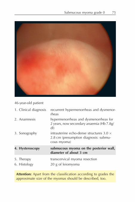
46-year-old patient
1. Clinical diagnosis recurrent hypermenorrheas and dysmenor-rheas
2. Anamnesis hypermenorrheas and dysmenorrheas for2 years, now secondary anaemia (Hb 7.8g/dl)
3. Sonography intrauterine echo-dense structures 3.0 '2.8 cm (presumption diagnosis: submu-cous myoma)
4. Hysteroscopy submucous myoma on the posterior wall,diameter of about 3 cm
5. Therapy transcervical myoma resection
6. Histology 20 g of leiomyoma
Attention: Apart from the classification according to grades theapproximate size of the myomas should be described, too.
Submucous myoma grade 0 75

52-year-old patient
1. Clinical diagnosis uterus myomatosus with bleeding disor-ders, recurrent Pap III D, cystocele II$
2. Anamnesis uterus myomatosus for 5 years, now grow-ing, and bleeding disorders, 3 times PapIII D during the last 12 months! vaginal hysterectomy with colporraphiaplanned
3. Sonography multiple intramural and submucous myo-mas
4. Hysteroscopy submucous myoma grade 0, diameter ofabout 4 cm
5. Therapy vaginal hysterectomy with morcellementsine adnexa with anterior colporraphia
6. Histology multiple leiomyomas (weight of theuterus: 320 g)
Submucous myoma grade 076

45-year-old patient
1. Clinical diagnosis permanent bleedings with uterus myoma-tosus
2. Anamnesis uterus myomatosus for 5 years, nowgrowth of multiple submucous and intra-mural myomas
3. Sonography 5 intramural myomas, one of them withsubmucous part
4. Hysteroscopy large submucous myoma on the left lateralwall with intramural part (about 20%)
5. Therapy laparoscopic supracervical hysterectomy
6. Histology uterus myomatosus (420 g)
Attention: With submucous myomas further myomas (intra-mural, subserous) should be searched for by sonography.
Submucous myoma grade 1 77

55-year-old patient
1. Clinical diagnosis postmenopausal bleeding
2. Anamnesis postmenopausal bleeding (menopause3 years ago), occasionally lower abdom-inal pain
3. Sonography intracavitary echo-dense structure 3.8 '3.2 cm, presumption diagnosis: polyp,myoma
4. Hysteroscopy vascular myoma with a diameter of 4cm, ex-tending from the right posterior lateral wall(with distinctive vessels on the surface)
5. Therapy hysteroscopic myoma resection
6. Histology parts of leiomyoma (32 g)
Attention: A differentiation between fibrosed polyp and submu-cous myoma is sonographically as well as hysteroscopically notpossible with certainty.
Submucous myoma grade 178

48-year-old patient
1. Clinical diagnosis uterus myomatosus
2. Anamnesis uterus myomatosus known for some years,growing, 1 and 2 years ago myoma embo-lisation, afterwards again growth and in-creasing discomfort/pain! planned LASH
3. Sonography submucous-intramural myoma on the pos-terior wall, size: 4.5 cm, endometriumthickness: 5mm
4. Hysteroscopy submucous-intramural myoma on theposterior wall, size: 5 cm, apart fromthat regular endometrium
5. Therapy LASH sine adnexa
6. Histology uterus myomatosus (460 g)
Attention: Recurrent myomas requiring therapy can developeven after myoma embolisation.
Submucous myoma grade 1 79

41-year-old patient
1. Clinical diagnosis uterus myomatosus with discomfort/painand bleeding disorders
2. Anamnesis for 3 years increasing abdominal pain andbleeding disorders with growing uterusmyomatosus, previously 2 caesarean sec-tions
3. Sonography multiple myomas, one large transmuralmyoma on the anterior wall
4. Hysteroscopy submucous-intramural myoma grade 2 onthe anterior wall (occupying more thanhalf of the cavity)
5. Therapy LASH
6. Histology multiple leiomyomas
Attention: With myomas of grade 2 the further therapy (organ-pre-serving versus hysterectomy) should be planned with special care.
Submucous myoma grade 280

40-year-old patient
1. Clinical diagnosis residual myoma after perforation duringmyoma resection
2. Anamnesis 4 months ago myoma resection (external)with perforation
3. Sonography submucous-intramural myoma parts nearthe isthmus, 1.7 ' 2.0 cm
4. Hysteroscopy myoma on the lateral wall grade 2, sub-mucous-intramural myoma parts (60%intramural)
5. Therapy transcervical myoma resection
6. Histology leiomyoma (15 g)
Residual myoma after uterus perfomation 81

Uterus subseptus and intramural myoma on the posterior wall82
52-year-old patient
1. Clinical diagnosis postmenopausal bleeding
2. Anamnesis postmenopausal bleeding, nullipara
3. Sonography endometrium thickness: 5 mmintrauterine structure 2.0 ' 1.2 echo-dense(¼ septum)
4. Hysteroscopy uterus subseptus, about 3.5 cm small sep-tum, right cavity half smaller than leftcavity half, intramural myoma on theposterior wall (grade 2) in the left cavity
5. Therapy dilatation/curettage (corpus curettage fromboth cavity halves)
6. Histology atrophic endometrium
Attention: Uterus malformations and myomas can also occurtogether. Under these circumstances the sonographic diagnos-tics is often difficult.
Uterus subseptus and intramural myoma on the posterior wall 83

Endometrial hyperplasia, endometrium sliding test84

40-year-old patient
1. Clinical diagnosis recurrent hypermenorrheas
2. Anamnesis for 3 years increasing hypermenorrheas,progestagen therapy without success
3. Sonography endometrium thickness: 10 mm(post menstruationem)
4. Hysteroscopy hyperplastic endometrium (see sliding testposterior wall)
5. Therapy endometrial resection during the same op-eration
6. Histology proliferative endometrium
Attention: When sliding the hysteroscope forward the differ-ence in the level of the endometrium may be seen (endome-trium sliding test).
Endometrial hyperplasia, endometrium sliding test 85

Endometrium sliding test
By sliding the hysteroscopic sheath in the hyperplastic endome-trium a difference in level between endometrium and myome-trium can be shown. This allows for a better assessment of theextent of the hyperplasia.
Attention: With high intrauterine pressure and evenly distribu-ted endometrial hyperplasia the endometrium is often wronglyclassified as atrophic or flat without the endometrium slidingtest.
endometrium
myometrium
hysteroscope
difference in levelendometrium thicknes
Endometrium sliding test86

Endometrial hyperplasia, perimenopause, endometrium sliding test 87

46-year-old patient
1. Clinical diagnosis bleeding after secondary amenorrhea
2. Anamnesis for 9 months recurrent metrorrhagias, pro-gestagen therapy without success
3. Sonography endometrium thickness: 12 mm
4. Hysteroscopy endometrial hyperplasia (especially ante-rior wall) (see sliding test)
5. Therapy dilatation/curettage
6. Histology glandular-cystic hyperplasia
Endometrial hyperplasia, perimenopause, endometrium sliding test88

Endometrial hyperplasia, endometrium sliding test 89

34-year-old patient
1. Clinical diagnosis recurrent hypermenorrhea and dysmenor-rhea
2. Anamnesis for 2 years recurrent hypermenorrhea, dys-menorrhea, 1 year ago hysteroscopy andcurettage, corpus polyp
3. Sonography endometrium thickness: 10 mm (post men-struationem), suspicion of adenomyosis
4. Hysteroscopy endometrial hyperplasia (see sliding testposterior wall)
5. Therapy dilatation/curettage
6. Histology polypoid endometrium
7. Recommendation levonorgestrel-IUS or LASH
Endometrial hyperplasia, endometrium sliding test90

44-year-old patient
1. Clinical diagnosis uterus myomatosus with bleeding disorders
2. Anamnesis uterus myomatosus
3. Sonography multiple intramural myomas, endome-trium thickness: 8mm (post menstruatio-nem)
4. Hysteroscopy polypoid endometrium posterior wall
5. Therapy LASH
6. Histology multiple leiomyomas, proliferative endo-metrium
Attention: Before a LASH with its necessary morcellation of thecorpus uteri a diagnostic hysteroscopy should always be per-formed to exclude premalignant or malignant changes.
Polypoid endometrium 91

38-year-old patient
1. Clinical diagnosis hyper- and dysmenorrhea
2. Anamnesis for 2 years increasing hyper- and dysme-norrhea (high consumption of analgesicsduring menstruation)
3. Sonography hyperplastic uterus (suspicion of adeno-myosis), endometrium thickness: 10 mm(9th day of the menstrual cycle)
4. Hysteroscopy focal endometrial hyperplasia posteriorwall, apart from that regular cavity
5. Therapy dilatation/curettage, laparoscopy (resec-tion of the endometriosis), later LASH(adenomyosis confirmed)
6. Histology glandular-cystic hyperplasia
Attention: Because of the estrogen-induced etiology adeno-myosis uteri and endometrial hyperplasia often occur together.
Endometrial hyperplasia92

52-year-old patient
1. Clinical diagnosis permanent bleedings
2. Anamnesis permanent bleeding for 13 days, beforethat already for 1 year metrorrhagias,known uterus myomatosus
3. Sonography endometrium thickness: 13 mm, multipleintramural myomas (up to a size of 3 cm)
4. Hysteroscopy hyperplastic endometrium with multiplesmall, bulging out myomas
5. Therapy dilatation/curettage, later vaginal hyster-ectomy with colporraphia
6. Histology hyperplastic endometrium
Endometral hyperplasia 93

67-year-old patient
1. Clinical diagnosis postmenopausal bleeding
2. Anamnesis 2 dilatations/curettages because of recur-rent postmenopausal bleedings (2 and6 years ago)
3. Sonography endometrium thickness: 8 mm
4. Hysteroscopy small focal hyperplasia, apart from thatregular cavity
5. Therapy dilatation/curettage, vaginal hysterectomy(at the request of the patient because ofrecurrent bleeding disorders)
6. Histology focal polypoid endometrium with initialformation of polyps
Attention: Most of the endometrial hyperplasias occur focally,demanding a targeted histological examination (target curet-tage).
Focal endometrial hyperplasia in the postmenopause94

61-year-old patient
1. Clinical diagnosis corpus polyp (sonographic suspicion)
2. Anamnesis at check-up sonographically suspiciousendometrium
3. Sonography endometrium thickness: 10 mm, definableintrauterine structure 15 ' 11 mm(suspicion of a corpus polyp)
4. Hysteroscopy large corpus polyp posterior wall, apartfrom that regular endometrium
5. Therapy dilatation/curettage and removal of thepolyp with grasping forceps, control hys-teroscopy: without pathological findingslater vaginal hysterectomy with adnexa
6. Histology ( corpus polyp with parts of an atypicaladenomatous hyperplasia
( uterus and ovaries without pathologicalfindings, no further parts of hyperplasia
Attention: About 7% of sonographically and hysteroscopicallynormal corpus polyps in the postmenopause show premalig-nant or malignant changes.
Adenomatous hyperplasia 95

Corpus polyps
1. Corpus polyps constitute one the most frequent causes ofbleedings (especially in the perimenopause).
2. With a dilatation/curettage without hysteroscopy polyps areoften not or incompletely removed.
3. Hysteroscopy makes the diagnostics of polyps possible, andthe complete removal can be checked during the intervention(control hysteroscopy).
4. With the complete removal of the polyps (by target curettage,grasping forceps or resection) most of the bleeding disordersare successfully treated.
5. The removed polyps must be carefully examined by histology.
Corpus polyps96

Sonographic finding with strong suspicion of a corpus polyp (con-firmed by hysteroscopy).

45-year-old patient
1. Clinical diagnosis bleeding disorders with uterus myomatosus
2. Anamnesis 1 year ago hysteroscopy and dilatation/curettagehistology: simple adenomatous hyperpla-sia
3. Sonography endometrium thickness: 12 mm (8th dayof menstrual cycle), 3 intramural myomas
4. Hysteroscopy large corpus polyp without pathologicalfindings
5. Therapy curettage and removal of the polyp withgrasping forceps after intraoperative histol-ogy – LASH
6. Histology fibroglandular polyp without malignancyuterus myomatosus
Corpus polyp98

85-year-old patient
1. Clinical diagnosis postmenopausal bleeding
2. Anamnesis postmenopausal bleeding(menopause 32 years ago)
3. Sonography endometrium thickness: 18 mm
4. Hysteroscopy large fibrosed corpus polyp posterior wall
5. Therapy dilatation/curettage and removal of thepolyp with grasping forceps, control hys-teroscopy: empty cavity
6. Histology fibrosed glandular-cystic corpus polyp
Corpus polyp 99

65-year-old patient
1. Clinical diagnosis recurrent postmenopausal bleeding
2. Anamnesis 1 year ago hysteroscopy and curettagewithout pathological findings
3. Sonography endometrium thickness: 8 mm
4. Hysteroscopy small corpus polyp right lateral walluterus arcuatus
5. Therapy resection of the polyp and of the endome-trium
6. Histology corpus polyp, apart from that atrophic en-dometrium
Uterus arcuatus, corpus polyp100

45-year-old patient
1. Clinical diagnosis recurrent permanent bleedings
2. Anamnesis metrorrhagias for 2–3 years, progestagentherapy only temporarily successful
3. Sonography endometrium thickness: 12 mm post men-struationem
4. Hysteroscopy large corpus polyp right lateral wall
5. Therapy dilatation/curettage and resection of thepolyp with the grasping forceps
6. Histology fibroglandular corpus polyp
Corpus polyp, perimenopause 101

61-year-old patient
1. Clinical diagnosis recurrent postmenopausal bleeding
2. Anamnesis 1 year ago hysteroscopy and curettagewith removal of the polyp, now again re-current postmenopausal bleeding withknown uterus myomatosus
3. Sonography 15 ' 13 mm intrauterine structure (suspi-cion of corpus polyp)
4. Hysteroscopy large corpus polyp extending from thefundal area
5. Therapy LASH
6. Histology corpus polyp, multiple leiomyomas
Corpus polyp, postmenopause102

77-year-old patient
1. Clinical diagnosis corpus polyp
2. Anamnesis sonographically suspect endometrium
3. Sonography endometrium thickness: 17 mm
4. Hysteroscopy corpus polyp posterior wallendometrium without pathologicalfindings
5. Therapy dilatation/curettage, resection of the polypwith grasping forceps, control hystero-scopy: cavity without findings
6. Histology fibroglandular corpus polyp, atrophic en-dometrium
Corpus polyp, postmenopause 103

Corpus polyp after dilatation/curettage104

80-year-old patient
1. Clinical diagnosis corpus polyp
2. Anamnesis sonographically suspect findings
3. Sonography endometrium thickness: 15 mm
4. Hysteroscopy cystic corpus polyp posterior wall
5. Therapy dilatation/curettage, resection of the polypwith grasping forceps, control hystero-scopy: empty cavity (see picture 2)
Attention: After the removal of the polyp an intraoperative con-trol hysteroscopy should always be performed.
Corpus polyp after dilatation/curettage 105

Corpus carcinoma
1. The incidence of corpus carcinomas rises in line with the in-creasing age of the patients.
2. With estrogen-dependent carcinomas sonography (endome-trium thickness >9 mm) is in most of the cases the method ofdiagnostics, whereas with de novo-carcinomas clinical signs(bleeding) constitute the only initial symptom.
3. Especially with corpus carcinomas hysteroscopy should be ex-tended to the cervix, too, to correct a too high staging (exten-sion to the cervix – stage 2) with the dilatation/curettage.
4. The potential transmission of tumour cells by hysteroscopy hasbeen disproved by some studies, especially since in most ofthe cases a simultaneous operative therapy of the corpus carci-noma is performed.
Sonographic picture of a thickened endometrium with strong suspi-cion of a corpus carcinoma (confirmed by hysteroscopy and histol-ogy).
Corpus carcinoma106

Corpus carcinoma 107

81-year-old patient
1. Clinical diagnosis postmenopausal bleeding
2. Anamnesis for 10 days postmenopausal bleeding
3. Sonography endometrium thickness: 23 mm
4. Hysteroscopy a lot of polypoid, suspect endometriumin the whole cavity (with free-runningvessels)
5. Therapy dilatation/curettage
6. Histology corpus carcinoma G2/G3 (mixed Mulleriantumour)! abdominal hysterectomy with bilateraladnexectomy, pelvic and para-aortal lym-phonodectomy
Final histology: Ib G3 N0 (0/42)
Attention: Insulated free-running vessels are a sign of a corpuscarcinoma.
Corpus carcinoma108

68-year-old patient
1. Clinical diagnosis suspect histological preliminary findings(polyp with atypias)
2. Anamnesis 9 months ago hysteroscopy and curettageexternal diagnosis: polyp with atypiasnow: referral to hysterectomy
3. Sonography endometrium thickness: 8 mm
4. Hysteroscopy necrotic, suspect, vascular endometrium
5. Therapy dilatation/curettage
6. Histology intraoperative histology: corpus carcinomaG2 , operation finished2nd session: LAVH with adnexectomy andpelvic and para-aortal lymphonodectomyFinal histology: corpus carcinoma, Ib G2N0 (0/38)
Attention: Histological findings with atypias should be oper-ated on as fast as possible to avoid progression.
Corpus carcinoma 109

65-year-old patient
1. Clinical diagnosis postmenopausal bleeding
2. Anamnesis obesity, for 5 years no check-up, no hor-mone replacement therapy
3. Sonography endometrium thickness: 21 mm
4. Hysteroscopy hyperplastic, partially necrotic endome-trium with free-running vessels in thewhole cavity, suspected corpus carcinoma
5. Therapy dilatation/curettagehysterectomy with adnexa and pelvic andpara-aortal lymphonodectomy, afterloading
6. Histology corpus carcinoma Ib G2Final diagnosis: T1b N0 (0/48) M0 G2
Attention: Large necrotic endometrial parts are also a sign of acorpus carcinoma.
Corpus carcinoma110

Procedure for bleeding disorders during use of oral contraceptives
anamnesisgynaecological examination
vaginal sonography
without pathological findings
hormonal therapy– change of drug– estrogen or progestagen substitution
(for 6 months at most)
persistent bleeding
outpatient minihysteroscopy
without pathological findings
again hormonal therapy, if necessaryalternative contraceptive methods
polyp, myoma
hysteroscopy (if necessary,resection of the polypor myoma)
myoma, polyp
Bleeding disorders during use of oral contraceptives 111

Bleeding disorders under hormone replacement therapy
1. Bleeding disorders under hormone replacement therapy mustbe adequately diagnosed but do not require more invasive di-agnostics than bleeding disorders without HRT because thehistological findings do not show any differences.
2. The invasive diagnostics of bleeding disorders under HRT re-duces the subsequent compliance.
3. Minihysteroscopy without anaesthesia in the practice is espe-cially suited for the diagnostics of bleeding disorders underHRT because the rate of compliance for HRT remains unaf-fected.
Table 5: Compliance with bleeding disorders under hormone replace-ment therapy depending on the diagnostic procedure
hysteroscopyþ curettagewith anaesthesia
minihysteroscopyþ biopsywithout anaesthesia
patients (n) 156 52subsequent compliance (n) 97 49continuation of HRT (%) 62 94
Bleeding disorders under hormone replacement therapy112
Bleeding disorders under HRT, corpus polyps 113

62-year-old patient
1. Clinical diagnosis sonographically suspect endometriumunder HRT
2. Anamnesis for 4 years continuous-combined HRT(Activelle!), no bleeding, no discomfort/pain
3. Sonography endometrium thickness: 13 mm (3 monthsago: 8 mm)
4. Hysteroscopy 2 corpus polyps anterior and posteriorwall
5. Therapy dilatation/curettage and control hystero-scopy: empty cavity
6. Histology glandular corpus polyps
Bleeding disorders under HRT, corpus polyps114

62-year-old patient
1. Clinical diagnosis permanent bleedings under HRT
2. Anamnesis for 2 years bleeding disorders under Acti-velle!, now permanent bleedings
3. Sonography endometrium thickness: 14 mm
4. Hysteroscopy large corpus polyp extending from theright lateral wall
5. Therapy dilatation/curettage, extraction of thepolyp with polyp forceps, control hystero-scopy: without findings
6. Histology glandular- cystic corpus polyp
Corpus polyps, bleeding disorders under HRT 115

60-year-old patient
1. Clinical diagnosis recurrent bleeding disorders under HRT
2. Anamnesis for 3 years HRT with Climodien!, for3 months acyclic breakthrough bleedings
3. Sonography endometrium thickness: 4 mm
4. Hysteroscopy slightly proliferative endometrium on theanterior wall
5. Therapy dilatation/curettage
6. Histology polypoid endometrium
Focal endometrial hyperplasia with bleeding disorders under HRT116

66-year-old patient
1. Clinical diagnosis bleeding disorders under HRT
2. Anamnesis for 3 years bleeding disorders under Gy-nodian Depot! and Uterogest, suspectedovarian fibroma on the right, knownuterus myomatosus
3. Sonography endometrium thickness: 10 mm
4. Hysteroscopy extended polypoid structures, posteriorwall with 2 insulated small polyps
5. Therapy dilatation/curettagelaparoscopy: uterus myomatosus, bothadnexa without findings
6. Histology simple adenomatous hyperplasia withoutatypias! LAVH with bilateral adnexectomy
Adenomatous hyperplasia, bleeding disorders 117

9. Hysteroscopy with sonographically suspectendometrial findings
Indications
1. Thickened endometrium in postmenopause (>9 mm) (see table 6)2. Endometrial hyperplasia in the perimenopause with negative
progestagen test (see page 119)3. Intrauterine finding (polyp, myoma) with discomfort/pain, or
gaining in size4. serometra with discomfort/pain, or gaining in size5. sonographically suspect endometrium under HRT (see table 6)6. sonographically suspect endometrium under Tamoxifen
Tab. 6: Recommendations for the diagnostics of asymptomatic wo-men with and without HRT (according to Roemer, Rabe, Duda, Foth2004), Guidelines of the German Society of Gynaecology and Obs-tetrics
double endometriumthicknes
cyclical HRTa) continuous-combined HRT
no HRT
hysteroscopic-histologicalexamination
"13 mmb) "9 mm "9 mm
check-up after2 to 3 months
9–12 mm 5–8 mm 5–8 mm
without consequence "8 mm "4 mm "4 mm
a) measurement after hormonally induced bleedingb) at least in 2 menstrual cycles

Sonographically supported progestagen test
Attention: If the endometrium thickness is not adequately re-duced after progestagen medication, further diagnostic assess-ment is necessary to exclude an endometrium carcinoma (mostfrequent cause: corpus polyp, endometrial hyperplasia resistantto therapy).
thickened endometrium (e.g. 12 mm)
2 mg Norethisteronacetate for 12 days
bleeding
control sonograpy after bleeding
endometrium thickness < 5 mm endometrium thickness > 5 mm
no further treatment necessary(if necessary, progestagenprophylaxis)
hysteroscopy and histology fordiagnostic assessment
Sonographic endometrial hyperplasia 119

Sonographic picture of endometrial hyperplasia.
Sonographic endometrial hyperplasia120

79-year-old patient
1. Clinical diagnosis sonographically suspect endometriumpostmenopause
2. Anamnesis menopause 25 years ago, no HRT
3. Sonography endometrium thickness: 14 mm
4. Hysteroscopy 2 plain fibrosed corpus polyps posteriorwall
5. Therapy dilatation/curettage and endometriumbiopsy, posterior wall; control hystero-scopy: without pathological findings
6. Histology fibrosed corpus polyps
Corpus polyp, postmenopause 121

Corpus polyp, postmenopause122

75-year-old patient
1. Clinical diagnosis corpus polyp, no bleedings
2. Anamnesis loss of weight,MRI: suspicion of cervical changescytology: without pathological findings
3. Sonography endometrium thickness: 18 mm
4. Hysteroscopy cervix without pathological findings,large corpus polyp posterior wall
5. Therapy dilatation/curettage and removal of polypwith grasping forceps
6. Histology glandular corpus polyp
Corpus polyp, postmenopause 123

85-year-old patient
1. Clinical diagnosis sonographically suspect endometrium inpostmenopause
2. Anamnesis menopause 30 years ago, now large, in-tracavitary findings well visualisable bysonography, no bleedings, no discomfort
3. Sonography endometrium thickness: 20 mm, echo-dense, vascular intracavitary structure
4. Hysteroscopy large corpus polyp occupying the wholecavity, partially with necrotic changes
5. Therapy attempt to remove polyp by grasping for-ceps (only partially successful)! hysteroscopic resection of the polyp
6. Histology corpus polyps with parts of an atypicaladenomatous hyperplasia! vaginal hysterectomy (without morcel-lement)
histology: no further parts of hyperplasia
Corpus polyp with adenomatous hyperplasia124

Corpus polyp, postmenopause 125
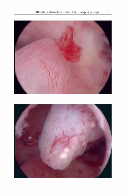
75-year-old patient
1. Clinical diagnosis sonographically suspect endometrium inpostmenopause
2. Anamnesis no bleeding, no discomfort/pain, 2 yearsago hysteroscopy and dilatation/curettageof corpus polyps
3. Sonography endometrium thickness: 10 mm (growingduring the last 6 months)
4. Hysteroscopy corpus polyps posterior wall
5. Therapy dilatation/curettage with intraoperativecontrol hysteroscopy without pathologicalfindings (see second picture)
6. Histology fibroglandular polyps without malignancy
Corpus polyp, postmenopause126

80-year-old patient
1. Clinical diagnosis sonographically suspect endometrium
2. Anamnesis no bleeding, no discomfort/pain
3. Sonography endometrium thickness: 18 mm
4. Hysteroscopy 2 well vascularised corpus polyps in thefundal area
5. Therapy dilatation/curettage and extraction of thepolyp with polyp forceps, intraoperativecontrol hysteroscopy: empty cavity
6. Histology glandular-cystic corpus polyps withoutmalignancy
Corpus polyp, postmenopause 127

83-year-old patient
1. Clinical diagnosis corpus polyp
2. Anamnesis sonographically suspect findings duringcheck-up, no bleedings, no discomfort
3. Sonography endometrium thickness: 18 mm
4. Hysteroscopy suspect corpus polyp (soft and crumbly)
5. Therapy dilatation/curettage and removal of thepolyp
6. Histology fibroglandular corpus polyps without ma-lignancy
Suspect corpus polyp, postmenopause128

69-year-old patient
1. Clinical diagnosis sonographically suspect endometrium
2. Anamnesis menopause 15 years ago
3. Sonography endometrium thickness: 6 mmintracavitary structure 1.2 ' 1.0 cm(suspicion of corpus polyp)
4. Hysteroscopy insulated hyperplastic vascular area rightlateral wall
5. Therapy dilatation/curettagelongitudinal laparotomy, hysterectomywith bilateral adnexectomy, pelvic andpara-aortal lymphonodectomy
6. Histology adenosquamous carcinoma G2final histology: corpus carcinoma Ib G2N0 (0/38)
Corpus carcinoma 129

Endometrium and Tamoxifen
1. Endometrial hyperplasias under Tamoxifen develop dependenton the dosage and the duration of medication.
2. Bleeding disorders under Tamoxifen require an intensive diag-nostic assessment.
3. Endometrial hyperplasias under Tamoxifen do not respond to aprogestagen therapy (reason: stromal hyperplasia).
4. With asymptomatic patients undergoing Tamoxifen therapy anannual sonographic check-up of the endometrium is recom-mended.
5. When the endometrium shows a tendency to grow (endome-trium thickness >12 mm) a diagnostic assessment is necessary.
6. Since by a simple dilatation/curettage endometrium for histolo-gical examination may often not be extracted, a segmental en-dometrial resection (by resectoscope) must be performed, ifnecessary.
Sonographic picture of an endometrial hyperplasia under Tamoxifentherapy.
Endometrium and Tamoxifen130

Endometrial hyperplasia under Tamoxifen 131

83-year-old patient
1. Clinical diagnosis endometrial hyperplasia under Tamoxifen
2. Anamnesis 2 years ago receptor-positive breast can-cer, since then Tamoxifen 20 mg/d
3. Sonography endometrium thickness: 15 mm
4. Hysteroscopy polypoid endometrium posterior wall,cervical stenosis with one adhesion
5. Therapy dilatation/curettage
6. Histology proliferative endometrium
Endometrial hyperplasia under Tamoxifen132

10. Hysteroscopy and lost IUD/IUS
1. Lost IUD is one of the classic indications for a hysteroscopy.2. At first the intrauterine evidence of the IUD/IUS should be
provided by sonography.3. The most frequent indication results from the tearing off of the
IUD- thread when trying to extract the IUD.4. The IUD can be extracted after the hysteroscopic evidence of
the IUD and by using a small grasping forceps. Alternatively, agrasping forceps may be introduced through the workingsheath and the IUD/IUS can then be extracted with the re-moval of the whole instrument.

35-year-old patient
1. Clinical diagnosis lost IUD
2. Anamnesis for 8 years IUD in situ, with extractionthreads were torn off
3. Sonography IUD correctly placed in the uterine cavity
4. Hysteroscopy IUD (type DANA) correctly placed in theuterine cavity
5. Therapy extraction by hysteroscope with graspingforceps, which is introduced through theworking sheath
Lost IUD134

38-year-old patient
1. Clinical diagnosis planned exchange of IUD during laparo-scopy
2. Anamnesis for 3 years copper-IUD, now suspecteddislocation
3. Sonography IUD dislocated
4. Hysteroscopy dislocated copper-IUD
5. Therapy IUD extraction and insertion of a newone
6. Histology none
Dislocated IUD 135

43-year-old patient
1. Clinical diagnosis bleeding disorders with IUD in situ(multiload)
2. Anamnesis for 3 years IUD in situ, for 6 monthsrecurrent spottings and hypermenorrheas
3. Sonography IUD dislocatedendometrium thickness: 10 mm
4. Hysteroscopy IUD transversely located in the uterinecavity, limited contraceptive safety
5. Therapy IUD extraction and dilatation/curettage
6. Histology proliferative endometrium
Attention: An assessment of the endometrium with bleedingdisorders in IUD-patients should always be performed beforeIUD-removal because otherwise artificial endometrial lesionscould confine diagnostics.
Bleeding disorders with IUD in situ136

42-year-old patient
1. Clinical diagnosis permanent bleedings under MIRENA
2. Anamnesis for 6 months MIRENA, recurrent perma-nent bleedings, progestagen therapy with-out success
3. Sonography MIRENA transversely located in the uter-ine cavity, endometrium thickness: 8 mm
4. Hysteroscopy MIRENA transversely located in the uter-ine cavity, endometrial hyperplasia pos-terior wall
5. Therapy IUS extraction and dilatation/curettage
6. Histology proliferative endometrium
7. Recommendation resection of the endometrium after fin-ished family planning
Bleeding disorders with IUS in situ (MIRENA) 137

Bleeding disorders with dislocated IUS (MIRENA)138

52-year-old patient
1. Clinical diagnosis bleeding disorders with MIRENA in situand large uterus (probe length ¼ 13.0 cm)
2. Anamnesis for 6 months recurrent permanent bleed-ings with MIRENA in situ with adiposepatientuterus clearly hyperplastic without insu-lated myomas
3. Sonography uterus hyperplasia, MIRENA in the largecavity clearly dislocated
4. Hysteroscopy MIRENA transversely located in the uter-ine cavity, endometrial hyperplasia
5. Therapy extraction of MIRENAdilatation/curettagenew insertion of MIRENA with hystero-scopic view (at urgent request of thepatient, who refuses a further operativetherapy; oral progestagens are contraindi-cated)
6. Histology simple hyperplasia
Bleeding disorders with dislocated IUS (MIRENA) 139

11. Special cases
Placental residuals
1. After some time placental residuals can become necrotic orcalcify.
2. With placental disorders (placenta accreta or increta) the re-moval without exact localisation can be difficult.
3. In these cases hysteroscopic diagnostics and the targeted re-moval (if necessary, even operatively) is the treatment of choice.
4. With very large solid residuals several sessions may be neces-sary.
Adhesions after IUS
Intrauterine adhesions after IUS are extremely rare and can onlybe explained by local inflammation of the endometrium.
Endometritis
Bleeding disorders are only rarely caused by endometritis, whichis in most of the cases an incidental finding.

33-year-old patient
1. Clinical diagnosis sonographically suspect placental resi-duals and persistent hCG-levels
2. Anamnesis missed abortion 13th week of pregnancyabortion curettage, after that sonographi-cally suspected placental residuals3 times hysteroscopy and dilatation/curet-tage with postoperatively persistent sono-graphic findings (performed externally),followed by 3 cycles of Methotrexate-ther-apy (because of increased hCG-levels)
3. Sonography clearly visible, partially calcified placentalresidual of 2.0 ' 2.0 cm
4. Hysteroscopy left tubal cornua and lateral wall partiallycalcified and necrotic placental residual
5. Therapy operative hysteroscopy: resection of theplacental residuals
6. Histology necrotic placental residuals, no malignancy
Placental residuals after missed abortion 141

24-year-old patient
1. Clinical diagnosis placental residuals with placenta accreta
2. Anamnesis spontaneous delivery 3 months ago, fol-lowed by a persistent large solid intracavi-tary finding, recurrent bleedings, 3 timesdilatations/curettages without success(performed externally)
3. Sonography solid intracavitary vascularised finding,size: 70 ' 60 mm, occupying the entirecavity
4. Hysteroscopy large, partially necrotic placental residualsoccupying the entire cavity
5. Therapy resection of the placenta by bipolar hys-teroscopy in two sessions
6. Histology necrotic placental residuals (240 g)
Placental residuals142

35-year-old patient
1. Clinical diagnosis secondary amenorrhea after extraction ofMIRENA 9 months ago
2. Anamnesis for 5 years MIRENA as method of contra-ception, after extraction secondary ame-norrhea,hormonal status: without pathologicalfindings, no induction of bleedings possi-ble in spite of estrogen substitution,suspicion of intrauterine adhesions
3. Sonography no endometrium visible
4. Hysteroscopy intrauterine adhesions grade 3, right lateralwall
5. Therapy intrauterine adhesiolysis
Intrauterine adhesions grade 3 after MIRENA 143

Endometritis
1. Indication postmenopausal bleeding
2. Hysteroscopy entire cavity reddened, endometriumtouch-sensitive and bleeds easily (seeposterior wall)
3. Diagnosis endometritis
Attention: If the entire cavity is reddened, there is a strong sus-picion of endometritis.
Endometritis144

12. Complications
1. endometritis/adnexitis 0.01%2. dysregulation of circulation 3 to 5% (without anaesthesia)3. via falsa (cervical canal) 2%4. uterus perforation 0.1%5. embolism (singular cases)6. dissemination of tumour cells
Attention: With all safety aspects considered the total rate ofcomplications is lower than 1 per mill.
Attention: A dissemination of tumour cells is excluded byCO2-hysteroscopy.
Attention: A dissemination of tumour cells by hysteroscopywith a fluid distending medium is very unlikely.

43-year-old patient
1. Clinical diagnosis recurrent hypermenorrhea and dysmenor-rhea
2. Anamnesis for 3 years increasing hyper- and dysme-norrhea, finished family planning
3. Sonography suspicion of adenomyosis(hyperplastic myometrium)endometrium thickness: 8 mm (post men-struationem)
4. Hysteroscopy via falsa on the posterior wall with cervi-cal stenosis, after withdrawal of the hys-teroscope the right direction becomesvisible at 11 o’clock
5. Therapy LASH
6. Histology adenomyosis uteri
Via falsa146

84-year-old patient
1. Clinical diagnosis postmenopausal bleeding
2. Anamnesis for 1 month increased vaginal bleeding,menopause 30 years ago
3. Sonography endometrium thickness: 14 mm
4. Hysteroscopy with craurosis cautious dilatation withthe smallest Hegar’s dilatators( insertion of the hysteroscope( perforation of the posterior wall fol-
lowed by bleeding
5. Therapy vaginal hysterectomy
6. Histology cervical myoma, corpus polyp
Perforation 147

38-year-old patient
1. Clinical diagnosis secondary uterine amenorrhea
2. Anamnesis spontaneous delivery 9 months ago withcurettage post partum because of placen-tal residuals (positive), after that secondaryamenorrhea
3. Sonography endometrium only partially visible, no he-matometra
4. Hysteroscopy cervical stenosisintrauterine adhesions grade 3(right half of the cavity completely ob-structed)slight perforation of the fundus
5. Therapy dilatation of the cervixtermination of the operationantibiotic prophylaxis! hysteroscopic adhesiolysis 4 monthsafter healing up of the perforation – with-out problems
6. Histology none
Intrauterine adhesions grade 3 (perforation)148

13. Summary
Advantages of diagnostic hysteroscopy
( simple method which can be performed as outpatient treatment( direct visualisation of the uterine cavity( exact localisation of pathological intrauterine findings( direct biopsy( high correspondence with histological findings( in the diagnostics of sterility far more effective than sonogra-
phy and hysterosalpingography( with abnormal uterine bleedings the hysteroscopy fills a diag-
nostic gap between vaginal sonography and dilatation/curettage( early information of the patient
Today, the diagnostic hysteroscopy is a standard method ingynaecology.

14. List of abbreviations
ESGE European Society of Gynaecological EndoscopyhCG human ChoriongonadotropinHRT hormone replacement therapyICSI intracytoplasmic sperm injectionIUA intrauterine adhesionsIUD intrauterine deviceIUS intrauterine systemIVF in-vitro fertilisationLASH laparoscopic supracervical hysterectomyLAVH laparoscopically assisted vaginal hysterectomyMRI magnet resonance imaging

www.degruyter.com
Operative hysteroscopy represents an important extension of operativegynecology. The benefits of this endoscopic technique, however, arebalanced by its high demands on the operator. It requires excellentknowledge of safety issues and possible complications. This book actsas guide and resource for the practical acquisition of this technique.This new edition is distinguished by the large number of improvedcolor illustrations and incorporation of the newest scientific findings.
• 2nd revised edition• guide and resource for the practical acquisition of operative
hysteroscopy
Thomas Römer
OPERATIVEHYSTEROSCOPYA Practical Guide
2nd ed. approx. viii, 150 pages.Paperback. ISBN 978-3-11-022499-3Series: Pocket Guides for GynaecologistsTo be published March 2010eBook: ISBN 978-3-11-022500-6

www.degruyter.com
Publication frequency: bi-monthly (6 issues per year).Approx. 500 pages per volume. ISSN 0300-5577ISSN (Internet) 1619-3997; CODEN JPEMAO2008 INCREASED IMPACT FACTOR: 1.234FIVE-YEAR IMPACT FACTOR: 1.315
Journal of Perinatal Medicine is a truly international forum covering theentire field of perinatal medicine. It is an essential news source for allobstetricians, neonatologists, perinatologists and allied health profession-als who wish to keep abreast of the latest developments in perinatal andrelated research.
All de Gruyter journals are hosted on Reference Global, de Gruyter’sintegrated platform. Please visit www.reference-global.com for moreinformation and free TOC alerts. Electronic sample copy atwww.degruyter.com/jpm
JOURNAL OFPERINATALMEDICINEOfficial journal of the WorldAssociation of PerinatalMedicine (WAPM)
Founded by Erich Z. Saling
Editor-in-Chief:Joachim W. Dudenhausen