Pneumonias - Kliinikum · characteristic of both community-acquired pneumonia ... management of the...
247
Pneumonias Alan Altraja Department of Pulmonary Medicine, University of Tartu
Transcript of Pneumonias - Kliinikum · characteristic of both community-acquired pneumonia ... management of the...

Pneumonias
Alan Altraja
Department of Pulmonary Medicine,University of Tartu

Lower respiratory tract infections (LRTI)Definition:•Acute illness (≤3 weeks) with:•Cough usually as the main symptom and•At least one other LRTI symptom: sputum production,
dyspnea, wheeze, or chest discomfort/pain and•No alternative explanation for the syndrome (e.g. sinusitis or asthma)
A. Altraja ©2017 Woodhead et al. Clin Microbiol Infect 2011; 17(Suppl. 6): E1-E59
In nature, LRTI are infectious inflammations of all levels or parts of the lower respiratory tract (i.e. parts of the respiratory tract below the vocal cords): tracheitis, bronchitis, bronchiolitis, and pneumonias and infectious inflammatory conditions that accompany structural lung diseases (incl. bronchiectasis, cystic malformations, cystic fibrosis etc.)

PneumoniaThe nature: acute infectious inflammation at the alveolar level (chronic pneumonia does not exist)
Definition: suspicion of pneumonia (preliminary diagnosis):•An acute illness with cough with:•At least one of new focal chest signs (chest discomfort/pain, pulse rate >100/min at rest, characteristic finding on auscultation, or wheeze) or•Fever of >4 days or dyspnea/tachypnea and•An absence of other obvious cause to explain this state
A. Altraja ©2017 Woodhead et al. Clin Microbiol Infect 2011; 17(Suppl. 6): E1-E59

Pneumonia: the final diagnosisDefinition: the initial diagnosis + radiographic changes•An acute illness with cough with:•At least one of new focal chest signs (chest discomfort/pain, pulse
rate >100/min at rest, characteristic finding on auscultation, or wheeze) or•Fever of >4 days or dyspnea/tachypnea and•An absence of other obvious cause to explain this state
•Radiographically supported by findings of lung shadowing that is/are likely to be new
•In the elderly people:•The presence of lung shadowing accompanied by acute clinical illness (unspecified) without other obvious cause
A. Altraja ©2017 Woodhead et al. Clin Microbiol Infect 2011; 17(Suppl. 6): E1-E59

PneumoniasA major part of the lower respiratory tract infections (in addition to acute bronchitis, acute exacerbations of chronic bronchitis, structural lung diseases (bronchiectasis, cystic fibrosis etc.))
Pneumonia is:•Among world’s commonest causes of illness•The commonest infections in general practice
•Holder of the 1st place for mortality due to infections
•Infection by nature, however, is not an infectious disease!
A. Altraja ©2017

Pneumonia nowadays•The need for hospitalization has decreased significantly: fixed criteria, based on assessment of patients’ risks•The proportion of elderly people has increased:•Among the overall patients with pneumonia•Among those, who need to be hospitalized•In the elderly, the course of pneumonia is often atypical that complicates the disease recognition and diagnostics (fever is low or absent etc.)What is significant:•LRTI are common, but pneumonia is not that frequent however•LRTI are self-limiting, whereas pneumonia needs antimicrobial treatment•Overuse of antibiotics resistance•The diagnosis of pneumonia out of hospital is complicated, especially in the absence of radiography•Primary care physicians support on symptoms and signs; diagnostic algorithms are of assistance
A. Altraja ©2017
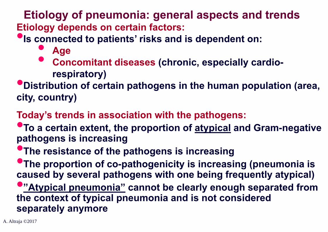
Etiology of pneumonia: general aspects and trends Etiology depends on certain factors:•Is connected to patients’ risks and is dependent on:• Age• Concomitant diseases (chronic, especially cardio-
respiratory) •Distribution of certain pathogens in the human population (area, city, country)
Today’s trends in association with the pathogens:•To a certain extent, the proportion of atypical and Gram-negative pathogens is increasing•The resistance of the pathogens is increasing•The proportion of co-pathogenicity is increasing (pneumonia is caused by several pathogens with one being frequently atypical)•”Atypical pneumonia” cannot be clearly enough separated from the context of typical pneumonia and is not considered separately anymore
A. Altraja ©2017

Etiology of pneumonia, trends in antimicrobial resistance I• β-lactams are still useful in treatment of extra-meningeal infections by Streprococcus pneumoniae•Moderately penicillin-resistant strains are treatable:• Strains having MIC90 8 mg/L: •G-penicillin 2 g i.v. 6 i.v.•Ceftriaxone 1 g 2 i.v.•Cefotaxime 2 g 4 i.v.•Amoxicillin + clavulanic acid 2 g/0,125 g 2 i.v.•Oral cephalosporin's are inadequate, if strains have penicillin MIC >2 mg/L•Erythromycin doesn’t work, if S. pneumoniae MIC90 >0,5 mg/L•H. influenzae and M. catarrhalis are moderately susceptible or
completely resistant•Community-acquired methicillin-resistant Staphylococcus aureus (MRSA) strains:• Are usually only β-lactam-resistant• Suppression of toxins by a bactericidal agent is desired• Vancomycin is not effective alone• Clindamycin or linezolid can be the optimal choice
A. Altraja ©2017 Woodhead et al. Clin Microbiol Infect 2011

Etiology of pneumonia, trends in antimicrobial resistance II
•Atypical pathogens:
Resistance against antibiotics is low and is not usually responsible to a treatment failure•Macrolide resistance in Mycoplasma pneumoniae is rising (in Japan); there are no sufficient data for Europe
A. Altraja ©2017 Woodhead et al. Clin Microbiol Infect 2011
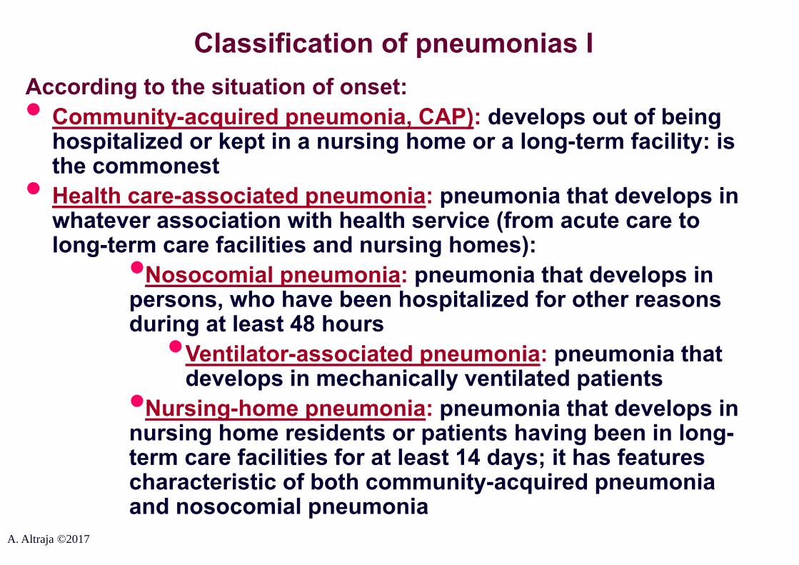
Classification of pneumonias IAccording to the situation of onset:• Community-acquired pneumonia, CAP): develops out of being
hospitalized or kept in a nursing home or a long-term facility: is the commonest• Health care-associated pneumonia: pneumonia that develops in whatever association with health service (from acute care to long-term care facilities and nursing homes):•Nosocomial pneumonia: pneumonia that develops in
persons, who have been hospitalized for other reasons during at least 48 hours•Ventilator-associated pneumonia: pneumonia that
develops in mechanically ventilated patients •Nursing-home pneumonia: pneumonia that develops in nursing home residents or patients having been in long-term care facilities for at least 14 days; it has features characteristic of both community-acquired pneumonia and nosocomial pneumonia
A. Altraja ©2017

Classification of pneumonias to typical and atypicalAccording to the clinical picture, course, respective etiology, and the management of the patient: this classification gradually looses its importance•Typical: ”classical” clinical picture (abrupt onset, severe symptoms etc.), the classical pathogen is Streptococcus pneumoniae•Atypical•Numerous specific peculiarities; the atypical pathogens
include: Mycoplasma pneumoniae, Chlamydophila pneumoniae, Legionella spp.•The classical symptoms and signs of pneumonia are not always present•The pathogens cannot be cultivated on routine media
There is not clear enough clinical difference between “atypical” and “typical” pneumonia, as is there no clear correlation with the respective pathogens: There is no basis and need for separation between typical and atypical pneumonias strictly as previously
A. Altraja ©2017

Classification of pneumonias IIIAccording to the extent and location: •Lobar or croupous pneumonia•Focal pneumonia or bronchopneumonia•Predominantly interstitial pneumoniaThis classification has almost no clinical practical significance nowadays
Aspiration pneumonia as a specific variant of pneumonia:• Inflammation that develops due to aspiration (most frequently, of the contents of the gastrointestinal tract):•3 significant mechanisms of injury: •Chemical inflammation (gastric juice, enzymes)•Obstruction or closure of a part of the conducting
respiratory tract by liquid or foreign bodies, followed by an obstruction due to mucosal edema•Infection component (as in CAP)
A. Altraja ©2017

Classification of pneumonias IV
According to severity: on the clinical significance’s point of view, it might be important to distinguish between severe and non-severe pneumonia•Non-severe (formerly mild + moderately severe)•Severe:
•There are criteria for classification of pneumonia as severe•Severe clinical course •High complication risk•High probability of lethal outcome
A. Altraja ©2017

The main facilitating factors for pneumonia
•Also virulent or aggressive bacteria •Common colds or viral infections of the lower conducting airways•Insufficient capability of the local defense mechanisms•Weakening of the defense mechanisms by the whole host organism•Age <2 years or ≥65 years•Alcoholism, impairment of consciousness•Airway obstruction (including local: foreign body, neoplasm etc.)•Structural lung disease
Everything that impairs:•The mucociliary clearance in the conducting airways•The bactericidal capacity of the enzymes in the respiratory secretions •The innate immune response at the alveolar leveland/or that facilitates:•Penetration of the infectious material to the lower respiratory tract in a sufficient amount
A. Altraja ©2017

Pathogenesis of pneumonia IHealthy persons have pathogen-free lower respiratory tract: this is guaranteed by:
•Mucociliary clearance•Enzymes in the respiratory secretions•Phagocyting cells (alveolar macrophages, polymorphonuclear leukocytes etc.)
Pneumonia needs special predisposing conditions to develop:1. Penetration and adhesion of the pathogens to the alveolar level•After aspiration (material from the upper respiratory tract)
•Via blood circulation•Via inhalation2. Predisposing factors: everything that is injurious to:•Mucociliary system•Functioning of the alveolar macrophages(acute infections of the conducting airways, smoking etc.)
A. Altraja ©2017

Aspiration of infected secretions•Upper respiratory tract has its microbiome: colonization, including that with the known pathogens of pneumonia•Aspiration of infected secretions can occur:•During all conditions of impaired consciousness:•In healthy individuals during sleep•During general anesthesia or other conditions of impaired consciousness•During alcohol etc. intoxications•In association with all invasive manipulations:•During use of nasogastric probes•During inflammatory conditions of the conducting airways:•In laryngeal edema•In neuromuscular conditions of the larynx•In association with injuries to the tracheobronchial tree•In local obstruction (tumors, foreign bodies etc.)•In inhalational injuries: after inhalation of irritant/corrosive gases/vapors or
infected aerosols•On the background of pre-existing conditions of the lung parenchyma itself:•In pulmonary edema associated with e.g. cardiac diseases or contusion of the chest•In pulmonary vascular diseases (pulmonary embolism and/or infarction)
A. Altraja ©2017

Pathogenesis of pneumonia IIIAdhesion of the microorganisms to the alveolar epithelium (according to a receptor-ligand principle) is the pre-requisite of bacterial colonization and development of infection)•During the initial phase: phagocytosis of the pathogens (by
alveolar macrophages and neutrophils)•Antigen presentation development of the innate and acquired immune responses
•Lymphocytes produce cytokines that participate in the development of inflammation, activate alveolar macrophages, stimulate migration of the phagocytes, and activate the complement system
In cases of insufficiency of the local defense mechanisms, the infection penetrates into the regional lymph nodes and further to the circulation bacteremiaBacteremia possibility of metastatic spread of infection
A. Altraja ©2017
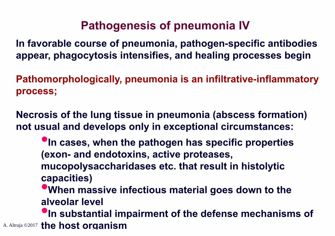
Pathogenesis of pneumonia IVIn favorable course of pneumonia, pathogen-specific antibodies appear, phagocytosis intensifies, and healing processes begin
Pathomorphologically, pneumonia is an infiltrative-inflammatory process;
Necrosis of the lung tissue in pneumonia (abscess formation) not usual and develops only in exceptional circumstances:
•In cases, when the pathogen has specific properties (exon- and endotoxins, active proteases, mucopolysaccharidases etc. that result in histolytic capacities)•When massive infectious material goes down to the alveolar level•In substantial impairment of the defense mechanisms of the host organismA. Altraja ©2017

Questions that need to be answered in the context of pneumonia
• Whom to suspect of having pneumonia?• Does the patient have pneumonia?• What is the differential diagnosis?• Does the patient need hospitalization or can he/she be treated as an outpatient?• What ancillary investigations are needed?• How and how long to treat the patient?• Management of symptoms and concomitant diseases• How can the treatment be guided, how to monitor patient’s condition, and how to assess the response to treatment?
A. Altraja ©2017 Woodhead et al. Clin Microbiol Infect 2011; 17(Suppl. 6): E1-E59

These patients should be suspected of having pneumonia:
• Acute cough and one of the following: •A new focal (unilateral) chest finding (on physical examination)•Dyspnea •Tachypnea•Pulse rate >100/min•Fever >4 days
• Serum CRP concentration >100 mg/L • S-CRP <20 mg/L makes the diagnosis of pneumonia very unprovable• In suspicion of pneumonia, chest X-ray (both postero-
anterior and lateral views) should be done to confirm the diagnosis
A. Altraja ©2017 Woodhead et al. Clin Microbiol Infect 2011; 17(Suppl. 6): E1-E59

How to interpret cough in the context of pneumonia?• An acute cough with or without dyspnea is a very common and
non-specific complaint• Resulting from location of the cough receptors, cough can, not surprisingly, be present as in non-infectious respiratory diseases, as well as in respiratory infections:•In upper respiratory tract infections (URTI) (sinusitis etc.)•In lower respiratory tract infections (LRTI)•Tracheitis and bronchitis: very similar
In pneumonia:• There is substantially more general weakness or fatigue, tachypnea, and tachycardia, as is there finding of alveolitis on auscultation (inspiratory fine crackles on the affected side)• Pneumonia definitely needs to be differentiated from less severe forms of LRTI, because there: •Is substantially higher complication risk•Are significantly more durable symptoms and
morbidity/mortalityA. Altraja ©2017 Metlay & Fine, 2003; Woodhead et al.; Eur Respir J 2005;26:1138-80

Symptoms of pneumoniaThe symptoms of pneumonia may vary between differentpathogens and different forms of pneumonia The following is characteristic of typical bacterial pneumonia:•Rapid onset with high fever, weakness, and chills•In focal pneumonia, clinical picture of an acute respiratory infection or an exacerbation of chronic bronchitis may precede•Cough is often present•Hemoptysis is rare, although in severe cases, reddish sputum may be expectorated (esp. if the pathogen is S. pneumoniae)•Often sharp (pleuritic) chest pain is present (in cases of affection of the parietal pleura by the inflammation)•Sputum preduction may occur, but is not characteristic, however (not at least diring the initial phase)•Breathlessness in cases of extended involvement of the lungs
A. Altraja ©2017

Objective findings in pneumoniaInspection:•Herpes labialis (in lobar pneumonia sometimes)•Breathlessness, cyanosis (in large pneumonias)•Less respiratory excursions of the involved side
Percussion:• Shortening of the respiratory sounds or dullness (in the presence of extended (lobar) pneumonia that reaches sufficiently close to the chest wall)
Auscultation:•Alveologenic crackles: fine crackles (formerly „crepitations“, but the use of this term is not more recommended in the context of pneumonia or overall lungs)•Lobar pneumonia: shortly in the beginning: „crepitatio indux“•When the alveoli get filled with exudate only bronchial breath sound is heard on the projection of pneumonia (consolidation)•During the resolution phase, again, „crepitatio redux“ is heard•In focal pneumonias located close to the chest wall “fine crackles”
A. Altraja ©2017

Inspection: Herpes labialis in severe pneumonia
A. Altraja ©2017
Objective findings in pneumonia (comments)
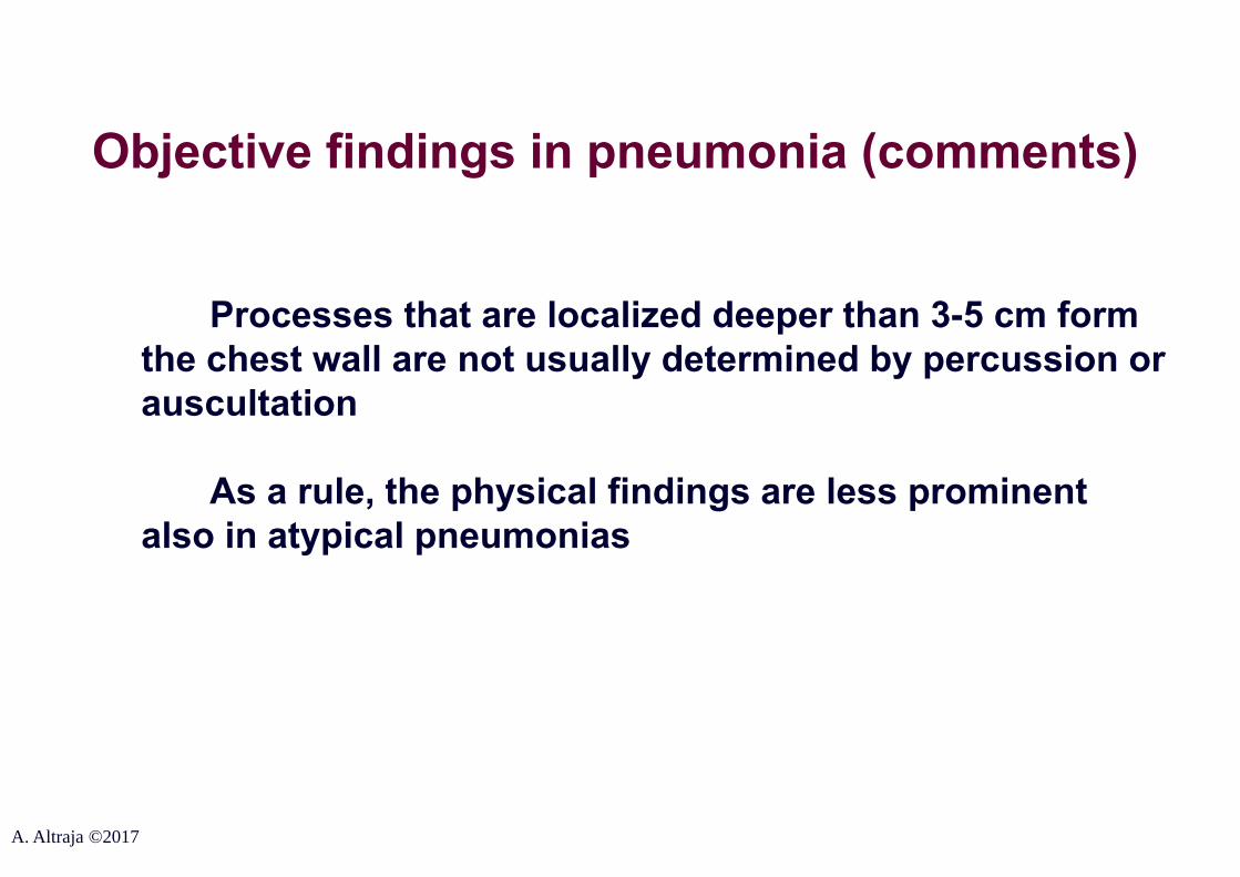
Processes that are localized deeper than 3-5 cm form the chest wall are not usually determined by percussion or auscultation
As a rule, the physical findings are less prominent also in atypical pneumonias
A. Altraja ©2017

Laboratory findings in pneumoniaBlood analyses:Changes that are characteristic of bacterial infections in general: •Increased concentration of serum C-reactive protein (CRP)•Other biomarkers [procalcitonin (PCT) etc.]•Leukocytosis with a shift to left•Accelerated erythrocyte sedimentation rate (ESR)•In atypical pneumonia, the mentioned changes are by far
less prominent
Sputum (if present):In typical pneumonia, mucoid, mucopurulent, or purulent sputum is characteristic•Rust-colored sputum often in extended lobar forms of pneumonia (often if S. pneumoniae is the pathogen)•Hemoptysis or hemoptoe is rare ( refers to lobar pneumonia)
A. Altraja ©2011

Biomarkers in pneumoniaMost often used: •C-reactive protein (CRP) •Procalcitonin (PCT)Others:•D-dimers•Carboxy-terminal provasopressin (CT-proAVO, copeptin)•Midregional proatrial natriuretic peptide (MR-pro-ANP)•Midregional proadrenomedullin (MR-ADM) (more promising)•Triggering receptor expressed on myeloid cells (TREM-1)•Markers of the adrenal response
Used for:•Markers for the presence of a bacterial infection or pathogen•Determining the severity of pneumonia•Making decisions to hospitalize•Risk assessment•Determining the correct length of therapy•Making decisions to prescribe antibiotics in non-pneumonic lower respiratory tract infections
A. Altraja ©2011 Woodhead et al. Clin Microbiol Infect 2011
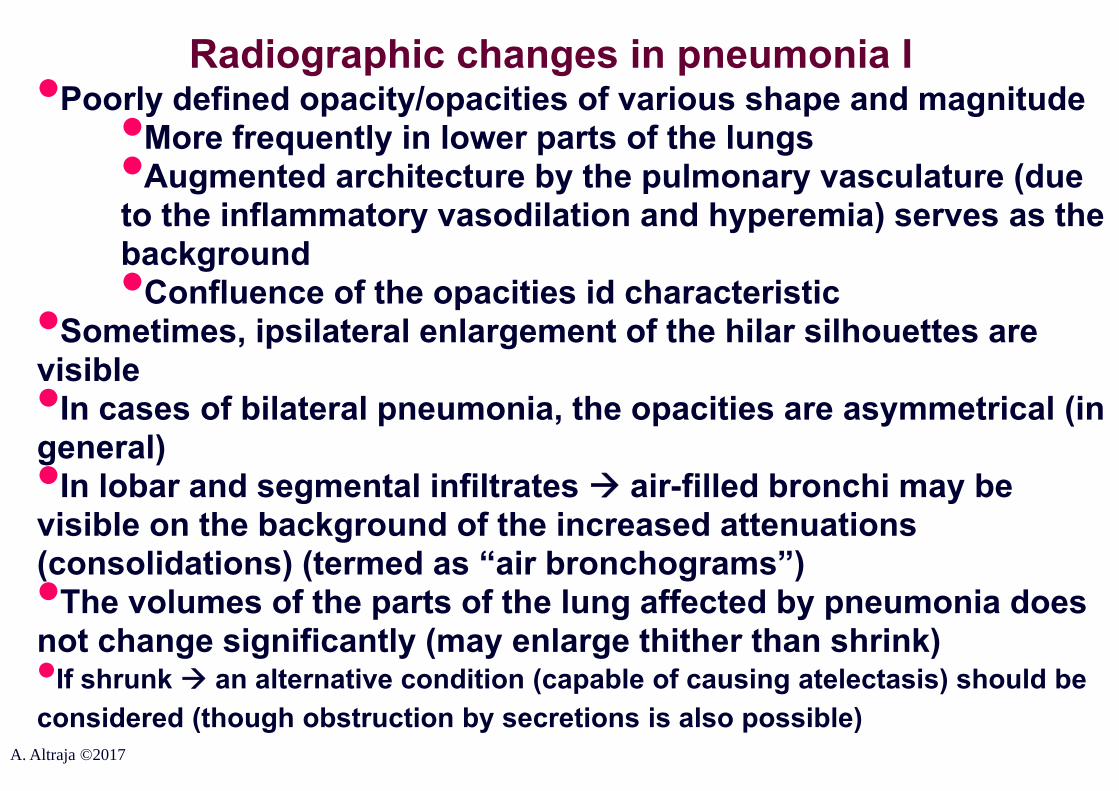
Radiographic changes in pneumonia I•Poorly defined opacity/opacities of various shape and magnitude•More frequently in lower parts of the lungs•Augmented architecture by the pulmonary vasculature (due to the inflammatory vasodilation and hyperemia) serves as the background•Confluence of the opacities id characteristic•Sometimes, ipsilateral enlargement of the hilar silhouettes are
visible •In cases of bilateral pneumonia, the opacities are asymmetrical (in general)•In lobar and segmental infiltrates air-filled bronchi may be visible on the background of the increased attenuations (consolidations) (termed as “air bronchograms”)•The volumes of the parts of the lung affected by pneumonia does not change significantly (may enlarge thither than shrink)•If shrunk an alternative condition (capable of causing atelectasis) should be considered (though obstruction by secretions is also possible)
A. Altraja ©2017

Radiographic changes in pneumonia depending on the etiology•Radiography of the chest cannot predict etiology with confidence•Associations with certain pathogens are found however (McFarlane et al. 1984)•Segmental and lobar infiltrates with air bronchograms: more likely in typical pneumonias•Combined alveolar-interstitial opacities can be present in atypical pneumonias•Aspiration pneumonia (chemical pneumonitis, respiratory tract occlusion, anaerobic pneumonia possible): infiltrates occur more often in the posterior parts of the right upper lobe (2nd segment), in the apical segment of the right lower lobe (6th segment), and in the posterior segment of the right lower lobe (10th segment); but also in the same segments of the left lung (depending on the position of the body during the witnessed or possible aspiration)•Infection via the bloodstream (S. aureus etc. from various tissues): several rounded opacities, often visually connected to the pulmonary vessels and with a destruction inside: S. aureus, P. aeruginosa, Enterobacteriaceae, Klebsiella, anaerobes
A. Altraja ©2017

Typical Streptococcus pneumoniae-related pneumonia
A. Altraja ©2017
A 51-year-old alcoholic male patient with typical Streptococcus pneumoniae-related pneumonia in the right upper lobe. A sharp limitation of the infiltrate to the (upper) lobe is visible in addition of the air bronchogram.

CT-scan in Streptococcus pneumoniae-related pneumonia
The same 51-year-old alcoholic male patient with typical Streptococcuspneumoniae-related pneumonia in the right upper lobe. A sharp limitation of the infiltrate to the (upper) lobe is visible along with the air bronchogram.
A. Altraja ©2017

Radiographic changes in pneumonia
A 77-year-old male patient: from sputum, both S. pneumoniae and H. Influenzae were isolated. Poorly defined, confluent focal opacities of various shapes and dimensions are visible peribronchially in the right middle and lower lobe.A. Altraja ©2017

Radiographic changes in pneumonia: a positive dynamics
The same 77-year-old male patient, as on the previous figure, but 9 days later.A. Altraja ©2017

Radiographic changes in pneumonia: a rapid positive dynamics (or „radiographic improvement“)
A 62-year-old female patient with community-acquired pneumonia (CAP). Typical pneumonia is frequently present in the lower lobes; in general, in patients <65 years, CAP has a good tendency for cure. Right panel: a rapid „radiographic improvement” is visible (after 1 week).
A. Altraja ©2017

Radiographic changes in pneumonia: a rapid positive dynamics (or „radiographic improvement“)
The same 62-year-old female patient with community-acquired pneumonia (CAP) that was on the previous figure. Typical pneumonia is frequently present in lower lobes; in general, in patients <65 years, CAP has a good tendency towards improvement. Right panel: a rapid „radiographic improvement” is visible (after 1 week).A. Altraja ©2017

Radiographic changes in pneumonia: upper lobe pneumonia
A 81-year-old female patient with pneumonia, mainly in the right upper lobe.A. Altraja ©2017

CT changes in pneumonia
The same 81-year-old female patient that was on the previous figure: pneumonia is located mainly to the right upper lobe. Air bronchogram is visible in addition to the right-sided pleural effusion.A. Altraja ©2017

Radiographic changes in the left lower lobe pneumonia
A. Altraja ©2017
A 29-year-old male patient with pneumonia in the left lower lobe. On the PA-film, the left contour of the heart silhouette is clearly distinguishable.
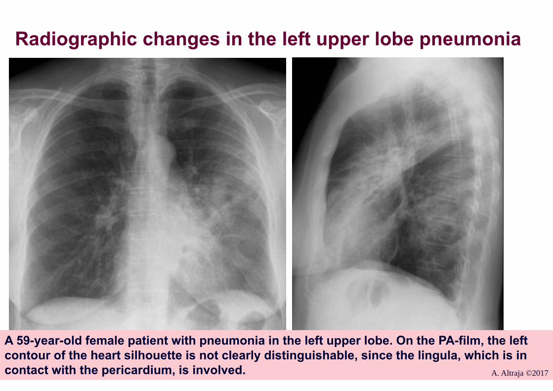
Radiographic changes in the left upper lobe pneumonia
A 59-year-old female patient with pneumonia in the left upper lobe. On the PA-film, the left contour of the heart silhouette is not clearly distinguishable, since the lingula, which is in contact with the pericardium, is involved. A. Altraja ©2017

Radiographic changes in the middle lobe pneumonia
A 50-year-old male patient with pneumonia in the right middle lobe. On the PA-film, the right contour of the heart silhouette is not clearly distinguishable, since the middle lobe, which is in contact with the pericardium, is involved. A. Altraja ©2017

Pneumonia caused by Staphylococcus aureus: a tendency to abscess formation
A 50-year-old female patient with bilateral, focal pneumonia that involves abscess formation. The pathogen is S. aureus, a hematogenic spread of infection from a suppurated pacemaker.
A. Altraja ©2017

Pneumonia in an immunocompromised person
A 72-year-old female patient with bilateral pneumonia. The patient has been on immunosuppressive therapy due to SLE; the pathogen is P. jirovecii diagnosed by the detection of the respective DNA in BAL fluid. Bilateral central and parabronchovascular ground glass opacities are visible along with thickening of the interlobular septa. A. Altraja ©2017

Radiographic changes in pneumonia IIIn destruction of the lung tissue (abscess formation)• Cavitation's appear inside the consolidations: they represent areas with decreased attenuation up to air density, with or without an horizontal air-fluid interface caused by (purulent) exudateAccumulation of fluid into the pleural cavity (pleural effusion)•Sometimes, the increased attenuation by pleural effusion can (partially) shade the consolidation of the lung tissueIn atypical pneumonia:•Increased interstitial attenuation and reticular changes•Sometimes, fine granular dissemination may be visible•Upper lung fields are involved with higher probability than in typical pneumoniaProgression of the radiographic changes may be a significant indicator of bad prognosis in any pneumonia! (especially, if the progression is accompanied by clinical worsening)
A. Altraja ©2017

Pneumonia:
A. Altraja ©2017
•Facilitating factors•Factors that refer to the presence of specific microflora (more aggressive or resistant)•Factors that determine the patient risks•Factors that determine the course of pneumonia

Radiographic changes in pneumonia: air bronchogram
A 39-year-old alcoholic male patient with pneumonia: fell ill after staying outdoors overnight at -25 °C.
A. Altraja ©2017

Radiographic changes in pneumonia: a significant radiographic improvement
The same 39-year-old alcoholic male patient with pneumonia, who fell ill after staying outdoors overnight at -25 °C. The patient survived thanks to intensive care. A. Altraja ©2017

Radiographically extended pneumonia: a consequence of alcohol addiction
A 52-year-old male patient with alcohol addiction. Consolidation characteristic of pneumonia is present in the 6th segment of the right lower lobe along with the finding of the same type in the right upper lobe. There is some pneumonic consolidation also in the right middle lobe.A. Altraja ©2017

Aspiration pneumonia: a consequence of alcohol addiction
64-year-old male alcohol addict with bronchopneumonia characteristic of aspiration pneumonia in the lower and posterior parts of the lungs. The illness developed after falling asleep with severe alcohol intoxication.A. Altraja ©2017

The diagnosis of pneumoniaThe pre-requisite for the diagnosis of pneumonia is satisfaction
of the requirements by the definition• Radiographically detectable new or progressive lung infiltrates with 1-2 other clinical elements
1. Data from the history: collection of the data should be directed to the symptoms and signs characteristic of pneumonia, as well as obtaining the data on the course of the disease
2. Results of the following investigations are needed: • Chest X-rays of the chest (2 projections)• Physical investigations• Clinical blood analysis with inflammatory markers (CRP)• In certain occasions, clarification of the causative
pathogen(s) (i.e. making the „etiological diagnosis)A. Altraja ©2017

Principles of revealing the etiology of pneumoniaRapid Nucleic Acid Amplification Tests (NAAT) have enabled to substantially increase the diagnostic yield in terms of the causative pathogens: up to 86.7% (together with viruses)• Clarification of the etiology with other methods was time-
consuming, expensive, unsuccessful (historically, only 30-50%), and did not often assist the choice or change of the treatment • Empiric treatment is/was nevertheless often efficacious in patients with non-severe pneumonia
Clarification of the etiology is important:• Often leads to adjustment of the treatment (in 82.9%: de-escalation in 77.2%; escalation in 5.9%)• In elderly patients (>65 years) • In patients with concomitant diseases• In severe cases with high risk of complications or death• In areas, where the composition and susceptibility pattern of the local microflora is unknown
A. Altraja ©2017 Gadsby et al. CID 2016;62:817

Recommendations for microbiological testing in LRTI in outpatient settings
3 main questions:• Is the LRTI in the patient caused by a bacterial pathogen/bacterial pathogens?• What is/are the bacterial pathogen(s)?• What is the susceptibility pattern of the bacterial pathogen(s)?
• There is no firm proof of the significance of the Gram stain in outpatient settings• Upper respiratory tract contamination may make the cultures non-specific• Other technical and organizatory issues
• Microbiological tests are generally not indicated in primary care• Concerns also detection of the S. pneumoniae antigen• In detection of respiratory viruses, the rRT-PCR-based methodologies are effective• Indications for treatment are based on clinical syndromes and assessment of their severity• Analysis of biomarkers (except S-CRP) is not generally recommended*
Woodhead et al. Eur Respir J 2005*Woodhead et al. Clin Microbiol Infect 2011A. Altraja ©2017

Recommendations for microbiological testing in LRTI in hospitalized patients I
Rapid tests based on NAAT technologies (rRT-PCR): have turned to being gold standards, they are applicable to all biomaterials incl. detection of viruses from nasopharyngeal or oropharyngeal aspirates
Microbiological investigations of sputum• Gram stain:• In cases, when it is possible to obtain specimens of purulent sputum and to process it in a timely manner• Presence of a predominant bacterial morphotype refers to a possible particular pathogen and facilitates interpretation of the culture results
Sputum cultures• A culture from a purulent sputum specimen of a bacterial species compatible with the morphotype in the Gram stain should be considered for confirmation of the pathogen identification and susceptibility testing• Sensitivity and specificity of the method may be lowered by eventual contamination by the upper respiratory tract microflora
Woodhead et al. Clin Microbiol Infect 2011A. Altraja ©2017

The primary microbiological test to guide the antimicrobial therapy
• Gram-positive diplococci (S. pneumoniae?)• Gram-positive cocci in chain (Streptococcus spp.?)• Gram-positive clustered cocci (Staphylococcus spp.?)• Gram-positive comma-shaped microorganisms (Nocardia
asteroides): rarely, in transplant recipients• Gram-negative diplococci (Neisseria spp.; kidney-shaped: M.
catarrhalis)• Gram-negative (pleomorphic) coccobacilli (Haemophilus spp.?)• Gram-negative rods (Enterobacter, Klebsiella spp. Pseudomonas
spp.): sometimes pairwise, connected by ends• If there are no dominating microorganisms: atypical pathogens,
viruses, or the material is inadequately collected
Gram stain of the sputum smear
A. Altraja ©2017

Recommendations for microbiological testing in LRTI in hospitalized patients II
Blood cultures: At least 2 series (sets) of blood cultures• From all hospitalized patients:•Streptococcus pneumoniae is found in 60% of the positive blood cultures•Haemophilus influenzae in 2-13%•Other microorganisms 1-14%:•Gram-negative aerobes, Streptococcus pyogenes,
Staphylococcus aureus and combinations of microorganisms•In the latter, it is always difficult to decide, whether there are „real“ pathogens or contaminants from the skin of the patient
Woodhead et al. Clin Microbiol Infect 2011A. Altraja ©2017

Recommendations for microbiological testing in LRTI in hospitalized patients III
Tests for antigens of selected pathogens (mainly form urine)
Streptococcus pneumoniae• Detection of S. pneumoniae antigen form urine•Recommended in patients hospitalized due to severe pneumonia• Sensitivity 65-100%, specificity 94%•Also from parapneumonic pleural effusion (if obtainable)• S. pneumoniae immunochromatographic test (ICT) from urine
Legionella pneumophila• Detection of L. pneumophila serogroup 1 antigen in urine•Recommended in patients hospitalized due to severe pneumonia• In patients, in whom L. pneumophila infection is probable on the clinical or epidemiological basis•Respiratory viruses: direct fluorescent antibody test; however, quantitative
molecular methods (rRT-PCR) have appeared as a gold standard
Woodhead et al. Clin Microbiol Infect 2011A. Altraja ©2017
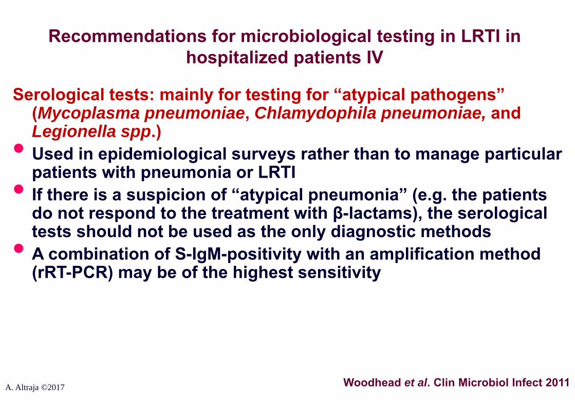
Recommendations for microbiological testing in LRTI in hospitalized patients IV
Serological tests: mainly for testing for “atypical pathogens” (Mycoplasma pneumoniae, Chlamydophila pneumoniae, and Legionella spp.)• Used in epidemiological surveys rather than to manage particular patients with pneumonia or LRTI• If there is a suspicion of “atypical pneumonia” (e.g. the patients do not respond to the treatment with β-lactams), the serological tests should not be used as the only diagnostic methods• A combination of S-IgM-positivity with an amplification method (rRT-PCR) may be of the highest sensitivity
Woodhead et al. Clin Microbiol Infect 2011A. Altraja ©2017

Recommendations for microbiological testing in LRTI in hospitalized patients V
Methods based on nucleic acid amplification methodologies (mainly to detect NDA or RNA of the respiratory pathogens) • Used in detection of the more common respiratory pathogens• Quantitative molecular methods (rRT-PCR):• Streptococcus pneumoniae (from sputum, blood), realistic also in
patients, in whom antibacterial treatment has already been started•Real-time qPCR (ompP6-based) to detect Homophiles influenza• Influenzaviruses and RSV: during the winter season•Atypical pathogens: often used, especially to detect L. pneumophila (in addition to detection of the antigen in urine)• Quantitative nucleic acid amplification methodologies (NAAT, Nucleic Acid
Amplification Test)•M. pneumoniae, C. pneumoniae, L. pneumophila, B. pertussis, preferably from sputum• “DNA panel of the respiratory tract bacteria”: B. pertussis, C. pneumoniae, H. influenzae, L. pneumophila, M. pneumoniae, S. pneumoniae •Other viruses: different coronaviruses, human metapneumovirus, bocavirus etc.
Woodhead et al. Clin Microbiol Infect 2011A. Altraja ©2017

Recommendations for microbiological testing in LRTI in hospitalized patients: invasive methods
Materials to all listed methods of analysisThoracocentesis (puncture of the pleural space)• Diagnostic thoracocentesis in all hospitalized patients with community-
acquired pneumonia, who present with significant pleural effusion
Bronchoscopy: in the context of managing the non-resolving pneumonia (see further)• Used more frequently in intubated patients/patients with tracheostomy, less frequently in non-intubated patients, if oxygenation of the patients allows• Bronchoalveolar lavage (BAL): a preferred method• Protected brush specimen (PSB)• Quantitative endotracheal aspiration (QEA)
Transthoracic needle aspiration (TTNA)• Because of a significant complication risk, only in rare individual cases• In severe pneumonia with focal infiltrates, when other methods have not appeared diagnostically successful
Woodhead et al. Clin Microbiol Infect 2011A. Altraja ©2017

How to write down the diagnosis of pneumonia
To be presented:• Radiographic and morphological type (lobar, focal)• Localization of the pneumonia (sides and lobes, sometimes also segments)• Etiology if known• The severity can be specified
A. Altraja ©2017

Differential diagnosis of pneumoniaVarious conditions that are able to present with pneumonia-like clinical or radiographic changes or both•Infectious diseases:•Acute bronchitis•Chronic bronchitis or acute exacerbations of chronic bronchitis or
obstructive pulmonary disease (COPD) •Pulmonary tuberculosis (especially infiltrative and focal, sometimes also disseminated pulmonary tuberculosis)•Non-infectious conditions:•Neoplasms of the lung (especially malignant, e.g. lung cancer)•Pulmonary embolism (in certain cases, infarction pneumonia)•Cardiac insufficiency•Eosinophilic pneumonias•Bronchiolitis obliterans•Diffuse parenchymal lung diseases (incl. idiopathic interstitial pneumonias: IPF, DIP, RB-ILD, AIP, NSIP, COP, LIP), pulmonary manifestations of systemic or other organs’ diseases)•Other rare pulmonary diseases
A. Altraja ©2017 Woodhead et al. Clin Microbiol Infect 2011

Pneumonia, acute (tracheo)bronchitis, or an acute exacerbation of chronic bronchitis
The respiratory symptoms are somewhat similar in these conditions
In bronchitis:•Radiographically, no infiltrative consolidation is present•On auscultation, no fine crackles•Clinically, weakness and fatigue is less prominent•Inflammatory shifts in blood analyses are significantly less prominent:•There is mild, if any, increase in serum CRP concentration•S-CRP >50 mg/L substantially increases the possibility of pneumonia (Melbye et al. 1992)
A. Altraja ©2017

The diagnosis of pneumonia without chest X-rayWith sufficiently high probability, one can omit radiographic investigations and exclude pneumonia if:•In formerly healthy persons of <65 years of age:• There are no changes in the vital indicators:• Heart rate <100/min• Respiratory rate <24/min• Orally taken body temperature <38 ºC• S-CRP <20 mg/L*• There is an absence of local or unilateral (asymmetric)
chest finding (fine crackles, pectoral fremitus, bronchophony (also known as bronchiloquy) etc.)
•Cough with a duration of at least 8 weeks “chronic cough” radiographic investigations are indicated to reveal the reasons
Snow et al. Ann Intern Med 2001*Woodhead et al. Clin Microbiol Infect 2011A. Altraja ©2017

Are radiographic investigations always mandatory for the diagnosis of pneumonia?
•Is the chest Xray the diagnostic gold standard of pneumonia?•IN certain circumstances, the diagnosis is allowed to be made without a radiographic confirmationIn out-patient practice, the diagnosis could be made on the basis of the following signs: •Cough + one sign of the LRTI:• New (formerly absent) local physical chest sign• + one general sign (sweats, chest pain, fever) • + these symptoms are not explainable otherwise• Pneumonias without fever (that occur frequently in elderly
people) are especially hard to diagnose without radiographic confirmation• The main differential diagnosis: congestive heart failure:particularly its exacerbation + acute virtual infection
Evidence-Based Respiratory Medicine, 2005 A. Altraja ©2017

Van Vugt’s model to evaluate the presence of pneumonia without radiographic investigations
•An absence of a runny nose•Presence of increased heart rate (>100/min)•Presence of breathlessness•Presence of fever >37,8 °C•Presence of fine crackles•Decreased vesicular breath sounds•Heightened S-CRP concentration (>30 mg/L)
Interpretation:•Score 0: 0.7%: pneumonia probability•Score 1-2: 3.8% probability•Score ≥3: 18.2% probability
Van Vugt et al. BMJ 2013; 346: f2450A. Altraja ©2017

Pulmonary tuberculosis as a differential diagnosis of pneumonia
Pulmonary tuberculosis may mimic pneumonia with a fairly similar clinical presentation; however, in pulmonary tuberculosis:•The patients tolerate fever much better•The finding on auscultation is not characteristic of pneumonia•Inflammatory changes in blood analyses (incl. increased S-CRP) are less prominent•Finding of Mycobacterium tuberculosis is the only specific sign!Radiographically, in pulmonary tuberculosis:•The infiltrates/opacities are located to more apical and posterior parts of the lungs•Fairly often, destruction (cavitation's) appear in the affected parts of the lungs•In the cavitary forms of tuberculosis, bronchogenic dissemination of the disease into the lower parts of the lungs is characteristic•The radiographic findings are often larger than assumed by the clinical presentation (e.g. symptoms) of the patient
A. Altraja ©2017
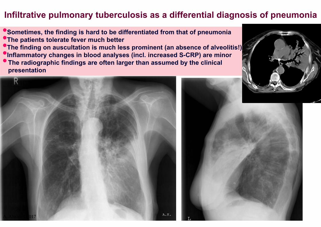
Infiltrative pulmonary tuberculosis as a differential diagnosis of pneumonia
•Sometimes, the finding is hard to be differentiated from that of pneumonia•The patients tolerate fever much better•The finding on auscultation is much less prominent (an absence of alveolitis!)•Inflammatory changes in blood analyses (incl. increased S-CRP) are minor•The radiographic findings are often larger than assumed by the clinical presentation
A. Altraja ©2017

Infiltrative pulmonary tuberculosis as a differential diagnosis of pneumonia
•Better tolerance of fever•The finding on auscultation is minor•Inflammatory changes in blood analyses (incl. increased S-CRP) are minor•Finding of Mycobacterium tuberculosis is the only specific sign•The infiltrates/opacities are located to more apical and posterior parts of the lungs•Destruction (cavitation's) are frequent•The radiographic findings are often larger than assumed by the clinical presentation (symptoms)
A. Altraja ©2017

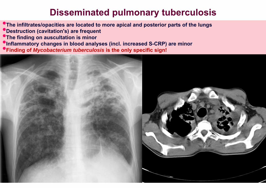
Disseminated pulmonary tuberculosis•The infiltrates/opacities are located to more apical and posterior parts of the lungs•Destruction (cavitation's) are frequent•The finding on auscultation is minor•Inflammatory changes in blood analyses (incl. increased S-CRP) are minor•Finding of Mycobacterium tuberculosis is the only specific sign!
A. Altraja ©2017

“Non-resolving pneumonia”: focal and infiltrative pulmonary tuberculosis•A 43-year-old woman with cough and progressive fever•Blood analyses: CRV <50 mg/L, slight leukocytosis•Progression of the radiographic findings within 5 days
A. Altraja ©2017

“Non-resolving pneumonia”: focal and infiltrative pulmonary tuberculosis
•The same 43-year-old woman with cough: after 9 more days•Fever >38,5 ºC•On auscultation: slightly coarse vesicular lung sounds•Blood analyses: CRV <50 mg/L •No response to conventional antibacterial treatment•The general condition is rather fair, the patient is active
A. Altraja ©2017

“Non-resolving pneumonia”: focal and infiltrative pulmonary tuberculosis
•The same 43-year-old woman with cough and fever >38,5 ºC•S-CRP <50 mg/L, no significant pathological lung sounds on auscultation•Progression of the CT findings with 14 days
A. Altraja ©2017

“Non-resolving pneumonia”•The same 43-year-old woman with cough and fever >38,5 ºC•S-CRP <50 mg/L, no significant pathological lung sounds on auscultation•interstitial lung disease? COP? DIP?•How to confirm the diagnosis?•To be treated? With prednisolone?
A. Altraja ©2017

“Non-resolving pneumonia” or pulmonary tuberculosis•Finding of Mycobacterium tuberculosis is the only specific sign! Smear microscopy, NAAT, or culture.
Disseminated pulmonary tuberculosis•The infiltrates/opacities are located to more apical and posterior parts of the lungs•Destruction (cavitation's) are frequent•The finding on auscultation is minor•Inflammatory changes in blood analyses (incl. increased S-CRP) are minor•Finding of Mycobacterium tuberculosis is the only specific sign!
A. Altraja ©2017

Non-infectious conditions that cause fever and lung infiltrates as a differential diagnosis of pneumonia
• Lung cancer (especially peripheral mucinous adenocarcinoma (formerly bronchioloalveolar carcinoma), also lung metastases• Pulmonary embolism (“infarction pneumonia”)• Eosinophilic pneumonias, acute and chronic (AEP, CEP)• Cryptogenic organizing pneumonia (COP) and bronchiolitis obliterans-organizing pneumonia (OP, formerly as BOOP)• Alveolar pulmonary edema• Hypersensitivity pneumonitis (HP, formerly allergic alveolitis)• Atelectasis• Adult respiratory distress syndrome (ARDS)• Pulmonary hemorrhage• Radiation pneumonitis• Adverse drug reactions (antibiotic fever)• Pulmonary vasculitis
A. Altraja ©2017 Winterbauer, 1995
Malignant neoplasms of the lung as a differential diagnosis of pneumonia•A pneumonia-specific clinical presentation is absent (no acute onset with high
fever and weakness)•In central neoplasm, the volume of the affected part of the lungs is often decreased: there is so-called obstructive-atelectatic pneumonitis or (at least partial) atelectasis•Frequently, there is a real (bacterial) pneumonia peripherally from the endobronchial obstacle/neoplasm (due to a failure to ventilate the periphery)•In cases of any suspicion of a central neoplasm bronchoscopy is indicated with biopsy and bronchial brush cytology•Often, the peripheral neoplasms (usually peripheral mucinous carcinoma) mimic pneumonia hard to differentiate from slowly resolving pneumonia: • Does not respond to antibacterial treatment, the radiographic changes are also different: air Broncho grams are not present because of the growth of the tumor);The morphological confirmation of the peripheral neoplasms of the lung:• Cytology of the bronchial aspirate• Transthoracic core needle biopsy (under CT or ultrasound guidance)• (Video) thoracoscopic or otherwise performed surgical lung biopsy
A. Altraja ©2017

•Epidermoid lung cancer in the right lower lobe in a 66-yar-old male patient, initially diagnosed as pneumonia. Right panel: 1 month later; the patient has received antibacterial treatment in the meantime.
A. Altraja ©2017

A. Altraja ©2017
•Epidermoid lung cancer in the right lower lobe in a 66-year-old male patient, initially diagnosed as pneumonia. Right panel: 1 month later; the patient has received antibacterial treatment in the meantime. The pneumonia component has been treated, but the neoplasm is present.

•Epidermoid lung cancer in the right lower lobe in a 66-year-old male patient, initially diagnosed as pneumonia. The same patient, as depicted on the two previous slides. CT scans that correspond to the later chest X-rays.
A. Altraja ©2017

CT-scan in pneumonia (left) and in peripheral lung cancer (right)
Left panel: a 81-year-old female patient with pneumonia in the right upper lobe; a consolidation with well-defined air bronchogram is visible. Right panel: a 67-year-old male patient with peripheral epidermoid lung cancer; there is no air bronchogram due to expansive and infiltrative growth of the tumor (including growth into the bronchial lumens).A. Altraja ©2017

CT-scan in pneumonia (left) and lung cancer (right)
Left panel: a 73-year-old male patient with pneumonia in the left lower lobe; a consolidation with well-defined air bronchogram is visible. Right panel: a 67-year-old male patient with peripheral epidermoid lung cancer; there is no air bronchogram due to expansive and infiltrative growth of the tumor (including growth into the bronchial lumens).A. Altraja ©2017

•Which of the patients has lung cancer?
A. Altraja ©2017

A. Altraja ©2017
•Which of the patients has lung cancer?

•A 62-year-old male patient with moderately severe COPD primarily diagnosed 2 months ago. Thereafter, hemoptysis appeared: a central bronchogenic lung cancer is present.
A. Altraja ©2017

•A 30-year-old male patient with pneumonia. Central opacity on the projection of the right hilar region that resembles, to some extent, a central neoplasm is visible on the posteroanterior view. On the lateral view, it is obvious that the opacity is located dorsally in the lower lobe. On the posteroanterior view, the silhouette of the right pulmonary artery is clearly distinguishable from the pneumonic consolidation. A. Altraja ©2017

•The same 30-year-old male patient with pneumonia. One month later. A significant radiographical improvement is visible.
A. Altraja ©2017

•A 73-year-old male patient•Bronchoscopy revealed a tumorous infiltration of mucosa at the terminal part of the left main bronchus with narrowing of the openings of the upper lobe bronchus and bronchus of the lingula
•DIAGNOSIS: Adenocarcinoma centrale pulmonis sinistri, stage IV (cT3N2M1)A. Altraja ©2017

Pneumonia abscedens: a frequent differential diagnosis of a neoplasm with destruction
A 43-year-old male patient with right-sided pneumonia and abscess formation already at diagnosis. The history characteristic of pneumonia was present. Right panel: 8 days later. Distinctly from destructive lung neoplasm, pneumonia, even with lung abscess, has a tendency to cure.
A. Altraja ©2017

Pneumonia abscedens: a frequent differential diagnosis of a neoplasm with destruction
A. Altraja ©2017
A 43-year-old male patient with right-sided pneumonia and abscess formation already at diagnosis. The history characteristic of pneumonia was present. Right panel: 8 days later. Distinctly from destructive lung neoplasm, pneumonia, even with lung abscess, has a tendency to cure.

Pulmonary embolism as a differential diagnosis of pneumonia
Pulmonary embolism (PE): may resemble severe pneumonia: abrupt onset with dyspnea, obvious chest pain, and/or consciousness impairment; hemoptysis may also occur•Clinically, the following speaks in favor of pulmonary embolism:• Occurrence of pulmonary embolism or deep venous thrombosis
(DVT) in the patient (in his/her history)• Presence of other risk factors for PE/DVT:• Immobilization within the last 4 weeks• Malignant neoplasms• Occurrence of preceding of surgical procedures or traumas•Radiographically:• Poorly defined but triangular opacity or consolidation with the basis contacting the visceral pleura or multiplicity of such foci• Pleural effusion (ipsilateral to the embolization)• Confirmation of the diagnosis of PE• CT- angiography of the lungs• Perfusion (+ ventilation) scintigraphy
A. Altraja ©2017 Woodhead et al. Clin Microbiol Infect 2011

CT in the diagnosis of pulmonary embolismSpiral CT with contrast media
in a 59-year-old female patient
• Large filling defects; the greatest in the left main pulmonary artery
• So-called “Wint-O-Green mint sign”
• Right-sided pleural effusion
A. Altraja ©2017

CT finding in “infarction pneumonia”• The same patient, as
on the previous figure• A large pyramid-
shaped consolidation with its basis residing on the visceral pleura that corresponds to the vascularization area of the embolized arterial branch
• Pleural effusion on the ipsilateral (right) side
A. Altraja ©2017

Congestive heart failure as the differential diagnosis of pneumonia
Left heart failure can be present in persons >65 years of age, who have (at least one of the following):•Orthopnoe•(Lateral and/or inferior) displacement of the apex beat•Myocardial infarction (in patient’s history)•Arterial hypertension(in patient’s history)•Atrial fibrillation (in patient’s history)
The following speaks against congestive cardiac insufficiency:•N-terminal propeptide of the brain natriuretic peptide (NT-proBNP (BNP) serum concentration <150 pg/mL•Brain natriuretic peptide (BNP) serum concentration <40 pg/mL•Just slight increase in serum inflammatory markers
A. Altraja ©2017 Woodhead et al. Clin Microbiol Infect 2011

Congestive heart failure as the differential diagnosis of pneumonia
• A 70-year-old male patient with an acute exacerbation of cardiac insufficiency. The patient was hospitalized to ordinary pulmonary ward due to a suspicion of pneumonia as a differential diagnosis. Both clinical and radiographic improvement occurred with 4 days (visible on the right chest X-ray) mainly with use of diuretics.
A. Altraja ©2017

Pulmonary edema as the differential diagnosis of pneumonia
Clinical signs:•Dyspnea and breathlessness•Presence of abundant watery and foamy sputum•Deterioration of respiratory insufficiency•Coarse crackles or moist rales on auscultation
Radiographically, in alveolar edema, bilateral, almost symmetrical opacities, located centrally, on the background of dilated pulmonary vessels; the periphery is relatively spared
A. Altraja ©2017

Pulmonary edema
A. Altraja ©2017
A 46-year-old female patient with pulmonary edema related to renal insufficiency

Eosinophilic pneumonias as the differential diagnosis of pneumonia
First of all acute eosinophilic pneumonia:•Clinically resembles pneumonia, from mild to severe•Manifestations of allergies in the history (especially to medicines)•Eosinophilia in the BAL fluid (>25%); in peripheral blood, eosinophilia may be absent in acute eosinophilic pneumonia•Bronchial obstruction is frequent•The condition responds rapidly to high-dose systemic glucocorticosteroidids or a removal of the agent (mainly the drug) that causes hypersensitivity•Radiographic finding: versatile•Diffuse confluent opacities•Can mimic pulmonary tuberculosis
A. Altraja ©2017

“Non-resolving pneumonia”: subacute eosinophilic pneumonia•A 41-year-old male patient with a history of mild-to-moderate asthma of 6-7 years•The problem developed after a flu-like illness•S-CRP 100-150 mg/L, frank leukocytosis•Slight alveolitis (fine crackles) on auscultation•Radiographic changes with a 2-day interval
A. Altraja ©2017

“Non-resolving pneumonia”: subacute eosinophilic pneumonia•The same 41-year-old male patient with a history of mild-to-moderate asthma of 6-7 years•The problem developed after a flu-like illness•S-CRP 100-150 mg/L, frank leukocytosis•Significant weakness, high fever (>39 ºC)•Antibacterial treatment remained without an effect
A. Altraja ©2017

Cryptogenic organizing pneumonia (COP) as the differential diagnosis of pneumonia
•Dry cough and dyspnea from some days to some months, sometimes weight loss•Fever is common, therefore, community-acquired pneumonia is often diagnosed (even cannot be excluded as a concomitant disease!)•The disease occurs in different age groups (20-80 years), whereas the mean age is 50 years •End-inspiratory fine crackles are present, but finger clubbing is rare (in a few % of cases)•Restrictive ventilatory pattern with decreased diffusing capacity of the lung in ca 80% of cases
A. Altraja ©2017

COP as the differential diagnosis of pneumonia
A. Altraja ©2017
A 50-year-old female patient

COP as the differential diagnosis of pneumonia
The same 50-year-old female patientA. Altraja ©2017

Obliterative bronchiolitis (OB) as the differential diagnosis of pneumonia
•Usually, OB appears as a result of inhalation of deleterious factors (e.g. chemical agents like ammonia etc.)
In obliterative bronchiolitis:•More breathlessness•Antibacterial treatment without an effect•Radiographically, diffuse infiltrative foci or consolidations with linear opacities
Differentiation is possible only morphologically (the main changes are located to the bronchioli)
A. Altraja ©2017

More frequent complications of pneumonia IParapneumonic exudative pleuritis•First, pleuritic chest pain appears•Breathlessness usually worsens after appearance of pleural exudate•Diagnosis: physical signs, ultrasonography, radiographic methods (e.g. chest X-rays with multiple views)Abscess formation•Expectoration of copious purulent sputum•Sometimes, the sputum is hemorrhagic•Recurrence or increase of the fever•Worsening of the general status of the patient/progression of the weakness is frequent•Radiographically, destructions are visible inside the consolidations
A. Altraja ©2017

Parapneumonic pleural effusion: a frequent complication of pneumonia
A male patient with pneumonia in the left upper lobe (lingula). On the lateral view, pleural effusion is seen in a better fashion (on posteroanterior view, the effusion is partly confluent with the lung infiltrate). Note also an elevation of the left hemidiaphragm. A. Altraja ©2017

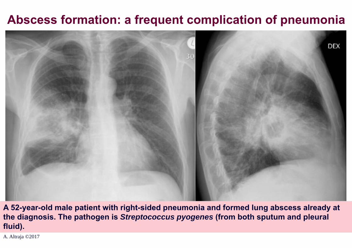
Abscess formation: a frequent complication of pneumonia
A 52-year-old male patient with right-sided pneumonia and abscess formation already at diagnosis. S. pyogenes was confirmed as the pathogen (isolated from both sputum and pleural fluid).A. Altraja ©2017

Abscess formation: a frequent complication of pneumonia
The same 52-year-old male patient with right-sided pneumonia and abscess formation already at diagnosis. S. pyogenes was confirmed as the pathogen (isolated from both sputum and pleural fluid).A. Altraja ©2017

Abscess formation: a frequent complication of pneumonia
A 27-year-old homeless man with pneumonia and abscess formation in the left upper lobe. An infiltrate with a clear destruction and a horizontal air-fluid interface inside is visible.
A. Altraja ©2017

Abscess formation: a frequent complication of pneumonia
47-year-old man with pneumonia and lung abscess. An infiltrate with extended cavitation and a horizontal air-fluid interface is visible in the right lung.
A. Altraja ©2017

More frequent complications of pneumonia IIPleural empyema•Typically develops via suppuration of the parapneumonic pleural exudate•Radiographic findings are identical to those seen in overall pleural effusions•Recurrence or increase of fever•Worsening of dyspnea and the overall general statusPyopneumothorax•Typically develops via breaking of the contents of lung abscess into the pleural cavity•Clinically:•Sudden appearance or worsening of chest pain•Breathlessness•Collapse and shock may accompany•Breath sounds are absent
Radiographically:•Pneumothorax with a horizontal air-fluid surfaceA. Altraja ©2017

Pleural empyema: a frequent complication of undertreated pneumonia or pneumonia left untreated
A male patient with right-sided pleural empyema
A. Altraja ©2017

Pleural empyema: a frequent complication of undertreated pneumonia or pneumonia left untreated
A male patient with right-sided pyopneumothorax that occurred as a complication of pneumonia with lung abscess. In addition to the pleural effusion divided into chambers by pleural adhesions, drainage inserted into the posterior part of the right pleural cavity and partial pneumothorax surrounded by adhesions are visible.A. Altraja ©2017
Abscess formation: a frequent complication of pneumonia
A 52-year-old male patient with right-sided pneumonia and formed lung abscess already at the diagnosis. The pathogen is Streptococcus pyogenes (from both sputum and pleural fluid).A. Altraja ©2017

Pyopneumothorax resulting from rupture of the pneumonic lung abscess into the pleural space
The same 52-year-old male patient with right-sided pneumonia and formed lung abscess already at the diagnosis. One day later than on the previous slide. Right panel: after insertion of the drainage and evacuation of the pus. The pathogen is Streptococcus pyogenes (from both sputum and pleural fluid). A. Altraja ©2017

Pyopneumothorax resulting from rupture of the pneumonic lung abscess into the pleural space
A. Altraja ©2017
The same 52-year-old male patient with right-sided pneumonia and formed lung abscess already at the diagnosis. One day later than on the previous slide. The pathogen is Streptococcus pyogenes (from both sputum and pleural fluid).

Pyopneumothorax: a frequent complication of pneumonia left untreated
A. Altraja ©2017
A 63-year-old male patient with parapneumonic pyopneumothorax: a result of a significant delay with seeking for medical care.

Pyopneumothorax: a frequent complication of pneumonia left untreated
The same 63-year-old male patient with parapneumonic pyopneumothorax. A result of delay with seeking for medical care. Deposition of bulky fibrin onto both visceral and parietal pleura has resulted in pleural thickening. Long-lasting air leak through the pleural drainage and failure of the lung to inflate are the main problems in such patients. Subcutaneous air emphysema is present as a complication of pleural drainage. A. Altraja ©2017

Pyopneumothorax: a frequent complication of pneumonia left untreated
A 63-year-old female patient with severe and extended parapneumonic pyopneumothorax. The patient fell ill almost 2 months ago, but did not seek for care. Right-sided massive pleural effusion with horizontal air-liquid interface is visible along with the shift of the mediastinum to the left. A. Altraja ©2017

Pyopneumothorax: a frequent complication of pneumonia left untreated
The same 63-year-old female patient with massive right-sided pyopneumothorax. The condition after placement of chest tube drainage and evacuation of the pus is shown. The drainage is visible together with the horizontal liquid level and partly collapsed and fixed into its shrinked position right lung. A. Altraja ©2017

Pyopneumothorax: a frequent complication of pneumonia left untreated
Chest CT scan of the same 63-year-old female patient with large right-sided pyopneumothorax. The condition after placement of chest tube drainage and evacuation of the pus is shown. The drainage is visible together with the partly collapsed and fixed into its shrunk position lower lobe of the right lung. The visceral pleura is obviously thickened. On the right panel, a relatively well preserved upper lobe of the right lung is seen.
A. Altraja ©2017

Pyopneumothorax: a frequent complication of pneumonia left untreated
A 48-year-old male patient with right-sided pyopneumothorax. He had a history of kidney transplantation due to chronic renal insufficiency resulting from glomerulonephritis. The pleural cavity has been partitioned by means of numerous adhesions and multiple horizontal air-fluid surfaces along with thickened visceral pleura are visible. A. Altraja ©2017

Pyopneumothorax: a frequent complication of pneumonia left untreated
CT scans of the chest in the same 48-year-old male patient with right-sided pyopneumothorax. The pleural cavity has been partitioned by means of numerous adhesions and multiple horizontal air-fluid surfaces along with thickened visceral pleura are visible.
A. Altraja ©2017

Pyopneumothorax: a frequent complication of pneumonia left untreated
A 31-year-old female patient with right-sided pyopneumothorax, HIV-infection, and a history of fever during 1 month. Multiple horizontal air-fluid surfaces are visible. Right panel: inflation of the right lung after placement of chest tube drainage. The cavitation (source of the pyopneumothorax) is visible along with numerous horizontal air-fluid surfaces (basally) and subcutaneous air emphysema as a complication of the pleural drainage. A. Altraja ©2017

Pyopneumothorax: a frequent complication of pneumonia left untreated
CT scans of the same 31-year-old female patient with right-sided pyopneumothorax. The abscess cavities with horizontal levels of pus, as well as free effusion in the pleural cavity. A shift of the mediastinum to the left is visible.
A. Altraja ©2017
More severe complications of pneumonia
•Sepsis and septic shock•Acute respiratory insufficiency•Acute respiratory distress syndrome (ARDS)•Acute cardiac insufficiency, pulmonary edema,•Acute renal insufficiency•Acute multiorgan insufficiency•Pyopneumothorax (discussed formerly)
A. Altraja ©2017

More severe complications of pneumonia: ARDS ARDS: Acute respiratory distress syndromeThe nature: Severe respiratory insufficiency resulting from non-cardiogenic, inflammatory pulmonary edema (due to increased permeability of the capillaries)
•Radiographically: bilateral lung infiltrates•Morphologically:• Interstitial and parenchymal edema, heterogeneous lung injury• Microthrombi in pulmonary capillaries, obliteration of their lumen• Collapse of the alveoli and peripheral parts of the conducting respiratory tract•Clinically: severe hypoxemia with its consequences• Diffusion-perfusion mismatch
A. Altraja ©2017

More severe complications of pneumonia: ARDS
•A 43-year-old male patient with ARDS on invasive ventilation
A. Altraja ©2017

Pneumonia-related ARDS
•The same 43-year-old male patient with ARDS on invasive ventilation
A. Altraja ©2017

More severe complications of pneumonia: septic shock
Clinical signs:•Acrocyanosis•Tachycardia•Tachypnea•Arterial hypotension (RR <80 mmHg)•Oligoanuria•Metabolic acidosis •Soporous condition•High fever
A. Altraja ©2017

Sepsis, severe sepsis, and septic shock •Systemic inflammatory response syndrome (SIRS):•The systemic response to a variety of factors manifested by at least 2 of the following: 1) body temperature >39°C or <36°C; 2) heart rate >90/min.; 3) respiratory rate >20/min., or PaCO2 <32 mmHg; 4) leukocytosis >12109/L, <4109/L, or >10% immature (band) forms•Sepsis:•The systemic response to infection manifested by at least 2 of the following as a result of infection: 1) body temperature >39°C or <36°C; 2) heart rate >90/min.; 3) respiratory rate >20/min., or PaCO2 <32 mmHg; 4) leukocytosis >12109/L, <4109/L, or >10% immature (band) forms •Severe sepsis:•Sepsis associated with organ dysfunction, hypoperfusion, or hypotension.Hypoperfusion and perfusion abnormalities may include, but are not limited tolactic acidosis, oliguria, or an acute alteration in mental status.•Septic shock:•Sepsis-induced with hypotension despite adequate fluid resuscitation with thepresence of perfusion abnormalities that may include, but are not limited to,lactic acidosis, oliguria, or an acute alteration in mental status. Patients on inotropic or vasopressor agents may not be hypotensive at the time thatperfusion abnormalities are measured. A. Altraja ©2017 ACCP/SCCM: Chest 1992;101:1644-55

Pulmonary edema as the differential diagnosis of pneumonia
Clinical signs:•Dyspnea and breathlessness•Presence of abundant watery and foamy sputum•Deterioration of respiratory insufficiency•Coarse crackles or moist rales on auscultation
Radiographically, in alveolar edema, bilateral, almost symmetrical opacities, located centrally, on the background of dilated pulmonary vessels; the periphery is relatively spared
A. Altraja ©2017
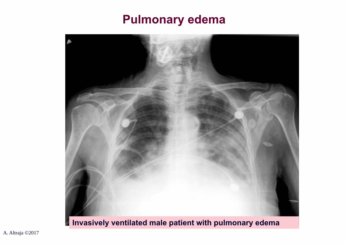
Pulmonary edema
Invasively ventilated male patient with pulmonary edemaA. Altraja ©2017
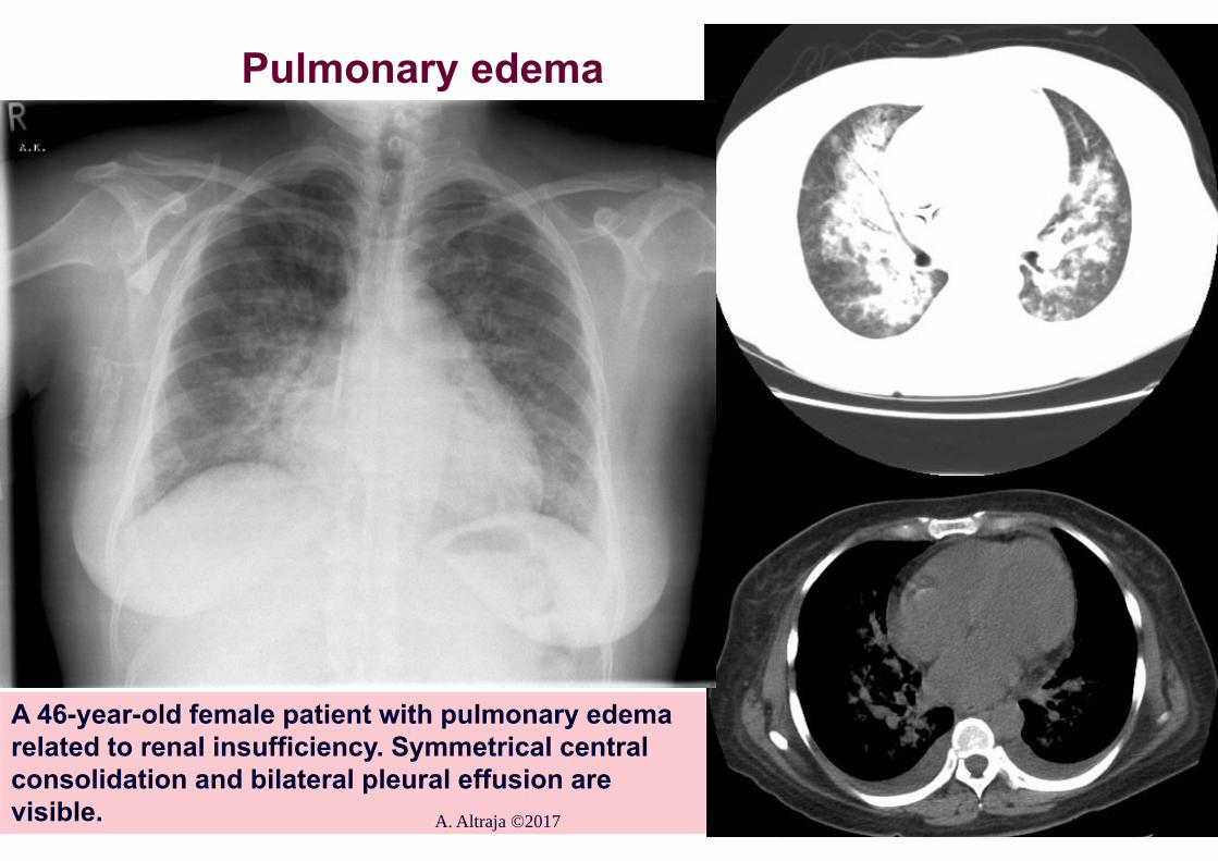
A 46-year-old female patient with pulmonary edema related to renal insufficiency. Symmetrical central consolidation and bilateral pleural effusion are visible.
Pulmonary edema
A. Altraja ©2017

Other complications of pneumonia
Myocarditis•Clinically fatigue, tachycardia•Of the investigations: exercise electrocardiogram or echocardiographyPericarditis•Clinically, sharp chest pain, dyspnea•Radiographically visible changes, ultrasonographyMeningitis•Permanent or worsening headache•Meningeal symptoms•Cerebrospinal fluid analyses
A. Altraja ©2017

The reasons of frequent recurrence of pneumoniaConcomitant diseases or various clinical situations (so-called structural changes in the respiratory or chest organs are frequently on the background):•Bronchiectasis•Bronchial carcinoma •Bronchial adenoma or other benign neoplasm•Recurrent aspiration•Pulmonary embolism (recurrent)•Foreign body in the lower conducting airways•Chronic bronchitis with obstruction•Reflux esophagitis•Esophageal achalasia•Immunosuppression (different forms)•Cystic fibrosis
A. Altraja ©2017

Etiology of pneumonia: general aspects and trends
Etiology depends on certain factors:•Is connected to patients’ risks and is dependent on:• Age• Concomitant diseases (chronic, especially cardio-respiratory) •Distribution of certain pathogens in the human population (area,
city, country)
Today’s trends in association with the pathogens:•To a certain extent, the proportion of atypical and Gram-negative pathogens is increasing•The resistance of the pathogens is increasing•The proportion of co-pathogenicity is increasing (pneumonia is caused by several pathogens with one being frequently atypical)
A. Altraja ©2017

Etiology of pneumoniaProbable pathogens in patients <65 years of age and formerly healthy:•Streptococcus pneumoniae•Mycoplasma pneumoniae•respiratory tract viruses (the gold standard of the diagnosis is a rRT-PCR-based methodology)•Chlamydophila pneumoniaeProbable pathogens in patients ≥65 years of age or with significant concomitant diseases:•S. pneumoniae (more resistance)•Viruses of the respiratory tract •H. influenzae (~20-40% -lactamase+)•C. pneumoniae•Aerobic Gram-negative rods•S. aureus•M. catarrhalis (~100% -lactamase+)•L. pneumophila
A. Altraja ©2017

Viruses in adult community-acquired pneumonia•The diagnostic gold standard is rRT-PCR:•The threshold value of cycles <40•Material from nasopharyngeal and oropharyngeal swabs
Viruses altogether (24.5%): •Human rhinovirus (hRV) (10.9%)•Human metapneumovirus (hMPV) (4.2%)•Coronaviruses (CoV) 2229E, HKU1, NL63, and OC43 (3,1%)•Influenza A and B (2.6%)•Respiratory-syncytial virus (RSV) (1.6%)•Adenovirus (AdV) (1.6%)•Parainfluenza virus (PIV) types 1, 2, and 3 (1.6%)
•References to the presence of influenza virus, RSV, and hMPV refer also to their roles as real pathogens; less in other viruses
A. Altraja ©2017 Self et al. J Infect Dis 2016; 231: 584

Viruses in community-acquired pneumonia in children
In children, viruses are significantly more important pathogens of community-acquired pneumonia than in adults
Viruses altogether (68.8%): •Respiratory-syncytial virus (RSV) (26.6%)•Human rhinovirus (hRV) (21.9%)•Human metapneumovirus (hMPV) (15.1%)•Adenovirus (AdV) (6.4%)•Parainfluenza viruses (PIV) types 1, 2, and 3 (4.7%)•Coronaviruses (CoV) 2229E, HKU1, NL63, and OC43 (4.5%)•Influenza A and B (3.4%)
A. Altraja ©2017 Self et al. J Infect Dis 2016; 231: 584

Factors that influence the etiology of pneumonia
•Advanced age: more frequently Gram-negative flora •Age <25 years: Mycoplasma pneumoniae, Chlamydophila pneumoniae
•Concomitant diseases (in addition to other pathogens):•Chronic bronchitis: Haemophilus influenzae, Moraxella catarrhalis (~100% -lactamase+), also S. pneumoniae•Alcoholism: K. pneumoniae, anaerobic bacteria, M. tuberculosis•Impaired consciousness, bad oral hygiene: anaerobes•Drug abusers: S. aureus, M. tuberculosis, and Pneumocystis jirovecii•Severe pneumonia: combined etiology (co-pathogenicity)
A. Altraja ©2017

Possible etiology in non-typical pneumoniaAtypical intracellular pathogens:• Mycoplasma pneumoniae• Chlamydophila pneumoniae• Legionella pneumophila
Non-typical course of pneumonia may be caused by common pathogens in cases of shifts in reactivity of the host:• Streptococcus pneumoniae• Haemophilus influenzae • Moraxella catarrhalis
A. Altraja ©2017

S. pneumoniae40%
M. catarrhalis3%
H. influenzae11%
S. aureus3%
Atypical30%
Others13%
Pathogens of community-acquired pneumonia
Cassiere & Niederman, Dis Mon 1998A. Altraja ©2017

Pathogens of the community-acquired pneumonia in Estonia (Tartu University Lung Clinic, 1999)
• Pathogens isolated in ~50% of cases; of these:• S. pneumoniae 24%• Gram-positives altogether 45%
• M. catarrhalis 11%• E. coli 9%• K. pneumoniae 9%• P. aeruginosa 6%• Enterobacter spp. 4%• Co-pathogenicity 18% (8% atypical)• Concomitant Candida spp. 20%
H. Leesik et al. 2000, 2013
•Atypical pathogens 16%
A. Altraja ©2017
Assessment of patient-associated risk is central in the management of pneumonia
• Risk of unfavorable course•Death•Complications
• The risk is strongly determined by:•Age 65 years•Concomitant diseases
• Risk assessment is the basis for hospitalization and treatment-related decisions
A. Altraja ©2017

Treatment of community-acquired pneumonia: what are the considerations?
• Mortality due to pneumonia: 1-15% if <65 years; 30% if >65 years
Mild
Non-severe
Severe
Incidence
Save money
Mortality
Save lives!
A. Altraja ©2017

Significant concomitant diseases that affect treatment and outcome of pneumonia
Cardiopulmonary concomitant diseases are especially important:• Chronic bronchitis, chronic obstructive pulmonary disease (COPD)• Cardiac insufficiency•>65-year-old people, orthopnea, (lateral and/or inferior)
displacement of the apex beat, myocardial infarction, hypertension, or atrial fibrillation
In addition:• Diabetes• Renal diseases (insufficiency)• Alcoholism • Malignancies• Liver diseases (incl. insufficiency)
A. Altraja ©2017 Woodhead et al. Clin Microbiol Infect 2011

Assessment of complication risk in out-patient pneumonia•In patients 65 years of age:•Concomitant diseases•COPD•Diabetes•Cardiac insufficiency•Hospitalization during the past 12 months•Therapy with oral glucocorticosteroids•Use of antibiotics during the last month•General weakness•Am absence of the upper respiratory tract symptoms/signs•Impaired consciousness •Pulse rate >100/min.•Body temperature >38ºC•Respiratory rate >30/min.•Arterial blood pressure <90/60 mmHg•Pneumonia is diagnosed by the primary care physician
A. Altraja ©2017 Woodhead et al. Clin Microbiol Infect 2011

Assessment of complication risk in out-patient pneumonia
•In patients <65 years of age:
•Diabetes•Asthma
•In all age groups:
•All severe concomitant diseases•Active malignancy•Severe liver disease•Severe renal disease (insufficiency)•Other concomitant diseases that may affect immune competence
A. Altraja ©2017 Woodhead et al. Clin Microbiol Infect 2011

When to hospitalize the outpatient with pneumonia?•Severely ill patients patients with suspicion of pneumonia (“an initial diagnosis”): the following symptoms and signs are particularly relevant:•Tachypnea•Tachycardia•Hypotension•Altered mental status (even minor disturbances)•If the patient with pneumonia does not respond sufficiently to treatment •Elderly patients (65 years) with heightened complication risks, especially those with the following concomitant diseases:•Diabetes•Cardiac insufficiency•Moderate-to-very severe COPD•Liver diseases (including insufficiency)•Renal diseases (insufficiency)•Active concomitant malignancies•Patients with suspected pulmonary embolism•Patients with suspected malignant neoplasm of the lung•If the home-based management is deemed improbable for other reasons
Woodhead et al. Clin Microbiol Infect 2011A. Altraja ©2017

A. Altraja ©2017
Pneumonia Severity Index (PSI/PORT) https://www.mdcalc.com/psi-port-score-pneumonia-severity-index-cap/Step 1: stratify to risk class I vs. risk classes II-VPresence of:
Over 50 years of age Yes/NoAltered mental status Yes/NoPulse ≥125/min. Yes/No
Respiratory rate >30/min. Yes/NoSystolic blood pressure <90 mm Hg Yes/NoTemperature <35°C or ≥40°C Yes/No
History of:Neoplastic disease Yes/No
Congestive heart failure Yes/No
Cerebrovascular disease Yes/NoRenal disease Yes/NoLiver disease Yes/No
If any "Yes", proceed to Step 2
If all "No“, assign to Risk Class I;
Mortality 0.1%; treat as an out-patient
Fine et al. NEJM 1997
Ste29.2p 2: Stratify to risk classes II-VDemographics Points
If male +Age (yr)If female +Age (yr) - 10Nursing home resident +10
ComorbiditiesNeoplastic disease +30Liver disease +20Congestive heart failure +10Cerebrovascular disease +10Renal disease +10
Physical exam findingsAltered mental status +20Pulse ≥125/minute +10Respiratory rate >30/minute +20Systolic blood pressure <90 mm Hg +20Temperature <35°C or ≥40°C +15
Laboratory and radiographic findingsArterial pH <7.35 +30Blood urea nitrogen ≥30 mg/dL (9 mmol/L) +20Sodium <130 mmol/L +20Glucose ≥250 mg/dL (14 mmol/L) +10Hematocrit <30% +10Partial pressure of arterial O2 <60mmHg +10Pleural effusion +10 Treat as:
Sum <70 = Risk Class II, mortality 0.6% Out-patientSum 71-90 = Risk Class III, mortality 2.8% Outp./hosp.
Sum 91-130 = Risk Class IV, mortality 8.2% Hospit.Sum >130 = Risk Class V, mortality 29.2% Hopsit.

CURB and CURB-65 scores• Four variables (in CURB-65, 5 variables) to be assessed • The score ranges between 0 and 4 (0 and 5); the score is got by
adding 1 point in the presence of one fact from the following:
• C: Mental Confusion (presence of)• U: Blood Urea Nitrogen >7 mmol/L • R: Respiratory Rate 30/min • B: Diastolic Blood Pressure 60 mmHg• 65: Age 65 years (if such an age is the case)
• CRB and CRB-65 also exist: no need for lab. analyses (urea)
A. Altraja ©2017 Lim et al. 2001
Hospitalization is indicated if:• C(U)RB or C(U)RB-65: 2 or• PSI: IV and V

C(U)RB and C(U)RB-65 in the assessment of risks in patients with pneumonia
•The use in practice is simpler than that of PSI•Comparable with PSI in terms of predicting mortality•In non-severe cases, there is no need for ancillary investigations•Avoid underestimation of the severity of pneumonia in young adults (assessment by PSI has such a risk)•The value in predicting the need for hospitalization into intensive care unit is unclear (Ewig et al. 1998)
•None of the systems is perfect: even non-severe pneumonia at the beginning can end up with fatal outcome•If even minor problems are perceived, a short-term hospitalization is recommended (e.g. for ancillary investigations)
Ewig et al. Eur Respir J 2006;Woodhead et al. Clin Microbiol Infect 2011A. Altraja ©2017

CURB score in making the decision to hospitalize in pneumonia
•0: Suitable to be treated as outpatient
•1: Needs ancillary data to decide on the need to hospitalize
•2: Presumes short-term hospitalization
•>2: There is severe pneumonia (mortality 26.7%)
•The scores correlate with the need for hospitalization, time to switch from intravenous to oral therapy, and time to be discharged from hospital
•If there are no issues, the 30-day mortality is 1%•In cases of the presence of 1-2 issues, the mortality 8%•In cases of the presence of 3-4 issues, the mortality 30%
Ewig et al. Eur Respir J 2006; Capalastegui et al. Eur Respir J 2006; 27:151-157A. Altraja ©2017

CRB-65 score in making decision to hospitalize in pneumonia: simpler and more precise in practice
0
CRB-65 score
1-2 3-4
Treatment as outpatient
(Short-term) hospitalization (if the age is not the
only criterion)
Immediate hospitalization
A. Altraja ©2017
Evidence-Based Respiratory Medicine, 2005;
Gibson et al. BMJ 2005;
Woodhead et al. Clin Microbiol Infect 2011

Criteria for hospitalization of the patient into intensive care unit in pneumonia•Clinical signs that refer to:•Acute respiratory insufficiency•Severe sepsis or septic shock•Significant progression of the infiltrates radiographically•Severe decompensation of the concomitant diseases•Presence of severe pneumonia: •Presence of at least 2 signs of the following:•Systolic blood pressure <90 mmHg•Severe respiratory insufficiency (PaO2/FIO2 <250)•Involvement of at least 2 lobes on chest X-ray (multilobar
involvement)•Presence of at least one sign of the following:•Need for mechanical ventilation•Need for vasopressor drugs for >4 hours (septic shock)•C(U)RB-65 and PSI scores do not unequivocally correlate with the need for intensive care
A. Altraja ©2017 Woodhead et al. Clin Microbiol Infect 2011

What is to be considered, when prescribing treatment for pneumonia
• An individual risk of death is considered• Antibacterial treatment is empiric• Antibacterial treatment cannot be delayedThe following is taken into account:• Is the patient treated in outpatient or hospital setting?• Is the patient hospitalized into intensive care unit?• What is the overall prognosis of the patient• Presence of concomitant diseases, especially cardio-pulmonary ones• Chronic obstructive pulmonary disease (COPD)•Congestive heart failure•Other diseases (diabetes, hepatic and/or renal insufficiency,
malignant neoplasms etc.)• Former treatments, hospitalizations, and immunosuppression• “Concomitant (modifying) factors”• Presence of drug-resistant S. pneumoniae• Presence of Gram-negative infection (nursing home resident etc.)• Presence of Pseudomonas aeruginosa• Other factors that influence the choice of antibacterial treatmentA. Altraja ©2017 Woodhead et al. 2011; Mandell et al. 2007; Woodhead et al. 2005; Niederman et al. 2001

Grouping of patients according to the performance of treatment: the general aspects
10 aspects form 3 areas are considered:• Prognostic assessment• Correct grouping• Assessment of factors determining selection of antimicrobials
A. Altraja ©2017 Woodhead et al. Clin Microbiol Infect 2011

Grouping of patients according to the performance of treatment: prognostic assessment
• Age: patients aged 65 years are subdivided into those with moderate/good ability and those who are severely disabled (according to an established score (e.g. ADL score)• Rough assessment: severely disabled patients are to be
defined as bedridden• General prognosis: patients with pneumonia as an expected
terminal event of a (known) severe co-morbidity should be managed according to the principles of palliative medicine
A. Altraja ©2017 Woodhead et al. Clin Microbiol Infect 2011
The initial treatment of pneumonia
• Rapid identification of the pathogen is not always possible (except application of the NA amplification technologies)• Identification of the pathogen is not successful in up to ~50% of cases• Isolation of the pathogen does not always allow adjustment of the treatment and provide optimal pharmacoeconomy• The spectrum of pathogens, as well as their resistance patterns are usually known in the given area • There are antibiotics or combinations of antimicrobials, based on the clinical-demographic properties of the patients, that warrant effective empiric treatment of pneumonia
• The initial treatment may be empiric
• The treatment should not be delayed! Antimicrobial treatment should be initiated immediately after the diagnosis
• In pneumonia patients with septic shock, the delay must not exceed 1 hour
A. Altraja ©2017 Woodhead et al. Clin Microbiol Infect 2011

Grouping of patients according to the performance of treatment: the correct grouping
• Previous hospitalizations and antimicrobial treatment:•Patients that have hospitalizations within <3 previous
months or have had repeated recent antimicrobial treatments should be classified as having nosocomial pneumonia and treated accordingly
• Patients with risk factors for severe immunosuppression (i.e. at risk of opportunistic pathogens): these should be managed following the guidelines for immunocompromized patients
A. Altraja ©2017 Woodhead et al. Clin Microbiol Infect 2011

Assessment of factors determining selection of the antimicrobial therapy in pneumonia
• Severity of pneumonia: although not a major determinant of the etiology, broad combination treatment is mandatory in severe cases to cover all potential pathogens and prevent excess mortality due to treatment failure
• Concomitant diseases: an independent influence on potential pathogens
• Residence of the patient: nursing home residence as such does not necessarily alter the patterns of pathogens
• Aspiration, either witnessed or suspected: correspond to gross or „silent“ aspiration
• Regional and local patterns of microbial prevalence and resistance
• Patient’s individual tolerability and toxicity of the antimicrobials
A. Altraja ©2017 Woodhead et al. Clin Microbiol Infect 2011

Grouping of patients according to the performance of treatment: setting of the treatment (ambulatory, hospital ward, or ICU)
I. Management outside hospital: patients without cardio-pulmonary concomitant diseases and modifying factors
II. Management inside hospital: when intensive care is not neededII. Management in the intensive care unit: (severe pneumonia)
A) without risk of P. aeruginosa as a pathogenB) with risk factors of having P. aeruginosa as a pathogen
In addition, special situations:• Aspiration pneumonia• Presence of a risk for community-acquired methicillin-resistant Staphylococcus aureus (CA-MRSA) (Mandell et al. 2007)
As a ”rule of thumb” (Lee et al. JAMA 2016;315:593-602): • Severe pneumonia: a β-lactam + a macrolide (to cover atypical)• Non-severe: a β-lactam alone
A. Altraja ©2017 Woodhead et al. 2011; Mandell et al. 2007; Woodhead et al. 2005; Lee et al. 2016
“Modifying factors” in the context of the etiology of pneumonia
• Risk factors for resistant S. pneumoniae as the pathogen• Risk factors for the presence of enteric Gram-negative rods as pathogens• Risk factors for the presence of Pseudomonas aeruginosa as the pathogen (under question mainly in intensive care settings)
A. Altraja ©2017 Mandell et al. 2007; Woodhead et al. 2005; Niederman et al. 2001

Risk factors for resistant S. pneumoniae as the pathogen
• Age 65 years• Treatment with a -lactam antibiotic within last 3 months• Alcoholism• Numerous (>1) concomitant diseases• A concomitant disease associated with immunosuppression or treatment with an immunosuppressive drug• Durable contact with children in kindergartens etc. preschool educational facilities for children
A. Altraja ©2017 Mandell et al. 2007; Woodhead et al. 2005; Niederman et al. 2001

Risk factors for the presence of enteric Gram-negative rods as pathogens
• Nursing home patient• Concomitant cardio-respiratory disease• Multiple (>1) concomitant diseases• Antibacterial treatment in the nearest past
A. Altraja ©2017 Mandell et al. 2007; Woodhead et al. 2005; Niederman et al. 2001

Risk factors for the presence of Pseudomonas aeruginosa as the pathogen (IDSA/ATS, 2001-2007, ERS Task Force, 2005)
• Frequent hospitalizations• Treatment with wide-spectrum antimicrobials (of >7 days duration) during the last 3 months• Frequent courses of antibacterial treatment (>4 per year)• Chronic treatment with glucocorticosteroids (with >10 mg of prednisolone or equivalent per day)• Bronchiectasis• Stage III-IV chronic obstructive pulmonary disease (FEV1 <30% predicted)• Former isolation of P. aeruginosa or documented colonization of the respiratory tract
A. Altraja ©2017 Mandell et al. 2007; Woodhead et al. 2005; Niederman et al. 2001

Risk factors for fluoroquinolone resistance
• Use of a fluoroquinolone in the nearest history • This discourages the use of a fluoroquinolone in the treatment of the current episode´of pneumonia
A. Altraja ©2017 Woodhead et al. 2011

Promptness of initiation of the antimicrobial treatment in pneumonia
•Antibiotic treatment should be initiated immediately after the diagnosis of CAP•In pneumonia patients with septic shock, the delay must not exceed 1 hour
A. Altraja ©2017 Woodhead et al. Clin Microbiol Infect 2011

•Amoxicillin (not effective against atypical pathogens, but is effective against M. catarrhalis and numerous enterobacteria)•A tetracycline (e.g. doxycycline)
•Antiviral treatment is not recommended in outpatients, unless the diagnosis is absolutely perfect•In cases of hypersensitivity to the previous antibacterials new generation macrolides (clarithromycin, azithromycin), but not spiramycin•The local sensitivity data should be considered, especially penicillin- and macrolide-resistance: not yet problematic in •If the resistance increases new generation “anti-pneumococcal” or “respiratory” fluoroquinolones (moxifloxcacin or levofloxcacin)
Treatment of pneumonia in outpatients (variants) I
A. Altraja ©2017
Woodhead et al. Eur Respir J 2005Woodhead et al. Clin Microbiol Infect 2011

•A clinical effect of antimicrobial treatment should be seen within 3 days•Patients should be instructed to contact their doctor if this is not the case•Severely ill patients (those with suspected pneumonia and elderly patients with a relevant co-morbidity) should be followed-up 2 days after the first visit):•High fever•Tachypnea•Dyspnea•Significant concomitant disease•Age 65 years•Patients should be advised to contact their doctor again, if their fever exceeds 4 days, dyspnea worsens, they stop drinking, or their consciousness is decreasing•Patients should be advised to return if the symptoms take >3 weeks to disappear
Guidance of the treatment and patient monitoring in outpatients with pneumonia
Woodhead et al. Eur Respir J 2005;Woodhead et al. Clin Microbiol Infect 2011A. Altraja ©2017
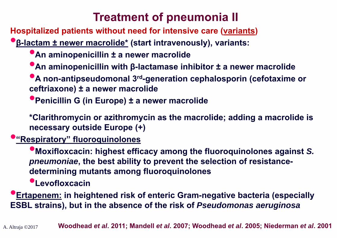
Hospitalized patients without need for intensive care (variants)•β-lactam ± newer macrolide* (start intravenously), variants:
•An aminopenicillin ± a newer macrolide •An aminopenicillin with β-lactamase inhibitor ± a newer macrolide•A non-antipseudomonal 3rd-generation cephalosporin (cefotaxime or ceftriaxone) ± a newer macrolide•Penicillin G (in Europe) ± a newer macrolide
*Clarithromycin or azithromycin as the macrolide; adding a macrolide is necessary outside Europe (+)
•“Respiratory” fluoroquinolones•Moxifloxcacin: highest efficacy among the fluoroquinolones against S. pneumoniae, the best ability to prevent the selection of resistance-determining mutants among fluoroquinolones•Levofloxcacin
•Ertapenem: in heightened risk of enteric Gram-negative bacteria (especially ESBL strains), but in the absence of the risk of Pseudomonas aeruginosa
Treatment of pneumonia II
A. Altraja ©2017 Woodhead et al. 2011; Mandell et al. 2007; Woodhead et al. 2005; Niederman et al. 2001

Treatment of pneumonia IIIA: patients in the intensive care unit, but with no risk of Pseudomonas aeruginosa infection
Alternatives:•A 3rd-generation non-antipseudomonal cephalosporin + a newer macrolide (not erythromycin or spiramycin)•A respiratory fluoroquinolone (moxifloxcacin or levofloxcacin ±a 3rd-generation non-antipseudomonal cephalosporin
A. Altraja ©2017 Woodhead et al. 2011
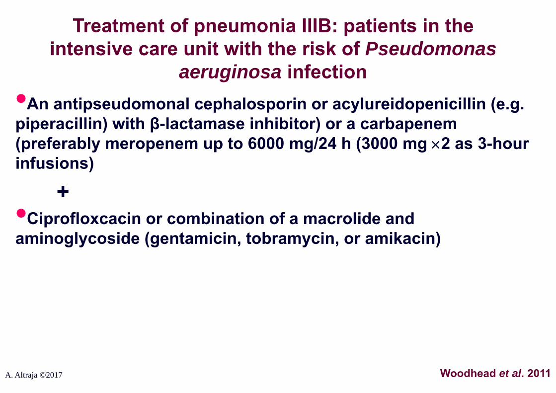
Treatment of pneumonia IIIB: patients in the intensive care unit with the risk of Pseudomonas
aeruginosa infection •An antipseudomonal cephalosporin or acylureidopenicillin (e.g. piperacillin) with β-lactamase inhibitor) or a carbapenem (preferably meropenem up to 6000 mg/24 h (3000 mg 2 as 3-hour infusions)
+•Ciprofloxcacin or combination of a macrolide and aminoglycoside (gentamicin, tobramycin, or amikacin)
A. Altraja ©2017 Woodhead et al. 2011

Duration of the antimicrobial treatment for pneumonia•Biomarkers have a significant role: PCT, CRP•In patients, who are adequately responding to treatment:
•The duration should not exceed 8 days•Usually it occurs to be 7 days •The same applies to nosocomial pneumonia
•Rehosipitalizations are mostly due to concomitant diseases, especially cardiopulmonary and neurological ones, and not to treatment failures due to inadequately short (i.v.) duration of treatment (Jasti et al. 2008)•An ongoing inflammatory process has been detected despite clinical improvement (Yende et al. 2008)
A. Altraja ©2017 Woodhead et al. 2011

How to choose/move between the oral and intravenous routes to administer antibiotics in pneumonia
•Oral route for treatment there, where possible•Intravenous (i.v.) route for treatment there, where necessary and as long as needed•In outpatients, 100% oral treatment from the very beginning•Even in some carefully selected inpatients, exclusively oral treatment may be used (patients with non-severe pneumonia, no sepsis, and no reason for impaired intestinal absorption)•In hospitalized patients, the treatment is begun with i.v. as a rule and continued with oral treatment (Marras et al. 2004):•Non-severe pneumonia•Absence of sepsis•Normal gastrointestinal functioning and absence of
impaired intestinal absorption;•However, in the most severely ill patients, i.v. treatment may be continued
A. Altraja ©2011 Woodhead et al. 2011

Switch to oral treatment in pneumonia•I.v. treatment is significantly more expensive2 variants:• Replacement of the intravenous antimicrobial agent by an oral formulation of the same agent that assures similar serum concentrations (“switch therapy”)•In the absence of an oral formulation of the same agent, it is replaced by another oral agent (or an agent with different pharmacokinetics) (“sequential therapy”)•There is no optimal time to switch to oral treatment; the decision should be guided by the resolution of the most prominent clinical features at admission•Usually, in the ordinary ward, this realizes after 3 days (after 3-7 days in elderly patients)•Switch to oral therapy is possible and indicated if (Mandell et al. 2007, strong recommendation):•The patient is improving clinically, the most prominent clinical features at admission have resolved•The patient is hemodynamically stable•The patient is able to take the medicines orally and to absorb them•The patient has normal gastrointestinal functioning
A. Altraja ©2011 Woodhead et al. 2011; Mandell et al. 2007

Indications for hospital discharge in pneumonia•Health care expenditures are 10-20 times less in outpatient settings than in hospitalized patients•Effective initial therapy id a pre-requisite for discharge•Keeping patients, who have been once switched to oral treatment, on the ward is generally neither indicated nor effective•Judgement upon the suitability for hospital discharge is based on the same simple clinical criteria, as used to monitor the treatment response:•Clinical stability is required•Body temperature•Respiratory (rate) and hemodynamic parameters (heart rate, blood
pressure)•Decrease in serum C-reactive protein content (should be measured usually on days 1 and 3-4)
Hospital discharge immediately after meeting the following criteria (Mandell et al. 2007, moderate recommendation):•Clinical improvement is detected in 48-72 hours•It is possible to switch from i.v. treatment to oral treatment•Patient’s condition is clinically stable (even in initially severe pneumonia)•Concomitant diseases are stable•All conditions for safe and successful treatment at home are met (as assessed)
A. Altraja ©2011 Woodhead et al. 2011; Mandell et al. 2007

Criteria for clinical stability i pneumonia•Tightly connected with the criteria for hospital discharge
Mandell et al. 2007:•Body temperature 37.8ºC•Heart rate 100 /min•Respiratory rate 24 /min•Systolic blood pressure 90 mmHg•SaO2 90% or PaO2 60 mmHg, when breathing room air•The patient is able to take and absorb oral medicines•The level of consciousness is sufficiently adequate
A. Altraja ©2017 Mandell et al. CID 2007

Non-antibiotic treatment in pneumonia•Early mobilization in all patients: already from the beginning of treatment: for at least 20 minutes per 24 h movement out of bed with change from horizontal to upright position•Low-molecular-weight heparin in patients with acute respiratory insufficiency•Non-invasive mechanical ventilation may be indicated in COPD and ARDS•Supportive measures, including intensive care, are needed in severe sepsis and septic shock•Cough is a defence reflex
Glucocorticosteroids: •Historically, these druga have been used in certain cases of meningitis, Pneumocystis jirovecii-infections, and influenza•The current evidence does not recommend!
Woodhead et al. Clin Microbiol Infect 2011; 2016A. Altraja ©2017

Symptomatic treatment in outpatients with lower respiratory tract infections (LRTI)
Cough, in outpatients*:•Cough suppressants, expectorants, mucolytics, antihistamines, inhaled glucocorticosteroids, and bronchodilators should not be prescribed in acute LRTI in primary care•Cough is a defence reflex; therefore, cough suppressants are not recommended, when the patient expectorates adequate amounts of sputum•In severe dry and disabling cough, medicines containing dextromethorphan (or codeine) can be prescribed•Expectorants, mucolytics, H1-antihistamines etc. are widely in use, but are without effect in the context of pneumonia (Schroeder et al. 2001)•The same concerns inhaled bronchodilators (unless otherwise indicated) (Smuncy et al. 2001)
*Woodhead et al. Clin Microbiol Infect 2011; Woodhead et al.; Eur Respir J 2005A. Altraja ©2017

Aspiration pneumonia: in whom to suspect and when?
• There is no final, agreed definition
• Aspiration pneumonia should be suspected in patients with community-acquired pneumonia, which either:
• Follows an episode of witnessed aspirationor• Occurs in the presence of risk factors for aspiration
such as:• Reduced level of consciousness• Dysphagia due to mechanical or neurological upper gastrointestinal tract dysfunction
A. Altraja ©2017 Woodhead et al. Clin Microbiol Infect 2011
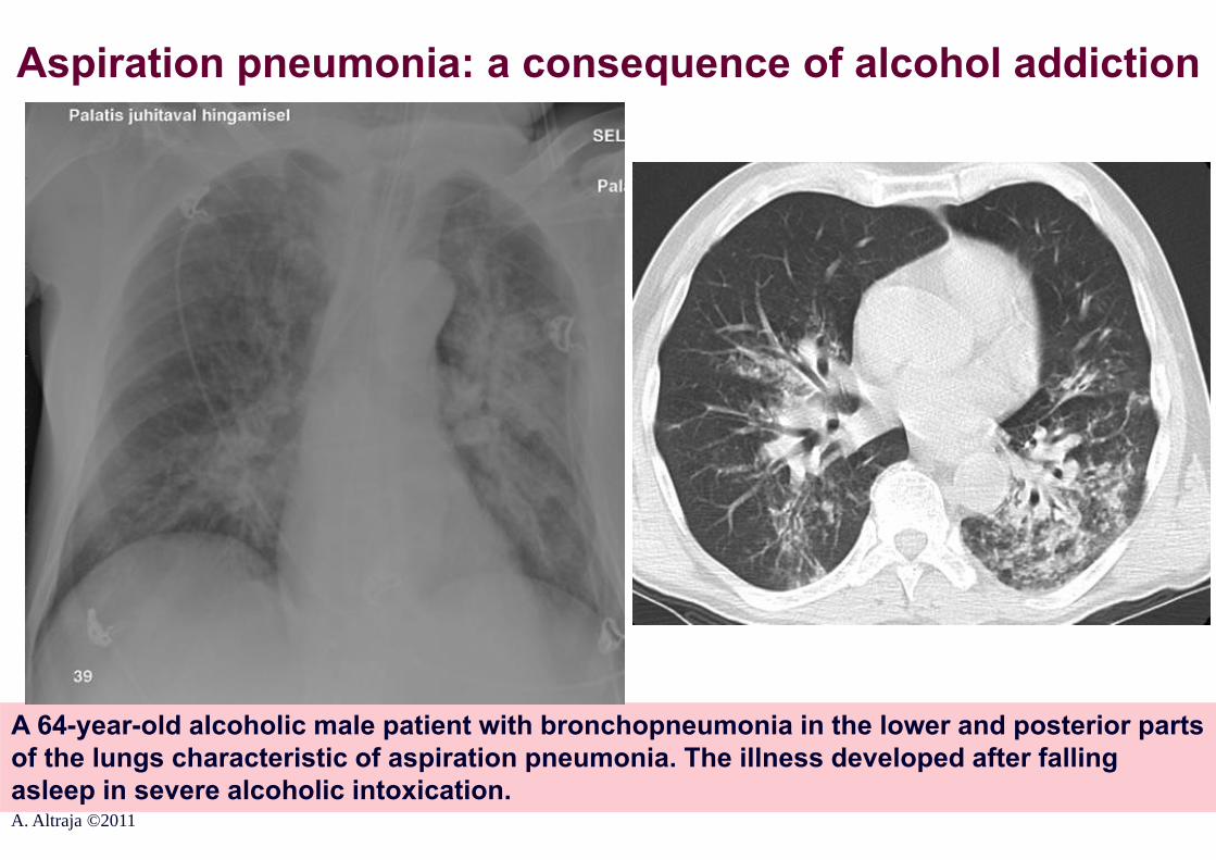
Aspiration pneumonia: a consequence of alcohol addiction
A 64-year-old alcoholic male patient with bronchopneumonia in the lower and posterior parts of the lungs characteristic of aspiration pneumonia. The illness developed after falling asleep in severe alcoholic intoxication.A. Altraja ©2011

Treatment of aspiration pneumoniaInpatient setting, hospitalized from their homes:• A β-lactam with β-lactamase inhibitoror • Clindamycinor• An i.v. cephalosporin + oral metronidazoleor • Moxifloxcacin
In an intensive care unit or hospitalized from a nursing home:• Clindamycin + a cephalosporinor• An i.v. cephalosporin + metronidazole
A. Altraja ©2017 Woodhead et al. Clin Microbiol Infect 2011

Treatment of pneumonia treatment in special circumstances
Pneumonia with a risk of community-acquired methicillin-resistant Staphylococcus aureus (CA-MRSA) infection:•Treatment according to the group (parameters) of the patient is necessary
in addition:•Vancomycin or linezolide
A. Altraja ©2011 Mandell et al. CID 2007

Assessment of the response to antibacterial treatment in pneumonia
Why is it important to assess response to therapy adequately and timely?
• To correct/change the treatment in time• To perform in time extensive ancillary diagnostic tests directed to either revealing the causative pathogen or make an alternative diagnosis• Non-effective treatment and complications have a very high price•The treatment is much more expensive
(complications)•Unexpectedly serious problems for the patient•Unfavorable (frequently fatal) course of the diseaseA. Altraja ©2017

Major criteria for adequacy of the antibacterial treatment in pneumonia
• Decrease in fever 48 hours• Decrease in serum CRV content 48-72
Factors that naturally slow down improvement in pneumonia:
• Age (65 years)• Significant concomitant disease(s)• Clinically severe pneumonia• Bacteremia course• In certain pathogens (Gram-negative, Legionella spp. etc.)
A. Altraja ©2017

Other indices of treatment efficacy in patients with pneumonia: usual time to obvious improvement
• Decrease in peripheral blood leukocytosis 2-4 days• Resolution of symptoms (cough, dyspnea,
breathlessness etc.) 3-5 days• Subjective improvement 3-5 days• Improvement of PaO2 in patients with
respiratory insufficiency 2-3 days• Duration of bacteremia 24-48 hours• Resolution of the finding on auscultation
(fine crackles) 7-14 days
A. Altraja ©2017

What to assess in the patient with pneumonia and how often?•Initial clinical assessment: based on the simplest clinical criteria, including body temperature, respiratory and haemodynamic parameters•Body temperature•Respiratory rate and hemodynamic parameters (heart rate, blood pressure)•Serum C-reactive protein should be measured on days 1 and 3/4, especially in patients with unfavorable clinical parameters•Also serum procalcitonin (PCT) level serves as a source of valuable information
•The same parameters are applied to judge suitability for hospital discharge (body temperature, S-CRP, respiratory rate and hemodynamic parameters)•Complete response, including radiographic resolution, requires more time
A. Altraja ©2017 Woodhead et al. Clin Microbiol Infect 2011

Resolution of the radiographic findings in pneumonia
• Significantly slower than the clinical improvement
Factors that slow down the radiographic improvement
• Patient-related:•Advanced age (65 a.)•Concomitant diseases (especially alcoholism)•Aspiration syndrome
• Pathogen-related:•Gram-negative pathogens (Klebsiella spp.)•Legionella spp.
A. Altraja ©2017

Resolution rate of the radiographic findings in pneumonia
Pathogen Initialworsening
Radiographicresolution
Residualchanges
Legionella spp. In the majority 2-6 months 25%
Fein et al. 1999
S. pneumoniae Sometimes 1-3 months Rare
S. pneumoniaewith bacteriemia
In the majority 3-5 months 25-35%
M. pneumoniae Rare 2-4 weeks Rare
H. influenzae Sometimes 1-5 months
Chlamydia spp. Rare 1-3 months 10-20%Occasional
A. Altraja ©2017
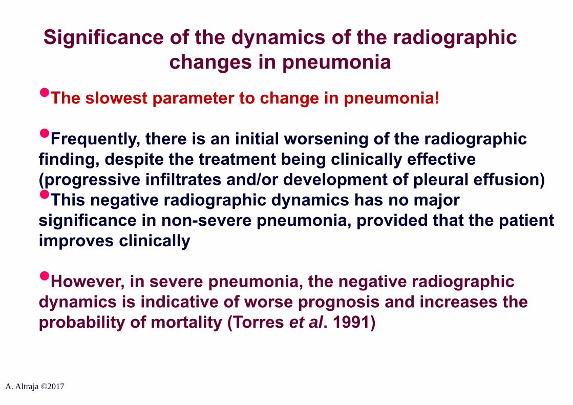
Significance of the dynamics of the radiographic changes in pneumonia
•The slowest parameter to change in pneumonia!
•Frequently, there is an initial worsening of the radiographic finding, despite the treatment being clinically effective (progressive infiltrates and/or development of pleural effusion)•This negative radiographic dynamics has no major significance in non-severe pneumonia, provided that the patient improves clinically
•However, in severe pneumonia, the negative radiographic dynamics is indicative of worse prognosis and increases the probability of mortality (Torres et al. 1991)
A. Altraja ©2017

Progression of the radiographic changes in pneumonia
A. Altraja ©2017
A 62-year-old male patient with prognostically significant radiographic progression of the pneumonic consolidations in 2 days

The need for radiographic investigations in the management of pneumonia
• The first chest X-ray for the diagnosis of pneumonia
• In cases of clinical improvement, the interval between control chest X-rays could be 1-2 weeks
• Just to document the resorption of pneumonic infiltrates• Because of the risk of lung cancer
Further, just the clinical monitoring is important
Longer radiographic follow-up is indicated in limited occasions (persons >40 years, smokers or former smokers):
A. Altraja ©2017

The need for radiographic investigations in the management of pneumonia
• Alternative diagnosis• Ineffectively treatment (non-responding patient)• Significant progression of pulmonary consolidation(s)• Dvelopment of lung destruction(s) (i.e. abscess formation:
purulent sputum)• Pleural effusion, pneumothorax or pyopneumothorax
(breathlessness, pleuritic chest pain)• ARDS or pulmonary edema• Sometimes necessary to test the function or to reveal the
position of the chest tube drainage
Repeated radiographic investigation is needed immediately, if there is a clinical suspicion of:
A. Altraja ©2017

An example of a full management algorithm for community-acquired pneumonia
Patient with pneumonia
Initial empiric treatment
Etiology not known
Pathogen isolated
Continue empiric
treatment
Narrower antimicrobial
spectrum
Clinicaleffect ?
Improvement ProgressionNo change
Oral treatment
Continue intravenously
! Extensivediagnostics

Reasons for failure in the treatment of pneumonia
Incorrect diagnosis
•Local factors•Complicat.•Deficient defence•Doesn’t take drugs
Due to the patient
Correct diagnosis
The condition does not change or deteriorates despite treatment
Due to the medicine
Due to the pathogen
•Incorrect drug•Incorrect dose or route•Side effects
•Resistant pathogens•Rare or non-bacterial pathogens
Differ. diagnosis•Neoplasm•Embolism•Atelectasis•Congestion•Lung fibrosis•ARDS, OP•Vasculitis•HemorrhageA. Altraja ©2017

Reasons behind the non-resolving pneumonia• The causative pathogen is resistant against the empirically selected
antibiotic(s) (may also refer to non-bacterial pathogen(s))• The causative pathogen is susceptible in vitro, but not in vivo• The selected antibiotic does not provide pharmacokinetically adequate
concentration in the inflamed lung (bad ”pulmonary pharmacokinetics”)• Abscess formation or empyema that is not drained• A condition caused by a foreign body or intraluminal neoplasm in the airways• The inflamed part of the lung is out of blood supply (abscess, necrosis,
pulmonary embolism, other causes of hypoperfusion)• Interactions between drugs (antagonism at whatever level that leads to
inactivation or lowered concentration of the antimicrobial(s))• Decrease of the effect of the antibiotic (e.g. because of low pH in the inflamed
focus)• Superinfection or concomitant co-infection by resistant pathogens (incl.
Additional or concomitant fungal infection, mycobacteriosis etc.)• A non-infectious disease or an infection not responding to the selected
antibiotic(s) (see “Alternative diagnosis”)
A. Altraja ©2017

“Non-resolving pneumonia”In practice, 2 types of treatment failures need to be distinguished:• Non-responding pneumonia
• It this occurs during the first 72 h of admission, it is usually due to antimicrobial resistance, an unusually virulent organism, or a host defence defect
• Non-response after 72 h is usually due to a complication• The evaluation of non-responding pneumonia depends on the
clinical condition• In unstable patients, full set of re-investigations followed by a
second empirical antimicrobial treatment regimen should be chosen
• In stable patients, change of the antimicrobial regimen is not always necessary
• Slowly resolving pneumonia • Re-investigations according to clinical needs, condition of the
patient, and individual risk factorsA. Altraja ©2017 Woodhead et al. Clin Microbiol Infect 2011

“Non-resolving pneumonia”•A 43-year-old woman with cough and progressive fever•Blood analyses: CRV <50 mg/L, slight leukocytosis•Progression of the radiographic findings within 5 days
A. Altraja ©2017

“Non-resolving pneumonia”
•The same 43-year-old woman with cough: after 9 more days•Fever >38,5 ºC•On auscultation: slightly coarse vesicular lung sounds•Blood analyses: CRV <50 mg/L •No response to conventional antibacterial treatment•The general condition is rather fair, the patient is active
A. Altraja ©2017

“Non-resolving pneumonia”
•The same 43-year-old woman with cough and fever >38,5 ºC•S-CRP <50 mg/L, no significant pathological lung sounds on auscultation•Progression of the CT findings with 14 days
A. Altraja ©2017

“Non-resolving pneumonia”
A. Altraja ©2017
•The same 43-year-old woman with cough and fever >38,5 ºC•S-CRP <50 mg/L, no significant pathological lung sounds on auscultation•interstitial lung disease? COP? DIP?•How to confirm the diagnosis?•To be treated? With prednisolone?

“Non-resolving pneumonia” or pulmonary tuberculosis?•Finding of Mycobacterium tuberculosis is the only specific sign! Smear microscopy, NAAT, or culture.

“Non-resolving pneumonia”: an alternative (non-infectious) diagnosis?
In practice, 4 major classes of diseases
• Neoplastic diseases• Immunopathological processes (vasculitis, eosinophilic pneumonias, sarcoidosis, AIP...)• Vascular pulmonary diseases (cardiac insufficiency, pulmonary embolism etc.)• Side effects of the drugs (pulmonary toxicity)
A. Altraja ©2017

Management of the “non-resolving pneumonia”
• Before rushing to change the antimicrobial regimen: de novo analysis of the full history, objective findings, the current course of the disease, and results of the investigations performed in terms of both complicating factors and alternative diagnoses
• Further diagnostic investigations
A. Altraja ©2017

Clinically applied diagnostic methods in “non-resolving pneumonia”
• CT and HRCT of the lungs• Bronchoscopy with ancillary methods• Video-assisted thoracoscopic surgery (VATS)• Transbronchial cryobiopsy (cTBB) or surgical lung biopsy (SLB)
A. Altraja ©2017

Non-responding pneumonia
•A 54-year-old male patient with a condition diagnosed as pneumonia, 4 weeks of antibacterial treatment without clinical-radiographic changes; a consolidation in the right middle lobe•On the CT-scan, consolidation + an endobronchial neoplasm in the right middle lobe bronchus ( )A. Altraja ©2017

Non-responding pneumonia: endobronchial hamartoma
•The same 54-year-old male patient with a condition diagnosed as pneumonia, 4 weeks of antibacterial treatment without clinical-radiographic changes; a consolidation in the right middle lobe•On the CT-scan, consolidation + an endobronchial neoplasm in the right middle lobe bronchus•On bronchoscopy, the neoplasm is of very elastic, but tenacious consistence A. Altraja ©2017

Non-responding pneumonia: endobronchial hamartoma
•The same 54-year-old male patient•Left panel: 11.7 cm endobronchial neoplasm, removed with use of a cryoprobe•Middle panel: branching of the middle lobe bronchus the segmental bronchi immediately after removal of the neoplasm by means of cryotherapy; histopathologically: hamartoma•Right panel: chest X-ray 1 day after removal of the neoplasm
A. Altraja ©2017
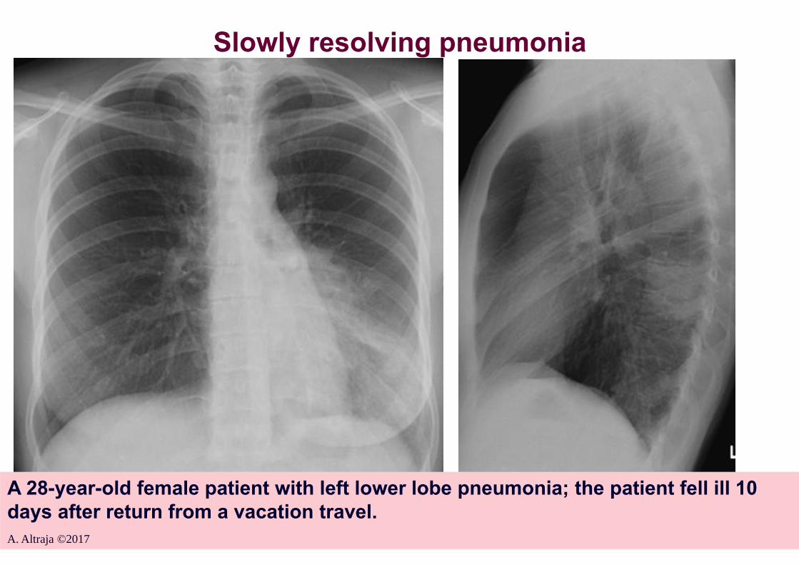
Slowly resolving pneumonia
A 28-year-old female patient with left lower lobe pneumonia; the patient fell ill 10 days after return from a vacation travel.A. Altraja ©2017

Radiographic interpretation of the resolution of pneumonia and ancillary investigations
A. Altraja ©2017
CT-scan: consolidation with air bronchogram
The same 28-year-old female patient with left lower lobe pneumonia; the improvement is slow despite treatment: 4 weeks from onset

Delayed resolving of pneumonia: ancillary investigations
The same 28-year-old female patient with left lower lobe pneumonia; improved, but not completely resolved. Bronchoscopy views of the left (inflammation) and right lower lobe bronchi (near-normal) at the same moment. (4 weeks from the diagnosis)A. Altraja ©2017

Vaccinations to prevent LRTI: vaccination against influenza
Influenza vaccine should be given yearly to persons atincreased risk of complications due to influenza; this measure decreases:• Incidence of pneumonia by 53% (Gross et al. 1995)• Hospitalizations due to pneumonia by 50% • Mortality due to pneumonia by 68% • Acute exacerbations of COPD and mortality in the elderly by
50%
Woodhead et al. Eur Respir J 2005; Woodhead et al. Clin Microbiol Infect 2011 A. Altraja ©2017

Indications of vaccinations against influenza• In adults, vaccination against influenza is indicated for the
following categories:• Age >65 years, institutionalization, chronic cardiac, pulmonary (COPD, IPF, PAH etc.), or renal diseases, diabetes mellitus, or hemoglobinopathies• Women, who will be in the second or third trimester of pregnancy during the influenza season• Medical co-workers• HIV-positives• Before travel to epidemic areas
Usage/application• Once a year, from September through the middle of November• Is cost-effective if used according to the indications
A. Altraja ©2017 Woodhead et al. Eur Respir J 2005; Woodhead et al. Clin Microbiol Infect 2011

Vaccination against S. pneumoniaeIndications:• Elderly people (65 a.)• Institutionalized patients• In patients with COPD of >65 years, it significantly reduces incidence and
mortality of pneumonia • <65 years, if concomitant diseases are present:• In patients with COPD of <65 years, it significantly reduces incidence of pneumonia in those, who have FEV1 <40% predicted• Dementia or seizure disorders• Heart failure, COPD, or cerebrovascular diseases• Chronic liver diseases or diabetes• Previous episodes of pneumonia • Functional or anatomical asplenia• Chronic cerebrospinal fluid leakage• The combination of 13-valent (Prevenar 13®) and (8 weeks later) 23-valent
polysaccharide pneumococcal vaccine (Pneumo 23®; Pneumovax®)• The 23-valent polysaccharide pneumococcal vaccine requires revaccination after 5-10 years, if the patient has been <65 years during the previous vaccination
Woodhead et al.; Eur Respir J 2005;26:1138-80; GOLD 2014-2016A. Altraja ©2017

Vaccination against Haemophilus influenzae
Indications:• Indications are similar to those for S. pneumoniae• Effective in prevention of pneumonia• Frequently in use in patients with chronic progressive
pulmonary diseases (COPD, IPF, PAH etc.)
A. Altraja ©2017 GOLD, 2014-2017
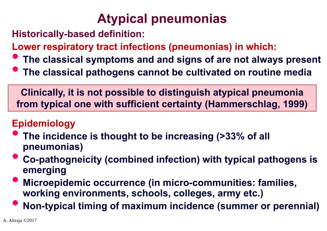
Atypical pneumoniasHistorically-based definition:Lower respiratory tract infections (pneumonias) in which:• The classical symptoms and and signs of are not always present• The classical pathogens cannot be cultivated on routine media
Epidemiology• The incidence is thought to be increasing (>33% of all pneumonias)• Co-pathogneicity (combined infection) with typical pathogens is emerging • Microepidemic occurrence (in micro-communities: families, working environments, schools, colleges, army etc.) • Non-typical timing of maximum incidence (summer or perennial)
Clinically, it is not possible to distinguish atypical pneumonia from typical one with sufficient certainty (Hammerschlag, 1999)
A. Altraja ©2017

Atypical pneumoniasMicrobiologyThe ”classical” “atypical pathogens”:• Mycoplasma pneumoniae, Chlamydophila pneumoniae,
Legionella pneumophila etc. Legionella species• In wider sense, ”atypicals” could be even other microorganisms that are capable of causing pulmonary infections (bacteria, mycobacteria, rickettsiae, viruses, protozoa, fungi etc.)
Clinical peculiarities• The clinical severity is varying, a milder course is more frequent, with lower fever etc. (except Legionella-pneumonia)• General and extra-pulmonary symptoms are prominent• Dry cough; sputum is very scarce• Bronchopneumonia or interstitial pneumonia, usually unilateral (except Legionella-pneumonia)• In the rare cases of pleuropneumonia, there is just little effusion
Forgie & Marrie, 2009A. Altraja ©2017

Possible pathogens of atypical pneumonias• Bacteria: Legionella spp. Francisella tularensis, Actinomyces
israelii, Nocardia asteroides, Mycobacterium spp.• Mycoplasmas: Mycoplasma pneumoniae• Chlamydiae: Chlamydophila pneumoniae, Chlamydia psittaci, Chlamydia trachomatis, Chlamydia pecorum• Rickettsiae: Coxiella burnetii• Viruses: Influenzavirus A, B; Parainfluenzavirus 1, 2, 3; RSV, Herpes simplex virus, Adenovirus, Hantavirus, Metapneumovirus, SARS Coronavirus (SARS Co-V), human Bocavirus• (Endemic) fungi: Histoplasma capsulatum, Blastomyces dermatitidis, Coccidioides immitis, Cryptococcus neoformans; Pneumocystis jirovecii• Protozoa: Toxoplasma gondii
A. Altraja ©2017 Forgie & Marrie, 2009

Atypical pneumonias
Laboratory findings and diagnostic work-up• The peripheral blood leukocyte count is normal or only slightly increased• Increase of the other inflammatory biomarkers is also slight• Diagnostics serology + DNA methods + cultures• There are usually no predominating microorganisms in sputum
Treatment• New (3rd generation) respiratory fluoroquinolones, macrolides, tetracyclines• -lactams do not work• The duration of treatment by the causative pathogens is not more regulated
A. Altraja ©2017

Pneumonia due to Legionella spp.: who are the patients • Rare in children, uncommon in younger adults• Mainly middle-aged and elderly people• In immunodeficiencies related to problems with T-lymphocytes
•Glucocorticosteroid therapy•Immunomodulation (azathioprine, cyclophosphamide etc.)•HIV-infection•Organ transplant recipients
• Smoking• Chronic respiratory diseases (incl. COPD)• Immediate post-operative period• Men : women = 2:1
A. Altraja ©2017 Forgie & Marrie, 2009
Younger and immunocompetent persons are also affected!
Cunha et al. 2007;

Pneumonia due to Legionella spp.: clinical picture and course• In the majority of cases, the patients need hospitalization because
of severity• Prodrome (common cold-like, may also be absent) 1-2 days: low
fever, loss of appetite etc.• Rapid development of high fever (>39.4°C)• Relative bradycardia (<100/min)• Weakness• Consciousness impairment, strong headache• Significant myalgias and arthralgias• Gastrointestinal complaints (nausea, vomiting, stomachache,
diarrhea)• Peri-, endo-, and myocarditis: may be present, though is rare• Pleuritis: chest pain, effusion is not abundant• Cough: non-productive, hemoptysis is rare
A. Altraja ©2017

Pneumonia due to Legionella spp.: radiographic changes
• Alveolar, rarely interstitial infiltrates• Both unilateral and bilateral• Both soft infiltrates, as well as thick consolidation• No predominant localization in the lungs• Rapid and asymmetric progression is characteristic• Pleural effusion is scarce, but is often the first sign• Cavitation (destruction, abscess formation) is rare
A. Altraja ©2017
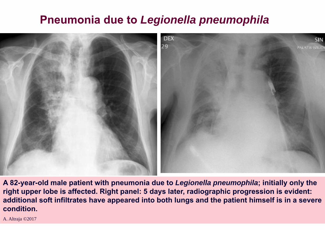
Pneumonia due to Legionella pneumophila
A 82-year-old male patient with pneumonia due to Legionella pneumophila; initially only the right upper lobe is affected. Right panel: 5 days later, radiographic progression is evident: additional soft infiltrates have appeared into both lungs and the patient himself is in a severe condition.A. Altraja ©2017

Pneumonia due to Legionella spp.: treatmentAntibiotics that are effective in all species:• So-called respiratory fluoroquinolones (superior to macrolides)
(Greenberg et al. Chest 2004)• Telithromycin• Doxycycline• New macrolides (not erythromycin)• Rifampicin 600 mg with macrolides is active, but has no
advantage in comparison with the above monotherapy
• The signs of improvement appear in 5-7 days• A decrease in fever• The relative bradycardia disappears• The other respiratory symptoms diminish
• Duration of treatment altogether 2-4 weeks• Radiographic resolution is the slowest among pneumonias!
Cunha et al. 2007A. Altraja ©2017
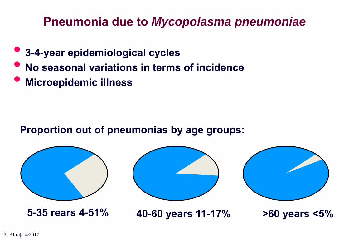
Pneumonia due to Mycopolasma pneumoniae
• 3-4-year epidemiological cycles• No seasonal variations in terms of incidence• Microepidemic illness
Proportion out of pneumonias by age groups:
5-35 rears 4-51% 40-60 years 11-17% >60 years <5%
A. Altraja ©2017

Pneumonia due to Mycopolasma pneumoniae: the clinical picture and course of the disease
• Incubation period 2-3 weeks• Pneumonia in 3-38% of cases: mild disease (hospit. 1.8%)• Asymptomatic onset or with pharyngitis, tracheitis, or bronchitis• Insidious onset, e.g. cough during many weeks• General symptoms: myalgias and arthralgias• Low fever <39°C• Extra-pulmonary symptoms
Respiratory symptoms:• Severe paroxysmal cough (ciliary dysfunction),• Breathlessness• Sputum is scarce, hemoptysis is very rare, no predominating
microorganisms found• Bronchoscopy: marked mucosal injection (hyperemia + edema)
A. Altraja ©2017
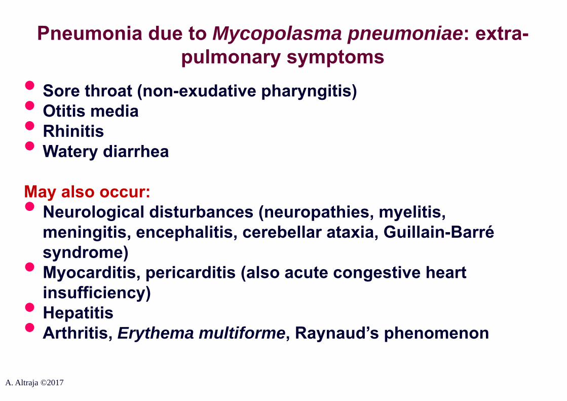
Pneumonia due to Mycopolasma pneumoniae: extra-pulmonary symptoms
• Sore throat (non-exudative pharyngitis)• Otitis media• Rhinitis• Watery diarrhea
May also occur:• Neurological disturbances (neuropathies, myelitis, meningitis, encephalitis, cerebellar ataxia, Guillain-Barrésyndrome)• Myocarditis, pericarditis (also acute congestive heart insufficiency)• Hepatitis• Arthritis, Erythema multiforme, Raynaud’s phenomenon
A. Altraja ©2017
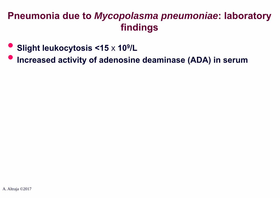
Pneumonia due to Mycopolasma pneumoniae: laboratory findings
• Slight leukocytosis <15 x 109/L• Increased activity of adenosine deaminase (ADA) in serum
A. Altraja ©2017
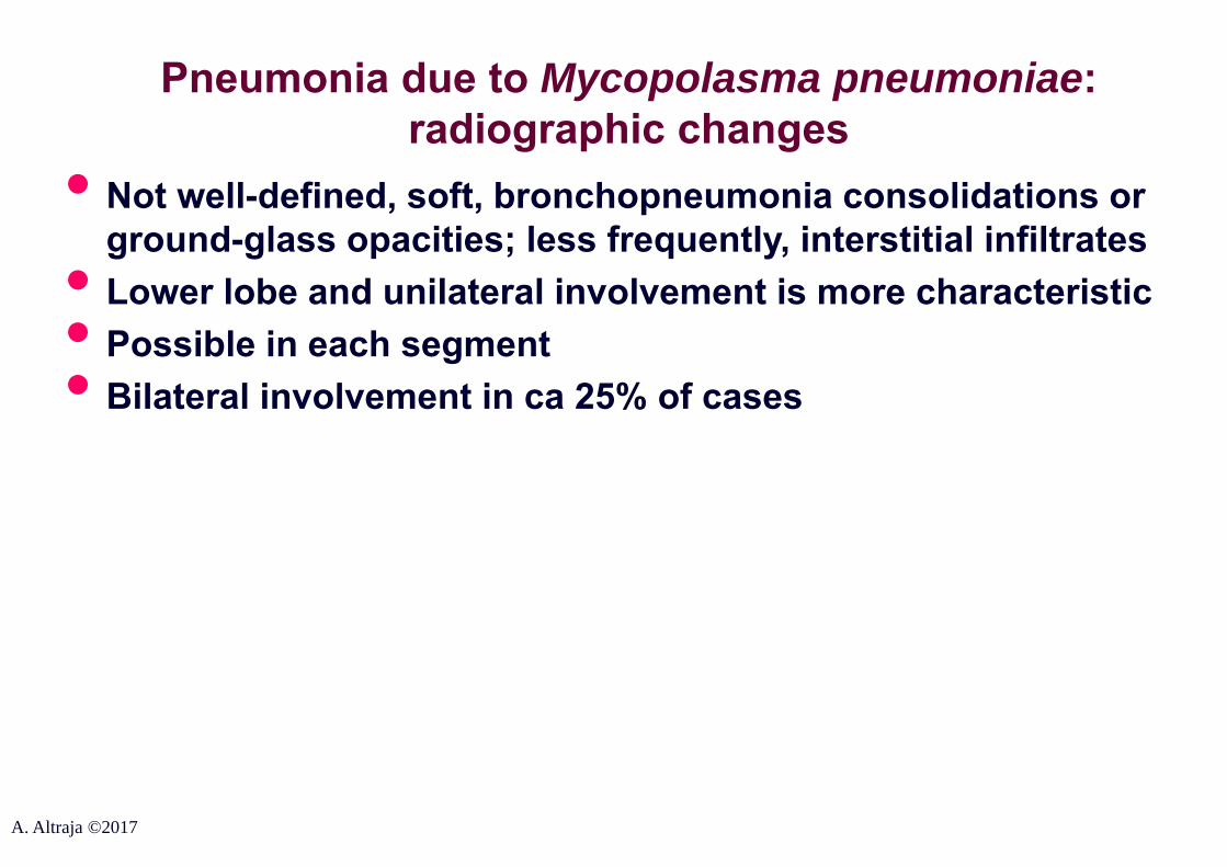
Pneumonia due to Mycopolasma pneumoniae: radiographic changes
• Not well-defined, soft, bronchopneumonia consolidations or ground-glass opacities; less frequently, interstitial infiltrates
• Lower lobe and unilateral involvement is more characteristic• Possible in each segment• Bilateral involvement in ca 25% of cases
A. Altraja ©2017

Pneumonia due to Mycopolasma pneumoniae
A 29-year-old female patient with pneumonia due to Mycoplasma pneumoniae in the left lung, mainly in the lingula. A characteristic clinical picture was present with rapid radiographic improvement (in 6 days) (right panel).

Pneumonia due to Mycopolasma pneumoniae
A 61-year-old female patient with pneumonia due to Mycoplasma pneumoniae. Bilateral soft consolidations are visible, the left lung is more affected.A. Altraja ©2017

Treatment of pneumonia due to M. pneumoniae• Empiric treatment after the analysis of the clinical picture, RT-
PCT tests (and increased titer of cold agglutinins)Effective antibiotics include:• Doxycycline• Macrolides and ketolides (incl. telithromycin)• Respiratory fluoroquinolones
• Subjective improvement is felt already in <72 hours• Dry cough persists for weeks, even after otherwise effective
treatment against Mycoplasma• Since the pathogen resides in the bronchial epithelium, at least
2-weeks treatment is necessary to reduce the risk of transmission or relapse
Cunha et al. 2007A. Altraja ©2017

Prognosis of pneumonia due to M. pneumoniae
• The course is usually mild and even self-limiting in immunocompetent inidviduals
• Severe course is possible:• Immunosuppression• Elderly people• Patients with severe concomitant lung diseases (e.g.
COPD)• Durable or persistent dry cough is a common complication• Bronchial hyperresponsiveness and post-M. pneumoniae-
asthma may develop
• Eradication of M. pneumoniae from the airways could be problematic
Cunha et al. 2007A. Altraja ©2017
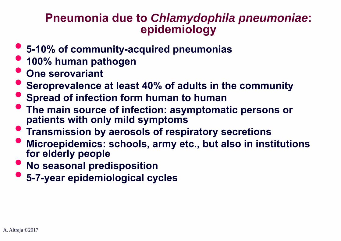
Pneumonia due to Chlamydophila pneumoniae:epidemiology
• 5-10% of community-acquired pneumonias• 100% human pathogen• One serovariant • Seroprevalence at least 40% of adults in the community• Spread of infection form human to human• The main source of infection: asymptomatic persons or patients with only mild symptoms• Transmission by aerosols of respiratory secretions• Microepidemics: schools, army etc., but also in institutions for elderly people• No seasonal predisposition• 5-7-year epidemiological cycles
A. Altraja ©2017

Pneumonia due to Chlamydophila pneumoniae: epidemiology
• Mainly causes pneumonia in younger people; however, a not infrequent pathogen also of nursing home pneumonias of the elderly persons
5-35 years: 5-15% >40 years: <6%
Proportion out of pneumonias by age groups:
A. Altraja ©2017

Pneumonia due to Chlamydophila pneumoniae: pathogenesis
• Obligate intracellular parasites • An average incubation period: 4 weeks• Intra-alveolar, less interstitial inflammation• Specific IgE (Emre et al. 1995)• Chlamydia family-specific protein (57 kDa): connected to a delayed
type hypersensitivity (Morrison RP 1989)
Elementary bodyCorpuscular body
Epithelial cell
A. Altraja ©2017

Pneumonia due to Chlamydophila pneumoniae: clinical picture and course of the disease
• “An illnesssimilar to Mycoplasma”: mild or moderately severe pneumonia, less frequently severe
• Clinical manifestation is dependent on age:•Mild pneumonia in children and young adults, frequently upper
respiratory tract diseases (bronchitis, sinusitis, pharyngitis, otitis media)
• In adults, the pneumonia is more severe; bronchitis and sinusitis are more durable (due to the immunopathogenesis)
• Extra-pulmonary manifestations•Mortality <1% (in complicated cases, up to 50%)
Extra-pulmonary manifestations:• Rhinitis, sore throat, coarseness may precede (in 20-50% of cases), up to 1-2
weeks before pneumonia• Fever usually <39,4°C• No relative bradycardia
Cunha et al. 2007A. Altraja ©2017

Pneumonia due to Chlamydophila pneumoniae: radiographic changes
• Usually unilateral, ill-defined coft pulmonary infiltrates• Dense consolidations, cavitations, and pleural effusion are
absent in most cases
Cunha et al. 2007A. Altraja ©2017

Pneumonia due to Chlamydophila pneumoniae
A. Altraja ©2017
A 66-year-old male patient with pneumonia due to Chlamydophila pneumoniae. An unilateral soft infiltrate is visible in the right lower lung field. No radiographically-specific clue exists to predict the pathogen here (Chlamydophila pneumoniae).

Pneumonia due to Chlamydophila pneumoniae: treatment
• The effective antibacterials include:•Doxycycline•Telithromycin•Respiratory fluoroquinolones•New macrolides (clarithromycin, azithromycin)• Monotherapy• Duration of the treatment usually 7-14 days• As a consequence of the acute infection, bronchial hyperresponsiveness and post-C. pneumoniae-asthma may develop
Cunha et al. 2007A. Altraja ©2017

Pneumonia due to Chlamydophila pneumoniae: prognosis
• The prognosis of non-severe pneumonia is very good• Severe community-acquired pneumonia occurs in:
•Elderly people•Persons with immunosuppression•Patients with severe concomitant pulmonary diseases
(COPD, cystic fibrosis etc.)
• Improper treatment or treatment of insufficient duration may result in the development of bronchial hyperresponsiveness and post-C. pneumoniae-asthma
• Re-infections are frequent• May turn to chronic, as eradication may me sometimes
problematic
A. Altraja ©2017

Atypical pneumonias in conclusion: the treatment
Treatments of choice:• Levofloxcacin 500 mg 1 i.v. for 1-2 weeksOr• Moxifloxcacin 400 mg 1 i.v. for 1-2 weeksOr• Doxycycline 200 mg 2 i.v. for the first 3 days, thereafter
100 mg 2 i.v. for 4-11 days
Cunha et al. 2007A. Altraja ©2017

Atypical pneumonias in conclusion: the treatmentAlternatives for parenteral treatment:• Azithromycin 500 mg 1 i.v. for 1-2 weeks (a minimum of
2 doses before switch to oral treatment)
Cunha et al. 2007A. Altraja ©2017

Oral treatment (or oral continuation):• Levofloxcacin 500 mg 1 p.o. for 1-2 weeksOr• Moxifloxcacin 400 mg 1 p.o. for 1-2 weeksOr• Gemifloxcacin 320 mg 1 p.o. for 1-2 weeksOr• Doxycycline 200 mg 2 p.o. for 3 days, thereafter 100 mg 2
p.o. for 4-11 daysOr• Clarithromycin XL 1000 mg 1 p.o. for 1-2 weeksOr• Azithromycin 500 mg 1 p.o. for 1-2 weeks
Cunha et al. 2007A. Altraja ©2017
Atypical pneumonias in conclusion: the treatment

Pneumonia due to H1N1 pandemic influenza (”swine influenza”)
A 47-year-old male patient with pneumonia due to H1N1 pandemic influenza. Multi-focal infiltrative opacities are visible mainly in the right upper lobe.
A. Altraja ©2017
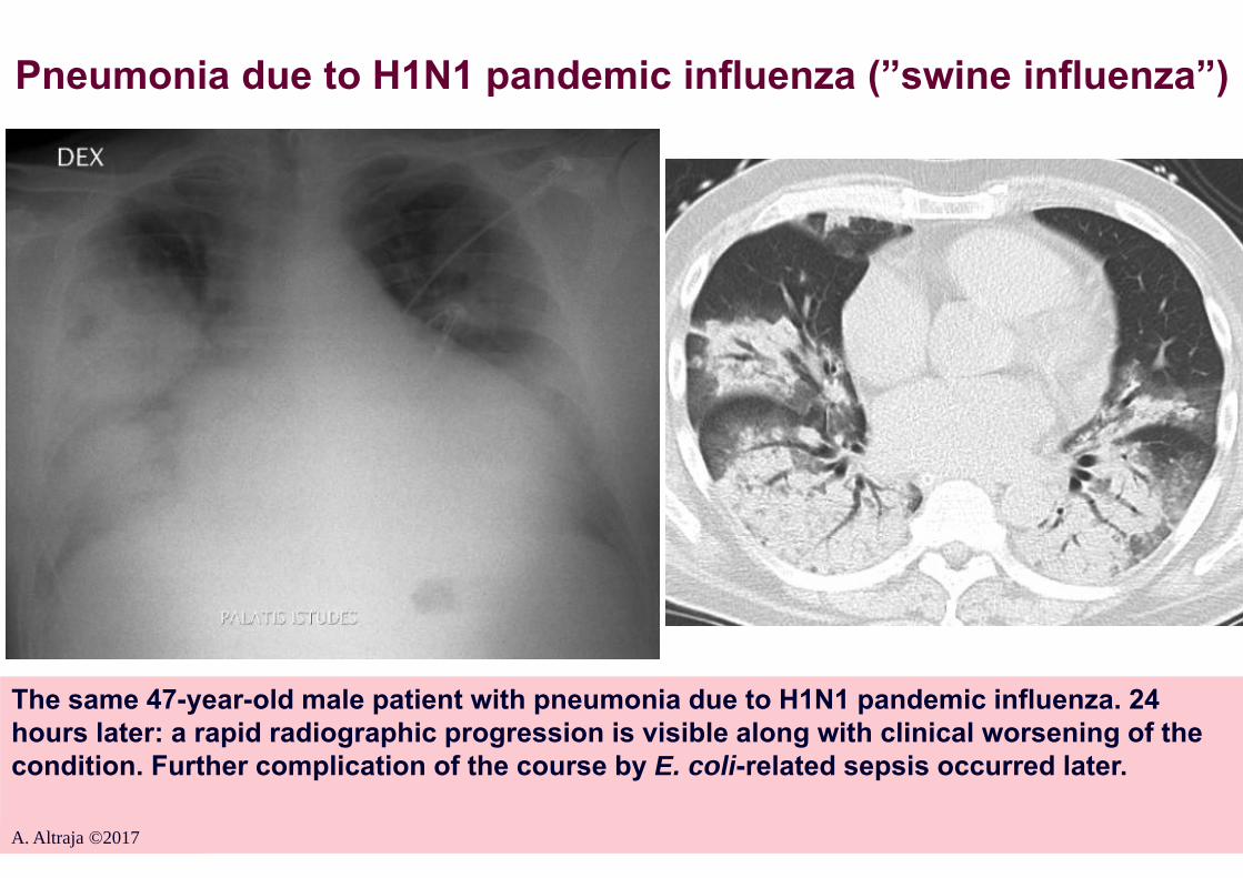
Pneumonia due to H1N1 pandemic influenza (”swine influenza”)
The same 47-year-old male patient with pneumonia due to H1N1 pandemic influenza. 24 hours later: a rapid radiographic progression is visible along with clinical worsening of the condition. Further complication of the course by E. coli-related sepsis occurred later.
A. Altraja ©2017