Pneumococcal Vaccine Response After Exposure to...
12
ARTICLE PEDIATRICS Volume 139, number 4, April 2017:e20162781 Pneumococcal Vaccine Response After Exposure to Parasites in Utero, in Infancy, or Mid-Childhood Monica Nayakwadi Singer, MD, MPH, a Claire Heath, PhD, b Jackson Muinde, MSc, c Virginia Gildengorin, PhD, a Francis M. Mutuku, PhD, d David Vu, MD, b Dunstan Mukoko, PhD, c Christopher L. King, MD, PhD, e Indu J. Malhotra, PhD, e Charles H. King, MD, MS, e A. Desirée LaBeaud, MD, MS b abstract BACKGROUND AND OBJECTIVE: Streptococcus pneumoniae is a leading cause of mortality before age 5, but few studies examine details of childhood response to pneumococcal vaccine in less-developed settings. Although malnutrition, HIV, and concurrent infections can impair response, evidence suggests that chronic parasitic infections can also contribute to poor vaccination results. The objective of this study was to determine whether response to pneumococcal vaccine varied among children either exposed to parasitic infections in utero, previously infected in infancy, or infected at the time of immunization. METHODS: Children from a 2006 to 2010 maternal–infant cohort were eligible for the current study. Children were screened for malaria, schistosomiasis, filariasis, intestinal helminths, and protozoa. Data on in utero exposure and early life infections were linked, and baseline antipneumococcal immunoglobulin G levels and nasopharyngeal carrier status were determined. Participants received decavalent pneumococcal vaccine, and 4 weeks later, serology was repeated to assess vaccine response. RESULTS: A total of 281 children were included. Preimmunity was associated with greater postvaccination increments in anti–pneumococcal polysaccharide immunoglobulin G, especially serotypes 4, 7, 9, 18C, and 19. Present-day growth stunting was independently associated with weaker responses to 1, 4, 6B, 7, 9V, and 19. Previous exposure to Trichuris was associated with stronger responses to 1, 5, 6B, 7, 18C, and 23, but other parasite exposures were not consistently associated with response. CONCLUSIONS: In our cohort, hyporesponsiveness to pneumococcal conjugate vaccine was associated with growth stunting but not parasite exposure. Parasite-related vaccine response deficits identified before age 3 do not persist into later childhood. a Division of Pediatric Infectious Diseases, UCSF Benioff Children’s Hospital Oakland and Children’s Hospital Oakland Research Institute, Oakland, California; b Division of Pediatric Infectious Diseases, Stanford School of Medicine, Palo Alto, California; c Division of Vector Borne and Neglected Tropical Diseases, Ministry of Public Health and Sanitation, Nairobi, Kenya; d Technical University of Mombasa, Mombasa, Kenya; and e Center for Global Health and Diseases, Case Western Reserve University, Cleveland, Ohio Drs Nayakwadi Singer, Malhotra, C. H. King, and LaBeaud conceptualized and designed the study, supervised and coordinated data collection at the study site, performed laboratory testing for this study, analyzed the study data, and drafted the initial manuscript; Drs Heath, Vu, and C. L. King performed laboratory testing for this study and reviewed and revised the manuscript; Mr Muinde and Drs Mutuku and Mukoko performed clinical enrollment and testing of the study subjects, supervised and coordinated data collection at the study site, and reviewed and revised the manuscript; Dr Gildengorin analyzed the study data and reviewed and revised the manuscript; and all authors approved the final manuscript as submitted. DOI: 10.1542/peds.2016-2781 NIH To cite: Nayakwadi Singer M, Heath C, Muinde J, et al. Pneumococcal Vaccine Response After Exposure to Parasites in Utero, in Infancy, or Mid-Childhood. Pediatrics. 2017;139(4):e20162781 WHAT’S KNOWN ON THIS SUBJECT: Streptococcus pneumoniae causes vaccine-preventable invasive disease and is a leading cause of child mortality. However, children in developing countries often do not respond appropriately to life-saving vaccines. Decreased vaccine responsiveness to multiple vaccines may be associated with parasite exposure. WHAT THIS STUDY ADDS: A pneumococcal conjugate vaccine is now available in Africa, but little is known about its effectiveness in real-world settings of polyparasitism. This study examined childhood pneumococcal vaccine response in the context of prenatal and postnatal exposure to multiple parasitic infections. by guest on April 21, 2018 http://pediatrics.aappublications.org/ Downloaded from
Transcript of Pneumococcal Vaccine Response After Exposure to...
ARTICLEPEDIATRICS Volume 139 , number 4 , April 2017 :e 20162781
Pneumococcal Vaccine Response After Exposure to Parasites in Utero, in Infancy, or Mid-ChildhoodMonica Nayakwadi Singer, MD, MPH, a Claire Heath, PhD, b Jackson Muinde, MSc, c Virginia Gildengorin, PhD, a Francis M. Mutuku, PhD, d David Vu, MD, b Dunstan Mukoko, PhD, c Christopher L. King, MD, PhD, e Indu J. Malhotra, PhD, e Charles H. King, MD, MS, e A. Desirée LaBeaud, MD, MSb
abstractBACKGROUND AND OBJECTIVE: Streptococcus pneumoniae is a leading cause of mortality before age 5, but few studies examine details of childhood response to pneumococcal vaccine in less-developed settings. Although malnutrition, HIV, and concurrent infections can impair response, evidence suggests that chronic parasitic infections can also contribute to poor vaccination results. The objective of this study was to determine whether response to pneumococcal vaccine varied among children either exposed to parasitic infections in utero, previously infected in infancy, or infected at the time of immunization.
METHODS: Children from a 2006 to 2010 maternal–infant cohort were eligible for the current study. Children were screened for malaria, schistosomiasis, filariasis, intestinal helminths, and protozoa. Data on in utero exposure and early life infections were linked, and baseline antipneumococcal immunoglobulin G levels and nasopharyngeal carrier status were determined. Participants received decavalent pneumococcal vaccine, and 4 weeks later, serology was repeated to assess vaccine response.
RESULTS: A total of 281 children were included. Preimmunity was associated with greater postvaccination increments in anti–pneumococcal polysaccharide immunoglobulin G, especially serotypes 4, 7, 9, 18C, and 19. Present-day growth stunting was independently associated with weaker responses to 1, 4, 6B, 7, 9V, and 19. Previous exposure to Trichuris was associated with stronger responses to 1, 5, 6B, 7, 18C, and 23, but other parasite exposures were not consistently associated with response.
CONCLUSIONS: In our cohort, hyporesponsiveness to pneumococcal conjugate vaccine was associated with growth stunting but not parasite exposure. Parasite-related vaccine response deficits identified before age 3 do not persist into later childhood.
aDivision of Pediatric Infectious Diseases, UCSF Benioff Children’s Hospital Oakland and Children’s Hospital
Oakland Research Institute, Oakland, California; bDivision of Pediatric Infectious Diseases, Stanford School of
Medicine, Palo Alto, California; cDivision of Vector Borne and Neglected Tropical Diseases, Ministry of Public
Health and Sanitation, Nairobi, Kenya; dTechnical University of Mombasa, Mombasa, Kenya; and eCenter for
Global Health and Diseases, Case Western Reserve University, Cleveland, Ohio
Drs Nayakwadi Singer, Malhotra, C. H. King, and LaBeaud conceptualized and designed the study,
supervised and coordinated data collection at the study site, performed laboratory testing for
this study, analyzed the study data, and drafted the initial manuscript; Drs Heath, Vu, and
C. L. King performed laboratory testing for this study and reviewed and revised the manuscript;
Mr Muinde and Drs Mutuku and Mukoko performed clinical enrollment and testing of the study
subjects, supervised and coordinated data collection at the study site, and reviewed and revised
the manuscript; Dr Gildengorin analyzed the study data and reviewed and revised the manuscript;
and all authors approved the fi nal manuscript as submitted.
DOI: 10.1542/peds.2016-2781
NIH
To cite: Nayakwadi Singer M, Heath C, Muinde J, et al. Pneumococcal Vaccine
Response After Exposure to Parasites in Utero, in Infancy, or Mid-Childhood.
Pediatrics. 2017;139(4):e20162781
WHAT’S KNOWN ON THIS SUBJECT: Streptococcus
pneumoniae causes vaccine-preventable invasive disease
and is a leading cause of child mortality. However,
children in developing countries often do not respond
appropriately to life-saving vaccines. Decreased vaccine
responsiveness to multiple vaccines may be associated
with parasite exposure.
WHAT THIS STUDY ADDS: A pneumococcal conjugate
vaccine is now available in Africa, but little is known about
its effectiveness in real-world settings of polyparasitism.
This study examined childhood pneumococcal vaccine
response in the context of prenatal and postnatal
exposure to multiple parasitic infections.
by guest on April 21, 2018http://pediatrics.aappublications.org/Downloaded from

NAYAKWADI SINGER et al
Vaccine-preventable diseases continue to kill ∼1 to 2 million children yearly despite mass vaccine campaigns that have significantly increased worldwide coverage. 1 –5 Studies have demonstrated that children in developing countries do not respond appropriately to life-saving vaccines, particularly bacille Calmette–Guérin, typhoid fever, measles, rotavirus, and polio vaccines. 6 – 12
Parasitic infections are endemic to low-resource communities, and young children are their most vulnerable hosts. Although poor vaccine response has been linked to nutritional and growth deficits, a growing body of evidence suggests that chronic parasitic infections also play a role in reduced responses to routine vaccination. 13 – 16 Specifically, among the Kenyan child cohort that participated in this study, Malhotra et al13 observed an association between parasitic exposure in utero and reduced vaccination responses to Haemophilus influenzae type b (Hib) and diphtheria antigens in early infancy.
Vaccine-preventable Streptococcus pneumoniae invasive disease (meningitis, pneumonia, and sepsis) is one of the leading worldwide causes of mortality under age 5. 17 Few studies have examined infant and child responses to pneumococcal vaccine in developing countries. Standard antipneumococcal vaccination was introduced into the Kenyan childhood vaccination schedule in 2011, yet there is a paucity of data about circulating pneumococcal serotypes and the effectiveness of immunization in the general population. This study’s goal was to evaluate pneumococcal vaccine response among at-risk children known to be exposed to parasites in utero or infected with parasites in early childhood or at the time of primary antipneumococcal immunization at age 4 to 7 years.
METHODS
Ethics Statement
Children who had participated in a 2006 to 2010 cohort study at the Msambweni District Hospital on the southern coast of Kenya 13, 18 were eligible for the current study of antipneumococcal vaccine response. The data sharing, follow-up assessment, and vaccine outcomes reported in the present article were performed after written reconsent under a newly approved study protocol supervised by Kenyatta National Hospital Ethical Review Committee (protocol P85/03/2013) and the Institutional Review Board for Human Studies at University Hospitals of Cleveland Case Medical Center (protocol 01-13-13).
Study Population
We located, reconsented, and enrolled eligible children from a maternal–infant study cohort developed in 2006 to 2010. 13, 18 During that period, pregnant mothers were tested for parasitic infections during their second and third trimesters and upon delivery of their infants. 13 The cohort children were followed prospectively every 6
months through age 3. Blood, urine, and stool were collected at each visit to test for parasitic infection. 13, 18
Inclusion and Exclusion Criteria
In January 2014, all available children from the 2006 to 2010 birth cohort, 13 now aged 4 to 7 years, were reenrolled in a follow-up study to assess their health status and test their immune response to a standard anti–pneumococcal polysaccharide (PPS) conjugate vaccine (Synflorix, GlaxoSmithKline, Brussels, Belgium). Children were excluded if they had moved from the study area, if reconsent was not provided, or if the child or family would be unable to return for the 4- to 6-week follow-up visit for postvaccination titers. Study design is summarized in Fig 1.
Clinical Study Procedures
For the 2014 resurvey and vaccine response testing, enrolled children were evaluated by physical examination including standardized anthropometrics and had blood, urine, and stool samples collected for diagnosis of parasitic infections. Children found to be positive for helminthic infection were treated
2
FIGURE 1Study design: Parasitic exposures in utero and during early infancy were abstracted from 2006 to 2010 cohort study records. In 2014, reenrolled subjects, 4 to 7 years old, were retested for infection and for anti-PPS IgG and S pneumoniae carriage. Subjects were then retested 4 to 6 weeks after their fi rst dose of decavalent conjugate vaccine.
by guest on April 21, 2018http://pediatrics.aappublications.org/Downloaded from
PEDIATRICS Volume 139 , number 4 , April 2017
with mebendazole or praziquantel. At the time of initial examination and at postvaccination follow-up, calcium alginate flexible aluminum swabs (Harmony Business Supplies, Inc, Garden Grove, CA) were used to obtain a nasopharyngeal sample, which was inoculated and stored in skim milk–tryptone–glucose–glycerol storage and transport medium (Sigma-Aldrich, St Louis, MO). Multiplex polymerase chain reaction was used to determine nasopharyngeal pneumococcal colonization, by serotype. 19 Caregivers were interviewed to provide interim health history and to complete socioeconomic status (SES) and nutrition questionnaires (Supplemental Information 1).
Parasitological Diagnosis
At all study time points, duplicate stool smears were examined to quantify ova of intestinal helminths and protozoa via the Ritchie Method. 20 Fresh urine was examined for Schistosoma hematobium eggs by membrane filtration of well-stirred 10-mL aliquots. 21 Blood hemoglobin was measured by point-of-care cassette technique of finger prick blood specimens (Hemocue, Ǟngelholm, Sweden). To increase detection of low-intensity infections, collected serum and plasma and red blood cells were stored at −80°C and later tested by enzyme-linked immunosorbent assay for evidence of circulating filarial OG4C3 antigen (TropBioMed, Townsville, Australia) and anti-Schistosoma soluble worm antigen proteins immunoglobulin G4, diagnostic of current or recent infection. 22 Red blood cell pellets underwent DNA extraction for polymerase chain reaction species-specific detection of malaria parasites. 23
Vaccination Procedures
The study cohort children were born before the 2011 introduction of pneumococcal conjugate vaccine
(PCV) in the Kenya/Gavi protocols for infant vaccination 24 and had not been previously immunized with pneumococcal vaccine. After completion of study examinations and parasitological testing, children enrolled in the current study received a 10-valent conjugated pneumococcal antigen vaccine (Synflorix) 25 that contained antigens from S pneumoniae serotypes 1, 4, 5, 6B, 7F, 9V, 14, 18C, 19F, and 23F. All serotypes except for 18C and 19F were conjugated to protein D carrier protein derived from a nontypable H influenzae. Serotype 18C was conjugated to tetanus toxoid and 19F to a diphtheria toxoid carrier protein. The immunization was administered in accordance with the Synflorix product monograph. 25 Four to 6 weeks after this primary immunization, study children underwent repeat nasopharyngeal swab and serum collection.
Testing for Serum Antipneumococcal Immunoglobulin G Levels
We measured antipneumococcal immunoglobulin G (IgG) levels before and after 1 dose of the decavalent vaccine. A fluorescent bead immunoassay performed on a MagPix (Bio-Rad, Hercules, CA) system was used for specimen-sparing, multiplex serological testing. Simultaneous measurement of IgG antibody levels against the 10 vaccine pneumococcal antigens was obtained as previously described. 26, 27 Antidiphtheria CRM197 levels were also measured to provide an internal control. Briefly, dilutions of serum specimens were simultaneously tested for antigen-specific antipneumococcal IgGs by being added to a mixture of PPS carboxyl-coupled microspheres (MagPlex Beads Bio-Rad, Hercules, CA) in a 96-well plate. Standard curves for anti–PPS IgG were generated via pneumococcal standard 007sp (Supplemental Information 2). 28 A subject’s individual levels of antigen-specific antipneumococcal IgGs were
determined from a 5-parameter logistic standard curve of the median fluorescent intensity against expected IgG concentration for 007sp 28 and converted to micrograms per milliliter. “Preimmunity” was defined as having a preimmunization antipneumococcal serotype specific IgG titer of >2 μg/mL.
Testing for Pneumococcal Carriage
Nasopharyngeal carriage of pneumococcal strains was detected by Streptococcus Laboratory protocols of National Center for Immunization and Respiratory Diseases/Division of Bacterial Diseases/Respiratory Diseases Branch, Centers for Disease Control and Prevention (Supplemental Information 2). 29, 30
Statistical Analysis
Primary outcomes evaluated in this study were subject’s increments in antigen-specific antipneumococcal IgG directed against the pneumococcal antigens included in the 10-valent conjugated vaccine (Synflorix). Initial groupwise differences in IgG responses were compared via Mann–Whitney U test. After bivariate exploratory analysis for significant associations, subsequent analysis of the relative impact of subject covariates was determined by multivariable linear regression of log-transformed IgG outcomes. For this analysis, children’s anthropometric measurements were converted to age-adjusted z scores in Anthro+ software (World Health Organization, Geneva, Switzerland). Growth stunting was defined as a height-for-age z score <−2, and nutritional wasting was defined as a BMI z score of <−2. Relative SES at birth and at the time of the 2014 examination was determined via principal component analysis of reported household inventory and monthly expenditure (ascertained on intake questionnaires), as previously described. 13 Other environmental factors and current or past exposure
3 by guest on April 21, 2018http://pediatrics.aappublications.org/Downloaded from

NAYAKWADI SINGER et al
to individual infections (eg, malaria, filaria, schistosomiasis, trichuriasis, ascariasis, hookworm infection) were entered as categorical values. For any potential explanatory variable, an association P value of <.1 was used to retain the variable in the model. Two-sided P values of <.05 were considered significant, and results are reported as multiply adjusted effect size estimated with 95% confidence intervals (CIs).
Because multiple immune response outcomes were available for each subject, to capture the aggregate effects of subject factors on vaccination outcomes, we also used a 2-step unsupervised clustering algorithm to categorize children into “high vaccine responder” and “low vaccine responder” classes, based on a combined, weighted score of their responses to all 10 pneumococcal antigens. SPSS version 22 software (IBM SPSS Statistics, IBM Corporation) was used to perform this cluster analysis and all other statistical testing.
RESULTS
Of the 547 infants initially enrolled in the 2006 to 2010 cohort, 385 children were available for the
current study and were assessed at a prevaccine visit ( Fig 2). A total of 341 children returned for the postimmunization visit, with 43 children lost to follow-up and 1 interval mortality from unrelated causes. Of these, 281 children had complete laboratory testing and were included in the final analysis. Because of an unfortunate laboratory accident, 60 of 341 participants did not have paired sera for analysis.
Table 1 presents the demographics of the initial 2006 to 2010 cohort (N = 547) compared with the 281 children who participated fully in this study. Although there were no significant differences between these groups with respect to sex distribution, SES at birth, or maternal parasitic infections during pregnancy, the average age of nonparticipants was older ( Table 1). That group also differed in that a larger percentage of their mothers had no formal education, and they had fewer parasitic infections detected during infancy (in the first 36 months of life), probably related to poor attendance with follow-up visits. For the 60 children who had complete examination and vaccination but not serological testing, prenatal infection
was significantly more common ( Table 1), but there were no other significant differences between the 281 study children and this missing subgroup in terms of age, sex, household factors, or parasite exposures.
Table 2 describes characteristics of study infants at delivery and in the 2014 follow-up study. Figure 3 summarizes the study subjects’ exposures to parasite infections during gestation, during infancy (6 to 36 months of age), and at the time of the current follow-up study when the pneumococcal vaccine was provided. For the study children, maternal infections involved prenatal exposures to hookworm, S hematobium, Wuchereria bancrofti, malaria, and Trichuris, in decreasing order of frequency. The most common infections during infancy were malaria, hookworm, and Trichuris. At the time of the follow-up study, Trichuris and hookworm were the most prevalent parasitic infections.
Measurable prevaccination antiantigen IgG (“preimmunity”) was detected for ≥1 vaccine antigen in 128 of 281 (46%) of studied children ( Fig 4). Seventy-eight of 281 (28%) had detectable preimmunity to the diphtheria CRM molecule used as one of the vaccine conjugate proteins. The greatest frequency of antigen-specific preimmunity was to pneumococcal serotypes 14, 19, 9V, 4, and 23. Forty-nine children (17%) were preimmune to ≥4 antigens, 24 (9%) were preimmune to 3 antigens, and 55 (20%) were preimmune to 1 or 2 antigens.
The children’s response to vaccination was assessed in 2 ways: as the absolute level of anti-PPS antigen IgG after vaccination and as the change in antiantigen IgG levels from before to after vaccination. The mean prevaccination and postvaccination serotype-specific IgG levels and the average incremental changes from preimmunization to
4
FIGURE 2Study enrollment and follow-up: Of 547 subjects enrolled in the 2006 to 2010 cohort, 450 completed full follow-up through 2010. Fifty-six subjects could not participate for reasons indicated. In 2014, 385 of 450 subjects were reconsented and reenrolled, 44 were lost to follow-up, and 281 subjects provided complete data for fi nal analysis.
by guest on April 21, 2018http://pediatrics.aappublications.org/Downloaded from
PEDIATRICS Volume 139 , number 4 , April 2017
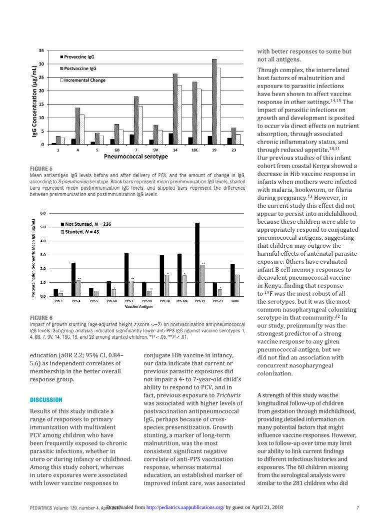
postimmunization can be seen in Fig 5. The strongest responses were to PPS 4, 7, 14, 18C, and 19.
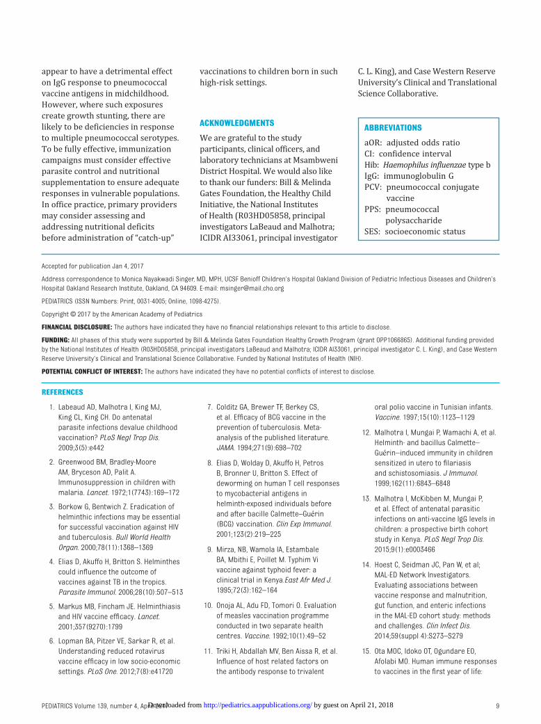
In our exploration of the associations between individual subject factors and the magnitude of postvaccination IgG responses, as expected, preimmunity to any specific antigen was associated with greater response to that antigen. Nasal carriage of S pneumoniae was not associated with differences in postvaccination antibody levels, nor did sex, younger age, anemia, severe anemia, or clinical wasting (BMI z score <−2) at the time of vaccination have significant associations with vaccine response outcomes. A prominent feature of reduced vaccine response was its association with growth stunting at the time of PCV. After vaccination, stunted children had significantly reduced geometric mean levels of anti-PPS IgG against strains 1, 4, 6B, 7, 9V, 14, 18C, 19, and 23 ( Fig 6 and Supplemental Information 3). Of note, children’s growth stunting was not associated with any concurrent parasitic infections (Supplemental Information 4). By contrast, Trichuris infection during infancy was associated with preimmunity to PPS
antigens 1, 5, 6B, 7, 9V, 14, 19, and 23 (Supplemental Information 5), and Trichuris infection at any point during infancy was associated with
significantly higher postvaccine IgG levels against antigens 1, 4, 5, 6B, 14, 18C, 19, and 23. Any previous maternal education was associated
5
TABLE 1 Characteristics of All Eligible Cohort Children and of the Participant and Nonparticipant Subgroups
Original 2006–
2010 Cohort
Current
Nonparticipants From
Original Cohort
Resurvey Participants
Enrolled
Completed Both
Study Visits and
Examinations
Not Fully IgG
Tested
Full Resurvey
Participation and
Laboratory Testing
N 547 162 385 341 60 281
Male: female ratio 1.2 1.5 1.1 1.1 1.2 1.0
Mean (SD) age at time of
2014 resurvey, y
6.2 (0.66) 6.3 (0.61)a 6.1 (0.67) 6.1 (0.66) 6.0 (0.63) 6.1 (0.67)
No formal maternal
education, %
36 43b 33 32 29 32
Lower SES at birth, % 59 60 59 58 53 59
Maternal parasitic
infections during
pregnancy, %
55 52 56 57 71c 54
Parasitic infections
detected during infancy
(≤36 mo), %
33 15b 41 41 43 40
Parasitic infections
detected at fi rst 2014
resurvey visit, %
— — 50 51 50 51
a Signifi cantly older than resurvey participant groups, P < .05 by analysis of variance and pairwise t testing.b Signifi cantly different from resurvey participants, by χ2 testing.c Signifi cantly different from fully tested subjects, P < .02.
TABLE 2 Study Participant Characteristics: Maternal Data From 2006–2010 Cohort Participation,
Infant Data at Delivery, and Participant Data From Current Study
Maternal characteristics (N = 545)
Maternal age at delivery, y
14–22 153 (28%)
23–30 210 (39%)
>30 87 (16%)
Unknown 95 (17%)
Education
None 100 (18%)
Lower primary 94 (17%)
Upper primary 263 (48%)
Secondary or more 86 (16%)
Unknown 2 (1%)
Household income (KSha per month)
<3000 322 (59%)
>3000 221 (40%)
Unknown 2 (1%)
Infant at delivery in 2007 (N = 545)
Male 288 (55%)
Female 236 (45%)
Average head circumference, cm 34.2 ± 1.4 (range 27.5–38)
Average length, cm 48.6 ± 2.5 (range 40–59.5)
Average weight, g 2949 ± 492 (range 1100–4350)
Children follow-up study in 2014 (N = 281 complete with data)
Average age, y 6.1 (range 4.8–7.9)
Female 138/281 (49%)
Mean hemoglobin, g/dL 9.7 (range 5.0–17.7)
Stunted (height-for-age z score <−2 SD below
median)
45/281 (16%)
Wasted (BMI <−2 SD below median) 14/281 (5%)
Current parasitic infections 144/281 (51%)
a 74 Kenya shillings = 1 US dollar in mid-2006; 82 Kenya shillings = 1 US dollar in mid-2010.
by guest on April 21, 2018http://pediatrics.aappublications.org/Downloaded from

NAYAKWADI SINGER et al
with improved responses to antigens 1, 4, 5, 9V, 18, and 19, although current SES and household monthly expenditure at the time of birth were not significant. Similarly, prenatal exposure to maternal parasitic infections and exposure to infection at the time of vaccination did not show a consistent association with IgG responses to PCV (Supplemental Information 6). As reported earlier, 13 hyporesponsiveness to Hib and diphtheria vaccines was noted in a subset of this cohort who had in utero exposure to malaria, hookworm, and filarial parasites.
However, there was no correlation between a child’s earlier responses to Hib during infancy and his or her ability to respond to pneumococcal antigens in the current study (Pearson R2 values for anti-Hib IgG antibody levels versus post-PCV anti-PPS IgG levels ranged from 0.00 to 0.011, not significant) (Supplemental Information 7).
In multivariable analysis, previous immunity to a serotype was consistently associated with a strong positive effect on serotype-specific postvaccination IgG levels
( Table 3). After adjustment for other potential modifying factors, the presence of growth stunting was independently associated with significantly decreased response to PPS serotypes 1, 4, 6B, 7, 9V, and 19. After adjustment, maternal education was associated with increased postvaccination IgG levels to PPS 4, 5, 7, and 19. Male sex was associated with an increased response to PPS 14, whereas younger age was associated with lowered response to the same antigen. With multiple adjustment for these factors, Trichuris infection during infancy remained independently associated with higher levels of postvaccination IgG against serotypes 1, 5, 6B, 7, 18C, and 23 ( Table 3). Not shown, additional multivariable analysis of the impact of infection exposure in utero or at the time of vaccination confirmed the significant associations of preimmunity, stunting, and maternal education with observed postvaccination IgG levels against multiple PPS antigens. However, there were no clear effects of parasite exposures at either of those time points on the children’s present-day pneumococcal vaccine responses.
In a global assessment of responsiveness to the PCV vaccination, by using an unsupervised, computer-based clustering analysis we identified a subgroup of 67 of 281 (24%) children who had consistently greater levels of IgG increments to multiple vaccine antigens ( Fig 7). Strongest weighting in this classification was based on responses to antigens 4, 18C, 7, 19, and 9V (in order of importance). Multivariable logistic regression for membership in the high responder category identified stunted status (adjusted odds ratio [aOR] 0.28; 95% CI, 0.10–0.83), Trichuris infection during infancy (aOR 2.0; 95% CI, 1.1–3.7), and maternal
6
FIGURE 3Prevalence of parasite exposures at 3 time points: in utero, during early infancy, and at midchildhood. Bars represent the percentage of participating children having S hematobium, hookworm, Trichuris trichiura, Ascaris lumbricoides, Strongyloides stercoralis, any STH, malaria, or fi laria (Wuchereria bancrofti). STH, soil-transmitted helminths (hookworm, Trichuris, Ascaris, or Strongyloides infection).
FIGURE 4Prevalence of signifi cant preimmunization anti–S pneumoniae IgG levels by vaccine serotype. The greatest frequency of vaccine-specifi c preimmunity in the Msambweni, Kenya cohort children was to pneumococcal serotypes 14, 19, 9V, 4, and 23.
by guest on April 21, 2018http://pediatrics.aappublications.org/Downloaded from
PEDIATRICS Volume 139 , number 4 , April 2017
education (aOR 2.2; 95% CI, 0.84–5.6) as independent correlates of membership in the better overall response group.
DISCUSSION
Results of this study indicate a range of responses to primary immunization with multivalent PCV among children who have been frequently exposed to chronic parasitic infections, whether in utero or during infancy or childhood. Among this study cohort, whereas in utero exposures were associated with lower vaccine responses to
conjugate Hib vaccine in infancy, our data indicate that current or previous parasitic exposures did not impair a 4- to 7-year-old child’s ability to respond to PCV, and in fact, previous exposure to Trichuris was associated with higher levels of postvaccination antipneumococcal IgG, perhaps because of cross-species presensitization. Growth stunting, a marker of long-term malnutrition, was the most consistent significant negative correlate of anti-PPS vaccination response, whereas maternal education, an established marker of improved infant care, was associated
with better responses to some but not all antigens.
Though complex, the interrelated host factors of malnutrition and exposure to parasitic infections have been shown to affect vaccine response in other settings. 14, 15 The impact of parasitic infections on growth and development is posited to occur via direct effects on nutrient absorption, through associated chronic inflammatory status, and through reduced appetite. 18, 31 Our previous studies of this infant cohort from coastal Kenya showed a decrease in Hib vaccine response in infants when mothers were infected with malaria, hookworm, or filaria during pregnancy.13 However, in the current study this effect did not appear to persist into midchildhood, because these children were able to appropriately respond to conjugated pneumococcal antigens, suggesting that children may outgrow the harmful effects of antenatal parasite exposure. Others have evaluated infant B cell memory responses to decavalent pneumococcal vaccine in Kenya, finding that response to 19F was the most robust of all the serotypes, but it was the most common nasopharyngeal colonizing serotype in that community. 32 In our study, preimmunity was the strongest predictor of a strong vaccine response to any given pneumococcal antigen, but we did not find an association with concurrent nasopharyngeal colonization.
A strength of this study was the longitudinal follow-up of children from gestation through midchildhood, providing detailed information on many potential factors that might influence vaccine responses. However, loss to follow-up over time may limit our ability to link current findings to different infectious histories and exposures. The 60 children missing from the serological analysis were similar to the 281 children who did
7
FIGURE 5Mean antiantigen IgG levels before and after delivery of PCV, and the amount of change in IgG, according to S pneumoniae serotype. Black bars represent mean preimmunization IgG levels, shaded bars represent mean postimmunization IgG levels, and stippled bars represent the difference between preimmunization and postimmunization IgG levels.
FIGURE 6Impact of growth stunting (age-adjusted height z score <−2) on postvaccination antipneumococcal IgG levels. Subgroup analysis indicated signifi cantly lower anti-PPS IgG against vaccine serotypes 1, 4, 6B, 7, 9V, 14, 18C, 19, and 23 among stunted children. *P < .05, **P < .01.
by guest on April 21, 2018http://pediatrics.aappublications.org/Downloaded from

NAYAKWADI SINGER et al
have complete data in terms of their age, sex, maternal and household factors, and current infection status, with 1 exception; more of their mothers had had infections during pregnancy. Loss of this group may have led to an underestimation of the impact of prenatal infections on the observed pneumococcal vaccine responses. The loss of 162 cohort participants between birth and the 2014 study reflected emigration and some mortality but also early dropouts due to parental refusal to continue in the cohort. Because more of their mothers were not formally educated, we believe that their loss may have masked the impact of this and other environmental factors in our analysis. Because fewer of these children had regular follow-up in infancy, the impact of infections during infancy may have been underestimated in our study. Furthermore, the intensity of helminth infections in our cohort was classified as “light” infection, and previous authors have suggested that greater infection intensity may have greater impact on growth and development. 33 Thus, another factor in our study may have diluted a measurable effect of parasitosis and vaccine responsiveness.
CONCLUSIONS
Chronic parasitic exposure or infection during early life does not
8
TABL
E 3
Mu
ltip
ly A
dju
sted
Eff
ects
of
Pre
imm
un
ity,
Gro
wth
Stu
nti
ng,
Hou
seh
old
Fac
tors
, an
d P
ast
Exp
osu
res
to P
aras
itic
Infe
ctio
n o
n P
ostv
acci
nat
ion
log 10
(Ig
G)
Leve
ls A
gain
st In
div
idu
al P
CV
Anti
gen
s
Cov
aria
teP
PS
1P
PS
4P
PS
5P
PS
6B
PP
S 7
PP
S 9
VP
PS
14
PP
S 1
8CP
PS
19
PP
S 2
3C
RM
Pre
viou
s
imm
un
ity
to
anti
gen
1.4
(0.8
4 to
1.9)
*
1.16
(0.
9 to
1.4)
*
1.52
(0.
95 t
o
2.1)
*
1.15
(0.
83 t
o
1.5)
*
1.10
(0.
77 t
o
1.4)
*
1.35
(1.
1 to
1.6)
*
1.13
(0.
98 t
o
1.3)
*
1.24
(0.
87 t
o
1.6)
*
1.23
(1.
0 to
1.4)
*
1.11
(0.
84 t
o
1.4)
*
1.05
(0.
9 to
1.2
)*
Stu
nti
ng
−0.3
5 (−
0.61
to −
0.97
)*
−0.2
6 (−
0.51
to 0
.00)
*
−0.3
3 (−
0.58
to −
0.08
)*
−0.4
3 (−
0.7
to
-0.1
6)*
−0.2
9 (−
0.53
to −
0.04
)*
−0.2
7 (−
0.56
to 0
.03)
−0.2
2 (−
0.43
to −
0.00
4)*
−0.2
3 (−
0.48
to 0
.015
)
−0.1
8 (−
0.40
to
0.03
)
Mat
ern
al
edu
cati
on
0.10
(−0
.01,
0.20
)
0.11
(0.
001
to
0.21
)*
0.12
(0.
02 t
o
0.22
)*
0.15
(0.
04 t
o
0.26
)*
0.23
(−0
.02
to
0.48
)
0.26
(−0
.04
to
0.56
)
0.27
(0.
05 t
o
0.48
)*
Mal
e se
x0.
18 (
−0.0
1 to
0.37
)
0.16
(0.
02 t
o
0.32
)*
0.17
(−0
.02
to
0.35
)
You
nge
r ag
e
grou
p
−0.1
6 (−
0.32
to −
0.01
)*
−0.1
8 (−
0.37
to 0
.01)
Tric
hu
ris
by
age
36 m
o
0.45
(0.
15 t
o
0.74
)*
0.28
(−0
.14
to
0.58
)
0.47
(0.
18 t
o
0.76
)*
0.31
(0.
02 t
o
0.60
)*
0.32
(0.
02 t
o
0.62
)*
0.28
(−0
.01
to
0.56
)
0.39
(0.
05 t
o
0.73
)*
0.23
(−0
.02
to
0.47
)
0.52
(0.
24 t
o
0.80
)*
Asca
ris
by
age
36 m
o
0.47
(−0
.03
to
0.96
)
0.37
(−0
.03
to
0.78
)
0.70
(0.
11 t
o
1.3)
*
Fila
ria
by
age
36 m
o
0.31
(−0
.01
to
0.62
)
0.41
(−0
.05
to
0.86
)
0.52
(0.
32 t
o
0.90
)*
Mal
aria
by
age
36 m
o
−0.3
1 (−
0.52
to −
0.09
)*
−0.2
6 (−
0.51
to −
0.01
)*
Hoo
kwor
m b
y
36 m
o
−0.2
4 (−
0.48
to −
0.01
)*
S h
aem
ato
biu
m
by
age
36 m
o
0.54
(0.
0 to
1.1)
*
Par
enth
eses
ind
icat
e th
e 95
% C
Is f
or t
he
effe
ct s
ize.
* Ad
just
ed P
val
ue
is <
.05.
FIGURE 7Differences between high- and low-responder subgroups identifi ed by cluster analysis of anti-PPS response across all serotypes. Shown are representative changes to 18C antigen before to after a single dose of 10-valent PCV. Children in the high-response group (N = 67) are shown in color. The low-response group (N = 214) is shown in black.
by guest on April 21, 2018http://pediatrics.aappublications.org/Downloaded from
PEDIATRICS Volume 139 , number 4 , April 2017
appear to have a detrimental effect on IgG response to pneumococcal vaccine antigens in midchildhood. However, where such exposures create growth stunting, there are likely to be deficiencies in response to multiple pneumococcal serotypes. To be fully effective, immunization campaigns must consider effective parasite control and nutritional supplementation to ensure adequate responses in vulnerable populations. In office practice, primary providers may consider assessing and addressing nutritional deficits before administration of “catch-up”
vaccinations to children born in such high-risk settings.
ACKNOWLEDGMENTS
We are grateful to the study participants, clinical officers, and laboratory technicians at Msambweni District Hospital. We would also like to thank our funders: Bill & Melinda Gates Foundation, the Healthy Child Initiative, the National Institutes of Health (R03HD05858, principal investigators LaBeaud and Malhotra; ICIDR AI33061, principal investigator
C. L. King), and Case Western Reserve University’s Clinical and Translational Science Collaborative.
REFERENCES
1. Labeaud AD, Malhotra I, King MJ,
King CL, King CH. Do antenatal
parasite infections devalue childhood
vaccination? PLoS Negl Trop Dis.
2009;3(5):e442
2. Greenwood BM, Bradley-Moore
AM, Bryceson AD, Palit A.
Immunosuppression in children with
malaria. Lancet. 1972;1(7743):169–172
3. Borkow G, Bentwich Z. Eradication of
helminthic infections may be essential
for successful vaccination against HIV
and tuberculosis. Bull World Health
Organ. 2000;78(11):1368–1369
4. Elias D, Akuffo H, Britton S. Helminthes
could infl uence the outcome of
vaccines against TB in the tropics.
Parasite Immunol. 2006;28(10):507–513
5. Markus MB, Fincham JE. Helminthiasis
and HIV vaccine effi cacy. Lancet.
2001;357(9270):1799
6. Lopman BA, Pitzer VE, Sarkar R, et al.
Understanding reduced rotavirus
vaccine effi cacy in low socio-economic
settings. PLoS One. 2012;7(8):e41720
7. Colditz GA, Brewer TF, Berkey CS,
et al. Effi cacy of BCG vaccine in the
prevention of tuberculosis. Meta-
analysis of the published literature.
JAMA. 1994;271(9):698–702
8. Elias D, Wolday D, Akuffo H, Petros
B, Bronner U, Britton S. Effect of
deworming on human T cell responses
to mycobacterial antigens in
helminth-exposed individuals before
and after bacille Calmette–Guérin
(BCG) vaccination. Clin Exp Immunol.
2001;123(2):219–225
9. Mirza, NB, Wamola IA, Estambale
BA, Mbithi E, Poillet M. Typhim Vi
vaccine against typhoid fever: a
clinical trial in Kenya.East Afr Med J.
1995;72(3):162–164
10. Onoja AL, Adu FD, Tomori O. Evaluation
of measles vaccination programme
conducted in two separate health
centres. Vaccine. 1992;10(1):49–52
11. Triki H, Abdallah MV, Ben Aissa R, et al.
Infl uence of host related factors on
the antibody response to trivalent
oral polio vaccine in Tunisian infants.
Vaccine. 1997;15(10):1123–1129
12. Malhotra I, Mungai P, Wamachi A, et al.
Helminth- and bacillus Calmette–
Guérin–induced immunity in children
sensitized in utero to fi lariasis
and schistosomiasis. J Immunol.
1999;162(11):6843–6848
13. Malhotra I, McKibben M, Mungai P,
et al. Effect of antenatal parasitic
infections on anti-vaccine IgG levels in
children: a prospective birth cohort
study in Kenya. PLoS Negl Trop Dis.
2015;9(1):e0003466
14. Hoest C, Seidman JC, Pan W, et al;
MAL-ED Network Investigators.
Evaluating associations between
vaccine response and malnutrition,
gut function, and enteric infections
in the MAL-ED cohort study: methods
and challenges. Clin Infect Dis.
2014;59(suppl 4):S273–S279
15. Ota MOC, Idoko OT, Ogundare EO,
Afolabi MO. Human immune responses
to vaccines in the fi rst year of life:
9
ABBREVIATIONS
aOR: adjusted odds ratioCI: confidence intervalHib: Haemophilus influenzae type bIgG: immunoglobulin GPCV: pneumococcal conjugate
vaccinePPS: pneumococcal
polysaccharideSES: socioeconomic status
Accepted for publication Jan 4, 2017
Address correspondence to Monica Nayakwadi Singer, MD, MPH, UCSF Benioff Children’s Hospital Oakland Division of Pediatric Infectious Diseases and Children’s
Hospital Oakland Research Institute, Oakland, CA 94609. E-mail: [email protected]
PEDIATRICS (ISSN Numbers: Print, 0031-4005; Online, 1098-4275).
Copyright © 2017 by the American Academy of Pediatrics
FINANCIAL DISCLOSURE: The authors have indicated they have no fi nancial relationships relevant to this article to disclose.
FUNDING: All phases of this study were supported by Bill & Melinda Gates Foundation Healthy Growth Program (grant OPP1066865). Additional funding provided
by the National Institutes of Health (R03HD05858, principal investigators LaBeaud and Malhotra; ICIDR AI33061, principal investigator C. L. King), and Case Western
Reserve University’s Clinical and Translational Science Collaborative. Funded by National Institutes of Health (NIH).
POTENTIAL CONFLICT OF INTEREST: The authors have indicated they have no potential confl icts of interest to disclose.
by guest on April 21, 2018http://pediatrics.aappublications.org/Downloaded from
NAYAKWADI SINGER et al
biological, socio-economic and
ethical issues—a viewpoint. Vaccine.
2013;31(21):2483–2488
16. Qadri F, Bhuiyan TR, Sack DA,
Svennerholm AM. Immune responses
and protection in children in
developing countries induced by oral
vaccines. Vaccine. 2013;31(3):452–460
17. O’Brien KL, Wolfson LJ, Watt JP, et al;
Hib and Pneumococcal Global Burden
of Disease Study Team. Burden of
disease caused by Streptococcus
pneumoniae in children younger than
5 years: global estimates. Lancet.
2009;374(9693):893–902
18. LaBeaud AD, Nayakwadi Singer M,
McKibben M, et al. Parasitism in
children aged three years and under:
relationship between infection and
growth in rural coastal Kenya. PLoS
Negl Trop Dis. 2015;9(5):e0003721
19. Dagan R, Givon-Lavi N, Greenberg D,
Fritzell B, Siegrist CA. Nasopharyngeal
carriage of Streptococcus pneumoniae
shortly before vaccination with
a pneumococcal conjugate
vaccine causes serotype-specifi c
hyporesponsiveness in early infancy.
J Infect Dis. 2010;201(10):1570–1579
20. Gamboa MI, Navone GT, Orden AB,
Torres MF, Castro LE, Oyhenart EE.
Socio-environmental conditions,
intestinal parasitic infections
and nutritional status in children
from a suburban neighborhood
of La Plata, Argentina. Acta Trop.
2011;118(3):184–189
21. Peters PAS, Mahmoud AAF, Warren KS,
Ouma JH, Siongok TKA. Field studies of
a rapid, accurate means of quantifying
Schistosoma haematobium eggs
in urine samples. Bull World Health
Organ. 1976;54(2):159–162
22. DuVall AS, Fairley JK, Sutherland L, et al.
Development of a specimen-sparing
multichannel bead assay to detect
antiparasite IgG4 for the diagnosis
of Schistosoma and Wuchereria
infections on the coast of Kenya. Am J
Trop Med Hyg. 2014;90(4):638–645
23. Malhotra I, Dent A, Mungai P, Muchiri
E, King CL. Real-time quantitative
PCR for determining the burden of
Plasmodium falciparum parasites
during pregnancy and infancy. J Clin
Microbiol. 2005;43(8):3630–3635
24. Scott JA, Ojal J, Ashton L, Muhoro A,
Burbidge P, Goldblatt D. Pneumococcal
conjugate vaccine given shortly
after birth stimulates effective
antibody concentrations and primes
immunological memory for sustained
infant protection. Clin Infect Dis.
2011;53(7):663–670
25. Synfl orix [package insert]. Version
GDS03/WHO Insert02 ed. Geneva,
Switzerland: World Health Organization;
2011:1–9
26. Whaley MJ, Rose C, Martinez J, et al.
Interlaboratory comparison of three
multiplexed bead-based immunoassays
for measuring serum antibodies to
pneumococcal polysaccharides. Clin
Vaccine Immunol. 2010;17(5):862–869
27. Schlottmann SA, Jain N, Chirmule
N, Esser MT. A novel chemistry
for conjugating pneumococcal
polysaccharides to Luminex
microspheres. J Immunol Methods.
2006;309(1–2):75–85
28. Goldblatt D, Plikaytis BD, Akkoyunlu M,
et al. Establishment of a new human
pneumococcal standard reference
serum, 007sp. Clin Vaccine Immunol.
2011;18(10):1728–1736
29. da Gloria Carvalho M, Pimenta
FC, Jackson D, et al. Revisiting
pneumococcal carriage by use of
broth enrichment and PCR techniques
for enhanced detection of carriage
and serotypes. J Clin Microbiol.
2010;48(5):1611–1618
30. Antonio M, Hakeem I, Sankareh K,
Cheung YB, Adegbola RA. Evaluation
of sequential multiplex PCR for direct
detection of multiple serotypes of
Streptococcus pneumoniae from
nasopharyngeal secretions. J Med
Microbiol. 2009;58(Pt 3):296–302
31. Stephenson LS, ed. Impact of Helminth
Infections on Human Nutrition. New
York, NY: Taylor & Francis; 1987:47–48
32. Muema DM, Nduati EW, Uyoga M, et al.
10-valent pneumococcal non-typeable
Haemophilus infl uenzae protein-D
conjugate vaccine (PHiD-CV) induces
memory B cell responses in healthy
Kenyan toddlers. Clin Exp Immunol.
2015;181(2):297–305
33. Oberhelman RA, Guerrero ES,
Fernandez ML, et al. Correlations
between intestinal parasitosis,
physical growth, and psychomotor
development among infants and
children from rural Nicaragua. Am
J Trop Med Hyg. 1998;58(4):470–475
10 by guest on April 21, 2018http://pediatrics.aappublications.org/Downloaded from

originally published online March 16, 2017; Pediatrics Malhotra, Charles H. King and A. Desirée LaBeaud
Francis M. Mutuku, David Vu, Dunstan Mukoko, Christopher L. King, Indu J. Monica Nayakwadi Singer, Claire Heath, Jackson Muinde, Virginia Gildengorin,
Infancy, or Mid-ChildhoodPneumococcal Vaccine Response After Exposure to Parasites in Utero, in
ServicesUpdated Information &
016-2781http://pediatrics.aappublications.org/content/early/2017/03/14/peds.2including high resolution figures, can be found at:
Supplementary Material
016-2781.DCSupplementalhttp://pediatrics.aappublications.org/content/suppl/2017/03/14/peds.2Supplementary material can be found at:
References
016-2781.full#ref-list-1http://pediatrics.aappublications.org/content/early/2017/03/14/peds.2This article cites 31 articles, 7 of which you can access for free at:
Subspecialty Collections
munization_subhttp://classic.pediatrics.aappublications.org/cgi/collection/vaccine:imVaccine/Immunizationdiseases_subhttp://classic.pediatrics.aappublications.org/cgi/collection/infectious_Infectious Diseasefollowing collection(s): This article, along with others on similar topics, appears in the
Permissions & Licensing
https://shop.aap.org/licensing-permissions/in its entirety can be found online at: Information about reproducing this article in parts (figures, tables) or
Reprintshttp://classic.pediatrics.aappublications.org/content/reprintsInformation about ordering reprints can be found online:
. ISSN:60007. Copyright © 2017 by the American Academy of Pediatrics. All rights reserved. Print
American Academy of Pediatrics, 141 Northwest Point Boulevard, Elk Grove Village, Illinois,has been published continuously since . Pediatrics is owned, published, and trademarked by the Pediatrics is the official journal of the American Academy of Pediatrics. A monthly publication, it
by guest on April 21, 2018http://pediatrics.aappublications.org/Downloaded from

originally published online March 16, 2017; Pediatrics Malhotra, Charles H. King and A. Desirée LaBeaud
Francis M. Mutuku, David Vu, Dunstan Mukoko, Christopher L. King, Indu J. Monica Nayakwadi Singer, Claire Heath, Jackson Muinde, Virginia Gildengorin,
Infancy, or Mid-ChildhoodPneumococcal Vaccine Response After Exposure to Parasites in Utero, in
http://pediatrics.aappublications.org/content/early/2017/03/14/peds.2016-2781located on the World Wide Web at:
The online version of this article, along with updated information and services, is
. ISSN:60007. Copyright © 2017 by the American Academy of Pediatrics. All rights reserved. Print
American Academy of Pediatrics, 141 Northwest Point Boulevard, Elk Grove Village, Illinois,has been published continuously since . Pediatrics is owned, published, and trademarked by the Pediatrics is the official journal of the American Academy of Pediatrics. A monthly publication, it
by guest on April 21, 2018http://pediatrics.aappublications.org/Downloaded from


![University of Groningen Pneumococcal conjugate vaccines ... › ws › files › 67431176 › Fortanier_et... · [Intervention Review] Pneumococcal conjugate vaccines for preventing](https://static.fdocuments.us/doc/165x107/5f03b7c17e708231d40a6d8d/university-of-groningen-pneumococcal-conjugate-vaccines-a-ws-a-files-a.jpg)