Pizza, polyps and pancreas - KRH• 8 week course of PPI is therapy of choice for symptom relief and...
62
PIZZA, POLYPS AND PANCREAS MARCH 7, 2017
Transcript of Pizza, polyps and pancreas - KRH• 8 week course of PPI is therapy of choice for symptom relief and...

PIZZA, POLYPS AND PANCREAS
MARCH 7, 2017

GERD/BARRETT’S/CANCER: STOPPING THE FREIGHT TRAIN OF DYSPEPSIA TO DISASTER
WILLIAM J. COBELL, MD


OUTLINE:
• GERD
• Epidemiology of GERD
• Risk factors of GERD
• Diagnosis and treatment of GERD
• Complications of GERD
• Barrett’s esophagus
• Risk factors for Barrett’s esophagus
• Diagnosis and treatment of Barrett’s esophagus
• Case studies

GERD DEFINED:
• Difficult to develop a standard definition for GERD, threshold of distinction between physiologic reflux and reflux disease is arbitrary
• Condition which develops when the reflux of stomach contents into the esophagus or beyond causes troubling symptoms and/or complications
• Symptoms are troublesome if they adversely affect an individual’s well-being
• GERD is further categorized into 2 types:
• Symptoms without erosions on endoscopic examination (non erosive disease or NERD)
• Symptoms with erosions present on endoscopy (ERD)

EPIDEMIOLOGY:
• Prevalence of GERD 10-20% of the Western world (lower prevalence in Asia), increasing
• 2/3 of adult population in US have had symptoms of GERD at some point
• Clinically troublesome heartburn in 6% of the population
• Patients with disruptive GERD symptoms have an increase in time off work and decrease in work productivity
• Lower scores on sleep scales compared to patients with less frequent symptoms
• Decrease in physical functioning
• Nocturnal GERD with greater impact on QOL compared to day time symptoms

COST OF GERD:
• According to the Centers for Disease Control and Prevention (CDC):
• GERD is the most common GI-related diagnosis
• Represents 8.9 million patient visits annually
• Outpatient upper endoscopy costs the healthcare system $32.4 billion annually
• According to the American College of Gastroenterology:
• GERD symptoms cost the U.S. nearly $2 billion each week in lost productivity

RISK FACTORS:
• Obesity, increased waist circumference
• Weight gain
• Tobacco use
• Alcohol use
• Pregnancy
• Connective tissue disorders (ie; scleroderma)
• Diabetes mellitus/Gastroparesis
• Medications, herbal supplements

PATHOPHYSIOLOGY:
• Impaired esophageal clearance
• Primary peristalsis failure resulting in impaired esophageal clearance
• Impaired mucosal defense
• LES incompetence in preventing retrograde passage of gastroduodenal contents
• Transient inappropriate LES relaxations
• Hiatal hernia
• Delayed gastric emptying

DIAGNOSIS:
• Symptom presentation:
• Heartburn and regurgitation most reliable to predict erosive esophagitis
• 30-76% sensitivity, 62-96% specificity
• Response to antisecretory therapy:
• 78% sensitivity, 54% specificity
• Objective testing with endoscopy
• Presence of Barrett’s esophagus or erosive esophagitis
• Ambulatory reflux monitoring
• pH Bravo

DIAGNOSIS:
• Presumptive diagnosis based on typical symptoms of heartburn and regurgitation
• Empiric therapy with PPI is recommended for typical symptoms of GERD
• Patients with chest pain suspected due to GERD should have full cardiac evaluation before institution of therapy or endoscopic investigation
• Barium radiographs should not be performed to diagnose GERD
• Routine distal esophageal biopsies should not be performed to diagnose GERD
• Esophageal manometry has no role in the diagnosis of GERD
• Endoscopy is not required to diagnose GERD, but recommended if alarm symptoms

PATHOLOGY: • Basal cell hyperplasia
• Mucosal eosinophils
• Papilla < 50% epithelial thickness
• Normal esophagus

ENDOSCOPY:

TREATMENT:
• Weight loss if overweight or have recent weight gain
• Lifestyle modification
• Elevate HOB 30 degrees
• Avoid eating 2-3 hours before bedtime
• Alcohol/tobacco cessation (weak evidence)
• Avoiding trigger foods (selective elimination)
• Routine global elimination of food that can trigger reflux (caffeine, chocolate, alcohol, acidic/spicy foods, etc) is not recommended

PPI THERAPY:
• 8 week course of PPI is therapy of choice for symptom relief and healing of esophagitis
• No major differences in efficacy of different PPIs
• PPI therapy should be initiated at once daily dosing, before 1st meal of day
• If partial response: dose can be increased vs BID dosing vs switch to different PPI
• PPIs are safe in pregnancy if clinically indicated (omeprazole class C, other PPIs class B)
• Non-responders to PPI should be referred for evaluation

OTHER MEDICATIONS:
• H2 receptor antagonist may be used as maintenance option in patients without erosive esophagitis, if they experience heartburn relief
• Can be added at night to daytime PPI therapy for refractory nocturnal symptoms
• H2 receptor antagonists associated with tachyphylaxis after only a short time of use
• Prokinetic therapy and/or baclofen should not be used in GERD patients without diagnostic evaluation
• No role for carafate in the non-pregnant GERD patient

ANTI-REFLUX SURGERY:
• Surgical therapy is an option for long-term therapy in GERD patients
• Surgical therapy is generally not recommended in patients who do not respond to PPI
• Pre-operative pH monitoring mandatory in patients w/o evidence of erosive esophagitis
• All patients should undergo pre-operative manometry to rule-out achalasia
• Surgical therapy as effective as medical therapy for carefully selected patients with chronic GERD when performed by an experienced surgeon
• Obese patients should be considered for bariatric surgery (gastric bypass preferred option)
• Current endoscopic txt or transoral incisionless fundoplication not recommended at this time

INDICATIONS FOR LONG TERM PPI USE:
• Patients who have symptoms after PPI is discontinued, refractory (abnormal pH study)
• Patients with acid related disorders, and/or complications:
• Erosive esophagitis
• Barrett’s esophagus
• Zollinger-Ellison syndrome
• Idiopathic ulcers
• Prophylaxis against bleeding in select patients
• Should be administered at lowest effective dose

POTENTIAL RISKS OF PPI USE:
• Consider switching PPI in the setting side effects (HA, diarrhea, dyspepsia, etc)
• Patients with known osteoporosis can remain on PPI
• PPI can be risk factor for C diff infection, should be used with care in patients at risk
• Short-term PPI use may increase risk of CAP, risk does not appear to be elevated in long term users
• PPI use does not need to be altered with concomitant clopidogrel (Plavix) use, does not appear to be increased risk for adverse cardiovascular events
• Various other risks recently in the media, weak observational or retrospective data:
• kidney failure (retrospective, VA), dementia (insurance data, Germany), Alzheimers, etc.

COMPLICATIONS:
• Erosive esophagitis
• LA classification system (grade A, B, C, D)
• Repeat EGD should be performed if moderate to severe esophagitis is observed (LA grade B, C or D) to exclude underlying Barrett’s esophagus
• Esophageal stricture/ring
• Treatment with PPI recommended after dilation of peptic stricture or Schatzki’s ring
• Barrett’s esophagus
• Screening for Barrett’s esophagus should be considered in patients with GERD who are at high risk based on epidemiological profile (male, age > 50, obese, FH Barrett’s/esophageal CA, chronicity, etc.)


BARRETT’S ESOPHAGUS DEFINED:
• Presence of > 1cm of metaplastic columnar epithelium that replaces the stratified squamous epithelium normally lining the distal esophagus
• Segments < 1cm are classified as “specialized intestinal metaplasia of the GEJ” and not Barrett’s due to high interobserver variability and low risk of cancer (EAC)
• Presence of intestinal metaplasia (IM) has traditionally been a requirement for diagnosis of Barrett’s esophagus in the US
• Guidelines from the UK suggest Barrett’s diagnosis with the presence of columnar metaplasia, regardless of the presence of IM
• Large population studies have demonstrated a substantially lower EAC risk in patients with columnar metaplasia without IM compared to those with IM

STRATIFIED SQUAMOUS (NORMAL ESOPHAGUS)
COLUMNAR EPITHELIUM WITH GOBLET CELLS (INTESTINAL METAPLASIA)

ANATOMY:
• Location of the GEJ:
• Where distal extent of tubular esophagus is in contact with proximal extent of the gastric folds
• Location of the proximal extent of the gastric folds can be affected by respiration, air insufflation during endoscopy and esophageal/gastric motility
• Diaphragmatic hiatus is identified as an indentation of the gastric folds that is apparent during upper endoscopy with respiration
• Barrett’s segment is described as long segment (> 3cm) or short segment (< 3cm)
• The extent of metaplastic change is described using the Prague classification


DIAGNOSIS:
• BE should be ruled-out when there is extension of salmon-colored mucosa into the tubular esophagus extending > 1cm proximal to the GEJ
• If BE is suspected on EGD: • At least 8 random biopsies should be obtained
• In patients with short segments (1-2cm), at least 4 biopsies per cm of circumferential BE and 1 biopsy per tongue of BE, should be obtained
• In patients with EGD findings highly suspicious for BE but lack of IM on histology, a repeat endoscopy should be considered in 1-2 years to rule-out BE

BARRETT’S ESOPHAGUS:
• IM of the cardia is very common, described in up to 20% of asymptomatic patients on routine upper endoscopy
• IM of the cardia is not more common in BE patients compared to controls
• Natural history of IM at the GEJ is associated with H pylori infection
• IM at the GEJ is not associated with EAC
• Takehome: Biopsy of the normal or irregular GEJ is not recommended

RISK FACTORS FOR BE:
• Chronic GERD symptoms (> 5 years) – BE detected in 15% of patients with chronic GERD
• Age > 50 years old
• Male gender
• Tobacco use
• Central obesity
• Caucasian
• BE is more common in 1st degree relatives of subjects with known BE
• Alcohol does not increase risk of BE, and wine may be protective
RISK FACTORS ASSOCIATED WITH DYSPLASIA/EAC:
• Advancing age
• Increasing length of BE
• Central obesity
• Tobacco usage
• Lack of NSAID use
• Lack of PPI use
• Lack of statin use


WHY DO WE CARE:
• EAC:
• Rising mortality
• Dismal 5-year survival rate
(10-15%)
• 50% of patients have incurable
disease at presentation

SCREENING FOR BE:
• Men with chronic GERD symptoms (>5 yrs) AND/OR frequent symptoms AND 2 or more risk factors (age > 50, Caucasian, central obesity, smoker, FH of BE or EAC)
• Given substantially lower risk of EAC in women with chronic GERD symptoms (compared to males), screening for BE in women is not recommended, should be considered on an individual basis
• Screening the general population is not recommended
• Before screening for BE, overall life expectancy should be considered and subsequent implications (such as need for periodic endoscopy and therapy) should be discussed

ALTERNATIVE SCREENING METHODS:
• Unsedated transnasal endoscopy (uTNE) can be considered as an alternative to conventional EGD (disposable sheath, non-physician providers can perform)
• Cytosponge is a novel gelatin-coated sponge attached to a string that expands to a sphere when swallowed, obtaining esophageal cytology samples (not prime time yet)
• Video capsule endoscopy of the esophagus?
• If initial EGD is negative for BE, repeating EGD for evaluation of BE is not recommended
• If esophagitis is observed (LA grade B, C or D), repeat endoscopy after PPI therapy x 8 weeks is recommended to rule-out underlying BE

RISK OF EAC IN BE, BASED ON DYSPLASIA:
• Nondysplastic BE: ~0.2 – 0.5% per year
• Low grade dysplasia (LGD): ~0.7% per year (1.7%/year when HGD included)
• High grade dysplasia (HGD): ~7% per year
• Majority of patients (> 90%) diagnosed with BE die of causes other than EAC

SURVEILLANCE:
• Before entering a surveillance program, patients should be counseled regarding the risks/benefits, including the limitations of surveillance endoscopy, as well as adhering to appropriate surveillance intervals
• Other considerations include: • Age
• Likelihood of survival over next 5 years
• Ability to tolerate interventions including endoscopic therapy, surgery, chemo/radiation
• Given the low risk of progression to cancer for most patients with BE and the data that most patients die of causes other than EAC, such counseling is important to help patients make an informed decision regarding therapeutic options

SURVEILLANCE:
• Goal of surveillance is detection of dysplasia
• Utilize high resolution, high definition, white light endoscopy
• Narrow band imaging (NBI) helpful
• Other techniques (methylene blue, acetic acid, indigo carmine, autoflourescence, confocal laser endomicroscopy, spectroscopy, molecular imaging, etc), interesting but not ready for prime time
• “Barrett’s inspection time” is key
• Includes retroflexed view of the GEJ
• Careful attention to right hemisphere of the segment (early cancer has predilection to develop)
• Biopsies should only be taken after active inflammation has been controlled with PPI therapy
• Standard: four quadrant biopsies at 2cm intervals along entire length of Barrett’s segment
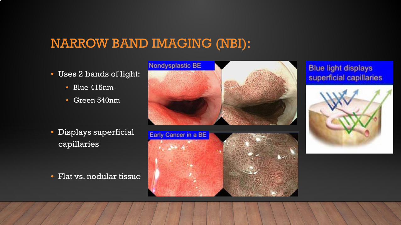
NARROW BAND IMAGING (NBI):
• Uses 2 bands of light:
• Blue 415nm
• Green 540nm
• Displays superficial capillaries
• Flat vs. nodular tissue

FUTURE:
• Confocal laser endomicroscopy
• Obtain real time in vivo histology during endoscopy

SURVEILLANCE:
• For BE without dysplasia: • Endoscopic surveillance every 3-5 years
• Patients diagnosed with BE on initial exam do not require a repeat endoscopy in 1 year
• Should receive once daily PPI, routine BID dosing is not recommended unless symptoms require
• Aspirin/NSAIDs should not be routinely prescribed to patients with BE as anti-neoplastic strategy
• Endoscopic ablative therapies should not be routinely applied to patients with nondysplastic BE
• For BE with dysplasia of any grade, review by 2 pathologists, at least one of whom has specialized experience in GI pathology is warranted (intraobserver variability)

THERAPY:
• For BE indefinite for dysplasia: • Repeat EGD after optimization of PPI for 3-6 months should be performed
• If indefinite for dysplasia is confirmed, then surveillance interval of 12 months is recommended
• BE with confirmed LGD: • Endoscopic therapy is the preferred treatment modality (radio frequency ablation – RFA)
• Endoscopic surveillance every 12 months is an acceptable alternative
• BE with confirmed HGD: • Endoscopic therapy is recommended (RFA)
• Endoscopic surveillance only if they have life-limiting comorbidity


THERAPY:
• Patients with nodularity within BE segment should undergo endoscopic mucosal resection (EMR) of the nodular lesion
• Histologic assessment of the EMR specimen will guide further therapy • Specimens with HGD or intramucosal carcinoma should be treated with endoscopic ablative
therapy (RFA) of the remaining BE
• Specimens demonstrating neoplasia at a deep margin should be assumed to have residual neoplasia and surgical, systemic or additional endoscopic therapies should be considered
• Patients with T1a EAC, endoscopic ablative therapy is preferred and effective
• Patients with T1b EAC, should be brought before tumor board before further endoscopic strategies are implemented (T1b sm1 can be considered for EMR, also consider functional status, etc)


SURGICAL THERAPY:
• Anti-reflux surgery should not be pursued in patients with BE as an anti-neoplastic measure
• Anti-reflux surgery can be considered in those patients with incomplete control of reflux symptoms, who are on optimized medical therapy
• Cases of EAC with invasion into the submucosa, especially mid – deep submucosa (T1b, sm2-3), esophagectomy and possibly neoadjuvant therapy is recommended
• Patients with T1a or T1b sm1 disease, with poor differentiation, lymphovascular invasion, or incomplete EMR should prompt consideration of surgical intervention

THERAPY:
• Radio frequency ablation (RFA)

THERAPY:
• Endoscopic mucosal resection (EMR)

CASE #1: JS
• 69 year old Caucasian Male with a long history of GERD
• Former smoker, BMI is 30 (central distribution), drinks 3-4 times/week
• No family history of esophageal or gastric CA
• Diagnosed with non-dysplastic Barrett’s esophagus in 2012, confirmed in 2013
• Surveillance EGD in 2016 was positive for Barrett’s with HGD, confirmed by 2nd pathologist at UW
• Referred to me for RFA


• LONG SEGMENT BARRETT’S ESOPHAGUS – C4M9
• 2 NODULES OBSERVED – EMR PERFORMED
• PATHOLOGY REVEALED HGD WITH NEGATIVE MARGINS

CASE #1: JS
• Has undergone 3 RFA treatments
• No major complications
CASE #2: NN
• 81 year old Caucasian Male with a long history of GERD
• Former smoker, drinks 1-2 times/week
• Diagnosed with long segment Barrett’s esophagus (12cm) in 2004, 2 yr recall recommended
• Underwent Nissen fundoplication in 2005, no follow-up EGD performed
• Presented with epigastric pain and dysphagia in 11/2015
• EGD positive for long segment Barrett’s esophagus, with a 5-6 cm circumferential nodular area within the Barrett’s containing a multilobulated polypoid mass (3-4cm), biopsies positive for HGD
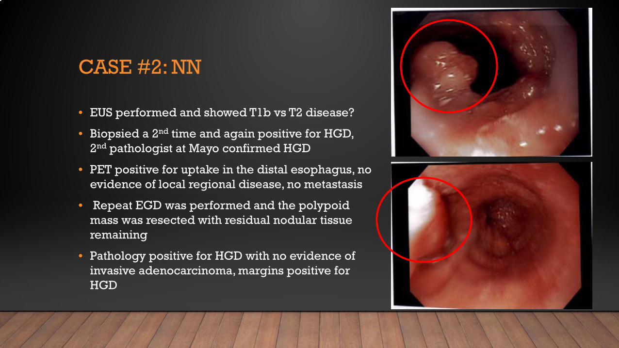
CASE #2: NN
• EUS performed and showed T1b vs T2 disease?
• Biopsied a 2nd time and again positive for HGD, 2nd pathologist at Mayo confirmed HGD
• PET positive for uptake in the distal esophagus, no evidence of local regional disease, no metastasis
• Repeat EGD was performed and the polypoid mass was resected with residual nodular tissue remaining
• Pathology positive for HGD with no evidence of invasive adenocarcinoma, margins positive for HGD


CASE #2: NN
• Rare entity called: polypoid dysplasia in Barrett’s esophagus
• Dysplastic polypoid lesions superimposed on Barrett’s esophagus
• Most reported cases of polypoid dysplasia in Barrett’s esophagus have been advanced on presentation and treated with esophagectomy
• Our patient, all biopsies including polyp resection were HGD, with no evidence of invasive disease

CASE #2: NN
• EGD 1/2016 showed long segment Barrett’s esophagus from 33-38cm (C5M5)
• Nodularity in a circumferential pattern from 33cm-36cm
• EMR x 2 was performed, with residual nodular tissue remaining
• Pathology positive for HGD

CASE #2: NN
• Underwent 6 EMR total
• All pathology positive for HGD

CASE #2: NN
• Subsequently, he formed 2 strictures from the EMR’s
• Undergone several balloon dilations

• He has undergone RFA x 3 with good results


REFERENCES:
• "American Gastroenterological Association Medical Position Statement on the Management of Barrett's Esophagus." Gastroenterology 140.3 (2011): 1084-091.
• Evans, John A., Dayna S. Early, Norio Fukami, et al. "The Role of Endoscopy in Barrett's Esophagus and Other Premalignant Conditions of the Esophagus." Gastrointestinal Endoscopy 76.6 (2012): 1087-094.
• Kahrilas, Peter J., Nicholas J. Shaheen, and Michael F. Vaezi. "American Gastroenterological Association Medical Position Statement on the Management of Gastroesophageal Reflux Disease." Gastroenterology 135.4 (2008).
• Katz, Philip O., Lauren B. Gerson, and Marcelo F. Vela. "Guidelines for the Diagnosis and Management of Gastroesophageal Reflux Disease." The American Journal of Gastroenterology 108.3 (2013): 308-28.
• Shaheen, Nicholas J., Gary W. Falk, Prasad G. Iyer, and Lauren B. Gerson. "ACG Clinical Guideline: Diagnosis and Management of Barrett’s Esophagus." The American Journal of Gastroenterology 111.1 (2015): 30-50.
• Sharma, Prateek, Richard E. Sampliner, and David Ilson. Esophageal Cancer and Barrett's Esophagus. Chichester, West Sussex: John Wiley and Sons, 2015.
• Stefanidis, Dimitrios, William W. Hope, Geoffrey P. Kohn, Patrick R. Reardon, William S. Richardson, and Robert D. Fanelli. "Guidelines for Surgical Treatment of Gastroesophageal Reflux Disease." Surgical Endoscopy 24.11 (2010): 2647-669.