Pim1 inhibition as a novel therapeutic strategy for ...
14
RESEARCH ARTICLE Open Access Pim1 inhibition as a novel therapeutic strategy for Alzheimer’ s disease Ramon Velazquez 1 , Darren M. Shaw 1,2 , Antonella Caccamo 1 and Salvatore Oddo 1,2* Abstract Background: Alzheimer’s disease (AD) is the most prevalent neurodegenerative disorder worldwide. Clinically, AD is characterized by impairments of memory and cognitive functions. Accumulation of amyloid-β (Aβ) and neurofibrillary tangles are the prominent neuropathologies in patients with AD. Strong evidence indicates that an imbalance between production and degradation of key proteins contributes to the pathogenesis of AD. The mammalian target of rapamycin (mTOR) plays a key role in maintaining protein homeostasis as it regulates both protein synthesis and degradation. A key regulator of mTOR activity is the proline-rich AKT substrate 40 kDa (PRAS40), which directly binds to mTOR and reduces its activity. Notably, AD patients have elevated levels of phosphorylated PRAS40, which correlate with Aβ and tau pathologies as well as cognitive deficits. Physiologically, PRAS40 phosphorylation is regulated by Pim1, a protein kinase of the protoconcogene family. Here, we tested the effects of a selective Pim1 inhibitor (Pim1i), on spatial reference and working memory and AD-like pathology in 3xTg-AD mice. Results: We have identified a Pim1i that crosses the blood brain barrier and reduces PRAS40 phosphorylation. Pim1i-treated 3xTg-AD mice performed significantly better than their vehicle treated counterparts as well as non-transgenic mice. Additionally, 3xTg-AD Pim1i-treated mice showed a reduction in soluble and insoluble Aβ 40 and Aβ 42 levels, as well as a 45.2 % reduction in Aβ 42 plaques within the hippocampus. Furthermore, phosphorylated tau immunoreactivity was reduced in the hippocampus of Pim1i–treated 3xTg-AD mice by 38 %. Mechanistically, these changes were linked to a significant increase in proteasome activity. Conclusion: These results suggest that reductions in phosphorylated PRAS40 levels via Pim1 inhibition reduce Aβ and Tau pathology and rescue cognitive deficits by increasing proteasome function. Given that Pim1 inhibitors are already being tested in ongoing human clinical trials for cancer, the results presented here may open a new venue of drug discovery for AD by developing more Pim1 inhibitors. Keywords: AD, Pim1 inhibitor, PRAS40, 3xTg-AD, Proteasome, Aβ, tau, mTOR, Aging, Working memory Background Alzheimer’ s disease (AD) is the most prevalent neurode- generative disorder worldwide. Clinically, AD is charac- terized by impairments of memory, cognitive and intellectual functions [1]. The buildup of amyloid-β (Aβ) plaques and neurofibrillary tangles (NFTs) are the two prominent pathologies contributing to the progression of cognitive deficits in AD [2]. Aβ is generated from the amyloid precursor protein (APP), which is sequentially cleaved by the β-site APP cleaving enzyme 1 (BACE-1) and the γ-secretase complex to liberate Aβ. NFTs consist of hyperphosphorylated and aggregated tau, a microtubule-binding protein. Over the next few decades, advancing age of the global population will dramatically increase the prevalence of AD to an estimated 20 million by 2050 in the US alone [3]. Currently, there are no effective treatment options to either prevent or re- duce the progression of AD. Therefore, there is an urgent need for novel, safe, and efficacious strategies to mitigate this disorder. Aging is the most important risk factor for AD; thus, it is possible that altered signaling pathways associated with aging may facilitate the development of AD and other age-dependent disorders [4, 5]. Overwhelming evidence has shown that the mammalian target of * Correspondence: [email protected] 1 Neurodegenerative Disease Research Center, Biodesign Institute, School of Life Sciences, Arizona State University, 727 E. Tyler Street, Tempe, AZ 85287-5001, USA 2 School of Life Sciences, Arizona State University, Tempe, AZ 85287, USA © 2016 The Author(s). Open Access This article is distributed under the terms of the Creative Commons Attribution 4.0 International License (http://creativecommons.org/licenses/by/4.0/), which permits unrestricted use, distribution, and reproduction in any medium, provided you give appropriate credit to the original author(s) and the source, provide a link to the Creative Commons license, and indicate if changes were made. The Creative Commons Public Domain Dedication waiver (http://creativecommons.org/publicdomain/zero/1.0/) applies to the data made available in this article, unless otherwise stated. Velazquez et al. Molecular Neurodegeneration (2016) 11:52 DOI 10.1186/s13024-016-0118-z
Transcript of Pim1 inhibition as a novel therapeutic strategy for ...

RESEARCH ARTICLE Open Access
Pim1 inhibition as a novel therapeuticstrategy for Alzheimer’s diseaseRamon Velazquez1, Darren M. Shaw1,2, Antonella Caccamo1 and Salvatore Oddo1,2*
Abstract
Background: Alzheimer’s disease (AD) is the most prevalent neurodegenerative disorder worldwide. Clinically, AD ischaracterized by impairments of memory and cognitive functions. Accumulation of amyloid-β (Aβ) and neurofibrillarytangles are the prominent neuropathologies in patients with AD. Strong evidence indicates that an imbalance betweenproduction and degradation of key proteins contributes to the pathogenesis of AD. The mammalian target of rapamycin(mTOR) plays a key role in maintaining protein homeostasis as it regulates both protein synthesis and degradation. A keyregulator of mTOR activity is the proline-rich AKT substrate 40 kDa (PRAS40), which directly binds to mTOR and reducesits activity. Notably, AD patients have elevated levels of phosphorylated PRAS40, which correlate with Aβ and taupathologies as well as cognitive deficits. Physiologically, PRAS40 phosphorylation is regulated by Pim1, a protein kinaseof the protoconcogene family. Here, we tested the effects of a selective Pim1 inhibitor (Pim1i), on spatial reference andworking memory and AD-like pathology in 3xTg-AD mice.
Results: We have identified a Pim1i that crosses the blood brain barrier and reduces PRAS40 phosphorylation.Pim1i-treated 3xTg-AD mice performed significantly better than their vehicle treated counterparts as well asnon-transgenic mice. Additionally, 3xTg-AD Pim1i-treated mice showed a reduction in soluble and insolubleAβ40 and Aβ42 levels, as well as a 45.2 % reduction in Aβ42 plaques within the hippocampus. Furthermore,phosphorylated tau immunoreactivity was reduced in the hippocampus of Pim1i–treated 3xTg-AD mice by38 %. Mechanistically, these changes were linked to a significant increase in proteasome activity.
Conclusion: These results suggest that reductions in phosphorylated PRAS40 levels via Pim1 inhibition reduceAβ and Tau pathology and rescue cognitive deficits by increasing proteasome function. Given that Pim1inhibitors are already being tested in ongoing human clinical trials for cancer, the results presented here mayopen a new venue of drug discovery for AD by developing more Pim1 inhibitors.
Keywords: AD, Pim1 inhibitor, PRAS40, 3xTg-AD, Proteasome, Aβ, tau, mTOR, Aging, Working memory
BackgroundAlzheimer’s disease (AD) is the most prevalent neurode-generative disorder worldwide. Clinically, AD is charac-terized by impairments of memory, cognitive andintellectual functions [1]. The buildup of amyloid-β (Aβ)plaques and neurofibrillary tangles (NFTs) are the twoprominent pathologies contributing to the progressionof cognitive deficits in AD [2]. Aβ is generated from theamyloid precursor protein (APP), which is sequentiallycleaved by the β-site APP cleaving enzyme 1 (BACE-1)
and the γ-secretase complex to liberate Aβ. NFTsconsist of hyperphosphorylated and aggregated tau, amicrotubule-binding protein. Over the next few decades,advancing age of the global population will dramaticallyincrease the prevalence of AD to an estimated 20 millionby 2050 in the US alone [3]. Currently, there are noeffective treatment options to either prevent or re-duce the progression of AD. Therefore, there is anurgent need for novel, safe, and efficacious strategiesto mitigate this disorder.Aging is the most important risk factor for AD; thus,
it is possible that altered signaling pathways associatedwith aging may facilitate the development of AD andother age-dependent disorders [4, 5]. Overwhelmingevidence has shown that the mammalian target of
* Correspondence: [email protected] Disease Research Center, Biodesign Institute, School ofLife Sciences, Arizona State University, 727 E. Tyler Street, Tempe, AZ85287-5001, USA2School of Life Sciences, Arizona State University, Tempe, AZ 85287, USA
© 2016 The Author(s). Open Access This article is distributed under the terms of the Creative Commons Attribution 4.0International License (http://creativecommons.org/licenses/by/4.0/), which permits unrestricted use, distribution, andreproduction in any medium, provided you give appropriate credit to the original author(s) and the source, provide a link tothe Creative Commons license, and indicate if changes were made. The Creative Commons Public Domain Dedication waiver(http://creativecommons.org/publicdomain/zero/1.0/) applies to the data made available in this article, unless otherwise stated.
Velazquez et al. Molecular Neurodegeneration (2016) 11:52 DOI 10.1186/s13024-016-0118-z

rapamycin (mTOR), a serine/threonine protein kinaseinvolved in the regulation of protein synthesis and deg-radation, plays a key role in regulating lifespan andhealth span [5–7]. mTOR is part of two major com-plexes, the mTOR complex 1 (mTORC1) and 2(mTORC2), which have different functions [8, 9].mTOR signaling is upregulated in AD [10–13]. Tothis end, we and others have shown that reducingmTOR activity pharmacologically, with rapamycin, orgenetically ameliorates Aβ plaque load and NFTs whileimproving cognitive deficits in multiple animal models ofAD [10, 14–17]. However, various studies have foundthat rapamycin has many side effects, necessitatingother treatment options to reduce mTOR hyperactivityin AD [18–20].The proline-rich Akt substrate 40 kDa (PRAS40) is a
constituent of the mTORC1, which physically binds tomTOR and inhibits its activity. Upon phosphorylation atThr246 by serine/threonine-specific protein kinase(AKT) or proto-oncogene serine/threonine-protein kin-ase Pim-1 (Pim1), PRAS40 detaches from mTORC1thereby releasing its inhibitory effects [21, 22]. We haveshown that intrahippocampal injection of naturallysecreted Aβ is sufficient to increase mTOR signaling inthe brains of wild type mice by increasing the phosphor-ylation of PRAS40 [23].Pim1 is a kinase member of the proto-oncogene Pim
kinase family [24–27]. Pim1 plays a role in cell survivaland proliferation; hence, its deregulation can easilytransform it to an oncogenic protein. Pim1 has gainedmuch attention due to its upregulation in a variety ofcancers [28, 29]. Of note, Pim1 knockout mice have nophenotype [30], suggesting that Pim1 might be a viabletherapeutic target. Crystallographic studies revealed aunique architecture within the Pim1 secondary struc-ture, which allowed the synthesis of highly specificinhibitors [31, 32]. Here, we sought to determine theeffects of reducing Pim 1 activity on AD-like pathologydeveloped by the 3xTg-AD mice.
ResultsPharmacological characterization of Pim1 inhibitor 1We have previously shown that the levels of PRAS40phosphorylated at Thr246 (pPRAS40) are increased in3xTg-AD mice [23]. To further determine the role ofPRAS40 in AD, we assessed the levels of pPRAS40 inthe inferior frontal gyrus of human AD brains (Table 1).We found that the levels of pPRAS40 are increased inhuman AD brains compared to age-matched controls(Fig. 1). We then sought to determine the effects ofreducing PRAS40 phosphorylation on the AD-like path-ology in 3xTg-AD mice. To achieve our goal, we targetedPim1, a kinase known to phosphorylate PRAS40 [33].Several Pim1 kinase inhibitors are commercially available,
and some are currently in clinical trial for the treatment ofcancer (ClinicalTrials.gov Identifier: NCT00848601). Thecommercially available Pim1 inhibitor, 3-Cyano-4-phenyl-6-(3-bromo-6-hydroxy) phenyl-2 (1H)-pyridone (hereinreferred to as Pim1i; Fig. 2a), exhibits highly specific com-petitive inhibition to Pim1; IC50 50 nM for Pim1 and2 μM for Pim2 [33]. We have shown that intracranial de-livery of this inhibitor effectively decreases pPRAS40 [23],a downstream substrate of Pim1. We first tested Pim1ibioavailability and measured its brain-to-plasma ratio byLC-MS/MS analysis. To this end, C57BL/6 mice receiveda single dose of 50 mg/kg Pim1i intraperitoneally (i.p.).We found that maximum plasma concentration of Pim1iwas 32166 ± 11220 ng/mL, which was observed at 1 h postdose with half-life of 1.02 h (Fig. 2b). Pim1i was also de-tectable up to 3 h post dose in brain tissue homogenatesamples (Fig. 2c). The maximum brain concentration of197 ± 48.5 ng/g was observed at 0.5 h post dose.To determine whether Pim1i engages the target, we
injected C57BL/6 mice with 100 mg/kg i.p. and har-vested brains at 1, 3, 6, 12 and 24 post injection (n = 5per time-point). Three additional mice were used as ourbaseline. We decided to increase the dose as no toxicitywas detected in mice receiving 50 mg/kg Pim1i. Wefound that the levels of pPRAS40 were significantlydifferent across the different time points (F(6, 26) = 11.09;p < 0.001; Fig. 2d). A Bonferroni’s multiple comparison
Table 1 Descriptive information of patients whose brain tissuewas utilized to measure pPRAS40 levels. Gel order indicates theorder in which protein samples were loaded in Fig. 1
Age at death Diagnosis MMSE score Braak stage Gel load order
86 AD 16 IV 1
82 AD 23 IV 2
93 CTL 30 I 3
89 CTL 29 II 4
92 CTL 26 III 5
79 CTL 29 II 6
82 AD 19 V 7
78 AD 0 VI 8
87 CTL 28 III 9
91 CTL 27 II 10
87 CTL 26 III 11
75 CTL 29 III 12
95 CTL 26 III 13
88 AD 9 VI 14
78 AD - IV 15
68 AD 0 VI 16
78 CTL 29 III 17
85 AD 23 VI 18
86 AD 20 IV 19
Velazquez et al. Molecular Neurodegeneration (2016) 11:52 Page 2 of 14
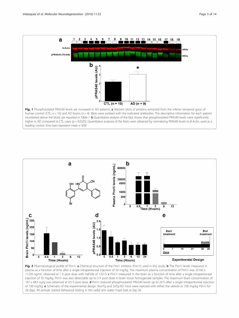
Fig. 1 Phosphorylated PRAS40 levels are increased in AD patients. a Western blots of proteins extracted from the inferior temporal gyrus ofhuman control (CTL; n = 10) and AD brains (n = 9). Blots were probed with the indicated antibodies. The descriptive information for each patient(numbered above the blots) are reported in Table 1. b Quantitative analysis of the blot shows that phosphorylated PRAS40 levels were significantlyhigher in AD compared to CTL cases (p = 0.0325). Quantitative analyses of the blots were obtained by normalizing PRAS40 levels to β-Actin, used as aloading control. Error bars represent mean ± SEM
Fig. 2 Pharmacological profile of Pim1i. a Chemical structure of the Pim1 inhibitor (Pim1i) used in this study. b The Pim1i levels measured inplasma as a function of time after a single intraperitoneal injection of 50 mg/kg. The maximum plasma concentration of Pim1i was 32166 ±11220 ng/mL observed at 1 h post dose with half-life of 1.02 h. c Pim1i measured in the brain as a function of time after a single intraperitonealinjection of 50 mg/kg. Pim1i was also detectable up to 3 h post dose in brain tissue homogenate samples. The maximum brain concentration of197 ± 48.5 ng/g was observed at 0.5 h post dose. d Pim1i reduced phosphorylated PRAS40 levels up to 24 h after a single intraperitoneal injectionof 100 mg/kg. e Schematic of the experimental design. NonTg and 3xTg-AD mice were injected with either the vehicle or 100 mg/kg Pim1i for28 days. All animals started behavioral testing in the radial arm water maze task at day 26
Velazquez et al. Molecular Neurodegeneration (2016) 11:52 Page 3 of 14

test indicated that pPRAS40 levels at the time points 1,3, 6, 12 and 24 h were significantly different than time 0,our baseline control. These data show that brain levelsof pPRAS40 were significantly reduced one hour follow-ing the Pim1i injections and stayed low for 24 h.
Pim1 inhibition reduces spatial reference and workingmemory deficits in 3xTg-AD miceGiven these data, we sought to determine the effects ofchronic administration of the Pim1i at a dosage of100 mg/kg on the AD-like pathology in 3xTg-AD mice.To this end, we used 7-month-old female 3xTg-AD andnon-transgenic (NonTg) mice. At this age, 3xTg-ADmice have mild cognitive deficits, which are associatedwith high soluble Aβ and tau levels [34]. Mice were ran-domly assigned to one of the following groups: 3xTg-ADVehicle (Veh), NonTg Veh, 3xTg-AD Pim1i, NonTgPim1i (n = 14/group) and received daily i.p. injections ofPim1i or vehicle for 28 days (Fig. 2e).We first assessed body weight of the mice at the be-
ginning of treatment and found that the average bodyweight was 24.21 ± 0.635 g for NonTg mice and 31.46 ±1.427 g for 3xTg-AD mice. The analysis of mean weightrevealed a significant main effect of Genotype (F(1, 42) =36.624, p < 0.0001), indicating that 3xTg-AD miceweighed significantly more than NonTg mice. Similarly,3xTg-AD mice (28.28 ± 1.05 g) weighed more than theNonTg mice (22.48 ± 0.46 g) at completion of treatment.The analysis of mean weight in grams revealed a signifi-cant main effect of Genotype (F(1, 42) = 28.689, p <0.0001) and Treatment (F(1, 42) = 5.364, p < 0.05), how-ever no significant Genotype by Treatment interaction(F(1, 42) = 1.117, p > 0.05). Together, these data indicatethat the Pim1i treated mice weighed less at the end oftreatment compared to the mice on vehicle, and this dif-ference was independent of genotype. Notably, duringthe 28 days of treatment, we lost mice in all four groups.The total numbers of mice that did not show any overttoxicity and were tested behaviorally are as follows:3xTg-AD Veh (n = 13), NonTg Veh (n = 12), 3xTg-ADPim1i (n = 7), NonTg Pim1i (n = 13).To determine the effect of 28 days of the Pim1i on
spatial reference and working memory, we tested all sub-jects on an 8-arm radial arm water maze (RAWM) task.Mice were tested for two consecutive days: on day one,mice received 15 trials, with trials alternating betweenvisible and hidden platform. On day 2, mice received 15additional trials but the platform was kept hiddenthroughout the trials. Entry into an incorrect arm wasscored as a spatial reference error. Reentry into a previ-ously visited arm within the trial was considered a work-ing memory error. The number of errors were averagedby block, with each block being equivalent to three trails.Using a mixed model ANOVA, we first examined mean
incorrect errors between day 1 and 2 to assess learning.We found a significant effect of Genotype (F(1,42) =13.610; p < 0.01), Treatment (F(1,42) = 18.790, p < 0.0001),day (F(1, 42) = 107.868, p < 0.0001) and a trend in Genotypeby Treatment interaction (F(1, 42) = 3.755, p = 0.0594;Fig. 3a). Specifically, all groups showed a significant reduc-tion in incorrect errors between day 1 and day 2, indicat-ing that all groups significantly learned the task.We next analyzed the number of errors during the
probe trials (day 2). For reference errors, we found a sig-nificant effect of Block (F(4, 42) = 3.882, p < 0.005, Fig. 3b),Genotype (F(1, 42) = 8.126, p < 0.01), Treatment (F(1, 42) =18.105, p < 0.001) and a significant Genotype by Treat-ment interaction (F(1, 42) = 4.496, p < 0.05). Post hoc testwith Bonferroni’s correction indicated that 3xTg-ADVeh mice committed a higher number of reference er-rors throughout the 5 blocks of testing when comparedto the NonTg Veh group (p < 0.05; Fig. 3b for errorsacross each block; 2C for mean total errors). Notably,3xTg-AD Pim1i mice made significantly fewer errorscompared to 3xTg-AD Veh treated mice, indicating im-proved spatial reference memory (p < 0.01). Further-more, 3xTg-AD Pim1i mice performed as well as NonTgVeh mice (p > 0.05), illustrating a full rescue of spatialreference memory. Interestingly, NonTg mice injectedwith Pim1i made significantly fewer errors than the vehicletreated NonTg mice (p < 0.05). These results show that28 days of Pim1i administration is sufficient to normalizethe spatial reference memory deficits in 3xTg-AD mice toNonTg levels, and to improve performance in NonTg mice.We then examined reentry errors into a previously en-
tered arm within a trial, which indicate working memoryerrors. When we compared the mice performance betweenthe two days of training, we found a significant effect ofGenotype (F(1, 42) = 8.045, p = 0.01), Treatment (F(1, 42) =10.381, p < 0.01), day (F(1, 42) = 13.693, p < 0.001), and aGenotype by Treatment interaction (F(1, 42) = 4.390, p <0.05; Fig. 3d). Notably, 3xTg-AD Veh mice had the samenumber of working memory errors between day 1 and day2 (p > 0.05), indicating that although they decreased thenumber of reference memory errors, they continued tocommit working memory errors on day 2. All other groupsshow a decrease in working memory errors between day 1and day 2. These data indicate that 3xTg-AD Pim 1i miceperformed significantly fewer working memory errorscompared to 3xTg-AD Veh. During the probe trials (day2), we found a significant effect of Genotype (F(1, 42) =5.255, p < 0.05), Treatment (F(1, 42) = 10.131, p < 0.01), andGenotype by Treatment interaction (F (1, 42) = 4.496, p <0.05; Fig. 3e). Post hoc test with Bonferroni’s correctionindicated that 3xTg-AD Veh mice committed a highernumber of working memory errors throughout the 5blocks of testing when compared to the NonTg-AD Vehmice (p = 0.0147, Fig. 3e for errors across each block; 3f for
Velazquez et al. Molecular Neurodegeneration (2016) 11:52 Page 4 of 14

mean total errors). Pim1i significantly reduced the numberof working memory errors in 3xTg-AD mice compared to3xTg-AD Veh mice (p < 0.05). Indeed, the number of work-ing memory errors made by 3xTg-AD Pim1i mice were notstatistically significant from errors made by NonTg Vehmice (p > 0.05). Collectively, these results show that 28 daysof the Pim1 inhibitor is sufficient to reduce spatial refer-ence and working memory deficits in 3xTg-AD mice.“Chaining” is a commonly used search strategy to
find the platform in the RAWM [35]. This methoddoes not require use of spatial cues and instead con-sists of sequential arm visits until the platform isfound. To further determine the nature of the cogni-tive impairment in 3xTg-AD mice, we examined theuse of a chaining strategy for navigation of the mazeduring the day 2 probe trials. For this analysis, a
chaining event was defined as three consecutive armentries in an anticlockwise or clockwise direction, anoperational definition used by [35]. The percentage ofchaining events committed per group were analyzedusing a chi-square test. We found that 3xTg-AD Vehmice show a significantly higher number of chainingevents compared to both NonTg groups and 3xTg-ADPim1i treated group (X2 = 7.8045, p < 0.01, Fig. 3g-h).These data suggest that Pim1 inhibition reduces theusage of the chaining search strategy in 3xTg-ADmice.
Pim1 inhibition lowers Aβ levels and hippocampal CP13immunoreactivity in 3xTg-AD miceAt the end of the behavioral testing, mice were sacrificedand their brains were used for neuropathological and
Fig. 3 Pim1 inhibition reduces spatial reference and working memory deficits in 3xTg-AD mice. NonTg Veh (n = 13), 3xTg-AD Veh (n = 13), NonTgPim1i (n = 12), 3xTg-AD Pim1i (n = 7) were tested in the radial arm water maze (RAWM). a Average reference memory errors on day 1 and day 2RAWM task. All groups show a decrease in total errors in day 2 indicating learning (F(1, 42) = 107.868, p < 0.0001). b Average reference memoryerrors across the 5 blocks of testing of day 2 (1 block = 3 trials). 3xTg-AD Veh mice committed a higher number of reference errors throughoutthe 5 blocks of testing compared to all the other groups. c Mean total errors of day 2. d Working memory errors on day 1 and day 2. Workingmemory errors are defined as a reentry into a previously visited arm. 3xTg-AD Veh mice had the same number of working memory errorsbetween day 1 and day 2 (p > 0.05), whereas all the other groups showed significant learning between the two days. e Average workingmemory errors across the 5 blocks of testing of day 2. 3xTg-AD Veh mice commit a higher number of working memory errors throughout the 5 blocksof testing compared to the other three groups. f. Mean working memory errors during day 2. g Pim1i significantly reduces the percentage of 3xTg-ADmice using a hippocampal-independent default strategy to find the hidden platform, known as chaining. The panel illustrates the percentage of micecommitting chaining events between all groups. h The panel illustrates the percentage of animals that committed chaining events betweenthe four groups. Pim 1i significantly reduced the percentage of chaining committed by the 3xTg-AD mice (X2 = 7.8045, p < 0.01). Error barsrepresent mean ± SEM
Velazquez et al. Molecular Neurodegeneration (2016) 11:52 Page 5 of 14

biochemical assessment. To determine whether periph-eral administration of Pim1i reached the brain, we usedsandwich enzyme linked immunosorbent assay to meas-ure the levels of pPRAS40 at Thr246, as this epitope isdirectly phosphorylated by Pim 1 [33]. We found a sig-nificant effect for Genotype (F(1, 20) = 4.501, p < 0.05),Treatment (F(1, 20) = 36.593, p < 0.0001), and a non-significant Genotype by Treatment interaction (F(1, 20) =1.767, p > 0.05; Fig. 4a). The effect of genotype indicatedthat 3xTg-AD mice show higher levels of pPRAS40 thanNonTg mice. Interestingly, the treatment effect revealsthat both 3xTg-AD and NonTg Pim1i mice have a re-duced pPRAS40 level. Together, these data further con-firm that Pim1i crosses the blood brain barrier anddecreases the levels of brain pPRAS40.One of the key neuropathological features of AD is the
accumulation of extracellular Aβ plaques [2]. Aβ peptidesconsist of 36–43 amino acids, where Aβ40 and Aβ42 arethe more abundant Aβ species. Aβ42 is more prone toaggregation and toxicity than the Aβ40 species [2]. We im-munostained sections from 3xTg-AD Veh (n = 6) and3xTg-AD Pim1i (n = 5) mice with an Aβ42-specific anti-body and found that Aβ42 immunoreactivity was signifi-cantly reduced in the brains of 3xTg-AD mice injectedwith Pim1i (Fig. 4b-c). Quantitative analysis of the overall
Aβ42 load indicated a significant decrease of 45.2 % in thebrain of 3xTg-AD Pim1i compared with 3xTg-AD mice(t (10) = 8.419, p < 0.05; Fig. 4d). We next measured Aβlevels by sandwich ELISA and found that both solubleand insoluble Aβ40 levels were significantly lower in3xTg-AD Pim1i mice compared with 3xTg-AD Vehmice (soluble Aβ40: t(10) =2.442, p < 0.05; insolubleAβ40: t(10) = 2.681, p = 0.0230; Fig. 5a-b). Furthermore,both soluble and insoluble levels of Aβ42 were signifi-cantly lower in 3xTg-AD Pim1i compared to 3xTg-ADVeh mice (soluble Aβ42: t(10) = 2.404, p = 0.0371; insol-uble Aβ42: t(10) = 3.932, p < 0.01; Fig. 5c-d).Another hallmark pathology associated with AD is the
accumulation of neurofibrillary tangles made of hyperpho-sphorylated tau [2]. To determine the effects of Pim1inhibition on tau pathology, we first immunostained sec-tions from 3xTg-AD Veh and 3xTg-Pim1-inh1 mice withCP13, an antibody that recognizes tau phosphorylated atserine 202. We found that CP13 immunoreactivity was re-duced in 3xTg-AD Pim1i compared with 3xTg-AD mice(Fig. 6a-b). Quantitative analysis indicated that CP13 im-munoreactivity was significantly decreased by 38 % in thebrain of 3xTg-AD Pim1i compared with 3xTg-AD Vehmice (t (10) = 2.785, p < 0.05; Fig. 6c). To further assess thetau pathology, we measured tau levels by western blot
Fig. 4 Pim1i decreases Aβ pathology. a The graph shows the phosphorylated levels of PRAS40 levels after 28 days of treatment, measured bysandwich ELISA. The Pim1 inhibitor significantly reduced the pPRAS40 levels of both the 3xTg-AD and NonTg mice as evident by a significanteffect of treatment (F(1, 42) = 36.593, p < 0.0001). b Representative microphotographs of brain sections from 3xTg-AD mice stained with an Aβ42specific antibody (Millipore, catalog number AB5078P). d Quantitative analysis of Aβ42 immunoreactivity by unpaired t-test reveals that 3xTg-ADmice treated with Pim1i had significantly fewer plaques compared to 3xTg-AD mice treated with vehicle (t(8) = 8.419, p < 0.05). Error barsrepresent mean ± SEM
Velazquez et al. Molecular Neurodegeneration (2016) 11:52 Page 6 of 14

using antibodies against total and phosphorylated tau. Wefound that the levels of total human tau (measured byHT7) were not significantly different between the Veh andPim1i-treated groups (p > 0.05; Fig. 6d-e). In contrast, wefound a main effect of Genotype for CP13 levels (F(1,15) =4.563, p < 0.05; Fig. 6d, f ). Post hoc analyses indicated that3xTg-AD mice had significantly higher levels than NonTgmice. The apparent contradiction between the CP13 dataobtained by immunohistochemistry and by western blot islikely due to the fact that the immunohistochemistry al-lows for single cell resolution while the western blot wasdone by homogenizing different cell types from differentbrain regions. Overall, these results show that Pim1 inhib-ition reduces hippocampal tau immunoreactivity in 3xTg-AD mice.
Pim1i increases proteasome activityOur results indicate that reductions in pPRAS40 viaPim1 inhibition is sufficient to reduce spatial referenceand working memory deficits in 3xTg-AD mice. Theseimprovements in cognitive function are associated witha reduction in Aβ levels and phosphorylated tau immu-noreactivity. To better elucidate the mechanisms under-lying these effects, we first assessed mTOR signaling bymeasuring the phosphorylation levels of the two majordownstream targets of mTOR: p70S6K1 and 4EBP1.
For the phosphorylation levels of p70S6K1, there wereno significant effects of Genotype (F(1,15) = 4.067, p >0.05), treatment (F(1,15) = 0.428, p > 0.05), and Genotypeby Treatment interaction (F(1,15) = 1.831, p > 0.05; Fig. 7a-b).Interestingly for the downstream target 4EBP1, there was amain effect of Genotype (F(1, 15) = 5.621, p = 0.05;Fig. 7a, c) where the NonTg mice had increased levelsof phosphorylated 4EBP1 compared to 3xTg-AD mice.There was no effect of the Pim1i treatment on thesetargets.To further understand what mechanisms may underlie
changes in AD pathology and improvements in behavior,we assessed autophagy induction and proteasome func-tion. We focused on these systems as they represent thetwo major cellular protein degradation systems and areknown to be involved in the turnover of Aβ and tau[36]. First, we measured the levels of LC3, Atg3, Atg5,and Atg12, key proteins involved in autophagy inductionwhose levels are routinely used to monitor it [37]. Wefound a significant main effect of Genotype (F(1, 15) =7.238, p < 0.05) but a non-significant effect of Treatment(F (1, 15) = 2.230, p > 0.05) or Genotype by Treatment inter-action (F(1, 15) = 0.00042, p > 0.05) for p62 (Fig. 8a-b). Inaddition, we found a significant main effect of Genotype(F(1, 15) = 10.851, p < 0.01) but a non-significant effect ofTreatment (F (1, 15) = 0.266, p > 0.05) and the Genotype by
Fig. 5 Pim1i decreases Aβ levels. Sandwich ELISA measurements of insoluble and soluble Aβ40 and Aβ42 levels in brain extracts from 3xTg-ADveh (n = 7) and 3xTg-AD Pim1i (n = 5) mice. a, b Soluble and insoluble Aβ42 levels were significantly lower in 3xTg-AD mice injected with Pim1i(Soluble Aβ40: t(10) =2.442, p < 0.05; insoluble Aβ40: t(10) = 2.681, p < 0.05). c, d Soluble and insoluble Aβ42 levels were significantly lower in 3xTg-ADmice treated with Pim1i (soluble Aβ42: t(10) = 2.404, p < 0.05; insoluble Aβ42: t(10) = 3.932, p < 0.01). Error bars represent mean ± SEM
Velazquez et al. Molecular Neurodegeneration (2016) 11:52 Page 7 of 14

Treatment interaction (F(1, 15) = 1.607, p > 0.05) for Atg3(Fig. 8a, c). In contrast, the levels of Atg5 and Atg12 weresimilar among the four groups (Fig. 8a, d and e).Next, we used the fluorogenic substrates Bz-VGR-AMC, Suc-LLVYAMC, and Z-LLE-AMC to measuretrypsin-like, chymotrypsin-like, and caspase-like activ-ities of the proteasome. We found an effect of Treat-ment for chymotrypsin-like (F(1, 20) = 10.127, p < 0.05,Fig. 8f ), trypsin-like (F(1, 20) = 16.18, p < 0.001, Fig. 8g)and caspase-like (F(1, 20) = 19.804, p < 0.001, Fig. 8h)activity. This finding indicates that the Pim1 inhibi-tor increases the activity of the three substrates forboth the 3xTg-AD and NonTg mice over the vehicle
treated groups. This suggests that the reductions inAβ pathology and tau immunoreactivity may be ac-complished by an increase in protein degradation.
DiscussionThe incidence of AD is quickly increasing and withno effective therapeutics to reduce the associatedpathologies, this disorder is bound to have a majorsocio-economic impact on our society. The data pre-sented here unambiguously show that inhibitingPim1 activity for 28 days, thereby reducing pPRAS40levels, has beneficial effects on AD-like pathology in3xTg-AD mice. Indeed, these data show that
Fig. 6 Reduced Tau pathology in 3xTg-AD Pim1i mice. a, b Representative microphotographs of CA1 hippocampal neurons from 3xTg-AD Vehand 3xTg-AD Pim1i mice stained with the anti-tau antibody CP13, which recognizes tau phosphorylated at Ser202. c Quantitative analysis of theCP13 immunoreactivity by unpaired t-test reveals that Pim1i significantly reduced tau immunoreactivity (t(9) = 2.785, p < 0.05). d Representativewestern blots of protein extracted from 3xTg-AD Veh, NonTg Veh, 3xTg-AD Pim1i, and NonTg Pim1i mice. Blots were probed with the indicatedantibodies. The HT7 antibody recognizes total human tau and CP13 antibody recognizes tau phosphorylated at Ser202. e, f Quantitative analysesof the blots. HT7 levels were not significantly different between the two groups (t(9) = 0.558, p > 0.05). For CP13, we found a Genotype effect(t(9) = 4.563, p < 0.05), revealing higher levels of CP13 in 3xTg-AD mice. Quantitative analyses of the blots were obtained by normalizing thelevels of the protein of interest to β-Actin, used as a loading control. Error bars represent mean ± SEM
Velazquez et al. Molecular Neurodegeneration (2016) 11:52 Page 8 of 14

inhibition of Pim1 reduced spatial reference andworking memory errors in 3xTg-AD mice, and im-proved performance to NonTg levels. This cognitiveimprovement was associated with a reduction in hip-pocampal Aβ pathology and hippocampal tauimmunoreactivity.PRAS40 is a component of the mTOR complex 1; it
physically binds to mTOR and negatively regulates itsactivity [33]. Upon phosphorylation, PRAS40 detachesfrom mTOR releasing its inhibitory effects. Indeed, highlevels of pPRAS40 have been associated with highmTOR activity [8, 33]. Notably, the levels of pPRAS40
are elevated in both humans AD patients and 3xTg-ADmice [23]. We have previously shown that the increasein pPRAS40 is due to a buildup in Aβ oligomers [23].Paradoxically, although we show that Pim1 inhibitionreduces the levels of pPRAS40, the activity of mTORappeared unaffected. Indeed, Pim1 inhibition did notchange the phosphorylation levels of p70S6K1 and4EBP1. These findings suggest that the reduction in Aβpathology and hippocampal tau immunoreactivity mayoccur through an mTOR independent pathway. To thisend, our data suggest that the mechanism underlyingthe reduced AD-like pathology is linked to an increasein proteasome activity as evident by the changes in thethree proteolytic activities, chymotrypsin-like, trypsin-like and caspase-like, which comprise the catalytic coreof the proteasome [38–40]. These results are consistentwith previous findings showing that modulation ofpPRAS40 levels alter proteasome function independentof mTOR activity [41, 42].Interestingly, the increase in three proteolytic activities
of the proteasome were observed in both the 3xTg-ADand NonTg mice and were associated with inhibition ofPim1 and reduced pPRAS40 levels. The increase in pro-teasome activity suggests an increase in degradation ofAβ and tau proteins. Proteasome dysfunction has beenlinked to AD pathology in both AD patients and mousemodels [43–45]. In particular, studies have shown thatproteasome activity is decreased in the hippocampus,which is more susceptible to AD pathology, and less inareas like the cerebellum, where minimal to no changesin proteasome functions have been detected in AD pa-tients [43]. To this end, work has found accumulation ofAβ and tau after direct inhibition of proteasome activityin the 3xTg-AD mice [45]. Consistent with these obser-vations, we and others have reported that increased pro-teasome activity is sufficient to degrade Aβ and relievedeficits in cognitive function [44, 46, 47].Diabetes is a major risk factor of AD [48–50]; however,
the molecular pathways linking diabetes to AD remainelusive. Physiologically, PRAS40 is activated by Pim1and by phosphatidylinositol 3-kinase/Akt (PI3K/Akt),which are further regulated by upstream tyrosine kinasereceptor growth factors, including insulin-like growthfactor 1 [51]. To this end, insulin signaling facilitatesPRAS40 phosphorylation at Thr246 [9, 52, 53]. Glucose-induced hyperphosphorylation of PRAS40 has been im-plicated in type 2 diabetes [54] and in the progression ofdiabetic nephropathy [55]. Higher pPRAS40 levels in-crease activation of mTORC1 and its downstream tar-gets [33]. At the same time, both PRAS40phosphorylation and mTOR activity are increased in ADpatients and in AD animal models, and contribute to thebuildup of Aβ and tau [6, 10, 15, 56]. While furtherstudies are needed, these data suggest that PRAS40
Fig. 7 Pim 1i does not alter mTORC1 signaling. a Representativewestern blots of the phosphorylated levels of downstream mTORC1targets 4EB-P1, p70S6K1 and β-Actin control. b, c Quantitative analysesof the blots show. We found no changes in p-p70S6K1 levels. Incontrast, we found that the NonTg groups had higher levels ofp4EBP1 compared to the 3xTg-AD groups (F(1, 15) = 5.621, p < 0.05).Quantitative analyses of the blots were obtained by normalizing thelevels of the protein of interest to β-Actin, used as a loading control.Error bars represent mean ± SEM
Velazquez et al. Molecular Neurodegeneration (2016) 11:52 Page 9 of 14

phosphorylation might represent a molecular link be-tween diabetes and AD.
ConclusionsIn conclusion, our data highlight the commerciallyavailable Pim1 inhibitor as a potential therapeutic toreduced AD-neuropathology and associated cognitivedeficits by decreasing elevated pPRAS40 levels and in-creasing proteasome activity in the 3xTg-AD mousemodel.
MethodsHuman and mouse tissueHuman tissue was obtained from the Brain and BodyDonation Program at the Banner Sun Health ResearchInstitute, an ongoing longitudinal clinicopathologicalstudy of normal aging and neurodegenerative disorders
[57]. Human cases were selected randomly by personnelof the Brain and Body Donation program among the tis-sue available. Groups were matched based on their clin-ical and neuropathological phenotype. The generation ofthe 3xTg-AD mice used in the current study has beendescribed previously [34]. All mice were housed 4–5 percage at 23 °C, kept on a 12 h light/dark cycle, and weregiven ad libitum access to food and water. In our colonyof 3xTg-AD mice, males show a large neuropathologicalvariability, even between littermates. In contrast, female3xTg-AD mice do not show such large variability andtheir phenotype changes as a function of age in a pre-dictable manner. Therefore, only female mice wereused for the experiments described here. All animalprocedures were approved by the Arizona State Univer-sity Institutional Animal Care and Use Committee(IACUC). All behavioral and experiments were performed
Fig. 8 Pim 1i increases proteasome function. a Representative western blots of protein extracted from treated and untreated 3xTg-AD and NonTgmice. Blots were probed with the indicated antibodies. b Quantitative analyses of the p62 blots show that p62 levels are reduced in both 3xTg-ADgroups compared to the NonTg groups, as indicated by a significant main effect of Genotype (F(1, 15) = 7.238, p < 0.05). These p62 changes wereindependent of Pim1 inhibition. c. Quantitative analyses of the ATG3 blots show that the ATG3 levels are reduced in both 3xTg-AD groups comparedto the NonTg groups, as indicated by a significant main effect of Genotype (F(1, 15) = 10.851, p < 0.01). d, e Quantitative analyses of the ATG5 andATG12 blots indicate no significant difference among the four groups. f-h Proteasome analyses of proteins extracted from the brains of the four groupsof mice revealed significant main effects of treatment for chymotrypsin-like (F(1, 20) = 10.127, p < 0.05), trypsin-like (F(1, 20) = 16.18, p < 0.001),and caspase-like (F(1, 20) = 19.804, p < 0.001) activities. Quantitative analyses of the blots were obtained by normalizing the levels of theprotein of interest to β-Actin, used as a loading control. Error bars represent mean ± SEM
Velazquez et al. Molecular Neurodegeneration (2016) 11:52 Page 10 of 14

with the experimenters blind to the genotype andtreatment.
Pim1i analysisAbsorption Systems (Philadelphia, PA) performed thedetection of the Pim1 inhibitor in the blood and brain ofC57BL/6 mice injected with a dosage of 50 mg/kg. Stan-dards were prepared in C57BL/6 mouse plasma contain-ing sodium heparin as an anticoagulant, or in blankhomogenized C57BL/6 mouse brain. The calibrationcurve was prepared to concentrations of 1000, 500, 250,100, 50, 10, 5, and 2.5 ng/mL by serial dilution. Standardsamples were treated identically to the study samples.Plasma and brain homogenate samples were extractedvia acetonitrile precipitation on a Tomtec Quadra 96-Model 320 liquid handling system in a 96-well plate for-mat. The procedure for sample extraction were as fol-lows; (1) Add 55 μL of samples or standards into 2 mLpolypropylene 96-well plate; (2) Using the Tomtec, add50 μL of sample to 150 μL of acetonitrile (containing100 ng/mL warfarin as an internal standard) that hasbeen pre-loaded onto a Sirocco Protein Precipitationplate (Waters Corp.); (3) Using the Tomtec, mix thesamples via air aspiration; (4) Apply vacuum and Capfor analysis.
8-arm radial arm water mazeThe radial arm water maze (RAWM) task is utilized toassess hippocampal-dependent spatial reference andworking memory [58, 59]. The task was performed in ablack maze of 66 cm in diameter, made of black ABSPlexiglas. The maze consists of eight radiating arms,which were filled with water kept at 23.5 °C. The waterwas made opaque with nontoxic white paint. An 8 cmwide platform was kept 1.5 cm under the surface of thewater at the end of the arm and was invisible to mice. Awhite ABS pipe 2.5 cm wide and plastic flag were usedfor visible trials. The location of the extramazal cues andplatform were kept in the same place in space through-out the testing period. Mice were tested between 9:00A.M. and 3:00 P.M. and started from a different pseudo-randomly chosen arm for each of the 15 daily trials. Onthe first trial of day one, mice were to locate the plat-form with the aid of a flag attached to the platform,making this trial visible. On trial 2, the flag was re-moved, forcing the mice to use extramazal cues (locatedthroughout the room) to find the escape platform. Theproceeding trials alternated from visible to hidden until12 trials were completed, followed by three hidden trialsto end day one. On day two, mice received 15 trials,which were all performed with the hidden platform. Ifmice failed to find the platform within 60 s, they weregently guided to the platform location and allowed tostay on it for 10 s. At the end of each trial, mice were
placed in a warm holding cage for 25 s before startingthe next trial. A video camera recorded each mouse, andthe experimenter, which was blind to the genotype andtreatment scored the entries into arms. The dependentvariables for learning were incorrect arm entries and re-entries in day one versus two, with a decrease number oferrors in day 2 versus day 1 interpreted as learning. Thedependent measures for day 2 were incorrect arm entries(reference memory errors), reentries (working memoryerrors) and chaining events (3 consecutive arm entriesinto adjacent arms).
Protein extractionHuman and mouse proteins were prepared as previouslydescribed [10]. Briefly, mice were sacrificed and theirbrains were removed and cut in half along the mediallongitudinal/sagittal fissure. One hemisphere of the brainwas post-fixed in 4 % paraformaldehyde for 48 h andused for immunohistochemical evaluation. The otherhemisphere was flash-frozen on dry ice and used for bio-chemical experiments and stored at −80 °C. The frozenmouse hemispheres as well as 0.1 g of human inferiorfrontal gyrus tissue were mechanically homogenized inice-cold T-PER protein extraction buffer (Thermo FisherScientific) containing complete protease inhibitor(Roche) and phosphatase inhibitor (Life Technologies).Brain homogenates were ultracentrifuged at 100,000 × gfor 1 h at 4 °C. The supernatant was recovered andstored at −80 °C until used for western blots and forboth pPRAS40 and soluble Aβ levels by ELISA. The pel-let was re-suspended in 70 % formic acid, mechanicallyhomogenized, and centrifuged as described above. Thesupernatant of this second centrifugation was recoveredand stored at −80 °C until used as the insoluble fractionfor ELISA experiments.
Western blotWestern blots were performed under reducing condi-tions using precast Novex gels (Life Technologies). Pro-teins were transferred to nitrocellulose membranes(iBlot2, Life Technologies) followed by incubation for60 min in 5 % nonfat powdered milk (Great Value) inTris-buffered saline with Tween (TBST: 0.1 M Tris,0.15 M NaCl, and 0.1 % Tween 20). Primary antibodiesspecific to the experiment were then applied overnightat 4 °C in 5 % milk in TBST. The following day, blotswere washed in TBST three times for 15 min and thenincubated in the appropriate fluorescent secondary anti-body for 1 h at room temperature. The blots were thenwashed as describe above, and imaged/quantified using aLICOR Odyssey CLx (LI-COR Biosciences) attached to aDell computer (OptiPlex 7010) running Windows 7 andImage Studio (version 1.0.11, LI-COR Biosciences).Quantitative analyses of the western blots were obtained
Velazquez et al. Molecular Neurodegeneration (2016) 11:52 Page 11 of 14

by normalizing the intensity of the protein of interestwith its loading control, β-actin.
HistologyFor immunohistochemistry analysis, hemispheres werefixed in 4 % paraformaldehyde for 48 h. Tissue was thensectioned (50 μm thick) using a sliding vibratome, andstored in 0.02 % sodium azide in PBS. The endogenousperoxidase activity was quenched with 3 % H2O2 in10 % methanol for 30 min. For Aβ42 staining, tissue wasincubated for 7 min in 95 % formic acid to retrieve theepitope. Then, tissue was incubated overnight at 4 °Cwith an appropriate primary antibody. Sections werewashed to remove excess primary antibody and incubatedin the appropriate secondary antibody for 1 h at roomtemperature. Excess secondary antibody was washed andsections were developed with diaminobenzidine substrateusing the avidin– biotin horseradish peroxidase system(Vector Labs).
ELISAThe commercially available sandwich ELISA assay byCell Signaling Technology was used to assess pPRAS40levels. Fractions of brain homogenates were processedand read in a plate reader (Bio Tek) at 450 nm in pre-coated, flat-bottom 96-well plates according to the kit’sinstructions. The concentration of pPRAS40 (picogramsper milliliter of sample) present in the homogenate wasthe dependent variable used for statistical analysis. Weused the Life Technologies ELISA kit to assess Aβ40 andAβ42 levels. Briefly, soluble or insoluble fractions ofbrain tissue homogenates were processed and read in aplate reader (BioTek) at 450 nm in precoated, flat-bottom 96-well plates according to the kit’s instructions.The range of Aβ detection was between 10 and 1000 pg/ml. For each assay kit, cross-reactivity with other speciesof Aβ, APP, or tau was negligible when concentrationswere <10 ng/ml. The concentration of Aβ (picogramsper milliliter of sample) present in the homogenate wasthe dependent variable used for statistical analysis.
Proteasome activityProteasome activity was assessed as previously described[10]. Briefly, 10 μl of brain homogenate were incubatedwith proteasomal substrates Suc-LLVY-AMC, Bz-VGR-AMC, and Z-LLE-AMC (Enzo Life Sciences), whichprobe for chymotrypsin-like, trypsin-like, and caspase-like activities, respectively. Reactions were performed in200 μl of assay buffer (25 mM HEPES, pH 7.5, 0.5 mMEDTA, 0.05 % NP-40) using black 96-well plates. Sub-strates were added immediately before readings. Kineticreadings were taken at 37 °C every 1.5 min for 60 min(excitation, 360 nm; emission, 460 nm) using the Syn-ergy HT multimode microplate reader with Gen5
software (BioTek). Readings were normalized to totalprotein concentrations assayed via a Coomassie ProteinAssay Kit (Bradford, Thermo Scientific) following themanufacturer’s instructions.
AntibodiesThe following antibodies were purchased from Cell Sig-naling Technology: pP70S6K1 Thr389 (dilution 1:1000,catalog #9204), p4EB-P1 Ser65 (dilution 1:1000, catalog#9451) β-actin (dilution 1:10000, catalog #3700), Atg3(dilution 1:1000, catalog #3415), Atg5 (dilution 1:1000,catalog #2630), Atg12 (dilution 1:1000, catalog #2010).The following antibodies were purchased from Millipore:anti-Aβ42 (dilution 1:200, catalog #AB5078P). ThermoFisher Scientific provided HT7 (dilution 1:3000, catalog#MN1000). CP13 (dilution 1:1000) was a gift from PeterDavies.
Statistical analysesExamination of the data evaluated via mixed (repeatedmeasures) model ANOVAs revealed no violations of anyassumptions that would warrant using a statistical testother than the ones described. Assumptions were testedvia conventional methods using SPSS 18 (IBM). Theseincluded normality (Shapiro–Wilk, p’s 0.10), homogen-eity of variance (Levene’s test, p’s 0.05), and sphericity(Mauchly’s test, p’s 0.26). Learning data which includedincorrect (reference) errors and working memory errorswere analyzed by a two-way mixed ANOVA, followed byBonferroni’s corrected post hoc tests using Statview forWindows Version 5.0.1. Chaining data were analyzedusing a 2x2 contingency Chi-square table. Proteasomeactivity was analyzed by an omnibus two-way ANOVA.A two-tailed unpaired t test was used to analyze selectpairwise comparisons (e.g., Western blots in humancases and histology, ELISA for soluble and insoluble Aβin 3xTg-AD mice), as specified in the results section.These analyses were performed using Stat view for Win-dows Version 5.0.1. A priori power analysis was not per-formed but our sample sizes are similar to thosereported in previously published papers (Ma et al., 2013;Caccamo et al., 2014; Caccamo et al., 2015). Whererepresentative images are shown, statistical analyseswere performed on the entire sample as indicated ineach figure legend.
AbbreviationsAD, Alzheimer’s disease; Aβ, amyloid beta; APP, amyloid precursor protein;BACE-1, beta-site APP-cleaving enzyme-1; ELISA, enzyme-linked immunosorb-ent assay; IRS-1, insulin-like growth factor 1; mTOR, mammalian target ofrapamycin; mTORC1, mTOR complex 1; NFT, neurofibrillary tangles; PBS,phosphate buffer saline; p, phosphorylation; PRAS40, proline-rick Akt sub-strate 40 kDa; Pim1, proto-oncogene serine/threonine-protein kinase Pim-1;RAWM, radial arm water maze; AKT, serine/threonine-specific protein kinase;TBST, tris-buffered saline with tween
Velazquez et al. Molecular Neurodegeneration (2016) 11:52 Page 12 of 14

AcknowledgementsWe thank Dr. Joshua Talboom with his help collecting the human data andMr. Mario Moreno for his help imaging the tau histology slices. This workwas supported by grants to S.O. from the National Institute on Aging (R01AG037637) and the Alzheimer’s Drug Discovery Foundation.
Authors contributionsRV carried out the dosing of the mice, the biochemical and histologicalexperiments, analyzed the data and wrote the manuscript. DMS performedthe behavioral experiments and contributed to drafting and revising/reviewing the manuscript. AC participated in the design of the study,performed the proteasome and ELISA experiments, and contributed todrafting and revising/reviewing of the manuscript. SO designed the study,analyzed the data and wrote the manuscript.All authors have read and approved the final version of the manuscript.
Competing interestsThe authors have no competing interests in the manuscript.
Received: 18 February 2016 Accepted: 2 July 2016
References1. LaFerla FM, Oddo S. Alzheimer’s disease: Abeta, tau and synaptic
dysfunction. Trends Mol Med. 2005;11:170–6.2. Querfurth HW, LaFerla FM. Alzheimer’s disease. N Engl J Med. 2010;362:329–44.3. Alzheimer’s A. Alzheimer’s disease facts and figures. Alzheimers Dement.
2015;2015(11):332–84.4. Moll L, El-Ami T, Cohen E. Selective manipulation of aging: a novel strategy
for the treatment of neurodegenerative disorders. Swiss Med Wkly. 2014;144:w13917.
5. Talboom JS, Velazquez R, Oddo S. The mammalian target of rapamycin atthe crossroad between cognitive aging and Alzheimer’s disease. NPJ AgingMech Dis. 2015;1:15008.
6. Johnson SC, Rabinovitch PS, Kaeberlein M. mTOR is a key modulator ofageing and age-related disease. Nature. 2013;493:338–45.
7. Richardson A, Galvan V, Lin AL, Oddo S. How longevity research can lead totherapies for Alzheimer’s disease: The rapamycin story. Exp Gerontol. 2015;68:51–8.
8. Wullschleger S, Loewith R, Hall MN. TOR signaling in growth andmetabolism. Cell. 2006;124:471–84.
9. Ma Y, Wu D, Zhang W, Liu J, Chen S, Hua B. Investigation of PI3K/PKB/mTOR/S6K1 signaling pathway in relationship of type 2 diabetes andAlzheimer’s disease. Int J Clin Exp Med. 2015;8:18581–90.
10. Caccamo A, Branca C, Talboom JS, Shaw DM, Turner D, Ma L, Messina A,Huang Z, Wu J, Oddo S. Reducing ribosomal protein S6 kinase 1 expressionimproves spatial memory and synaptic plasticity in a mouse model ofAlzheimer’s disease. J Neurosci. 2015;35:14042–56.
11. An WL, Cowburn RF, Li L, Braak H, Alafuzoff I, Iqbal K, Iqbal IG, Winblad B,Pei JJ. Up-regulation of phosphorylated/activated p70 S6 kinase and itsrelationship to neurofibrillary pathology in Alzheimer’s disease. Am J Pathol.2003;163:591–607.
12. Pei JJ, Bjorkdahl C, Zhang H, Zhou X, Winblad B. p70 S6 kinase and tau inAlzheimer’s disease. J Alzheimers Dis. 2008;14:385–92.
13. Chang RC, Wong AK, Ng HK, Hugon J. Phosphorylation of eukaryoticinitiation factor-2alpha (eIF2alpha) is associated with neuronal degenerationin Alzheimer’s disease. Neuroreport. 2002;13:2429–32.
14. Caccamo A, De Pinto V, Messina A, Branca C, Oddo S. Genetic reduction ofmammalian target of rapamycin ameliorates Alzheimer’s disease-likecognitive and pathological deficits by restoring hippocampal geneexpression signature. J Neurosci. 2014;34:7988–98.
15. Caccamo A, Majumder S, Richardson A, Strong R, Oddo S. Molecularinterplay between mammalian target of rapamycin (mTOR), amyloid-beta,and Tau: effects on cognitive impairments. J Biol Chem. 2010;285:13107–20.
16. Majumder S, Richardson A, Strong R, Oddo S. Inducing autophagy byrapamycin before, but not after, the formation of plaques and tanglesameliorates cognitive deficits. PLoS One. 2011;6:e25416.
17. Spilman P, Podlutskaya N, Hart MJ, Debnath J, Gorostiza O, Bredesen D,Richardson A, Strong R, Galvan V. Inhibition of mTOR by rapamycinabolishes cognitive deficits and reduces amyloid-beta levels in a mousemodel of Alzheimer’s disease. PLoS One. 2010;5:e9979.
18. Boers-Doets CB, Epstein JB, Raber-Durlacher JE, Ouwerkerk J, Logan RM,Brakenhoff JA, Lacouture ME, Gelderblom H. Oral adverse events associatedwith tyrosine kinase and mammalian target of rapamycin inhibitors in renalcell carcinoma: a structured literature review. Oncologist. 2012;17:135–44.
19. Hille U, Soergel P, Makowski L, Dork-Bousset T, Hillemanns P. Lymphedemaof the breast as a symptom of internal diseases or side effect of mTorinhibitors. Lymphat Res Biol. 2012;10:63–73.
20. Ersoy A, Koca N. Everolimus-induced lymphedema in a renal transplantrecipient: a case report. Exp Clin Transplant. 2012;10:296–8.
21. Sancak Y, Thoreen CC, Peterson TR, Lindquist RA, Kang SA, Spooner E, CarrSA, Sabatini DM. PRAS40 is an insulin-regulated inhibitor of the mTORC1protein kinase. Mol Cell. 2007;25:903–15.
22. Wang L, Harris TE, Roth RA, Lawrence Jr JC. PRAS40 regulates mTORC1kinase activity by functioning as a direct inhibitor of substrate binding.J Biol Chem. 2007;282:20036–44.
23. Caccamo A, Maldonado MA, Majumder S, Medina DX, Holbein W, Magri A,Oddo S. Naturally secreted amyloid-beta increases mammalian target ofrapamycin (mTOR) activity via a PRAS40-mediated mechanism. J Biol Chem.2011;286:8924–32.
24. Cuypers HT, Selten G, Quint W, Zijlstra M, Maandag ER, Boelens W, vanWezenbeek P, Melief C, Berns A. Murine leukemia virus-induced T-celllymphomagenesis: integration of proviruses in a distinct chromosomalregion. Cell. 1984;37:141–50.
25. Hoover D, Friedmann M, Reeves R, Magnuson NS. Recombinant human pim-1protein exhibits serine/threonine kinase activity. J Biol Chem. 1991;266:14018–23.
26. Saris CJ, Domen J, Berns A. The pim-1 oncogene encodes two relatedprotein-serine/threonine kinases by alternative initiation at AUG and CUG.EMBO J. 1991;10:655–64.
27. van der Lugt NM, Domen J, Verhoeven E, Linders K, van der Gulden H, AllenJ, Berns A. Proviral tagging in E mu-myc transgenic mice lacking the Pim-1proto-oncogene leads to compensatory activation of Pim-2. EMBO J. 1995;14:2536–44.
28. Amson R, Sigaux F, Przedborski S, Flandrin G, Givol D, Telerman A. Thehuman protooncogene product p33pim is expressed during fetalhematopoiesis and in diverse leukemias. Proc Natl Acad Sci U S A.1989;86:8857–61.
29. van Lohuizen M, Verbeek S, Krimpenfort P, Domen J, Saris C, RadaszkiewiczT, Berns A. Predisposition to lymphomagenesis in pim-1 transgenic mice:cooperation with c-myc and N-myc in murine leukemia virus-inducedtumors. Cell. 1989;56:673–82.
30. Laird PW, van der Lugt NM, Clarke A, Domen J, Linders K, McWhir J,Berns A, Hooper M. In vivo analysis of Pim-1 deficiency. Nucleic AcidsRes. 1993;21:4750–5.
31. Jacobs MD, Black J, Futer O, Swenson L, Hare B, Fleming M, Saxena K. Pim-1ligand-bound structures reveal the mechanism of serine/threonine kinaseinhibition by LY294002. J Biol Chem. 2005;280:13728–34.
32. Kumar JK, Ping RY, Teong HF, Goh S, Clement MV. Activation of a non-genomicPim-1/Bad-Pser75 module is required for an efficient pro-survival effect of Bcl-xLinduced by androgen in LNCaP cells. Int J Biochem Cell Biol. 2011;43:594–603.
33. Zhang F, Beharry ZM, Harris TE, Lilly MB, Smith CD, Mahajan S, Kraft AS.PIM1 protein kinase regulates PRAS40 phosphorylation and mTOR activity inFDCP1 cells. Cancer Biol Ther. 2009;8:846–53.
34. Oddo S, Caccamo A, Shepherd JD, Murphy MP, Golde TE, Kayed R,Metherate R, Mattson MP, Akbari Y, LaFerla FM. Triple-transgenic model ofAlzheimer’s disease with plaques and tangles: intracellular Abeta andsynaptic dysfunction. Neuron. 2003;39:409–21.
35. Lovasic L, Bauschke H, Janus C. Working memory impairment in atransgenic amyloid precursor protein TgCRND8 mouse model ofAlzheimer’s disease. Genes Brain Behav. 2005;4:197–208.
36. Vilchez D, Saez I, Dillin A. The role of protein clearance mechanisms inorganismal ageing and age-related diseases. Nat Commun. 2014;5:5659.
37. Mizushima N. Methods for monitoring autophagy. Int J Biochem Cell Biol.2004;36:2491–502.
38. Craiu A, Gaczynska M, Akopian T, Gramm CF, Fenteany G, Goldberg AL,Rock KL. Lactacystin and clasto-lactacystin beta-lactone modify multipleproteasome beta-subunits and inhibit intracellular protein degradation andmajor histocompatibility complex class I antigen presentation. J Biol Chem.1997;272:13437–45.
39. Heinemeyer W, Fischer M, Krimmer T, Stachon U, Wolf DH. The active sitesof the eukaryotic 20 S proteasome and their involvement in subunitprecursor processing. J Biol Chem. 1997;272:25200–9.
Velazquez et al. Molecular Neurodegeneration (2016) 11:52 Page 13 of 14

40. Layfield R, Cavey JR, Lowe J. Role of ubiquitin-mediated proteolysis in thepathogenesis of neurodegenerative disorders. Ageing Res Rev. 2003;2:343–56.
41. Wiza C, Chadt A, Blumensatt M, Kanzleiter T, Herzfeld De Wiza D, Horrighs A,Mueller H, Nascimento EB, Schurmann A, Al-Hasani H, Ouwens DM.Over-expression of PRAS40 enhances insulin sensitivity in skeletalmuscle. Arch Physiol Biochem. 2014;120:64–72.
42. Wiza C, De Wiza Herzfeld D, Nascimento EB, Lehr S, Al-Hasani H, OuwensDM. Knockdown of PRAS40 inhibits insulin action via proteasome-mediateddegradation of IRS1 in primary human skeletal muscle cells. Diabetologia.2013;56:1118–28.
43. Keller JN, Hanni KB, Markesbery WR. Impaired proteasome function inAlzheimer’s disease. J Neurochem. 2000;75:436–9.
44. Oddo S. The ubiquitin-proteasome system in Alzheimer’s disease. J Cell MolMed. 2008;12:363–73.
45. Tseng BP, Green KN, Chan JL, Blurton-Jones M, LaFerla FM. Abeta inhibitsthe proteasome and enhances amyloid and tau accumulation. NeurobiolAging. 2008;29:1607–18.
46. Hong L, Huang HC, Jiang ZF. Relationship between amyloid-beta and theubiquitin-proteasome system in Alzheimer’s disease. Neurol Res. 2014;36:276–82.
47. Medina DX, Caccamo A, Oddo S. Methylene blue reduces abeta levels andrescues early cognitive deficit by increasing proteasome activity. BrainPathol. 2011;21:140–9.
48. Leibson CL, Rocca WA, Hanson VA, Cha R, Kokmen E, O’Brien PC, PalumboPJ. The risk of dementia among persons with diabetes mellitus: apopulation-based cohort study. Ann N Y Acad Sci. 1997;826:422–7.
49. Ott A, Stolk RP, van Harskamp F, Pols HA, Hofman A, Breteler MM.Diabetes mellitus and the risk of dementia: the Rotterdam study.Neurology. 1999;53:1937–42.
50. Qiu CX, Winblad B, Fratiglioni L. Risk factors for dementia and Alzheimer’ sdisease-findings from a community-based cohort study in Stockholm,Sweden. Zhonghua Liu Xing Bing Xue Za Zhi. 2005;26:882–7.
51. Wang YF, Khan M, van den Berg HA. Interaction of fast and slow dynamicsin endocrine control systems with an application to beta-cell dynamics.Math Biosci. 2012;235:8–18.
52. Wang H, Zhang Q, Wen Q, Zheng Y, Lazarovici P, Jiang H, Lin J, Zheng W.Proline-rich Akt substrate of 40 kDa (PRAS40): a novel downstream target ofPI3k/Akt signaling pathway. Cell Signal. 2012;24:17–24.
53. Vander Haar E, Lee SI, Bandhakavi S, Griffin TJ, Kim DH. Insulin signalling tomTOR mediated by the Akt/PKB substrate PRAS40. Nat Cell Biol. 2007;9:316–23.
54. Nascimento EB, Fodor M, van der Zon GC, Jazet IM, Meinders AE, Voshol PJ,Vlasblom R, Baan B, Eckel J, Maassen JA, et al. Insulin-mediatedphosphorylation of the proline-rich Akt substrate PRAS40 is impaired ininsulin target tissues of high-fat diet-fed rats. Diabetes. 2006;55:3221–8.
55. Dey N, Ghosh-Choudhury N, Das F, Li X, Venkatesan B, Barnes JL, KasinathBS, Ghosh Choudhury G. PRAS40 acts as a nodal regulator of high glucose-induced TORC1 activation in glomerular mesangial cell hypertrophy. J CellPhysiol. 2010;225:27–41.
56. Yates SC, Zafar A, Hubbard P, Nagy S, Durant S, Bicknell R, Wilcock G,Christie S, Esiri MM, Smith AD, Nagy Z. Dysfunction of the mTOR pathway isa risk factor for Alzheimer’s disease. Acta Neuropathol Commun. 2013;1:3.
57. Beach TG, Sue LI, Walker DG, Roher AE, Lue L, Vedders L, Connor DJ,Sabbagh MN, Rogers J. The Sun Health Research Institute Brain DonationProgram: description and experience, 1987–2007. Cell Tissue Bank. 2008;9:229–45.
58. Alamed J, Wilcock DM, Diamond DM, Gordon MN, Morgan D. Two-dayradial-arm water maze learning and memory task; robust resolution ofamyloid-related memory deficits in transgenic mice. Nat Protoc. 2006;1:1671–9.
59. Penley SC, Gaudet CM, Threlkeld SW. Use of an eight-arm radial water mazeto assess working and reference memory following neonatal brain injury.Journal of visualized experiments: JoVE. 2013:50940.
• We accept pre-submission inquiries
• Our selector tool helps you to find the most relevant journal
• We provide round the clock customer support
• Convenient online submission
• Thorough peer review
• Inclusion in PubMed and all major indexing services
• Maximum visibility for your research
Submit your manuscript atwww.biomedcentral.com/submit
Submit your next manuscript to BioMed Central and we will help you at every step:
Velazquez et al. Molecular Neurodegeneration (2016) 11:52 Page 14 of 14