
Pim-1Kinase-DependentPhosphorylationofp21Cip1/WAF1...
15
Pim-1 Kinase-Dependent Phosphorylation of p21 Cip1/WAF1 Regulates Its Stability and Cellular Localization in H1299 Cells Yandong Zhang, Zeping Wang, and Nancy S. Magnuson, School of Molecular Biosciences, Washington State University, Pullman, Washington Abstract Previous studies from our laboratory showed that p21 Cip1/WAF1 can be phosphorylated by Pim-1 kinase in vitro , implying that part of the function of Pim-1 might involve influencing the cell cycle. In the present study, site-directed mutagenesis and phosphorylated-specific antibodies were used as tools to identify the sites phosphorylated by Pim-1 and the consequences of this phosphorylation. What we found was that Pim-1 can efficiently phosphorylate p21 on Thr 145 in vitro using recombinant protein and in vivo in intact cells. Unexpectedly, we found that Ser 146 is a second site that is phosphorylated in vivo , but this phosphorylation event seems to be an indirect result of Pim-1 expression. More importantly, the consequences of phosphorylation of either Thr 145 or Ser 146 are distinct. When p21 is phosphorylated on Thr 145 , it localizes to the nucleus and results in the disruption of the association between proliferating cell nuclear antigen and p21. Furthermore, phosphorylation of Thr 145 promotes stabilization of p21. On the other hand, when p21 is phosphorylated on Ser 146 , it localizes primarily in the cytoplasm and the effect of phosphorylation on stability is minimal. Cotransfection of wild-type Pim-1 with p21 increases the rate of proliferation compared with cotransfection of p21 with kinase-dead Pim-1. Knocking down Pim-1 expression greatly decreases the rate of proliferation of H1299 cells and their ability to grow in soft agar. These data suggest that Pim-1 overexpression may contribute to tumorigenesis in part by influencing the cellular localization and stability of p21 and by promoting cell proliferation. (Mol Cancer Res 2007;5(9):909 – 22) Introduction Pim-1 was originally identified as a frequently activated gene by the preferential integration of the Moloney leukemia virus into its 3¶-untranslated region (1). It codes for a serine/ threonine protein kinase (2) and was later identified as a proto- oncogene because, when expressed from the EA enhancer in transgenic mice, it induced lymphomas, albeit at a low inci- dence and with a long latency (3). Pim-1, together with its two homologues, Pim-2 and Pim-3, belongs to a small group of kinases that do not require posttranslational modification to be activated because they are naturally constitutively active (4). This means that the level of kinase activity is dependent on the absolute amount of protein present in a cell. In fact, Pim-1 levels are tightly controlled at the transcriptional, posttranscrip- tional, translational, and posttranslational levels (5). Knock- ing out of all Pim kinases leads to reduced body size (6), indicating that Pim kinases may play very important roles in growth factor signaling. Up until now, substrates identified for Pim-1 kinase include BAD (7), NuMa (8), Socs (9, 10), Cdc25A (11), C-TAK1 (12), NFATc (13), HP-1 (14), PAP-1 (15), and p21 Cip1/WAF1 (hereafter referred to as p21; ref. 16), indicating that Pim-1 functions in a variety of cellular events, such as cell proliferation, differentiation, and cell survival (5). Most notably, Pim-1 can strongly synergize with c-Myc to rapidly cause B-cell lymphomas (3, 17, 18). The synergism is believed to originate from antiapoptotic activity promoted by Pim-1 (19), although the underlying mechanism for this remains unclear. With regards to tumor formation, recent findings showed that Pim-1 was highly expressed in tumors of prostate cancer patients (20, 21). Interestingly, in Myc-driven prostate cancers, Pim-1 was also shown to be up-regulated (20, 21), suggesting a synergism in cancer induction beyond the hematopoietic lineages. The p53-inducible cell cycle inhibitor p21 is important in cell cycle control and cell survival (22-24). Its expression is both p53 dependent and p53 independent (25). Originally identified as a binding partner for cyclin D1 and cyclin-dependent kinase 2, p21 can form ternary complexes with cyclins D/E and related cyclin-dependent kinases (26). Therefore, it can inhibit cyclin- dependent kinases from phosphorylating the downstream targets, the retinoblastoma proteins. This event causes cell cycle arrest at G 1 -S phase. p21 has also been found to be involved in a variety of cellular events, such as proliferation (27), differentiation (22), senescence (28), cell motility and tumor metastasis (29), as well as cell survival (30). The involvement of p21 in multiple cellular functions underscores its importance and that its precise regulation is crucial to the maintenance of the normal cellular function. The p21 protein level is mainly controlled at the trans- criptional level by a variety of transcription factors (31). However, phosphorylation and association with other cellular proteins are also important factors that regulate p21 stability posttranslationally and, therefore, protein level. Normally, p21 Received 11/28/06; revised 4/20/07; accepted 5/17/07. Grant support: NIH grant R01-CA 104470. The costs of publication of this article were defrayed in part by the payment of page charges. This article must therefore be hereby marked advertisement in accordance with 18 U.S.C. Section 1734 solely to indicate this fact. Requests for reprints: Nancy S. Magnuson, School of Molecular Biosciences, Washington State University, Pullman, WA 99163. Phone: 509-335-0966; Fax: 509-335-1907. E-mail: [email protected] Copyright D 2007 American Association for Cancer Research. doi:10.1158/1541-7786.MCR-06-0388 Mol Cancer Res 2007;5(9). September 2007 909 Research. on January 27, 2019. © 2007 American Association for Cancer mcr.aacrjournals.org Downloaded from
Transcript of Pim-1Kinase-DependentPhosphorylationofp21Cip1/WAF1...

Pim-1 Kinase-Dependent Phosphorylation of p21Cip1/WAF1
Regulates Its Stability and Cellular Localization inH1299 Cells
Yandong Zhang, Zeping Wang, and Nancy S. Magnuson,
School of Molecular Biosciences, Washington State University, Pullman, Washington
AbstractPrevious studies from our laboratory showed that
p21Cip1/WAF1 can be phosphorylated by Pim-1 kinase
in vitro , implying that part of the function of Pim-1 might
involve influencing the cell cycle. In the present study,
site-directed mutagenesis and phosphorylated-specific
antibodies were used as tools to identify the sites
phosphorylated by Pim-1 and the consequences of this
phosphorylation. What we found was that Pim-1 can
efficiently phosphorylate p21 on Thr145 in vitro using
recombinant protein and in vivo in intact cells.
Unexpectedly, we found that Ser146 is a second site that
is phosphorylated in vivo, but this phosphorylation
event seems to be an indirect result of Pim-1 expression.
More importantly, the consequences of phosphorylation
of either Thr145 or Ser146 are distinct. When p21 is
phosphorylated on Thr145, it localizes to the nucleus and
results in the disruption of the association between
proliferating cell nuclear antigen and p21. Furthermore,
phosphorylation of Thr145 promotes stabilization of p21.
On the other hand, when p21 is phosphorylated on
Ser146, it localizes primarily in the cytoplasm and the
effect of phosphorylation on stability is minimal.
Cotransfection of wild-type Pim-1 with p21 increases the
rate of proliferation compared with cotransfection of
p21 with kinase-dead Pim-1. Knocking down Pim-1
expression greatly decreases the rate of proliferation of
H1299 cells and their ability to grow in soft agar. These
data suggest that Pim-1 overexpression may contribute
to tumorigenesis in part by influencing the cellular
localization and stability of p21 and by promoting cell
proliferation. (Mol Cancer Res 2007;5(9):909–22)
IntroductionPim-1 was originally identified as a frequently activated
gene by the preferential integration of the Moloney leukemia
virus into its 3¶-untranslated region (1). It codes for a serine/
threonine protein kinase (2) and was later identified as a proto-
oncogene because, when expressed from the EA enhancer in
transgenic mice, it induced lymphomas, albeit at a low inci-
dence and with a long latency (3). Pim-1, together with its two
homologues, Pim-2 and Pim-3, belongs to a small group of
kinases that do not require posttranslational modification to be
activated because they are naturally constitutively active (4).
This means that the level of kinase activity is dependent on the
absolute amount of protein present in a cell. In fact, Pim-1
levels are tightly controlled at the transcriptional, posttranscrip-
tional, translational, and posttranslational levels (5). Knock-
ing out of all Pim kinases leads to reduced body size (6),
indicating that Pim kinases may play very important roles in
growth factor signaling. Up until now, substrates identified for
Pim-1 kinase include BAD (7), NuMa (8), Socs (9, 10),
Cdc25A (11), C-TAK1 (12), NFATc (13), HP-1 (14), PAP-1
(15), and p21Cip1/WAF1 (hereafter referred to as p21; ref. 16),
indicating that Pim-1 functions in a variety of cellular events,
such as cell proliferation, differentiation, and cell survival (5).
Most notably, Pim-1 can strongly synergize with c-Myc to
rapidly cause B-cell lymphomas (3, 17, 18). The synergism is
believed to originate from antiapoptotic activity promoted by
Pim-1 (19), although the underlying mechanism for this
remains unclear. With regards to tumor formation, recent
findings showed that Pim-1 was highly expressed in tumors of
prostate cancer patients (20, 21). Interestingly, in Myc-driven
prostate cancers, Pim-1 was also shown to be up-regulated
(20, 21), suggesting a synergism in cancer induction beyond
the hematopoietic lineages.
The p53-inducible cell cycle inhibitor p21 is important in cell
cycle control and cell survival (22-24). Its expression is both
p53 dependent and p53 independent (25). Originally identified
as a binding partner for cyclin D1 and cyclin-dependent kinase
2, p21 can form ternary complexes with cyclins D/E and related
cyclin-dependent kinases (26). Therefore, it can inhibit cyclin-
dependent kinases from phosphorylating the downstream
targets, the retinoblastoma proteins. This event causes cell
cycle arrest at G1-S phase. p21 has also been found to be
involved in a variety of cellular events, such as proliferation
(27), differentiation (22), senescence (28), cell motility and
tumor metastasis (29), as well as cell survival (30). The
involvement of p21 in multiple cellular functions underscores its
importance and that its precise regulation is crucial to the
maintenance of the normal cellular function.
The p21 protein level is mainly controlled at the trans-
criptional level by a variety of transcription factors (31).
However, phosphorylation and association with other cellular
proteins are also important factors that regulate p21 stability
posttranslationally and, therefore, protein level. Normally, p21
Received 11/28/06; revised 4/20/07; accepted 5/17/07.Grant support: NIH grant R01-CA 104470.The costs of publication of this article were defrayed in part by the payment ofpage charges. This article must therefore be hereby marked advertisement inaccordance with 18 U.S.C. Section 1734 solely to indicate this fact.Requests for reprints: Nancy S. Magnuson, School of Molecular Biosciences,Washington State University, Pullman, WA 99163. Phone: 509-335-0966; Fax:509-335-1907. E-mail: [email protected] D 2007 American Association for Cancer Research.doi:10.1158/1541-7786.MCR-06-0388
Mol Cancer Res 2007;5(9). September 2007 909
Research. on January 27, 2019. © 2007 American Association for Cancermcr.aacrjournals.org Downloaded from

is a short-lived protein with a half-life of <30 min (32). The
major mode of degradation involves ubiquitination and targeting
to the proteasome, but it has also been shown that p21 can be
directly targeted to the proteasome without ubiquitination. This
occurs through binding of its COOH terminus with the C8
subunit of the 20S core of the proteasome (33). It was found that
protein kinase C~ (PKC~) can phosphorylate Ser146 in the
COOH terminus, promoting p21 degradation (34). However, it
was also found that Akt phosphorylation of the same site
resulted in stabilization of p21 (35). In addition, it has been
shown that p21 can bind to cyclin E through its COOH-terminal
cyclin-binding site 2, leading to phosphorylation by cyclin-
dependent kinase 2 at Thr130, which results in p21 degradation
(36). On the other hand, binding of cyclin D1 to p21 stabilizes it
(37). Most recently, it was shown that chaperons also play an
important role in controlling the stability of p21. In this regard,
data suggest that newly synthesized p21 binds to chaperon
WISp3 and Hsp90 through its NH2 terminus, leading to dramatic
stabilization of p21 (38).
In the present study, we were surprised to find that
phosphorylation of both Thr145 and Ser146 of p21 occurred
when Pim-1 was expressed in vivo . However, in vitro with the
full-length p21 protein, only Thr145 is efficiently phosphory-
lated by Pim-1. This suggests that in vivo another kinase
Table 1. Consensus Sequences for Pim-1, PKC, and Akt asa Comparison with a Peptide in the COOH Terminus of p21
p21 peptide R-K-R-R-Q-T145-S146-MAkt (PKB) R-X-R-X-X-S/T
* *
p21 peptide R-K-R-R-Q-T145-S146-MPKC R/K-X-R/K-R/K-X-S/T-F-R/K-R/K
* *
p21 peptide R-K-R-R-Q-T145-S146-MPim-1 R/K-R/K-R-R/K-X-S/T-X
* * * *
NOTE: A p21 COOH-terminal peptide contains two potential sites (Thr145 andSer146) that could be phosphorylated by Pim-1 and other related kinases. Asshown, PKC phosphorylates Ser146 and Akt phosphorylates Thr145. The asteriskrepresents conserved basic residues.
FIGURE 1. Identification of phosphorylation sites of p21Cip1/WAF1 by Pim-1 in vitro using site-directed mutagenesis and phosphorylated-specificantibodies.A. Site-directed mutagenesis was used to identify Thr145 as the preferential site on p21 phosphorylated by Pim-1. WT, mutant GST-p21 (T/A, S/A,and AA), and GST were affinity purified by glutathione-Sepharose beads. Each GST substrate protein (2 Ag) was incubated with 0.2 Ag of Pim-1 (WT or KD)in the kinase buffer. After reactions, autoradiography was done as described in Materials and Methods. Top, GST proteins were subsequently stained byCoomassie blue; middle, 32P-labeled GST-p21 by autoradiography; bottom, densitometry was also done to quantify the 32P-labeled GST-p21. Columns,mean of three separate experiments; bars, SD. B. Western blot with two phosphorylated-specific antibodies (anti-pT145-p21 and anti-pS146-p21) showsonly Thr145 of WT p21 is phosphorylated in vitro . Each of WT or mutant GST-p21 proteins (4 Ag) was incubated with 0.5 Ag of WT Pim-1 or KD Pim-1 kinasein 100 AL of kinase buffer containing 10 Amol/L ATP. Reaction was done at room temperature for 20 min followed by analysis with the two phosphorylated-specific antibodies. Total p21 protein was detected by Coomassie blue staining as shown in the bottom blot. C. In vitro kinase assay with p21 peptides showsthat phosphorylation on Thr145 does not increase Ser146 phosphorylation. The p21 peptide RKRRQTSM was synthesized on campus, and the phosphorylatedp21 peptide RKRRQpTSM and phosphomimic peptide RKRRQDSM were synthesized by GenScript Corp. Kinase assays were done as described previously(40). Phosphorylation was quantified on a Packard 1900 TR liquid scintillation analyzer. Points, mean of triplicate; bars, SD.
Zhang et al.
Mol Cancer Res 2007;5(9). September 2007
910
Research. on January 27, 2019. © 2007 American Association for Cancermcr.aacrjournals.org Downloaded from
might be involved in the phosphorylation of Ser146. With
LY294002 that inhibits Pim-1 kinase activity (39) and
wortmannin that inhibits phosphatidylinositol 3-kinase/Akt
pathway, we found that inhibition of Pim-1 activity transiently
does not decrease the phosphorylation of Ser146, suggesting
that another kinase may be regulated by Pim-1 and
responsible for the direct phosphorylation of Ser146. Overall,
we find that Pim-1 phosphorylation of p21 stabilizes it and
results in a shift in the subcellular localization of p21 with a
significant amount localizing in the cytoplasm in H1299 cells.
With regard to functional consequences, the component of the
phosphorylated p21 that remains nuclear promotes the
dissociation of p21 from proliferating cell nuclear antigen
(PCNA), which correlates with cell proliferation. In addition,
we found that knocking down Pim-1 protein levels in this
cell line significantly affects the rate of proliferation and
growth in soft agar characteristics of malignant growth,
providing further evidence for its contribution to promoting
tumorigenesis.
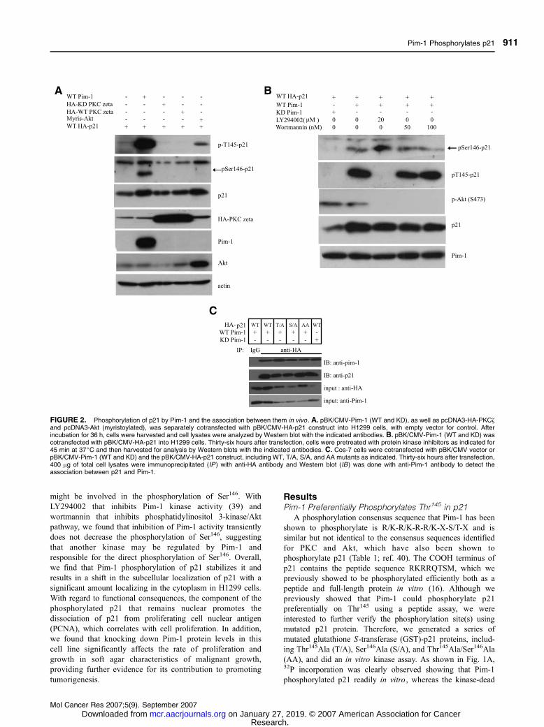
ResultsPim-1 Preferentially Phosphorylates Thr145 in p21
A phosphorylation consensus sequence that Pim-1 has been
shown to phosphorylate is R/K-R/K-R-R/K-X-S/T-X and is
similar but not identical to the consensus sequences identified
for PKC and Akt, which have also been shown to
phosphorylate p21 (Table 1; ref. 40). The COOH terminus of
p21 contains the peptide sequence RKRRQTSM, which we
previously showed to be phosphorylated efficiently both as a
peptide and full-length protein in vitro (16). Although we
previously showed that Pim-1 could phosphorylate p21
preferentially on Thr145 using a peptide assay, we were
interested to further verify the phosphorylation site(s) using
mutated p21 protein. Therefore, we generated a series of
mutated glutathione S-transferase (GST)-p21 proteins, includ-
ing Thr145Ala (T/A), Ser146Ala (S/A), and Thr145Ala/Ser146Ala
(AA), and did an in vitro kinase assay. As shown in Fig. 1A,32P incorporation was clearly observed showing that Pim-1
phosphorylated p21 readily in vitro , whereas the kinase-dead
FIGURE 2. Phosphorylation of p21 by Pim-1 and the association between them in vivo . A. pBK/CMV-Pim-1 (WT and KD), as well as pcDNA3-HA-PKC~and pcDNA3-Akt (myristoylated), was separately cotransfected with pBK/CMV-HA-p21 construct into H1299 cells, with empty vector for control. Afterincubation for 36 h, cells were harvested and cell lysates were analyzed by Western blot with the indicated antibodies. B. pBK/CMV-Pim-1 (WT and KD) wascotransfected with pBK/CMV-HA-p21 into H1299 cells. Thirty-six hours after transfection, cells were pretreated with protein kinase inhibitors as indicated for45 min at 37jC and then harvested for analysis by Western blots with the indicated antibodies. C. Cos-7 cells were cotransfected with pBK/CMV vector orpBK/CMV-Pim-1 (WT and KD) and the pBK/CMV-HA-p21 construct, including WT, T/A, S/A, and AA mutants as indicated. Thirty-six hours after transfection,400 Ag of total cell lysates were immunoprecipitated (IP ) with anti-HA antibody and Western blot (IB ) was done with anti-Pim-1 antibody to detect theassociation between p21 and Pim-1.
Pim-1 Phosphorylates p21
Mol Cancer Res 2007;5(9). September 2007
911
Research. on January 27, 2019. © 2007 American Association for Cancermcr.aacrjournals.org Downloaded from

(KD) form of Pim-1 did not. We found that, of the two
potential target sites, Thr145 and Ser146, Thr145 was efficiently
phosphorylated relative to Ser146. When both sites have been
mutated to alanine, phosphorylation was essentially eliminated,
indicating that there are no other sites in p21 phosphorylated
by Pim-1. Further analysis by Western blot with two
phosphorylated-specific antibodies confirmed this preference
(Fig. 1B). In this experiment, the two phosphorylated-specific
antibodies to p21 were anti-pS146-p21 and anti-pT145-p21.
Results for the wild-type (WT) GST-p21 showed that only
Thr145 was phosphorylated, whereas Ser146 phosphorylation
was undetectable. However, when Thr145 was mutated to
alanine, detectable phosphorylation occurred on Ser146 as
shown in the lane with T/A. This indicates that Thr145 is the
preferential site for phosphorylation by Pim-1. To further
determine if phosphorylation on Thr145 had any effect on
phosphorylation of Ser146, we designed two peptides. The first
one included a phosphate group on Thr145 and the second
peptide contained an aspartic acid to mimic phosphorylation.
As illustrated in Fig. 1C, WT peptide was efficiently
phosphorylated. However, if Thr145 was already phosphory-
lated, incorporation of phosphate was dramatically reduced.
FIGURE 3. Phosphorylation on Thr145 ofp21 by Pim-1 promotes its stabilization.A. H1299 cells were transiently cotrans-fected with pBK/CMV, pBK/CMV-WT-Pim-1,or pBK/CMV-KD-Pim-1 together with pBK/CMV-HA-p21 (WT). Following a previouslypublished protocol to analyze protein half-lifeassay (35), 24 h after transfection, cellswere treated with cycloheximide (CHX) at aconcentration of 25 Ag/mL for the indicatedtimes. Cells were then harvested and ana-lyzed by Western blot with anti-p21 antibodyto detect p21. B. Quantitation of p21amounts was determined using ImageJsoftware. The plot shows the degradationof p21 after cycloheximide addition.C. H1299 cells were transfected with equalamounts of HA-tagged p21 constructs,including WT, T/D, S/D, DD, T/A, S/A, andAA to determine the transfection efficiency,and cells were cotransfected with equalamount of pEGFP-C1, which allows visual-ization by fluorescence microscopy to checkfor transfection efficiency. Thirty-six hoursafter transfection, cells were harvested andnormalized for evaluating the steady levelsof these transfected proteins. The mem-brane was reprobed with anti-actin antibodyto verify equal loading. D. H1299 cells weretransfected with HA-tagged p21 plasmids(WT, T/D, S/D, DD, and AA). Twenty-fourhours after transfection, cells were treatedwith cycloheximide (25 Ag/mL). At theindicated time, the cells were lysed and50 Ag of cell lysates were used to detect theexogenous p21 degradation rates by West-ern blot using anti-p21 antibody. Membraneswere reprobed with anti-actin to ensureequal loading. E. Quantitation of the p21protein amounts in D and plotted as proteinremaining after cycloheximide addition withtime.
Zhang et al.
Mol Cancer Res 2007;5(9). September 2007
912
Research. on January 27, 2019. © 2007 American Association for Cancermcr.aacrjournals.org Downloaded from
Our result also shows that a spartic acid at position 145
influences phosphorylation of Ser146 in a very similar manner
to the peptide with the phosphorylated Thr145.
Pim-1 Phosphorylates p21 on Thr145 and IndirectlyPromotes Phosphorylation on Ser146 In vivo
To identify the phosphorylation sites of p21 by Pim-1
in vivo , we cotransfected pBK/CMV-Pim-1 (both WT and KD)
and the pBK/CMV-HA-p21 constructs into the p53-null H1299
cells. For phosphorylation controls, we also carried out
cotransfection of the pBK/CMV-HA-p21 with pcDNA3-HA-
PKC~ and pcDNA3-myristoylated-Akt because PKC~ and Akt
are two known kinases that can phosphorylate p21 at either
Thr145 or Ser146. An empty vector was used as a control. As
illustrated in Fig. 2A, using phosphorylated-specific antibodies,
we found that in the presence of Pim-1 both Thr145 and Ser146
are phosphorylated. On the other hand, Akt only phosphor-
ylates Thr145, whereas PKC~ seems to phosphorylate neither
Thr145 nor Ser146 at least under our experimental conditions.
From the total p21 levels, it seems that overexpression of Pim-1
caused p21 levels to be increased. The fact that the presence of
Pim-1 leads to phosphorylation of Ser146 in vivo was surprising
because Ser146 is not an efficient phosphorylation site for Pim-1
in vitro . This prompted us to think that another kinase could
be activated by Pim-1 and might be responsible for the direct
phosphorylation on Ser146. To test this hypothesis, we
transiently inhibited Pim-1 kinase activity with an inhibitor to
determine if phosphorylation on Ser146 might be reduced.
Currently, there is only one inhibitor called LY294002 that is
reported to inhibit Pim-1 kinase. We pretreated the cells with
LY294002 for 45 min at 37jC and then harvested the cells for
analysis as shown in Fig. 2B. We observed that phosphorylation
on Thr145 is diminished at 20 Amol/L, but phosphorylation on
Ser146 is not affected at all. Rather, it seems that phosphory-
lation on Ser146 is increased a bit. This suggests that the
mechanism for phosphorylation at each site is different. To
eliminate the possibility of the involvement of phosphatidyli-
nositol 3-kinase/Akt in the phosphorylation of either site, we
also used wortmannin as an inhibitor because LY294002 inhi-
bits not only Pim kinase but also Akt at certain concentrations.
We found that wortmannin at 100 nmol/L, which inhibits
phosphatidylinositol 3-kinase/Akt, does not influence phos-
phorylation of either Thr145 or Ser146 compared with cells
without inhibitors. Therefore, most likely, Ser146 is phosphor-
ylated by another kinase and not directly by Pim-1, although the
presence of Pim-1 promotes the phosphorylation on Ser146,
whereas KD Pim-1 does not.
The Association of Pim-1 with p21 Can Be DetectedIn vivo
Phosphorylation of p21 by Pim-1 means that there is an
enzyme-substrate interaction. Coimmunoprecipitation was done
on cotransfected Cos-7 cells with p21 and Pim-1 (Fig. 2C). In
this case, hemagglutinin (HA)-tagged p21 expression vectors
were used expressing either WT, T/A, S/A, or the double-
mutant AA protein. These vectors were cotransfected with WT
Pim-1. The KD Pim-1 was also cotransfected with WT HA-
p21. Anti-p21 antibody was used to pull down Pim-1 along
with p21. We found association between the two proteins even
when the two sites were mutated. This indicates that binding
between Pim-1 and p21 does not require a form of p21 that can
be phosphorylated. We also observed association of the KD
Pim-1 with p21.
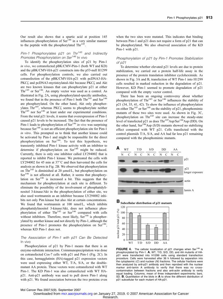
Phosphorylation of p21 by Pim-1 Promotes Stabilizationof p21
To determine whether elevated p21 levels are due to protein
stabilization, we carried out a protein half-life assay in the
presence of the protein translation inhibitor cycloheximide. As
shown in Fig. 3A and B, transfection of WT Pim-1 into H1299
cells resulted in marked reduction in the degradation of p21.
However, KD Pim-1 seemed to promote degradation of p21
compared with the empty vector control.
There has been an ongoing controversy about whether
phosphorylation of Thr145 or Ser146 influences the stability of
p21 (34, 35, 41, 42). To show the influence of phosphorylation
on either Thr145 or Ser146 on the stability of p21, phosphomimic
mutants of these two sites were used. As shown in Fig. 3C,
phosphorylation on Thr145 site can increase the steady-state
level of transfected p21 as does Thr145Asp/Ser146Asp (DD). On
the other hand, Ser146Asp (S/D) mutants showed no stabilizing
effect compared with WT p21. Cells transfected with the
control plasmids T/A, S/A, and AA had far less p21 remaining
compared with the phosphomimic mutants.
FIGURE 4. The cellular localization of p21 changes when Ser146 isphosphorylated by Pim-1. A. WT, T/D, S/D, DD, and AA mutants of HA-p21 were transfected into H1299 cells using standard transfectionprocedure. Cells were harvested after 36 h followed by separation intothe cytoplasmic (C) and nuclear (N ) fractions. The cellular fractions werethen analyzed by anti-p21 antibody and then reprobed with the nuclearmarker anti-lamin A antibody to verify that there was no cross-contamination between fractions and also anti-actin antibody to verifyequal loading. Columns, mean of three independent experiments; bars,SD. B. Quantitation of the blots in A to show the different distribution ofp21 subcellular for each mutant of HA-p21.
Pim-1 Phosphorylates p21
Mol Cancer Res 2007;5(9). September 2007
913
Research. on January 27, 2019. © 2007 American Association for Cancermcr.aacrjournals.org Downloaded from

We also examined the protein half-life of these p21 mutants
while inhibiting protein translation with cycloheximide. As
shown in Fig. 3D and E, the phosphomimic mutant Thr145Asp
(T/D) was more stable compared with the WT. The DD mutant
was found to be similarly stable compared with the T/D
mutant. The S/D mutant seems to be a little less stable than
WT. These findings indicate that phosphorylation on Thr145
has an effect on the stability of p21. Taken together, our data
show that phosphorylation by Pim-1 preferentially on Thr145
stabilizes p21.
Phosphorylation of p21 by Pim-1 Changes Its SubcellularLocalization
The residue phosphorylated on p21 by Pim-1 occurs in the
COOH terminus where the nuclear localization signal is
located. Potentially, this phosphorylation could influence the
subcellular localization of p21. In fact, there is some con-
troversy over this issue and in some cases may be a result of cell
type used in the experiments (27, 43). For example, in HER2/
neu 3T3 cells in which Akt kinase was constitutively activated,
the phosphorylation of Thr145 on p21 caused its transloca-
tion from the nucleus to the cytoplasm (43). However, in human
umbilical vein endothelial cells, this did not occur (27). In our
studies, phosphomimic mutants of p21 at both sites were
analyzed in H1299 cells. As shown in Fig. 4A and B, com-
paring the total distribution of p21 in cytosolic and nuclear
fractions for WT and phosphomimic mutants of HA-p21, we
found that phosphorylation of Thr145 generally does not change
the nuclear localization of p21, which is what is observed
similar to the WT HA-p21. In contrast, phosphorylation of
Ser146 seems to cause the distribution of p21 to shift from the
nucleus to the cytoplasm as shown by the decrease of the
nuclear localization of p21. The DD mutant was distributed
similarly to the S/D mutant but there seems to be overall higher
p21 levels for the DD mutant presumably because of the
increased stability promoted by the mimicked phosphorylation.
We also examined the AA mutant and found that most of
the p21 in these cells is observed in the nucleus as would be
predicted, although the total amount is low as might also be
predicted.
To confirm this finding, we examined each of the mutants
and WT HA-p21 by confocal microscopy in H1299 cells.
NIH3T3 cells were also examined for comparison. It has been
reported that the T/D mutant of p21 has more cytoplasmic
FIGURE 5. H1299 (left ) or NIH3T3 (right ) cells were transfected with WT, T/D, S/D, DD, or AA mutants of HA-p21 and 36 h after transfection, cells werewashed with PBS, fixed with 4% paraformaldehyde, permeabilized with 0.3% Triton X-100, and incubated with anti-HA antibody and with Oregon Green goatanti-mouse antibody. Subsequently, the nucleus was stained with propidium iodide (PI ) and the cell preparation was examined by confocal microscopy.
Zhang et al.
Mol Cancer Res 2007;5(9). September 2007
914
Research. on January 27, 2019. © 2007 American Association for Cancermcr.aacrjournals.org Downloaded from
staining in these cells (44). NIH3T3 cells are murine fibroblast
cells, whereas H1299 cells are lung carcinoma cells. Different
localization patterns of p21 in these two cell types may reflect
the different metabolism of different cellular lineages. To show
that a difference exists for T/D or S/D mutants, we carried out
experiments with WT, T/D, S/D, DD, and AA mutants of p21
in H1299 and NIH3T3 cells. A typical field for each of the HA-
p21 mutants is shown in Fig. 5. As reported for NIH3T3 cells,
T/D causes nuclear p21 to redistribute in the cytoplasm. The
S/D mutant seems to be less efficient in the translocation to the
cytoplasm. However, for H1299 cells, the T/D mutant of p21
seems to be more localized in the nucleus than in the cytoplasm
and overall has a stronger fluorescence signal compared with
the WT HA-p21. The S/D mutant, in contrast, is mostly
localized throughout the cells. DD is similar to SD, and AA
mutant localized mostly in the nucleus. These observations
support the subcellular distribution of p21 mutants revealed by
Western blotting.
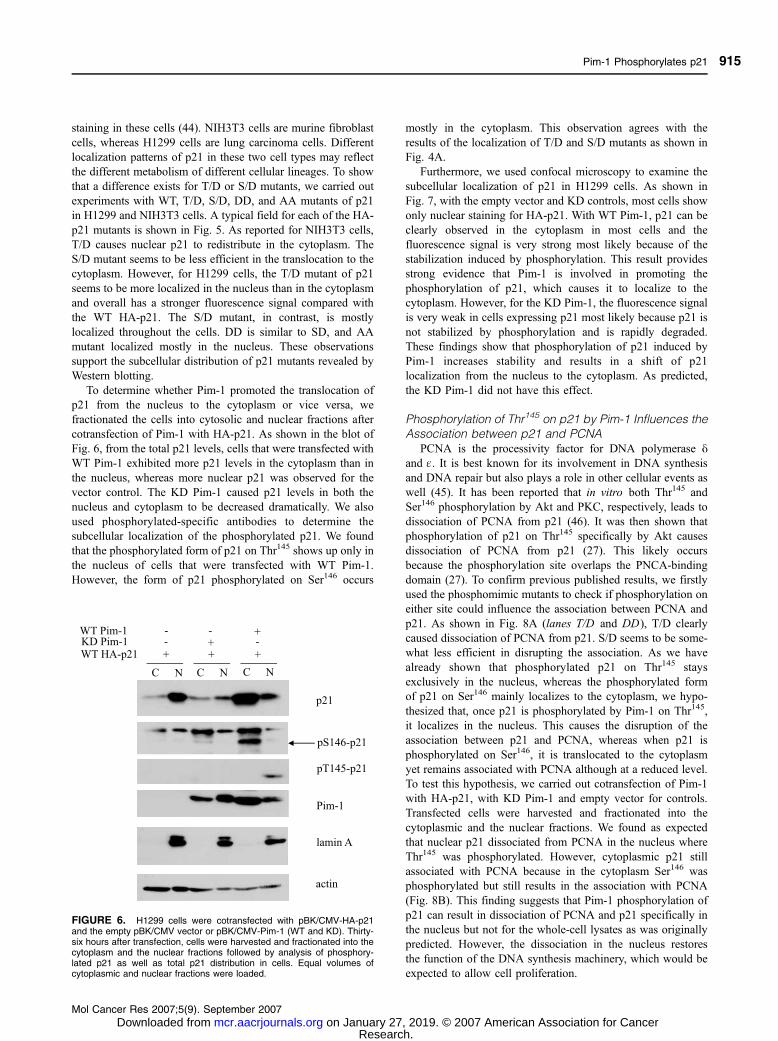
To determine whether Pim-1 promoted the translocation of
p21 from the nucleus to the cytoplasm or vice versa, we
fractionated the cells into cytosolic and nuclear fractions after
cotransfection of Pim-1 with HA-p21. As shown in the blot of
Fig. 6, from the total p21 levels, cells that were transfected with
WT Pim-1 exhibited more p21 levels in the cytoplasm than in
the nucleus, whereas more nuclear p21 was observed for the
vector control. The KD Pim-1 caused p21 levels in both the
nucleus and cytoplasm to be decreased dramatically. We also
used phosphorylated-specific antibodies to determine the
subcellular localization of the phosphorylated p21. We found
that the phosphorylated form of p21 on Thr145 shows up only in
the nucleus of cells that were transfected with WT Pim-1.
However, the form of p21 phosphorylated on Ser146 occurs
mostly in the cytoplasm. This observation agrees with the
results of the localization of T/D and S/D mutants as shown in
Fig. 4A.
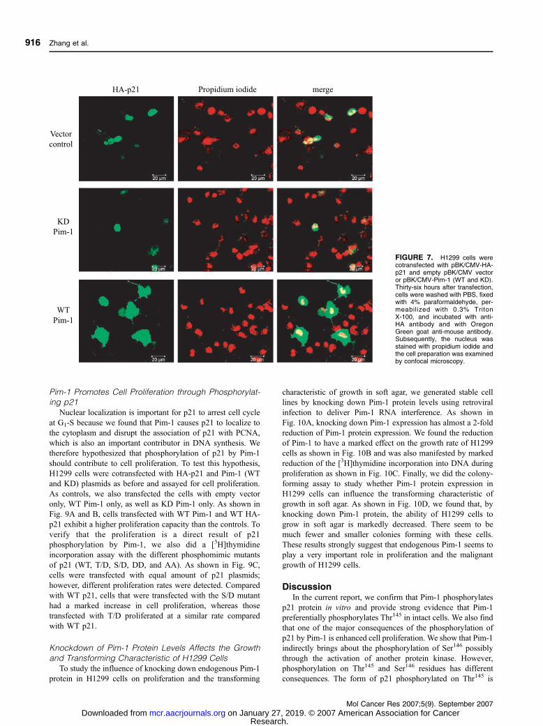
Furthermore, we used confocal microscopy to examine the
subcellular localization of p21 in H1299 cells. As shown in
Fig. 7, with the empty vector and KD controls, most cells show
only nuclear staining for HA-p21. With WT Pim-1, p21 can be
clearly observed in the cytoplasm in most cells and the
fluorescence signal is very strong most likely because of the
stabilization induced by phosphorylation. This result provides
strong evidence that Pim-1 is involved in promoting the
phosphorylation of p21, which causes it to localize to the
cytoplasm. However, for the KD Pim-1, the fluorescence signal
is very weak in cells expressing p21 most likely because p21 is
not stabilized by phosphorylation and is rapidly degraded.
These findings show that phosphorylation of p21 induced by
Pim-1 increases stability and results in a shift of p21
localization from the nucleus to the cytoplasm. As predicted,
the KD Pim-1 did not have this effect.
Phosphorylation of Thr145 on p21 by Pim-1 Influences theAssociation between p21 and PCNA
PCNA is the processivity factor for DNA polymerase yand q. It is best known for its involvement in DNA synthesis
and DNA repair but also plays a role in other cellular events as
well (45). It has been reported that in vitro both Thr145 and
Ser146 phosphorylation by Akt and PKC, respectively, leads to
dissociation of PCNA from p21 (46). It was then shown that
phosphorylation of p21 on Thr145 specifically by Akt causes
dissociation of PCNA from p21 (27). This likely occurs
because the phosphorylation site overlaps the PNCA-binding
domain (27). To confirm previous published results, we firstly
used the phosphomimic mutants to check if phosphorylation on
either site could influence the association between PCNA and
p21. As shown in Fig. 8A (lanes T/D and DD), T/D clearly
caused dissociation of PCNA from p21. S/D seems to be some-
what less efficient in disrupting the association. As we have
already shown that phosphorylated p21 on Thr145 stays
exclusively in the nucleus, whereas the phosphorylated form
of p21 on Ser146 mainly localizes to the cytoplasm, we hypo-
thesized that, once p21 is phosphorylated by Pim-1 on Thr145,
it localizes in the nucleus. This causes the disruption of the
association between p21 and PCNA, whereas when p21 is
phosphorylated on Ser146, it is translocated to the cytoplasm
yet remains associated with PCNA although at a reduced level.
To test this hypothesis, we carried out cotransfection of Pim-1
with HA-p21, with KD Pim-1 and empty vector for controls.
Transfected cells were harvested and fractionated into the
cytoplasmic and the nuclear fractions. We found as expected
that nuclear p21 dissociated from PCNA in the nucleus where
Thr145 was phosphorylated. However, cytoplasmic p21 still
associated with PCNA because in the cytoplasm Ser146 was
phosphorylated but still results in the association with PCNA
(Fig. 8B). This finding suggests that Pim-1 phosphorylation of
p21 can result in dissociation of PCNA and p21 specifically in
the nucleus but not for the whole-cell lysates as was originally
predicted. However, the dissociation in the nucleus restores
the function of the DNA synthesis machinery, which would be
expected to allow cell proliferation.
FIGURE 6. H1299 cells were cotransfected with pBK/CMV-HA-p21and the empty pBK/CMV vector or pBK/CMV-Pim-1 (WT and KD). Thirty-six hours after transfection, cells were harvested and fractionated into thecytoplasm and the nuclear fractions followed by analysis of phosphory-lated p21 as well as total p21 distribution in cells. Equal volumes ofcytoplasmic and nuclear fractions were loaded.
Pim-1 Phosphorylates p21
Mol Cancer Res 2007;5(9). September 2007
915
Research. on January 27, 2019. © 2007 American Association for Cancermcr.aacrjournals.org Downloaded from
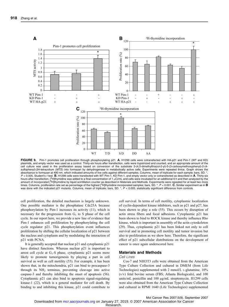
Pim-1 Promotes Cell Proliferation through Phosphorylat-ing p21
Nuclear localization is important for p21 to arrest cell cycle
at G1-S because we found that Pim-1 causes p21 to localize to
the cytoplasm and disrupt the association of p21 with PCNA,
which is also an important contributor in DNA synthesis. We
therefore hypothesized that phosphorylation of p21 by Pim-1
should contribute to cell proliferation. To test this hypothesis,
H1299 cells were cotransfected with HA-p21 and Pim-1 (WT
and KD) plasmids as before and assayed for cell proliferation.
As controls, we also transfected the cells with empty vector
only, WT Pim-1 only, as well as KD Pim-1 only. As shown in
Fig. 9A and B, cells transfected with WT Pim-1 and WT HA-
p21 exhibit a higher proliferation capacity than the controls. To
verify that the proliferation is a direct result of p21
phosphorylation by Pim-1, we also did a [3H]thymidine
incorporation assay with the different phosphomimic mutants
of p21 (WT, T/D, S/D, DD, and AA). As shown in Fig. 9C,
cells were transfected with equal amount of p21 plasmids;
however, different proliferation rates were detected. Compared
with WT p21, cells that were transfected with the S/D mutant
had a marked increase in cell proliferation, whereas those
transfected with T/D proliferated at a similar rate compared
with WT p21.
Knockdown of Pim-1 Protein Levels Affects the Growthand Transforming Characteristic of H1299 Cells
To study the influence of knocking down endogenous Pim-1
protein in H1299 cells on proliferation and the transforming
characteristic of growth in soft agar, we generated stable cell
lines by knocking down Pim-1 protein levels using retroviral
infection to deliver Pim-1 RNA interference. As shown in
Fig. 10A, knocking down Pim-1 expression has almost a 2-fold
reduction of Pim-1 protein expression. We found the reduction
of Pim-1 to have a marked effect on the growth rate of H1299
cells as shown in Fig. 10B and was also manifested by marked
reduction of the [3H]thymidine incorporation into DNA during
proliferation as shown in Fig. 10C. Finally, we did the colony-
forming assay to study whether Pim-1 protein expression in
H1299 cells can influence the transforming characteristic of
growth in soft agar. As shown in Fig. 10D, we found that, by
knocking down Pim-1 protein, the ability of H1299 cells to
grow in soft agar is markedly decreased. There seem to be
much fewer and smaller colonies forming with these cells.
These results strongly suggest that endogenous Pim-1 seems to
play a very important role in proliferation and the malignant
growth of H1299 cells.
DiscussionIn the current report, we confirm that Pim-1 phosphorylates
p21 protein in vitro and provide strong evidence that Pim-1
preferentially phosphorylates Thr145 in intact cells. We also find
that one of the major consequences of the phosphorylation of
p21 by Pim-1 is enhanced cell proliferation. We show that Pim-1
indirectly brings about the phosphorylation of Ser146 possibly
through the activation of another protein kinase. However,
phosphorylation on Thr145 and Ser146 residues has different
consequences. The form of p21 phosphorylated on Thr145 is
FIGURE 7. H1299 cells werecotransfected with pBK/CMV-HA-p21 and empty pBK/CMV vectoror pBK/CMV-Pim-1 (WT and KD).Thirty-six hours after transfection,cells were washed with PBS, fixedwith 4% paraformaldehyde, per-meabilized with 0.3% TritonX-100, and incubated with anti-HA antibody and with OregonGreen goat anti-mouse antibody.Subsequently, the nucleus wasstained with propidium iodide andthe cell preparation was examinedby confocal microscopy.
Zhang et al.
Mol Cancer Res 2007;5(9). September 2007
916
Research. on January 27, 2019. © 2007 American Association for Cancermcr.aacrjournals.org Downloaded from

stabilized and localizes to the nucleus, which leads to disruption
of the association between PCNA and p21. On the other hand,
when p21 is phosphorylated on Ser146, this causes p21 to
localize to the cytoplasm, yet maintaining its association
with PCNA.
Our data show that a major increase in stability of p21
occurs when Thr145 is phosphorylated, whereas phosphoryla-
tion of Ser146 seems to have little effect on stability. The
increase in stability of p21 when Thr145 is phosphorylated is
consistent with a previous report in which the death-
associated kinase Zip can phosphorylate p21 on Thr145 and
cause its stabilization (41). The COOH terminus of p21 has
previously been shown to be very important in controlling its
stability. This occurs with the C8 subunit of proteasome
binding to it, which then leads to ubiquitin-independent
degradation (32). On the other hand, PCNA and cyclin D1
have also been shown to cause stabilization of p21 (37, 47).
The molecular mechanism for the increased stability may
involve the phosphate at Thr145 hindering the ubiquitination
of Lys141 (48).
Contrary to what has been reported previously, we found
that not Thr145 phosphorylation but rather Ser146 phosphoryla-
tion is what leads to p21 translocation to the cytoplasm from the
nucleus. We found that p21 phosphorylated on Thr145 localizes
in the nucleus. Although this is in contrast to what has been
previously found with fibroblasts and cancer cells where
phosphorylation of p21 by Akt on Thr145 changes its
subcellular localization (43), it also suggests that the cell type
could be an important factor in the results one obtains from
such experiments. For example, another group did not observe
the translocation effect with p21 in endothelial cells with Akt.
Rather, it was observed that the T/D mutant tended to localize
in the nucleus (27). Here, we present evidence that, in the lung
carcinoma cell line H1299, phosphorylated p21 on Thr145
is localized to the nucleus, whereas the form of p21 phos-
phorylated on Ser146 mainly localizes in the cytoplasm. Our
data with the phosphomimic mutants of p21 are consistent with
this observation. Because both Thr145 and Ser146 are part of the
nuclear localization signal sequence of p21, the different
localization patterns might be a result of different binding
partners that may vary from cell type to cell type and have a
preference for one or the other phosphorylated forms of p21.
Because we found that phosphorylation on Thr145 and Ser146
seems to occur by different pathways (Fig. 2B), the possibility
exists that Pim-1 might activate another kinase in vivo and
cause Ser146 to be phosphorylated or Pim-1 may influence
the activity of phosphatases in vivo (11, 12, 49) and thereby
affect the phosphorylation status on Ser146.
Although dissociation of p21 from PCNA should allow cell
cycling to resume, apparently cellular localization and stability
of p21 play more important roles for reasons that are not clear
at this time. In vivo , we detected phosphorylation of both
Thr145 and Ser146, and each of these forms of phosphorylated
p21 localized differently. In addition to the DD mutant being
very stable and localizing to both the nucleus and cytoplasm
(Fig. 4A), it is not entirely surprising that proliferation
promoted by the DD mutant of p21 seems to be similar to that
of the WT p21. It is possible that the double charge results in
canceling some of the effects observed for each of the T/D
and S/D mutants. Because the AA mutant is not very stable
(Fig. 4A), the proliferation observed for it was similar to that
of the WT.
Taken together, our data show that overexpression of Pim-1
results in an increase in the total level of p21 with cytoplasmic
localization of p21 in general. Through modulating p21
localization and its association with PCNA, Pim-1 promotes
cell proliferation. Knocking down Pim-1 protein expression
dramatically decreases the proliferation rate of H1299 cells and
their ability to grow in soft agar. These results suggest that Pim-1
probably plays a very important role in the malignant growth of
H1299 cells as shown in vitro . Pim-1 has been thought to play
an important role in the transduction of mitogenic signals from
cytokines because Pim-1 expression is rapidly induced after
cytokine stimulation (50-52). The proliferative response to
cytokines is impaired in cells from pim-1 –deficient mice (52).
The function of Pim kinases in proliferation is also clearly
shown by the compound Pim knockout mice, which show much
reduced body size at birth and throughout postnatal life (6). It
was reported that, in smooth muscle cells, infection of
adenovirus encoding KD Pim-1 remarkably reduced cell
growth (53). Pim-1 was also found to be highly expressed in
prostate cancer cells, and overexpression of Pim-1 kinase
dramatically enhances the growth of tumor cells (54). Despite
the many lines of evidence indicating the function of Pim-1 in
FIGURE 8. Phosphorylation of p21 by Pim-1 leads to disruption of theassociation between p21 and PCNA.A. HA-p21 constructs (WT, T/D, S/D,DD, T/A, S/A, and AA) were transiently transfected into H1299 cells.Thirty-six hours after transfection, cells were lysed and cell lysates wereimmunoprecipitated with anti-HA antibody and immunoblotted with anti-PCNA antibody. Membranes were then reprobed with anti-p21 antibody.Five percent of input cell lysates were also analyzed with anti-PCNAantibody. B. Thirty-six hours after transfection of indicated plasmids,H1299 cells were harvested and fractionated into cytoplasmic and nuclearfractions followed by immunoprecipitation with anti-HA antibody for each ofthe cytoplasmic and the nuclear lysates; immunocomplexes wereanalyzed by anti-PCNA and anti-p21, and 5% of input cell lysates werealso analyzed with anti-lamin A and anti-PCNA.
Pim-1 Phosphorylates p21
Mol Cancer Res 2007;5(9). September 2007
917
Research. on January 27, 2019. © 2007 American Association for Cancermcr.aacrjournals.org Downloaded from
cell proliferation, the detailed mechanism is largely unknown.
One possible mediator is the phosphatase Cdc25A because
phosphorylation by Pim-1 increases its activity (11), which is
necessary for the progression from G1 to S phase of the cell
cycle. In our report here, we provide a new line of evidence that
Pim-1 enhances cell proliferation by phosphorylating the cell
cycle regulator p21. This phosphorylation event influences
proliferation by shifting the cellular localization of p21 between
the nucleus and cytoplasm and by modulating the interaction of
p21 with PCNA.
It is generally accepted that nuclear p21 and cytoplasmic p21
have distinct functions. Whereas nuclear p21 is important to
arrest cell cycle at G1-S phase, cytoplasmic p21 seems more
likely to promote tumorigenesis by playing a part in cell
survival as well as cell motility (55). For example, it has been
shown that, in the mitochondria, p21 can bind to procaspase-3
through its NH2 terminus, preventing cleavage into active
caspase-3 and thereby inhibiting the onset of apoptosis (56).
Cytoplasmic p21 can also bind to apoptosis signal-regulating
kinase-1 (22), which is a general mediator for cell death. By
binding to and inhibiting this kinase, p21 could contribute to
cell survival. In terms of cell motility, cytoplasmic localization
of cyclin-dependent kinase inhibitors, such as p21 and p27, has
been shown to play a role (55). This occurs by disruption of
actin stress fibers and focal adhesions. Cytoplasmic p21 has
been shown to bind to ROCK kinase and thereby influence Rho
kinase, which is important in assembly of the actin cytoskeleton
(29). Thus, cytoplasmic p21 has been linked not only to cell
survival and in promoting cell motility and tumor invasion but
also to proliferation as we show here. Therefore, the significant
effect of p21 subcellular distributions on the development of
cancer is once again underscored here.
Materials and MethodsCell Lines
Cos-7 and NIH3T3 cells were obtained from the American
Type Culture Collection and cultured in DMEM (from Life
Technologies) supplemented with 2 mmol/L L-glutamine, 10%
(v/v) fetal bovine serum (FBS; Atlanta Biologicals), and 100
units/mL penicillin and 100 Ag/mL streptomycin. H1299 cells
were also obtained from the American Type Culture Collection
and cultured in RPMI 1640 (Life Technologies) supplemented
FIGURE 9. Pim-1 promotes cell proliferation through phosphorylating p21. A. H1299 cells were cotransfected with HA-p21 and Pim-1 (WT and KD)plasmids, and empty vector was used as a control. Thirty-six hours after transfection, cells were trypsinized and counted, and an appropriate amount of thecell culture was used in the proliferation assay based on conversion of the substrate 3-(4,5-dimethylthiazol-2-yl)-5-(3-carboxymethoxyphenyl)-2-(4-sulfophenyl)-2H-tetrazolium (MTS ) into formazan by dehydrogenase in metabolically active cells. Experiments were repeated thrice. Graph shows theabsorbance to formazan at 490 nm, which indicated amounts of live cells against different samples. Columns, mean of triplicate for each sample; bars, SD. *,P < 0.005, Student’s t test. B. H1299 cells were transfected with WT Pim-1, KD Pim-1, and empty vector only or cotransfected as described in A. Thirty-sixhours after transfection, [3H]thymidine was added to a final concentration of 1 ACi/mL and cells were incubated for an additional 6 h and then analyzed by theamount of incorporated [3H]thymidine by liquid scintillation counter as described in Materials and Methods. Experiments were repeated for at least two moretimes. Columns, proliferation rate set as percentage of the highest [3H]thymidine incorporated samples; bars, SD. *, P < 0.001. C. Similar experiment as in Bwas done with the indicated p21 mutants. Columns, mean of triplicate; bars, SD. *, P < 0.005, statistically significant difference from controls.
Zhang et al.
Mol Cancer Res 2007;5(9). September 2007
918
Research. on January 27, 2019. © 2007 American Association for Cancermcr.aacrjournals.org Downloaded from

with 10% FBS and 100 units/mL penicillin and 100 Ag/mL
streptomycin. GP2-293 cells were purchased from Clontech and
cultured in DMEM supplemented with 10% FBS and anti-
biotics as above. All cells were maintained at 37jC in a
humidified atmosphere of 95% air and 5% CO2.
Transfection and Stable Cell Line GenerationCos-7 and H1299 cells were transfected using Lipofect-
amine 2000 (Invitrogen) following the manufacturer’s instruc-
tion. For NIH3T3 cells, transfectin from Bio-Rad was used
following the manufacturer’s instructions. To generate retro-
virus to infect target cells, GP2-293 cells were cotransfected
with pVSV-G (from Clontech) and pSIREN (from Clontech),
which contains an oligonucleotide coding for human Pim-1
small interfering RNA to knock down human pim-1 gene by the
method of calcium phosphate precipitation (CalPhos Mamma-
lian Transfection kit was purchased from Clontech). The
sequence of the oligonucleotide was kindly provided by
Dr. Xueke You (Cornell University, Ithaca, NY). It contains a
sense 19-nucleotide strand (5¶-GATCTCTTCGACTTCATCA)
sequence followed by a spacer (5¶-TTCAAGAGA) and its
reverse complementary strand followed by five thymidines as a
RNA polymerase III transcriptional stop signal. In parallel,
empty vector pSIREN was also cotransfected with pVSV-G to
generate the control cell line. Forty-eight hours after transfec-
tion, supernatant was collected and filtered with 0.45-Am unit
and target cells were infected in the presence of 8 Ag/mL
polybrene. Spinoculation was done at 750 � g for 2 h at 32jC
to enhance the infection efficiency. Thirty-six hours after
infection, cells were put under selection with 2 Ag/mL
puromycin for 7 days to establish the stable Pim-1 RNA
interference–expressing cell line and the control cell line.
Plasmids and ConstructsUsing standard PCR protocols, an NH2-terminal HA-tag was
added to the cDNA of WT human p21 and subcloned into a
modified pBK/CMV vector in which LacZ promoter was
deleted. To generate mutated p21, a PCR-based site-directed
mutagenesis was carried out to generate T/A, S/A, and AA
mutant and also T/D, S/D, and DD mutants. To generate GST-
p21 with the desired mutations, p21 cDNA was subcloned into
pGEX-2T and used as a template to generate mutated GST-p21
with T/A, S/A, and AA. COOH-terminal 6� His-tagged WT
Pim-1 and KD Pim-1 were subcloned into pET30a digested
with NdeI/KpnI double enzymes. Sequence analysis was done
to confirm the correct sequences. pcDNA3-myristoylated-Akt
was purchased from Addgene.
Antibodies and ReagentsAntibodies used included anti-p21 SXM30 monoclonal anti-
body (BD Pharmingen), anti-actin (Sigma), anti-HA antibody
and anti-pAkt-S473 (Cell Signaling), anti-pT145-p21 and anti-
pS146-p21 (Santa Cruz Biotechnology), anti-PCNA (Transduc-
tion Laboratories), and anti-lamin A and anti-Akt (BioLegend).
Anti-Pim-1 1140p polyclonal antibody was produced in our
laboratory using full-length recombinant Pim-1 expressed in
Escherichia coli . Reagents used include cycloheximide (Sig-
ma), LY294002 (Calbiochem), wortmannin (Calbiochem),
puromycin (Clontech), and noble agar (U.S. Biochemical).
Recombinant Protein PurificationPlasmids pGEX-2T-p21 (WT or mutants) were transformed
into E. coli strain BL-21pLysS (DE3), and 0.1 mmol/L isopropyl-
L-thio-B-D-galactopyranoside was used to induce the recombinant
FIGURE 10. Knockdown of Pim-1 protein decreases the proliferationand transforming characteristic of H1299 cells. A. H1299 cells eitherinfected with control retrovirus or retrovirus that knocks down Pim-1protein expression and selected with 2 Ag/mL puromycin for 7 d to set upstable Pim-1 knockdown cell lines. Western blotting with Pim-1 antibody(1140p) was done to analyze Pim-1 protein expression. Actin was usedfor protein loading control. B. Approximately 6.0 � 104 cells were seededinto 24-well plates in quadruplicates for both the stable Pim-1 knockdowncell line and the control cell line. Live cells were then counted every24 h with trypan blue. A growth curve was generated for each cell lineafter 6 d. Points, mean of the quadruplicates; bars, SD. C. Cells (5 � 104)were plated into 24-well plates for both the control and knockdown celllines. Cells were then serum starved for 24 h and then stimulated to growwith RPMI 1640/10% FBS. One microcurie per milliliter [3H]thymidine(methyl-) was added and incubated for an additional 6 h. Cells were thenharvested and analyzed for radioactivity as shown above. *, P < 0.001,Student’s t test, there is statistically significant difference. D. Soft agarwas used to compare the transforming characteristic between the controlH1299 cells and Pim-1 knockdown cells. Approximately 1 � 104 cellswere plated into each well of a six-well plate in triplicate for each sample.Two weeks later, colonies were photographed and images were capturedat �10 magnification.
Pim-1 Phosphorylates p21
Mol Cancer Res 2007;5(9). September 2007
919
Research. on January 27, 2019. © 2007 American Association for Cancermcr.aacrjournals.org Downloaded from

protein expression. Cells were sonicated and centrifuged, and the
supernatants were incubated with PBS-equilibrated Sepharose-
glutathione 4B beads followed by extensive washing. Protein
was then eluted with 20 mmol/L glutathione in 100 mmol/L Tris
buffer followed by dialysis against Tris buffer without
glutathione at 4jC overnight. To prepare recombinant Pim-1
kinases (WT and KD), pET30(a)-Pim-1 plasmids were trans-
formed into E. coli strain BL-21pLysS (DE3), and 1 mmol/L
isopropyl-L-thio-B-D-galactopyranoside was used to carry out
the induction. The protein was affinity purified with nickel-
nitrilotriacetic acid beads and eluted with 300 mmol/L imidazole.
Protein was subsequently dialyzed against Tris buffer at 4jC
overnight.
Cycloheximide TreatmentH1299 cells were plated on six-well tissue culture plates
1 day before transfection. Standard protocol was followed to
transfect these cells using LipofectAMINE 2000 reagent.
Twenty-four hours after transfection, cells were treated with
cycloheximide with a final concentration of 25 Ag/mL with the
indicated time.
In vitro Kinase AssayFor kinase assay analyzed by 32P autoradiography, each
GST substrate protein (2 Ag) was incubated with 0.2 Ag Pim-1
(WT or KD) in 50 AL kinase buffer [20 mmol/L MOPS (pH
7.4), 150 mmol/L NaCl, 12.5 mmol/L MgCl2, 1 mmol/L
MnCl2, 1 mmol/L EGTA, 1 mmol/L DTT, 10 Amol/L ATP]
containing 20 ACi [g-32P]ATP. The reactions were carried out at
room temperature for 20 min and stopped with 2� Laemmli
buffer, samples were boiled for 10 min, and then proteins were
isolated in SDS-PAGE and transferred to polyvinylidene
difluoride membrane until exposed to X-film. For the kinase
assay without the radiolabel, each of WT or mutant GST-p21
proteins (4 Ag) was incubated with 0.5 Ag of WT Pim-1 or KD
Pim-1 kinase in 100 AL of kinase buffer containing 10 Amol/L
unlabeled ATP, reactions were stopped as above, and samples
were analyzed by Western blot with the indicated antibodies.
For the kinase assay using the peptide substrates, reactions were
carried in the same buffer but with 0.5 mmol/L peptides as
substrates. After reactions, 100 Ag bovine serum albumin was
added and then trichloroacetic acid was added to a final
concentration of 2.5% to stop the reactions. The samples were
kept in ice for 30 min and then centrifuged at 12,000 rpm at
4jC for 15 min. An aliquot of the supernatant was then spotted
on a phosphocellulose filter (2.1 cm diameter, Whatman P-81)
and washed with 15 mmol/L phosphoric acid extensively. Filter
papers were then air dried and put in scintillation counting vials
for analysis.
Cell Lysate Preparation and Western BlottingCells were trypsinized and harvested, washed with PBS once,
and resuspended in cell lysis buffer containing 25 mmol/L
Tris-HCl (pH 7.5), 1% (w/v) NP40, 1 mmol/L EDTA, 1 mmol/L
activated sodium orthovanadate, 5 mmol/L sodium fluoride,
protease inhibitor cocktail set I (Calbiochem), and 150 mmol/L
NaCl. After brief sonication, cell lysates were centrifuged at
13,000 rpm for 10 min. Protein concentration was determined
and equivalent amounts of lysate based on protein concentra-
tion were added to an equal volume of 2� Laemmli buffer
and boiled for 10 min. For Western blot analysis, protein was
separated by SDS-PAGE and transferred to polyvinylidene
difluoride membrane. Membranes were subsequently blocked
with 5% nonfat dry milk-PBS-Tween 20 for 1 h at room
temperature and incubated with primary antibody at optimized
dilution for 2 h at room temperature. Membranes were then
washed, incubated with horseradish peroxidase–conjugated
secondary antibody (Santa Cruz biotechnology) at 1:10,000
for 1 h, washed, treated with enhanced chemiluminescence
reagent (Pierce), and exposed to hyperfilm. To quantitate the
signals in Western blots, ImageJ software was downloaded
from the Web site1 and used to carry out the densitometry
for blots.
Cell FractionationCytoplasmic/nuclear fractions were made using NE-PER
from Pierce according to the manufacturer’s instructions.
ImmunoprecipitationsCells were harvested as described previously and then
resuspended in the cell lysis buffer and incubated on ice for
10 min before centrifugation. The cell lysates were then
precleared with protein G-agarose beads (Roche Diagnostics)
followed by incubation with 2.5 Ag of antibody at 4jC
overnight. A volume of 20 AL of protein G beads equilibrated
with PBS was then added and incubated at 4jC for an
additional 2 h. Protein G beads were then centrifuged down at
5,000 rpm for 2 min followed by PBS washes.
Confocal MicroscopyNIH3T3/H1299 cells were grown on coverslips in 24-well
plates and then transfected with the appropriate plasmids. After
treatment for 24 h, cells were fixed in 4% paraformaldehyde for
30 min, washed in three changes of PBS, and then
permeabilized in 0.3% Triton X-100 for 20 min followed by
three changes of PBS. Cells were then blocked for 30 min with
3% bovine serum albumin/PBS at room temperature and then
incubated with primary antibody (1:100 anti-HA) for 2 h at
room temperature. The coverslips were washed with three
changes of PBS and then incubated with the secondary
antibody (1:200 anti-mouse conjugated with Oregon Green)
for 1 h at room temperature in the dark. The cells were washed
with three changes of PBS and then mounted onto cover glass
using mounting medium with propidium iodide and examined
using confocal microscope after overnight staining.
Cell Proliferation AssayH1299 cells were plated in 35-mm plate 1 day before
transfection so that, on the day of transfection, cells were at
about 60% to 70% confluence and transfected with the
indicated plasmids. For the 3-(4,5-dimethylthiazol-2-yl)-5-
(3-carboxymethoxyphenyl)-2-(4-sulfophenyl)-2H-tetrazolium
1 http://rsb.info.nih.gov/ij/
Zhang et al.
Mol Cancer Res 2007;5(9). September 2007
920
Research. on January 27, 2019. © 2007 American Association for Cancermcr.aacrjournals.org Downloaded from

assay (CellTiter96 Aqueous, Promega) that measures the
number of viable cells, 36 h after transfection, cells were
trypsinized and counted. An appropriate amount of cells was
plated in triplicate into microtiter plate wells in 100 AL RPMI
1640. Controls using the same medium without cells were set
up in parallel. 3-(4,5-Dimethylthiazol-2-yl)-5-(3-carboxyme-
thoxyphenyl)-2-(4-sulfophenyl)-2H-tetrazolium (20 AL) was
added to the wells. Two hours after adding 3-(4,5-dimethylth-
iazol-2-yl)-5-(3-carboxymethoxyphenyl)-2-(4-sulfophenyl)-2H-
tetrazolium, the plates were read in a microplate autoreader at
490 nm wavelength. Alternatively, cell proliferation was
analyzed by [3H]thymidine incorporation. Thirty-six hours
after transfection, [3H]thymidine (methyl-) was added to a final
concentration of 1 ACi/mL and cells were incubated for an
additional 6 h. For analysis with the stable cell lines, an equal
number of cells were seeded into 24-well plates and then serum
starved for 24 h followed by addition of complete medium with
10% FBS. Cells were then pulsed with 1 ACi/mL [3H]thymidine
(methyl-) and incubated for an additional 6 h. Cells were then
washed with PBS once and treated with ice-cold 5% trichloro-
acetic acid at 4jC for 30 min. Cells were then washed with PBS
once and DNA was extracted with 0.5 mol/L NaOH/0.5% SDS
solution and added into scintillation counting vials containing
scintillation fluid for counting.
Colony-Forming Assay
To study the transforming activity of H1299 cells, 1.0 � 104
cells were mixed in 2.0 mL 0.3% agar/1� RPMI 1640/10%
FBS as the top agar and plated into six-well plates with 1.5 mL
0.6% agar/1� RPMI 1640/10% FBS as the base agar. Plates
were incubated at 37jC and checked every 3 days and 0.5 mL
of fresh complete RPMI 1640/10% FBS was added. Two weeks
later, colonies were photographed.
Statistical Analysis
The statistical significance between the means of the
unpaired values was determined by Student’s t test. Results
were considered significant if P < 0.05.
AcknowledgmentsWe thank Christine Davitt for the technical support in confocal microscopy,Dr. Jim Woods (University of Washington, Seattle, WA) for providing theplasmids of pCDNA3-HA-PKC~ (WT and KD), and Dr. Bert Vogelstein (JohnHopkins University, Baltimore, MD) for providing the cDNA of WT human p21.
References1. Cuypers HT, Selten G, Quint W, et al. Murine leukemia virus-induced T-celllymphomagenesis: integration of proviruses in a distinct chromosomal region.Cell 1984;37:141 – 50.
2. Hoover D, Friedmann M, Reeves R, Magnuson NS. Recombinant humanpim-1 protein exhibits serine/threonine kinase activity. J Biol Chem 1991;266:14018 – 23.
3. van Lohuizen M, Verbeek S, Krimpenfort P, et al. Predisposition tolymphomagenesis in pim-1 transgenic mice: cooperation with c-myc andN-myc in murine leukemia virus-induced tumors. Cell 1989;56:673 – 82.
4. Qian KC, Wang L, Hickey ER, et al. Structural basis of constitutive activityand a unique nucleotide binding mode of human Pim-1 kinase. J Biol Chem 2005;280:6130 – 7.
5. Wang Z, Bhattacharya N, Weaver M, et al. Pim-1: a serine/threonine kinasewith a role in cell survival, proliferation, differentiation and tumorigenesis. J VetSci 2001;2:167 – 79.
6. Mikkers H, Nawijn M, Allen J, et al. Mice deficient for all PIM kinasesdisplay reduced body size and impaired responses to hematopoietic growthfactors. Mol Cell Biol 2004;24:6104 – 15.
7. Aho TL, Sandholm J, Peltola KJ, Mankonen HP, Lilly M, Koskinen PJ. Pim-1kinase promotes inactivation of the pro-apoptotic Bad protein by phosphorylatingit on the Ser112 gatekeeper site. FEBS Lett 2004;571:43 – 9.
8. Bhattacharya N, Wang Z, Davitt C, McKenzie IF, Xing PX, Magnuson NS.Pim-1 associates with protein complexes necessary for mitosis. Chromosoma2002;111:80 – 95.
9. Chen XP, Losman JA, Cowan S, et al. Pim serine/threonine kinases regulatethe stability of Socs-1 protein. Proc Natl Acad Sci U S A 2002;99:2175 – 80.
10. Peltola KJ, Paukku K, Aho TL, Ruuska M, Silvennoinen O, Koskinen PJ.Pim-1 kinase inhibits STAT5-dependent transcription via its interactions withSOCS1 and SOCS3. Blood 2004;103:3744 – 50.
11. Mochizuki T, Kitanaka C, Noguchi K, Muramatsu T, Asai A, Kuchino Y.Physical and functional interactions between Pim-1 kinase and Cdc25Aphosphatase. Implications for the Pim-1-mediated activation of the c-Mycsignaling pathway. J Biol Chem 1999;274:18659 – 66.
12. Bachmann M, Hennemann H, Xing PX, Hoffmann I, Moroy T. Theoncogenic serine/threonine kinase Pim-1 phosphorylates and inhibits the activityof Cdc25C-associated kinase 1 (C-TAK1): a novel role for Pim-1 at the G2/M cellcycle checkpoint. J Biol Chem 2004;279:48319 – 28.
13. Rainio EM, Sandholm J, Koskinen PJ. Cutting edge: Transcriptional activityof NFATc1 is enhanced by the Pim-1 kinase. J Immunol 2002;168:1524 – 7.
14. Koike N, Maita H, Taira T, Ariga H, Iguchi-Ariga SM. Identification ofheterochromatin protein 1 (HP1) as a phosphorylation target by Pim-1 kinaseand the effect of phosphorylation on the transcriptional repression function ofHP1(1). FEBS Lett 2000;467:17 – 21.
15. Maita H, Harada Y, Nagakubo D, et al. PAP-1, a novel target protein ofphosphorylation by pim-1 kinase. Eur J Biochem 2000;267:5168 – 78.
16. Wang Z, Bhattacharya N, Mixter PF, Wei W, Sedivy J, Magnuson NS.Phosphorylation of the cell cycle inhibitor p21Cip1/WAF1 by Pim-1 kinase.Biochim Biophys Acta 2002;1593:45 – 55.
17. Selten G, Cuypers HT, Zijlstra M, Melief C, Berns A. Involvement of c-mycin MuLV-induced T cell lymphomas in mice: frequency and mechanisms ofactivation. EMBO J 1984;3:3215 – 22.
18. van Lohuizen M, Verbeek S, Scheijen B, Wientjens E, van der GH, Berns A.Identification of cooperating oncogenes in E mu-myc transgenic mice by provirustagging. Cell 1991;65:737 – 52.
19. Amaravadi R, Thompson CB. The survival kinases Akt and Pim as potentialpharmacological targets. J Clin Invest 2005;115:2618 – 24.
20. Dhanasekaran SM, Barrette TR, Ghosh D, et al. Delineation of prognosticbiomarkers in prostate cancer. Nature 2001;412:822 – 6.
21. Ellwood-Yen K, Graeber TG, Wongvipat J, et al. Myc-driven murine prostatecancer shares molecular features with human prostate tumors. Cancer Cell 2003;4:223 – 38.
22. Asada M, Yamada T, Ichijo H, et al. Apoptosis inhibitory activity ofcytoplasmic p21(Cip1/WAF1) in monocytic differentiation. EMBO J 1999;18:1223 – 34.
23. Weiss RH. p21Waf1/Cip1 as a therapeutic target in breast and other cancers.Cancer Cell 2003;4:425 – 9.
24. Dotto GP. p21(WAF1/Cip1): more than a break to the cell cycle? BiochimBiophys Acta 2000;1471:M43 – 56.
25. Gartel AL, Tyner AL. Transcriptional regulation of the p21((WAF1/CIP1))gene. Exp Cell Res 1999;246:280 – 9.
26. Harper JW, Elledge SJ, Keyomarsi K, et al. Inhibition of cyclin-dependentkinases by p21. Mol Biol Cell 1995;6:387 – 400.
27. Rossig L, Jadidi AS, Urbich C, Badorff C, Zeiher AM, Dimmeler S. Akt-dependent phosphorylation of p21(Cip1) regulates PCNA binding and prolifer-ation of endothelial cells. Mol Cell Biol 2001;21:5644 – 57.
28. Brown JP, Wei W, Sedivy JM. Bypass of senescence after disruption ofp21CIP1/WAF1 gene in normal diploid human fibroblasts. Science 1997;277:831 – 4.
29. Lee S, Helfman DM. Cytoplasmic p21Cip1 is involved in Ras-inducedinhibition of the ROCK/LIMK/cofilin pathway. J Biol Chem 2004;279:1885 – 91.
30. Mattiussi S, Turrini P, Testolin L, et al. p21(Waf1/Cip1/Sdi1) mediates shearstress-dependent antiapoptotic function. Cardiovasc Res 2004;61:693 – 704.
31. Gartel AL, Radhakrishnan SK. Lost in transcription: p21 repression,mechanisms, and consequences. Cancer Res 2005;65:3980 – 5.
32. Sheaff RJ, Singer JD, Swanger J, Smitherman M, Roberts JM, Clurman BE.
Pim-1 Phosphorylates p21
Mol Cancer Res 2007;5(9). September 2007
921
Research. on January 27, 2019. © 2007 American Association for Cancermcr.aacrjournals.org Downloaded from

Proteasomal turnover of p21Cip1 does not require p21Cip1 ubiquitination. MolCell 2000;5:403 – 10.
33. Touitou R, Richardson J, Bose S, Nakanishi M, Rivett J, Allday MJ. Adegradation signal located in the C-terminus of p21WAF1/CIP1 is a binding sitefor the C8 alpha-subunit of the 20S proteasome. EMBO J 2001;20:2367 – 75.
34. Scott MT, Ingram A, Ball KL. PDK1-dependent activation of atypical PKCleads to degradation of the p21 tumour modifier protein. EMBO J 2002;21:6771 – 80.
35. Li Y, Dowbenko D, Lasky LA. AKT/PKB phosphorylation of p21Cip/WAF1enhances protein stability of p21Cip/WAF1 and promotes cell survival. J BiolChem 2002;277:11352 – 61.
36. Zhu H, Nie L, Maki CG. Cdk2-dependent Inhibition of p21 stability via aC-terminal cyclin-binding motif. J Biol Chem 2005;280:29282 – 8.
37. Coleman ML, Marshall CJ, Olson MF. Ras promotes p21(Waf1/Cip1) proteinstability via a cyclin D1-imposed block in proteasome-mediated degradation.EMBO J 2003;22:2036 – 46.
38. Jascur T, Brickner H, Salles-Passador I, et al. Regulation of p21(WAF1/CIP1) stability by WISp39, a Hsp90 binding TPR protein. Mol Cell 2005;17:237 – 49.
39. Jacobs MD, Black J, Futer O, et al. Pim-1 ligand-bound structures reveal themechanism of serine/threonine kinase inhibition by LY294002. J Biol Chem2005;280:13728 – 34.
40. Friedmann M, Nissen MS, Hoover DS, Reeves R, Magnuson NS.Characterization of the proto-oncogene pim-1: kinase activity and substraterecognition sequence. Arch Biochem Biophys 1992;298:594 – 601.
41. Burch LR, Scott M, Pohler E, Meek D, Hupp T. Phage-peptide displayidentifies the interferon-responsive, death-activated protein kinase family as anovel modifier of MDM2 and p21WAF1. J Mol Biol 2004;337:115 – 28.
42. Rossig L, Badorff C, Holzmann Y, Zeiher AM, Dimmeler S. Glycogensynthase kinase-3 couples AKT-dependent signaling to the regulation of p21Cip1degradation. J Biol Chem 2002;277:9684 – 9.
43. Zhou BP, Liao Y, Xia W, Spohn B, Lee MH, Hung MC. Cytoplasmiclocalization of p21Cip1/WAF1 by Akt-induced phosphorylation in HER-2/neu-overexpressing cells. Nat Cell Biol 2001;3:245 – 52.
44. Rodriguez-Vilarrupla A, Jaumot M, Abella N, et al. Binding of calmodulin to
the carboxy-terminal region of p21 induces nuclear accumulation via inhibition ofprotein kinase C-mediated phosphorylation of Ser153. Mol Cell Biol 2005;25:7364 – 74.
45. Maga G, Hubscher U. Proliferating cell nuclear antigen (PCNA): a dancerwith many partners. J Cell Sci 2003;116:3051 – 60.
46. Scott MT, Morrice N, Ball KL. Reversible phosphorylation at the C-terminalregulatory domain of p21(Waf1/Cip1) modulates proliferating cell nuclear antigenbinding. J Biol Chem 2000;275:11529 – 37.
47. Cayrol C, Ducommun B. Interaction with cyclin-dependent kinases andPCNA modulates proteasome-dependent degradation of p21. Oncogene 1998;17:2437 – 44.
48. Wang W, Nacusi L, Sheaff RJ, Liu X. Ubiquitination of p21Cip1/WAF1 bySCFSkp2: substrate requirement and ubiquitination site selection. Biochemistry2005;44:14553 – 64.
49. Bachmann M, Kosan C, Xing PX, Montenarh M, Hoffmann I, Moroy T.The oncogenic serine/threonine kinase Pim-1 directly phosphorylates andactivates the G2/M specific phosphatase Cdc25C. Int J Biochem Cell Biol2006;38:430 – 43.
50. Dautry F, Weil D, Yu J, Dautry-Varsat A. Regulation of pim and myb mRNAaccumulation by interleukin 2 and interleukin 3 in murine hematopoietic celllines. J Biol Chem 1988;263:17615 – 20.
51. Domen J, van der Lugt NM, Acton D, Laird PW, Linders K, Berns A. Pim-1levels determine the size of early B lymphoid compartments in bone marrow.J Exp Med 1993;178:1665 – 73.
52. Domen J, van der Lugt NM, Laird PW, et al. Impaired interleukin-3 responsein Pim-1-deficient bone marrow-derived mast cells. Blood 1993;82:1445 – 52.
53. Katakami N, Kaneto H, Hao H, et al. Role of pim-1 in smooth muscle cellproliferation. J Biol Chem 2004;279:54742 – 9.
54. Chen WW, Chan DC, Donald C, Lilly MB, Kraft AS. Pim family kinasesenhance tumor growth of prostate cancer cells. Mol Cancer Res 2005;3:443 – 51.
55. Denicourt C, Dowdy SF. Cip/Kip proteins: more than just CDKs inhibitors.Genes Dev 2004;18:851 – 5.
56. Suzuki A, Ito T, Kawano H, et al. Survivin initiates procaspase 3/p21complex formation as a result of interaction with Cdk4 to resist Fas-mediated celldeath. Oncogene 2000;19:1346 – 53.
Zhang et al.
Mol Cancer Res 2007;5(9). September 2007
922
Research. on January 27, 2019. © 2007 American Association for Cancermcr.aacrjournals.org Downloaded from
2007;5:909-922. Mol Cancer Res Yandong Zhang, Zeping Wang and Nancy S. Magnuson CellsRegulates Its Stability and Cellular Localization in H1299
Cip1/WAF1Pim-1 Kinase-Dependent Phosphorylation of p21
Updated version
http://mcr.aacrjournals.org/content/5/9/909
Access the most recent version of this article at:
Cited articles
http://mcr.aacrjournals.org/content/5/9/909.full#ref-list-1
This article cites 56 articles, 30 of which you can access for free at:
Citing articles
http://mcr.aacrjournals.org/content/5/9/909.full#related-urls
This article has been cited by 20 HighWire-hosted articles. Access the articles at:
E-mail alerts related to this article or journal.Sign up to receive free email-alerts
Subscriptions
Reprints and
To order reprints of this article or to subscribe to the journal, contact the AACR Publications
Permissions
Rightslink site. (CCC)Click on "Request Permissions" which will take you to the Copyright Clearance Center's
.http://mcr.aacrjournals.org/content/5/9/909To request permission to re-use all or part of this article, use this link
Research. on January 27, 2019. © 2007 American Association for Cancermcr.aacrjournals.org Downloaded from


![AppNote - PIM - Mitigating Ext. Sources of PIM [11410-00756A]](https://static.fdocuments.us/doc/165x107/55cf9b4e550346d033a5882d/appnote-pim-mitigating-ext-sources-of-pim-11410-00756a.jpg)
