Piggybacking pycnogonids and parasitic narcomedusae on Pandea
8
Introduction Pycnogonids are marine arthropods that are usually ben- thic in habitat. However, occasionally they are observed swimming in coastal surface waters (Clark & Carpenter 1977) or are found in plankton samples collected in upper waters (Lebour 1916, Ohshima 1933, Franc 1951). Records of pycnogonids in deep-water plankton samples are unusual. Hedgpeth (1962) summarized only six records of bathypelagic pycnogonids, almost all concerning the occurrence of Pallenopsis (Bathypallenopsis) calcanea Stephensen in Atlantic and Pacific waters. Stock (1964) de- scribed P. juttingae from a plankton sample collected in the Bay of Biscay. Mauchline (1984) found P. calcanea, P. sco- paria Fage and P. tritonis Hoek in bathypelagic samples from the northeastern Atlantic Ocean. Recently, the occur- rence of P. tritonis, P. calcanea, P. juttingae, P. mollisima (Hoek), P. scoparia and perhaps P. tydemani Loman has been reported from mesopelagic depths in the North At- lantic (Bamber 2002a, 2000b). Our discovery of a young male of P. tritonis in a plankton sample collected at mesopelagic depths in the Weddell Sea is therefore notable as it is the first pelagic record of a pycnogonid from the Southern Ocean. Hedgpeth (1962) suggested that bathypelagic pycnogo- nids are parasites or commensals upon larger organisms, possibly medusae, as previous observations had shown that the larval stages of pycnogonids can be parasitic on hy- droidomedusae in coastal waters (Lebour 1916, Oshima 1933, Okuda 1940). Later, Child and Harbison (1986) demonstrated, from in situ observations and material col- lected at mesopelagic depths by a manned submersible, that P. calcanea parasitizes the scyphomedusan Periphylla peri- phylla (Péron and Lesueur). They considered it probable that other Pallenopsis species would be found living in the water column and that the long, pointed distal extremities of the legs in this subgenus suggested parasitism on plank- tonic cnidarians. The two observations of pycnogonids rest- ing on the exumbrella of the anthomedusan Pandea rubra at mesopelagic depths in Japanese waters and their simultane- ous videotaping by a remotely-operated vehicle (ROV) allow us to infer with confidence that the juvenile specimen of P. tritonis collected in the same plankton sample as P. rubra in the Weddell Sea, was originally associated with this medusan substratum. Pandea rubra is known to occur throughout the world’s oceans, with the exception of the Arctic, usually in deep waters. To report this co-occurrence, we describe the immature specimen of Pallenopsis tritonis and the Pandea rubra specimen, and propose possible origins of this pycnogonid- Piggybacking pycnogonids and parasitic narcomedusae on Pandea rubra (Anthomedusae, Pandeidae) FRANCESC P AGÈS 1† , JORDI CORBERA 2 &DHUGAL LINDSAY 3 * 1 Institut de Ciències del Mar (CSIC), Passeig Marítim de la Barceloneta 37–49, 08003, Barcelona, Catalunya, Spain 2 Carrer Gran 90, 08310 Argentona, Catalunya, Spain 3 Marine Biology and Ecology Research Program, Extremobiosphere Research Center, Japan Agency for Marine-Earth Science and Technology (JAMSTEC), 2–15 Natushima-cho, Yokosuka, 237–0061, Japan † Deceased Received 26 October 2006; Accepted 30 January 2007 Abstract: Associations between pycnogonids and the mesopelagic anthomedusan Pandea rubra are reported from two in situ video footage records off the Pacific coast of northern Japan, and from a plankton sample collected in the Weddell Sea (one juvenile of the pycnogonid Pallenopsis (Bathypallenopsis) tritonis). This is the first pelagic record of a pycnogonid in the Southern Ocean and the first record of an association between pycnogonids and a hydroidomedusa at mesopelagic depths. Taxonomic descriptions of both host and associate are given. Two early stages of a parasitic nar- comedusa adhered to the medusan subumbrella are also reported. Possible origins for the pycnogonid-medusa associa- tion are postulated. Key words: Antarctica, anthomedusa, association, Japan, mesopelagic, pycnogonid Plankton Benthos Res 2(2): 83–90, 2007 * Corresponding author: Dhugal J. Lindsay; E-mail, [email protected] Plankton & Benthos Research © The Plankton Society of Japan
Transcript of Piggybacking pycnogonids and parasitic narcomedusae on Pandea

Introduction
Pycnogonids are marine arthropods that are usually ben-thic in habitat. However, occasionally they are observedswimming in coastal surface waters (Clark & Carpenter1977) or are found in plankton samples collected in upperwaters (Lebour 1916, Ohshima 1933, Franc 1951). Recordsof pycnogonids in deep-water plankton samples are unusual. Hedgpeth (1962) summarized only six records of bathypelagic pycnogonids, almost all concerning the occurrence of Pallenopsis (Bathypallenopsis) calcaneaStephensen in Atlantic and Pacific waters. Stock (1964) de-scribed P. juttingae from a plankton sample collected in theBay of Biscay. Mauchline (1984) found P. calcanea, P. sco-paria Fage and P. tritonis Hoek in bathypelagic samplesfrom the northeastern Atlantic Ocean. Recently, the occur-rence of P. tritonis, P. calcanea, P. juttingae, P. mollisima(Hoek), P. scoparia and perhaps P. tydemani Loman hasbeen reported from mesopelagic depths in the North At-lantic (Bamber 2002a, 2000b). Our discovery of a youngmale of P. tritonis in a plankton sample collected atmesopelagic depths in the Weddell Sea is therefore notableas it is the first pelagic record of a pycnogonid from theSouthern Ocean.
Hedgpeth (1962) suggested that bathypelagic pycnogo-nids are parasites or commensals upon larger organisms,possibly medusae, as previous observations had shown thatthe larval stages of pycnogonids can be parasitic on hy-droidomedusae in coastal waters (Lebour 1916, Oshima1933, Okuda 1940). Later, Child and Harbison (1986)demonstrated, from in situ observations and material col-lected at mesopelagic depths by a manned submersible, thatP. calcanea parasitizes the scyphomedusan Periphylla peri-phylla (Péron and Lesueur). They considered it probablethat other Pallenopsis species would be found living in thewater column and that the long, pointed distal extremitiesof the legs in this subgenus suggested parasitism on plank-tonic cnidarians. The two observations of pycnogonids rest-ing on the exumbrella of the anthomedusan Pandea rubra atmesopelagic depths in Japanese waters and their simultane-ous videotaping by a remotely-operated vehicle (ROV)allow us to infer with confidence that the juvenile specimenof P. tritonis collected in the same plankton sample as P.rubra in the Weddell Sea, was originally associated withthis medusan substratum. Pandea rubra is known to occurthroughout the world’s oceans, with the exception of theArctic, usually in deep waters.
To report this co-occurrence, we describe the immaturespecimen of Pallenopsis tritonis and the Pandea rubraspecimen, and propose possible origins of this pycnogonid-
Piggybacking pycnogonids and parasitic narcomedusaeon Pandea rubra (Anthomedusae, Pandeidae)
FRANCESC PAGÈS1†, JORDI CORBERA2 & DHUGAL LINDSAY3*
1 Institut de Ciències del Mar (CSIC), Passeig Marítim de la Barceloneta 37–49, 08003, Barcelona, Catalunya, Spain2 Carrer Gran 90, 08310 Argentona, Catalunya, Spain3 Marine Biology and Ecology Research Program, Extremobiosphere Research Center, Japan Agency for Marine-Earth Scienceand Technology (JAMSTEC), 2–15 Natushima-cho, Yokosuka, 237–0061, Japan†Deceased
Received 26 October 2006; Accepted 30 January 2007
Abstract: Associations between pycnogonids and the mesopelagic anthomedusan Pandea rubra are reported fromtwo in situ video footage records off the Pacific coast of northern Japan, and from a plankton sample collected in theWeddell Sea (one juvenile of the pycnogonid Pallenopsis (Bathypallenopsis) tritonis). This is the first pelagic record ofa pycnogonid in the Southern Ocean and the first record of an association between pycnogonids and a hydroidomedusaat mesopelagic depths. Taxonomic descriptions of both host and associate are given. Two early stages of a parasitic nar-comedusa adhered to the medusan subumbrella are also reported. Possible origins for the pycnogonid-medusa associa-tion are postulated.
Key words: Antarctica, anthomedusa, association, Japan, mesopelagic, pycnogonid
Plankton Benthos Res 2(2): 83–90, 2007
* Corresponding author: Dhugal J. Lindsay; E-mail, [email protected]
Plankton & Benthos Research
© The Plankton Society of Japan

anthomedusan association. Likewise, two vesicle-shapestructures adhered to the medusan subumbrella and identi-fied as parasitic narcomedusan larvae are described.
Materials and Methods
Antarctic specimens
One immature specimen of the pycnogonid Pallenopsistritonis and one specimen of the anthomedusan Pandearubra were collected by a Multinet (0.25 m2 open mouth,100 mm mesh size) in the Weddell Sea on 15 January 1993during the Polarstern cruise Antarktis X/7. The haul depthrange was 1,000–500 m and bottom depth was 1,434 m. Thegeographic position was 68°38.7�S 55°27.67�W, off theLarsen Ice Shelf. Water temperature ranged from �0.4 to0.4°C, and salinity from 34.60 to 34.64 (Boehme et al.1995). Both specimens were preserved in 4% formalin.
Japanese specimens
In situ high definition video footage of two pycnogonidsperched on the exumbrella of a specimen of P. rubra (Fig.1A) was taken south of Hokkaido Island, Japan(41°00.37�N, 144°41.27�E), below Oyashio Current-de-rived waters at 14:22 by the ROV HyperDolphin (JAM-STEC) at 792 m depth on Dive 98 (22 April 2002). Sam-pling was attempted but was unsuccessful. Physico-chemi-cal parameters of the environment were as follows: temper-ature 2.96°C, salinity 34.25, dissolved oxygen 3.67 ml L�1,sigma-t 27.29 kg m�3. A second individual of this anthome-dusan species (Fig. 1B) was observed and sampled at 15:18at 868 m depth (temperature 2.86°C, salinity 34.29, dis-solved oxygen 3.54 ml L�1, sigma-t 27.33 kg m�3) duringthe same dive and the in situ identification of the medusanwas confirmed on board the vessel.
A second observation of two pycnogonids piggybackingon P. rubra (Fig. 1C, D) was made off the east coast of
84 F. PAGÈS, J. CORBERA & D. LINDSAY
Fig. 1. In situ images of the mesopelagic anthomedusa Pandea rubra Bigelow, 1913; A, aboral view taken at 792 m depth dur-ing ROV HyperDolphin Dive 98 with one adult and one juvenile (?) pycnogonid attached; B, at 868 m depth within a gate sampler(Lindsay 2003) during ROV HyperDolphin Dive 98; C, at 913 m depth during ROV HyperDolphin Dive 105 with one adult andone juvenile (?) pycnogonid attached; D, another frame grab of the same individuals as in C.
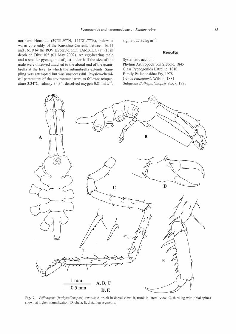
northern Honshuu (39°51.97�N, 144°21.77�E), below awarm core eddy of the Kuroshio Current, between 16:11and 16:19 by the ROV HyperDolphin (JAMSTEC) at 913 mdepth on Dive 105 (01 May 2002). An egg-bearing maleand a smaller pycnogonid of just under half the size of themale were observed attached to the aboral end of the exum-brella at the level to which the subumbrella extends. Sam-pling was attempted but was unsuccessful. Physico-chemi-cal parameters of the environment were as follows: temper-ature 3.34°C, salinity 34.34, dissolved oxygen 0.81 ml L�1,
sigma-t 27.32 kg m�3.
Results
Systematic accountPhylum Arthropoda von Siebold, 1845Class Pycnogonida Latreille, 1810Family Pallenopsidae Fry, 1978Genus Pallenopsis Wilson, 1881Subgenus Bathypallenopsis Stock, 1975
Pycnogonids and narcomedusae on Pandea rubra 85
Fig. 2. Pallenopsis (Bathypallenopsis) tritonis; A, trunk in dorsal view; B, trunk in lateral view; C, third leg with tibial spinesshown at higher magnification; D, chela; E, distal leg segments.

Pallenopsis (Bathypallenopsis) tritonis Hoek, 1883
Description of the Antarctic specimen
Immature. Trunk segmented, smooth (Fig. 2A). Lateralprocesses slightly longer than their diameters, without tu-bercles or spines, and separated by more than half their di-ameters (Fig. 2B). Neck rounded. Ocular tubercle at the an-terior extreme of segment, bearing four eyes and two latero-distal papillae. Proboscis two thirds of trunk length, widerproximally and slightly oriented downwards (Fig. 2B). Ab-domen almost horizontal, with few, small setae extendingbeyond distal rim of first coxa of fourth leg.
Chelifores long, scape consisting of 2 well-articulatedsegments, second segment slightly longer than the first,with some small distal setae (Fig. 2B). Chela palm with fewsetae near the movable finger (Fig. 2D). Fingers wellcurved, longer than the palm, tips overlapping. Lateral palpbuds short. Oviger not fully developed, 9-segmented, with-out setae.
Second coxa of the third leg longer than both first andthird coxa together (Fig. 2C). Femur with some smallspines that increase in length and number towards the sec-ond tibia, the latter being the longest segment. Tibial spineswith distal denticulation. Tarsus very short with a long ven-tral spine and a few short setae (Fig. 2E). Propodus straight,with two strong proximal spines on the heel and five smallspines on the sole. Claw large, longer than three-fourths ofthe propodal length; auxiliaries about one third of mainclaw (Fig. 2E).
Measurements (in mm)
Trunk length (from chelifore insertion to the tip of fourthlateral process): 2.67; trunk width (across second lateralprocesses): 1.42; proboscis length: 1.97; chelifore scape 1:0.75; chelifore scape 2: 0.8; abdomen length: 0.77; thirdleg, coxa 1: 0.35; coxa 2: 0.95; coxa 3: 0.5; femur: 2.22;tibia 1: 2.07; tibia 2: 2.45; tarsus: 0.12; propodus: 0.72;main claw: 0.6; auxiliary claw 0.2.
Remarks
This specimen belongs to the subgenus Bathypallenopsisaccording to the shape of the chela, the articulation of thescape and the reduced auxiliary claws. Within this sub-genus, it may be assigned to the tydemani-group (sensuStock 1975) by its styliform proboscis. Two species of Pal-lenopsis (Bathypallenopsis) have been recorded fromAntarctic waters, namely P. longiseta Turpaeva and P.meridionalis Hodgson (Child 1995); both belong to themollisima-group (Stock 1975). Our specimen does not fitwith the description of either of these species but the mor-phological features agree with those of P. tritonis within therange of variation observed by Bamber (2002a), except forthe ratios scape 1: scape 2 and the abdomen length:ab-domen width that are slightly lower (0.93 compared with
0.95–1.23 and 3 compared with 3.31–6.55, respectively).However, the tibial spines are denticulated distally as ob-served in specimens of P. tritonis from the northern hemi-sphere and the above mentioned differences may be due tothe early stage of development. It is also similar to P. cal-canea, but this last species does not have auxiliary claws.
Phylum Cnidaria Verrill, 1865Class Hydroidomedusae Claus, 1877Sub-class Anthomedusae Haeckel, 1879Order Filifera Kühn, 1913Family Pandeidae Haeckel, 1879Pandea rubra Bigelow, 1913
Description of the Antarctic specimen
Umbrella dome-shaped, 15 mm in height, 14 mm wide(Fig. 3A). Mesogloea thin but consistent. Exumbrella trans-parent, smooth, without cnidocyst tracks. Stomach brown-ish in preserved specimen, cruciform, with wide base,hanging down to one fourth of the subumbrellar cavity.Mouth with four lips very crenulated. Mesenteries short.Radial canals four, wide, jagged. Gonads not discernible.Fourteen tentacles, all equal, with wide triangular basalbulbs and abaxial spurs. Velum medium.
Remarks
The specimen fits the description for juveniles of Pandearubra given by Russell (1953). In looking for signs of pre-dation by the pycnogonid, two vesicle-shape structures set-tled side by side in the outer wall of the stomach and identi-fied as parasitic narcomedusae were discovered (Figs.3A–D). The smallest one (Fig. 3D) detached itself after ashort time while the other one (Fig. 3B) was detached laterfor better microscopic examination (Fig. 3C). The descrip-tions of both early stages are as follows:
Sacculiform larval narcomedusan (Fig. 3C): 0.87 mm inlength, 0.73 mm maximum diameter. The distal circularopening is 0.35 mm in diameter. There are six proximalpoints of attachment, about 0.15 mm in length each.
Pyriform larval narcomedusan (Fig. 3D): 0.75 mm inlength, 0.45 mm maximum diameter. The distal circularopening is 0.25 mm in diameter. There is one proximalpoint of attachment, about 0.1 mm in length.
Description of the Japanese specimens
Sampled specimen description as for the Antarctic speci-men except for size (30 mm height, 38 mm wide in partiallycontracted state), tentacle number (16), and that the stom-ach colour was red in the live animal, brown when pre-served. Unsampled individuals had 24 (Fig. 1A) and 18(Fig. 1C, D) tentacles respectively but size was unable to beestimated.
86 F. PAGÈS, J. CORBERA & D. LINDSAY

Discussion
The co-occurrence of the pycnogonid and the anthome-dusan in the same plankton sample from the Weddell Seasuggested that Pandea rubra was the pelagic substratum towhich Pallenopsis tritonis was attached. Both specimenswere the largest organisms by far in the mesozooplanktonsample, which was numerically dominated by small cope-pods. The volume of water filtered was relatively small(125 m3) and, because of the rarity of both of these speciesin plankton samples, the probability that these two large,rare animals were collected independently by chance in thesame net tow was considered extremely low. This assump-tion was further validated by the two in situ observations ofpycnogonids in association with P. rubra at mesopelagicdepths in Japanese waters. To date, nine pycnogonid specieshad been found attached to ten medusae species (Table 1)but the possibility that any of these interspecific associa-tions are exclusive has not yet been addressed.
Pallenopsis tritonis is widespread in the north Atlanticand the north Pacific, at depths ranging from 344 to 7,280m, and it is associated with gelatinous plankton (Bamber
2002a). The present record is the first for the southernhemisphere. P. rubra can attain 10 cm in height (Pagès un-published data) and it lives mainly in mesopelagic waters ofall oceans except the Arctic (Kramp 1961). In waters offJapan it has previously been reported, as Hydroidomedusaesp. A, from 703 m depth in Sagami Bay (35°00.07�N,139°14.07�E; temperature 4.8°C, salinity 34.25, dissolvedoxygen 1.6 ml L�1) (Hunt & Lindsay 1999) and more re-cently as P. rubra (Lindsay & Hunt 2005, Lindsay 2006). Inthe Weddell Sea, it has been collected mostly in the 500–1,000 m depth range and once in the 1,000–2,000 m depthrange (Pagès et al. 1994), always in very low densities (upto 0.66 specimens 104 m�3). In the same waters, a more dis-crete vertical sampling collected specimens in three consec-utive 100-m range hauls between 700 and 1,000 m depth(up to 0.30 specimens 104 m�3 respectively) (Pugh et al.1997). In the Antarctic Polar Frontal Zone it has been col-lected in the 600–800 and 800–1,000 m depth ranges, re-spectively (up to 0.12 specimens 104 m�3) (Pagès et al.1996).
Child & Harbison (1986) concluded that P. scoparia par-asitized Periphylla periphylla because most of the jellyfish
Pycnogonids and narcomedusae on Pandea rubra 87
Fig. 3. Pandea rubra; A, view of the specimen with the subumbrella everted. Note the whitish spot that is the larger and sac-culiform parasitic narcomedusan. B, location of the sacculiform parasitic narcomedusan attached to the stomach. C, view of thesacculiform parasitic narcomedusan after detachment. D, view of the pyriform parasitic narcomedusan after detachment,scale�0.15 mm.

tentacles had been eaten and the gut diverticulae of the pyc-nogonid were filled with cnidocysts. Was P. tritonis para-sitizing P. rubra? The tentacles of the Antarctic anthomedu-san were contracted and no signs of predation could be dis-cerned on them, or on any other parts of the body, appar-ently indicating that the association was not parasitic. Noobvious signs of predation were seen on the medusae fromthe in situ observation either. However, this is not conclu-sive evidence as the gut diverticulae of P. tritonis were notexamined due to the tiny size of the specimen, and thespecimens observed by the ROV were unable to be sam-pled.
Staples & Watson (1987), in reviewing the associationsbetween hydroids and pycnogonids, referred to the occur-rence of pycnogonid larvae (protonymphons) encapsulatedin highly modified polyps named “fusiform bodies”, “pyri-form sacs” “pyriform vesicles” and “galls”. The life cycleof Pallenopsis is poorly known, particularly whether Pal-lenopsis develops through larval encystment. Adult pycno-gonids mate and their eggs are then carried by the maleuntil they hatch as typical protonymphon larvae (Bain2003). It is assumed that both the adults and larvae of pyc-nogonids feed on the medusae but the only evidence is thatdemonstrated by Child & Harbison (1986).
How do the pycnogonids reach the anthomedusan? Child& Harbison (1986) suggested two mechanisms of host loca-tion, namely generation of benthic polyps or resting of themedusae on the bottom (as they had previously observedsuch an event). The first explanation is not plausible for Pe-riphylla periphylla as Jarms et al. (1999) have demon-strated that this species is holoplanktonic.
The medusa stage of P. rubra has neither been observedstaying on the sea bottom nor has it been reported frombentho-pelagic samples. The polyp stage is believed to beCampaniclava clionis Vanhöffen (Rees 1967), and is anepizoic associate of the euthecosome pteropod Clio recurva(Childern) (see Lalli & Gilmer 1989, p. 127), which is
found throughout the world’s temperate and tropical oceans.Also attached to the shell of another pteropod, Clio cuspi-data (Bosc), lives the polyp stage of P. conica, the only con-generic medusa species (Picard 1956), if Pandea cybelesAlvariño 1988 is an invalid species as suspected (Pagès, un-published information). Likewise four little-known polypspecies, two of them also pandeids, have been identified at-tached to four different euthecosome pteropod species(Lalli & Gilmer 1989, p. 128). A hypothesis arises fromthese coincidences. Like many epibenthic pycnogonids thatlive on benthic hydroids (e.g. Staples & Watson 1987), thelarval stages (protonymphons) of P. tritonis could developand grow on hydroids epizoic on pteropods. Then,protonymphons could ride piggyback on medusae buds be-fore these are released from the hydrorhiza, the hydroidstolon. The key question is if Pallenopsis eggs orprotonymphons could become encysted in the hydroids thatare epizoic on Clio. The present two in situ observationssuggest that the pycnogonids at least reach adulthood ontheir host, but whether the protonymphons are transferreddirectly to adult medusae by the egg-bearing male pycnogo-nid after hatching or somehow find their way to polypcolonies growing on euthecosome pteropods remains to beverified. A report (Larson et al. 1991) of pycnogonids at-tached to Voragonema pedunculata, a trachymedusa with-out a polyp stage in its life cycle, in combination with thereport concerning parasitism of the scyphomedusa P. peri-phylla (Child & Harbison 1986), also holoplanktonic, sug-gests that the former hypothesis, not involving an interme-diate polyp host, is more likely. Perhaps the protonymphonlarvae have larval legs with greatly elongated distal spines,as has been reported for Phoxichilidium femoratum, whichis also a member of the family Phoxichilidiidae to whichPallenopsis has been proposed to belong by some authors(Stock 1978, Arnaud & Bamber 1987). They may also havecement glands on the chelifores that can secrete elongatefilaments, also reported for P. femoratum, and either of
88 F. PAGÈS, J. CORBERA & D. LINDSAY
Table 1. Records of pycnogonid species associated with planktonic medusae.
Pycnogonid Medusae Reference
Ammothea sp. Polyorchis karafutoensis Uchida 1927Ammothea alaskensis Polyorchis karafutoensis Okuda 1940Anoplodactylus petiolatus Clytia hemisphaerica Amphinema dinema Franc 1951Anoplodactylus petiolatus Obelia sp. Dogiel 1913, Lebour 1945Anoplodactylus petiolatus Obelia sp., Cosmetira pilosella, Lebour 1916
Leuckartiara octona, Clytia hemisphaericaAnoplodactylus virescens Sarsia eximia Dogiel 1913, Lebour 1945Endeis spinosa Obelia sp. Lebour 1916Pallenopsis calcanea ? Hedgpeth 1962Pallenopsis scoparia Periphylla periphylla Child & Harbison 1986Pallenopsis tritonis Pandea rubra This studyPhloxichilidium femoratum Sarsia eximia Dogiel 1913, Lebour 1945Unidentified juvenile Periphylla periphylla Mauchline 1984Unidentified species Voragonema (Benthocodon) pedunculata Larson et al. 1991
Aeginura grimaldii

these could aid in passive distribution of the larvae similarto the strategy used by baby spiders or lamellibranchpostveligers until they are “captured” by their new hosts.Alternatively the larvae-bearing male pycnogonid may“jump ship” when a possible new host for its young comesnear, as short bouts of swimming have been reported forsome species.
The parasitic narcomedusan larvae (Fig. 3, C–D) couldnot be identified to species level owing to their early stageof development. Similar larvae parasitizing medusae hadbeen described and drawn (e.g. Uchida 1928, Bouillon1987) but we provide the first photographs. This is also thefirst record involving a medusa species of the highly diversefamily Pandeidae.
Hopefully, future in situ observations on midwatermedusae with piggybacking Pallenopsis, and intensivesearches of polyp colonies on euthecosome pteropods forprotonymphon larvae will shed more light on this biologicalassociation and the life cycle of these intriguing arthropods.
Acknowledgements
We would like to express our appreciation for the effortsof the operations team of the ROV HyperDolphin and thecaptain and crew of both the R/V Kaiyo and R/VPolarstern. Roger N. Bamber has kindly confirmed theidentification of the juvenile Pallenopsis tritonis. We appre-ciate the opportunity given DL for a 5-month sabbatical atCSIC, Barcelona, by the Japan Society for the Promotion ofScience.
References
Alvariño A (1988). Pandea cybeles, a new medusa from the Sar-gasso Sea (Coelenterata: Anthomedusae: Pandeidae). Proc BiolSoc Wash 101: 102–108.
Arnaud F, Bamber RN (1987) The biology of Pycnogonida. AdvMar Biol 24: 1–86.
Bain BA (2003) Larval types and a summary of postembryonicdevelopment within the pycnogonids. Invertebr Reprod Dev 43:193–222.
Bamber RN (2002a) Morphological variation in and a redescrip-tion of Pallenopsis (Bathypallenopsis) tritonis Hoek, 1883(Arthropoda : Pycnogonida). J Nat Hist 36: 157–172.
Bamber RN (2002b) Bathypelagic pycnogonids (Arthropoda, Pyc-nogonida) from the Discovery cruises. J Nat Hist 36: 715–727.
Boehme T, Voet JC v., Fahrbach E, Fischer H, Hamann R, Kolb L,Latten A, Rohardt G, Schütt E, Seiß G, Strass V, Witte H,Zwein F (1995) Water masses and circulation in the WeddellSea. Ber Polarfosrch 135: 135–156.
Bouillon J (1987) Considérations sur le développement des nar-coméduses et sur leur position phylogénétique. Indo-MalayZool 4: 189–278.
Child AC (1995) Antarctic and Subantarctic Pycnogonida. V. Thefamilies Pycnogonidae, Phoxichilidiidae, Endeididae and Calli-pallenidae, including the genus Pallenopsis. Biology of theAntarctic Seas XXIV. Ant Res Ser 69: 113–160.
Child AC, Harbison GR (1986) A parasitic association between apycnogonid and a scyphomedusa in midwater. J Mar Biol AssUK 66: 113–117.
Clark WC, Carpenter A (1977) Swimming behaviour in a pycno-gonid. N Z J Mar Freshw Res 11: 613–615.
Dogiel V (1913) Embryologische Studien an Pantopoden. Z WissZool 107: 576–741.
Franc A (1951) Le zooplancton de la région de Dinard-SaintMalo. Bull Lab Marit Dinard 34: 25–40.
Hedgpeth JW (1962) A bathypelagic pycnogonid. Deep-Sea Res9: 487–491.
Hunt JC, Lindsay DJ (1999) Methodology for creating an observa-tional database of midwater fauna using submersibles: resultsfrom Sagami Bay, Japan. Plankton Biol Ecol 46: 75–87.
Jarms G, Båmstedt U, Tiemann H, Martinussen MB, Fosså JH(1999) The holopelagic life cycle of the deep-sea medusa Peri-phylla periphylla. Sarsia 84: 55–65.
Lalli CM, Gilmer RW (1989) Pelagic snails, the biology of holo-planktonic gastropod mollusks. Stanford Press. 275 pp.
Larson RJ, Mills CE, Harbison GR (1991) Western Atlantic mid-water hydrozoan and scyphozoan medusae: in situ studies usingmanned submersibles. Hydrobiologia 216/217: 311–317.
Lebour, MV (1916) Notes on the life cycle of Anaphia petiolata(Kröyer). J Mar Biol Ass UK 11: 51–56.
Lebour MV (1945) Notes on the Pycnogonida off Plymouth. JMar Biol Ass UK 26: 139–165.
Lindsay DJ (2003) Bioluminescence in the mesopelagic realm.Kaiyo Monthly, Special Edition 35: 606–612.
Lindsay DJ, Hunt JC (2005) Biodiversity in midwater cnidariansand ctenophores: submersible-based results from deep-waterbays in the Japan Sea and North-western Pacific. J Mar BiolAssoc UK 85: 503–517.
Lindsay DJ (2006) A checklist of midwater cnidarians andctenophores from Sagami Bay—species sampled during sub-mersible surveys from 1993–2004. Bull Plank Soc Jpn 53(2):104–110.
Kramp PL (1961) Synopsis of the medusae of the world. J MarBiol Ass UK 40: 7–469.
Mauchline J (1984) Pycnogonids caught in bathypelagic samplesfrom the Rockall Trough, northeastern Atlantic Ocean. J NatHist 18: 315–322.
Ohshima H (1933) Pycnogonids taken with a tow-net. Annot ZoolJap 14: 211–220.
Okuda S (1940) Metamorphosis of a pycnogonid parasitic in a hy-dromedusa. J Fac Sci Hokkaido Univ (ser 6 Zool) 7: 73–86.
Pagès F, Pugh PR, Gili JM (1994) Macro-and megaplanktoniccnidarians collected in the eastern part of the Weddell Gyre dur-ing summer 1979. J Mar Biol Ass UK 74: 873–894.
Pagès F, White MG, Rodhouse MG (1996) Abundance of gelati-nous carnivores in the nekton community of the Antarctic PolarFront in summer 1994. Mar Ecol Prog Ser 141: 139–147.
Picard (1956) Le premier stade de l’Hydroméduse Pandea conica,issu de l’Hydropolype Campaniclava cleodorae. Bull InstOcéanogr. Monaco 52(1086): 1–11.
Pugh PR, Pagès F, Boorman B (1997) Vertical distribution ofpelagic cnidarians in the eastern part of the Weddell Sea,Antarctica. J Mar Biol Ass UK 77: 342–360.
Rees WJ (1967) A brief survey of the symbiotic association of
Pycnogonids and narcomedusae on Pandea rubra 89
Cnidaria with Mollusca. Proc Malacol Soc Lond 37: 214–231.Russell, FS (1953) The medusae of the British Isles. Cambridge
University Press. 530 pp.Staples DA, Watson JE (1987) Associations between pycnogonids
and hydroids In: Modern trends in the systematics, ecology andevolution of hydroids and hydromedusae. (eds Bouillon J,Boero F, Cicogna F, Cornelius PFS). Oxford University Press,pp. 215–226.
Stock JH (1964) Deep-sea Pycnogonida collected by the Cirrus inthe north Atlantic. Beaufortia 11: 45–52.
Stock JH (1975) Pycnogonida from the continental shelf, slopeand deep sea of the tropical Atlantic and East Pacific. Biologi-cal results of the University of Miami deep-sea expeditions.Bull Mar Sci 24: 957–1092.
Stock JH (1978) Abyssal Pycnogonida from the north-eastern At-lantic basin, 1. Cah Biol Mar 19: 397–413.
Uchida T (1927) Studies on Japanese Hydromedusae. J Fac SciImp Univ Tokyo 1: 145–241.
Uchida T (1928) Studies on Japanese Hydromedusae. 2. Tra-chymedusae and Narcomedusae. Jpn J Zool 2: 73–97.
90 F. PAGÈS, J. CORBERA & D. LINDSAY