Phytochemical Investigation and Antimicrobial Activity of ... · PDF filePhytochemical...
13
Available online at www.scholarsresearchlibrary.com Scholars Research Library Der Pharmacia Lettre, 2010, 2(4): 284-296 (http://scholarsresearchlibrary.com/archive.html) ISSN 0975-5071 USA CODEN: DPLEB4 284 Scholar Research Library Phytochemical Investigation and Antimicrobial Activity of Leucas Cephalotes Roth. Spreng Whole Herb Katara Antariksh 1* , Pradhan Chandan Kumar 1 , Tyagi Amit Kumar 2 , Singh Pradeep 3 1 Ram-Eesh Institute of Voc. & Tech. Education, Greater Noida, India. 2 Meerut College, CCS University, Meerut, India. 3 Teerthankar Mahaveer College of Pharmacy, TMU, Moradabad, India. ______________________________________________________________________________ ABSTRACT Leucas cephalotes Roth. Spreng (Lamiaceae) is the well known herb in the Ayurvedic and Modern systems of medicine, to cure various disorders. Commonly known as Dronpushpi or Guma is mainly a rainy season weed found through out India, The plant is authentified by Taxonomist Dr. Anjula Pandey and specimen (NHCP/NBPGR 2009-9/900) has preserved in NBPGR, Pusa Campus, Delhi. Powdered plant material analyzed for the two major attributes, Pharmacognostic parameters and Phyto-chemical characterization followed by the quantitative analysis of Tannins and Flavonoids. Antimicrobial activity on the Toluene and Methanolic extract was also performed in which Methanolic extract have shown the 59% and 41% zone of inhibition on the bacterial strains and on the fungal strains resp. when compared to the standards. Key words: Dronpushpi/Guma, Pharmacognosy, Florescence analysis and Antimicrobial activity. ______________________________________________________________________________ INTRODUCTION Leucas cephalotes (Roth) Spreng. Syn. Phlomis cephalotes Hindi- Guma, Sanskrit- Dronapushpi (family- lamiaceae) is an erect, scabrous or pubescent, stout annual herb, 30-100 cm high and found as a common weed throughout the India.[1] Leaves are yellowish-green; de ovate or ovate lanceolate, subacute, more or less pubescent. Stem is light greenish yellow, surface rough, hairy, quadrangular with four prominent furrows. Roots are cylindrical, zig-zag, smooth, long with
Transcript of Phytochemical Investigation and Antimicrobial Activity of ... · PDF filePhytochemical...
Available online at www.scholarsresearchlibrary.com
Scholars Research Library
Der Pharmacia Lettre, 2010, 2(4): 284-296
(http://scholarsresearchlibrary.com/archive.html)
ISSN 0975-5071 USA CODEN: DPLEB4
284
Scholar Research Library
Phytochemical Investigation and Antimicrobial Activity of Leucas Cephalotes Roth. Spreng Whole Herb
Katara Antariksh 1*, Pradhan Chandan Kumar1, Tyagi Amit Kumar 2, Singh Pradeep3
1 Ram-Eesh Institute of Voc. & Tech. Education, Greater Noida, India. 2 Meerut College, CCS University, Meerut, India.
3 Teerthankar Mahaveer College of Pharmacy, TMU, Moradabad, India. ______________________________________________________________________________ ABSTRACT Leucas cephalotes Roth. Spreng (Lamiaceae) is the well known herb in the Ayurvedic and Modern systems of medicine, to cure various disorders. Commonly known as Dronpushpi or Guma is mainly a rainy season weed found through out India, The plant is authentified by Taxonomist Dr. Anjula Pandey and specimen (NHCP/NBPGR 2009-9/900) has preserved in NBPGR, Pusa Campus, Delhi. Powdered plant material analyzed for the two major attributes, Pharmacognostic parameters and Phyto-chemical characterization followed by the quantitative analysis of Tannins and Flavonoids. Antimicrobial activity on the Toluene and Methanolic extract was also performed in which Methanolic extract have shown the 59% and 41% zone of inhibition on the bacterial strains and on the fungal strains resp. when compared to the standards. Key words: Dronpushpi/Guma, Pharmacognosy, Florescence analysis and Antimicrobial activity. ______________________________________________________________________________
INTRODUCTION
Leucas cephalotes (Roth) Spreng. Syn. Phlomis cephalotes Hindi- Guma, Sanskrit- Dronapushpi (family- lamiaceae) is an erect, scabrous or pubescent, stout annual herb, 30-100 cm high and found as a common weed throughout the India.[1] Leaves are yellowish-green; de ovate or ovate lanceolate, subacute, more or less pubescent. Stem is light greenish yellow, surface rough, hairy, quadrangular with four prominent furrows. Roots are cylindrical, zig-zag, smooth, long with

Katara Antariksh et al Der Pharmacia Lettre, 2010, 2(4):284-296 ______________________________________________________________________________
285
Scholar Research Library
numerous wiry. Inflorescence is sessile, white, crowded in dence, globose, surrounded by numerous foliaceous bracts. Fruits are schizocarpic carcerule, nutlets smooth and brown. Seeds are oblong, trigonous, smooth, dark brown. [2] Basically Guma is a rainy season weed and commonly found ascending up to 600 - 1,800 m. in Himalayas, mainly in the hilly regions of Nepal, India, Pakistan, Bhutan and Bangladesh. [3] In the East Asia this plant is found in Afghanistan to Western China in open areas at elevation of 1,700 m. [4] The other species of leucas genus are Leucas aspera (willd.) spreng., L. linifolia spreng., L. lanata Benth., L. stelligera Wall., L. urticaefolia R. Br. L. Biflora (Vahl.) Benth., L. ciliata Wall ex Benth., L. indica (L.) R. Br., L. Zeylanica (L.) R. Br. are commonly found in the Asian Countries. [5, 6] A decoction of the plant is used in the treatment of malarial fever and a domestic medicine for Snake bite. The dried inflorescences are smoked and the smoke exhaled through the nose to treat nose bleeds. [7, 8] Dried leaves along with tobacco (1:3) are smoked to treat bleeding as well as itching piles and fresh leaves eaten as a potent herb. [1, 3, 9] The juice of leaves used topically in psoriasis, skin eruptions, scabies and internally for the treatment of urinary complaints. The flowers are administered in the form of syrup or with honey as a domestic remedy for cough and colds.[6, 10] Whole herb of Dronapushpi was found to be hepatoprotective in CCl4 induced hepatotoxicity in mice and rats, juice of this plant act as an antibilious in herbal therapy for Jaundice. [11, 12] The whole plant powder in the proportion of 70% in the herbal composition is patented to cure Epileptic convulsions and Cerebral function disorders. [13] This plant has antipyretic action and also considered to be stimulant, expectorant, aperient, diaphoretic, insecticidal and emmenagogue. [6, 14] Leucas cephalotes (Roth) Spreng whole herb contains new labdane, norlabdane and abietane type diterpenes and protostane type triterpenes, together with common triterpene, five sterols and eight flavones. [15] A rarely found Laballenic acid (octadeca-5, 6-dienoic acid) and others luaric acid, glutaric acid, adipic acid and tridecanoic acid from the seed oil. [16] A major compound β-sitosterol and its glucoside also isolated from this plant. [17] The volatile compounds of inflorescence and seeds were found caryophyllene oxide 26.56%; delta-fenchene 12.02%; α-cardinal 2.13%; 1-hepten-3-ol 6.53%; menthol 6.30%; deca hydro naphthalene 5.15% and trans-caryophyllene 4.05%. [18]
MATERIALS AND METHODS: Collection of Plant Material The plant material was collected from waste lands of Industrial area of Distt. Haridwar, Uttrakhand. The plant was authentified as Leucas cephalotes (Roth) Spreng. F- Lamiaceae by the Dr. Anjula Pandey (Taxonomist), National Bureau of Plant Genetic Resources (NBPGR), Pusa Campus, New Delhi. A voucher specimen (Specimen No: NHCP/NBPGR 2009-9/900) is preserved in Herbarium section of Taxonomic Deptt. of NBPGR, New Delhi.
Katara Antariksh et al Der Pharmacia Lettre, 2010, 2(4):284-296 ______________________________________________________________________________
286
Scholar Research Library
Pharmacognostic parameters: Morphological Characters: The macroscopy of a drug includes its visual appearance to the naked eye. Macroscopic identity of a medicinal plant material is based on shape, size, colour, taste, surface characteristic, texture, fracture characteristic and appearance of cut surface. [19] Microscopical Characters: The fresh whole herb was boiled with chloral hydrate for several minutes for completely clarified. The size, shape and relative positions of the different cells and tissues, chemical nature of the cell walls and of the cell contents are determined in Microscope. [19] Foreign Matter Analysis: The 100 gms sample was weighed then spread in a thin layer and foreign matter was sorted into groups either by visual inspection, using a magnifying lens or with the help of a suitable sieve according to the requirements. The remainder of the sample was sifted through a sieve no. 250; dust regarded as mineral admixture. The portions of sorted foreign matter were weighed. [19] Crude Fiber Determination: 2 gm of powder drug was taken in a beaker; add 50 ml of 10% v/v HNO3. Boil with constant stirring. Strain through fine cotton cloth on Buchner funnel and residue washed with boiling water. Transfer the residue from cloth to a beaker and 50 ml of 2.5% v/v NaOH solution was added and heat to boil, maintain at boiling point with constant stirring. Strain and washed the residue with hot water, transfer in cleaned & dried crucible for quantitative determination. Weigh the residue. [20] Phytochemical Investigation: The diff. extracts of the whole plant were subjected to preliminary phytochemical screening for the detection of various phytoconstituents: 1. Tests for Alkaloids a) Dragendroff’s test: Dragendroff’s Reagent (potassium bismuth iodide) gives reddish brown precipitate. b) Mayer’s test: Mayer’s Reagent (Potassium mercuric iodide) gives cream precipitate. c) Wagner’s test: Wagner’s Reagent (Iodine in potassium iodide) gives reddish brown precipitate. d) Hager’s test: Hager’s Reagent (Saturated picric acid solution) gives yellow precipitate indicated. 2. Tests for Glycosides a) Keller-killiani Test (for deoxy sugar): 1 ml of glacial acetic acid containing traces of FeCl3 and 1 ml of conc. H2SO4 were added. A reddish-brown colour formed at the junction of two layers and the upper layer turned bluish green.
Katara Antariksh et al Der Pharmacia Lettre, 2010, 2(4):284-296 ______________________________________________________________________________
287
Scholar Research Library
b) Legal test (for cardinolides): Conc. Ethanolic extract was made alkaline with few drops of 10% NaOH and then add freshly prepared Sod. Nitroprusside solution. Blue colouration observed. c) Baljet test: Picric acid is added to the extract and made alkaline, gives a stable orange color. d) Borntrager’s test: Dil. HCl is added to powdered drug and heated for 5 mins then filtered and add equal volume of CHCl3, shake well and collect the lower org. layer of CHCl3, add NH3
half of its volume, shake well, lower ammonical layer turned rose pink colour. 3. Tests for Saponins a) Foam Test: Powdered drug residue was taken in a test tube and shaken vigorously with a small amount of NaHCO3 and water. Characteristic honeycomb like froth obtained. 4. Tests for Steroids a) Salkowaski reaction: Residue of extract taken in 2 ml of CHCl3 and 2 ml of conc. H2SO4
added from the side of the test-tube and then shaken for few minutes. Red colour developed in the CHCl3 layer. 5. Tests for Tannins and Phenolic Compounds a) Ferric chloride reagent: FeCl3 sol. added to test sol. Dark green or deep blue colour is obtained. b) Lead acetate test: A 10% w/v solution of basic lead acetate in distilled water was added to test sol. Precipitation occurred. c) Gelatin solution test: 1% w/v solution of gelatin in water, with 10% sodium chloride and then added to test sol. White precipitate is obtained. 6. Tests for Flavonoids a) Shinoda Test: Test residue dissolved in 5 ml ethanol (95% v/v) and reacted with few drops of conc. HCl and 0.5 g of Mg metal. The pink, crimson or magenta colour is developed. b) Ammonia Test: Filter paper strips were dipped in the alcoholic extract and ammoniated. Strips turned yellow. 7. Tests for Amino Acids & Proteins a) Ninhydrin test: 0.1% w/v solution of Ninhydrine in n-butanol and then added to test sol. A violet or purple colour is developed. b) Cysteine test: To the 5 ml of test sol., 2 ml of 40% w/v NaOH and 2 drops of 10% w/v lead acetate solution, resulting solution is boiled for few minutes to obtain black precipitate. c) Biuret test: Residue taken in water and add 4% NaOH solution then add few drops of 1% CuSO4 sol. Violet or pink colour is obtained. d) Xanthoprotein test (for tyrosine and tryptophan): Test sol. added with 1ml of conc. H2SO4, white precipitate is observed, boil and add NH4OH and precipitate turns yellow. e) Millon’s test (Mercuric Nitrate solution): Aq. residue taken with 5 ml of Million’s reagent. White precipitate gently warmed to turns pink or red.

Katara Antariksh et al Der Pharmacia Lettre, 2010, 2(4):284-296 ______________________________________________________________________________
288
Scholar Research Library
8. Tests for Sugars a) Molisch test: Test solution with few drops of α-naphthol and then conc. H2SO4. Violet colour ring was formed at the junction of two layers. b) Fehling’s test: Fehling A and Fehling B solution were mixed in equal amount and boiled then added with test solution and again heated. Yellow to brick red colour. c) Benedict’s test: Test solution and Benedict’s reagent was mixed and heated in water bath for 5-10 minutes. Yellow to red or green colour. d) Tollen’s phloroglucinol test for galactose: 2.5 ml of conc. HCl and 4 ml of 0.5 % phloroglucinol added to test sol. then heated on water bath. Yellow to red colour. [21] Fluorescence Analysis Fluorescence analysis of the drug was conducted on the powder to observe diff. types of fluorescent characteristics. Subsequently, powdered sample was treated with 1-N Sodium hydroxide in Methanol, 1-N Sodium hydroxide in water, 50% Sulphuric acid, 50% nitric acid, Different solvents and observed under day light, short wavelength UV and long wavelength UV light. The observations shown in Table no. 03. [22] Quantitative Determination: Any estimation method applied to plant tissue should allow determination of a particular compound or of a group of compounds in weight percent. Total Flavonoids Content: 10 g of the powdered crude sample was extracted repeatedly with 100 ml of 80% aqueous methanol at room temperature. The whole solution was filtered through Whatman filter paper No 42 (125 mm). The filtrate was later transferred into a crucible and evaporated into dryness over a water bath and weighed to a constant weight. [20] Total Tannins Content: 1 gm of sample/crude powdered drug was extracted with 100 ml of water by shake for one hour and left for overnight, allowed the solid material to settle and filtered the liquid through a Whatman no. 1 filter paper , the first 20 ml of filtrate was discarded and then 10 ml of the filtrate taken in to conical flask. To it 750 ml of water and 25 ml of Indigo suphuric acid sol. was added and titrated with 0.1 N KMnO4 solution. The flask was shaken vigorously till a golden yellow end point was reached. A blank determination was also performed and percentage calculated. Each ml of KMnO4 solution is equivalent to 0.004157 gm of total tannins. [23] Antimicrobial activity The fresh whole plant of Leucas cephalotes were collected and shade dried at room temperature for 2 weeks. Dried part then stored at 20°C before using them individually to solvent extraction procedures. The antimicrobial activity was tested against different bacterial and fungal strains which were obtained from Arbro Industrial Testing Lab., Kirti Nagar, New Delhi. The microbial strains with their laboratory registration number were: Bacillus cereus (ATCC 11778), Shigella flexineri (MTCC 1457), Candida albicans (MTCC 227). The Cup Plate method was used to test the antimicrobial activity in present investigation ie adopted from Rose & Miller 1939. The antibacterial activity of plant extract was tested on Assay Medium no. 11 (No. 11) (Himedia-
Katara Antariksh et al Der Pharmacia Lettre, 2010, 2(4):284-296 ______________________________________________________________________________
289
Scholar Research Library
M004) and antifungal activity was tested on Sabouraud Dextrose Agar (SDA) (Himedia-MU063). The No. 11 & SDA plates were prepared by pouring a few ml of molten media into sterile Petri plates. The plates were allowed to solidify for 5 min and appropriate amount of inoculum’s suspension (0.1 %) was swabbed uniformly on agar medium. A sterile disc was placed on the surface of medium and an appropriate concentration of extracts (5 mg/disc) was loaded on it. The extract was allowed to diffuse for 5 min and the plates were kept for incubation at 37°C for 18-20 h. Inhibition zones formed around the disc were measured with transparent ruler in millimeter (mm). Gentamycin (10 µg/ml), Amikacin (10 µg/ml) and Nystatin (100 µg/ml) were used as standard for antibacterial and antifungal activity testing resp. [24, 25]
RESULTS AND DISCUSSIONS
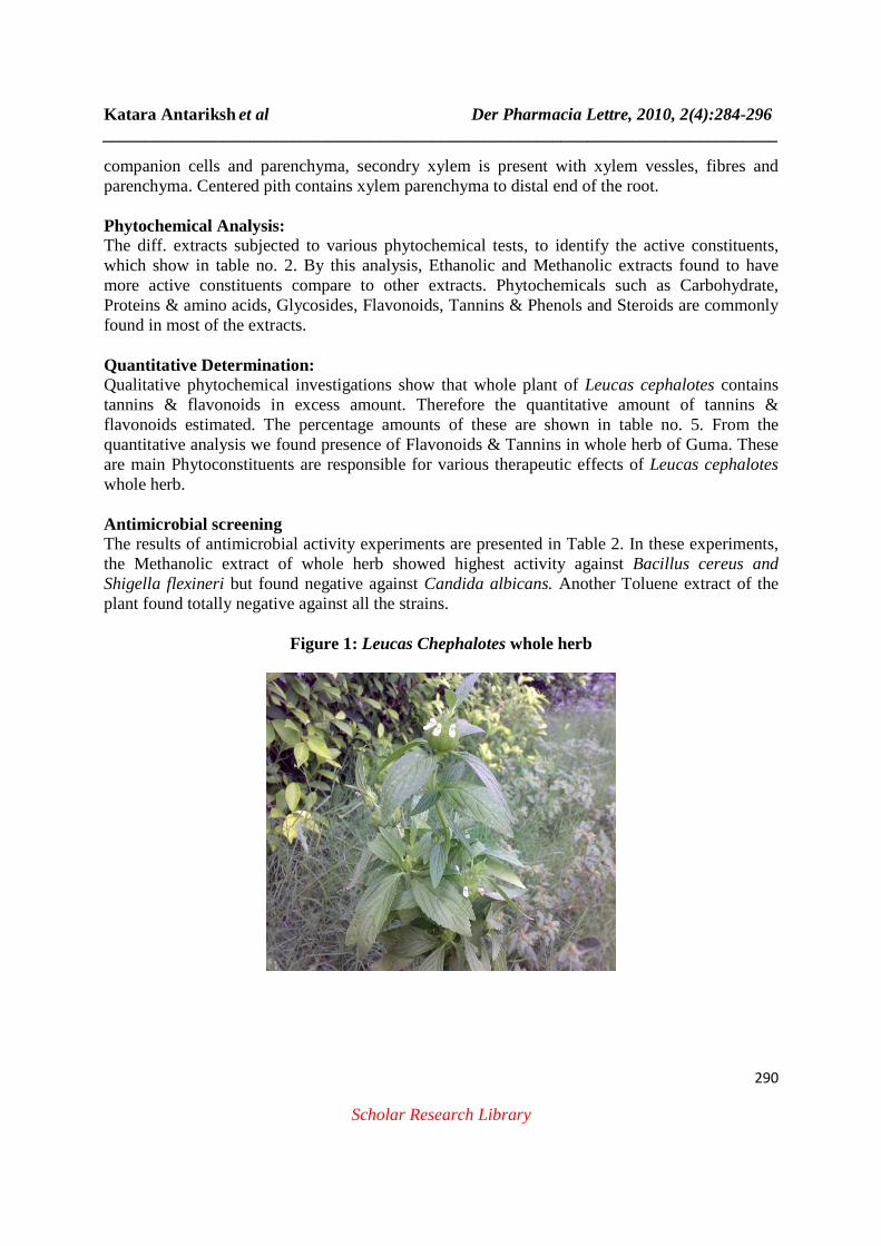
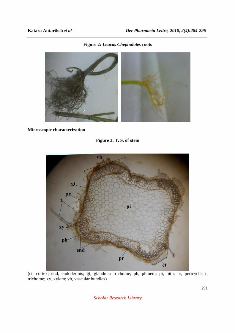
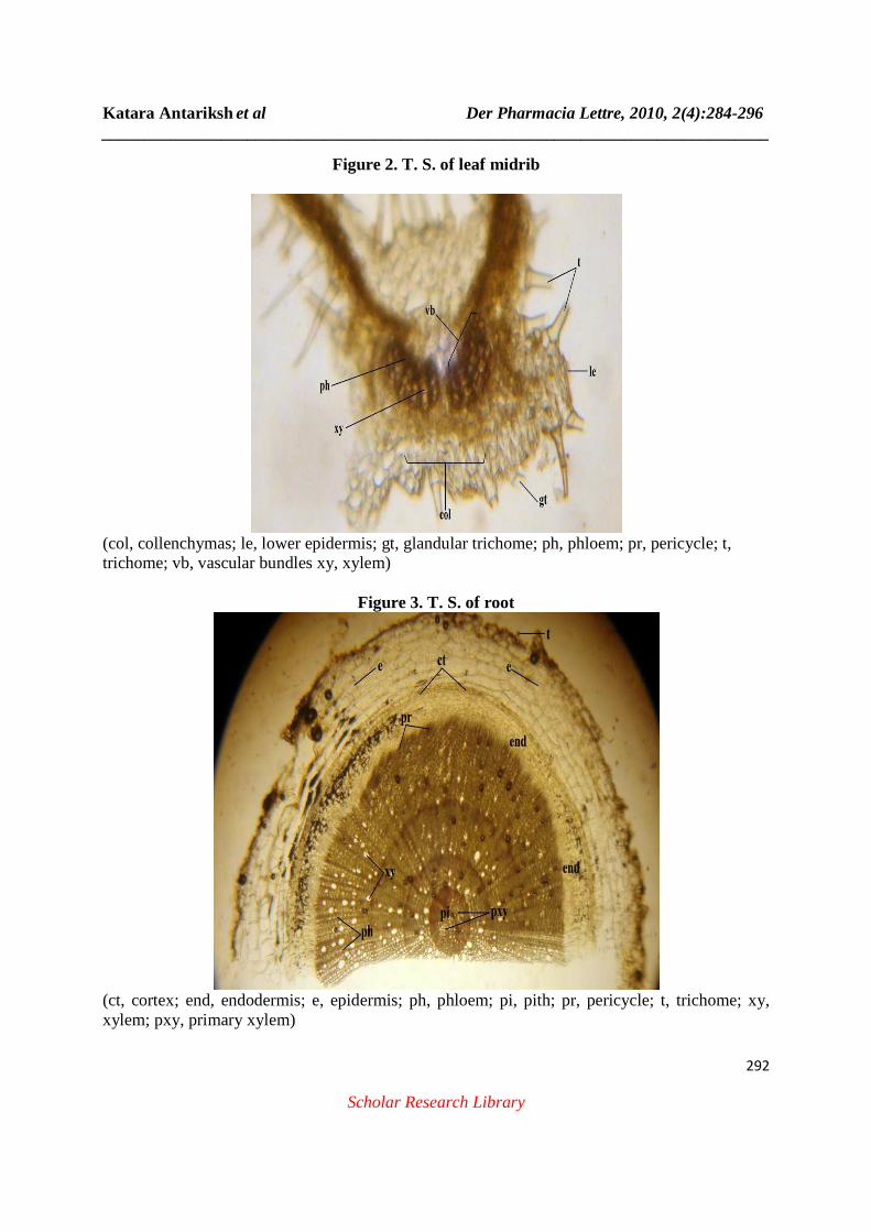
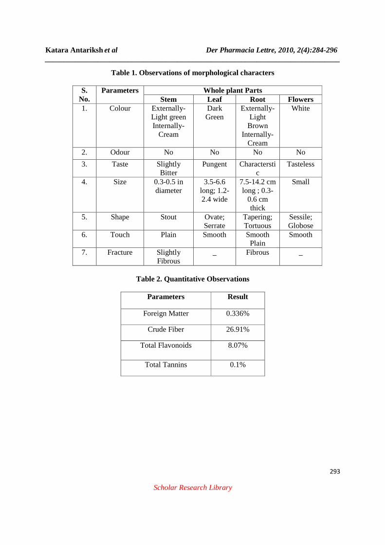
Pharmacognostic Parameters: Macroscopy: Macroscopic characters are useful in the identification of Organoleptic features such as colour, odour, taste, size, shape, touch and fracture were analyzed on the different plant parts. Stems were usually stout, slightly fibrous and light green, odourless and slightly bitter in taste. Leaves were ovate, serrate, dark green, odourless and having pungent taste. Flowers were white in colour, sessile, globose, surrounded by foliaceous bracts, odoureless and tasteless. Roots are tapering, tourtuous, fibrous nature, odourless and having characteristic taste. These characters shown in Table No. 01 and Figure No. 1 & 2. Microscopy: The TS of stem, leaf and root were observed and different histological parameters determined under microscope. A: Transverse Section of Stem (Figure. 3) TS of Stem found to be quadrangular and having outlined ridges covered with thick cuticle and with cortex parenchymal cells forms 3 to 7 rows. Vascular bundles are bicollatoral and found with in the ridges. Xylem fully developed, contains trachides and parenchyma. Phloem contains narrow circles surrounded by xylem vessels and fixing towards the pith cells. Pericyle and endodermis is present and outer layer consist diff. types of trichomes such as single cell, double cell, covering and glandular trichomes. Pith cells are collenchymatous and contain prismatic shaped crystals. B: Transverse Section of Leaf (Figure. 4) TS of midrib of leaf contain diff. types of trichomes in a big mass ie glandular, non-glandular and multilayered with the single layered palisade cells. Vascular bundles are bicollatoral, mesophyll cells are parenchymatous and central part is collenchymatous. Midrib contains deep groove on upper side and wide ridges on lower side. C: Transverse Section of Root (Figure. 5) TS of root contain out line of thick walled epidermis cells in 1-3 layers, very few simple trichomes are present, cortex parechymatous cells with 8-10 layered, tangentially elongated. Endodermis elongated 4-6 layered parenchymatous cells, phloem consist of sieve tubes,
Katara Antariksh et al Der Pharmacia Lettre, 2010, 2(4):284-296 ______________________________________________________________________________
290
Scholar Research Library
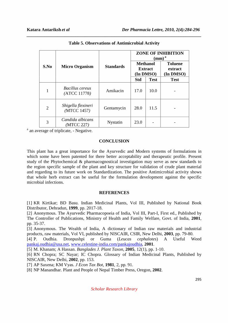
companion cells and parenchyma, secondry xylem is present with xylem vessles, fibres and parenchyma. Centered pith contains xylem parenchyma to distal end of the root. Phytochemical Analysis: The diff. extracts subjected to various phytochemical tests, to identify the active constituents, which show in table no. 2. By this analysis, Ethanolic and Methanolic extracts found to have more active constituents compare to other extracts. Phytochemicals such as Carbohydrate, Proteins & amino acids, Glycosides, Flavonoids, Tannins & Phenols and Steroids are commonly found in most of the extracts. Quantitative Determination: Qualitative phytochemical investigations show that whole plant of Leucas cephalotes contains tannins & flavonoids in excess amount. Therefore the quantitative amount of tannins & flavonoids estimated. The percentage amounts of these are shown in table no. 5. From the quantitative analysis we found presence of Flavonoids & Tannins in whole herb of Guma. These are main Phytoconstituents are responsible for various therapeutic effects of Leucas cephalotes whole herb. Antimicrobial screening The results of antimicrobial activity experiments are presented in Table 2. In these experiments, the Methanolic extract of whole herb showed highest activity against Bacillus cereus and Shigella flexineri but found negative against Candida albicans. Another Toluene extract of the plant found totally negative against all the strains.
Figure 1: Leucas Chephalotes whole herb
Katara Antariksh et al Der Pharmacia Lettre, 2010, 2(4):284-296 ______________________________________________________________________________
291
Scholar Research Library
Figure 2: Leucas Chephalotes roots
Microscopic characterization
Figure 3. T. S. of stem
(ct, cortex; end, endodermis; gt, glandular trichome; ph, phloem; pi, pith; pr, pericycle; t, trichome; xy, xylem; vb, vascular bundles)
Katara Antariksh et al Der Pharmacia Lettre, 2010, 2(4):284-296 ______________________________________________________________________________
292
Scholar Research Library
Figure 2. T. S. of leaf midrib
(col, collenchymas; le, lower epidermis; gt, glandular trichome; ph, phloem; pr, pericycle; t, trichome; vb, vascular bundles xy, xylem)
Figure 3. T. S. of root
(ct, cortex; end, endodermis; e, epidermis; ph, phloem; pi, pith; pr, pericycle; t, trichome; xy, xylem; pxy, primary xylem)
Katara Antariksh et al Der Pharmacia Lettre, 2010, 2(4):284-296 ______________________________________________________________________________
293
Scholar Research Library
Table 1. Observations of morphological characters
S. No.
Parameters Whole plant Parts Stem Leaf Root Flowers
1. Colour Externally- Light green Internally-
Cream
Dark Green
Externally-Light Brown
Internally- Cream
White
2. Odour No No No No
3. Taste Slightly Bitter
Pungent Characterstic
Tasteless
4. Size 0.3-0.5 in diameter
3.5-6.6 long; 1.2-2.4 wide
7.5-14.2 cm long ; 0.3-
0.6 cm thick
Small
5. Shape Stout Ovate; Serrate
Tapering; Tortuous
Sessile; Globose
6. Touch Plain Smooth Smooth Plain
Smooth
7. Fracture Slightly Fibrous
_ Fibrous _
Table 2. Quantitative Observations
Parameters Result
Foreign Matter 0.336%
Crude Fiber 26.91%
Total Flavonoids 8.07%
Total Tannins 0.1%

Katara Antariksh et al Der Pharmacia Lettre, 2010, 2(4):284-296 ______________________________________________________________________________
294
Scholar Research Library
Table 3. Observations of Fluorescence Analysis
S.No. Materials / Treatments
Observations Under UV Cabinet Day Light At Short
Wavelength At Long
Wavelength 1. Drug Powder Brownish
green Green Dark Green
2. Drug powder rubbed on filter paper
Light Brown Light green Green
3. Powder treated with 1 N NaOH in
Methanol
Yellowish green
Light green Yellow
4. Powder treated with 1 N NaOH in Water
Brownish green
Florescent green Green
5. Powder treated with 1 N HCl Solution
Colourless Colourless Light green
6. Powder treated with 50% HNO3
Yellow Light florescent green
Dark green
7. Powder treated with 50% H2SO4
Light brownish
green
Light florescent green
Light green
8. Methanolic Extract Yellowish green
Florescent green Yellow
9. Water Extract Brown Florescent green Light flor. green
10. Chloroform Extract Green Yellowish Brown Green 11. Pet. Ether Extract Yellow Florescent green Orange
Table 4. Phytochemical Analysis
Phytochemicals Extracts
PE T CH Et. Mt W Alkaloids - - - - - - Glycosides - - - + + + Saponins - - - - - - Steroids + + + + + -
Tannins & Phenols - - - + + + Flavonoids - - - + + +
Amino acid & Proteins - - - + + - Carbohydrates - - + + + +
Legends: + Positive, - Negative, PE Pet ether, T Toluene, CH Chloroform, Et Ethanol, Mt Methanol, W Water.
Katara Antariksh et al Der Pharmacia Lettre, 2010, 2(4):284-296 ______________________________________________________________________________
295
Scholar Research Library
Table 5. Observations of Antimicrobial Activity
a an average of triplicate, - Negative.
CONCLUSION
This plant has a great importance for the Ayurvedic and Modern systems of formulations in which some have been patented for there better acceptability and therapeutic profile. Present study of the Phytochemical & pharmacognostical investigation may serve as new standards to the region specific sample of the plant and key structure for validation of crude plant material and regarding to its future work on Standardization. The positive Antimicrobial activity shows that whole herb extract can be useful for the formulation development against the specific microbial infections.
REFERENCES
[1] KR Kirtikar; BD Basu. Indian Medicinal Plants, Vol ΙΙΙ, Published by National Book Distributor, Dehradun, 1999, pp. 2017-18. [2] Anonymous. The Ayurvedic Pharmacopoeia of India, Vol ΙΙΙ, Part-Ι, First ed., Published by The Controller of Publications, Ministry of Health and Family Welfare, Govt. of India, 2001, pp. 35-37. [3] Anonymous. The Wealth of India, A dictionary of Indian raw materials and industrial products, raw materials, Vol VI, published by NISCAIR, CSIR, New Delhi, 2003, pp. 79-80. [4] P. Oudhia. Dronpushpi or Guma (Leucas cephalotes) A Useful Weed [email protected], www.celestine-india.com/pankajoudhia, 2001. [5] M. Khanam; A Hassan. Banglades J. Plant Taxon, 2005, 12(1), pp. 1-10. [6] RN Chopra; SC Nayar; IC Chopra. Glossary of Indian Medicinal Plants, Published by NISCAIR, New Delhi, 2002, pp. 153. [7] AP Saxena; KM Vyas. J Econ Tax Bot, 1981, 2, pp. 91. [8] NP Manandhar. Plant and People of Nepal Timber Press, Oregon, 2002.
S.No Micro Organism Standards
ZONE OF INHIBITION (mm) a
Methanol Extract
(In DMSO)
Toluene extract
(In DMSO) Std Test Test
1 Bacillus cereus (ATCC 11778)
Amikacin 17.0 10.0 -
2 Shigella flexineri
(MTCC 1457) Gentamycin 28.0 11.5 -
3 Candida albicans
(MTCC 227) Nystatin 23.0 - -
Katara Antariksh et al Der Pharmacia Lettre, 2010, 2(4):284-296 ______________________________________________________________________________
296
Scholar Research Library
[9] UN Kanjilal; S Das; PC Knajilal. and RN De. Flora of Assam, 1939, 3, pp. 497-530. [10] JF Caius. The medicinal and poisonous plants of India. Scientific Publ., Jodhpur, India 1986, pp. 397-99. [11] N Singh; R Nath; ML Gupta; RP Kohli. Quarterly Journal of Crude Drug Research, 1978, 16, (1), pp. 8-16. [12] DC Bhatt; KD Mitaliya; NA Pandya; US Baxi. Advances in Plant Sciences, 2001, 14, pp. 123-25. [13] Patents: JP Singh. Indian Pat. Appl., INXXBQ IN 2005KO00589 A 20070202 (2007). [14] AK Gupta. Quality Standards of Indian Medicinal Plants, ICMR, New Delhi, 2003, Vol II, pp. 146-54. [15] Y Miyaichi; A Segawa; T Tomimori. Chemical & Pharmaceutical Bulletin (Tokyo), 2006, 54, pp. 1370-79. [16] S Sinha; AA Ansari; SM Osman. Chemistry & Industry (London, United Kingdom), 1978, 2, pp 67. [17] KD Bahadur; AB Sen. Quarterly Journal of Crude Drug Research, 1969, 9, pp. 1453-4. [18] AR Chowdhary; S Tripathi. Indian Perfumer, 2001, 45(2), pp. 81-82. [19] Anonymous. Quality Control Methods for Medicinal Plants Materials, WHO, Geneva, AITBS publisher, Delhi, 2002, pp. 8-21. [20] P Pradhan; L Joseph; M George; N. Kaushik; R Chulet. Journal of Pharmacy Research 2010, 3(4), pp. 776-80. [21] KR Khandelwal. Practical Pharmacognosy, Nirali Prakashan, Pune. 2005, pp. 149-53. [22] CR Chase; R Pratt. Am. J. Pharm. Sci. Assoc. (Sci.ed.), 1949, 38, pp. 324-30. [23] Anonymous. Indian Herbal Pharmacopoeia, IDMA, Mumbai, 2002, pp. 7-9. [24] S. Brandt Rose; Ruth E. Miller. J. Bacteriol, 1939, 38, pp. 525-37. [25] S Bose; A Bose. Ind Jour of Pharm Sci, 2008, 70(6), pp. 821-23.