Physiology of Vision: a swift overview 16-721: Advanced Machine Perception A. Efros, CMU, Spring...
75
Physiology of Vision: a swift overview 16-721: Advanced Machine Perceptio A. Efros, CMU, Spring 200 Some figures from Steve Palmer
-
Upload
dwain-burns -
Category
Documents
-
view
218 -
download
3
Transcript of Physiology of Vision: a swift overview 16-721: Advanced Machine Perception A. Efros, CMU, Spring...
Physiology of Vision: a swift overview
16-721: Advanced Machine PerceptionA. Efros, CMU, Spring 2006
Some figures from Steve Palmer

Class Introductions
• Name:• Research area / project / advisor• What you want to learn in this class?• When I am not working, I ______________• Favorite fruit:
Class mailing list…

Image Formation
Digital Camera
The Eye
Film
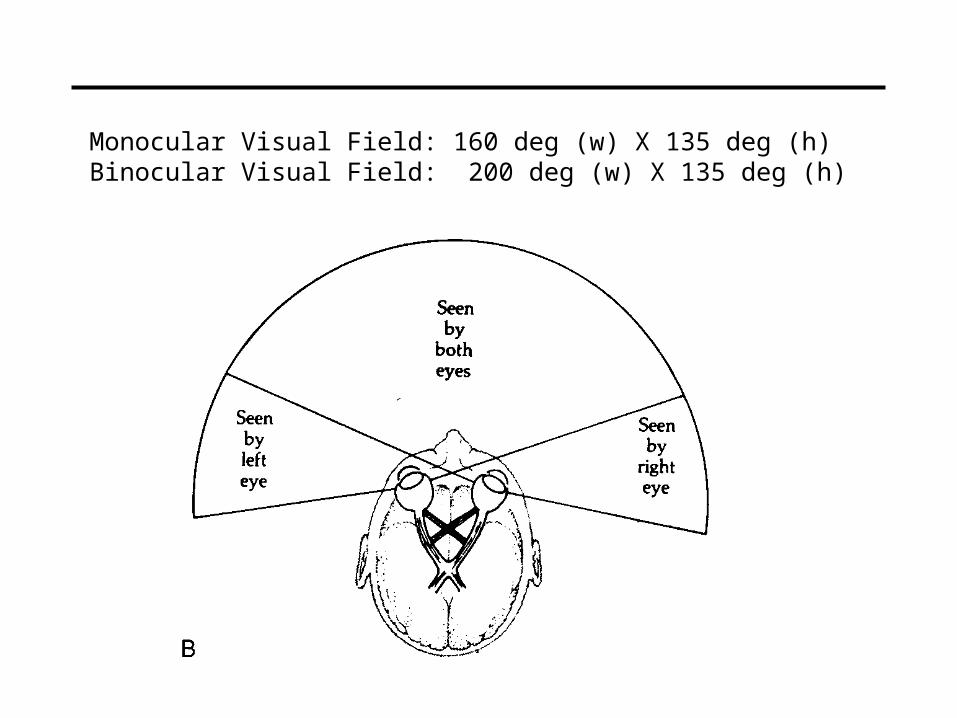
Monocular Visual Field: 160 deg (w) X 135 deg (h)Binocular Visual Field: 200 deg (w) X 135 deg (h)
Point of observation
Figures © Stephen E. Palmer, 2002
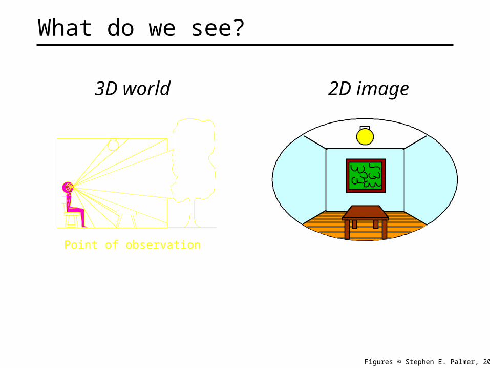
What do we see?
3D world 2D image
Point of observation
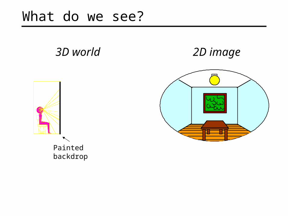
What do we see?
3D world 2D image
Painted backdrop

The Plenoptic Function
Q: What is the set of all things that we can ever see?
A: The Plenoptic Function (Adelson & Bergen)
Let’s start with a stationary person and try to parameterize everything that he can see…
Figure by Leonard McMillan

Grayscale snapshot
is intensity of light • Seen from a single view point
• At a single time
• Averaged over the wavelengths of the visible spectrum
(can also do P(x,y), but spherical coordinate are nicer)
P()
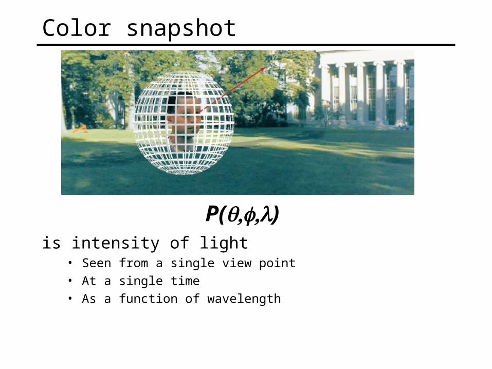
Color snapshot
is intensity of light • Seen from a single view point
• At a single time
• As a function of wavelength
P()
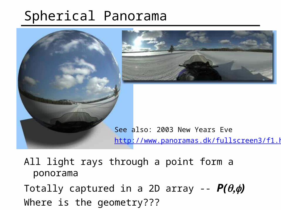
Spherical Panorama
All light rays through a point form a ponorama
Totally captured in a 2D array -- P()Where is the geometry???
See also: 2003 New Years Eve
http://www.panoramas.dk/fullscreen3/f1.html

A movie
is intensity of light • Seen from a single view point
• Over time
• As a function of wavelength
P(,t)
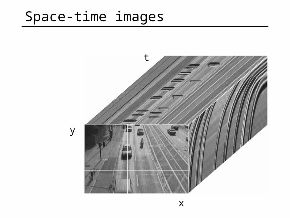
Space-time images
x
y
t

Holographic movie
is intensity of light • Seen from ANY viewpoint
• Over time
• As a function of wavelength
P(,t,VX,VY,VZ)

The Plenoptic Function
• Can reconstruct every possible view, at every moment, from every position, at every wavelength
• Contains every photograph, every movie, everything that anyone has ever seen! it completely captures our visual reality! Not bad for a function…
P(,t,VX,VY,VZ)
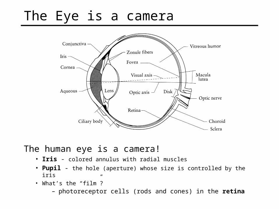
The Eye is a camera
The human eye is a camera!• Iris - colored annulus with radial muscles
• Pupil - the hole (aperture) whose size is controlled by the iris
• What’s the “film”?– photoreceptor cells (rods and cones) in the retina

The Retina
Cross-section of eye
Ganglion cell layer
Bipolar cell layer
Receptor layer
Pigmentedepithelium
Ganglion axons
Cross section of retina
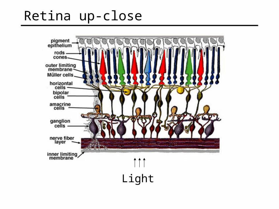
Retina up-close
Light
© Stephen E. Palmer, 2002
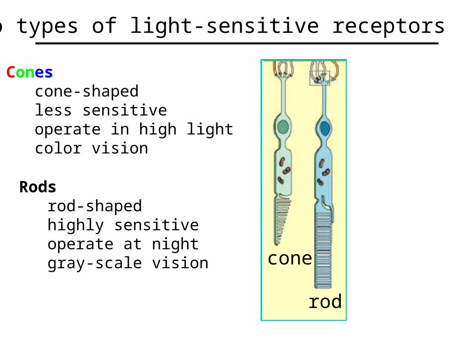
Cones cone-shaped less sensitive operate in high light color vision
Two types of light-sensitive receptors
cone
rod
Rods rod-shaped highly sensitive operate at night gray-scale vision
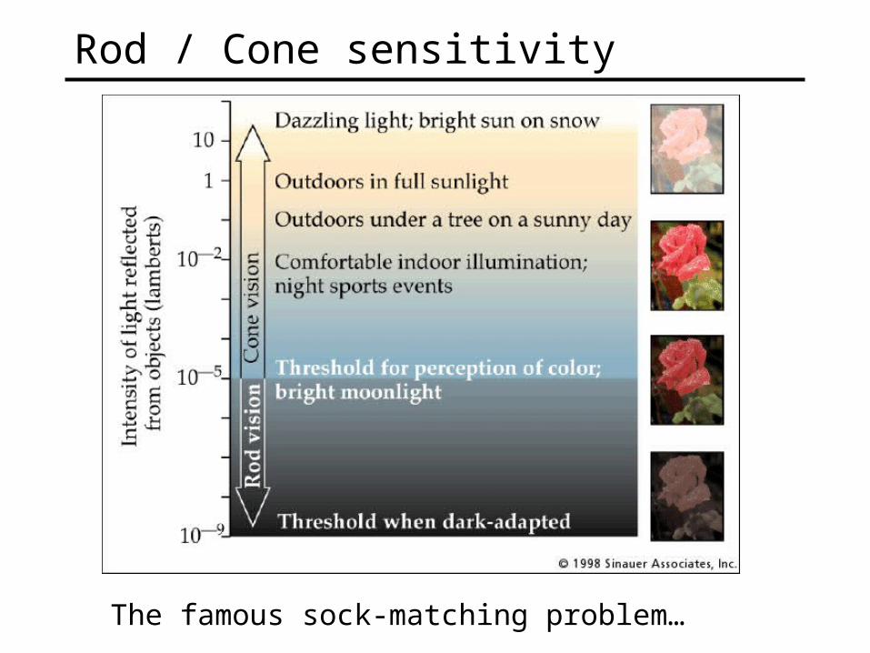
Rod / Cone sensitivity
The famous sock-matching problem…

© Stephen E. Palmer, 2002
Distribution of Rods and Cones.
0
150,000
100,000
50,000
020 40 60 8020406080
Visual Angle (degrees from fovea)
Rods
Cones Cones
Rods
FoveaBlindSpot
# R
ecep
tors
/mm
2
Night Sky: why are there more stars off-center?
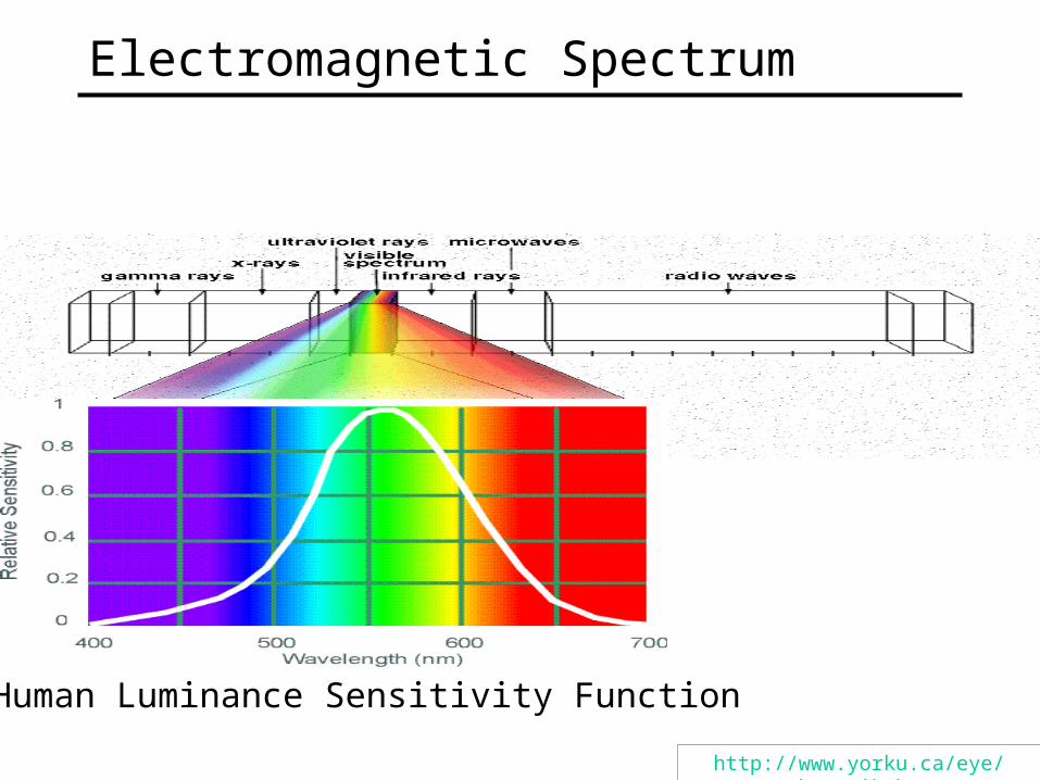
Electromagnetic Spectrum
http://www.yorku.ca/eye/photopik.htm
Human Luminance Sensitivity Function
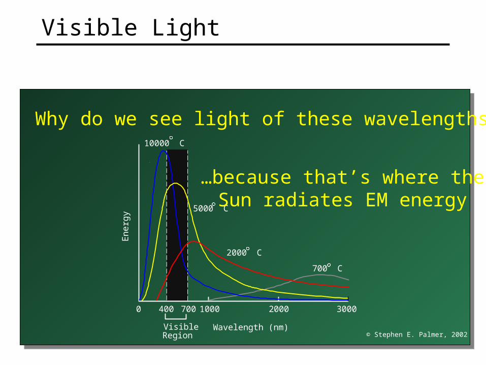
Why do we see light of these wavelengths?
© Stephen E. Palmer, 2002
.
0 1000 2000 3000
En
erg
y
Wavelength (nm)
400 700
700 C
2000 C
5000 C
10000 C
VisibleRegion
…because that’s where theSun radiates EM energy
Visible Light
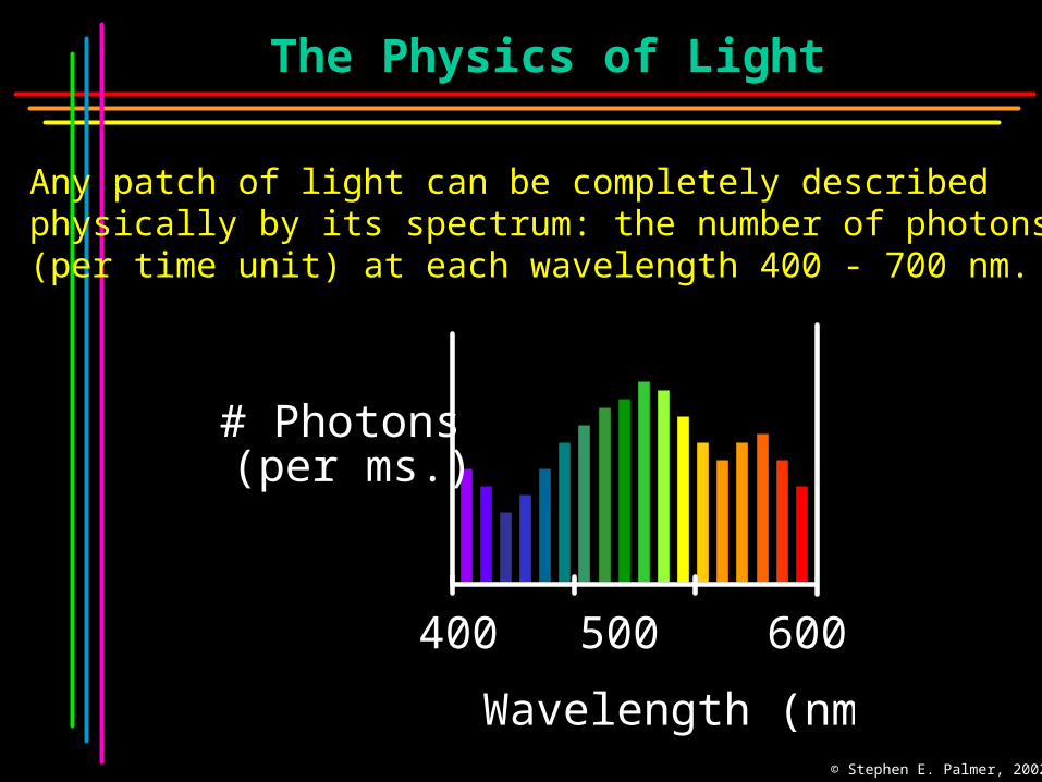
The Physics of Light
Any patch of light can be completely describedphysically by its spectrum: the number of photons (per time unit) at each wavelength 400 - 700 nm.
400 500 600 700
Wavelength (nm.)
# Photons(per ms.)
© Stephen E. Palmer, 2002

The Physics of Light
.
# P
ho
ton
s
D. Normal Daylight
Wavelength (nm.)
B. Gallium Phosphide Crystal
400 500 600 700
# P
ho
ton
s
Wavelength (nm.)
A. Ruby Laser
400 500 600 700
400 500 600 700
# P
ho
ton
s
C. Tungsten Lightbulb
400 500 600 700
# P
ho
ton
s
Some examples of the spectra of light sources
© Stephen E. Palmer, 2002

The Physics of Light
Some examples of the reflectance spectra of surfaces
Wavelength (nm)
% P
hoto
ns R
efle
cted
Red
400 700
Yellow
400 700
Blue
400 700
Purple
400 700
© Stephen E. Palmer, 2002
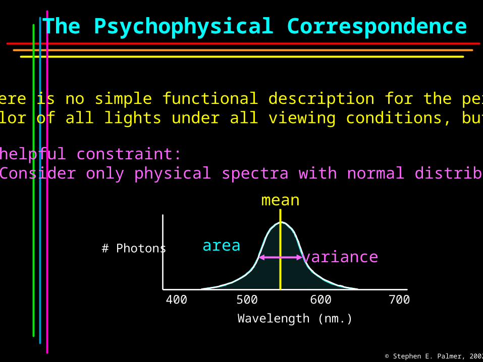
The Psychophysical Correspondence
There is no simple functional description for the perceivedcolor of all lights under all viewing conditions, but …...
A helpful constraint: Consider only physical spectra with normal distributions
area
Wavelength (nm.)
# Photons
400 700500 600
mean
variance
© Stephen E. Palmer, 2002

The Psychophysical Correspondence
Mean Hue
yellowgreenblue
# P
hoto
ns
Wavelength
© Stephen E. Palmer, 2002

The Psychophysical Correspondence
Variance Saturation
Wavelength
high
medium
low
hi.
med.
low# P
hoto
ns
© Stephen E. Palmer, 2002

The Psychophysical Correspondence
Area Brightness#
Pho
tons
Wavelength
B. Area Lightness
bright
dark
© Stephen E. Palmer, 2002
© Stephen E. Palmer, 2002
.
400 450 500 550 600 650
RE
LAT
IVE
AB
SO
RB
AN
CE
(%
)
WAVELENGTH (nm.)
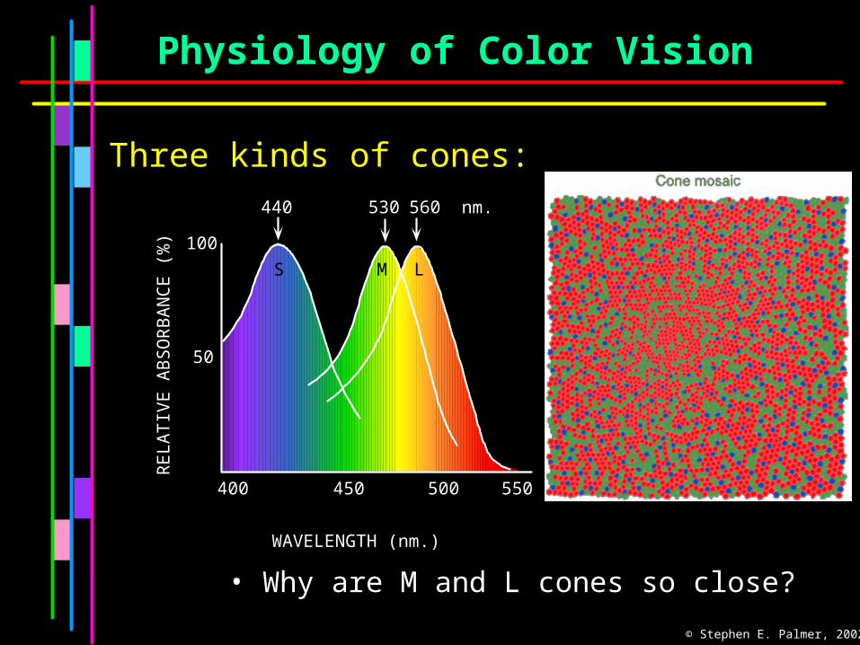
100
50
440
S
530 560 nm.
M L
Three kinds of cones:
Physiology of Color Vision
• Why are M and L cones so close?
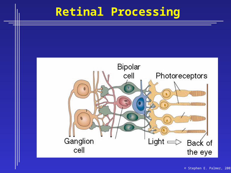
Retinal Processing
© Stephen E. Palmer, 2002
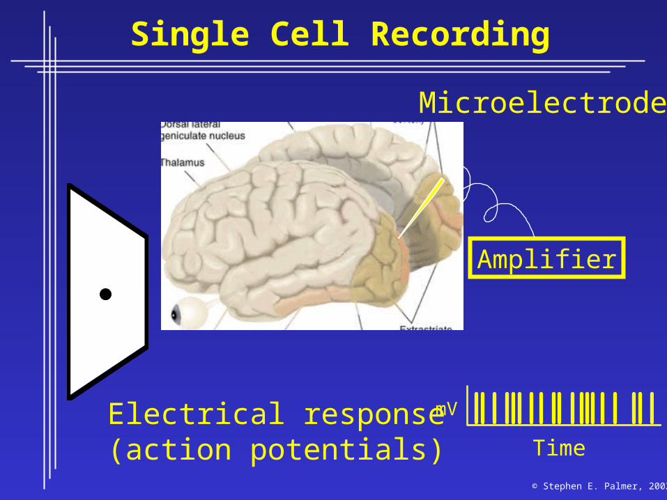
Single Cell Recording
Microelectrode
Amplifier
Time
Electrical response(action potentials)
mV
© Stephen E. Palmer, 2002
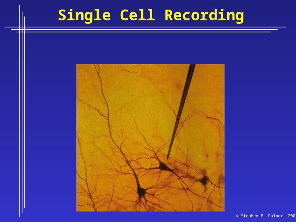
Single Cell Recording
© Stephen E. Palmer, 2002

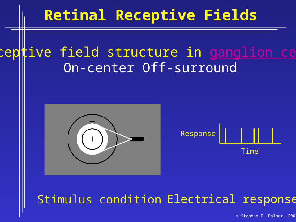
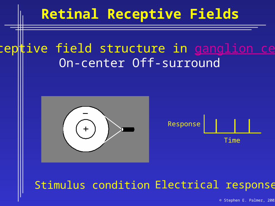
Retinal Receptive Fields
Receptive field structure in ganglion cells:On-center Off-surround
Stimulus condition Electrical response
Time
Response
© Stephen E. Palmer, 2002

Receptive field structure in ganglion cells:On-center Off-surround
Stimulus condition Electrical response
Time
Response
Retinal Receptive Fields
© Stephen E. Palmer, 2002
Receptive field structure in ganglion cells:On-center Off-surround
Stimulus condition Electrical response
Time
Response
Retinal Receptive Fields
© Stephen E. Palmer, 2002
Receptive field structure in ganglion cells:On-center Off-surround
Stimulus condition Electrical response
Time
Response
Retinal Receptive Fields
© Stephen E. Palmer, 2002

Receptive field structure in ganglion cells:On-center Off-surround
Stimulus condition Electrical response
Time
Response
Retinal Receptive Fields
© Stephen E. Palmer, 2002

Receptive field structure in ganglion cells:On-center Off-surround
Stimulus condition Electrical response
Time
Response
Retinal Receptive Fields
© Stephen E. Palmer, 2002

RF of On-center Off-surround cells
Receptive FieldNeural Response
Center
Surround
On Off
Response Profile
on-center
off-surround
Horizontal Position
FiringRate
Retinal Receptive Fields
© Stephen E. Palmer, 2002

RF of Off-center On-surround cells
Receptive Field
Horizontal Position
on-surround
off-center
Response Profile
FiringRate
Retinal Receptive Fields
© Stephen E. Palmer, 2002
Center
Surround
On Off
Neural Response
Surround
Center

Retinal Receptive Fields

Receptive field structure in bipolar cells
Light
Retinal Receptive Fields
© Stephen E. Palmer, 2002
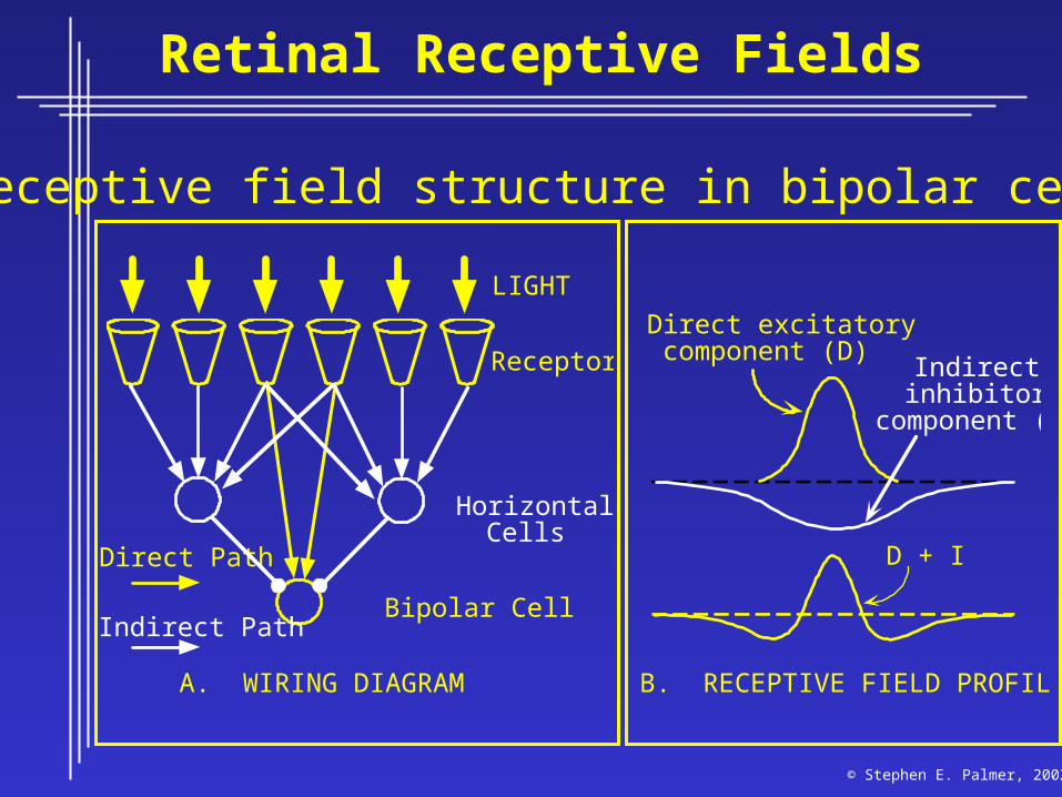
Receptive field structure in bipolar cells
Receptors
Bipolar Cell
A. WIRING DIAGRAM
HorizontalCells
Direct excitatory component (D)
B. RECEPTIVE FIELD PROFILES
LIGHT
Direct Path
Indirect Path
Indirectinhibitory
component (I)
D + I
Retinal Receptive Fields
© Stephen E. Palmer, 2002

© Stephen E. Palmer, 2002
Visual CortexVisual Cortex
aka:Primary visual cortexStriate cortexBrodman’s area 17
Cortical Area V1
ThalamusLGN
Striatecortex(V1)
DorsalStream
Parietalvisualcortex
Temporalvisualcortex
Eye Opticnerve
Extrastriatecortex
VentralStream
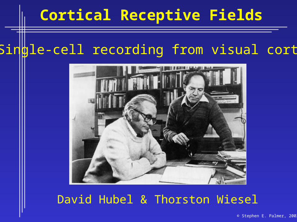
Cortical Receptive Fields
Single-cell recording from visual cortex
David Hubel & Thorston Wiesel© Stephen E. Palmer, 2002

Cortical Receptive Fields
Single-cell recording from visual cortex
TimeTime
© Stephen E. Palmer, 2002
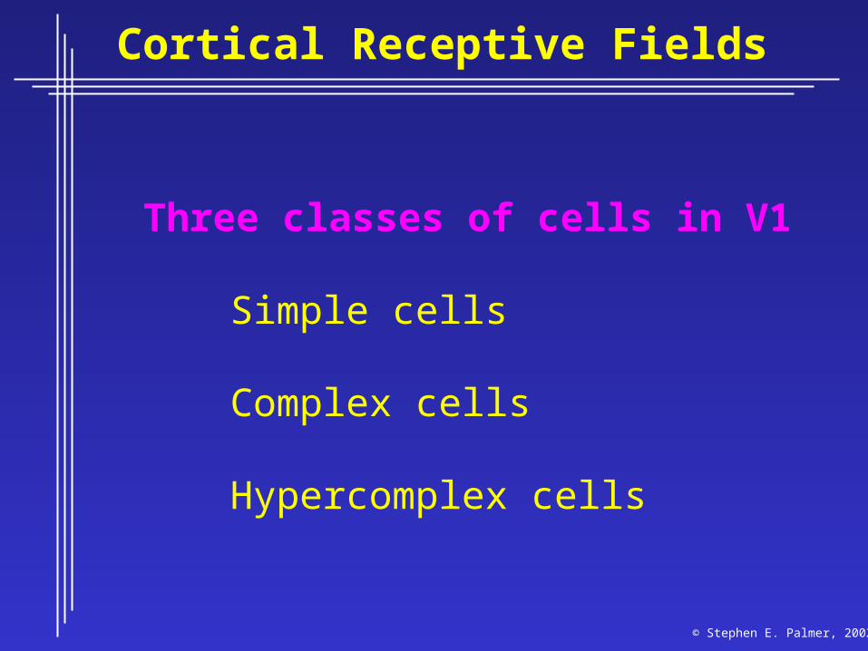
Cortical Receptive Fields
Three classes of cells in V1
Simple cells
Complex cells
Hypercomplex cells
© Stephen E. Palmer, 2002
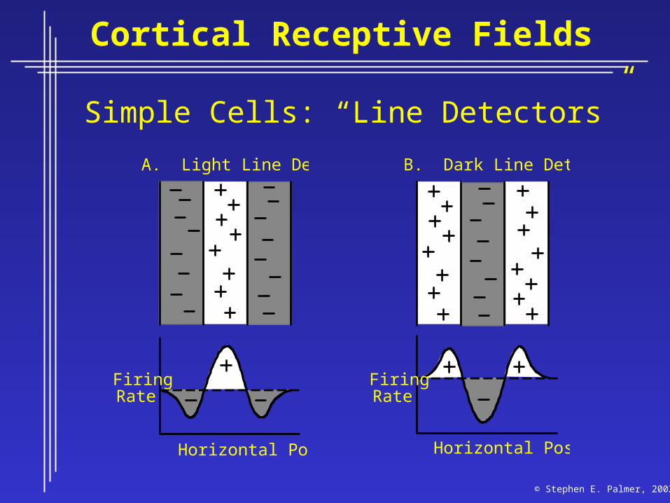
Cortical Receptive Fields
Simple Cells: “Line Detectors”
A. Light Line Detector
Horizontal Position
FiringRate
B. Dark Line Detector
Horizontal Position
FiringRate
© Stephen E. Palmer, 2002
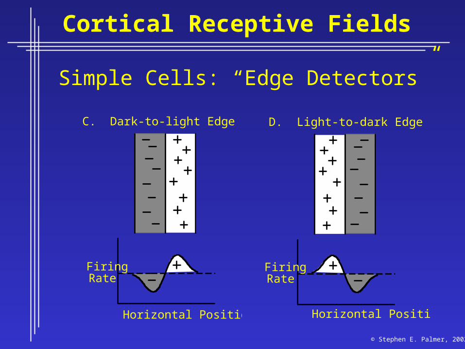
Cortical Receptive Fields
Simple Cells: “Edge Detectors”
C. Dark-to-light Edge Detector
Horizontal Position
FiringRate
D. Light-to-dark Edge Detector
Horizontal Position
FiringRate
© Stephen E. Palmer, 2002

Cortical Receptive Fields
Constructing a line detector
Receptive Fields
Retina LGN
Center-Surround Cells
Simple Cell
CorticalArea V1
© Stephen E. Palmer, 2002
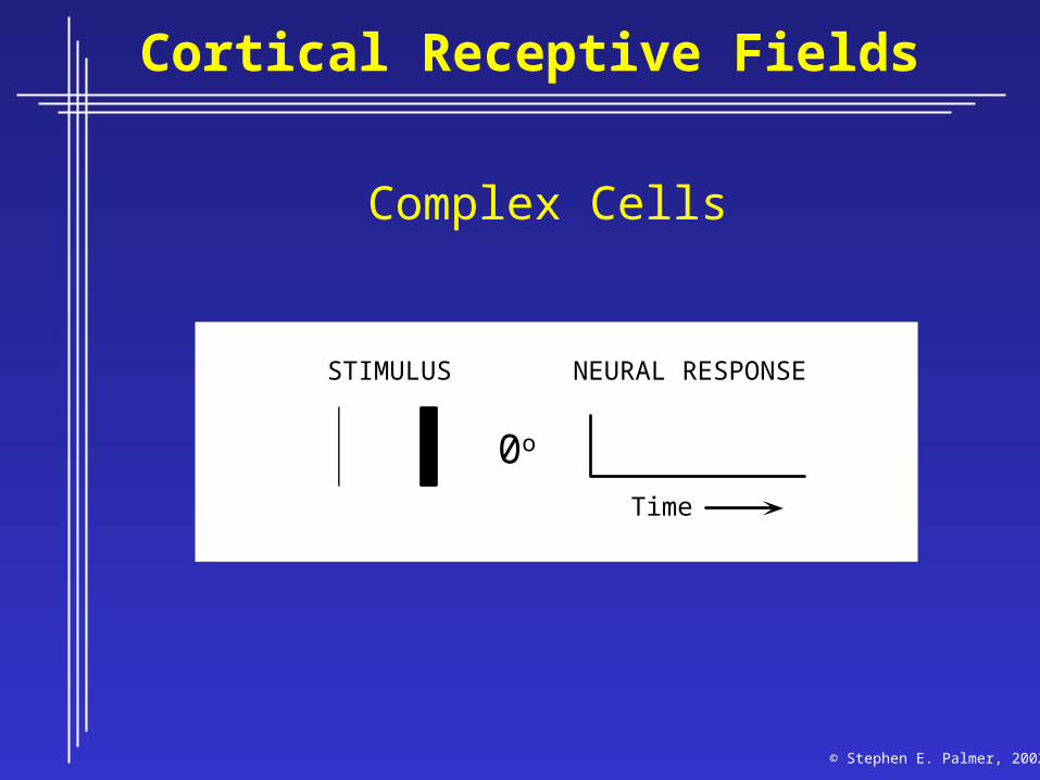
Cortical Receptive Fields
Complex Cells
STIMULUS NEURAL RESPONSE
Time
00o
© Stephen E. Palmer, 2002
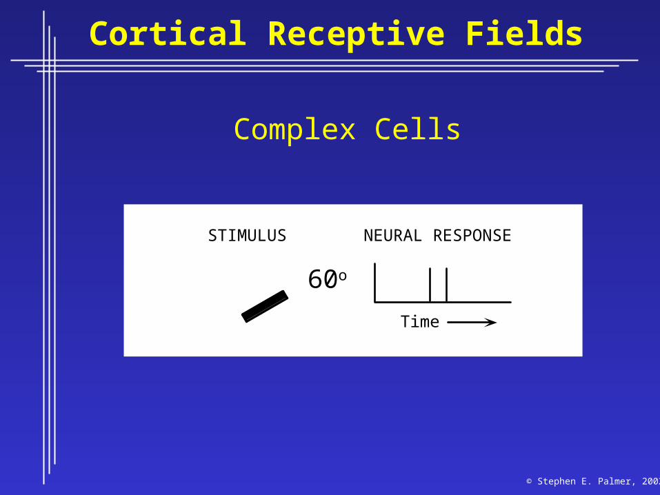
Cortical Receptive Fields
Complex Cells
STIMULUS NEURAL RESPONSE
Time
060o
© Stephen E. Palmer, 2002

Cortical Receptive Fields
Complex Cells
STIMULUS NEURAL RESPONSE
Time
090o
© Stephen E. Palmer, 2002

Cortical Receptive Fields
Complex Cells
STIMULUS NEURAL RESPONSE
Time
0120o
© Stephen E. Palmer, 2002

Cortical Receptive Fields
Constructing a Complex Cell
Simple Cells
Cortical Area V1
Complex CellReceptive Fields
Retina
© Stephen E. Palmer, 2002

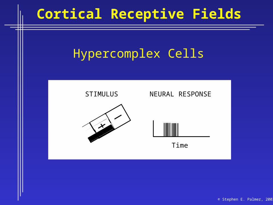
Cortical Receptive Fields
Hypercomplex Cells
Time
STIMULUS NEURAL RESPONSE
© Stephen E. Palmer, 2002
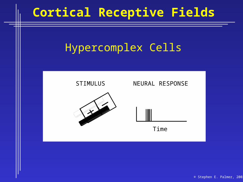
Cortical Receptive Fields
Hypercomplex Cells
Time
STIMULUS NEURAL RESPONSE
© Stephen E. Palmer, 2002
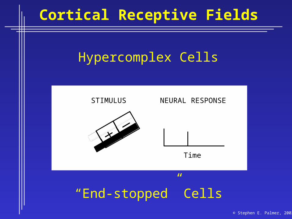
Cortical Receptive Fields
Hypercomplex Cells
Time
STIMULUS NEURAL RESPONSE
© Stephen E. Palmer, 2002
Cortical Receptive Fields
Hypercomplex Cells
Time
STIMULUS NEURAL RESPONSE
“End-stopped” Cells© Stephen E. Palmer, 2002
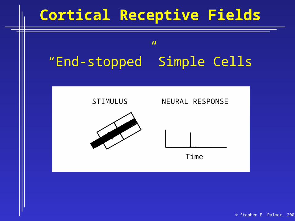
Cortical Receptive Fields
Time
STIMULUS NEURAL RESPONSE
“End-stopped” Simple Cells
© Stephen E. Palmer, 2002
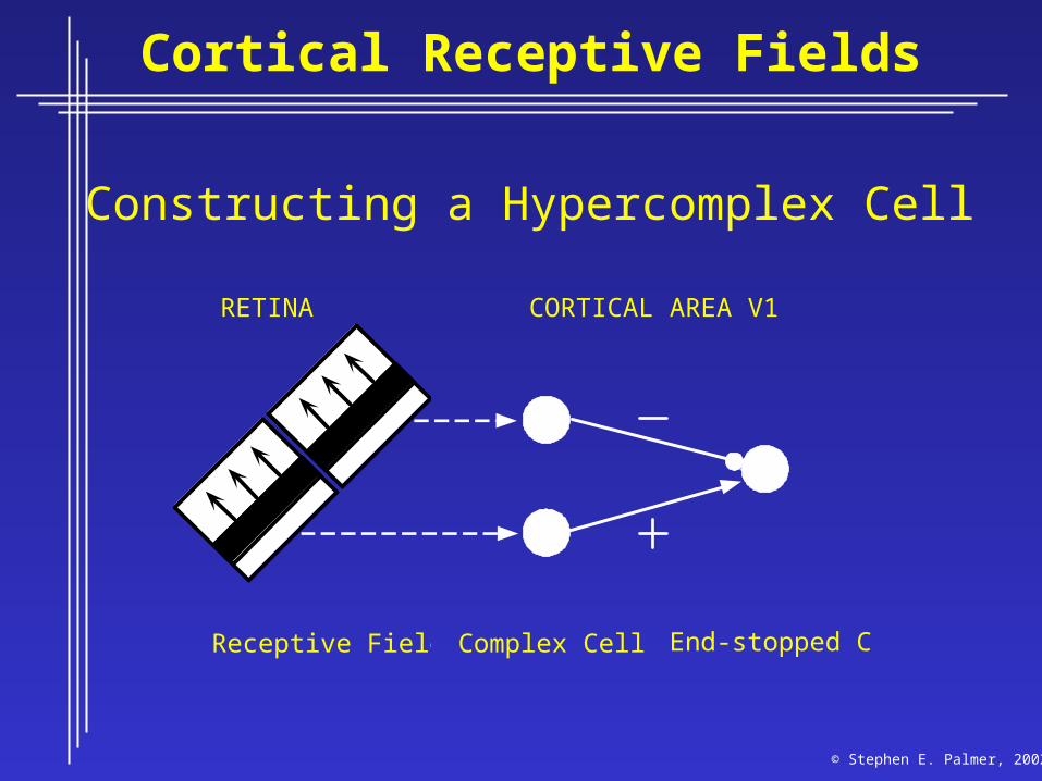
Cortical Receptive Fields
Constructing a Hypercomplex Cell
Receptive Fields
RETINA CORTICAL AREA V1
Complex Cell End-stopped Cell
© Stephen E. Palmer, 2002
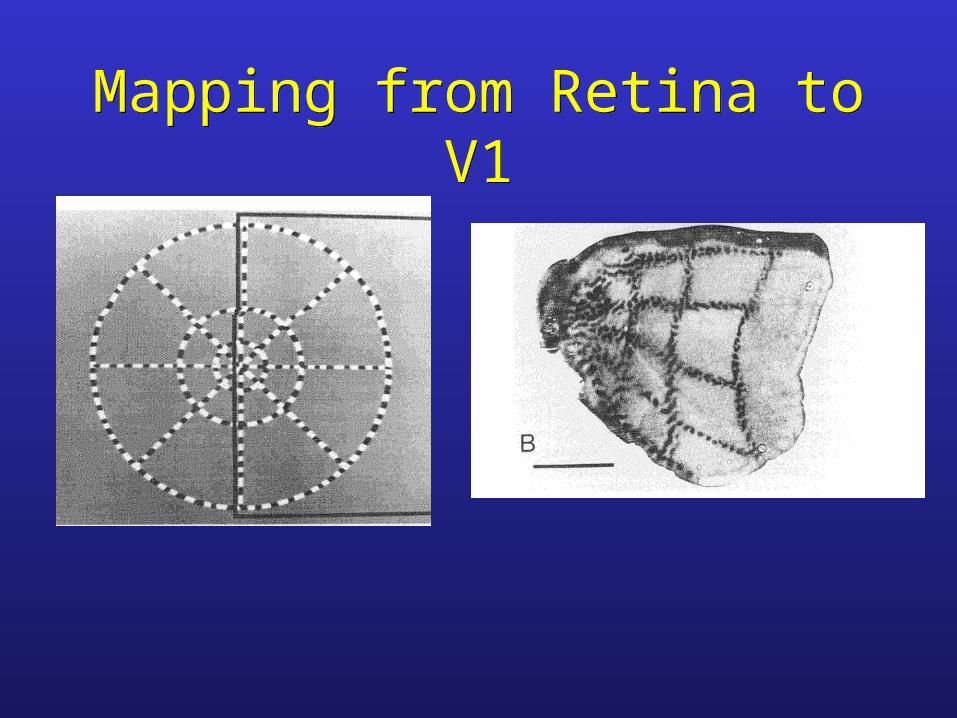
Mapping from Retina to V1Mapping from Retina to V1
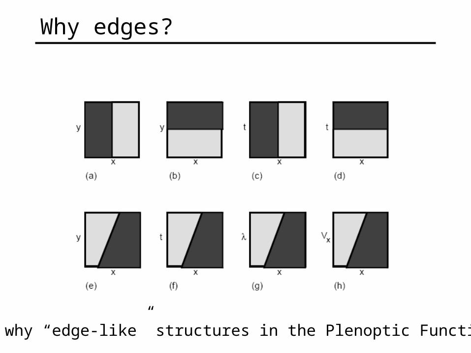
Why edges?
So, why “edge-like” structures in the Plenoptic Function?
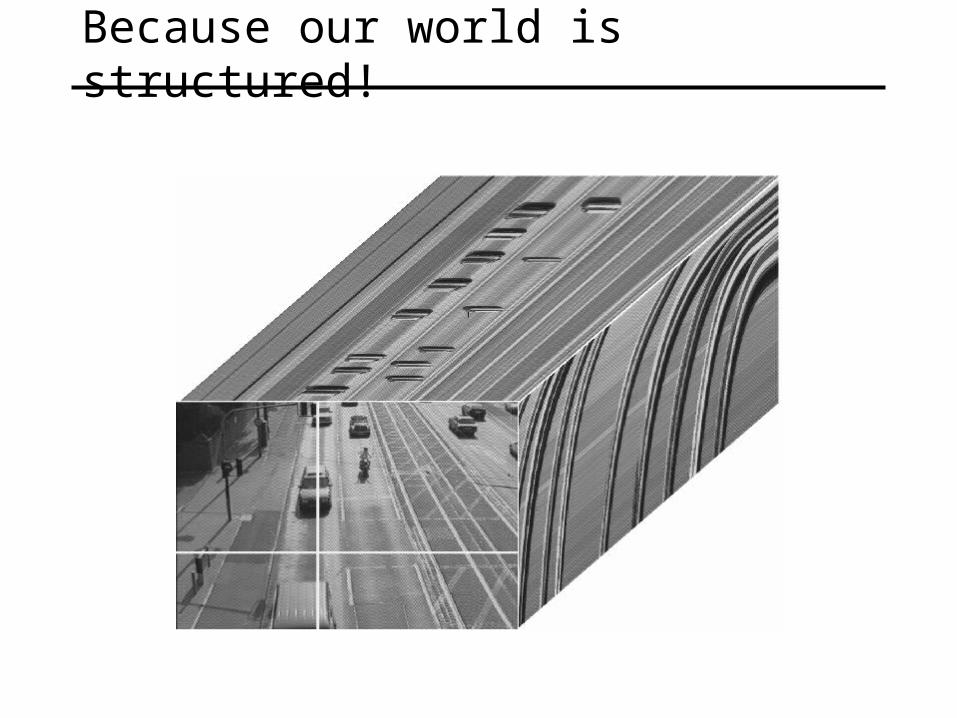
Because our world is structured!
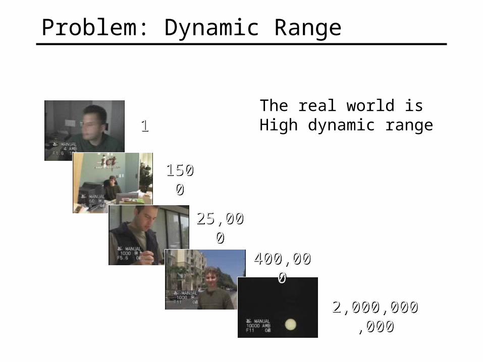
Problem: Dynamic Range
15001500
11
25,00025,000
400,000400,000
2,000,000,0002,000,000,000
The real world isHigh dynamic range
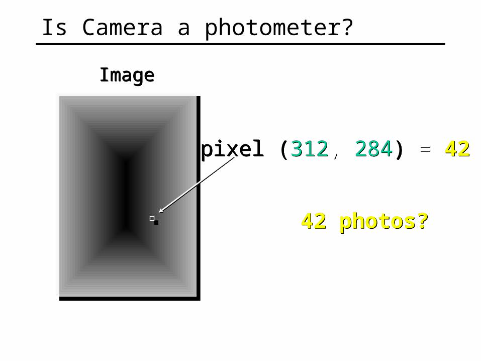
pixel (312, 284) = 42pixel (312, 284) = 42
ImageImage
42 photos?42 photos?
Is Camera a photometer?
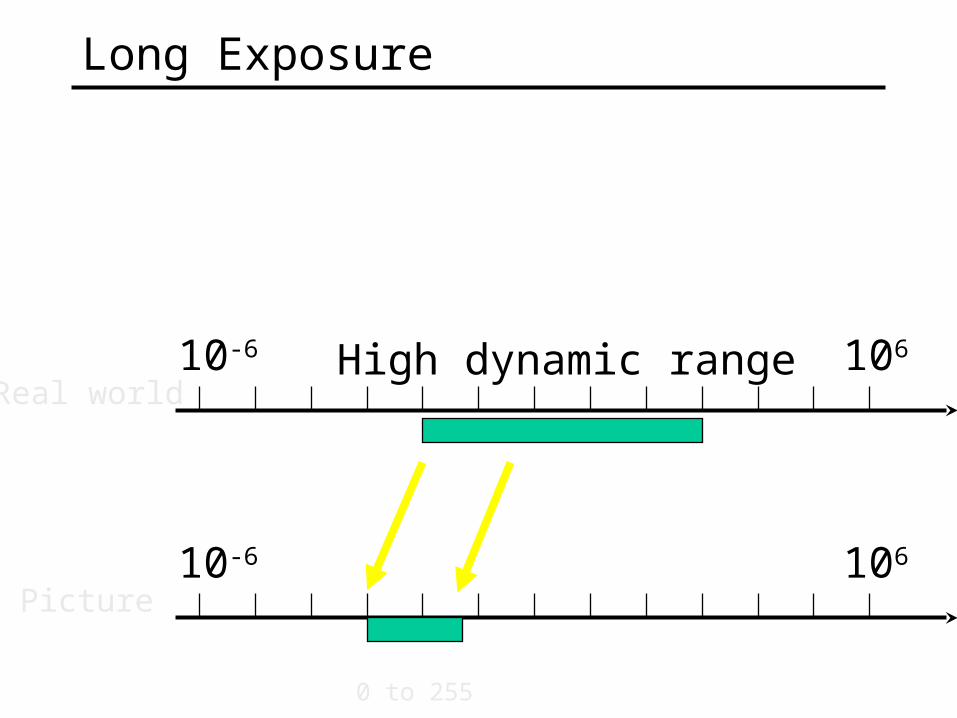
Long Exposure
10-6 106
10-6 106
Real world
Picture
0 to 255
High dynamic range
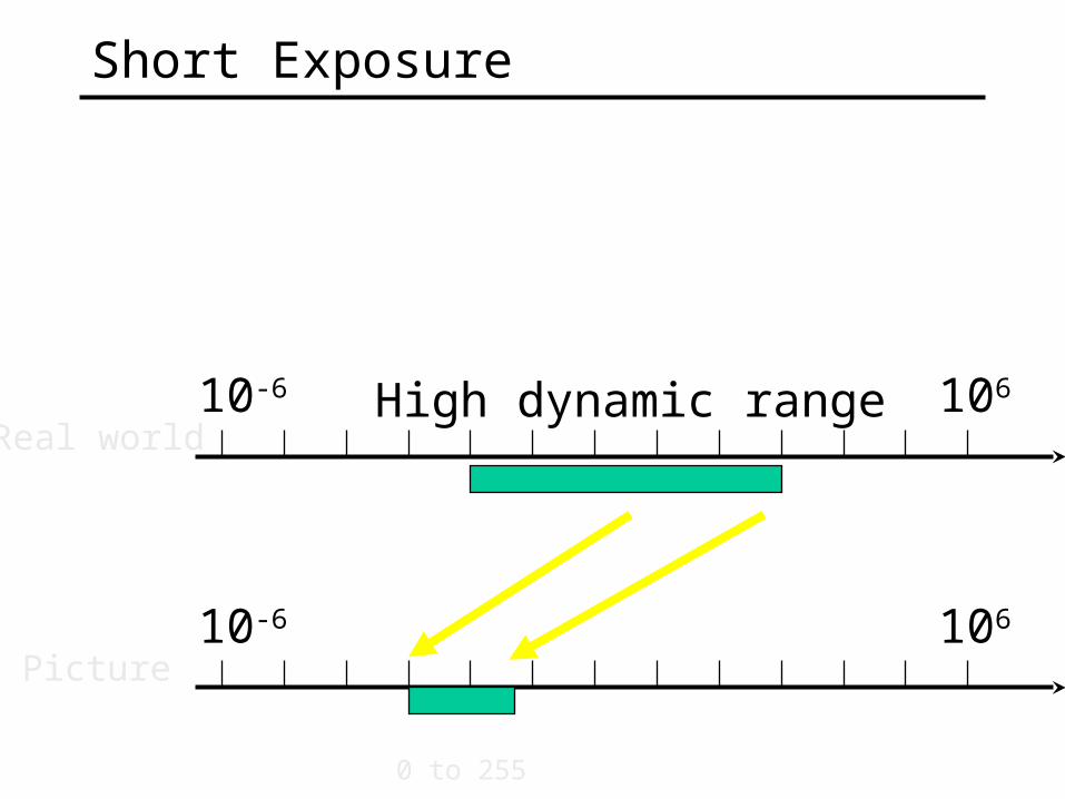
Short Exposure
10-6 106
10-6 106
Real world
Picture
0 to 255
High dynamic range
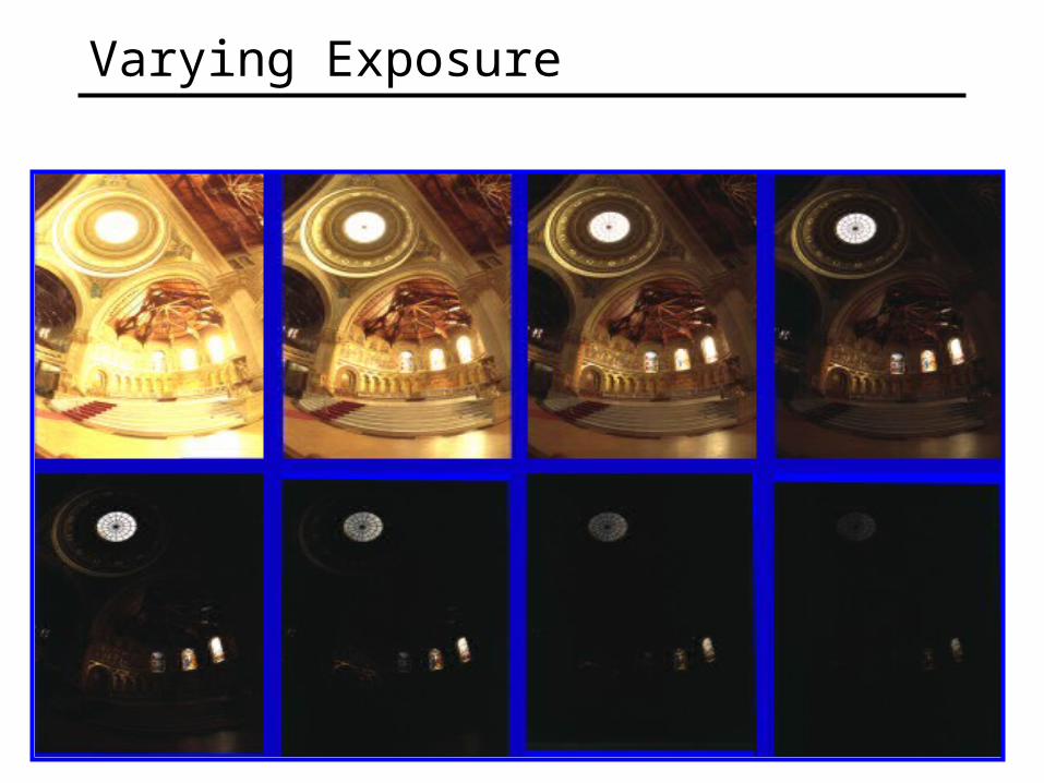
Varying Exposure
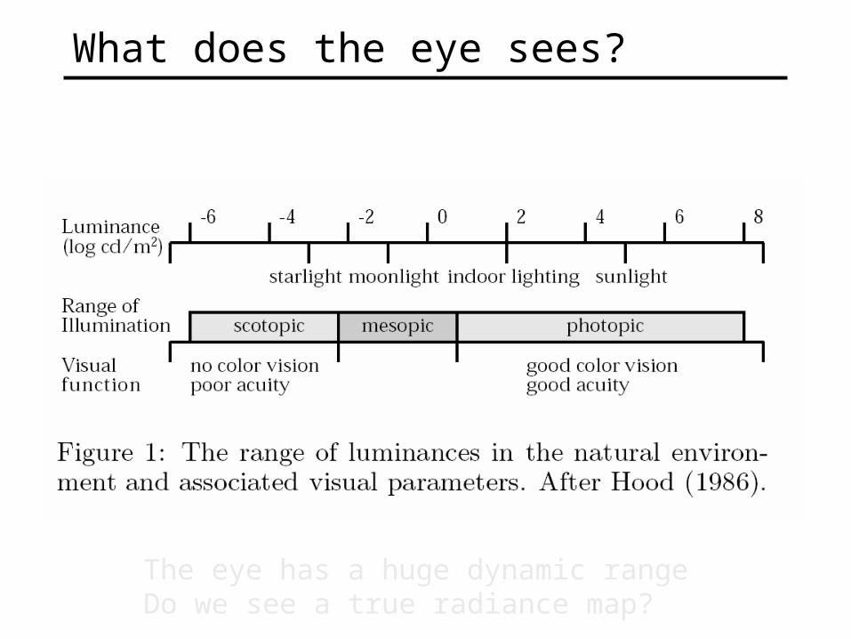
What does the eye sees?
The eye has a huge dynamic rangeDo we see a true radiance map?

Eye is not a photometer!
"Every light is a shade, compared to the higher lights, till you come to the sun; and every shade is a light, compared to the deeper shades, till you come to the night."
— John Ruskin, 1879
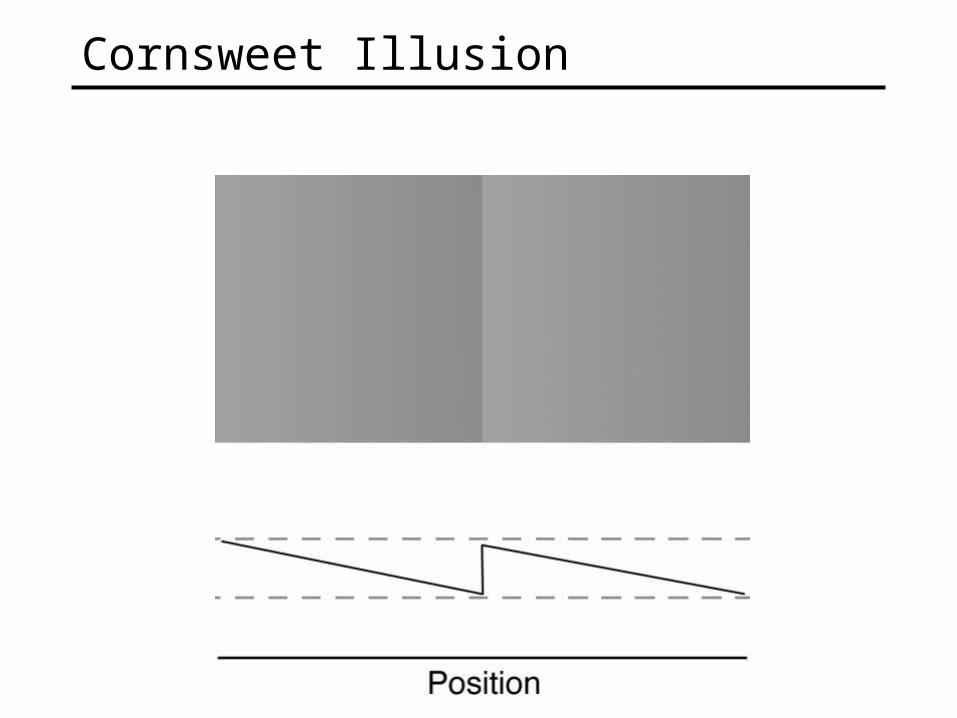
Cornsweet Illusion
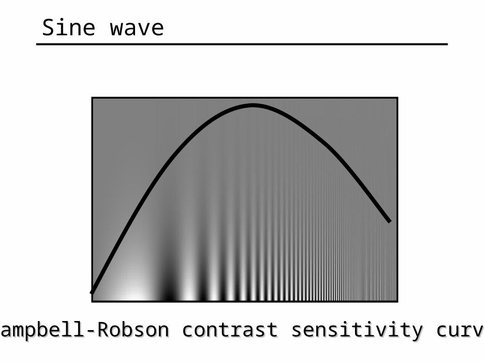
Campbell-Robson contrast sensitivity curveCampbell-Robson contrast sensitivity curve
Sine wave

Metamers
Eye is sensitive to changes(more on this later…)