![Electrical Vestibular Stimulation after Vestibular ......electrical stimulation of the vestibular system to one ear [4,5,9]. However studies have also reported vestibular responses](https://static.fdocuments.us/doc/165x107/60f6b0762ca1b41e91018b73/electrical-vestibular-stimulation-after-vestibular-electrical-stimulation.jpg)
physiology, function and physics of the vestibular system Barany… · physiology, function and...
85
physiology, function and physics of the vestibular system“ Herman Kingma, department of ORL, Maastricht University Medical Centre Maastricht Research Institute Mental Health and Neuroscience Faculty of Biomedical Technology, Technical University Eindhoven
Transcript of physiology, function and physics of the vestibular system Barany… · physiology, function and...

physiology, function and physics of the vestibular system“
Herman Kingma, department of ORL, Maastricht University Medical Centre
Maastricht Research Institute Mental Health and Neuroscience
Faculty of Biomedical Technology, Technical University Eindhoven

can we learn from what happens if
something goes wrong in the
vestibular system ?
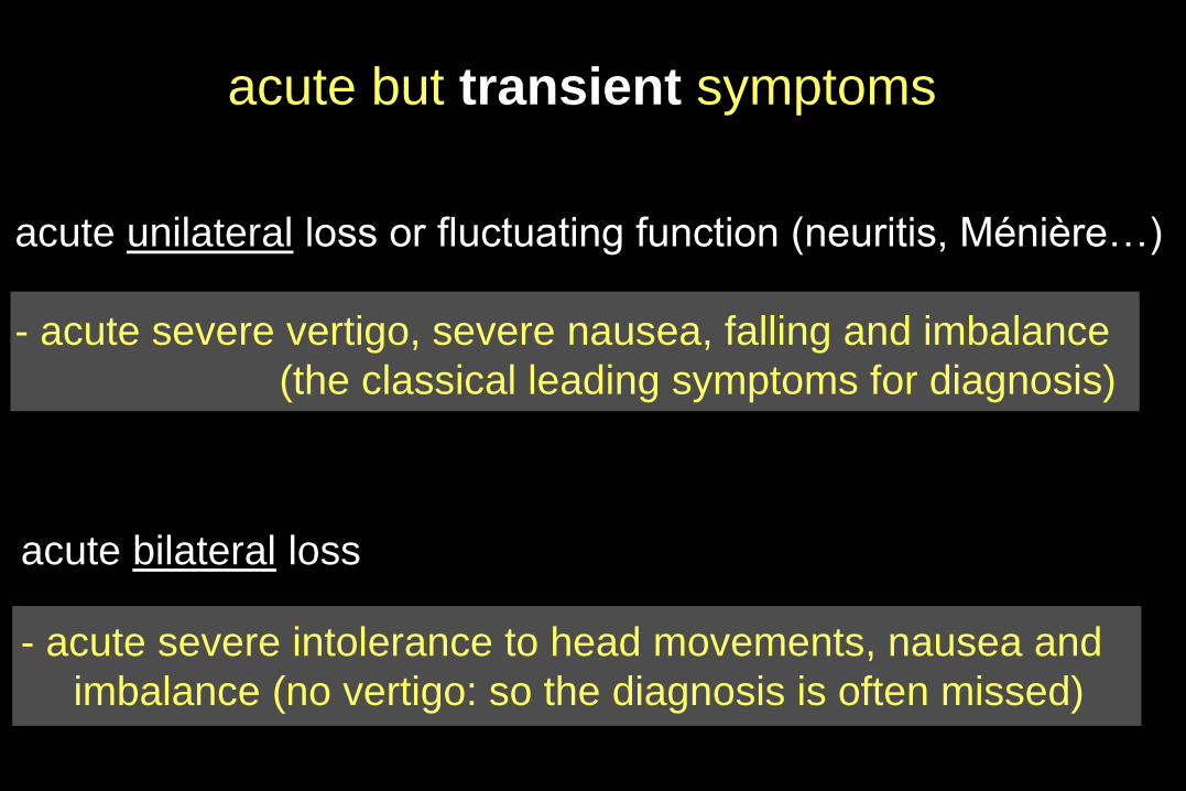
acute unilateral loss or fluctuating function (neuritis, Ménière…)
- acute severe vertigo, severe nausea, falling and imbalance
(the classical leading symptoms for diagnosis)
acute bilateral loss
- acute severe intolerance to head movements, nausea and
imbalance (no vertigo: so the diagnosis is often missed)
acute but transient symptoms
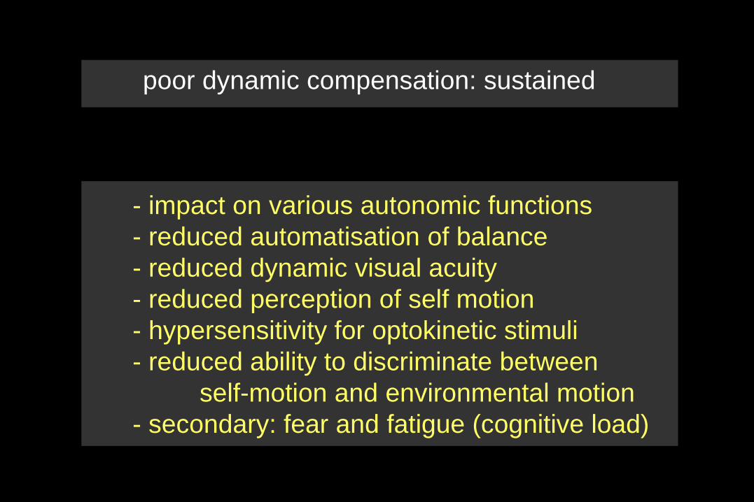
poor dynamic compensation: sustained
- impact on various autonomic functions
- reduced automatisation of balance
- reduced dynamic visual acuity
- reduced perception of self motion
- hypersensitivity for optokinetic stimuli
- reduced ability to discriminate between
self-motion and environmental motion
- secondary: fear and fatigue (cognitive load)
which complaints are related to vestibular deficits ?
which complaints are related to natural limitations ?
which complaints are related to vestibular deficits ?
which complaints are related to natural limitations ?
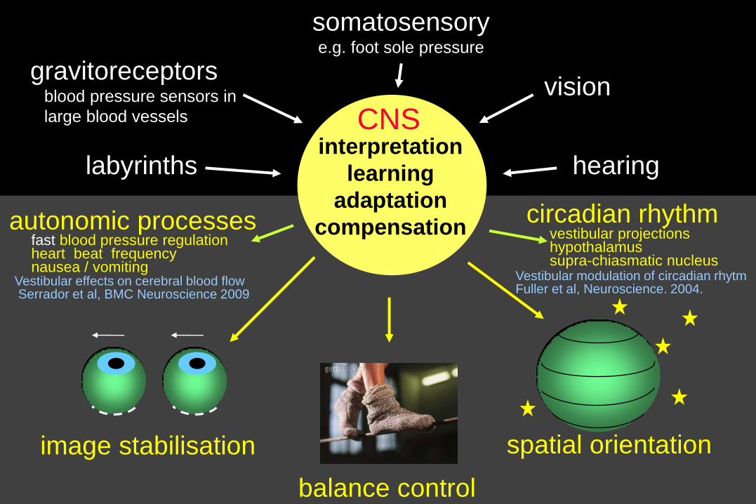
image stabilisation
balance control
spatial orientation
interpretation
learning
adaptation
compensation
CNS
labyrinths
vision gravitoreceptors blood pressure sensors in
large blood vessels
hearing
somatosensory e.g. foot sole pressure
circadian rhythm vestibular projections hypothalamus supra-chiasmatic nucleus
autonomic processes fast blood pressure regulation heart beat frequency nausea / vomiting Vestibular effects on cerebral blood flow Serrador et al, BMC Neuroscience 2009
Vestibular modulation of circadian rhytm Fuller et al, Neuroscience. 2004.

complaints related to vestibular dysfunction
acute loss or fluctuating function
transient: vertigo, nausea, falling / imbalance
remaining peripheral vestibular function loss
sustained:
- enhanced neuro-vegetative sensitivity

image stabilisation
balance control
spatial orientation
interpretation
learning
adaptation
compensation
CNS
labyrinths
vision gravitoreceptors blood pressure sensors in
large blood vessels
hearing
somatosensory e.g. foot sole pressure
circadian rhythm vestibular projections
hypothalamus
supra-chiasmatic nucleus
autonomic processes blood pressure regulation heart beat frequency respiration rate nausea / vomiting

complaints related to vestibular dysfunction
acute loss or fluctuating function
transient: vertigo, nausea, falling / imbalance
remaining peripheral vestibular function loss
sustained:
- reduced perception of self motion
- hypersensitivity for optokinetic stimuli
- reduced ability to discriminate between
self-motion and environmental motion

base of support
Centre Of Mass
vestibular impact
upon postural control
- regulation of muscle tone
relative to gravity
- regulation of Centre of Mass
relative to base of support
balancing
correction steps
- labyrinths important for
learning motor activities
and fast feed back
→ automatisation

vn
cer
hippocampus
basal ganglia
spinal pattern generator
visual
cortex
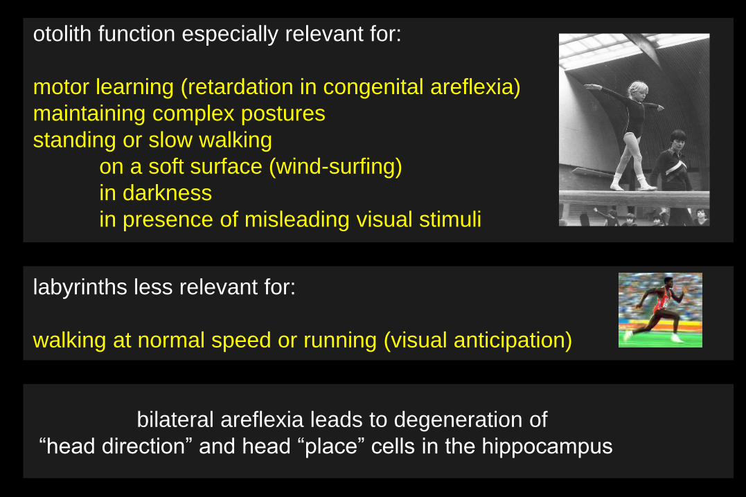
otolith function especially relevant for:
motor learning (retardation in congenital areflexia)
maintaining complex postures
standing or slow walking
on a soft surface (wind-surfing)
in darkness
in presence of misleading visual stimuli
labyrinths less relevant for:
walking at normal speed or running (visual anticipation)
bilateral areflexia leads to degeneration of
“head direction” and head “place” cells in the hippocampus
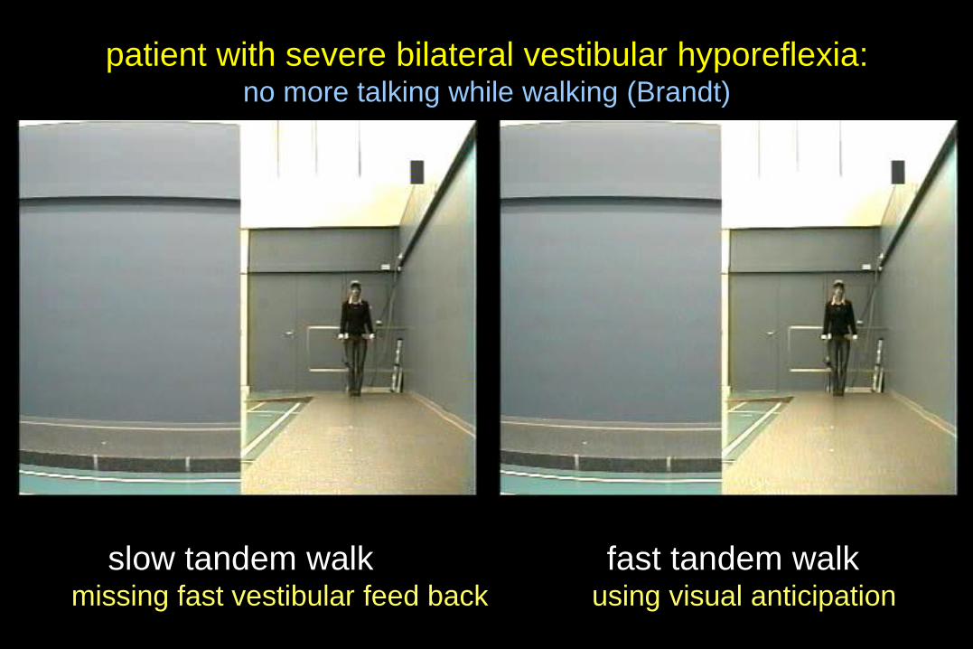
patient with severe bilateral vestibular hyporeflexia: no more talking while walking (Brandt)
slow tandem walk fast tandem walk missing fast vestibular feed back using visual anticipation
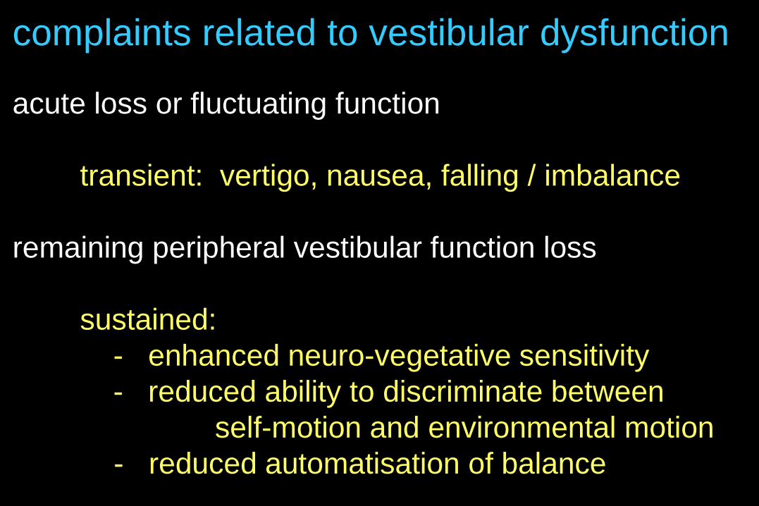
complaints related to vestibular dysfunction
acute loss or fluctuating function
transient: vertigo, nausea, falling / imbalance
remaining peripheral vestibular function loss
sustained:
- enhanced neuro-vegetative sensitivity
- reduced ability to discriminate between
self-motion and environmental motion
- reduced automatisation of balance

image stabilisation
balance control
spatial orientation
interpretation
learning
adaptation
compensation
CNS
labyrinths
vision gravitoreceptors blood pressure sensors in
large blood vessels
hearing
somatosensory e.g. foot sole pressure
circadian rhythm vestibular projections
hypothalamus
supra-chiasmatic nucleus
autonomic processes blood pressure regulation heart beat frequency respiration rate nausea / vomiting
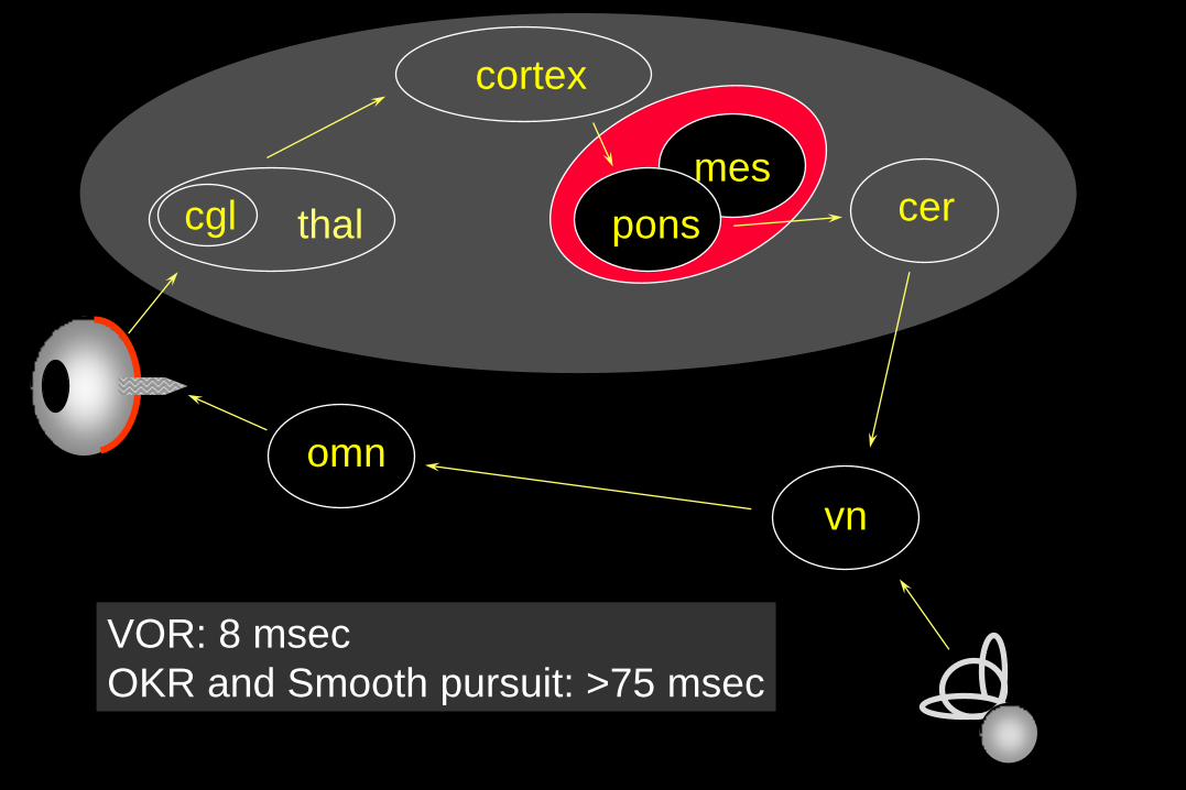
mes
thal
cortex
pons cer
vn
omn
cgl
VOR: 8 msec
OKR and Smooth pursuit: >75 msec
head impulse test in unilateral loss
standard video (50 Hz)
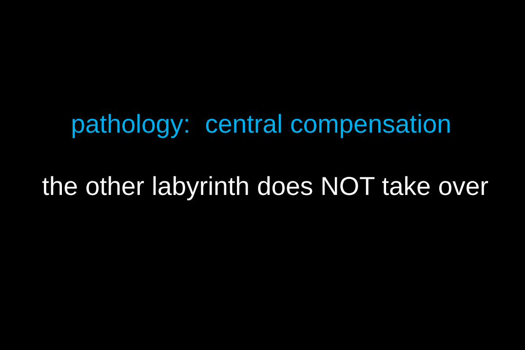
pathology: central compensation
the other labyrinth does NOT take over

simulation of oscillopsia reduced dynamic visual acuity
in case of bilateral vestibular areflexia

Dynamic Visual Acuity (VA) measurement
treadmill: 2, 4 and 6 km/h
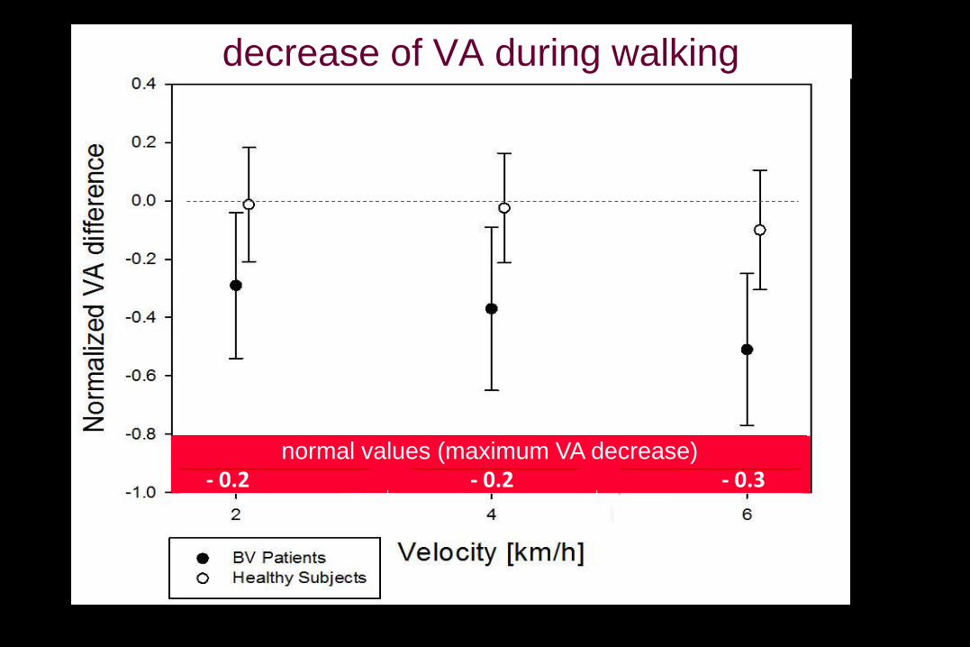
decrease of VA during walking
- 0.2 - 0.2 - 0.3 normal values (maximum VA decrease)
which complaints are related to vestibular deficits ?
which complaints are related to natural limitations ?

acute unilateral:
- vertigo, imbalance, nystagmus
sustained:
- impact on various autonomic functions
- reduced automatisation of balance
- reduced dynamic visual acuity
- reduced perception of self motion
- hypersensitivity for optokinetic stimuli
- reduced ability to discriminate between
self-motion and environmental motion
- secondary: fear and fatigue (cognitive load)

which complaints are related to vestibular deficits ?
which complaints are related to natural limitations ?

vestibular labyrinth senses low frequency motions: movement
cochlear labyrinth senses high frequency motions: sound
labyrinth function and limitations
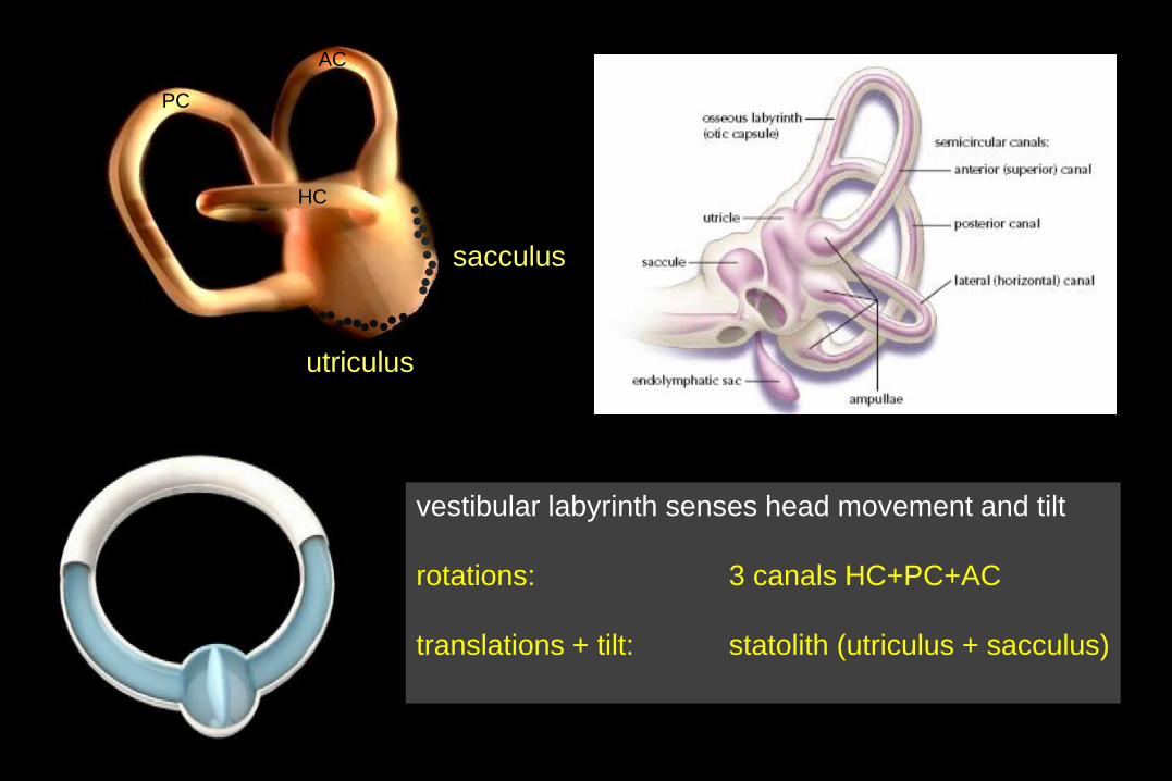
sacculus
utriculus
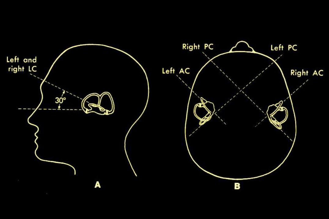
HC
AC
PC
vestibular labyrinth senses head movement and tilt
rotations: 3 canals HC+PC+AC
translations + tilt: statolith (utriculus + sacculus)
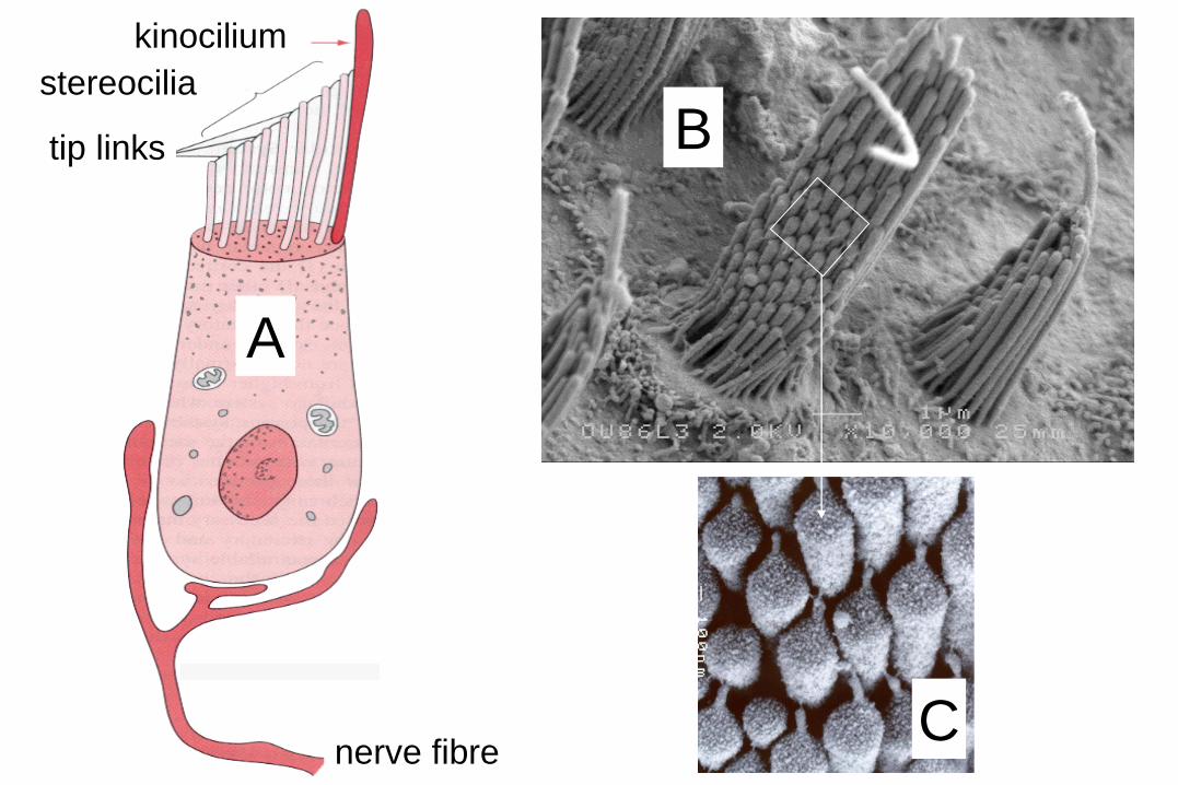
nerve fibre
kinocilium
stereocilia
tip links
A
B
C

80 mV 60 mV 120 mV
ion channels
myosine
filaments
action potentials
sensitive less sensitive
Ewald’s 2nd Law: asymmetry

Ewald’s 2nd Law:
asymmetry
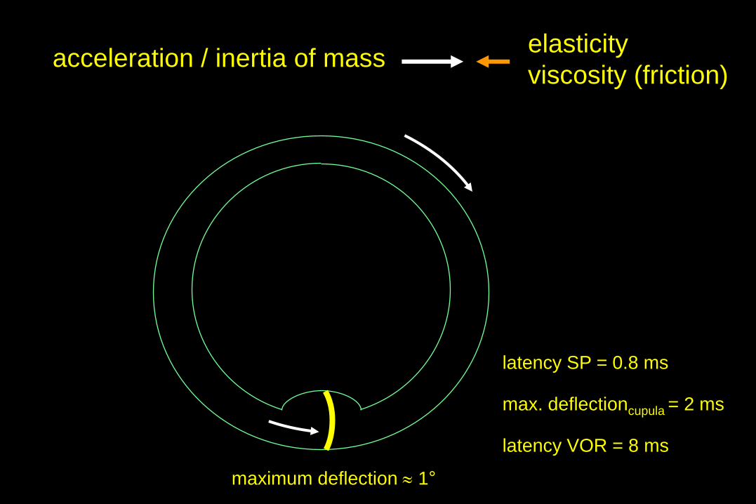
acceleration / inertia of mass elasticity
viscosity (friction)
latency SP = 0.8 ms
max. deflectioncupula = 2 ms
latency VOR = 8 ms
maximum deflection 1°

elasticity viscosity (friction)
mass
backcupula = 20 s
canals are insensitive for constant rotations
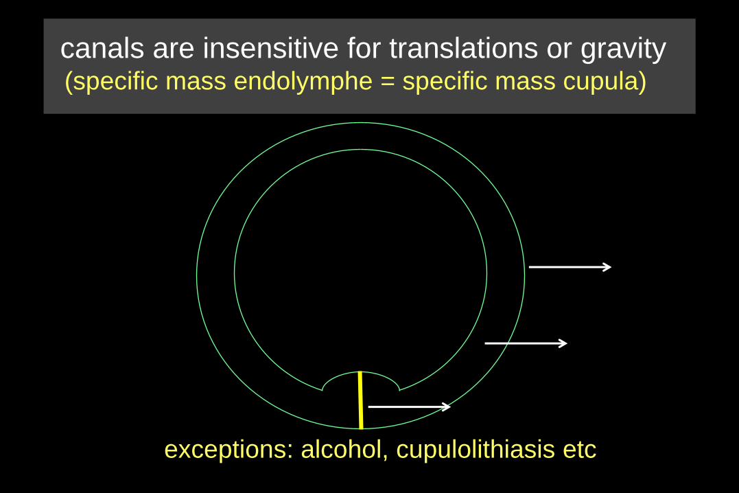
canals are insensitive for translations or gravity (specific mass endolymphe = specific mass cupula)
exceptions: alcohol, cupulolithiasis etc

canals are insensitive for translations or gravity (specific mass endolymphe = specific mass cupula)
exceptions: alcohol, cupulolithiasis etc
Fg

cer
vn
omn
nph
velocity storage mechanism
= integration
- increase of sensitivity
- calculation of velocity
duration 20 s 60 s

labyrinth or retina OKAN nph + nodulus
nystagmus still nystagmus

integration
labyrinth
labyrinth
+ VS
threshold
threshold
position velocity acceleration
differentiation differentiation
integration integration
- canal detects head acceleration
- brain calculates head velocity
- brain matches head and eye velocity = SPV

durationdeflection cupula = 2 ms
durationcupula back = 20 s
durationvelocity storage = 60 s
durationcentral adaptation > 300 s
velocity storage: mainly for horizontal canals
0 100 200 300 sec
25
50
75
100
0

maximal
minimal
Ewald’s 1st Law: optimal sensitivity we need 3 dimensions: 3 canals
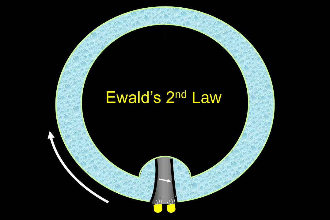
Ewald’s 2nd Law

loss of gaze stabilisation (towards bad-side) especially for fast head movements
The German Experience
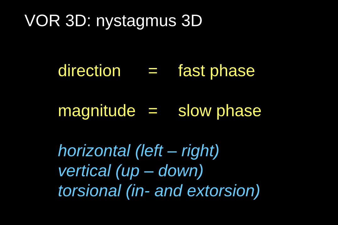
VOR 3D: nystagmus 3D
direction = fast phase
magnitude = slow phase
horizontal (left – right)
vertical (up – down)
torsional (in- and extorsion)
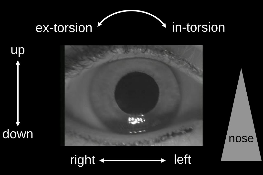
in-torsion ex-torsion
nose
left right
up
down
PC-AD PC-AS
HC-AD HC-AS
AC-AD AC-AS
OD OS
nystagmus direction
nose
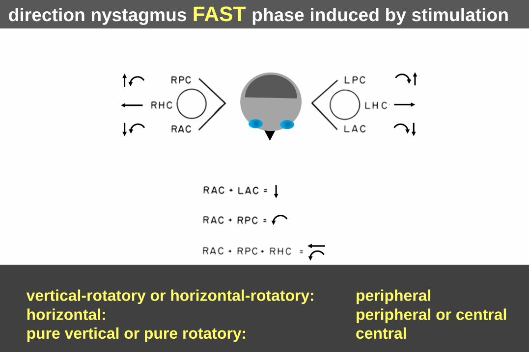
direction nystagmus FAST phase induced by stimulation
vertical-rotatory or horizontal-rotatory: peripheral
horizontal: peripheral or central
pure vertical or pure rotatory: central

frequency dependence
semicircular canals ?
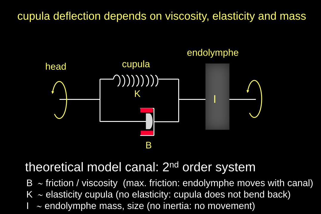
theoretical model canal: 2nd order system
B
head
endolymphe
K
B friction / viscosity (max. friction: endolymphe moves with canal)
K elasticity cupula (no elasticity: cupula does not bend back)
I endolymphe mass, size (no inertia: no movement)
cupula
I
cupula deflection depends on viscosity, elasticity and mass

theoretical model canal: 2nd order system
leads to the following differential equation
q = Θ + Θ + Θ
B K
I I
q angle head rotation
Θ angle cupula deflection
I endolymphe mass, size
B friction (viscosity)
K elasticity cupula frequency frequency
gain phase
cupula deflection depends on viscosity, elasticity and mass
0.1 Hz 10 Hz sensitivity
frequency (Hz)
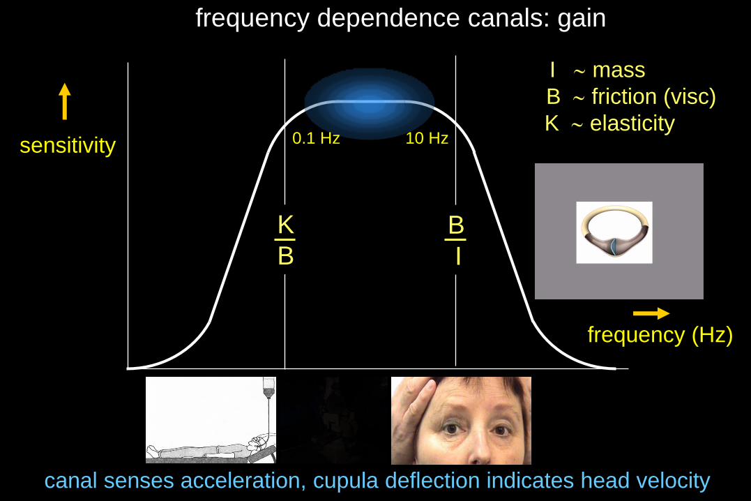
frequency dependence canals: gain
B
I
K
B
I mass
B friction (visc)
K elasticity
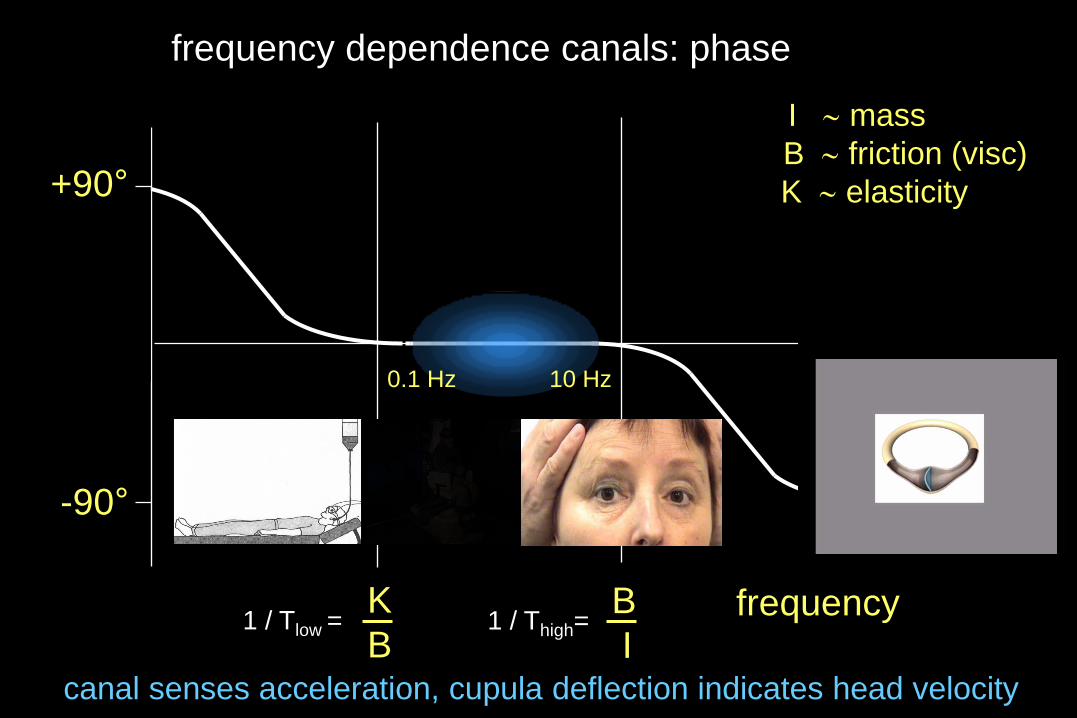
canal senses acceleration, cupula deflection indicates head velocity
0.1 Hz 10 Hz sensitivity
frequency (Hz)
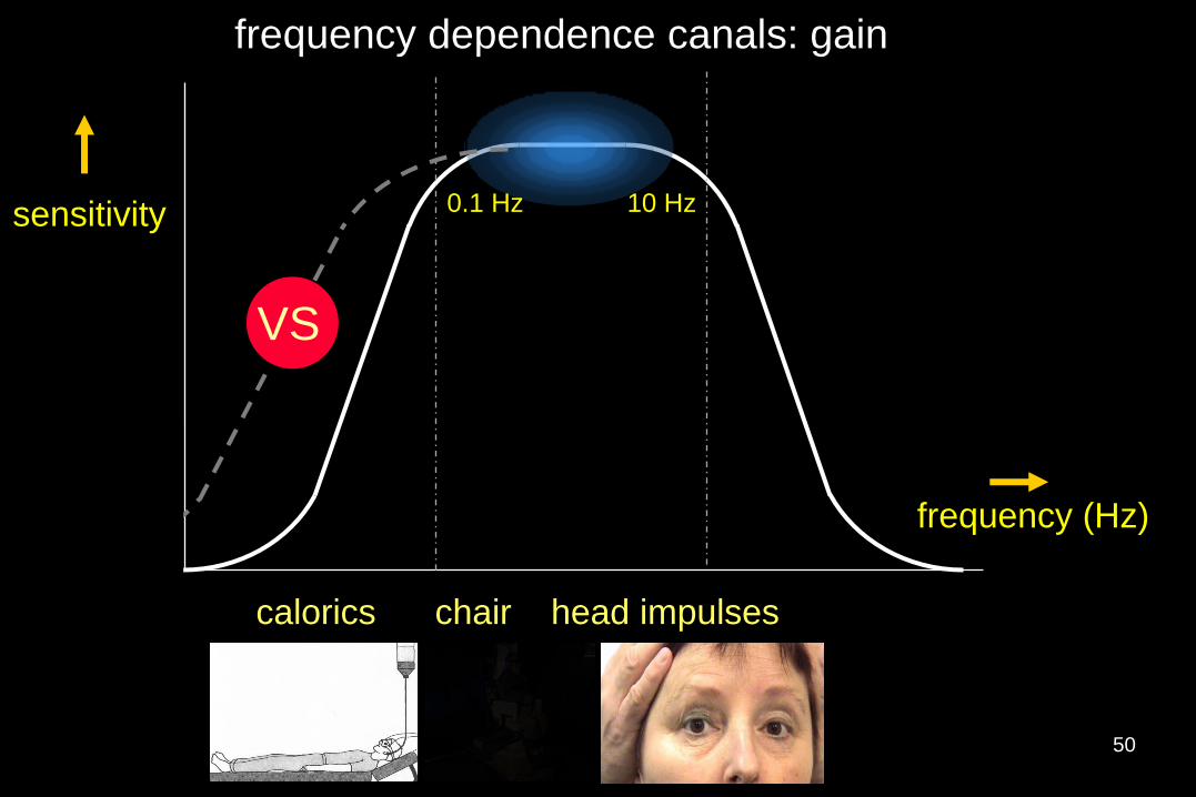
frequency dependence canals: gain
calorics chair head impulses
VS
50
B
I
K
B frequency
-90°
+90°
1 / Tlow = 1 / Thigh=
0.1 Hz 10 Hz
canal senses acceleration, cupula deflection indicates head velocity
frequency dependence canals: phase
I mass
B friction (visc)
K elasticity

-90°
+90°
0.1 Hz 10 Hz
frequency dependence canals: phase (≈ time constant)
calorics chair head impulses
frequency
VS
52

impact viscosity B and elasticity K on canal function
• mechanical changes
viscosity B
elasticity K
specific mass (e.g. alcohol intake, canaloliths)
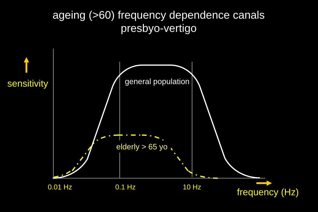
0.01 Hz 0.1 Hz 10 Hz
sensitivity
frequency (Hz)
ageing (>60) frequency dependence canals
presbyo-vertigo
general population
elderly > 65 yo

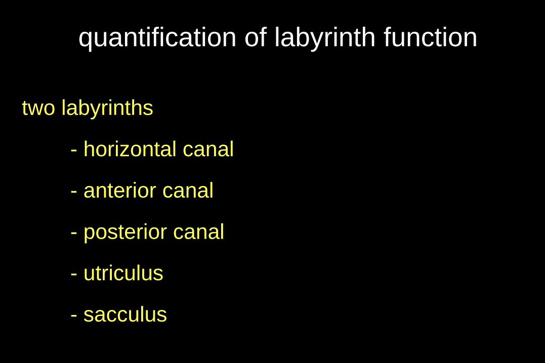
quantification of labyrinth function
two labyrinths - horizontal canal - anterior canal - posterior canal - utriculus - sacculus
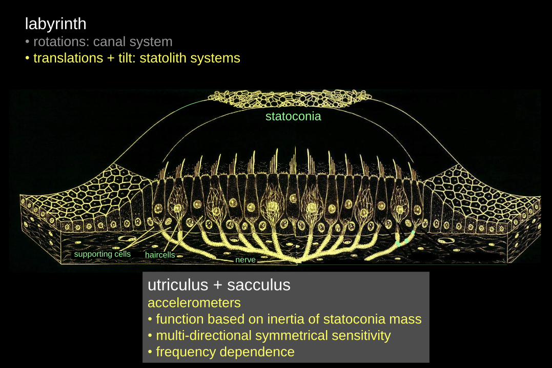
labyrinth • rotations: canal system
• translations + tilt: statolith systems
statoliths statoconia
supporting cells haircells nerve
utriculus + sacculus accelerometers
• function based on inertia of statoconia mass
• multi-directional symmetrical sensitivity
• frequency dependence

Fg
0
velocity
Fg
no discrimination between translation and tilt possible
constant velocity
acceleration deceleration

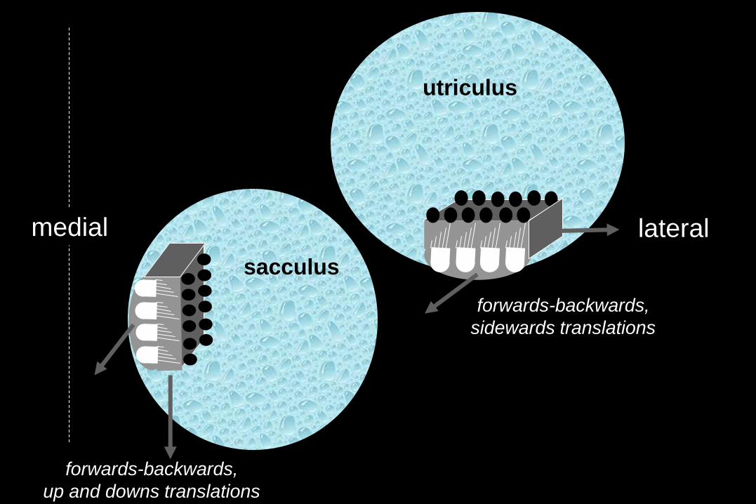
utriculus
sacculus
lateral medial
forwards-backwards,
up and downs translations
forwards-backwards,
sidewards translations

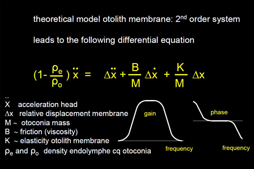
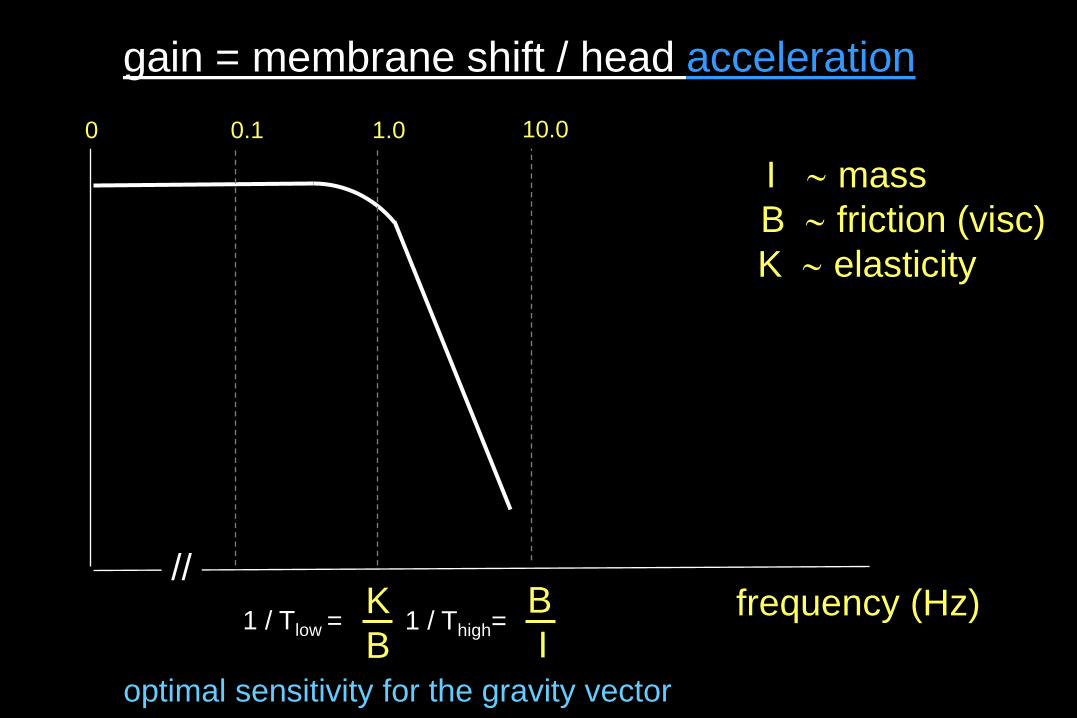
gain = membrane shift / head acceleration
frequency (Hz) 1 / Tlow =
I mass
B friction (visc)
K elasticity
K
B
B
I 1 / Thigh=
1.0 10.0
optimal sensitivity for the gravity vector
0.1
//
0
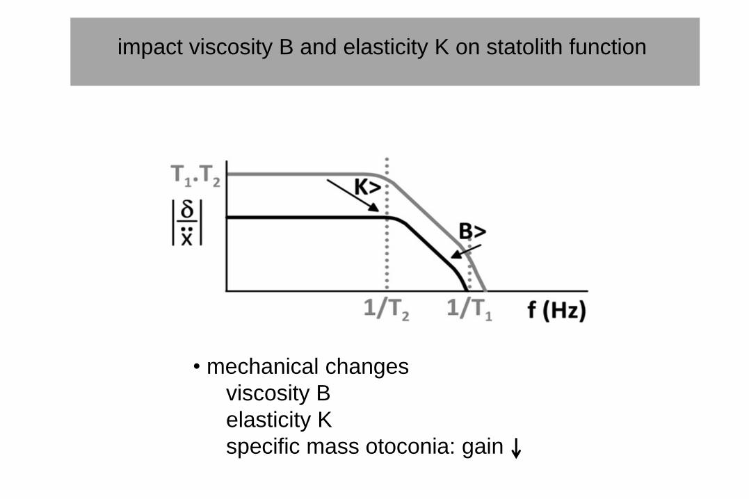
impact viscosity B and elasticity K on statolith function
• mechanical changes
viscosity B
elasticity K
specific mass otoconia: gain

0.2 Hz 2 Hz 20 Hz
sensitivity
frequency (Hz)
correct tilt or translation
statolith
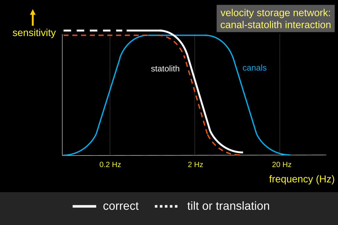
0.2 Hz 2 Hz 20 Hz
sensitivity
frequency (Hz)
canals
correct tilt or translation
statolith
velocity storage network:
canal-statolith interaction

0.2 Hz 2 Hz 20 Hz
sensitivity
frequency (Hz)
canals statolith vision and/or
propriocepsis
correct tilt or translation
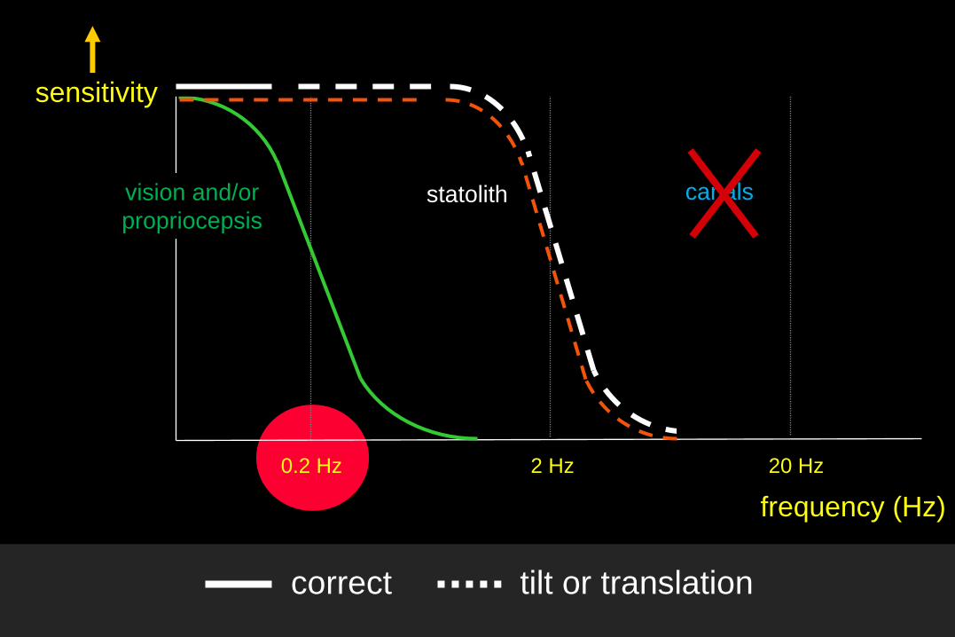
0.2 Hz 2 Hz 20 Hz
sensitivity
frequency (Hz)
canals statolith vision and/or
propriocepsis
correct tilt or translation
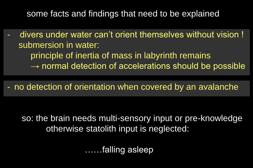
some facts and findings that need to be explained
- divers under water can’t orient themselves without vision !
submersion in water:
principle of inertia of mass in labyrinth remains
→ normal detection of accelerations should be possible
- no detection of orientation when covered by an avalanche
so: the brain needs multi-sensory input or pre-knowledge
otherwise statolith input is neglected:
……falling asleep
which complaints are related to vestibular deficits ?
which complaints are related to natural limitations ?

canals: orientation in space: constant rotation or stand still ?
statoliths: orientation in space: constant translation or stand still ?
orientation relative to gravity: tilt or translation ?
when correct interpretation fails (gravity / selfmotion)
motion sickness
- almost all subjects are susceptible with correct stimulus
unless a low neuro-vegetative sensitivity
training / adaptation helps
- a (partly) working labyrinth is prerequisite for Motion Sickness:
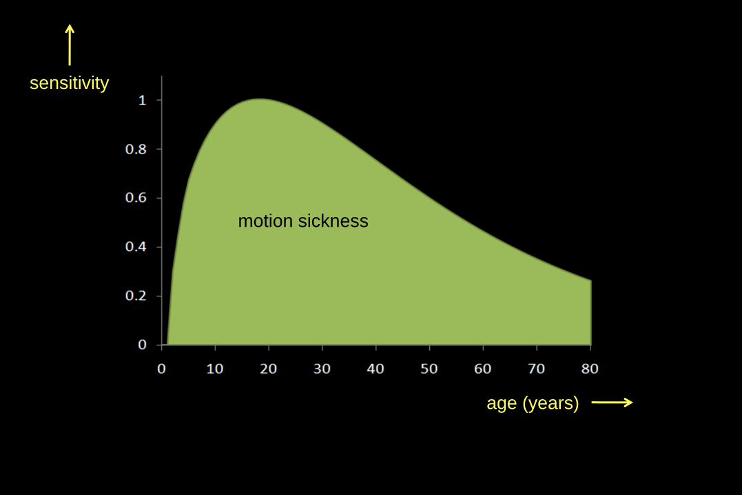
sensitivity
age (years)
motion sickness
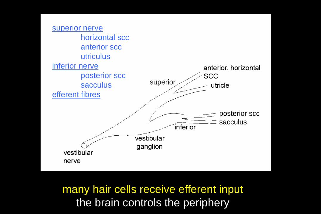
many hair cells receive efferent input
the brain controls the periphery
Effernt fibres
superior nerve
horizontal scc
anterior scc
utriculus
inferior nerve
posterior scc
sacculus
efferent fibres
superior
posterior scc
sacculus
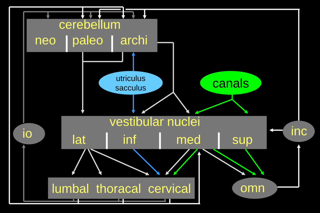
canals utriculus
sacculus
inf lat med sup
vestibular nuclei
omn lumbal thoracal cervical
cerebellum
neo paleo archi
inc io

memories and integration in the brain of
signals from the labyrinth (accelerometer)
aim:
- image stabilisation after head motion
- increase of sensitivity
- calculation of head velocity

cer
vn
omn
nph
velocity storage mechanism - integration
- canal-statolith interaction
- increase of sensitivity
- calculation of velocity
duration 20 s 60 s
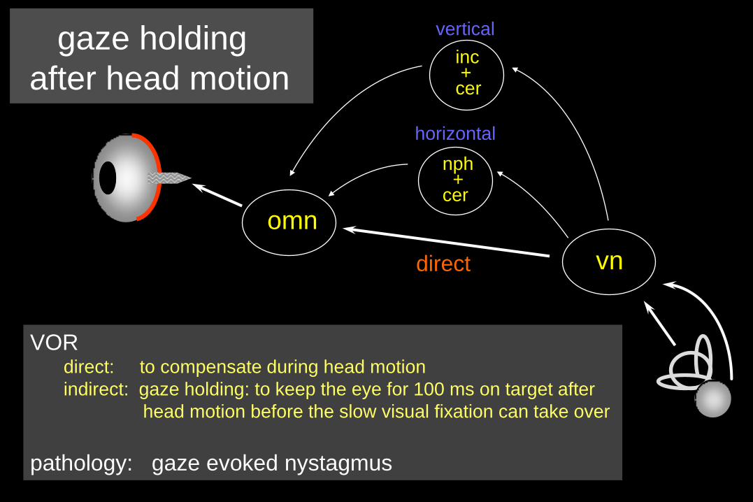
vn
omn
inc + cer
VOR direct: to compensate during head motion
indirect: gaze holding: to keep the eye for 100 ms on target after
head motion before the slow visual fixation can take over
pathology: gaze evoked nystagmus
horizontal
vertical
direct
nph + cer
gaze holding
after head motion
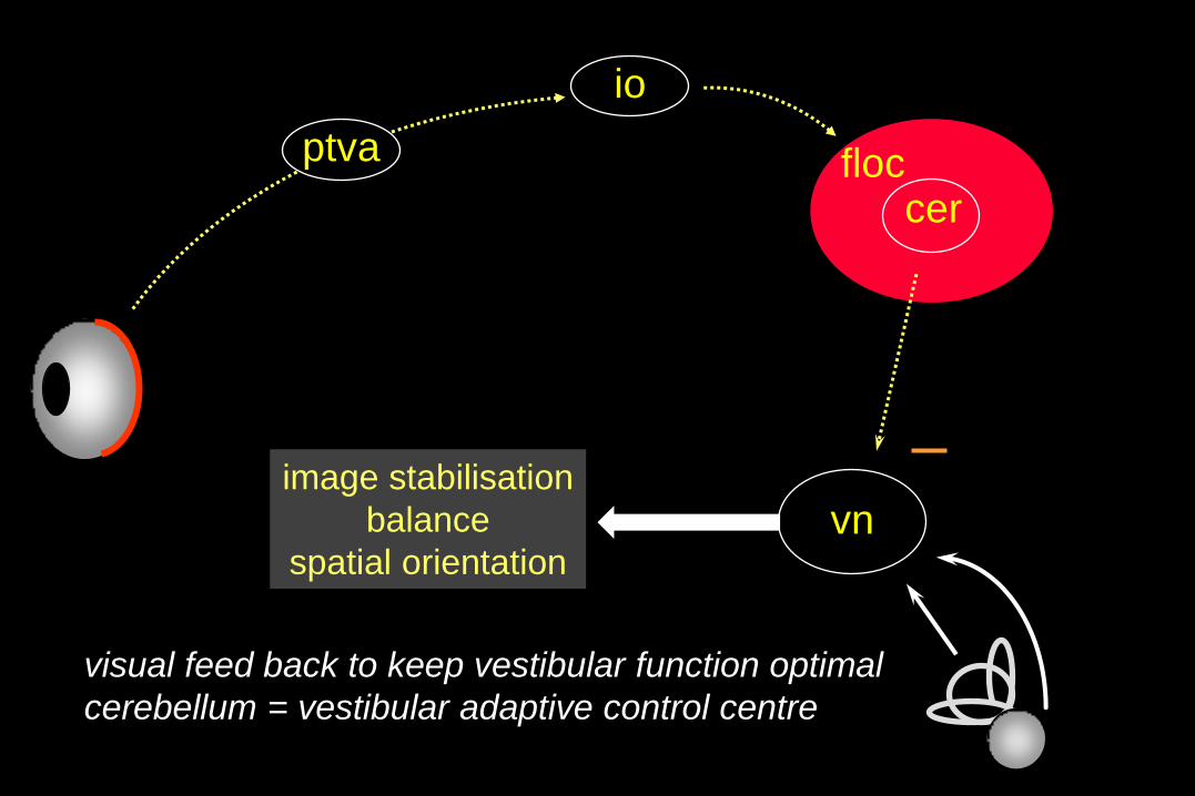
vn
io
cer
visual feed back to keep vestibular function optimal
cerebellum = vestibular adaptive control centre
ptva floc
image stabilisation
balance
spatial orientation

mes
thal
cortex
pons cer
omn
cgl
vn
VOR
Vestibulo-Collic, Cervico-Collic,
Vestibulo-spinal
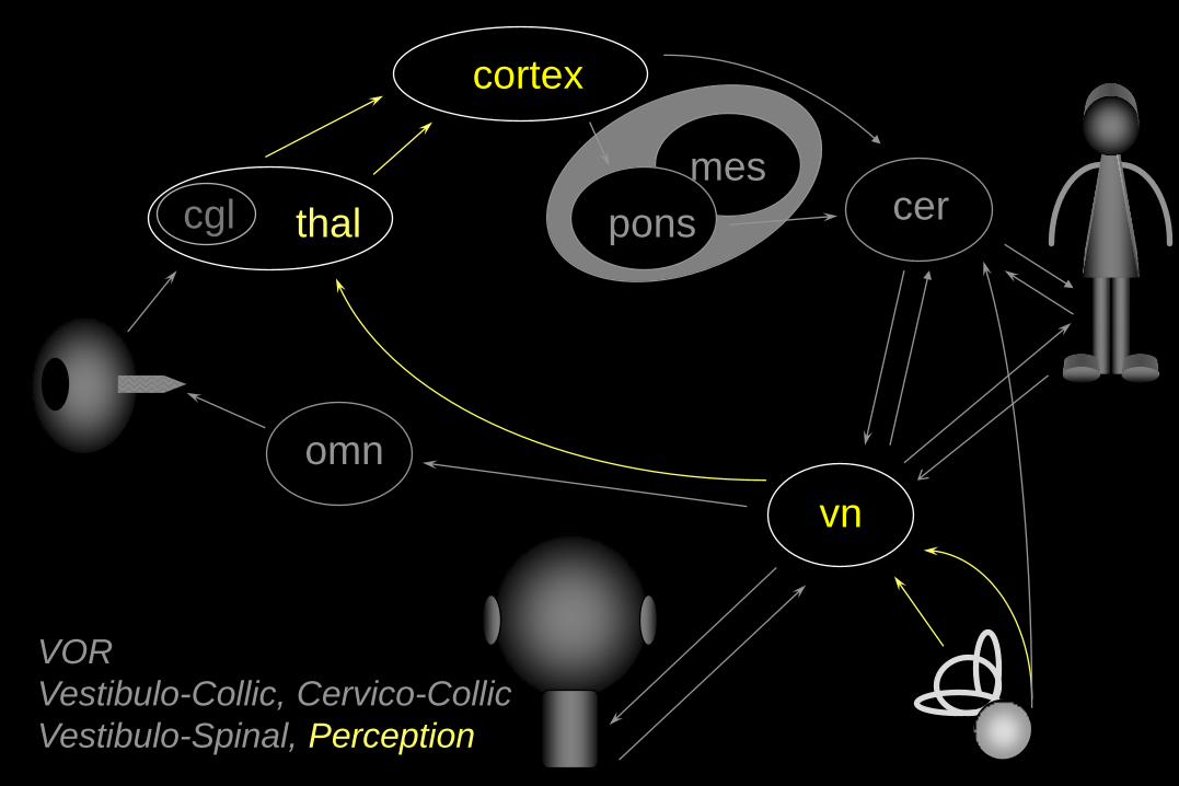
mes
pons
omn
VOR
Vestibulo-Collic, Cervico-Collic
Vestibulo-Spinal, Perception
vn
cer thal cgl
cortex
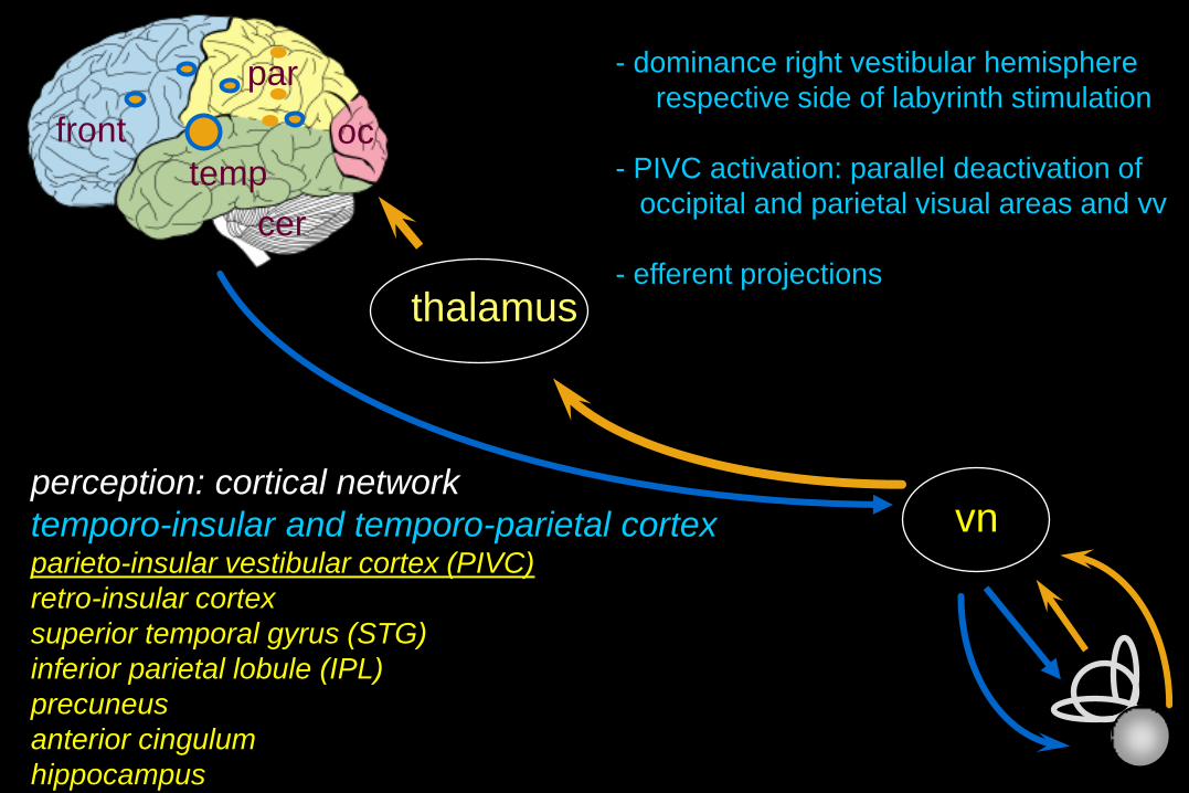
thalamus
vn perception: cortical network
temporo-insular and temporo-parietal cortex parieto-insular vestibular cortex (PIVC)
retro-insular cortex
superior temporal gyrus (STG)
inferior parietal lobule (IPL)
precuneus
anterior cingulum
hippocampus
oc
par
front
temp
cer
- dominance right vestibular hemisphere
respective side of labyrinth stimulation
- PIVC activation: parallel deactivation of
occipital and parietal visual areas and vv
- efferent projections

thank you for your kind attention

EyeSeeCam® (München)
ICS Impulse® (Sydney)

-5
0
5
10
15
20
25
-10 -5 0 5 10 15 20
-25
-20
-15
-10
-5
0
5
-10 -5 0 5 10 15 20
-5
0
5
10
15
20
25
-10 -5 0 5 10 15 20
-25
-20
-15
-10
-5
0
5
-10 -5 0 5 10 15 20
-25
-20
-15
-10
-5
0
5
-10 -5 0 5 10 15 20
small VOR
correction
saccade
head
latency
eye
head
eye
some patients:
covert saccades: sensory substitution
head
eye
normal
VOR
normal dynamic vision
normal head impulse test
poor dynamic vision
abnormal head impulse test
normal dynamic vision
seemingly normal head impulse test

unilateral or bilateral peripheral vestibular loss
head impulse test
- often 1-2 big correction saccades
- some patients compensate with many covert saccades
normal test by observation: does not exclude function loss