Physiological Systems - Waterford Mott...
6
Chapter 44: Osmoregulation and Excretion AP Biology 2013 Physiological Systems Operate in a fluid environment Concentrations of water and solutes must be maintained within fairly narrow limits Freshwater animals have adaptations to reduce water uptake and conserve solutes Desert and marine animals face desiccating environments with the potential to quickly deplete the body water Osmoregulation - regulates solute concentrations and balances the gain and loss of water Excretion - gets rid of metabolic wastes Fig. 44.1 Osmoregulation Based on controlled movement of solutes between internal fluids and external environment Osmosis - cells require a balance between osmotic gain and loss of water; water uptake and loss are balanced by various mechanisms of osmoregulation in different environments Osmoconformers (marine animals) - isoosmotic with surroundings and do not regulate osmolarity Osmoregulators - expend energy to control water uptake and loss Stenohaline - cannot tolerate substantial changes in external osmolarity Euryhaline - can survive large fluctuations in external osmolarity 1 2 3
Transcript of Physiological Systems - Waterford Mott...
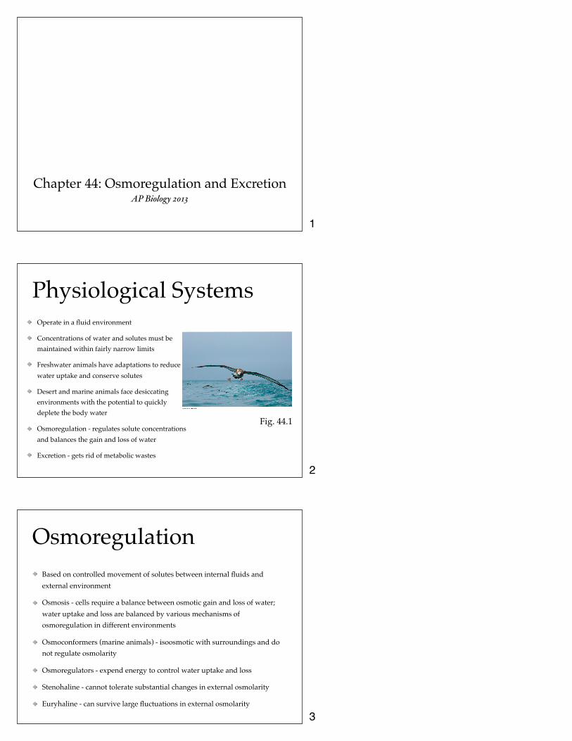
Chapter 44: Osmoregulation and Excretion AP Biology 2013
Physiological SystemsOperate in a fluid environment
Concentrations of water and solutes must be maintained within fairly narrow limits
Freshwater animals have adaptations to reduce water uptake and conserve solutes
Desert and marine animals face desiccating environments with the potential to quickly deplete the body water
Osmoregulation - regulates solute concentrations and balances the gain and loss of water
Excretion - gets rid of metabolic wastes
Fig. 44.1
OsmoregulationBased on controlled movement of solutes between internal fluids and external environment
Osmosis - cells require a balance between osmotic gain and loss of water; water uptake and loss are balanced by various mechanisms of osmoregulation in different environments
Osmoconformers (marine animals) - isoosmotic with surroundings and do not regulate osmolarity
Osmoregulators - expend energy to control water uptake and loss
Stenohaline - cannot tolerate substantial changes in external osmolarity
Euryhaline - can survive large fluctuations in external osmolarity
1
2
3

Marine AnimalsMost invertebrates are osmoconformers; most vertebrates are osmoregulators
Marine bony fish are hyperosmotic to sea water and lose water by osmosis and gain salt by both diffusion and food
Balance water loss by drinking seawater and excreting salts Fig. 44.3
(a) Osmoregulation in a marine fish
Gain of water and salt ions from food
Excretion of salt ions from gills
Osmotic water loss through gills and other parts of body surface
Gain of water and salt ions from drinking seawater
Excretion of salt ions and small amounts of water in scanty urine from kidneys
Key
Water Salt
Freshwater AnimalsConstantly take in water from their hypoosmotic environment
Lose salts by diffusion
Maintain water balance by excreting large amounts of dilute urine
Salts lost by diffusion are replaced by foods and uptake across gills Fig. 44.3
(b) Osmoregulation in a freshwater fish
Gain of water and some ions in food
Uptake of salt ions by gills
Osmotic water gain through gills and other parts of body surface
Excretion of salt ions and large amounts of water in dilute urine from kidneys
Key
Water Salt
Other Animals
Some aquatic invertebrates living in temporary ponds can lose almost all their body water and survive in a dormant state (anhydrobiosis)
Land animals - manage water budgets by drinking and eating moist foods and using metabolic water
Desert animals get water savings from simple anatomical features Fig. 44.6
(a) Hydrated tardigrade (b) Dehydrated tardigrade
50 µm
50 µm
Fig. 44.5
Water balance in a kangaroo rat (2 mL/day)
Water balance in a human (2,500 mL/day)
Ingested in food (0.2)
Ingested in food (750)
Ingested in liquid (1,500) Water
gain (mL)
Water loss (mL)
Derived from metabolism (1.8)
Derived from metabolism (250)
Feces (0.09)
Urine (0.45)
Feces (100)
Urine (1,500)
Evaporation (1.46) Evaporation (900)
4
5
6

Transport Epithelia
Specialized cells that regulate solute movement
Essential components of osmotic regulation and metabolic waste disposal
Arranged in complex tubular networks
Ex. salt glands of marine birds (remove excess NaCl from blood)
Fig. 44.7
Nasal salt gland
Ducts
Nostril with salt secretions
(a) Location of nasal glands in a marine bird
Nasal gland
(b) Secretory tubules
Capillary Secretory tubule Transport epithelium
Vein Artery
Central duct
Nasal gland
Key
Salt movement Blood flow
(c) Countercurrent exchange
Secretory cell of transport epithelium
Lumen of secretory tubule
Salt ions
Blood flow Salt secretion
Nitrogenous Wastes
Different animals excrete nitrogenous waste in different forms
Ammonia - need access to lots of water; release across body surface or through gills
Urea (mammals and adult amphibians) - liver converts ammonia to less toxic urea which is carried to the kidneys, concentrated and excreted with minimal water loss
Uric acid (insects, land snails, reptiles and birds) - uric acid is largely insoluble in water and can be secreted as a paste with little water loss
Fig. 44.8
Proteins Nucleic acids
Amino acids
Nitrogenous bases
—NH2 Amino groups
Most aquatic animals, including most bony fishes
Mammals, most amphibians, sharks,
some bony fishes
Many reptiles (including birds),
insects, land snails
Ammonia Urea Uric acid
Excretory SystemsRegulate solute movement between internal fluids and external environment
Most produce urine by refining a filtrate derived from body fluids
Key functions: filtration, reabsorption, secretion, and excretion Fig. 44.10
Capillary Filtration
Excretory tubule
Reabsorption
Secretion
Excretion
Filtrate U
rine
2
1
3
4
7
8
9

Types of Excretory Systems
Built around a complex network of tubes
Protonephridium - network of dead-end tubules lacking internal openings
Tubules branch throughout the body and smallest are capped by a cellular unit called a flame bulb that excrete a dilute fluid
Metanephridia - each earthworm segment has a pair of open-ended metanephridia that consist of tubules that collect coelomic fluid and produce dilute urine
Figs. 44.11 $ 44.12
Tubules of protonephridia
Tubule
Flame bulb
Nucleus of cap cell
Cilia
Interstitial fluid flow
Opening in body wall
Tubule cell
Components of a metanephridium:
Collecting tubule
Internal opening
Bladder
External opening
Coelom Capillary network
Types of Excretory Systems
Malpighian Tubules - insects and terrestrial arthropods
Remove nitrogenous wastes from hemolymph
Produce dry waste matter (adaptation to terrestrial life)
Kidneys - excretory organs of vertebrates
Fig. 44.13
Digestive tract
Midgut (stomach)
Malpighian tubules
Rectum Intestine Hindgut
Salt, water, and nitrogenous
wastes
Feces and urine
Malpighian tubule
To anus
Rectum Reabsorption
HEMOLYMPH
KidneysPrincipal site of water balance and salt regulation in mammals
Each kidney is supplied with blood by a renal artery and drained by a renal vein
Urine exists the kidney through a duct called the ureter
Both ureters drain into a common urinary bladder
Kidney has two regions: outer renal cortex and inner renal medulla
The nephron consists of a single long tubule and a ball of capillaries called the glomerulus
10
11
12

Kidney FiltrationOccurs as blood pressure forces fluid from the blood in the glomerulus into the lumen of the Bowman’s capsule
Filtration of small molecules is nonselective and the filtrate mixture in the Bowman’s capsule mirrors the concentrations of solutes in the blood plasma
From the Bowman’s capsule the filtrate passes through three regions of the nephron: proximal tubule, loop of Henle, and the distal tube
Fluid from several nephrons flows into a collecting duct
Nephron Organization Afferent arteriole from renal artery Glomerulus
Bowman’s capsule
Proximal tubule
Peritubular capillaries
Distal tubule
Efferent arteriole from glomerulus
Collecting duct
Branch of renal vein
Vasa recta
Descending limb
Ascending limb
Loop of
Henle
Fig. 44.14
Nephron Blood VesselsEach nephron is supplied with blood by an afferent arteriole (branch of the renal artery that subdivides into capillaries)
Capillaries converge as they leave the glomerulus (forming an efferent arteriole)
Vessels subdivide again forming peritubular capillaries which surround the proximal and distal tubules
Filtrate becomes urine as it flows through the mammalian nephron and collecting duct
Fig. 44.14
Proximal tubule Distal tubule
Filtrate CORTEX
Loop of Henle
OUTER MEDULLA
INNER MEDULLA
Key Active transport Passive transport
Collecting duct
Nutrients NaCl
NH3
HCO3- H2O K+
H+
NaCl H2O
HCO3-
K+ H+
H2O NaCl
NaCl
NaCl H2O
Urea
Secretion and Reabsorption Secretion and reabsorption in the proximal tubule alters the volume and composition of filtrate
Reabsorption of water continues as filtrate moves into the descending limb of the loop of Henle
As filtrate travels through the ascending limb of the loop of Henle salt diffuses out of the tubule into the interstitial fluid
Distal tubule plays a key role in regulating K+ and NaCl concentration of body fluids
Collecting duct carries the filtrate through the medulla to the renal pelvis and reabsorbs NaCl
13
14
15
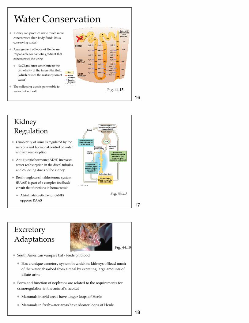
Water ConservationKidney can produce urine much more concentrated than body fluids (thus conserving water)
Arrangement of loops of Henle are responsible for osmotic gradient that concentrates the urine
NaCl and urea contribute to the osmolarity of the interstitial fluid (which causes the reabsorption of water)
The collecting duct is permeable to water but not salt Fig. 44.15
Osmolarity of interstitial
fluid (mOsm/L)
Key
Active transport Passive transport
INNER MEDULLA
OUTER MEDULLA
CORTEX NaCl H2O
H2O
H2O
H2O
H2O
H2O
H2O
NaCl
NaCl
NaCl
NaCl
NaCl
NaCl
NaCl
NaCl
H2O
H2O
H2O
H2O
H2O
H2O
H2O
Urea
Urea
Urea
1,200
1,200 1,200
900
600
400
300 300
400
600
100
100
200
400
700 900
600
400
300
300 300
Kidney Regulation
Osmolarity of urine is regulated by the nervous and hormonal control of water and salt reabsorption
Antidiuretic hormone (ADH) increases water reabsorption in the distal tubules and collecting ducts of the kidney
Renin-angiotensin-aldosterone system (RAAS) is part of a complex feedback circuit that functions in homeostasis
Atrial natriuretic factor (ANF) opposes RAAS
Thirst Hypothalamus
ADH
Pituitary gland
Osmoreceptors in hypothalamus trigger
release of ADH.
STIMULUS: Increase in blood
osmolarity (for instance, after
sweating profusely)
Homeostasis: Blood osmolarity
(300 mOsm/L)
Drinking reduces blood osmolarity
to set point.
H2O reab- sorption helps prevent further
osmolarity increase.
Increased permeability
Distal tubule
Collecting duct
Fig. 44.20
Excretory Adaptations
South American vampire bat - feeds on blood
Has a unique excretory system in which its kidneys offload much of the water absorbed from a meal by excreting large amounts of dilute urine
Form and function of nephrons are related to the requirements for osmoregulation in the animal’s habitat
Mammals in arid areas have longer loops of Henle
Mammals in freshwater areas have shorter loops of Henle
Fig. 44.18
16
17
18