PHYSIOLOGICAL AND PHYLOGENETIC STUDIES OF THE …
81
Abstract PHYSIOLOGICAL AND PHYLOGENETIC STUDIES OF THE BIOGEOGRAPHY OF ALKALIPHILIC HETEROTROPHIC BACTERIA FROM SERPENTINIZING HABITATS Alyssa N. Kloysuntia August 24, 2014 Director: Dr. Jeffrey McKinnon Department: Biology Serpentinization occurs when ultramafic rocks containing the mineral olivine react with water to produce highly reducing conditions, which are commonly coincident with high concentrations of hydrogen. Hydrogen provides an energy source for microbial metabolism and in combination with mineral catalysts can lead to the abiogenic synthesis of small organic compounds, such as methane. Serpentinite habitats contain abundant potential electron donors, but microorganisms presumably have limited accessibility to electron acceptors. A consequence of serpentinization is the production of highly alkaline (pH 10-12), highly reducing fluids. The stress exerted on microorganisms in such ecosystems can make fundamental cellular processes such as maintaining a proton motive force or stabilizing RNA a difficult task and limit microbial diversity in these environments. It is reputed that the microbial communities in these extreme habitats are able to adjust to the ultrabasic conditions through biochemical and metabolic adaptations. However, it remains to be discovered how microorganisms have physiologically adapted to the serpentinite environment.
Transcript of PHYSIOLOGICAL AND PHYLOGENETIC STUDIES OF THE …

Abstract
PHYSIOLOGICAL AND PHYLOGENETIC STUDIES OF THE BIOGEOGRAPHY OF ALKALIPHILIC HETEROTROPHIC BACTERIA FROM SERPENTINIZING
HABITATS
Alyssa N. Kloysuntia
August 24, 2014
Director: Dr. Jeffrey McKinnon
Department: Biology
Serpentinization occurs when ultramafic rocks containing the mineral olivine
react with water to produce highly reducing conditions, which are commonly coincident
with high concentrations of hydrogen. Hydrogen provides an energy source for microbial
metabolism and in combination with mineral catalysts can lead to the abiogenic synthesis
of small organic compounds, such as methane. Serpentinite habitats contain abundant
potential electron donors, but microorganisms presumably have limited accessibility to
electron acceptors. A consequence of serpentinization is the production of highly
alkaline (pH 10-12), highly reducing fluids. The stress exerted on microorganisms in such
ecosystems can make fundamental cellular processes such as maintaining a proton motive
force or stabilizing RNA a difficult task and limit microbial diversity in these
environments. It is reputed that the microbial communities in these extreme habitats are
able to adjust to the ultrabasic conditions through biochemical and metabolic adaptations.
However, it remains to be discovered how microorganisms have physiologically adapted
to the serpentinite environment.
Taxonomic characterization of novel isolates from different serpentinite habitats
allows us to examine microbial biodiversity and assess whether certain microorganisms
are found only at a particular site or whether their occurrence is widespread and
correlated with specific environmental conditions. Comparisons of microbial diversity
of the numerous serpentine sites our laboratory group is working on in the United States,
Canada, and Italy to previously published studies provides insight into the biogeography
of alkaliphiles in serpentinizing habitats at a global scale. Detailed physiological studies
focused upon a high-resolution environmental data set from the Coast Range Ophiolite
Microbial Observatory (CROMO) site in California have been used to relate
physiological data from a subset of new isolates to environmental characteristics of the
environments from which they were sampled.
Alkaliphilic microorganisms were isolated and grown aerobically from samples
collected from three different serpentinite habits located in California, Italy, and Canada
between 2009 and 2013. Total genomic DNA from the isolates was used to sequence
their 16S rRNA gene to establish the taxa present in each serpentinite habitat as well as in
“background” soils and water. Culture-dependent analyses on a subset of isolates
investigated the physiology of the isolated alkaliphilic heterotrophs. This research
contributes to our understanding of microbial life in ultrabasic habitats associated with
serpentinization, explores the biodiversity of the subsurface microorganisms, and
characterizes their physiological adaptations.


PHYSIOLOGICAL AND PHYLOGENETIC STUDIES OF THE BIOGEOGRAPHY
OF ALKALIPHILIC HETEROTROPHIC BACTERIA
FROM SERPENTINIZING HABITATS
A Thesis Presented to
The Faculty of the Department of Biology
East Carolina University
In Partial Fulfillment
of the Requirements for the Degree
Masters of Science Molecular Biology and Biotechnology
By Alyssa N. Kloysuntia
August 24, 2014

@ 2014
Alyssa N. Kloysuntia
All Rights Reserved

PHYSIOLOGICAL AND PHYLOGENETIC STUDIES OF THE BIOGEOGRAPHY OF ALKALIPHILIC HETEROTROPHIC BACTERIA
FROM SERPENTINIZING HABITATS
By
Alyssa N. Kloysuntia
APPROVED BY:
DIRECTOR OF THESIS:___________________________________________________ Matthew Schrenk, Ph.D.
COMMITTEE MEMBER:__________________________________________________
Carol Goodwillie, Ph.D.
COMMITTEE MEMBER:__________________________________________________ Jason Gee, Ph.D.
COMMITTEE MEMBER:__________________________________________________
Tom Rickenbach, Ph.D. CHAIR OF THE DEPARTMENT OF BIOLOGY:_______________________________
Jeffrey McKinnon, Ph.D.
DEAN OF THE GRADUATE SCHOOL:______________________________________ Paul J. Gemperline, Ph.D.

ACKNOWLEDGEMENTS
Foremost, I would like to thank Dr. Mathew O. Schrenk for giving me the chance
to work and conduct research in a geomicrobiology lab. Also, I would like to thank Dr.
Schrenk for being a great mentor who provided constant advice and guidance given to
help me accomplish this program. I would like to thank Dr. Gee, Dr. Goodwillie, and Dr.
Rickenbach for their willingness to give vital input and support to my project.
I would also like to recognize my fellow lab mates Dr. Melitza Crespo-Medina,
Crystal George, and Katrina Twing for helping provide samples for this project, giving
valuable advice, and for the contributions made to this lab. I would also like to extend
my gratitude to Dr. Igor Tiago, who gave fundamental assistance on the physiological
work done for this project. I would like to thank Denise Mayer for conducting the
sequencing work for this project. I would also like to recognize Dr. William Brazelton,
who was able to contribute data on Hydrogenophaga and for also providing valuable
guidance on this project. Lastly, I would like to thank my friends and family for their
encouragement during this program. I would especially like to thank my Papa, who was
my main motivator and constant supporter.
Funding for this this project was provided to Dr. Schrenk from the Division of
Research and Graduate Studies at East Carolina University, from the NASA Astrobiology
Institute through the Carnegie Institution for Science, Deep Carbon Observatory, and the
Alfred P. Sloan Foundation.
TABLE OF CONTENTS
Title Page ..................................................................................................................................... IV
Copyright ....................................................................................................................................... V
Signature Page ............................................................................................................................. VI
Acknowledgements ................................................................................................................... VII
Chapter 1: Review of Literature ................................................................................................. 1
Serpentinization .......................................................................................................................... 3
Microbial Biogeography and Serpentinizing Environments ...................................................... 6
Microbial Adaptations to High pH ........................................................................................... 10
Chapter 2: Research Objectives ................................................................................................ 14
Document Alkaliphilic Heterotrophs in Serpentinite Samples ................................................. 14
Investigate Patterns in Microbial Distribution .......................................................................... 14
Understanding Relationships Between Microbial Physiology and the Environment ............... 15
Chapter 3: Central Hypotheses ................................................................................................. 16
Chapter 4: Methods .................................................................................................................... 18
Isolation of Novel Microorganisms .......................................................................................... 18
Determining Colony Forming Units in CROMO Wells ........................................................... 20
Extraction of Total Genomic DNA ........................................................................................... 21
PCR Based Assays to Amplify 16S rRNA genes ..................................................................... 22
Construction of Phylogenetic Trees .......................................................................................... 23
Determining Optimum Temperature and pH ............................................................................ 23

Oxidase Test .............................................................................................................................. 24
Catalase Test ............................................................................................................................. 25
Chapter 5: Microbial Biodiversity and Biogeography of Serpentinizing Habitats .............. 26
Total Microbial Biodiversity ..................................................................................................... 26
Microbial Biogeography ........................................................................................................... 28
Phylogenetic Relatedness of Isolates ........................................................................................ 30
Serpentine Soils vs. Fluids ........................................................................................................ 31
Divergence from Known Alkaliphiles ...................................................................................... 32
Chapter 6: Experimental Physiology vs. The Environment ................................................... 45
Coast Range Ophiolite Microbial Observatory, California ...................................................... 45
Experimental Optimum pH and Temperature of Isolates ......................................................... 47
Oxidase Test Results ................................................................................................................. 51
Catalase Test Results ................................................................................................................ 52
Chapter 7: Discussion ................................................................................................................. 54
References .................................................................................................................................... 61
Appendix A: Taxonomic & Physiological Data of Isolates ..................................................... 64

LIST OF TABLES
1. Geochemical Characteristics of Serpentinite Sites ............................................................. 4
2. Bacterial 16S rRNA Primers ............................................................................................. 22
3. Number of Isolates at Serpentinite Sites ........................................................................... 27
4. Comparison of Isolates at the Genus Level ...................................................................... 29
5. Environmental Data from CROMO Wells ....................................................................... 45
6. Microbial Abundance from CROMO Wells ..................................................................... 46
7. Microbial Diversity from CROMO Wells ........................................................................ 47
8. Example of Determining Experimental Optimum pH and Temperature .......................... 48
LIST OF FIGURES
1. Map of CROMO Core Shed Well (CSW) Site ................................................................. 18
2. Microbial Enrichment Culture .......................................................................................... 20
3. CROMO Pre-Existing Well Site ....................................................................................... 21
4. Sector Plates ...................................................................................................................... 24
5. Catalase Test ..................................................................................................................... 25
6. Serpentinite Microbial Diversity ....................................................................................... 28
7. Phylogenetic Tree of Isolates ............................................................................................ 33
8. Actinobacteria Phylogenetic Tree ..................................................................................... 34
9. Dietzia Phylogenetic Tree ................................................................................................. 35
10. Microcella Phylogenetic Tree ........................................................................................... 36
11. Bacillus Phylogenetic Tree ............................................................................................... 39
12. Hydrogenophaga Phylogenetic Tree ................................................................................ 42
13. Hydrogenophaga Heat Map .............................................................................................. 44
14. Environmental pH vs. Experimental pH ........................................................................... 49
15. Environmental Temperature vs. Experimental Temperature ............................................ 50
16. Oxidase Test Graph ........................................................................................................... 52
17. Catalase Test Graph .......................................................................................................... 53

CHAPTER 1: Review of Literature
Microorganisms are the most diverse living entities on Earth, and have adapted to a vast
array of environments. Due to their flexibility to acclimate to varying surroundings and their
extensive history microbial populations are often phenotypically and genotypically distinct from
one another, even over very small scales. There are such a broad spectrum of microbial species
and conditions that favor their growth that major portions of microbial biodiversity have not been
thoroughly characterized. Despite these challenges, it is important to find ways to describe
microbial communities in nature in order to understand their biogeography and relationships to
their environment.
Since the early days of Microbiology, many studies have focused on microbial diversity
at the surface of our planet, influenced by higher taxonomic groups, by photosynthetically
derived organic carbon, the atmosphere, and the water cycle. Until relatively recently little
investigation has gone into characterizing the subsurface microbiome, in particular in rocks
originating from the deep subsurface. As a result, microbial activities and distributions in the
subsurface are not altogether understood. However, with recent technological advances in gene
sequencing techniques, it has been more accessible to characterize microorganisms that have yet
to be cultured due to the extreme environments they live in. Typically “normal” environments
that are moderate in terms of pH, nutrient availability, salinity, etc., support relatively high
microbial abundances and community diversity. In contrast, an extreme environment may limit
the diversity of microorganisms present, although it does not necessarily suggest a low
abundance of microorganisms if they are sufficiently adapted to meet the environmental
challenges. An example of an extreme environment would be one undergoing active
2
serpentinization, often characterized by its high pH’s and low oxidation-reduction potential (Eh).
Serpentinite sites can exert specific evolutionary selective pressures on the microorganisms
present, and are often coincident with low cell abundances and microbial diversity. Due to their
ubiquity and antiquity, and the low diversity present in the serpentinite sites, they are an ideal
location to investigate microbial evolution and physiological adaptations.
What environmental factors are the driving forces for determining the microbial diversity
of a particular environment (Fierer et al., 2006)? A famous statement by the Dutch
Microbiologist Lourens Baas Becking states “everything is everywhere, but the environment
selects”, if there are no dispersal barriers and the habitats are physically and chemically similar;
we should see the same groups of microorganisms at all sites (Wit et al., 2006). Recent work by
Reno, et al. on the biogeography of the archaeon Sulfolobus islandicus pangenome suggests that
there are some contradictions to Bass Becking’s broad hypothesis (Reno et al., 2009). The
authors examined the biogeographical distribution of multiple genes in several genomes of
closely related S. islandicus strains from varying geographical locations. It was found that in S.
islandicus the core genome between strains were highly similar, while the variable genome
shows changes between lineages that were indicative of strains evolving independently from one
another based on their geographical distribution (Reno et al., 2009). However, there remains
little evidence of whether microorganisms are site-specific and the role of physiological traits in
defining distinct species (Rutger et al., 2006). The research presented in this dissertation focuses
on the microbial biogeography of highly alkaline habitats associated with serpentinization as
another type of model system to study microbial diversity and biogeography.

3
Serpentinization
Serpentinization starts with ultramafic rocks, which are a major part of the Earth’s
lithosphere and upper mantle and are enormous reservoirs for carbon (Brazelton et al., 2012).
Serpentinization occurs when ultramafic rock is exposed to water during tectonic uplift or from
fluid flow of underground, producing different serpentinites (Brazelton et al., 2012). Exposure to
water results in the hydration and oxidation of the minerals comprising the ultramafic rock
(Sleep et al., 2004). Exposure frequently occurs in tectonically active areas near mountain belts
or slow spreading mid-ocean ridges. As plate tectonics progresses, partially serpentinized
oceanic rocks are accreted onto the continental margins to form structures known as ophiolites.
Ultramafic rocks continue to form and are exposed by tectonic activities today on modern-day
Earth (Sleep et al., 2004). The oxidation of minerals in ultramafic rocks results in the formation
of molecular hydrogen and methane, and leads to high alkalinity (Muntener et al., 2010). Mineral
composition, pressure, and temperature of the environment results in some variability between
serpentinite sites, however the products of serpentinization are consistent between sites (Schulte
et al., 2006). Serpentinization at low temperatures often results in a high pH environment,
typically over 10 (Schrenk et al., 2013). This alkaline environment is a result of released Ca2+
and OH- into the fluid systems of actively serpentinizing habitats can result in the precipitation of
calcium carbonate upon reaction with atmospheric CO2 (Tiago et al., 2004). Minerals, such as
olivine at these serpentinite sites are able to buffer metamorphic fluids, which result in highly
reducing conditions that produce an abundance of molecular hydrogen gas (Sleep et al., 2004).
Additionally, magnetite and iron-nickel composites can catalyze the reaction to enhance the
amount of hydrogen, hydrocarbons, and methane produced (Muntener et al., 2010). The
4
abiogenic hydrogen and organic compounds may be a critical energy source for microorganisms
in such settings (Schrenk et al., 2013).
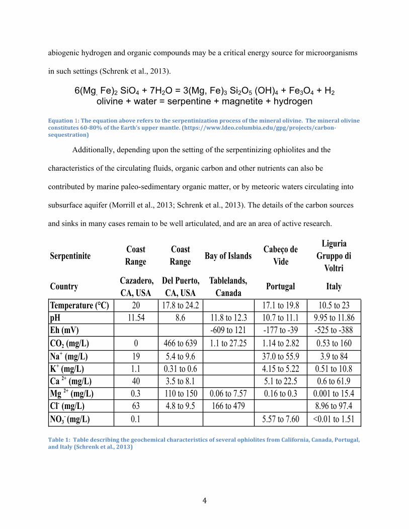
6(Mg, Fe)2 SiO4 + 7H2O = 3(Mg, Fe)3 Si2O5 (OH)4 + Fe3O4 + H2 olivine + water = serpentine + magnetite + hydrogen
Equation 1: The equation above refers to the serpentinization process of the mineral olivine. The mineral olivine constitutes 60-‐80% of the Earth’s upper mantle. (https://www.ldeo.columbia.edu/gpg/projects/carbon-‐sequestration)
Additionally, depending upon the setting of the serpentinizing ophiolites and the
characteristics of the circulating fluids, organic carbon and other nutrients can also be
contributed by marine paleo-sedimentary organic matter, or by meteoric waters circulating into
subsurface aquifer (Morrill et al., 2013; Schrenk et al., 2013). The details of the carbon sources
and sinks in many cases remain to be well articulated, and are an area of active research.
Table 1: Table describing the geochemical characteristics of several ophiolites from California, Canada, Portugal, and Italy (Schrenk et al., 2013)
SerpentiniteCoast Range
Coast Range Bay of Islands
Cabeço de Vide
Liguria Gruppo di
Voltri
Country Cazadero, CA, USA
Del Puerto, CA, USA
Tablelands, Canada
Portugal Italy
Temperature (°C) 20 17.8 to 24.2 17.1 to 19.8 10.5 to 23pH 11.54 8.6 11.8 to 12.3 10.7 to 11.1 9.95 to 11.86Eh (mV) -609 to 121 -177 to -39 -525 to -388CO2 (mg/L) 0 466 to 639 1.1 to 27.25 1.14 to 2.82 0.53 to 160Na+ (mg/L) 19 5.4 to 9.6 37.0 to 55.9 3.9 to 84K+ (mg/L) 1.1 0.31 to 0.6 4.15 to 5.22 0.51 to 10.8Ca 2+ (mg/L) 40 3.5 to 8.1 5.1 to 22.5 0.6 to 61.9Mg 2+ (mg/L) 0.3 110 to 150 0.06 to 7.57 0.16 to 0.3 0.001 to 15.4Cl- (mg/L) 63 4.8 to 9.5 166 to 479 8.96 to 97.4NO3
- (mg/L) 0.1 5.57 to 7.60 <0.01 to 1.51

5
Cell densities in fluids of many subsurface serpentinite habitats are less than 1,000 cells
per milliliter (Schrenk et al., 2013). However, the interface between the energy rich subsurface
fluids and surface waters in serpentinites can support higher cell densities of 105 cells ml-1 or
greater (Brazelton et al., 2013). Microbial communities were found to correlate with the
subsurface ultrabasic fluids that supported them and communities differed from one another
based upon the depth of the groundwater it inhabited (Suzuki et al., 2013; Brazelton et al., 2013).
Serpentinite sites create numerous challenges for the support of microbial life, by having an
environment that is high in pH, with few electron acceptors, and low nutrient availability
(Schrenk et al., 2013). These inhospitable conditions beg the question; “What kinds of
microorganisms are found in these extreme habitats?” and “What mechanisms do they utilize to
enable them to grow in such settings?”
An interesting point-of-comparison to habitats associated with serpentinization are the
much better studied alkaline ecosystems associated with soda lakes. These lakes are, closed
basins of water that results in a concentration of anions such as CO32- and Cl-.due to extensive
evaporation (Jones et al., 1999). Soda lake ecosystems, such as those found in Mono Lake, CA
and in northern Africa have been well studied and yielded numerous strains of alkaliphilic
microorganisms that have been shown to depend upon the high concentration of Na+ to aid in the
transfer of various substrates across cell membranes (Horikoshi et al., 1999). In contrast,
serpentinite environments are non-saline alkaline environments, and therefore the resident
microorganisms cannot rely on the extensive availability of sodium ions to cope with alkalinity.

6
Microbial Biogeography and Serpentinizing Environments
Due to the range of methods available to microbiologists in the modern era, it is possible
to investigate the biogeography of microorganisms from a number of different perspectives,
using both cultivation-dependent and -independent approaches (Green et al., 2008). Studies since
the early days of Microbiology have employed enrichment culture approaches and the isolation
of distinct species to quantify both the number of different types of bacteria and to characterize
their physiology and biochemistry. In the past several years, sequence based approaches have
sought to obtain an unbiased picture of microbial diversity and distributions. However, the
benefits of the wealth of sequence data are offset by the frustration that many gene sequences
serve unknown functions. Nevertheless, substantial advances have been made in understanding
microbial activities and biogeographic distributions. A study by Reno, et al. examined the
genomic sequences of several closely related strains of the hyperthermophilic archaeon
Sulfolobus islandicus from geographically distant sites in the USA and Russia (Reno, et al.,
2009). The authors were able to identify both a “core genome” common to all strains, and
adaptive genomes specific to each site. They also found that the extent of sequence divergence
corresponded to geographic distance. In contrast, a study by Fierer, et al. (2006) using molecular
methods found differences in the composition and diversity of microbial communities in soils
worldwide, with the physical-chemical characteristics of the soils being a huge controlling factor.
Similar approaches are being applied to the study of various habitats of the human body through
the Human Microbiome Project (http://commonfund.nih.gov/hmp/index).
The subsurface habitats associated with serpentinization provide an interesting
comparison to the previously mentioned studies. Although serpentinizing habitats are found
throughout the world, their characteristic harsh conditions make them distinct from intervening
7
habitats. The fact that many of these organisms exist in subsurface aquifers, often in
discontinuous fracture networks, may further limit the dispersal and proliferation of
microorganisms within such habitats. In near surface environments microbial populations are
presumed to disperse via aerosols in the atmosphere or via the water cycle. It is unknown
whether subsurface populations, particularly those that are adapted to extreme environmental
conditions follow such a pattern. Microbial adaptation to highly alkaline environments can be a
result of extensive evolution of specific lineages of bacteria found just in these serpentinite sites
or the occurrence of horizontal gene transfer between microorganisms containing adaptive genes.
Overall, there have been very few studies on microbial diversity and abundances at
serpentinite sites. Previous work by Oline et al., sought to determine if serpentinite –hosted
microbial populations differed from non-serpentinite microorganisms (Oline et al., 2006).
Microbial communities were observed using culture-independent techniques and compared to
adjacent non-serpentinite microbial communities in serpentine soils of northern California and
southern Oregon. It was concluded in this study that serpentinite soil microbial communities are
more compositionally similar to each other than they are to their non-serpentinite counterparts
(Oline et al., 2006). It was suggested that perhaps the unique chemistry of the serpentinite soils
resulted in constraints upon the microbial community (Oline el al., 2006). The previously
mentioned paper by Fierer and colleagues acknowledged that environmental pH as a major
predictor of microbial diversity at a continental scale (Fierer et al., 2006). Environmental pH
was found to be more influential on microbial diversity than geographic distance or temperature.
Microbial diversity was found to be higher in more neutral habitats than in the acidic soils
studied.

8
Tiago and colleagues conducted a study of ultrabasic groundwater wells in Portugal using
a cultivation-dependent approach where heterotrophic alkaliphiles in a non-saline serpentine
environment were isolated and characterized (Tiago et al., 2004). It was found that a majority of
the microorganisms found in the non-saline serpentinite environment were unrelated to other
alkaliphilic species found in high pH habitats (Tiago et al., 2004). Tiago also found that under
microorganisms that were isolated from high pH serpentinite samples were unable to proliferate
at the same pH under lab conditions. The number of cultivatable bacteria from these sites in a
lab setting was also relatively low and were predominantly best suited to a medium that was
slightly alkaline or neutral in pH. It seemed that when replicated in the lab, the alkaliphiles
preferred more moderate pH levels (Tiago et al., 2004). This may be the result of picking up
microorganisms from the serpentinizing environment that have the capability to tolerate growth
at a high pH, but may not be growing optimally in this type of environment.
Unlike their microbial counterparts there has been extensive investigation of
macroorganisms in serpentinite environments, such as the plant communities that inhabit such
areas. Through previous research serpentinite soils are known to reduce the abundance and
diversity of the plants that inhabit these sites (DeGrood et al., 2004). Plants present in
serpentinite habitats exhibit metal hyper accumulation and high levels on endemicity, related to
the abundance of toxic heavy metals and low nutrient availability. These unusual plant
characteristics are thought to be a result of the evolutionary pressures that the serpentine habitat
exhibits on the plants. Perhaps similar evolutionary forces allow for the microbial communities
in the serpentinite sites to also have unique characteristics found just in these habitats (Oline et
al., 2006).

9
Previous research also suggests that there are different microbial communities within the
serpentinite ecosystem, depending on whether you look at serpentine soil or fluid habitats. With
regard to the microbial communities established in serpentinite soils there was dominance in the
phylum Actinobacteria at geographically distinct sites (DeGrood et al., 2004; Tiago et al., 2004;
Daae et al., 2013). In the alkaline fluid systems of the Del Puerto Ophiolite, California clone
libraries of 150 16S rRNA gene sequences were evaluated from microbial mats located by
nearby springs that found microbial communities were dominated respectively by phyla
Proteobacteria, Bacteroidetes, and Firmicutes (Blank et al., 2009). Similar results were found in
Tiago’s 2013 culture independent study of the Cabeço de Vide Aquifer in Portugal, where
Proteobacteria and Bacteroidetes were found to be prominent. Microbial populations found at
Cabeço de Vide were surprisingly diverse and strongly indicated the utilization of metabolisms
associated with H2, sulfur, and methane (Tiago et al., 2013).
Particularly in the mixing zones of the fluid systems at serpentinite sites there is a
predominance of Betaproteobacteria within the Genus Hydrogenophaga (Brazelton et al., 2013;
Tiago et al., 2013; Ishii et al., 2013; Daee et al. 2013). These mixing zones consist of the
hydrogen rich serpentinite fluid mixing with the oxygenated surface water (Brazelton et al.,
2013). Hydrogenophaga is a type of facultative autotrophic hydrogen-oxidizing bacterium,
characterized as chemoorganotrophic or chemolithoautotrophic (Willems et al., 1989).
Hydrogenophaga can use hydrogen as an electron donor and oxygen as its electron acceptor-
facilitated by hydrogenase, an enzyme that results in the oxidation of hydrogen. This enzyme
occurs in three different forms in Hydrogenophaga species, membrane-bound, cytoplasmic
soluble, and regulatory hydrogenase, although it is unknown which variety is preferred at high
pH (Yoon et al., 2008). Hydrogenophaga species are known to be rod-shaped Gram-negative
10
bacteria, that are catalase variable and oxidase positive. This genus can be found in a variety of
habitats such as soil, geothermal, freshwater/marine, and rhizosphere ecosystems (Yoon et al.,
2008). It can be hypothesized that the reason that Hydrogenophaga exists in many of the fluid
systems of these serpentinite sites are that they are known hydrogen oxidizers who take
advantage of the release of hydrogen from active serpentinization. In fact metagenomic research
on serpentinite hosted microbial communities found hydrogen oxidation genes belonging to
Hydrogenophaga (Brazelton 2012). Past research suggests that evidence of hydrogen oxidizing
microbial communities indicate that hydrogen production at active serpentinite sites sustains the
microbial life present and that the presence of Hydrogenophaga can be an indicator of active
serpentinization (Daae et al., 2013; Brazelton et al., 2013). As more studies accumulate, it will be
essential to understand if these microorganisms are just found where serpentinization appears or
can they be discovered everywhere and have just adapted to the alkaline conditions.
Microbial Adaptations to High pH
One of the major challenges for microorganisms to survive in a serpentinite environment
is the extreme pH. This alkaline environment of serpentinizing habitats is caused by the release
of Ca2+ and OH- during serpentinization and the associated precipitation of calcium carbonate
(Tiago et al., 2004). The calcium carbonate not only contributes to an extremely alkaline
environment, but also sequesters inorganic carbon away from the microbial communities. A
recent study on several strains of Serpentinomas, a new genus being proposed that is highly
related to Hydrogenophaga, has been found to employ the novel ability to utilize solid calcium
carbonate as its sole carbon source. These Serpentinomas strains were found to be highly
adapted to the serpentinizing environment in the sense that they use the serpentinite product
hydrogen as an electron donor and calcium carbonate as carbon source in an environment
11
otherwise deficient in carbon (Suzuki et al., 2014). In addition to the challenges of limited carbon
availability extremely alkaline conditions external to the cytoplasm interfere with the pH
homeostasis of microbial cells, which in turn affects protein stability, enzymatic process,
metabolism, and other biochemical processes. The internal pH of most microorganisms is nearly
neutral and cannot deviate far from the optimum cytoplasmic pH. For example, Bacillus subtilis
can proliferate and grow only if the cytoplasmic pH range stays between 7.4-7.8, while the
external pH can range anywhere from 5.0 all the way to 9.0. Mechanisms in which bacteria
maintain their cytoplasmic pH within that narrow range can vary between different species of
bacteria, which could require alkaliphiles to consume more energy than neutrophiles to maintain
homeostasis due to the extreme difference of the internal and external pH (Slonczewski et al.,
2009; Sorokin et al., 1999).
There are several mechanisms microorganisms are known to employ in order to adapt to
an alkaline environment, an increase in acid production through use of urease activity,
morphological changes in the cell wall to retain cytoplasmic protons or to increase production of
anionic phospholipids, protection from sudden shifts in pH, and an increase in the number of
cation/proton antiporters (Padan et al., 2005; Slonczewski et al., 2009). Most of these microbial
adaptations are to acclimate to the effects of alkalinity on ATP production. The production of
ATP is dependent upon the electrochemical potential of the pH gradient across the microbial cell
membrane. Typically in neutrophiles the transmembrane pH gradient is more acidic outside the
membrane, due to the constant pumping of hydrogen ions in the electron transport chain, while
inside the membrane remains more basic. This relationship is reversed in alkaliphiles where
outside the cell membrane there is an extremely alkaline environment, when compared to inside
the cell membrane. This reversed pH gradient interferes with the pumping of hydrogen ions into
12
the cell membrane through ATP synthase to produce the ATP (Yumoto et al., 1997). Unlike
microorganisms that inhabit alkaline soda lakes, serpentinite-hosted microorganisms have a more
limited ability to used alternative cations such as Na+ and K+ to help pump protons across the
cell membrane since high concentrations of cations are not readily available (Slonczewski et al.,
2009; Schrenk et al., 2013).
It is also found that in many alkaliphiles there is an increase in cytochrome expression,
when compared to neutrophiles. Cytochromes assist in the transfer of electrons from the electron
donor to the electron acceptor, thus playing a central role in creating an electrochemical potential
across the cellular membrane during ATP production (Yumoto et al., 1997). Once protons are
pumped outside into the alkaline environment, there is evidence that there may be mechanisms to
locally sequester them, in which the protons stay rooted near the membrane to be pushed through
ATP synthase (Slonczewski et al., 2009). Many of the mechanisms mentioned previously such
as utilizing Na+ to pump more hydrogen ions, antiporters, and increased cytochrome expression,
help compensate for the deficiency of the electrochemical potential across the cell membrane in
highly alkaline environments other than serpentinite sites. It is unknown whether microbial
adaptations in serpentinizing environments are similar to other alkaline habitats or whether these
microorganisms have entirely different methods of adaptation.
In addition to the high pH, one of the trademarks of serpentinite soil systems are the high
concentration of heavy metals (chromium, nickel, etc.) and the deficiencies in nutrients such as
nitrogen, phosphorus, and potassium (DeGrood et al., 2004). Organic carbon sources may also
be present, either as the byproduct of chemosynthesis by autotrophic microbial communities,
such as the Hydrogenophaga species mentioned earlier, as the alteration products of ancient
organic materials, or as the product of abiogenic synthesis processes influenced by
13
serpentinization. The microbial populations in serpentinizing ecosystems must expend additional
energy to compensate for these challenges, An important source of this energy are the iron
containing minerals of the ultramafic rock, which undergo oxidation when exposed to water
which results in the release of hydrogen (Okland et al., 2012). As hydrogen is generated and
mixed with more oxidized fluids, it can act as a potential electron donor for chemoautotrophic
organisms. Hydrogen can also be combined with the surrounding carbon dioxide by
methanogenic bacteria, known to inhabit certain serpentinite sites, to produce methane (Schulte
et al., 2006). Hydrogen is one of the primary energy sources for the microbial life that live at
these serpentinite sites and may select for microbial species that are able to utilize this electron
donor (Daae et al., 2013). When the hydrogen rich fluids mix with the atmosphere or with
oxidized surface fluids they create an energy-rich interface that microbial communities can
harness and use to cope with a variety of environmental stressors. Due to the paucity of studies
on serpentinites, it is unknown whether the microorganisms in these ecosystems have evolved
similar mechanisms those from soda lakes or if they utilizing other, unknown adaptations.
CHAPTER 2: Research Objectives
The emphasis of my research is to understand the identity and distribution of alkaliphilic
heterotrophic bacteria in serpentinite habitats and to characterize the physiology of
microorganisms isolated from these environments. The research plan can be summarized by
three complementary objectives, described below.
Document the Biodiversity of Alkaliphilic Heterotrophs in Serpentinites
This research characterized and isolated microorganisms from several distinct
serpentinite locations. Tables 3A-3C describe the number of isolates obtained from each of the
sampling sites at serpentinizing habitats in Italy, Canada, and California. Isolated microbial
cultures had their DNA extracted, and their 16S rRNA gene sequenced to identify the isolate.
The 16S rRNA gene is a convenient taxonomic identifier used for most prokaryotes, because it is
highly conserved within microbial genomes and of a sufficient length for comparative studies.
This objective provides baseline data to relate the microbial diversity of cultivable alkaliphiles
from serpentinites to other published work.
Investigate Patterns in Microbial Distribution
The sequenced bacterial isolates provide an opportunity to investigate, which species are
living in a particular serpentinite habitat, and compare them in a phylogenetic sense both locally
and globally. Essentially this project aims to distinguish whether the same organisms are seen
across all serpentinite sites sampled.
The samples used in this study include a variety of soils, rocks, and fluids. Identification
of isolates from these various sites has provided an improved understanding of the microbial
biodiversity of serpentinizing habitats. The identity and phylogenetic diversity of the isolates
has allowed us to visualize how particular subsets of bacteria vary over a global scale in similar
15
environments and whether there are geographic or environmental patterns. Phylogenetic analysis
of the 16S rRNA gene allows better resolution to describe any relationships between the isolates
from the same serpentinizing habitat or in geographically distinct separate serpentinizing
habitats. It is important to understand the geographical distribution of microorganisms from
these serpentinizing habitats, since there have been very few studies on the topic although
serpentinization is a major ongoing process on Earth.
Understanding Relationships Between Microbial Physiology and the Environment
Culturing novel isolates provides working cultures that can be manipulated for
physiological and biochemical characterization. Several phenotypic traits of the isolated
organisms have been characterized, to note if there are any trends in physiology between sites.
This was used as a way to distinguish if microbial community composition is heavily based upon
the attributes of the environment it lives in or if there are geographic factors influencing species
occurrence.
With water samples from various serpentinite wells on the California field site, the Coast
Range Ophiolite Microbial Observatory (CROMO), I have characterized the phylogeny of new
isolates and conducted tests of their physiology to establish whether there are trends between
species occurrence and environmental characteristics of these habitats. Test of the microbial
physiology in terms of pH, temperature, and oxygen usage have been used to determine if the
isolates possess adaptations to live in the serpentinizing environment. The physiological data
collected from the isolates has been compared to the environmental data such as pH,
temperature, Eh, dissolved oxygen, and conductivity of each of the well sites. A detailed table
displaying characteristics of the isolates are found in Appendix A.

CHAPTER 3: Hypotheses
Microbial Diversity will be Low in Serpentinizing Habitats
Due to the selectivity of the alkaline serpentinite environment, lineages will be
constrained in number, which will result in low microbial diversity. The serpentinite habitat is a
harsh environment with low nutrient availability, limited electron acceptors, and high alkalinity.
The combination of these three components will present physiological challenges which likely
impact the types of microorganisms found in serpentinite habitats and the overall diversity of
microbial communities.
Serpentinite Environments will have Similar Microbial Communities
Serpentinite environments of geographically distinct locations are expected to share the
same microbial communities. If indeed there are no barriers to microbial dispersal, as Baas
Becking predicted, environments with similar environmental characteristics should be expected
to have similar microbial community diversity. Serpentinite environments are so harsh that there
are only select microorganisms that can survive here. There should be the same groups of
microorganisms found throughout all serpentinizing habitats, with very few differences in the
microbial communities associated with each particular habitat.
Microbial Physiology will Reflect Environmental Characteristics of Serpentinites
Physiological characteristics of the alkaliphilic heterotrophs will show a strong
relationship with the characteristics of the serpentinite habitat. For example, it is hypothesized
that a sample site that is more neutral in pH should in turn have more microbial diversity and that
the isolates would grow best in more neutral pH environment. If this hypothesis is valid, the
experimental physiological data of the isolates should closely relate to the environmental data of
the CROMO well it is isolated from. Physiological testing can also be used to determine if the

17
microbial populations are flexible enough to adapt to other environmental stresses (i.e. they are
“tolerant”) or if they have evolved to be so adapted to their serpentinite habitat (they are “-
philic”). Describing the physiology of different isolates and comparing it to the environmental
data has been used to investigate relationships between microbial adaptations and habitat
characteristics.
Essentially this project aims to document the diversity of cultivable microorganisms in
serpentinite habitats and whether the same organisms are seen across all sampled sites in Canada,
Italy, and California. Subsets of the isolated organisms have been physiologically characterized,
to note if there are any trends in physiology between sites. This was used as a way to distinguish
if microbial community composition is heavily based upon the attributes of the environment it
lives in or if there are local and global geographic factors influencing species occurrence.

CHAPTER 4: Methods
Isolation of Novel Microorganisms
Rock cores, soil, and water samples were collected from serpentinite sites in Liguria,
Italy, Tablelands, Canada, and the Coast Range Ophiolite Microbial Observatory, California
between 2009 and 2013. Soil samples from College Hill and the Howell Science Building at East
Carolina University Greenville, NC were taken in November 2012 and used as background
samples for comparative purposes. Samples were sent to our lab at East Carolina University and
stored at 4°C until use.
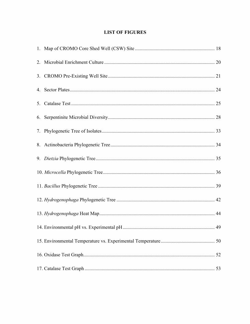
Figure 1: Map showing displaying several of the well sites located at the Coast Range Ophiolite Microbial Observatory, California USA
CROMO2:'Core'Shed'Wells'
CSW1,5'
OLD'CSW'
CSW1,1'CSW1,2'
CSW1,3'
CSW1,4'

19
To promote growth from water samples approximately 45 ml of sample was centrifuged
at 8,000-10,000 × g for 20 minutes at 4°C and the resulting supernatant was removed. The
pelleted biomass was re-suspended in 4.5 ml of pH 11 yeast-peptone liquid media comprised of
0.5% wt of yeast extract, 0.5% wt of peptone in 0.1M sodium carbonate buffer at pH 11. Rock
and soil samples were homogenized and re-suspended in 4.5 ml of pH 11 yeast-peptone media.
Approximately 500 µl of sub-samples were suspended in 1000 µl of pH 11 yeast-peptone media.
Following suspension anywhere from 100-500 µl, dependent upon cell density of the sample, of
the mixture was aseptically spread-plated onto pH 11 yeast-peptone agar plates liquid media
comprised of 0.5%% wt of yeast extract, 0.5%% wt of peptone, 1.5% bacto agar and 0.1M
sodium carbonate buffer at pH 11. Plates were incubated at room temperature (~25°C), in the
dark, for two weeks aerobically and anaerobically. High pH was used as a selective pressure to
enrich for any alkaliphile or alkalitolerant organisms that were present in the sample. Colonies
that grew were isolated and sub-sampled based upon differences in colony shape and color onto
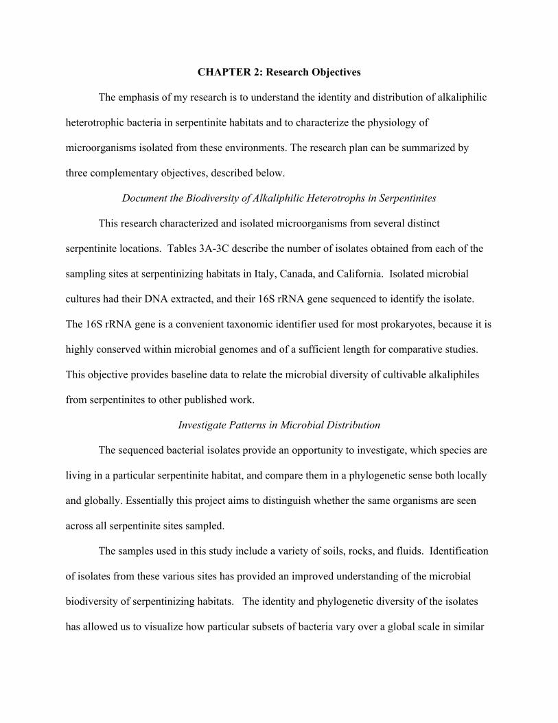
a separate pH 11 enriched media plate for further analysis (Figure 2). A chosen colony was
picked with a sterilized loop and aseptically transferred to a separate pH 11 media plate using the
4-quadrant streaking method to streak for isolation and ensure a pure culture (Appendix A.)
20
Figure 2: Mixed microbial culture from a water sample at the serpentinizing environment CROMO California, USA. A colony was chose to be isolated based upon different morphologies and colors. Arrows indicate colonies that would be considered for isolation.
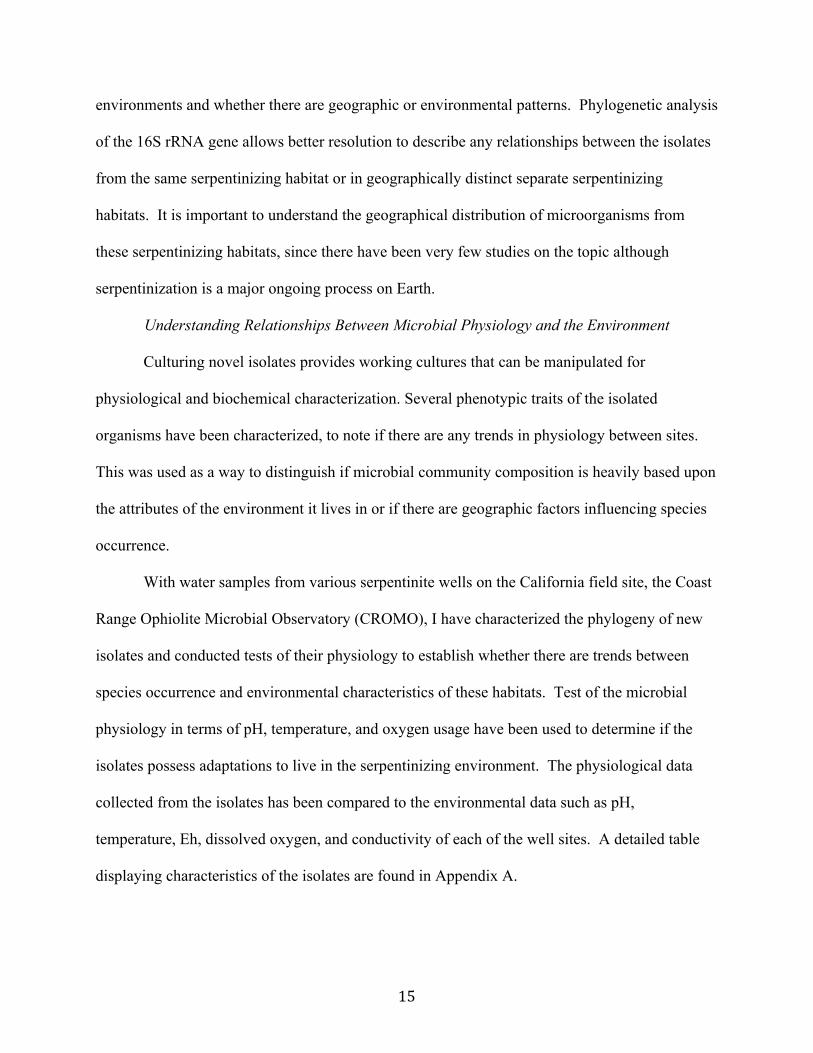
Determining Colony Forming Units of CROMO Wells
Samples were sent to our lab at East Carolina University and stored at 4°C. To promote
growth from water samples approximately 45 ml of sample was centrifuged at 8,000-10,000 × g
for 20 minutes at 4°C and the resulting supernatant was removed. The pelleted biomass was re-
suspended in 4.5 ml of pH 11 yeast-peptone liquid media comprised of 0.5 % wt of yeast, 0.5 %
wt of peptone, and 0.1M sodium carbonate buffer at pH 11. Approximately 150 µl of the
suspension, a 10-fold dilution, was aseptically spread plated once onto the pH 11 yeast-peptone
plates and aerobically/anaerobically cultured in the dark at room temperature for 2 weeks.
Colonies were counted to calculate the Colony Forming Units per ml (CFU/ml) of each well to
determine microbial abundance and the numbers of cells per milliliter obtained by direct
microscopic counts are also included for each well for comparison (Table 6).
21
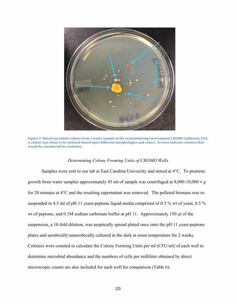
Figure 3: An image of alkaline groundwater wells at the CROMO site in California.
Extraction of Total Genomic DNA
Following isolation of the cultures, DNA was extracted from each individual isolate
using the MoBio Ultra Clean Microbial DNA isolation kit (MoBio Labs: Carlsbad, CA)
following the manufacturer’s protocol. Microbial cell biomass used for the DNA isolation kit
was harvested from the isolated sub-cultured bacteria from the pH 11 yeast-peptone agar plates
using a sterilized loop; several “loopfuls” of biomass was used for sufficient extraction.
Extracted DNA was quantified using a NanoDrop 2000 UV-Vis spectrophotometer. The nucleic
acid content and the absorbance at 260 and 280 nm, which determine sample purity, were
recorded for each isolate’s extracted DNA. Approximately 30 ng/µl DNA was needed per PCR
reaction for each sample. However, due to the prolific growth of these cultures on organic-rich
media, the nucleic acid content of the extracts often exceeded 100 ng/µl.
22
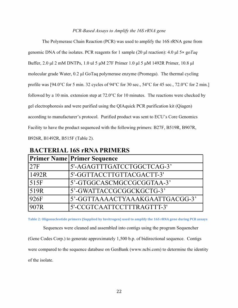
PCR-Based Assays to Amplify the 16S rRNA gene
The Polymerase Chain Reaction (PCR) was used to amplify the 16S rRNA gene from
genomic DNA of the isolates. PCR reagents for 1 sample (20 µl reaction): 4.0 µl 5× goTaq
Buffer, 2.0 µl 2 mM DNTPs, 1.0 ul 5 µM 27F Primer 1.0 µl 5 µM 1492R Primer, 10.8 µl
molecular grade Water, 0.2 µl GoTaq polymerase enzyme (Promega). The thermal cycling
profile was [94.0°C for 5 min. 32 cycles of 94°C for 30 sec., 54°C for 45 sec., 72.0°C for 2 min.]
followed by a 10 min. extension step at 72.0°C for 10 minutes. The reactions were checked by
gel electrophoresis and were purified using the QIAquick PCR purification kit (Qiagen)
according to manufacturer’s protocol. Purified product was sent to ECU’s Core Genomics
Facility to have the product sequenced with the following primers: B27F, B519R, B907R,
B926R, B1492R, B515F (Table 2).
Table 2: Oligonucleotide primers (Supplied by Invitrogen) used to amplify the 16S rRNA gene during PCR assays
Sequences were cleaned and assembled into contigs using the program Sequencher
(Gene Codes Corp.) to generate approximately 1,500 b.p. of bidirectional sequence. Contigs
were compared to the sequence database on GenBank (www.ncbi.com) to determine the identity
of the isolate.
BACTERIAL 16S rRNA PRIMERSPrimer Name Primer Sequence 27F 5'-AGAGTTTGATCCTGGCTCAG-3’ 1492R 5'-GGTTACCTTGTTACGACTT-3' 515F 5’-GTGGCASCMGCCGCGGTAA-3’ 519R 5’-GWATTACCGCGGCKGCTG-3’ 926F 5’-GGTTAAAACTYAAAKGAATTGACGG-3’ 907R 5'-CCGTCAATTCCTTTRAGTTT-3'

23
Construction of Phylogenetic Trees
Phylogenetic trees were made to compare the 16S rRNA gene of the alkaliphilic isolates
to each other and to determine their relatedness. Following the assembly of contigs and sequence
editing, FASTA files from isolates were exported out of the program Sequencher (Gene Codes
Corp.), and grouped into three different phyla, Actinobacteria, Bacilli, and Proteobacteria. Each
phylum was then aligned with reference sequences on Silva, a comprehensive ribosomal RNA
database, http://www.arb-silva.de/aligner. After isolated sequences were aligned with three
“reference” sequences for each isolate, they were exported out of Silva and imported into the
program MEGA, molecular evolutionary genetic analysis (Tamura et al., 2013). Imported
aligned sequences were then used to construct a maximum likelihood tree through a MEGA
utility. Pairwise distances of the 16S rRNA sequences were also constructed through the
computation of the MEGA utility (data not shown).
Determining Optimum Temperature and pH
Nutrient rich plates were made as previously described at a range of different pH
conditions. Different buffer solutions were made for each pH range. These included pH 7
Buffer: 1M monobasic sodium phosphate and 1M dibasic sodium phosphate, made into a 50.0 ml
buffer solution and measured with a pH meter. pH 8 Buffer: 1M TRIS and 1M HCL, made into a
50.0 ml buffer solution and measured with a pH meter. pH 9 Buffer: 1M sodium carbonate and
1M sodium bicarbonate, made into a 50.0 ml buffer solution and measured with a pH meter. pH
10 Buffer: 1M sodium carbonate and 1M sodium bicarbonate, made into a 50.0 ml buffer
solution and measured with a pH meter.
Plates were sectored individually into 6 different segments to hold 6 different isolates
(Figure 4). Isolates were aseptically streaked onto the sector plates in their designated segment.

24
Each isolate was streaked on a pH 7, 8, 9, and 10 plates for three sets of each pH. Therefore for
each isolate there were a total of 12 plates. For the three plates per pH, each plate was stored at a
different temperatures of 20°C, 30°C, and 37°C. Subsequently, this was done for each range of
pH the isolate was streaked on. Growth was monitored for a week and optimum growth
conditions were determined based upon how fast the microbe grew and how dense the growth
was.
Figure 4: An example of the sector plates used to determine optimum pH and temperature. Each wedge represents a different isolate culture.
Oxidase Test Microbial cultures grown for a maximum of two weeks on yeast-peptone agar plates were
sampled with a sterile swab. A drop of BD Oxidase reagent was applied to the inoculated swab.
If the reagent turned a dark purple color within a 30 second time period, the isolate was denoted
25
as oxidase positive. The oxidase test is an indicator if the organism has cytochrome c oxidase,
which helps facilitate the transfer of electrons to the electron acceptor oxygen.
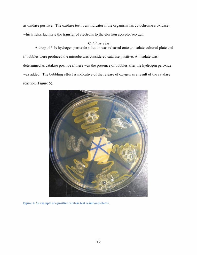
Catalase Test A drop of 3 % hydrogen peroxide solution was released onto an isolate cultured plate and
if bubbles were produced the microbe was considered catalase positive. An isolate was
determined as catalase positive if there was the presence of bubbles after the hydrogen peroxide
was added. The bubbling effect is indicative of the release of oxygen as a result of the catalase
reaction (Figure 5).
Figure 5: An example of a positive catalase test result on isolates.
CHAPTER 5: Microbial Biodiversity and Biogeography of Serpentinite Habitats
Total Microbial Biodiversity
Alkaliphilic microorganisms have been isolated and identified from samples collected
from three different serpentinite habitats located in California, Italy, and Canada (Table 3).
Ultrabasic waters and soil samples were collected from Canada’s Tablelands Ophiolite, which
has been estimated to have been accreted to the North American continent nearly 500 Ma. The
site has extensive travertine deposits that indicate past serpentinization, alkaline springs at pH
ranging from 11-12 and highly reducing conditions. The 200 Ma Liguria Springs in Italy are
generally similar to the Tablelands site in the sense that it too has a pH ranging from 11-12, is
highly reducing, and rich in H2 and CH4. The youngest serpentinite habitat is the Coast Range
Ophiolite in northern California at 65 Ma. The Coast Range Ophiolite site is unique in that fluid
and sediment samples have been obtained directly from the serpentinizing subsurface and a long-
term microbial observatory has been established. Samples collected in California include soil and
water samples from the Quarry Valley (QV) and Core Shed Well (CSW) sites.
To examine the microbial diversity of these serpentinite habitats, it was imperative to
examine if all habitats share common microbial communities. From the three geographically
distinct serpentinite sites there was an overall dominance of the phyla Firmicutes, Actinobacteria,
and Proteobacteria from the isolated cultures (Figure 6). It can also be noted that several isolates
from the phylum Bacteroidetes were found at the Canadian serpentinite environment. Similar
results were found through previous research on microbial diversity at serpentinite sites through
culture dependent analysis (Tiago et al., 2004) although similar results have been observed in
culture independent sequencing studies as well (DeGrood et al., 2004; Daae et al., 2013; Blank et
al., 2009; Tiago et al., 2013). When comparing total distribution of phyla from all the isolates at

27
the three serpentinite sites, Firmicutes were found to make up 54.0%, Proteobacteria 22.8%,
Actinobacteria 21.0% and 0.02% Bacteroidetes. Individual serpentinite sites also showed a
strong dominance in Firmicutes, with varying distributions of Proteobacteria and Actinobacteria
(Figure 6). This indicates that there are overall similarities in the phyla of microorganisms that
can be found at these serpentinizing habitats, even though they are geographically distinct from
one another. That perhaps these dominant phyla are physiologically more suited to the
serpentinizing environment than other microbial phyla. Previous research done on the microbial
diversity of soils show there to be at least 32 phyla groups, with as many 52 phyla can be
present. In typical soils Proteobacteria, followed by Acidobacteria, dominate a majority of the
soil composition, while Firmicutes contribute very little to the typical soil microbial community
(Janssen 2006). This is interesting in that Firmicutes make up a large majority of these
serpentinite systems, which compels the idea that perhaps these Firmicutes have adaptations that
allow them to survive in these harsh serpentinite habitats.
Table 3: 3A-‐C describes the number of isolates obtained from sampling sites in the serpentinizing environments of Italy, Canada, and California.
A. Liguria Springs, Italy B. Tablelands, Canada C. CROMO, California
Site Type Number
of Isolates
Site Type Number
of Isolates
Site Type Number
of Isolates
Gor 2.1 water 1 TLED water 2 CSW 1.1 water 7Gor 2.2 water 1 WHC2A water 9 CSW 1.2 water 2Gor 2.4 water 1 WHC2 water 8 CSW 1.3 water 4Gor 3.A water 1 B30-2 water 1 CSW 1.5 water 5Gor 3.B water 1 DLB-1 water 1 CSW 2 core 1CllO water 1 A4A-2 water 1 CSW 20 core 2L43 water 1 A4D-2 water 1 CSW 22 core 1LA1 water 1 A4A-1 water 1 CSW 23 core 2BR2 water 1 CSW 5 core 2BA2 water 1 CSW 9 core 10LER20 water 4 CSW OLD water 17
NO8A water 3NO8B water 8NO8C water 1QV 1.1 water 8QV 1.2 water 7QV 1.3 water 5QV 10 core 2QV 18 core 2QV 33 core 1QV 41 core 1QV 8 core 2

28
Figure 6: Displays the three dominant phyla from isolated cultures from Italy, Canada, and California
Microbial Biogeography
Magnified at the genus level, we can compare similarities of the serpentinite-hosted
microbial communities more thoroughly between the different sites. For comparison,
background soil samples from Greenville, NC were included and were isolated by the same
procedure used for serpentinite soils. The isolates were categorized into the appropriate groups
at a Genus level by looking at the 16S rRNA gene sequence similarity through NCBI BLAST
(Table 4). Bacillus was the only genus found throughout all three serpentinizing habitats, the
background sample, and data from the Portuguese hyperalkaline wells from Tiago 2004. It is
interesting that Bacillus is found throughout all of the serpentinite sites studied and that it was
even present in the background sampling. Bacilli are spore-forming microorganisms, which
allow them to be better suited for dispersal, and perhaps more adaptive which may be why they
are common in all of the sites.
0" 10" 20" 30" 40" 50" 60" 70"
Proteobacteria"
Ac4nobacteria"
Firmicutes"
Number'of'Isolates'
Phyla
'Serpen5nite'Microbial'Diversity'at'the'Phylum'Level'
Total""
Italy"
Canada"
California"

29
Table 4: Comparison of isolates at the genus level between the three serpentinite sites, background samples, and previous data from Tiago 2004.
When comparing the overlap of different genera between the three serpentinite sites
studied, there were more similarities between the isolates found in Canada and California than
when either was compared to Italian isolates. Bacillus, Dietzia, Geomicrobium,
Hydrogenophaga, Microcella, and Paenibacillus were found both in the serpentinizing
environments in Canada and California. Isolates from Italy were most similar to the data from
Tiago 2004 with comparable results in the genus Arthrobacterium, Bacillus, and
Microbacterium. Broad similarities between the populations found in Canada/California
compared with Italy/Portugal suggest that serpentinizing sites being on the same continental
Phylum Genus Canada California ItalyPortugal data
from Tiago 2004
Greenville, NC
Actinobacteria Arthrobacterium 1 XActinobacteria Brachybacterium 1Actinobacteria Cellulosimicrobium 1Actinobacteria Dietzia 2 7 XActinobacteria Kocuria 1Actinobacteria Microbacterium 1 2 XActinobacteria Microcella 2 5Actinobacteria Micrococcus 3 XBacteroidetes Aquiflexum 2Firmicutes Bacillus 13 25 6 X 6Firmicutes Erysipelothrix 2Firmicutes Exiguobacterium 4Firmicutes Geomicrobium 3 5Firmicutes Paenibacillus 1 5Firmicutes Planococcus 1Firmicutes Planomicrobium 1Firmicutes Solibacillus 1Firmicutes Sporosarcina 2Proteobacteria Alishewanella 8Proteobacteria Agrobacterium 1Proteobacteria Hydrogenophaga 1 10Proteobacteria Malika 1Proteobacteria Pseaudomonas 7Proteobacteria Thauera 1
Total: 24 88 13 6
30
plate may harbor more similar organisms. In can be possible that perhaps geographical distance
and dispersal barriers do play a role in the microbial distribution of certain microorganisms
between similar environmental habitats. However, this result can also be a result of under
sampling and further analysis will rectify when the same trend is found with more isolates.
In contrast, the genera Bacillus and Microbacterium can be found both in the serpentinite
sites of Italy and California, consistent with the earlier discussion about alkalitolerant organisms
enriched from background soils and the possibility that some species are better suited for
dispersal, due to the ability to form spores. Perhaps with more exhaustive sampling and isolation
efforts there will be more similarities among the genera found between the serpentinite sites.
There is a strong bias towards the California serpentinites in the current data set, and under
sampling of the Canadian and Italian serpentinite sites. Further sampling will remedy that bias
and provide better resolution for comparisons between microbial communities. The current study
identified what microorganisms resided at these various serpentinite sites through conventional
culturing and sequencing approaches. For future analyses, sequencing the genomes of the
closely related isolates will lead to greater resolution of both local and global effect of the
evolution of microbial populations.
Phylogenetic Relatedness of Isolates
To examine whether isolates were genetically more related to other isolates from the
same geographic region, a phylogenetic tree was constructed to understand the relationships
among all of the isolates (Figure 7). There was a relatively even distribution between isolates of
the same genus, but from geographically distinct separate serpentinite habitats. There are few
cases where distinct genera represented entirely by one serpentinite environment; in cases where
we see this instance are probably results of under sampling. In general we see the same genera

31
present at all serpentinite habitats. Thus, reinforces Bass Becking’s hypothesis that dispersal
barriers do not account for microorganism. Perhaps the serpentinite environment is selecting for
particular microbes based upon genetic adaptions that allow for survival at these serpentinite
habitats, which is why you see the same types of microbes in separate environments in
California, Canada, and Italy. However variations and geographic separation between the
serpentinite sites may have allowed for genetic evolution to be recorded in the organism’s whole
genomes, not just in the 16S rRNA gene. It would be interesting to examine the presence of
genes in isolates that may be related to survival in the serpentinite environment, to see if these
organisms are truly adapted to the habitat or merely just tolerant. Evaluation of the isolates
genomes would allow for better resolution to see if the same relatedness is found between
isolates, when looking at genes beyond 16S rRNA. This would be similar to the previous work
discussed earlier Reno 2009. The resolution of similarities between the isolates would be better
visualized by looking at whole genomic sequences, as was done in the Reno 2009 study. By
comparing the genomic content of isolates the biogeographic patterns of the microbial isolates
would be more apparent, and this is the intent of follow on work with these isolates.
Serpentine Soils vs. Fluids
Previous studies through culture-independent research have determined that microbial
communities in serpentinite systems varied from the serpentinizing deep subsurface
environments. It was found that H2 oxidizing Betaproteobacteria dominated the fluid systems
and Clostridia, containing [FeFe]-hydrogenases used to help catalyze hydrogen production, are
prevalent in the deep subsurface (Brazelton et al., 2012; Brazelton et al., 2013). Phylogenetic
relationships of isolates were compared to one another based upon if they originated from
serpentinite rock/soils or waters (Figure 7). While there are higher instances of soil/rock isolates
32
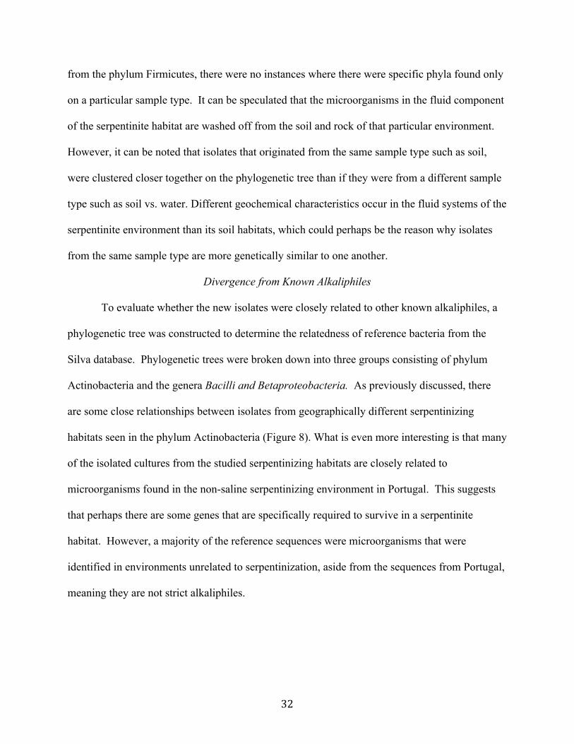
from the phylum Firmicutes, there were no instances where there were specific phyla found only
on a particular sample type. It can be speculated that the microorganisms in the fluid component
of the serpentinite habitat are washed off from the soil and rock of that particular environment.
However, it can be noted that isolates that originated from the same sample type such as soil,
were clustered closer together on the phylogenetic tree than if they were from a different sample
type such as soil vs. water. Different geochemical characteristics occur in the fluid systems of the
serpentinite environment than its soil habitats, which could perhaps be the reason why isolates
from the same sample type are more genetically similar to one another.
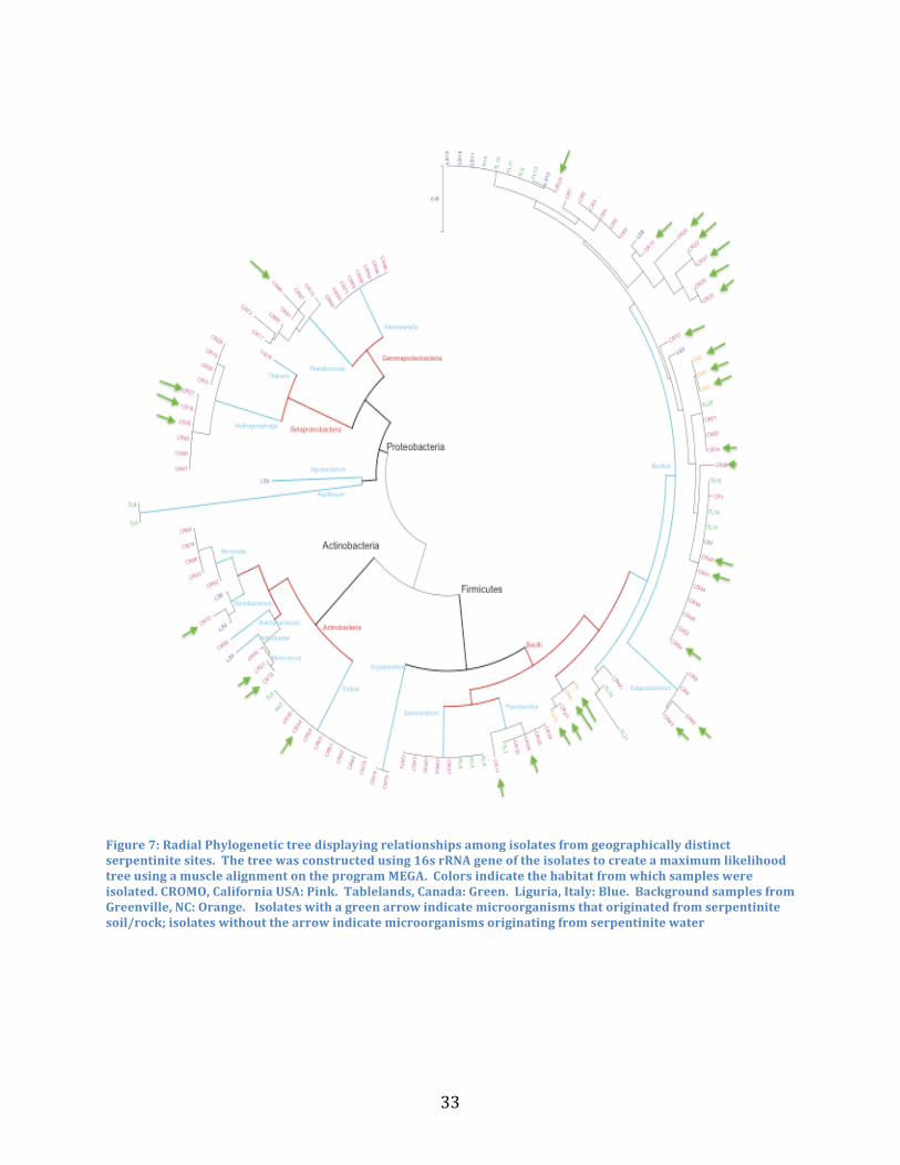
Divergence from Known Alkaliphiles
To evaluate whether the new isolates were closely related to other known alkaliphiles, a
phylogenetic tree was constructed to determine the relatedness of reference bacteria from the
Silva database. Phylogenetic trees were broken down into three groups consisting of phylum
Actinobacteria and the genera Bacilli and Betaproteobacteria. As previously discussed, there
are some close relationships between isolates from geographically different serpentinizing
habitats seen in the phylum Actinobacteria (Figure 8). What is even more interesting is that many
of the isolated cultures from the studied serpentinizing habitats are closely related to
microorganisms found in the non-saline serpentinizing environment in Portugal. This suggests
that perhaps there are some genes that are specifically required to survive in a serpentinite
habitat. However, a majority of the reference sequences were microorganisms that were
identified in environments unrelated to serpentinization, aside from the sequences from Portugal,
meaning they are not strict alkaliphiles.
33
Figure 7: Radial Phylogenetic tree displaying relationships among isolates from geographically distinct serpentinite sites. The tree was constructed using 16s rRNA gene of the isolates to create a maximum likelihood tree using a muscle alignment on the program MEGA. Colors indicate the habitat from which samples were isolated. CROMO, California USA: Pink. Tablelands, Canada: Green. Liguria, Italy: Blue. Background samples from Greenville, NC: Orange. Isolates with a green arrow indicate microorganisms that originated from serpentinite soil/rock; isolates without the arrow indicate microorganisms originating from serpentinite water
34
Figure 8: Phylogenetic tree of isolates from the phylum Actinobacteria. Colors indicate the serpentinite site from which the isolated originated. California, USA: Pink. Liguria, Italy: Blue. Tablelands, Canada: Green. Sequences that are purple correlate to microorganisms originating from the serpentinizing habitat in Portugal.
CR-12 Contig
CR-21 Contig
JF274940.1.1497 BacteriaActinobacteriaActinobacteriaMicrococcalesMicrococcaceaeMicrococcusMicrococcus sp. PX9 S1
AJ717368.1.1502 BacteriaActinobacteriaActinobacteriaMicrococcalesMicrococcaceaeMicrococcusMicrococcus luteus
EF198243.1.1518 BacteriaActinobacteriaActinobacteriaMicrococcalesMicrococcaceaeMicrococcusMicrococcus sp. MACL03
DQ490458.1.1486 BacteriaActinobacteriaActinobacteriaMicrococcalesMicrococcaceaeMicrococcusMicrococcaceae bacterium KVD-1921-02
AB188213.1.1515 BacteriaActinobacteriaActinobacteriaMicrococcalesMicrococcaceaeMicrococcusMicrococcus sp. TUT1210
AJ717366.1.1506 BacteriaActinobacteriaActinobacteriaMicrococcalesMicrococcaceaeCitricoccusCitrococcus sp. AC7
CR56 Contig
EF143420.1.1492 BacteriaActinobacteriaActinobacteriaMicrococcalesMicrococcaceaeCitricoccusMicrococcus sp. WP02-2-225
EU305672.1.1490 BacteriaActinobacteriaActinobacteriaMicrococcalesMicrococcaceaeCitricoccusCitricoccus sp. FS24
LS-4 Contig
HM222680.1.1485 BacteriaActinobacteriaActinobacteriaMicrococcalesMicrococcaceaeArthrobacterArthrobacter sp. 0713C4-1
HQ728389.1.1489 BacteriaActinobacteriaActinobacteriaMicrococcalesMicrococcaceaeArthrobacterbacterium 7(2011)
GQ472811.1.1484 BacteriaActinobacteriaActinobacteriaMicrococcalesMicrococcaceaeArthrobacteruncultured actinobacterium
CR63 Contig
AY741723.1.1504 BacteriaActinobacteriaActinobacteriaMicrococcalesDermabacteraceaeBrachybacteriumBrachybacterium sp. SKJH-25
X91030.1.1513 BacteriaActinobacteriaActinobacteriaMicrococcalesDermabacteraceaeBrachybacteriumBrachybacterium conglomeratum
EU660345.1.1505 BacteriaActinobacteriaActinobacteriaMicrococcalesDermabacteraceaeBrachybacteriumBrachybacterium paraconglomeratum
CR-30 Contig
CR58 Contig
JN882137.1.1478 BacteriaActinobacteriaActinobacteriaCorynebacterialesDietziaceaeDietziauncultured bacterium
CR60 Contig
CR61 Contig
CR62 Contig
CR70 Contig
CR82 Contig
TL5 Contig
KL3 Contig
AB298545.1.1511 BacteriaActinobacteriaActinobacteriaCorynebacterialesDietziaceaeDietziaDietzia natronolimnaea
JQ687118.1.1445 BacteriaActinobacteriaActinobacteriaCorynebacterialesDietziaceaeDietziaDietzia sp. 02SU1
JF500182.1.1475 BacteriaActinobacteriaActinobacteriaCorynebacterialesDietziaceaeDietziauncultured bacterium
CR88 Contig
CR93 Contig
CR80 Contig
CR74 Contig
AJ717385.1.1505 BacteriaActinobacteriaActinobacteriaMicrococcalesMicrobacteriaceaeMicrocellaMicrocella alkaliphila
JQ323147.1.1479 BacteriaActinobacteriaActinobacteriaMicrococcalesMicrobacteriaceaeMicrocellauncultured Microcella sp.
AJ717388.1.1497 BacteriaActinobacteriaActinobacteriaMicrococcalesMicrobacteriaceaeMicrocellaMicrocella putealis
AJ717387.1.1509 BacteriaActinobacteriaActinobacteriaMicrococcalesMicrobacteriaceaeMicrocellaMicrocella putealis
CR92 Contig
LS5 Contig
AY649756.1.1481 BacteriaActinobacteriaActinobacteriaMicrococcalesMicrobacteriaceaeMicrobacteriumMicrobacterium arborescens
HM045843.1.1486 BacteriaActinobacteriaActinobacteriaMicrococcalesMicrobacteriaceaeMicrobacteriumMicrobacterium sp. WJ19
JN644505.1.1513 BacteriaActinobacteriaActinobacteriaMicrococcalesMicrobacteriaceaeMicrobacteriumMicrobacterium arborescens
AM181505.1.1509 BacteriaActinobacteriaActinobacteriaMicrococcalesMicrobacteriaceaeMicrobacteriumMicrobacterium kitamiense
AB013919.1.1526 BacteriaActinobacteriaActinobacteriaMicrococcalesMicrobacteriaceaeMicrobacteriumMicrobacterium kitamiense
CR-15 Contig
AB013907.1.1526 BacteriaActinobacteriaActinobacteriaMicrococcalesMicrobacteriaceaeMicrobacteriumMicrobacterium kitamiense
JQ684245.1.1495 BacteriaActinobacteriaActinobacteriaMicrococcalesMicrobacteriaceaeMicrobacteriumMicrobacterium phyllosphaerae
DQ985063.1.1486 BacteriaActinobacteriaActinobacteriaMicrococcalesMicrobacteriaceaeMicrobacteriumMicrobacterium sp. JL1103
LS3 Contig
FJ999574.1.1515 BacteriaActinobacteriaActinobacteriaMicrococcalesMicrobacteriaceaeMicrobacteriumMicrobacterium sp. HLSB285
0.01
Micrococcus
Citrococcus
Arthrobacter
Brachybacterium
Dietzia
Microcella
Microbacterium
[olive mill waste water]
[potatoe]
[volcanic deposits]
[bioreactor saline waste-water]
[deep sea sediments]
[arabidopsis thaliana leaves]
[Bering sea]
[gruyere and beufort cheese]
[coral from Gulf of Mannar]
[crude oil]
[Gangyang Bay seawater]
[Chesapeake Bay deep biosphere terrestrial sediments]
[industrial circulating cooling water]
[insect gut bacteria]
[citrus]
[mosquito]
[waste water beet sugar factory]
[permafrost soil]
[waste water beet sugar factory]
[marine]
[sponge from the South China Sea]
MICROCOCCUS
CITROCOCCCUS
ARTHROBACTER
BRACHYBACTERIUM
DIETZIA
MICROCELLA
MICROBACTERIUM
35
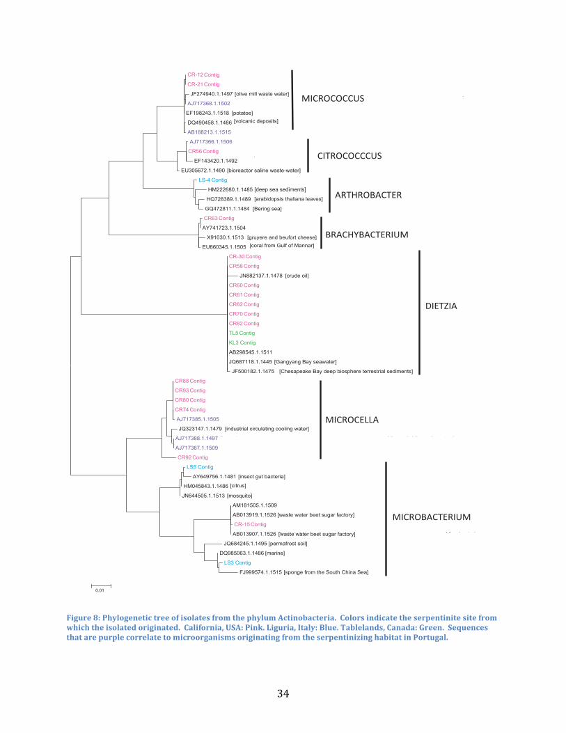
Isolated cultures may be able to tolerate the high alkalinity of the serpentinite sites, but
may not be able to grow optimally in these habitats. There is a prominence in the number of
isolates from the genera Dietzia and Microcella, which could imply that these microorganisms
may contain physiological traits that enable adaptation to the serpentinite environment as
compared with isolates from other taxa. The data from the phylogenetic tree of the phylum
Actinobacteria suggests that you do in fact see some of the same microbes at all the serpentinite
sites. These serpentinite isolates are also more closely related to other serpentinite microbes of
geographically distinct habitats than they are to the references sequences that did not come from
a serpentinizing habitat. This same relationship is found when looking at the phylogenetic
relationships at the genus level through separate phylogenetic analysis (Fig 9, 10).
Figure 9: Phylogenetic tree of the genus Dietzia. Pink: California isolates, Green: Canada isolates, Purple: Portugal isolates
CR82_Contig
KF306378.1.1479_Bacteria;Actinobacteria;Actinobacteria;Corynebacteriales;Dietzi
CR62_Contig
CR58_Contig
gi|56542022|emb|AJ717373.1|_Dietzia_natronolimnaea_16S_rRNA_gene,_isolate_CV46
TL5_Contig
gi|56542023|emb|AJ717374.1|_Dietzia_natronolimnaea_16S_rRNA_gene,_isolate_CV34
CR60_Contig
gi|56542021|emb|AJ717372.1|_Dietzia_natronolimnaea_16S_rRNA_gene,_isolate_AC21r
CR-30_Contig
CR34_Contig
KL3_Contig
CR61_Contig
CR70_Contig
JQ687118.1.1445_Bacteria;Actinobacteria;Actinobacteria;Corynebacteriales;Dietzi
AB266601.1.1470_Bacteria;Actinobacteria;Actinobacteria;Corynebacteriales;Dietzi
AOSR01000052.3685.5181_Bacteria;Actinobacteria;Actinobacteria;Corynebacteriales
FJ435352.1.1461_Bacteria;Actinobacteria;Actinobacteria;Corynebacteriales;Dietzi
AGFF01000033.3678.5176_Bacteria;Actinobacteria;Actinobacteria;Corynebacteriales
EU375845.1.1515_Bacteria;Actinobacteria;Actinobacteria;Corynebacteriales;Dietzi
0.002
[Spain oil spill, culture]
[Gangyang Bay seawater, clone]
[China, culture]
[Artic Ocean marine sediment, culture]
[culture]
[culture]
[culture]
[horned beetle larvae, culture]

36
These relationships suggest that the serpentinite isolates may have genetic differences
from their non-serpentinite counterparts that may or may not allow them to adapt to the
serpentinite environment. That because of the way some of the same microorganisms are
dispersed, you start to see evolutionary changes that occur due to geographical distance at the
same types of habitats. It is also possible that genetic drift may play a role; where you would see
some divergence from the reference sequences. Overall, the data suggest that you do see some
of the same microbes at geographically distinct serpentinite habitat, but they are perhaps
genetically attuned to the particular environment they are isolated from.
Figure 10: A phylogenetic tree of the genus Microcella. Pink: California isolates and Purple: Portugal isolates
AJ717387.1.1509_Bacteria;Actinobacteria;Actinobacteria;Micrococcales;Microbacte
AJ717388.1.1497_Bacteria;Actinobacteria;Actinobacteria;Micrococcales;Microbacte
gi|56542035|emb|AJ717386.1|_Microcella_putealis_16S_rRNA_gene,_strain_AC30
CR92_Contig
JQ323147.1.1479_Bacteria;Actinobacteria;Actinobacteria;Micrococcales;Microbacte
KC734524.1.1628_Bacteria;Actinobacteria;Actinobacteria;Micrococcales;Microbacte
AJ717385.1.1505_Bacteria;Actinobacteria;Actinobacteria;Micrococcales;Microbacte
CR74_Contig
DD147545.5.1456_Bacteria;Actinobacteria;Actinobacteria;Micrococcales;Microbacte
CR93_Contig
CR88_Contig
KC683257.1.1516_Bacteria;Actinobacteria;Actinobacteria;Micrococcales;Microbacte
0.002
[fly ash landfill, culture]
[culture][culture]
[culture]
[culture]

37
As previously discussed the phylum Firmicutes were dominant in the serpentinite sites in
California, Canada, and Italy. In particular the genus Bacillus was present in all three of the
serpentinite sites and even in the background samples taken from neutral pH soil in Greenville,
NC. To examine the relationship of the isolated Bacillus, sequences from the isolates and
reference sequences from the Silva database were used to construct a maximum likelihood tree.
In particular the genus Bacillus was investigated due to the fact that it was present in all of the
serpentinite sites (Figure 11). It was curious to find that not only are the Bacillus which are able
to live in the alkaline environment of serpentinizing habitats, distributed across all the sites, they
were also genetically very similar to one another. The Bacillus isolates are very similar in the
16S rRNA gene, which can be shown by the short branch lengths on the phylogenetic tree
(Figure 11). Comparing the 16S rRNA gene may not be enough to make comparisons between
the Bacillus isolates and employing other genes may provide better insight. A previous study
done by Kopac et al. 2014, looks at the biogeography of a group of Bacillus of the same species
by examining different ecotype formation events that occur between species. The study found
that when only conducting a three-gene analysis between the Bacillus species there were far
fewer ecotype formations than when comparing entire genomes. There were more ecotype
formation events when comparing Bacillus genomes by a factor of 192, which is far more
effective than just looking at three genes (Kopac et al., 2014). This may be the same case with
the serpentinite Bacillus isolates, that we are just not getting enough resolution in one gene to
make adequate biogeographical comparisons.
Bacilli are known spore formers, which may allow them to be better adapted for
microbial dispersal (Errington, 1993). Microbial isolates were more phylogenetically similar to
one another than the background isolates and the reference sequences, shown by the distinct

38
branching on the phylogenetic tree (Figure 11). Perhaps microbial isolates share similar adaptive
genes for the serpentinite environments, despite living in geographically distinct locations.
However, since some of the isolates from California, Italy, and Canada are genetically closely
tied to one another, it can be said that dispersal barriers do not apply to microorganisms. Since,
we see the background samples related to other serpentinites, but with more genetic distance, it
can be said that the serpentinite environment requires some genetic differences for a
microorganism to live in that habitat. That there are microorganisms that contain the genetic
capability to maybe not thrive in serpentinite habitats, but have the capability to survive in those
harsh conditions.
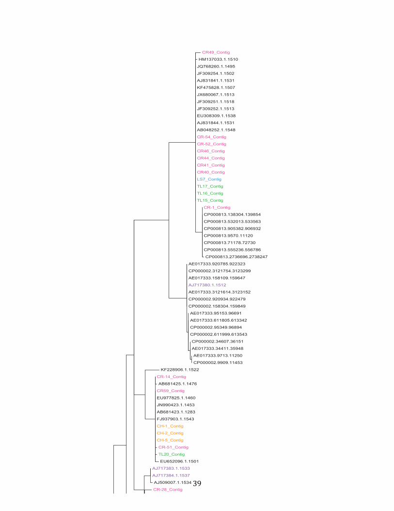
39
CR49_Contig
HM137033.1.1510
JQ768260.1.1495
JF309254.1.1502
AJ831841.1.1531
KF475828.1.1507
JX680067.1.1513
JF309251.1.1518
JF309252.1.1513
EU308309.1.1538
AJ831844.1.1531
AB048252.1.1548
CR-54_Contig
CR-52_Contig
CR46_Contig
CR44_Contig
CR41_Contig
CR40_Contig
LS7_Contig
TL17_Contig
TL16_Contig
TL15_Contig
CR-1_Contig
CP000813.138304.139854
CP000813.532013.533563
CP000813.905382.906932
CP000813.9570.11120
CP000813.71178.72730
CP000813.555236.556786
CP000813.2736696.2738247
AE017333.920785.922323
CP000002.3121754.3123299
AE017333.158109.159647
AJ717380.1.1512
AE017333.3121614.3123152
CP000002.920934.922479
CP000002.158304.159849
AE017333.95153.96691
AE017333.611805.613342
CP000002.95349.96894
CP000002.611999.613543
CP000002.34607.36151
AE017333.34411.35948
AE017333.9713.11250
CP000002.9909.11453
KF228906.1.1522
CR-14_Contig
AB681425.1.1476
CR59_Contig
EU977825.1.1460
JN990423.1.1453
AB681423.1.1283
FJ937903.1.1543
CH-1_Contig
CH-2_Contig
CH-5_Contig
CR-51_Contig
TL20_Contig
EU652096.1.1501
AJ717383.1.1533
AJ717384.1.1537
AJ509007.1.1534
CR-28_Contig
GQ903392.1.1514
GQ903397.1.1514
GQ903388.1.1514
ALEG01000035.70.1600
AWXY01000136.9.1558
FR733689.1.1539
KF879101.1.1515
AJ717382.1.1534
AB062678.1.1545
GQ284392.1.1492
JN882078.1.1512
LS-9_Contig
KC494318.1.1512
CP003017.4782908.4784459
gi|56542030|emb|AJ717381.1|
CP003017.4536652.4538204
CP003017.4578163.4579714
CP003017.4972960.4974511
CP003017.356444.357995
CP003017.4392779.4394330
CP003017.4839215.4840766
CP003017.4918877.4920428
CP003017.4738540.4740093
CP003017.4759089.4760642
CR-7_Contig
EU817573.1.1502
EU530591.1.1513
AB055093.1.1513
JQ906325.1.1509
AJ786819.1.1488
KC734530.1.1489
CR-2_Contig
CR-3_Contig
CR-4_Contig
CR-5_Contig
CR6_Contig
ACPC01000001.16975.18506
AB043862.1.1535
X76443.1.1529
AB043865.1.1521
GU385860.1.1518
FN436100.1.1519
JF970581.1.1533
CR-26_Contig
CR-25_Contig
JN178144.1.1564
EF101552.1.1525
TL9_Contig
CP000903.9027.10571
CP000903.728333.729877
CP000903.88000.89544
EF093131.1.1531
CP000903.4698641.4700185
CP000903.82288.83832
ACMU01000002.45.1584
CP003687.4893190.4894729
CP000903.515954.517498
CP000903.299789.301333
CP000903.275634.277178
CP000903.247808.249352
CP000903.151105.152649
CP000903.28861.30405
BAUY01000093.36.1590
CP000903.307053.308597
CP000903.288925.290469
CP000903.268373.269917
AB021199.1.1531
LS-14_Contig
LS-13_Contig
LS-11_Contig
Tl-14_Contig
TL12_Contig
TL11_Contig
LS-12_Contig
FJ957773.1.1516
CP003889.305524.307063
CP003889.298372.299911
CP003889.282956.284495
CP003687.4832843.4834382
CP003687.5194734.5196273
CP003687.4678194.4679732
CP003889.784157.785696
ACMR01000003.66.1605
CR20_Contig
CP003889.4874127.4875666
CP003889.243920.245459
CP003687.4865377.4866916
FN433029.1.1544
AHEB01000008.25.1564
AHCO01000003.60.1599
CP003889.82042.83581
CP003687.5409201.5410740
CP003889.9017.10556
CP003687.4741287.4742826
CP003687.4043516.4045055
BD180986.4.1536
CP003687.4917463.4919002
CP003687.4885990.4887529
CP003889.87752.89291
CP003687.4903715.4905254
CP003687.4625673.4627212
CP003687.4925021.4926560
CP003687.4872653.4874192
CP003889.28761.30300
CP003889.150933.152472
CP003889.267037.268576
TL-13_Contig
CP003889.525396.526935
CP003687.4606092.4607631
CP003889.274271.275810
CR-23_Contig
AF137020.1.1507
CH-3_Contig
X76438.1.1505
HQ121170.1.1522
HQ120209.1.1522
CH-4_Contig
H4-_Contig
JN178831.1.1548
AB637115.1.1511
AJ717379.1.1527
JN178013.1.1561
JN177837.1.1564
AB637114.1.1512
JN178386.1.1559
CR66_Contig
AB637108.1.1511
TL19_Contig
AB437410.1.1513
JN178396.1.1559
JN178568.1.1547
HQ697807.1.1515
TL21_Contig
AB044748.1.1563
EU186648.1.1527
FR691453.1.1520
JN038220.1.1531
0.01

40
CR49_Contig
HM137033.1.1510
JQ768260.1.1495
JF309254.1.1502
AJ831841.1.1531
KF475828.1.1507
JX680067.1.1513
JF309251.1.1518
JF309252.1.1513
EU308309.1.1538
AJ831844.1.1531
AB048252.1.1548
CR-54_Contig
CR-52_Contig
CR46_Contig
CR44_Contig
CR41_Contig
CR40_Contig
LS7_Contig
TL17_Contig
TL16_Contig
TL15_Contig
CR-1_Contig
CP000813.138304.139854
CP000813.532013.533563
CP000813.905382.906932
CP000813.9570.11120
CP000813.71178.72730
CP000813.555236.556786
CP000813.2736696.2738247
AE017333.920785.922323
CP000002.3121754.3123299
AE017333.158109.159647
AJ717380.1.1512
AE017333.3121614.3123152
CP000002.920934.922479
CP000002.158304.159849
AE017333.95153.96691
AE017333.611805.613342
CP000002.95349.96894
CP000002.611999.613543
CP000002.34607.36151
AE017333.34411.35948
AE017333.9713.11250
CP000002.9909.11453
KF228906.1.1522
CR-14_Contig
AB681425.1.1476
CR59_Contig
EU977825.1.1460
JN990423.1.1453
AB681423.1.1283
FJ937903.1.1543
CH-1_Contig
CH-2_Contig
CH-5_Contig
CR-51_Contig
TL20_Contig
EU652096.1.1501
AJ717383.1.1533
AJ717384.1.1537
AJ509007.1.1534
CR-28_Contig
GQ903392.1.1514
GQ903397.1.1514
GQ903388.1.1514
ALEG01000035.70.1600
AWXY01000136.9.1558
FR733689.1.1539
KF879101.1.1515
AJ717382.1.1534
AB062678.1.1545
GQ284392.1.1492
JN882078.1.1512
LS-9_Contig
KC494318.1.1512
CP003017.4782908.4784459
gi|56542030|emb|AJ717381.1|
CP003017.4536652.4538204
CP003017.4578163.4579714
CP003017.4972960.4974511
CP003017.356444.357995
CP003017.4392779.4394330
CP003017.4839215.4840766
CP003017.4918877.4920428
CP003017.4738540.4740093
CP003017.4759089.4760642
CR-7_Contig
EU817573.1.1502
EU530591.1.1513
AB055093.1.1513
JQ906325.1.1509
AJ786819.1.1488
KC734530.1.1489
CR-2_Contig
CR-3_Contig
CR-4_Contig
CR-5_Contig
CR6_Contig
ACPC01000001.16975.18506
AB043862.1.1535
X76443.1.1529
AB043865.1.1521
GU385860.1.1518
FN436100.1.1519
JF970581.1.1533
CR-26_Contig
CR-25_Contig
JN178144.1.1564
EF101552.1.1525
TL9_Contig
CP000903.9027.10571
CP000903.728333.729877
CP000903.88000.89544
EF093131.1.1531
CP000903.4698641.4700185
CP000903.82288.83832
ACMU01000002.45.1584
CP003687.4893190.4894729
CP000903.515954.517498
CP000903.299789.301333
CP000903.275634.277178
CP000903.247808.249352
CP000903.151105.152649
CP000903.28861.30405
BAUY01000093.36.1590
CP000903.307053.308597
CP000903.288925.290469
CP000903.268373.269917
AB021199.1.1531
LS-14_Contig
LS-13_Contig
LS-11_Contig
Tl-14_Contig
TL12_Contig
TL11_Contig
LS-12_Contig
FJ957773.1.1516
CP003889.305524.307063
CP003889.298372.299911
CP003889.282956.284495
CP003687.4832843.4834382
CP003687.5194734.5196273
CP003687.4678194.4679732
CP003889.784157.785696
ACMR01000003.66.1605
CR20_Contig
CP003889.4874127.4875666
CP003889.243920.245459
CP003687.4865377.4866916
FN433029.1.1544
AHEB01000008.25.1564
AHCO01000003.60.1599
CP003889.82042.83581
CP003687.5409201.5410740
CP003889.9017.10556
CP003687.4741287.4742826
CP003687.4043516.4045055
BD180986.4.1536
CP003687.4917463.4919002
CP003687.4885990.4887529
CP003889.87752.89291
CP003687.4903715.4905254
CP003687.4625673.4627212
CP003687.4925021.4926560
CP003687.4872653.4874192
CP003889.28761.30300
CP003889.150933.152472
CP003889.267037.268576
TL-13_Contig
CP003889.525396.526935
CP003687.4606092.4607631
CP003889.274271.275810
CR-23_Contig
AF137020.1.1507
CH-3_Contig
X76438.1.1505
HQ121170.1.1522
HQ120209.1.1522
CH-4_Contig
H4-_Contig
JN178831.1.1548
AB637115.1.1511
AJ717379.1.1527
JN178013.1.1561
JN177837.1.1564
AB637114.1.1512
JN178386.1.1559
CR66_Contig
AB637108.1.1511
TL19_Contig
AB437410.1.1513
JN178396.1.1559
JN178568.1.1547
HQ697807.1.1515
TL21_Contig
AB044748.1.1563
EU186648.1.1527
FR691453.1.1520
JN038220.1.1531
0.01

41
Figure 11: The tree displays isolates and reference sequences from the genus Bacillus. Isolates are color coordinated according to the serpentinite site they originated from. California, USA: Pink. Liguria, Italy: Blue. Tablelands, Canada: Green, Portugal: Purple. Background samples from Greenville, NC: Orange.
CR49_Contig
HM137033.1.1510
JQ768260.1.1495
JF309254.1.1502
AJ831841.1.1531
KF475828.1.1507
JX680067.1.1513
JF309251.1.1518
JF309252.1.1513
EU308309.1.1538
AJ831844.1.1531
AB048252.1.1548
CR-54_Contig
CR-52_Contig
CR46_Contig
CR44_Contig
CR41_Contig
CR40_Contig
LS7_Contig
TL17_Contig
TL16_Contig
TL15_Contig
CR-1_Contig
CP000813.138304.139854
CP000813.532013.533563
CP000813.905382.906932
CP000813.9570.11120
CP000813.71178.72730
CP000813.555236.556786
CP000813.2736696.2738247
AE017333.920785.922323
CP000002.3121754.3123299
AE017333.158109.159647
AJ717380.1.1512
AE017333.3121614.3123152
CP000002.920934.922479
CP000002.158304.159849
AE017333.95153.96691
AE017333.611805.613342
CP000002.95349.96894
CP000002.611999.613543
CP000002.34607.36151
AE017333.34411.35948
AE017333.9713.11250
CP000002.9909.11453
KF228906.1.1522
CR-14_Contig
AB681425.1.1476
CR59_Contig
EU977825.1.1460
JN990423.1.1453
AB681423.1.1283
FJ937903.1.1543
CH-1_Contig
CH-2_Contig
CH-5_Contig
CR-51_Contig
TL20_Contig
EU652096.1.1501
AJ717383.1.1533
AJ717384.1.1537
AJ509007.1.1534
CR-28_Contig
GQ903392.1.1514
GQ903397.1.1514
GQ903388.1.1514
ALEG01000035.70.1600
AWXY01000136.9.1558
FR733689.1.1539
KF879101.1.1515
AJ717382.1.1534
AB062678.1.1545
GQ284392.1.1492
JN882078.1.1512
LS-9_Contig
KC494318.1.1512
CP003017.4782908.4784459
gi|56542030|emb|AJ717381.1|
CP003017.4536652.4538204
CP003017.4578163.4579714
CP003017.4972960.4974511
CP003017.356444.357995
CP003017.4392779.4394330
CP003017.4839215.4840766
CP003017.4918877.4920428
CP003017.4738540.4740093
CP003017.4759089.4760642
CR-7_Contig
EU817573.1.1502
EU530591.1.1513
AB055093.1.1513
JQ906325.1.1509
AJ786819.1.1488
KC734530.1.1489
CR-2_Contig
CR-3_Contig
CR-4_Contig
CR-5_Contig
CR6_Contig
ACPC01000001.16975.18506
AB043862.1.1535
X76443.1.1529
AB043865.1.1521
GU385860.1.1518
FN436100.1.1519
JF970581.1.1533
CR-26_Contig
CR-25_Contig
JN178144.1.1564
EF101552.1.1525
TL9_Contig
CP000903.9027.10571
CP000903.728333.729877
CP000903.88000.89544
EF093131.1.1531
CP000903.4698641.4700185
CP000903.82288.83832
ACMU01000002.45.1584
CP003687.4893190.4894729
CP000903.515954.517498
CP000903.299789.301333
CP000903.275634.277178
CP000903.247808.249352
CP000903.151105.152649
CP000903.28861.30405
BAUY01000093.36.1590
CP000903.307053.308597
CP000903.288925.290469
CP000903.268373.269917
AB021199.1.1531
LS-14_Contig
LS-13_Contig
LS-11_Contig
Tl-14_Contig
TL12_Contig
TL11_Contig
LS-12_Contig
FJ957773.1.1516
CP003889.305524.307063
CP003889.298372.299911
CP003889.282956.284495
CP003687.4832843.4834382
CP003687.5194734.5196273
CP003687.4678194.4679732
CP003889.784157.785696
ACMR01000003.66.1605
CR20_Contig
CP003889.4874127.4875666
CP003889.243920.245459
CP003687.4865377.4866916
FN433029.1.1544
AHEB01000008.25.1564
AHCO01000003.60.1599
CP003889.82042.83581
CP003687.5409201.5410740
CP003889.9017.10556
CP003687.4741287.4742826
CP003687.4043516.4045055
BD180986.4.1536
CP003687.4917463.4919002
CP003687.4885990.4887529
CP003889.87752.89291
CP003687.4903715.4905254
CP003687.4625673.4627212
CP003687.4925021.4926560
CP003687.4872653.4874192
CP003889.28761.30300
CP003889.150933.152472
CP003889.267037.268576
TL-13_Contig
CP003889.525396.526935
CP003687.4606092.4607631
CP003889.274271.275810
CR-23_Contig
AF137020.1.1507
CH-3_Contig
X76438.1.1505
HQ121170.1.1522
HQ120209.1.1522
CH-4_Contig
H4-_Contig
JN178831.1.1548
AB637115.1.1511
AJ717379.1.1527
JN178013.1.1561
JN177837.1.1564
AB637114.1.1512
JN178386.1.1559
CR66_Contig
AB637108.1.1511
TL19_Contig
AB437410.1.1513
JN178396.1.1559
JN178568.1.1547
HQ697807.1.1515
TL21_Contig
AB044748.1.1563
EU186648.1.1527
FR691453.1.1520
JN038220.1.1531
0.01
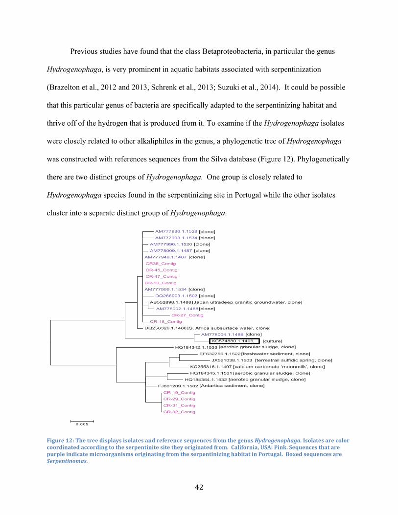
42
Previous studies have found that the class Betaproteobacteria, in particular the genus
Hydrogenophaga, is very prominent in aquatic habitats associated with serpentinization
(Brazelton et al., 2012 and 2013, Schrenk et al., 2013; Suzuki et al., 2014). It could be possible
that this particular genus of bacteria are specifically adapted to the serpentinizing habitat and
thrive off of the hydrogen that is produced from it. To examine if the Hydrogenophaga isolates
were closely related to other alkaliphiles in the genus, a phylogenetic tree of Hydrogenophaga
was constructed with references sequences from the Silva database (Figure 12). Phylogenetically
there are two distinct groups of Hydrogenophaga. One group is closely related to
Hydrogenophaga species found in the serpentinizing site in Portugal while the other isolates
cluster into a separate distinct group of Hydrogenophaga.
Figure 12: The tree displays isolates and reference sequences from the genus Hydrogenophaga. Isolates are color coordinated according to the serpentinite site they originated from. California, USA: Pink. Sequences that are purple indicate microorganisms originating from the serpentinizing habitat in Portugal. Boxed sequences are Serpentinomas.
AM777986.1.1528_Bacteria;Proteobacteria;Betaproteobacteria;Burkholderiales;Coma
AM777993.1.1534_Bacteria;Proteobacteria;Betaproteobacteria;Burkholderiales;Coma
AM777990.1.1520_Bacteria;Proteobacteria;Betaproteobacteria;Burkholderiales;Coma
AM778009.1.1487_Bacteria;Proteobacteria;Betaproteobacteria;Burkholderiales;Coma
AM777949.1.1487_Bacteria;Proteobacteria;Betaproteobacteria;Burkholderiales;Coma
CR35_Contig
CR-45_Contig
CR-47_Contig
CR-50_Contig
AM777999.1.1534 Bacteria;Proteobacteria;Betaproteobacteria;Burkholderiales;Comamonadaceae;Hydrogenophaga;uncultured bacterium
DQ266903.1.1503 Bacteria;Proteobacteria;Betaproteobacteria;Burkholderiales;Comamonadaceae;Hydrogenophaga;uncultured bacterium
AB552898.1.1488_Bacteria;Proteobacteria;Betaproteobacteria;Burkholderiales;Coma
AM778002.1.1488_Bacteria;Proteobacteria;Betaproteobacteria;Burkholderiales;Coma
CR-27_Contig
CR-18_Contig
DQ256326.1.1488_Bacteria;Proteobacteria;Betaproteobacteria;Burkholderiales;Coma
AM778004.1.1486_Bacteria;Proteobacteria;Betaproteobacteria;Burkholderiales;Coma
KC574880.1.1496_Bacteria;Proteobacteria;Betaproteobacteria;Burkholderiales;Coma
HQ184342.1.1533 Bacteria;Proteobacteria;Betaproteobacteria;Burkholderiales;Comamonadaceae;Hydrogenophaga;uncultured Comamonadaceae bacterium
EF632756.1.1522 Bacteria;Proteobacteria;Betaproteobacteria;Burkholderiales;Comamonadaceae;Hydrogenophaga;uncultured bacterium
JX521038.1.1503 Bacteria;Proteobacteria;Betaproteobacteria;Burkholderiales;Comamonadaceae;Hydrogenophaga;uncultured bacterium
KC255316.1.1497_Bacteria;Proteobacteria;Betaproteobacteria;Burkholderiales;Coma
HQ184345.1.1531_Bacteria;Proteobacteria;Betaproteobacteria;Burkholderiales;Coma
HQ184354.1.1532_Bacteria;Proteobacteria;Betaproteobacteria;Burkholderiales;Coma
FJ801209.1.1502_Bacteria;Proteobacteria;Betaproteobacteria;Burkholderiales;Coma
CR-19_Contig
CR-29_Contig
CR-31_Contig
CR-32_Contig
0.005
[S. Africa subsurface water, clone]
[Japan ultradeep granitic groundwater, clone]
[aerobic granular sludge, clone]
[Antartica sediment, clone][aerobic granular sludge, clone]
[calcium carbonate ‘moonmilk’, clone]
[aerobic granular sludge, clone]
[freshwater sediment, clone]
[terrestrail sulfidic spring, clone]
[clone][clone]
[clone]
[clone]
[clone]
[clone]
[clone]
[clone]
[clone]
[culture]

43
Both groups of Hydrogenophaga are hydrogen-oxidizing bacteria, as referenced by their
genus name, which may be why they are so prevalent in the hydrogen producing serpentinite
environment (Figure 12). The ubiquity of Hydrogenophaga at serpentinite sites is also reflected
in culture independent data produced using next generation sequencing approaches (Sukuzi et al.,
2013; Brazelton et al., 2013, Daee et al., 2013). Through culture independent surveys of bacterial
populations the abundance of various Hydrogenophaga species at serpentinite sites in
geographically different regions was evaluated. The heat map shown in Figure 13 includes some
of the cultured isolates of Hydrogenophaga, in which the group of isolates that were closely
related to Tiago’s isolates from the serpentinite site in Portugal and to Serpentimonas, were in
high abundance at many of the serpentinizing habits. While the other group of Hydrogenophaga,
which were not closely related to the Portugal isolates, were not as abundant in many of these
serpentinizing habitats.
This data suggest the isolated Hydrogenophaga can be categorized into two different
groups, a group that has adapted to live successfully in a serpentinizing environment and a group
that can be found more commonly in other habitats besides the serpentines. It would seem that
one group of Hydrogenophaga has genetically evolved to become better optimized for the
surviving the harsh serpentinite environment. This group of Hydrogenophaga may be part of a
new proposed genus called Serpentinomas. Serpentinomas, as previously discussed, are close
relatives to Hydrogenophaga. Serpentinomas were isolated from the serpentinizing habitat in
The Cedars, California (Suzuki et al., 2014). The Serpentinomas isolates were able to grow
heterotrophically in lab conditions, consistent with the current work. However, the
Serpentinomas were found to couple hydrogen oxidation, typical for Hydrogenophaga species,

44
and utilize solid calcium carbonate from the serpentinizing habitat as a carbon source (Suzuki et
al., 2014).
These Serpentinomas can be found through a wide range of alkaline habitats, whether
they are artificial or naturally occurring. (Suzuki et al., 2014) With this proposed new genus and
the presence of two distinctive groups of Hydrogenophaga from my isolates it is likely that one
of the groups of Hydrogenophaga are actually part of the Serpentinomas genus which are highly
adapted to serpentinizing habitats.
Figure 13: Figure made by Dr. W. Brazelton (University of Utah). The figure depicts the relative abundance of 16S rRNA tag sequences in environmental samples from different clades of Hydrogenophaga and their closest relatives. The figure above uses next generation tag sequencing of the 16S rRNA gene that shows the abundance of difference species of Hydrogenophaga at different serpentinite sites. Cultures that I have isolated and identified in the lab are also displayed in the figure as follows CR29, CR 31, CR 19, CR 47, CR 18, CR 45, and CR 50. Based upon where my isolates fall on the phylogenetic tree, there appears to be two different groups of Hydrogenophaga isolated.
CR-31
CR-29
CR-19
Leka clone JN003032
Hydrogenophaga sp. CL3
Hydrogenophaga taeniospiralis
Hydrogenophaga palleronii
Leka clone JN003042
Hydrogenophaga atypica
CR-47
CR-18
CR-45
CR-50
Malikia granosa
Hydrogenophaga flava
Hydrogenophaga bisanensis
Ralstonia insidiosa
Ralstonia pickettii
LC1524B-50 (DQ270636)
LC1537B-16 (DQ270638)
Ralstonia syzygii
Ralstonia solanacearum
Ralstonia mannitolilytica 0.03
8A_A
pr8A
.Apr
8A_A
ug8A
.Aug
8B_A
pr8B
.Apr
C8B
.Apr
D8B
.Aug
CSW
old.
Nov
CSW
oldA
CSW
11CS
W11
.Nov
ACS
W11
.Nov
BCS
W11
.DNA
2QV
11.D
NA1
QV11
.DNA
2QV
11.D
NA3
QV11
ABQV
11C
QV11
DE3u
mCS
W13
ACS
W14
ABQV
12A
QV13
RQV
13R.
who
leQV
21R
QV21
R.w
hole
LS10
_7LS
11_1
17LS
11_6
5LS
11_8
3BR
2up.
DNA1
BR2u
p.DN
A2TL
10_5
7W
HC2B
.201
0TL
11_4
6W
HC2C
.DNA
1TL
11_2
4TL
11_7
1TL
11_7
2TL
11_3
WHB
up.D
NA2
LCY1
LCY3
LCY5
LCY7
LC34
CV
A, P
ort
ug
alCoast Range, California
Tablelands,Newfoundland
Liguria,Italy
Lost City,M.A.R.
0 % 0.02% 0.1% 0.3% 1% 5% 20% 80%
Percentage of bacterial community
Abundance of 16S rRNA Tag Sequences Mapped to Hydrogenophaga Phylogeny
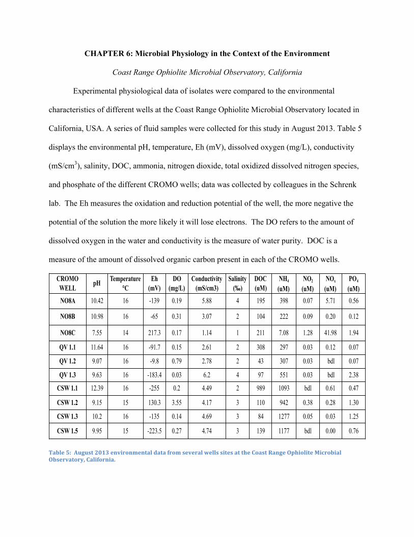
CHAPTER 6: Microbial Physiology in the Context of the Environment
Coast Range Ophiolite Microbial Observatory, California
Experimental physiological data of isolates were compared to the environmental
characteristics of different wells at the Coast Range Ophiolite Microbial Observatory located in
California, USA. A series of fluid samples were collected for this study in August 2013. Table 5
displays the environmental pH, temperature, Eh (mV), dissolved oxygen (mg/L), conductivity
(mS/cm3), salinity, DOC, ammonia, nitrogen dioxide, total oxidized dissolved nitrogen species,
and phosphate of the different CROMO wells; data was collected by colleagues in the Schrenk
lab. The Eh measures the oxidation and reduction potential of the well, the more negative the
potential of the solution the more likely it will lose electrons. The DO refers to the amount of
dissolved oxygen in the water and conductivity is the measure of water purity. DOC is a
measure of the amount of dissolved organic carbon present in each of the CROMO wells.
Table 5: August 2013 environmental data from several wells sites at the Coast Range Ophiolite Microbial Observatory, California.
CROMO WELL
pH Temperature °C
Eh (mV)
DO (mg/L)
Conductivity (mS/cm3)
Salinity (‰)
DOC (uM)
NH4 (uM)
NO2 (uM)
NOx (uM)
PO4 (uM)
NO8A 10.42 16 -139 0.19 5.88 4 195 398 0.07 5.71 0.56
NO8B 10.98 16 -65 0.31 3.07 2 104 222 0.09 0.20 0.12
NO8C 7.55 14 217.3 0.17 1.14 1 211 7.08 1.28 41.98 1.94
QV 1.1 11.64 16 -91.7 0.15 2.61 2 308 297 0.03 0.12 0.07
QV 1.2 9.07 16 -9.8 0.79 2.78 2 43 307 0.03 bdl 0.07
QV 1.3 9.63 16 -183.4 0.03 6.2 4 97 551 0.03 bdl 2.38
CSW 1.1 12.39 16 -255 0.2 4.49 2 989 1093 bdl 0.61 0.47
CSW 1.2 9.15 15 130.3 3.55 4.17 3 110 942 0.38 0.28 1.30
CSW 1.3 10.2 16 -135 0.14 4.69 3 84 1277 0.05 0.03 1.25
CSW 1.5 9.95 15 -223.5 0.27 4.74 3 139 1177 bdl 0.00 0.76
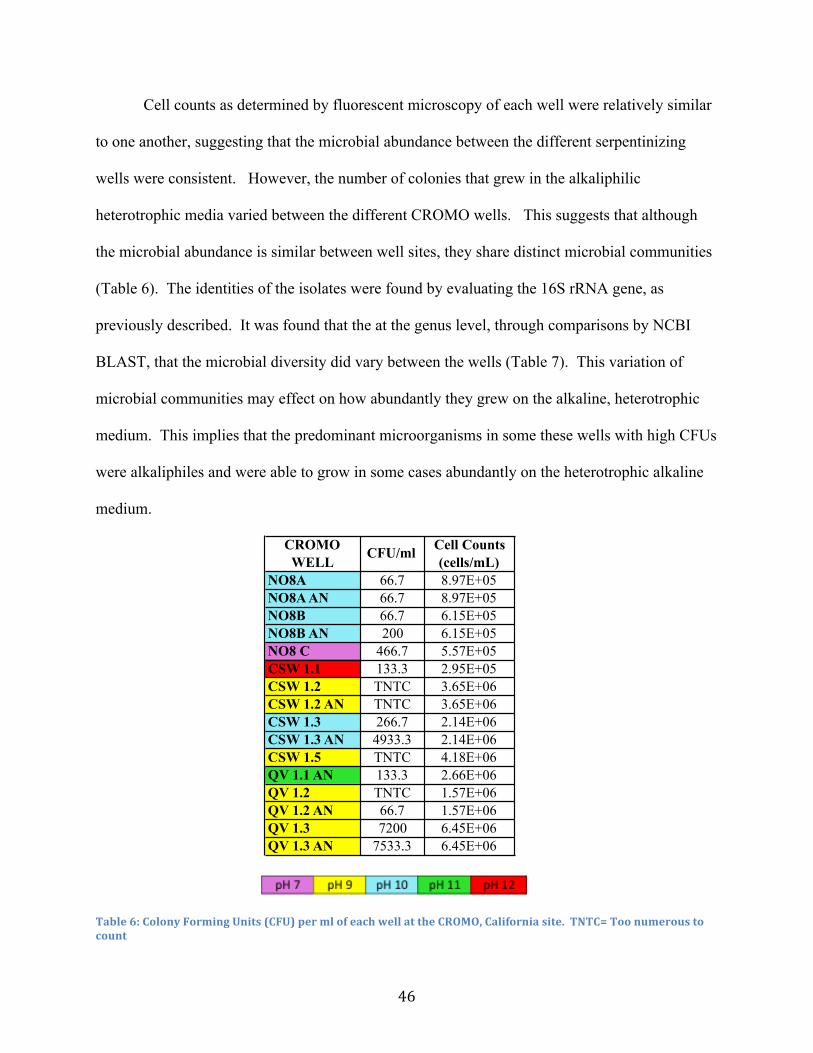
46
Cell counts as determined by fluorescent microscopy of each well were relatively similar
to one another, suggesting that the microbial abundance between the different serpentinizing
wells were consistent. However, the number of colonies that grew in the alkaliphilic
heterotrophic media varied between the different CROMO wells. This suggests that although
the microbial abundance is similar between well sites, they share distinct microbial communities
(Table 6). The identities of the isolates were found by evaluating the 16S rRNA gene, as
previously described. It was found that the at the genus level, through comparisons by NCBI
BLAST, that the microbial diversity did vary between the wells (Table 7). This variation of
microbial communities may effect on how abundantly they grew on the alkaline, heterotrophic
medium. This implies that the predominant microorganisms in some these wells with high CFUs
were alkaliphiles and were able to grow in some cases abundantly on the heterotrophic alkaline
medium.
Table 6: Colony Forming Units (CFU) per ml of each well at the CROMO, California site. TNTC= Too numerous to count
CROMO WELL
CFU/ml Cell Counts (cells/mL)
NO8A 66.7 8.97E+05NO8A AN 66.7 8.97E+05NO8B 66.7 6.15E+05NO8B AN 200 6.15E+05NO8 C 466.7 5.57E+05CSW 1.1 133.3 2.95E+05CSW 1.2 TNTC 3.65E+06CSW 1.2 AN TNTC 3.65E+06CSW 1.3 266.7 2.14E+06CSW 1.3 AN 4933.3 2.14E+06CSW 1.5 TNTC 4.18E+06QV 1.1 AN 133.3 2.66E+06QV 1.2 TNTC 1.57E+06QV 1.2 AN 66.7 1.57E+06QV 1.3 7200 6.45E+06QV 1.3 AN 7533.3 6.45E+06

47
Table 7: Table displaying the number of isolates obtained from each serpentinizing CROMO well and the microbial diversity of each well. Identity of QV 1.1 isolate was unable to be identified due to issues with DNA extraction.
Experimental Optimum pH and Temperature of Isolates
Microbial isolates were then obtained on a separate pH 11 nutrient agar plate.
Approximately 1-5 isolates were obtained from each water sample. Each isolate was then plated
onto 3 sets of nutrient agar plates at a pH of 7, 8, 9, or 10. Microbial isolate sets plated at a
specific pH were then individually incubated at three different temperatures 20°C, 30°C, and
37°C. Plates were monitored and observed daily for a week (Figure 4). Based upon how fast the
isolated culture grew and how dense the growth determines the qualitative optimum pH and
temperature of the alkaliphilic microbe. Growth was measured on a + scale, meaning + barely
any growth, ++ for medium growth, +++ for high growth; the faster and the more dense the
culture the more optimized the microbe is for that temperature or pH (Table 8)
Genus NO8A
NO8B
NO8C
QV 1.1
QV 1.2
QV 1.3
CSW 1.1
CSW 1.2
CSW 1.3
CSW 1.2
Alishewanella 2 1 2 3Bacillus 1Dietzia 1 1Erysipelothrix 2Geomicrobium 1 1 1 1Microcella 1 1 1 1 2Pseudomonas 2 1Thauera
Total: 2 2 1 1 7 5 2 2 4 5

48
Table 8: An example table showing how optimum temperature and pH was determined. The date beside ‘Sample” describes the start of the experiment. Each set of pH 7, 8, 9, and 10 plates were grown at 20°C in this case and +, ++, +++, describes the start and density of microbial growth a number of days after inoculation
Experimental physiological data of each isolate were then compared to the environmental
data of the CROMO well it was isolated from. Figure 14 depicts the relationship between the
environmental pH of each CROMO well and the average experimental optimum pH of the
isolates from each well. Each plot represents each serpentinizing well at the CROMO site.
Based upon the data there seems to be a strong relationship between the environmental pH and
the experimental optimum pH of the isolates. To test the correlation of the environmental and
experimental pH a linear regression line was made on the plots and the value of R2 = 0.09772
indicates there is a strong relationship between experimental and environmental pH. The data
suggest that the isolated microorganisms, selecting specifically for alkaliphiles, are true
alkaliphilic bacteria or microorganisms that have adapted to the alkalinity of the serpentinite
environment. Microbial isolates from the wells may have several of the previously named
mechanisms that allow them to adapt to the alkaline conditions, which may be the reason for the
preference of alkaline conditions in a lab cultured setting.
Temperature 20°CSample 9/16/13 pH7 pH8 pH9 pH10CR 64 ++ Day 1 ++ Day 1CR 65 ++ Day 1 ++ Day 1CR 66 + Day 4 + Day 4 + Day 1 + Day 1CR 67 ++ Day 1 ++ Day 1CR 68 + Day 2 ++ Day 1 +++ Day 1CR 69 ++ Day 1 + Day 1CR 70 + Day 2 + Day 2 + Day 1 + Day 1CR 71 ++ Day 2 ++ Day 1 +++ Day 1CR 72 + Day 2 + Day 7 + Day 1 + Day 1CR 73 ++ Day 1 ++ Day 1CR 74 + Day 7 + Day 4 + Day 1 ++ Day 1
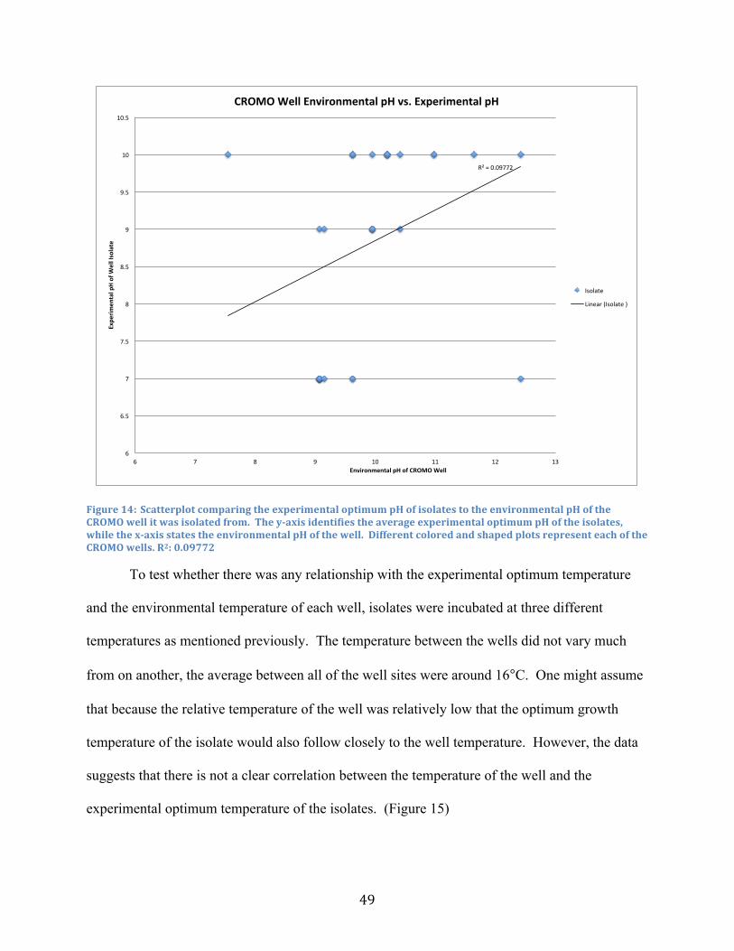
49
Figure 14: Scatterplot comparing the experimental optimum pH of isolates to the environmental pH of the CROMO well it was isolated from. The y-‐axis identifies the average experimental optimum pH of the isolates, while the x-‐axis states the environmental pH of the well. Different colored and shaped plots represent each of the CROMO wells. R2: 0.09772
To test whether there was any relationship with the experimental optimum temperature
and the environmental temperature of each well, isolates were incubated at three different
temperatures as mentioned previously. The temperature between the wells did not vary much
from on another, the average between all of the well sites were around 16°C. One might assume
that because the relative temperature of the well was relatively low that the optimum growth
temperature of the isolate would also follow closely to the well temperature. However, the data
suggests that there is not a clear correlation between the temperature of the well and the
experimental optimum temperature of the isolates. (Figure 15)
R²#=#0.09772#
6#
6.5#
7#
7.5#
8#
8.5#
9#
9.5#
10#
10.5#
6# 7# 8# 9# 10# 11# 12# 13#
Expe
rimen
tal,pH,of,W
ell,Isolate,
Environmental,pH,of,CROMO,Well,
CROMO,Well,Environmental,pH,vs.,Experimental,pH,,
Isolate##
Linear#(Isolate#)#

50
There was no correlation between the environmental temperature and the average
experimental temperature of the isolates, R2 = 0.02635. When grown in a lab setting isolates
preferred warmer growing conditions. It can be noted that the samples were cultured and
isolated using a selective filter of ~25°C, thus isolates should grow relatively well at this
temperature. The optimum experimental growing temperatures of a majority of the isolates
were typically higher than 25°C. There was no obvious correlation between the temperature
effects on the growth of the isolates and their sample temperature. This suggests that
temperature is not a major selecting factor for the microorganisms that inhabit the CROMO
wells. It can also be said that because the experimental optimum temperature varied so
differently from the well temperature, that these microorganisms may not be growing optimally
in their natural environment.
Figure 15: Scatterplot comparing the experimental optimum temperature of isolates to the environmental temperature of the CROMO well it was isolated from. The y-‐axis identifies the average experimental optimum temperature of the isolates, while the x-‐axis states the environmental temperature of the well. Different colored and shaped plots represent each of the CROMO wells. R2-‐value: 0.02635
R²#=#0.02635#
0#
5#
10#
15#
20#
25#
30#
35#
40#
14.8# 15# 15.2# 15.4# 15.6# 15.8# 16# 16.2# 16.4# 16.6# 16.8#
Expe
rimen
tal,Tem
perature,of,W
ell,Isolate,
Environmental,Temperature,of,CROMO,Well,
CROMO,Well,Environmental,Temperature,vs.,Experimental,Temperature,
Isolate#
Linear#(Isolate)#
51
Oxidase Test Results
Microorganisms from the CROMO wells were isolated and cultured under aerobic
conditions. The relative amounts of dissolved oxygen in each of the wells were relatively
consistent with one another (Table 5). Most aerobic organisms use the cytochrome c oxidase
enzyme to transfer electrons from the electron donors to the electron acceptor oxygen. It has
been shown in previous studies that the levels of cytochromes are elevated in alkaliphiles.
Microorganisms that are oxidase positive are typically aerobic, however that does not mean they
are strict aerobes. The data from the oxidase test of the isolates show there was slightly more
oxidase positive isolates 58.6 % that there was oxidase negative isolates 41.4% (Figure 16).
These data may support the idea that cytochromes are more prominent in alkaliphiles. However,
there seems to be no trend between the alkalinity and the amount of dissolved oxygen in relation
to whether the isolate contained cytochrome c oxidase. The results suggest that some isolates
have the ability to use oxygen as an electron acceptor in the electron transport chain or that they
are using some other molecule as their electron acceptor or they use alterative oxygen detox
mechanisms. Hydrogenophaga sp. is known to use oxygen as an electron acceptor while
Bacillus sp. can also use nitrate and Actinobacteria can use nitrite (Suzuki et al., 2014; Nakano et
al., 2007; Ventura et al., 2007). There are so few electron acceptors in the serpentinite
environment it would be interesting to determine the range of alternative electron acceptors are
being utilized by the isolates in this particular serpentinite environment. It could be possible
based upon the NOx (uM) levels (Table 5) of the CROMO wells that different nitrogen species
(nitrate, nitrite, etc.) are being used as electron acceptors.

52
Figure 16: The number of oxidase +/-‐ isolates for each CROMO well site. The light blue bar indicates the number of oxidase positive isolates for the particular well, while the dark blue bar indicates the number of oxidase negative organisms
Catalase Test
As a product of redox reactions happening inside aerobic microorganisms hydrogen
peroxide is constantly accumulating. Hydrogen peroxide can result in higher mutation rates,
growth defects, and possibly death. Hydrogen peroxide is toxic to these organisms, so
microorganisms produce enzymes such as catalase to keep the toxin levels relatively low. The
catalase enzyme is present in many aerobic bacteria that break down hydrogen peroxide into
molecular oxygen and water (Mishra et al., 2012). Based upon the experimental results most of
the organisms isolated were catalase positive 89.7% versus 10.3% of the catalase negative
0" 1" 2" 3" 4" 5"
NO8A"
NO8B"
NO8C"
QV"1.1"
QV"1.2"
QV"1.3"
CSW"1.2"
CSW"1.3"
CSW"1.5""
Number'of'Isolates'
CROMO'W
ell'Site
'
Oxidase'Test'Results'
Oxidase"9""
Oxidase"+""

53
isolates (Figure 17). There does not seem to be any relationship between the number of catalase
positive organisms with the amount of dissolved oxygen in each well or the number of
experimentally oxidase positive organisms. It is common to see catalase in most aerobic bacteria
and since these microorganisms were isolated aerobically it is not surprising to see that most are
catalase positive. Isolates were also grown in anaerobic conditions and it was found that a large
majority was able to grow aerobically and anaerobically (Appendix A). This suggests that these
isolates were not strict aerobes or anaerobes, but perhaps were facultative anaerobes that required
oxygen detox mechanisms such as the enzyme catalase when grown under oxic conditions.
Figure 17: The number of catalase +/-‐ isolates for each CROMO well site. The light blue bar indicates the number of catalase positive isolates for the particular well, while the dark blue bar indicates the number of catalase negative organisms.
0" 1" 2" 3" 4" 5" 6" 7"
NO8A"
NO8B"
NO8C"
QV"1.1"
QV"1.2"
QV"1.3"
CSW"1.2"
CSW"1.3"
CSW"1.5""
Number'of'Isolates'
CROMO'W
ell'Site'
Catalase'Test'Results'
Catalase":"
Catalase"+"

CHAPTER 7: Discussion
Microbial diversity of the serpentinite subsurface has not been intensively studied and the
number of cultured bacteria from these serpentinite sites is low. This study focuses on
examining the biodiversity of alkaliphilic heterotrophic bacteria from several serpentinite sites in
California, Canada, and Italy in comparison to previous studies. This study also evaluates the
experimental microbial physiology of isolated alkaliphilic heterotrophic microbial cultures in
relation to the characteristics of the environment from which they were isolated.
When examining microbial diversity at the phylum level we see three major phyla
present at each of the three serpentinite sites, Firmicutes, Actinobacteria, and Proteobacteria.
Similar studies on microbial diversity have also examined this phenomenon, where they find the
same three phyla present at additional serpentinite habitats (Tiago et al., 2004), DeGrood et al.,
2004, Daae et al., 2013, Blank et al., 2009, Tiago et al., 2013). Firmicutes not only made up a
large portion of the total isolates from all the serpentinite sites, but also dominated in abundance
at each individual site. This may indicate that the phylum Firmicutes are more adapted to being
geographically dispersed and are better suited to this serpentinizing environment. Proteobacteria
was the second most prevalent phylum when examining the isolates in their entirety, however for
each individual serpentinite it was comparable in relative abundance with the phylum
Actinobacteria. This predominance of Firmicutes and Proteobacteria from the total isolates
could indicate that these phyla are more suited to the serpentinite environment; especially since
their respective genera Clostridia and Hydrogenophaga have been previously found prominently
in the deep subsurface and fluid systems of these serpentinite habitats (Schrenk et al., 2013).
Microbial diversity was then evaluated at the genus level of the microbial isolates. We
find that microbial diversity is very low, indicated by the total number of genera, not only when
55
looking at the total number of isolates, but when examining the microbial diversity at each
individual serpentinite site. Even when examining the microbial diversity of the sample plates, it
was evident that the amount of diversity was very low despite the fact they were cultured on
nutrient rich heterotrophic medium and concentrated 10-100 times. This indicates that the
serpentinite habitat, because of its low nutrient availability, lack of electron acceptors, and
extreme alkalinity, is selective on what types of microorganisms are able to survive its harsh
habitat. It was found that there was overlap between genera in all of the serpentinite sites, in
particular with the genus Bacillus that was present at every site including the background-
sampling site. Bacilli are known spore formers and may be able to survive geographical
dispersion better than microorganisms of another genera (Errington, 1993). It could also be
possible that they reason we see Bacillus at every site is that they are very alkalitolerant genus of
microorganism, as supported by previous data (Padan et al., 2005).
This supports the idea that microorganisms do not have any dispersal barriers, as most
macroorganisms do. However, there is some variation between genera at each serpentinite site
that were not present at other sites. Similar genera of each site were found to be more common
between serpentinite habitats that were on the same continental plate. We see that these
microorganisms may not have any dispersal barriers, since we see the same organisms at all
serpentinizing habits. Isolates from geographically distant serpentinite sites were closely related
to one another, with no distinct phylogenetic branching although they were from an entirely
separate geographical location. This may be a consequence of just examining the highly
conserved 16S rRNA gene of these isolates and it would be interesting to see if we find the same
types of relationships with other genes. It could be possible that with further examination of the
isolates genomes that we will see variation between core and variable genes between isolates of

56
the same species, as previously found in Reno’s 2009 study. Overall, serpentinite isolates were
phylogenetically more similar to one another than to the reference 16S rRNA sequences found in
non-serpentinite habitats from the Silva database.
These data from both gene sequencing and physiological characterization suggest that
serpentinite isolates have genetically evolved to become more adapted to survive in a
serpentinite site and that they may also be genetically unique to the particular serpentinite site
that they were isolated. This may be an indication that although we see the same organisms
everywhere there are microbial adaptations to the environment through perhaps mutations, gene
exchange, or even genetic drift. It would be interesting to sequence the genomes of all the
isolates to make more thorough comparisons on the genetic content of the microorganisms and
examine the genetic variations that occur between different serpentinite habitats. It is also
imperative to get a more statistically representative number of isolates from more serpentinizing
habitats.
Microbial communities were phylogenetically evaluated in the soil and water systems of
the serpentinite sites to examine whether there were relationships between the isolates in the
serpentinite microenvironment. There were no distinct genera that were more present in either
the soil or fluid systems of the serpentinite habitats. There were also no instances of distinct
phylogenetic differences between isolates from soil or fluid systems. Since there were no
divergent microbial sub-communities in the soil or fluid systems it can be inferred that they have
the similar microbial communities; perhaps due to microorganisms washing off of the soils in to
the serpentinite fluids.
Previous studies have indicated that the genus Hydrogenophaga dominated the fluid
systems of many serpentinite sites. All isolates of Hydrogenophaga were obtained from samples

57
of water were predominantly from the fluid systems of the CROMO site in California, with the
exception of a single isolate from Canada. Since, Hydrogenophaga are so prominent in the
mixing zones of these serpentinite habits, it is important to evaluate how they are
phylogenetically related to other Hydrogenophaga isolated from a separate serpentinite habitat
and to other non-serpentinite Hydrogenophaga. The isolated Hydrogenophaga from the
CROMO site was determined to belong to two different Hydrogenophaga groups; one that was
more adapted to the serpentinite environment and the other that was supposedly more “common”
Hydrogenophaga. The group of Hydrogenophaga that seemed to be more suited to the
serpentinite environment was closely related to previously characterized Hydrogenophaga found
at the serpentinite site in Portugal. These serpentinite Hydrogenophaga were also found in
greater abundance at various serpentinite habitats than their more common counterparts. These
data may suggest that this group of serpentinite Hydrogenophaga may actually belong to a newly
proposed genus Serpentinomas (Suzuki et al., 2014).
Serpentinomas are a group of microorganisms that are known to grow heterotrophically
like Hydrogenophaga and also have the hydrogen oxidation capability. However,
Serpentinomas can also utilize calcium carbonate, an abundant mineral in the highly alkaline
serpentinite environment, and a carbon source in an otherwise carbon-limited environment.
These new Serpentinomas strains are metabolically versatile, in the sense that they can utilize
inorganic carbon as well as organic carbon sources. Utilization of calcium carbonate makes
Serpentinomas well adapted to the serpentinizing habitat and it is possible that the serpentinite
group of Hydrogenophaga is part of this proposed new genus (Suzuki et al., 2014).
To determine what physiological attributes of serpentinite isolates contribute to the
microbial diversity present at these habitats, basic physiological testing of isolates from the

58
CROMO well sites in California was performed. To evaluate whether environmental pH played
a role in determining microbial abundance, the number of colony forming units of alkaliphilic
heterotrophs per milliliter of serpentinite water was determined for each well. Cells were
concentrated for the enrichment of alkaliphiles by spread plating onto organic-rich pH 11
medium. It was surprising that the highest CFUs and microbial abundances were seen in
serpentinite wells with a pH of 9. There was no relationship between the cell counts of the
CROMO wells, which were relatively consistent throughout the wells, and the number of
colonies that grew from sampled well water. The more neutral or extremely alkaline wells were
found to have lower CFU counts. This suggests that perhaps the microbes existing in these wells
neither are not strict alkaliphiles nor are they neutrophiles, but may be alkalitolerant. It would be
interesting to also isolate microorganisms at a pH of 7 to analyze the colony forming units per
milliliter and compare them to the microorganisms isolated at a pH of 11. These data would give
insight to whether there is a predominance of alkaliphiles in these serpentinite wells or whether
there were neutrophiles present that are dormant or growing suboptimally in these habitats.
The number of colony forming units from each well sample may actually be tied to the
microbial diversity of each CROMO well. Identification of the genera present in each of the
CROMO wells proved that the microbial diversity of each well site was relatively low and that
the diversity between the wells also varied. Microbial diversity seemed to show similarities
amongst each well site in NO8 and in CSW when comparing what genera are present in each
well. However, there seems to be no similarities in microbial diversity amongst the QV well
sites. This may indicate that each particular well site NO8, CSW, and QV has as specific
microbial community specifically adapted to that particular environment. The microbial
community of each well site may differ in the number of alkaliphiles or alkalitolerant

59
microorganisms, which may influence the number of colony forming units that formed on the
alkaliphilic heterotrophic medium.
To determine which physiological characteristics played a role in the microbial
abundance and diversity of the CROMO wells, the experimental optimum pH, temperature,
oxidase and catalase test were determined for the isolates. Based upon the data there seems to be
a strong correlation between the environmental pH of a particular well and the experimental
optimum pH of the microorganism from which it was isolated. The fact that alkaliphiles were
predominantly found in these wells, reinforces the idea that pH is a major selecting factor. The
data suggest that the isolates are alkaliphilic or alkalitolerant and that the pH of the environment
is a major selecting factor for the microbial diversity of the CROMO wells. It was also found
that temperature was not a selecting factor for these serpentinite environments, since no
correlation was found between the experiment optimum temperature and the temperature of the
wells.
To determine the number of isolates that use oxygen as an electron acceptor by the use of
cytochromes, the oxidase test was conducted. There were more oxidase positive isolates than
oxidase negative, suggesting that some of the isolates are using oxygen as an electron acceptor.
In a previous study it was discussed that alkaliphiles had elevated cytochrome levels to help
maintain inter-membrane homeostasis and the presence of more oxidase positive isolates may be
an indicator for the abundance of alkaliphiles present (Sorokin et al., 1999). As a by-product of
redox reactions hydrogen peroxide is often produced. So it was unsurprising to see more
catalase positive organisms than catalase negative organisms, since there were also more oxidase
positive isolates. In fact there were very few catalase negative organisms, but this may expected

60
since the microorganisms were isolated aerobically. There was also no correlation between the
amount of dissolved oxygen in the well and the number of catalase positive isolates.
Overall this study found that similar microorganisms were found at all serpentinizing
sites and that they were adapted to the harsh habitat. Although, we see the same microorganisms
globally at these serpentinizing habitats in most cases they are genetically most similar to other
organisms from the same site, suggesting that microbial biogeography influenced by dispersal
barriers does play a role. It was also determined that pH was the only selecting factor measured
that was related to the physiology of microorganisms isolated from serpentinites habitats,
although other physiological and phenotypic factors are also likely to play a role.

61
References
1. Blank, J.G., S.J. Green, D. Blake, J.W. Valley, N.T. Kita, A. Treiman, and P.F. Dobson. "An Alkaline Spring System within the Del Puerto Ophiolite (California, USA): A Mars Analog Site." Planetary and Space Science 57.5-6 (2009): 533-40.
2. Brazelton, W. J., P. L. Morrill, N. Szponar, and M. O. Schrenk. "Bacterial Communities
Associated with Subsurface Geochemical Processes in Continental Serpentinite Springs." Applied and Environmental Microbiology 79.13 (2013): 3906-916.
3. Brazelton, W. J., B. Nelson, and M. O. Schrenk. "Metagenomic Evidence for H2 Oxidation
and H2 production by Serpentinite-hosted Subsurface Microbial Communities." Frontiers in Microbiology 2 (2012): 1-16.
4. Daae, F. L., I. Økland, H. Dahle, S. L. Jørgensen, I. H. Thorseth, and R. B. Pedersen.
"Microbial Life Associated with Low-temperature Alteration of Ultramafic Rocks in the Leka Ophiolite Complex." Geobiology 11.4 (2013): 318-39.
5. Degrood, S. H., V. P. Claassen, and K. M. Scow. "Microbial Community Composition on
Native and Drastically Disturbed Serpentine Soils." Soil Biology and Biochemistry 37.8 (2005): 1427-435.
6. Errington, J. "Bacillus Subtilis Sporulation: Regulation of Gene Expression and Control of
Morphogenesis." Microbiology Reviews 57 (1993): 1-33. 7. Fierer, N., and R. B. Jackson. "The Diversity and Biogeography of Soil Bacterial
Communities." Proceedings of the National Academy of Sciences 103.3 (2006): 626-31 8. Green, J. L., B. J. M. Bohannan, and R. J. Whitaker. "Microbial Biogeography: From
Taxonomy to Traits." Science 320.5879 (2008): 1039-043. 9. Horikoshi, K. "Alkaliphiles: Some Applications of Their Products for Biotechnology."
Microbiology and Molecular Biology Reviews (1999): 735-750. 11. Janssen, P. H. "Identifying the Dominant Soil Bacterial Taxa in Libraries of 16S rRNA and
16S rRNA Genes." Applied and Environmental Microbiology 72.3 (2006): 1719-728. 11. Jones, B. E., and W. D. Grant. "Microbial Diversity and Ecology of the Soda Lakes of East
Africa." Ecology and Diversity of Extremophiles (1999): 681-687. 12. Kopac, S., Z. Wang, J. Wiedenbeck, J. Sherry, M. Wu, and F. Cohan. "Genomic
Heterogeneity and Ecological Speciation within One Subspecies of Bacillus Subtilis." Applied and Environmental Microbiology 80.16 (2014): 4842-853.

62
13. Mishra, S., and J. Imlay. "Why Do Bacteria Use so Many Enzymes to Scavenge Hydrogen
Peroxide?" Archives of Biochemistry and Biophysics 525.2 (2012): 145-60. 14. Morrill, P. L., J. G. Kuenen, O. J. Johnson, S. Suzuki, A. Rietze, A. L. Sessions, M. L.
Fogel, and K. H. Nealson. "Geochemistry and Geobiology of a Present-day Serpentinization Site in California: The Cedars." Geochimica Et Cosmochimica Acta 109 (2013): 222-40.
15. Muntener, O. "Serpentine and Serpentinization: A Link between Planet Formation and Life."
Geology 38.10 (2010): 959-60. 16. Nakano, M. M., Y. P. Dailly, P. Zuber, and D. P. Clark. "Characterization of Anaerobic
Fermentative Growth of Bacillus Subtilis: Identification of Fermentation End Products and Genes Required for Growth." Journal of Bacteriology 179.21 (1997): 6749-755.
17. Oline, D. K. "Phylogenetic Comparisons of Bacterial Communities from Serpentine and
Nonserpentine Soils." Applied and Environmental Microbiology 72.11 (2006): 6965-6971.
18. Padan, E., E. Bibi, M. Ito, and T. A. Krulwich. "Alkaline pH Homeostasis in Bacteria: New
Insights." Biochimica et Biophysica Acta (BBA) - Biomembranes 1717.2 (2005): 67-88. 19. Reno, M. L., N. L. Held, C. J. Fields, P. V. Burke, and R. J. Whitaker. "Biogeography of the
Sulfolobus Islandicus Pan-genome." Proceedings of the National Academy of Sciences 106.21 (2009): 8605-610.
20. Schrenk, M. O., B. Y. Nelson, and W. J. Brazelton. "The Serpentinite Subsurface
Microbiome." Reading. American Geophysical Union. SAO/NASA Astrophysics Data System. Smithsonian Astrophysical Observatory, Dec. 2011.
21. Schrenk, M. O., W. J. Brazelton, and S. Q. Lang. "Serpentinization, Carbon, and Deep Life."
Reviews in Mineralogy and Geochemistry (2013): 575-606 22. Schulte, M., D. Blake, T. Hoehler, and T. McCollom. "Serpentinization and Its Implications
for Life on the Early Earth and Mars." Astrobiology 6.2 (2006): 364-76. 23. Sleep, N. H., A. Meibom, Th. Fridriksson, R. G. Coleman, and D. K. Bird. "H2-rich Fluids
from Serpentinization: Geochemical and Biotic Implications." PNAS. (2004): 12818-12823.
24. Slonczewski, J.L., M. Fujisawa, M. Dopson, and T.A. Krulwich. "Cytoplasmic pH
Measurement and Homeostasis in Bacteria and Archaea." Advanced Microbial Physiology 55 (2009): 1-79.
63
25. Suzuki, S., J. G. Kuenen, K. Schipper, S. Van Der Velde, S. Ishii, A. Wu, D. Y. Sorokin, A. Tenney, X. Meng, P. L. Morrill, Y.Kamagata, G. Muyzer, and K.H. Nealson. "Physiological and Genomic Features of Highly Alkaliphilic Hydrogen-utilizing Betaproteobacteria from a Continental Serpentinizing Site." Nature Communications (2014): 1-12.
26. Tamura K, Stecher G, Peterson D, Filipski A, and Kumar S (2013) MEGA6: Molecular
Evolutionary Genetics Analysis Version 6.0. Molecular Biology and Evolution 30: 2725-2729.
27. Tiago, I., A. P. Chung, and A. Verissimo. "Bacterial Diversity in a Nonsaline Alkaline
Environment: Heterotrophic Aerobic Populations." Applied and Environmental Microbiology 70.12 (2004): 7378-387.
28. Tiago, I., and A. Veríssimo. "Microbial and Functional Diversity of a Subterrestrial High PH
Groundwater Associated to Serpentinization." Environmental Microbiology 15.6 (2013): 1687-706.
29. Ventura, M., C. Canchaya, A. Tauch, G. Chandra, G. F. Fitzgerald, K. F. Chater, and D. Van
Sinderen. "Genomics of Actinobacteria: Tracing the Evolutionary History of an Ancient Phylum." Microbiology and Molecular Biology Reviews 71.3 (2007): 495-548.
30. Willems, A., J. Busse, M. Goor, B. Pot, E. Falsen, E. Jantzen, B. Hoste, M. Gillis, K.
Kersters, G. Auling, and J. De Ley. "Hydrogenophaga, a New Genus of Hydrogen-Oxidizing Bacteria That Includes Hydrogenophaga Flava Comb. Nov. (Formerly Pseudomonas Flava), Hydrogenophaga Palleronii (Formerly Pseudomonas Palleronii), Hydrogenophaga Pseudoflava (Formerly Pseudomonas Pseudoflava and "Pseudomonas Carboxydoflava"), and Hydrogenophaga Taeniospiralis (Formerly Pseudomonas Taeniospiralis)." International Journal of Systematic Bacteriology 39.3 (1989): 319-33.
31. Wit, R. De, and T. Bouvier. "'Everything Is Everywhere, But, the Environment Selects';
What Did Baas Becking and Beijerinck Really Say?" Environmental Microbiology 8.4 (2006): 755-58.
32. Yumoto, I., K. Nakajima, and K. Ikeda. "Comparative Study on Cytochrome Content of
Alkaliphilic Bacillus Strains." Journal of Fermentation and Bioengineering 83.5 (1997): 466-69.

64
Appendix A: Taxonomic & Physiological Data of Isolate
Canada%SampleRe,named Accession%Number Max%Identity Site Type Year BLAST%Result
TL#1 NR_042286.1 99% TLED water 2009 Bacillus=herbersteinensisTL#2 JF505968.1 99% TLED water 2009 Microcella=alkaliphilaTL#3 JF505968.1 99% WHC2A water 2009 Microcella=alkaliphilaTL#4 WHC2A water 2009TL#5 AJ717373.1 99% WHC2A water 2009 Dietzia=natronolimnaea,=isolate=CV46TL#6 NR_025634.1 96% WHC2A water 2009 Aquiflexum=balticum=strain=BA160=TL#7 KF011690.1 99% WHC2A water 2009 Paenibacillus=TL#8 NR_025634.1 96% WHC2 water 2009 Aquiflexum=balticum=strain=BA160=TL#9 FJ263011.1 99% WHC2 water 2011 Bacillus=sp.=RA5TL#10 FJ263011.1 99% WHC2 water 2011 Bacillus=sp.=RA51TL#11 HM567001.1 99% WHC2 water 2011 Bacillus=sp.=DU120(2010)TL#12 FJ263011.1 99% WHC2 water 2011 Bacillus=sp.=RA51TL#13 CP001186.1 99% water 2011 Bacillus=cereus=G9842TL#14 DQ490379.1 100% water 2011 Bacillaceae=bacterium=KVD#1700#03TL#15 KC172059.1 99% WHC2A water 2010 Bacillus altitude strain SH164TL#16 JX266376.1 99% WHC2A water 2010 Bacillus=sp.=B2066TL#17 JX266376.1 99% WHC2A water 2010 Bacillus=sp.=B2066TL#18 AB300163.1 97% WHC2A water 2010 Hydrogenophaga=sp=AH#24TL#19 AB437410.1 98% WHC2 Bacillus sp. JAEA TL#20 AB043854.1 99% WHC2 Bacillus sp. N6 TL#21 AB044748.1 99% WHC2 Bacillus sp KSM-K38KL3 AB298545.1 99% 2013 Dietzia=natronolimnaeaKL4 KF040368.1 99% 2013 GeomicrobiumKL5 KF040368.1 99% 2013 GeomicrobiumKL6 KF040368.1 99% 2013 GeomicrobiumKL7 A4A#1
Italy&SamplesRe-named Accession&Number Max&Identity Site Type Year BLAST&Result
LS#1 GQ352404.1 99% water 2010 Kocuria9sp.9104LS#2 water 2010LS#3 DQ270677.1 99% water 2010 Microbacterium9sp.9B#1001LS#4 HM222680.1 99% water 2010 Arthrobacter9sp.90713C4#1LS#5 JN099789.1 99% water 2010 Microbacterium9arborescens9strain9BAB#264LS#6 FJ666055.1 99% water 2010 Agrobacterium9tumefaciensLS#7 HQ222345.1 99% L43 water 2010 Bacillus9sp.9H6LS#8 GU570653.1 99 water 2010 PlanococcusLS#9 GQ284392.1 99% BR2 water 2010 Bacillus9niabensis9strain9THWCSN9LS#10 AB188222.1 99% water 2010 Cellulosimicrobium9sp.9TUT1242LS#11 CP000903.1 100% LER20 water 2011 Bacillus9weihenstephanensis9KBAB4LS#12 FJ957641.1 100% LER20 water 2011 Uncultured9Bacillus9sp.9clone9JPL#S3_K17LS#13 CP000903.1 100% LER20 water 2011 Bacillus9weihenstephanensis9KBAB4LS#14 GU385872.1 100% LER20 water 2011 Bacillaceae9bacterium9QT26

65
California)SamplesRe0named Accession)Number Max)
IdentitySite Type Year BLAST)Result
CR#1 CP000813.1 100% N08A water 2011 Bacillus9pumilusCR#2 FJ607051.1 100% N08B water 2011 Bacillus9sp.942#7CR#3 DQ448755.1 100% N08B water 2011 Bacillus9sp.9CNJ759CR#4 GQ304778.1 99% N08B water 2011 Bacillus9horikoshiiCR#5 FJ607051.1 100% N08B water 2011 Bacillus9sp.942#7CR#6 FJ607051.1 100% N08B water 2011 Bacillus9sp.942#7CR#7 AB362829.1 99% N08B water 2011 Bacilllus9sp.9YT0027CR#8 JN644574.1 99% OldCSW water 2011 Exiguobacterium9aurantiacumCR#9 NR_043478.1 99% OldCSW water 2011 Exiguobacterium9aurantiacum9strain9DSM96208CR#10 NR_042259.1 97% CSW5 core 2011 PlanomicrobiumCR#11 HM439456.1 97% CSW5 core 2011 Paenibacillus9lautus9strainCR#12 AJ717367.1 99% CSW22 core 2011 Micrococcus9luteusCR#13 HQ697771.1 98% QV41 core 2011 Unculutred9bacterium9clone9Bms_CK242CR#14 FR749913.1 99% QV10 core 2011 Bacillus9horneckiaCR#15 AB013919.2 99% QV33 core 2011 Microbacterium9kitamienseCR#16 JF766689.1 99% OldCSW water 2011 Pseudomonas9sp.9BIHBCR#17 AF439803.1 99% OldCSW water 2011 Pseudomonas9anguillisepticaCR#18 AB300163.1 97% CSW9 core 2011 Hydrogenophaga9sp.9AH#249CR#19 DQ986320.1 99% OldCSW water 2011 Hydrogenophaga9sp.9CL3CR#209 AB334763.1 99% CSW9 core 2011 Bacillus9thuringiensisCR#21 GU213502.1 99% QV10 core 2011 MicrococcusCR#22 JF970589.1 99% CSW9 core 2011 Sporosarcina9globisporaCR#23 X76438.1 99% CSW9 core 2011 Bacillus9sp.9(DSM98714)CR#24 AP012157.1 100% CSW9 core 2011 Solibacillus9silvestrisCR#25 EF101552.1 99% CSW9 core 2011 Bacillus9psychrodurans9strain9KOPRI24934CR#26 GU385871.1 99% CSW9 core 2011 Bacillus9psychrodurans9strain9QT25CR#27 NR_040904.1 97% CSW9 core 2011 Malikia9spinosa9strain9IAMCR#28 EU685820.1 97% CSW9 core 2011 Bacillus9sp.9PK#1CR#29 DQ986320.1 99% OldCSW water 2011 Hydrogenophaga9sp.9CL3CR#30 EU373398.1 99% OldCSW water 2011 Dietzia9natronolimnaea9strain9TPL19CR#31 NR_028716.1 99% OldCSW water 2011 Hydrogenophaga9taeniospiralis9strain92K1CR#32 DQ986320.1 99% OldCSW water 2011 Hydrogenophaga9sp.9CL39CR#33 EU558286.1 99% OldCSW water 2011 Paenibacillus9sp.9B17aCR#34 AB300163.1 97% CSW20 core 2011 Hydrogenophaga9sp9AH#24CR#35 AB300163.1 97% CSW20 core 2011 Hydrogenophaga9sp9AH#24CR#36 CSW23 core 2011CR#37 JF970589.1 99% CSW9 core 2011 Sporosarcina9globisporaCR#38 EU558286.1 99% CSW23 core 2011 Paenibacillus9sp.9B17aCR#39 KF562253.1 99% OldCSW water 2011 PaenibacillusCR#40 KC466132.1 100% QV18 core 2011 Bacillus9sp9H69CR#41 EU880532.1 99% QV18 core 2011 Bacillus9pumilusCR#42 HM030747.1 98% QV8 core 2011 Exiguobacterium9aurantiacum9strain9M#4CR#43 AM398212.1 99% QV8 core 2011 Exiguobacterium9sp.9EP03CR#44 KF475828.1 100% QV91,1 water 2012 Bacillus9altitudinisCR#45 AB300163.1 97% QV91,1 water 2012 Hydrogenophaga9sp.9AH#249geneCR#46 EU880532.1 99% QV91,1 water 2012 Bacillus9pumilusCR#47 NR_028717.1 97% QV91,1 water 2012 Hydrogenophaga9pseudoflava9strain9GA3CR#48 HM030754.1 97% CSW2 core 2011 Pseudomonas9stutzeri9strain9M16#9#4

66
California)SamplesRe0named Accession)
NumberMax)Identity Site Type Year BLAST)Result
CR#49 KF475828.1 99% CSW11,1 water 2012 Bacillus1altitudinisCR#50 AB300163.1 98% CSW11,1 water 2012 Hydrogenophaga1sp.1AH#241CR#51 FR749913.1 99% CSW11,1 water 2012 Bacillus1horneckiae1partialCR#52 EU880532.1 99% CSW11,1 water 2012 Bacillus1pumilus1strain1PRE14CR#53 AY289507.1 99% CSW11,1 water 2012 Paenibacillus1sp.1IDA5358CR#54 EU880532.1 99% CSW14 core 2011 Bacillus1pumilus1strain1PRE141CR55 QV1.1CR56 EU330348.1 99% QV1.1 MicrococcusCR57 KF040368.1 99% QV1.1 GeomicrobiumCR58 AB298545.1 99% CSW1OLD Dietzia1sp.1P27#19CR59 FR749913.1 98% CSW1OLD Bacillus1horneckiaeCR60 AB298545.1 100% CSW1OLD Dietzia1natronolimnaeaCR61 AB298545.1 99% CSW1OLD Dietzia1natronolimnaeaCR62 AB298545.1 99% CSW1OLD Dietzia1natronolimnaeaCR63 AY741723.1 99% CSW1OLD BrachybacteriumCR64 KF040368.1 99% NO8A water 2013 GeomicrobiumCR651 KF040368.1 99% NO8C water1 2013 GeomicrobiumCR66 JQ661108.1 99% CSW11.1 water 2013 Bacillus1sp1G47CR67 KF040368.1 99% CSW11.1 water1 2013 GeomicrobiumCR68 KC166730.1 99% CSW11.5 water 2013 Uncultured1AlishewanellaCR69 KC166730.1 99% CSW11.5 water1 2013 Uncultured1AlishewanellaCR70 AB298545.1 99% CSW11.5 water 2013 Dietzia natronolimnaea CR71 KC166730.1 100% CSW11.5 water1 2013 Uncultured1AlishewanellaCR72 EF554919.1 97% CSW11.5 water 2013 Pseudomonas1sp.1G#R2A7CR73 KF040368.1 99% NO8B1 water1 2013 GeomicrobiumCR174 NR_042275.1 99% NO8B water1 2013 Microcella1alkaliphilaCR175 DQ234215.2 91% QV11.2 water1 2013 ErysipelothrixCR76 QV11.2 water1 2013CR77 QV11.2 water1 2013CR78 HQ132455.1 98% QV11.2 water1 2013 ThaueraCR79 AB055907.1 90% QV11.2 water1 2013 Erysipelothrix rhusiopathiae CR80 NR_042275.1 99% NO8A water1 2013 Microcella alkaliphila CR81 QV11.1 water1 2013CR82 AB298545.1 99% QV1.21 water1 2013 Dietzia natronolimnaea CR83 QV11.2 water1 2013CR84CR85CR86 KC166730.1 100% QV11.3 water1 2013 AlishewanellaCR87 AB284047.1 99% QV11.3 water1 2013 PseudomonasCR88 NR_042275.1 99% CSW11.3 water1 2013 Microcella alkaliphila CR89 KC166730.1 100% CSW11.31 water1 2013 AlishewanellaCR90 KC166730.1 100% QV11.3 water1 2013 AlishewanellaCR91 AB284047.1 99% QV11.3 water1 2013 PseudomonasCR92 JQ323147.1 99% QV11.3 water1 2013 MicrocellaCR93 NR_042275.1 99% CSW11.3 water1 2013 Microcella alkaliphila CR94 KC166730.1 100% CSW11.3 water1 2013 AlishewanellaCR95 JX945764.1 99% CSW11.2 water1 2013 Pseudomonas sp. LARP66CR96 EU817498.2 99% CSW11.2 water1 2013 Alishewanella jeotgali strain
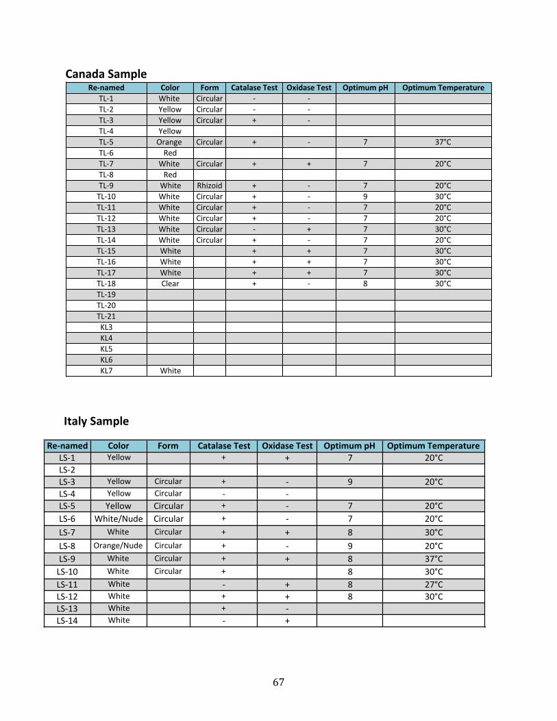
67
Re#named Color Form Catalase/Test Oxidase/Test Optimum/pH Optimum/TemperatureLS#1 Yellow *+* *+* 7 20°CLS#2LS#3 Yellow Circular *+* *#* 9 20°CLS#4 Yellow Circular *#* *#*LS#5 Yellow Circular *+* *#* 7 20°CLS#6 White/Nude Circular *+* *#* 7 20°CLS#7 White* Circular *+* *+* 8 30°CLS#8 Orange/Nude* Circular *+* *#* 9 20°CLS#9 White* Circular *+* *+* 8 37°CLS#10 White* Circular *+* 8 30°CLS#11 White *#* *+* 8 27°CLS#12 White *+* *+* 8 30°CLS#13 White *+* *#*LS#14 White *#* *+*
Canada%SampleRe,named Color Form Catalase%Test Oxidase%Test Optimum%pH Optimum%Temperature
TL#1 White* Circular *#* *#*TL#2 Yellow Circular *#* *#*TL#3 Yellow Circular *+* *#*TL#4 Yellow *TL#5 Orange* Circular *+* *#* 7 37°CTL#6 RedTL#7 White* Circular *+* *+* 7 20°CTL#8 RedTL#9 White Rhizoid *+* *#* 7 20°CTL#10 White* Circular *+* *#* 9 30°CTL#11 White* Circular *+* *#* 7 20°CTL#12 White* Circular *+* *#* 7 20°CTL#13 White* Circular *#* *+* 7 30°CTL#14 White* Circular *+* *#* 7 20°CTL#15 White *+* *+* 7 30°CTL#16 White *+* *+* 7 30°CTL#17 White *+* *+* 7 30°CTL#18 Clear *+* *#* 8 30°CTL#19TL#20TL#21KL3KL4KL5KL6KL7 White
Italy Sample

68
California)SamplesRe0named Color Form Catalase)Test Oxidase)Test Optimum)pH Optimum)Temperature Aerobic Anaerobic)
CR#1 Yellow Circular /+/ /#/ 7 37°CCR#2 White/ Circular /+/ /#/ 10 30°CCR#3 White/ Circular /+/ /#/ 9 30°CCR#4 White /+/ /#/ 9 20°CCR#5 White/ Circular /+/ /#/ 9 37°CCR#6 White Circular /+/ /#/ 7 37°CCR#7 White /+/ /+/ 7 30°CCR#8 Orange/ Circular /+/ /#/ 9 30°CCR#9 Orange Circular /+/ /#/ 9 37°CCR#10 Orange/ Circular /+/ /#/ 9 30°CCR#11 White/ Circular /#/ /+/ 7 30°CCR#12 Yellow Circular /+/ /#/ 7 37°CCR#13 White/ Circular /#/ /#/ 10 30°CCR#14 White Irregular /+/ /+/CR#15 Orange/Red Circular /+/ /#/CR#16 White /+/CR#17 White /+/ /+/CR#18 Yellow /+/ /#/ 7 30°CCR#19 White /#/ /+/ 7 20°CCR#20/ White/ /+/ /#/ 7 37°CCR#21 Black 7 20°CCR#22 White/ /+/ /+/CR#23 White /#/ /+/ 9 20°CCR#24 White /+/ /+/ 7 20°CCR#25 White /+/ /#/ 7 37°CCR#26 White /+/ /+/ 7 20°CCR#27 Yellow /+/ /#/ 10 30°CCR#28 White /+/ /+/ 7 20°CCR#29 Orange/White /+/ /#/ 10 30°CCR#30 Orange /+/ /#/ 7 37°CCR#31 Orange/White /+/ /#/ 7 30°CCR#32 Orange/White /+/ /#/ 7 30°CCR#33 White /#/ /+/ 2 20°CCR#34 White /+/ /#/CR#35 WhiteCR#36 White /#/ /+/ 7 20°CCR#37 White /+/ /+/ 8 20°CCR#38 White /#/ /+/ 7 20°CCR#39 White /#/ /+/ 7 20°CCR#40 White /+/ /+/ 8 30°CCR#41 White /+/ /+/ 8 30°CCR#42 Orange /+/ /#/ 7 20°CCR#43 Orange /+/ /+/ 7 37°CCR#44 White /+/ /+/ 7 37°CCR#45 Clear /#/ /#/ 9 37°CCR#46 White /+/ /+/ 7 37°CCR#47 Clear /#/ /+/ 9 37°CCR#48 Orange /+/ /+/ 10 20°C
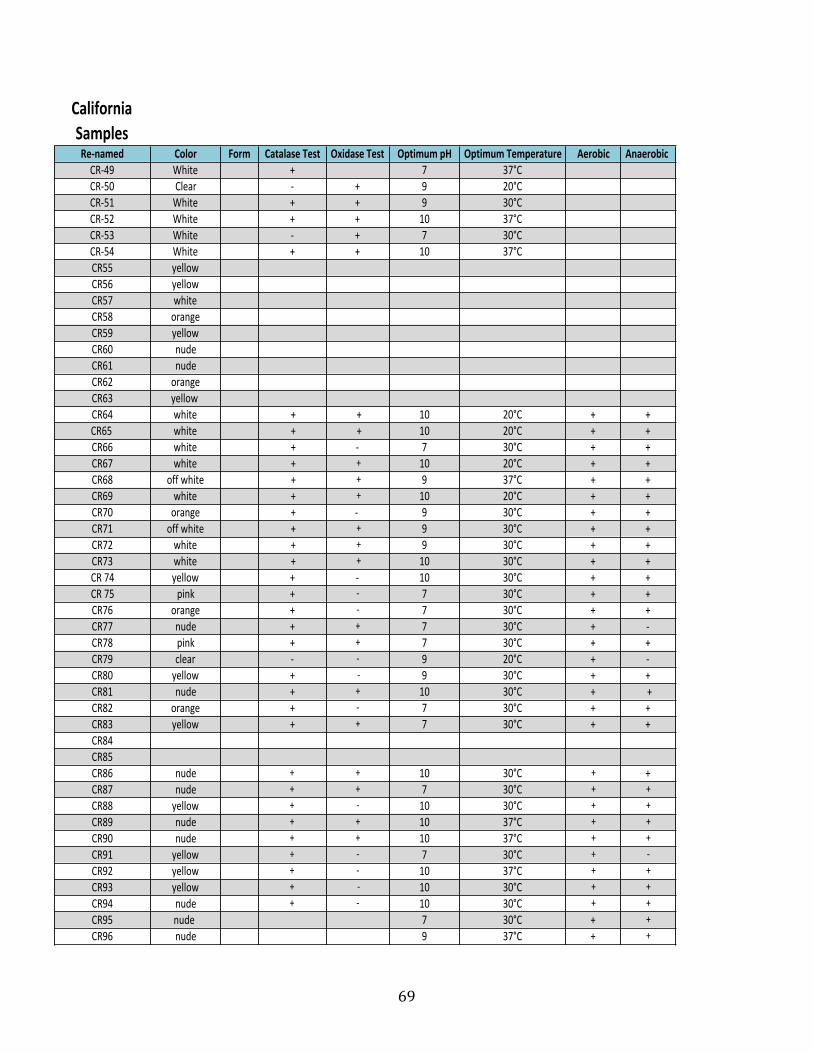
69
California)SamplesRe0named Color Form Catalase)Test Oxidase)Test Optimum)pH Optimum)Temperature Aerobic Anaerobic)CR#49 White +++ 7 37°CCR#50 Clear +#+ +++ 9 20°CCR#51 White +++ +++ 9 30°CCR#52 White +++ +++ 10 37°CCR#53 White +#+ +++ 7 30°CCR#54 White +++ +++ 10 37°CCR55 yellowCR56 yellowCR57 whiteCR58 orangeCR59 yellowCR60 nudeCR61 nudeCR62 orangeCR63 yellow+CR64 white ++ ++ 10 20°C +++ +++CR65+ white ++ ++ 10 20°C +++ +++CR66 white ++ +#+ 7 30°C +++ +++CR67 white ++ ++ 10 20°C +++ +++CR68 off+white ++ ++ 9 37°C +++ +++CR69 white ++ ++ 10 20°C +++ +++CR70 orange ++ +#++ 9 30°C +++ +++CR71 off+white ++ ++ 9 30°C +++ +++CR72 white ++ ++ 9 30°C +++ +++CR73 white ++ ++ 10 30°C +++ +++CR+74 yellow +++ +#+ 10 30°C +++ +++CR+75 pink +++ +#+ 7 30°C +++ +++CR76 orange +++ +#+ 7 30°C +++ +++CR77 nude +++ +++ 7 30°C +++ +#+CR78 pink +++ +++ 7 30°C +++ +++CR79 clear +#+ +#+ 9 20°C +++ +#+CR80 yellow +++ +# 9 30°C +++ +++CR81 nude +++ +++ 10 30°C +++ ++CR82 orange +++ +#+ 7 30°C +++ +++CR83 yellow +++ +++ 7 30°C +++ +++CR84CR85CR86 nude +++ +++ 10 30°C +++ +++CR87 nude +++ +++ 7 30°C +++ +++CR88 yellow +++ +#+ 10 30°C +++ +++CR89 nude +++ +++ 10 37°C +++ +++CR90 nude +++ +++ 10 37°C +++ +++CR91 yellow +++ +#+ 7 30°C +++ +#+CR92 yellow +++ +#+ 10 37°C +++ +++CR93 yellow +++ +# 10 30°C +++ +++CR94 nude +++ +#+ 10 30°C +++ +++CR95 nude+ 7 30°C +++ +++CR96 nude 9 37°C +++ +++

70