GFI - "Illicit Financial Flows fromDeveloping Countries Overthe Decade Ending 2009"

Proc. Natl. Acad. Sci. USAVol. 86, pp. 8665-8668, November 1989Biochemistry
Physical map of the genome of sonchus yellow net virus, a plantrhabdovirus with six genes and conserved gene junction sequences
(genome map of negative-strand virus/rhabdovirus evolution/rhabdovirus intergenic sequence)
Louis A. HEATON*, BRADLEY I. HILLMANt, BRENDA G. HUNTER, DOUWE ZUIDEMAt,AND ANDREW 0. JACKSON§Department of Plant Pathology, University of California, Berkeley, CA 94720
Communicated by Myron K. Brakke, August 14, 1989 (received for review April 27, 1989)
ABSTRACT We provide evidence that a plant rhabdovi-rus, sonchus yellow net virus (SYNV), is similar to most animalrhabdoviruses in the order of structural genes and in thenucleotide sequences at the gene junctions but that it differs inthe presence and location of a putative nonstructural gene.From the patterns of hybridization of a library of recombinantDNA clones, we have shown that the SYNV genome is tran-scribed into a short 3'-terminal "leader RNA" and six mRNAs.The proteins encoded by the SYNV mRNAs, in order of theappearance of their genes in the SYNV genome, are designated3'-N-M2-sc4-M1-G-L-5' (N, nucleoprotein; M, matrix protein;sc, protein encoded by SYNV complementary RNA; G, glyco-protein; L, large protein). The intergenic and flanking genesequences are conserved and consist of a central core of 14nucleotides (3'-UUCUUUUUGGUUGU/A-5') whose sequenceis similar to the sequence at the gene junctions of vesicularstomatitis and rabies viruses. The SYNV core consists of an8-nucleotide (3'-UUCUUUUU-5') transcription terminationsignal at the 5' terminus of each gene, a dinucleotide (GG)spacer whose complement does not appear in mRNA, and atetranucleotide (3'-UUGU/A-5') that is complementary to thefirst four nucleotides at the 5' terminus of the SYNV mRNAs.These results, when compared with structural informationavailable on animal rhabdoviruses, suggest that organization ofstructural genes and maintenance of signals thought to playimportant roles in regulation of transcription have been con-served during evolution in plant, insect, and vertebrate hosts.However, differences in number and location of putativenonstructural genes reveal some flexibility in genome organi-zation that may be important in deducing taxonomic andevolutionary relationships among viruses causing diseases inphylogenetically diverse hosts.
The Rhabdovirideae represent one of only two virus familieswith members that infect and cause serious diseases in bothplants and animals (1). The members of the family infect hoststhat are widely distributed throughout both kingdoms andhave been isolated from many species of plants, mammals,fish, insects, and other invertebrates (2, 3). The prototyperhabdovirus, vesicular stomatitis virus (VSV), has been thesubject of intensive investigation over the past 20 years and,as such, it has become a useful model that is now a standardagainst which the physicochemical properties of other rhab-doviruses are compared (4). Virions of VSV and otherrhabdoviruses are of a complex bullet-shaped or bacilliformmorphology and contain lipid (20%), carbohydrate (8%),RNA (3%), and five structural proteins. The RNAs of therhabdoviruses consist ofan 11- to 13-kilobase single-strandedRNA of negative-sense polarity. The five structural proteinsof VSV include a large protein (L), a glycoprotein (G), a
nucleoprotein (N), a matrix protein (M), and a phosphopro-tein (P), which for historical reasons is often designated NS(4). The structural proteins of other rhabdoviruses have beengiven similar designations, but they vary slightly in size andrelative abundance. Comparative analyses of the plant andanimal rhabdoviruses therefore provide rare opportunities tostudy the replication and evolution of viruses within a singlefamily that infect hosts in both the plant and animal king-doms.Sonchus yellow net virus (SYNV) has become the most
highly characterized of the plant rhabdoviruses (2, 5-8). Wehave previously identified five discrete SYNV-complemen-tary (sc) RNAs, or viral-encoded mRNAs, that were desig-nated scRNAs 1-5 and have postulated that these mRNAsencode the five structural proteins L, G, N, Ml, and M2,respectively (5). The putative locations of these proteinswithin the virion and their electrophoretic patterns (9) areconsistent with the nomenclature proposed for rhabdoviruses(10), but the genome organization and sequences at thejunctions of all of the genes have not previously beendetermined.
In contrast to SYNV and other plant rhabdoviruses, thegenomes of three animal rhabdoviruses have been mapped,and VSV and rabies-RNAs have been subjected to detailednucleotide sequence analyses. More than a decade ago,Abraham and Banerjee (11) and Ball and White (12) deter-mined that the gene order of VSV is 3'-N-P(NS)-M-G-L-5'.Subsequently, elucidation of the complete primary structureof the VSV (IND) genome (13-16) verified the gene order andconfirmed the presence of five genes, although a previouslyunidentified protein product has recently been shown to beexpressed from the phosphoprotein mRNA by a uniquemechanism (17). Variation in the number ofgenes encoded byrhabdovirus genomes was first demonstrated by the findingthat six discrete mRNAs are transcribed by infectious hem-atopoietic necrosis virus (IHNV), a fish rhabdovirus (18, 19).Five of the proteins are structural and the sixth is nonvirion(NV), since it is induced in infected cells but has not beendetected in mature virions. Further differences among rhab-doviruses were revealed by analysis of the rabies virusgenome, which is composed of five structural genes but alsocontains a long intergenic sequence between the G and Lgenes (20). Thus, from the analyses of a relatively few
Abbreviations: VSV, vesicular stomatitis virus; SYNV, sonchusyellow net virus; scRNA, SYNV complementary RNA; IHNV,infectious hematopoietic necrosis virus; NV, nonvirion; gcDNA,genomic cDNA.*Present address: Department of Plant Pathology, ThrockmortonHall, Kansas State University, Manhattan, KS 66056.
tPresent address: Department of Plant Pathology, Cook College,Rutgers University, New Brunswick, NJ 08903.tPresent address: Department of Virology, Agricultural University,Binnenhaven 11, 6709 PD Wageningen, The Netherlands.§To whom reprint requests should be addressed.
8665
The publication costs of this article were defrayed in part by page chargepayment. This article must therefore be hereby marked "advertisement"in accordance with 18 U.S.C. §1734 solely to indicate this fact.
Proc. Natl. Acad. Sci. USA 86 (1989)
rhabdoviruses, it has become evident that members of thefamily vary in the number of genes and in their organization.
Despite the variation in genome organization, the nucleo-tide sequences at the intergenic and flanking gene regions oftwo animal rhabdoviruses, VSV and rabies virus, are strik-ingly similar. VSV (IND) contains a highly conserved 23-nucleotide gene junction consensus sequence (21), and anearly identical, although slightly more variable, consensus ispresent at the rabies virus gene junctions (20). Because theseputative regulatory signals at the gene junctions of theseviruses are so highly conserved and because the number ofmRNAs transcribed from different animal rhabdovirus ge-nomes such as VSV and IHNV differ, we have extended ouranalyses of the organization and expression of the SYNVgenome. We have determined the gene order of SYNV,detected six mRNA species encoded by the SYNV genome,and sequenced the intergenic and flanking regions at eachgene junction. The results now permit us to compare theorganization of a plant rhabdovirus genome with those of theanimal rhabdoviruses.
MATERIALS AND METHODSGeneral Procedures. The procedures used for SYNV
(ATCC PV-263) propagation, maintenance, purification, andRNA extraction have been described (22, 23). Isolation ofpoly(A) RNA and hybridization analyses were as outlined byRezaian et al. (5). Construction of cDNA libraries fromSYNV RNA and from the poly(A) fraction of SYNV-infectedtobacco plants and methods used for nucleic acid sequenceanalysis were as described by Zuidema et al. (7).
Identification of cDNA Clones. Approximately 4800 ampi-cillin-resistant colonies with cDNA inserts derived from thepoly(A) RNA of infected tobacco leaves were hybridizedwith randomly labeled SYNV genomic RNA. Plasmids pu-rified from 50 positive colonies were arbitrarily designatedpAS 1-50. The inserts were found to range from 300 to 1500base pairs and were grouped according to scRNA specificityby screening with recombinant plasmids that hybridized tospecific scRNAs (5). The largest recombinant plasmid fromeach scRNA specificity group was chosen to screen thegenomic cDNA (gcDNA) library described below.A gcDNA library was constructed essentially as described
for the pAS library except that purified virion RNA was usedas a template and reverse transcription was accomplished viarandom self-priming (13). Approximately 600 ampicillin-resistant colonies were probed with randomly labeled SYNVgenomic RNA, and the plasmids purified from the 260positive colonies were arbitrarily designated pGL 1-260. Theinserts in these plasmids varied from 500 to 1800 base pairs.One hundred of the newly constructed gcDNA clones weregrouped according to scRNA specificity by screening withappropriate inserts from the pAS library.Mapping Genes Encoding the scRNAs. To differentiate the
scRNAs on RNA blots and to determine the order of geneson the SYNV genome, selected gcDNA plasmids were nick-translated and used as hybridization probes of blots ofpoly(A) RNA from SYNV-infected tobacco leaves. If a singlegcDNA plasmid hybridized to two scRNAs, then the plasmidcontained intergenic and flanking gene sequences, and thegenes encoding the two scRNAs were concluded to beadjacent. Such plasmids were called "gene junction" clones.Other plasmids that hybridized to a single scRNA wereassigned to the corresponding scRNA specificity group. Anoverlapping group of the cDNA and gcDNA inserts wasobtained by DNA blot hybridization by using appropriateplasmid probes to identify inserts derived from adjacentregions of the genome. With this strategy, we aligned a set ofoverlapping inserts that represented SYNV RNA from the 3'terminus into the L gene.
RESULTS AND DISCUSSIONThe order of genes that encode the scRNAs, as determinedfrom the hybridization patterns of SYNV-specific probes, isshown in Fig. 1. The cumulative sizes of the cDNA insertsand their hybridization to individual scRNAs constitute ev-idence that six distinct mRNAs are transcribed from theSYNV genome instead of the previously estimated five (5).The additional scRNA, which we now designate scRNA 6,moves only slightly in advance of scRNAs 4 and 5 in agarosegels tFig. 1), and the difficulties in resolving this RNAcontributed to our failure to identify it previously. Ourpresent results with five gcDNA plasmids, each of whichhybridize to two scRNAs, show that scRNAs 3 and 5, 5 and4, 4 and 6, 6 and 2, and 2 and 1, respectively, are encoded byadjacent genes (Fig. 1). These conclusions have been verifiedby sequence analyses of individual genes (data to be pre-sented elsewhere). The order of the RNAs relative to theSYNV genome is 3'-scRNA3-scRNA5-scRNA4-scRNA6-scRNA2-scRNA1-5'.Assignment of Proteins Encoded by the scRNAs. We (6) have
previously shown that the 144-nucleotide "leader RNA"gene ("I") is located at the 3' terminus of the SYNV genomicRNA and precedes the nucleocapsid protein gene from whichscRNA 3 is transcribed (7). The mRNA, scRNA 5, which istranscribed from the next downstream gene, encodes the M2protein, which has limited amino acid sequence relatednessto phosphoproteins of other rhabdoviruses (8). Identificationof both the scRNA 3 and scRNA 5 gene products wasobtained by protein blot analyses with antisera elicited byfusion proteins constructed from sequenced cDNA that wasderived from the respective scRNA (7, 8). Based on ourunpublished data from similar serological experiments,scRNA 6 is the mRNA for the Ml protein, which we believeis the matrix protein. We have been unable to detect astructural polypeptide encoded by scRNA 4 and thereforeprovisionally designate its putative translation product"sc4." We have not yet determined whether sc4 is a non-structural polypeptide-that is synthesized at a specific stageof replication. From comparisons of their coding capacitieswith the molecular weights of their putative protein productsand from relatedness of amino acid sequence deduced fromnucleotide sequence data, scRNAs 1 and 2 encode the L andG proteins, respectively (unpublished data). Therefore, the
N %I, (1 NI I G; Id
scLIM2Ml,
FIG. 1. Construction of a genome map for SYNV. SelectedSYNV cDNA clones derived from genomic RNA or poly(A) RNAwere nick-translated and used to probe blots of poly(A) RNA fromSYNV-infected tobacco leaves. The gene order was deduced fromthe hybridization patterns of overlapping cDNA clones. The lanes tothe far left and far right were probed with randomly labeled SYNVgenomic RNA and serve as markers for SYNV mRNAs. The far rightlane was overexposed to reveal the L protein mRNA, which is presentat -5-fold lower abundance than scRNAs 2-6. L, scRNA 1; G, scRNA2; N, scRNA 3; sc 4, scRNA 4; M2, scRNA 5; Ml, scRNA 6.
8666 Biochemistry: Heaton et al.
I
Proc. Natl. Acad. Sci. USA 86 (1989) 8667
gene order of SYNV is 3'-"l"-N-M2(P?)-sc4-M1(M?)-G-L-5'.Comparison of our results with those previously published
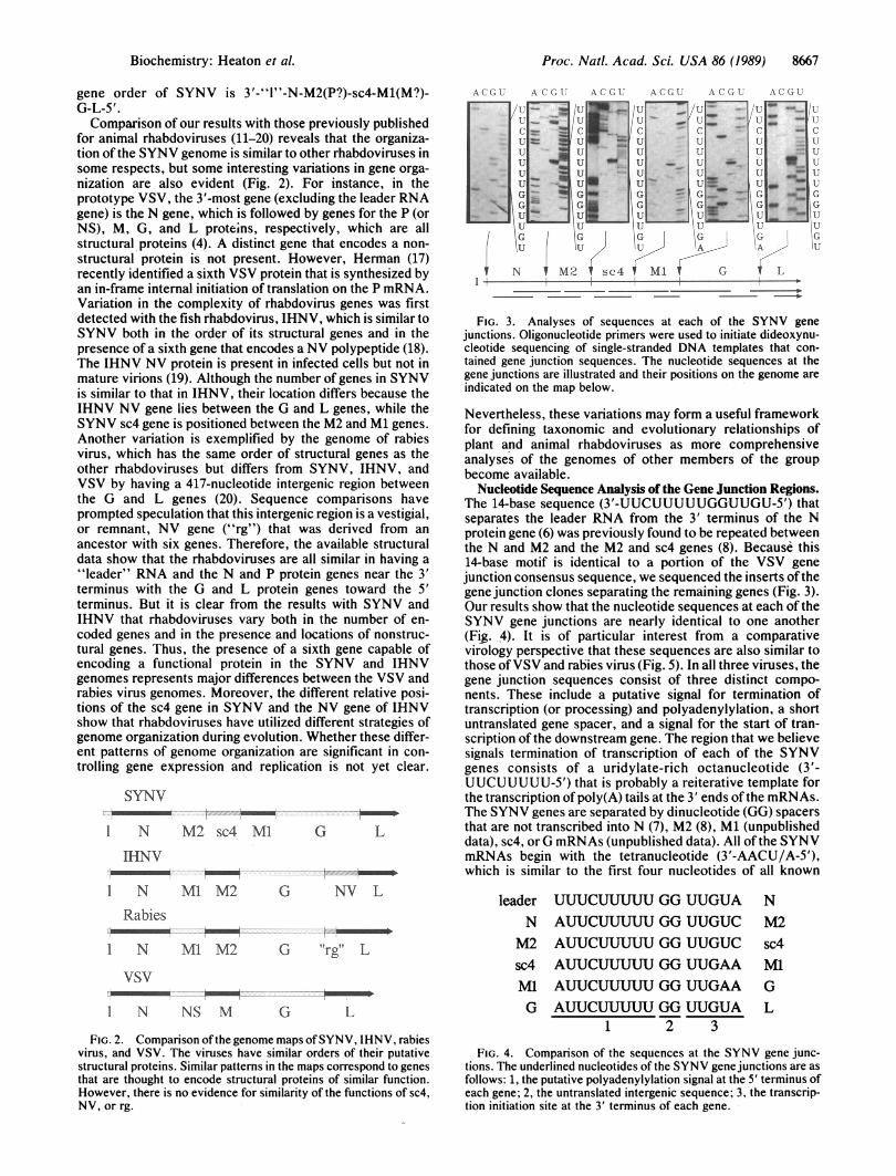
for animal rhabdoviruses (11-20) reveals that the organiza-tion of the SYNV genome is similar to other rhabdoviruses insome respects, but some interesting variations in gene orga-nization are also evident (Fig. 2). For instance, in theprototype VSV, the 3'-most gene (excluding the leader RNAgene) is the N gene, which is followed by genes for the P (orNS), M, G, and L proteins, respectively, which are allstructural proteins (4). A distinct gene that encodes a non-structural protein is not present. However, Herman (17)recently identified a sixth VSV protein that is synthesized byan in-frame internal initiation of translation on the P mRNA.Variation in the complexity of rhabdovirus genes was firstdetected with the fish rhabdovirus, IHNV, which is similar toSYNV both in the order of its structural genes and in thepresence of a sixth gene that encodes a NV polypeptide (18).The IHNV NV protein is present in infected cells but not inmature virions (19). Although the number of genes in SYNVis similar to that in IHNV, their location differs because theIHNV NV gene lies between the G and L genes, while theSYNV sc4 gene is positioned between the M2 and Ml genes.Another variation is exemplified by the genome of rabiesvirus, which has the same order of structural genes as theother rhabdoviruses but differs from SYNV, IHNV, andVSV by having a 417-nucleotide intergenic region betweenthe G and L genes (20). Sequence comparisons haveprompted speculation that this intergenic region is a vestigial,or remnant, NV gene ("rg") that was derived from anancestor with six genes. Therefore, the available structuraldata show that the rhabdoviruses are all similar in having a"leader" RNA and the N and P protein genes near the 3'terminus with the G and L protein genes toward the 5'terminus. But it is clear from the results with SYNV andIHNV that rhabdoviruses vary both in the number of en-coded genes and in the presence and locations of nonstruc-tural genes. Thus, the presence of a sixth gene capable ofencoding a functional protein in the SYNV and IHNVgenomes represents major differences between the VSV andrabies virus genomes. Moreover, the different relative posi-tions of the sc4 gene in SYNV and the NV gene of IHNVshow that rhabdoviruses have utilized different strategies ofgenome organization during evolution. Whether these differ-ent patterns of genome organization are significant in con-trolling gene expression and replication is not yet clear.
SYNV., _ ....... ....~~~~~~~~~~~.............
1 N M 2sc4 Ml G L
IHNV-. --.
I N M1 M2 G NV L
Rabies-_---1 N M1 M2 6 '"rg"lL
vsV
1 N NS M G L
FIG. 2. Comparison of the genome maps ofSYNV, IHNV, rabiesvirus, and VSV. The viruses have similar orders of their putativestructural proteins. Similar patterns in the maps correspond to genesthat are thought to encode structural proteins of similar function.However, there is no evidence for similarity of the functions of sc4,NV, or rg.
:
I1-
U
C-UuuuuGGuu|GU
U
A C G; I
_
_ _
A (C G
L: r
lu -UU
GII
C.
u .....u
IGUU
UG
2 1 sc4
A (1U
cU
U -U
'UiUMG'M1
G
;i
cC-
uu
| GI~&
1U
U
A (;GU
=.
GA-
I G
AC -..
3- _U =
u _U
G VW
. _-
C;
IL
U
UG,u1='&
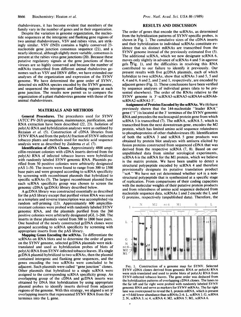
FIG. 3. Analyses of sequences at each of the SYNV genejunctions. Oligonucleotide primers were used to initiate dideoxynu-cleotide sequencing of single-stranded DNA templates that con-tained gene junction sequences. The nucleotide sequences at thegene junctions are illustrated and their positions on the genome areindicated on the map below.
Nevertheless, these variations may form a useful frameworkfor defining taxonomic and evolutionary relationships ofplant and animal rhabdoviruses as more comprehensiveanalyses of the genomes of other members of the groupbecome available.
Nucleotide Sequence Analysis of the Gene Junction Regions.The 14-base sequence (3'-UUCUUUUUGGUUGU-5') thatseparates the leader RNA from the 3' terminus of the Nprotein gene (6) was previously found to be repeated betweenthe N and M2 and the M2 and sc4 genes (8). Because this14-base motif is identical to a portion of the VSV genejunction consensus sequence, we sequenced the inserts of thegene junction clones separating the remaining genes (Fig. 3).Our results show that the nucleotide sequences at each of theSYNV gene junctions are nearly identical to one another(Fig. 4). It is of particular interest from a comparativevirology perspective that these sequences are also similar tothose ofVSV and rabies virus (Fig. 5). In all three viruses, thegene junction sequences consist of three distinct compo-nents. These include a putative signal for termination oftranscription (or processing) and polyadenylylation, a shortuntranslated gene spacer, and a signal for the start of tran-scription of the downstream gene. The region that we believesignals termination of transcription of each of the SYNVgenes consists of a uridylate-rich octanucleotide (3'-UUCUUUUU-5') that is probably a reiterative template forthe transcription of poly(A) tails at the 3' ends of the mRNAs.The SYNV genes are separated by dinucleotide (GG) spacersthat are not transcribed into N (7), M2 (8), Ml (unpublisheddata), sc4, or G mRNAs (unpublished data). All of the SYNVmRNAs begin with the tetranucleotide (3'-AACU/A-5'),which is similar to the first four nucleotides of all known
leader UUUCUUUUU GG UUGUA NN AUUCUUUUU GG UUGUC M2M2 AUUCUUUUU GG UUGUC sc4sc4 AUUCUUUUU GG UUGAA MlMl AUUCUUUUU GG UUGAAG AUUCUUUUU GG UUGUA
1 2 3
GL
FIG. 4. Comparison of the sequences at the SYNV gene junc-tions. The underlined nucleotides of the SYNV gene junctions are asfollows: 1, the putative polyadenylylation signal at the 5' terminus ofeach gene; 2, the untranslated intergenic sequence; 3, the transcrip-tion initiation site at the 3' terminus of each gene.
Biochemistry: Heaton et al.
Proc. Natl. Acad. Sci. USA 86 (1989)
SYNV AUUCUUUUU G G UUGAA
VSV ACUUUUUUU C U UUGUCRiGWUC( GRabies ACUUUUUUU C (N) UUGUAG
1 2 3
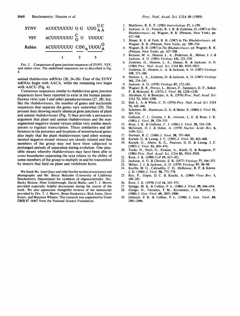
FIG. 5. Comparison of gene junction sequences of SYNV, VSV,and rabies virus. The underlined sequences are as described in Fig.4.
animal rhabdovirus mRNAs (20, 24-26). Four of the SYNVmRNAs begin with AACA, while the remaining two beginwith AACU (Fig. 4).Consensus sequences similar to rhabdovirus gene junction
sequences have been reported to exist in the human parain-fluenza virus type 3 and other paramyxoviruses (27, 28), but,like the rhabdoviruses, the number of genes and nucleotidesequences that separate the genes vary somewhat (29). Thepresent data showing nearly identical gene junctions of plantand animal rhabdoviruses (Fig. 5) thus provide a persuasiveargument that plant and animal rhabdoviruses and the non-segmented negative-strand viruses utilize very similar mech-anisms to regulate transcription. These similarities and dif-ferences in the presence and locations of nonstructural genesalso imply that the plant rhabdoviruses (and other nonseg-mented negative-strand viruses) are closely related and thatmembers of the group may not have been subjected toprolonged periods of separation during evolution. One plau-sible means whereby rhabdoviruses may have been able tocross boundaries separating the taxa relates to the ability ofsome members of the group to multiply in and be transmittedby insects that feed on plant and vertebrate hcsts.
We thank Ms. Joan Quay and Julie Sun for technical assistance andphotography and Mr. Bruce Malcolm (University of CaliforniaBiochemistry Department) for synthesis of oligonucleotides. Drs.Becky Boston, Peter Goldsbrough, David Marks, and T. J. Morrisprovided especially helpful discussions during the course of thework. We also appreciate thoughtful reviews of the manuscriptprovided by Drs. T. J. Morris, Brian Staskawicz, Rick Jones, DaveKnorr, and Maureen Whalen. This research was supported by GrantDMB-87 16467 from the National Science Foundation.
1. Matthews, R. E. F. (1982) Intervirology 17, 1-199.2. Jackson, A. O., Francki, R. 1. B. & Zuidema, D. (1987) in The
Rhabdoviruses, ed. Wagner, R. R. (Plenum, New York), pp.427-508.
3. Shope, R. E. & Tesh, R. B. (1987) in The Rhabdoviruses, ed.Wagner, R. R. (Plenum, New York), pp. 509-534.
4. Wagner, R. R. (1987) in The Rhabdoviruses, ed. Wagner, R. R.(Plenum, New York), pp. 427-508.
5. Rezaian, M. A., Heaton, L. A., Pederson, K., Milner, J. J. &Jackson, A. 0. (1983) Virology 131, 221-229.
6. Zuidema, D., Heaton, L. A., Hanau, R. & Jackson, A. 0.(1986) Proc. Natl. Acad. Sci. USA 83, 5019-5023.
7. Zuidema, D., Heaton, L. A. & Jackson, A. 0. (1987) Virology159, 373-380.
8. Heaton, L. A., Zuidema, D. & Jackson, A. 0. (1987) Virology161, 234-241.
9. Jackson, A. 0. (1978) Virology 87, 172-181.10. Wagner, R. R., Prevec, L., Brown, F., Summers, D. F., Sokol,
F. & Mcleoud, R. (1972) J. Virol. 10, 1228-1230.11. Abraham, G. & Baneijee, A. K. (1976) Proc. Natl. Acad. Sci.
USA 73, 1054-1508.12. Ball, L. A. & White, C. N. (1976) Proc. Natl. Acad. Sci. USA
73, 442-446.13. Schubert, M., Harmison, G. G. & Meier, E. (1984)J. Virol. 51,
505-514.14. Gallione, C. J., Greene, J. R., Iverson, L. E. & Rose, J. K.
(1981) J. Virol. 39, 529-535.15. Rose, J. K. & Gallione, C. J. (1981) J. Virol. 39, 519-528.16. McGeoch, D. J. & Dolan, A. (1979) Nucleic Acids Res. 6,
3199-3211.17. Herman, R. C. (1986) J. Virol. 58, 797-804.18. Kurath, G. & Leong, J. C. (1985) J. Virol. 53, 462-468.19. Kurath, G., Ahern, K. G., Pearson, G. D. & Leong, J. C.
(1985) J. Virol. 53, 469-476.20. Tordo, N., Poch, O., Ermine, A., Kieth, G. & Rougeon, F.
(1986) Proc. Natl. Acad. Sci. USA 83, 3914-3918.21. Rose, J. K. (1980) Cell 19, 415-421.22. Jackson, A. 0. & Christie, S. R. (1977) Virology 77, 344-355.23. Milner, J. J. & Jackson, A. 0. (1979) Virology 97, 90-99.24. Kurilla, M. G., Cabradilla, C. D., Holloway, B. P. & Keene,
J. D. (1984) J. Virol. 50, 773-778.25. Roy, P., Gupta, D. C. & Kiuchi, A. (1984) Virus Res. 1,
189-202.26. Rose, J. K. (1978) Cell 14, 345-353.27. Spriggs, M. K. & Collins, P. L. (1986) J. Virol. 59, 646-654.28. Elango, N., Varsanyi, T. M., Kovamees, J. & Norrby, E.
(1988) J. Gen. Virol. 69, 2893-2900.29. Johnson, P. R. & Collins, P. L. (1988) J. Gen. Virol. 69,
2901-2906.
8668 Biochemistry: Heaton et al.