PHYLOGENY AND MOLECULAR IDENTIFICATION OF
97
PHYLOGENY AND MOLECULAR IDENTIFICATION OF CRONOBACTER STRAINS ISOLATED FROM SOUTH AFRICAN FOOD PRODUCTS AMY STRYDOM Thesis presented in partial fulfilment of the requirements for the degree of MASTER OF SCIENCE IN FOOD SCIENCE at the Stellenbosch University Department of Food Science Faculty of AgriSciences Study Leader: Prof. R.C. Witthuhn Co-study Leader: Dr. M. Cameron March 2011
Transcript of PHYLOGENY AND MOLECULAR IDENTIFICATION OF
PHYLOGENY AND MOLECULAR IDENTIFICATION OF CRONOBACTER STRAINS
ISOLATED FROM SOUTH AFRICAN FOOD PRODUCTS
AMY STRYDOM
Thesis presented in partial fulfilment of the requirements for the degree of
MASTER OF SCIENCE IN FOOD SCIENCE
at the Stellenbosch University
Department of Food Science
Faculty of AgriSciences
Study Leader: Prof. R.C. Witthuhn
Co-study Leader: Dr. M. Cameron
March 2011
ii
DECLARATION
By submitting this thesis electronically, I declare that the entirety of the work contained
therein is my own, original work, that I am the sole author thereof (save to the extent
explicitly otherwise stated), that reproduction and publication thereof by Stellenbosch
University will not infringe any third party rights and that I have not previously in its entirety
or in part submitted it for obtaining any qualification.
---------------------------
Amy Strydom
---------------------------
Date
Copyright © 2011 Stellenbosch University
iii
ABSTRACT
The genus Cronobacter (Enterobacter sakazakii) contains opportunistic pathogens that can
cause a severe form of neonatal meningitis, necrotising enterocolitis and septicaemia.
Cronobacter infections have been reported in all age groups, however, immuno-
compromised infants are more susceptible to these infections. Furthermore, Cronobacter
strains have been reported to show differences in sensitivity to antibiotics and virulence.
These differences led to the reclassification of Cronobacter and currently the genus
contains five distinct species, namely Cronobacter sakazakii, Cronobacter malonaticus,
Cronobacter turicensis, Cronobacter dublinensis and Cronobacter muytjensii. As this
reclassification was only accepted recently, there are not many typing methods optimised
for differentiation between the five Cronobacter species. Typing of Cronobacter strains are
important as the species may be diverse regarding their virulence.
Cronobacter strains have been isolated from infant formula milk (IFM), the
environment of an IFM processing facility and fresh produce in South Africa. However,
little is known about the phylogeny and prevalence of these strains. The aim of this study
was to classify 24 South African Cronobacter strains (previously identified as E. sakazakii)
and to evaluate the phylogeny of the isolates based on the 16S ribosomal RNA (rRNA) and
rpoA genes. All 24 South African strains were identified as Cr. sakazakii despite a wide
variety of isolation sources. Other studies have also found that irrespective of the isolation
source, the majority of Cronobacter strains are identified as Cr. sakazakii. The South
African strains were found to be phylogenetically closely related. However, two distinct
clusters separated at a 93 % confidence level were observed in the Cr. sakazakii group
based on the 16S rRNA gene analysis.
Strains of Cr. sakazakii, Cr. dublinensis, Cr. turicensis and Cr. muytjensii were
differentiated from each other with sequence data of the 16S rRNA and rpoA genes, but it
was not possible to differentiate between Cr. sakazakii and Cr. malonaticus. The
phylogram based on the rpoA gene sequences did separate Cr. malonaticus and Cr.
sakazakii strains, however, the clusters were separated with a low bootstrap value of 70 %.
Phylogenetic analysis based on the rpoA and 16S rRNA genes were, therefore, not
sufficient to distinguish between all the Cronobacter species. The sequence data of these
two genes can be used to differentiate between the Cronobacter strains when used in
combination with malonate utilisation analysis.
iv
A PCR-RFLP method was subsequently developed to facilitate the simultaneous
differentiation between all five Cronobacter species. The PCR-RFLP assay was based on
the amplification of the rpoB gene followed by the combined digestion with restriction
endonucleases Csp6I and HinP1I. Unique profiles for each of the five Cronobacter species
were obtained and it was also possible to differentiate between Enterobacteriaceae and
Cronobacter strains. Furthermore, two strains which were identified as Cr. sakazakii with
sequencing based on the 16S rRNA and rpoA genes had PCR-RFLP profiles identical to
that of Cr. malonaticus. Sequencing based on the rpoB gene and additional biochemical
analysis with malonate broth confirmed the identities of these two strains as Cr.
malonaticus. This PCR-RFLP assay is, therefore, an accurate typing method that ensures
rapid differentiation between the five species of Cronobacter.
v
OPSOMMING
Die Cronobacter genus (Enterobacter sakazakii) bevat opportunistiese patogene wat 'n
ernstige vorm van neonatale meningitis, enterokolitis en septisemie kan veroorsaak.
Cronobacter infeksies is al in alle ouderdomsgroepe aangemeld, maar
immuungekompromitteerde babas is die meeste vatbaar vir hierdie infeksies. Verder toon
Cronobacter spesies verskille in virulensie en sensitiwiteit vir antibiotika. Hierdie verskille
het gelei tot die herklassifikasie van Cronobacter en tans bestaan die genus uit vyf
afsonderlike spesies, naamlik Cronobacter sakazakii, Cronobacter malonaticus,
Cronobacter turicensis, Cronobacter dublinensis en Cronobacter muytjensii. Aangesien
hierdie herklassifikasie slegs onlangs aanvaar is, is daar nie baie metodes wat geskik is vir
onderskeiding tussen die vyf Cronobacter spesies nie. Onderskeiding tussen Cronobacter
spesies is belangrik omdat die spesies verskillend kan wees met betrekking tot hulle
virulensie.
Cronobacter is geisoleer uit baba formule melk (BFM), die omgewing van 'n BFM
fabriek en vars produkte in Suid-Afrika. Daar is egter nie baie bekend oor die filogenie en
voorkoms van hierdie isolate nie. Die doel van hierdie studie was om 24 Suid-Afrikaanse
Cronobacter stamme (voorheen geïdentifiseer as E. sakazakii) te klassifiseer en die
filogenie van die isolate te evalueer gebaseer op die 16S ribosomale RNS (rRNS) en rpoA
gene. Al 24 Suid-Afrikaanse stamme is geïdentifiseer as Cr. sakazakii ten spyte van 'n
wye verskeidenheid isolasie bronne. Ander studies het ook gevind dat, ongeag die isolasie
bron, die meerderheid van Cronobacter stamme as Cr. sakazakii geïdentifiseer word. In
hierdie studie is gevind dat die Suid-Afrikaanse stamme filogeneties nou verwant is. Op
grond van die 16S rRNA geen analise is die Cr. sakazakii stamme egter in twee
afsonderlike groepe gedeel met 'n 93% vertrouens vlak.
Dit was moontlik om stamme van Cr. sakazakii, Cr. dublinensis, Cr. turicensis en Cr.
muytjensii van mekaar te onderskei met die DNS volgorde data van die 16S rRNA en rpoA
gene, maar geen onderskeid tussen Cr. sakazakii en Cr. malonaticus stamme was
moontlik nie. Die filogram gebaseer op die rpoA DNS volgorde data het aparte takke vir Cr.
malonaticus en Cr. sakazakii stamme getoon, maar die twee takke is met ‘n lae vertrouens
waarde van slegs 70 % geskei. Filogenetiese analise gebaseer op die rpoA en 16S rRNA
gene is dus nie voldoende om te onderskei tussen al die Cronobacter spesies nie. Die
DNS volgorde data van hierdie twee gene sou egter gebruik kon word om te onderskei
vi
tussen die Cronobacter spesies wanneer dit gebruik word in kombinasie met
malonaatbenutting-analises.
'n Polimerase ketting reaksie (PKR) beperkings fragment lengte polimorfisme
(BFLP) metode is ontwikkel om die gelyktydige onderskeiding tussen al vyf Cronobacter
spesies te fasiliteer. Die PKR-BFLP metode is gebaseer op die vermeerdering van die
rpoB geen gevolg deur die gesamentlike vertering met die beperkingsensieme, Csp6I en
HinP1I. Unieke profiele vir elk van die vyf Cronobacter spesies is verkry en dit was ook
moontlik om tussen Enterobacteriaceae en Cronobacter spesies te onderskei. Verder het
twee stamme wat as Cr. sakazakii geïdentifiseer is met DNS volgordebepaling van die 16S
rRNA en rpoA gene, PKR-BFLP profiele identies aan dié van Cr. malonaticus getoon.
DNS volgordebepaling van die rpoB geen en ‘n addisionele biochemiese toets met
malonaat sop het die identiteit van hierdie twee stamme as Cr. malonaticus bevestig.
Hierdie PKR-BFLP is dus 'n akkurate metode wat vinnige onderskeid tussen die vyf
spesies van Cronobacter kan verseker.
vii
CONTENTS
Chapter Page
Declaration ii
Abstract iii
Uittreksel v
Acknowledgements viii
1 Introduction 1
2 Literature review 6
3 Phylogenetic analysis of Cronobacter isolates based on the 40
rpoA and 16S rRNA genes
4 PCR-RFLP analysis of the rpoB gene to distinguish between the 60
five Cronobacter species
5 General discussion and conclusions 84
Language and style used in this thesis are in accordance with the requirements of the
International Journal of Food Science and Technology. This thesis represents a
compilation of manuscripts where each chapter is an individual entity and some repetition
between chapters has, therefore, been unavoidable.

viii
ACKNOWLEDGEMENTS
I would like to express my sincere gratitude to the following individuals and institutions for
their valuable contributions to the successful completion of this research:
My study leaders, Prof. R.C. Witthuhn and Dr. M. Cameron, for their expert guidance,
knowledge, support, positive criticism, skilled practical assistance and general
awesomeness during this study, I loved working with you;
SAMPRO, FoodBev Seta, Milk SA, CGCSA, the University of Stellenbosch and the Ernst
and Ethel Eriksen Trust for financial support;
Prof K. Jacobs for teaching me the art of drawing a tree, who knew it would be that
complicated?;
Donna Cawthorn for advice and interest in my project, even when you hated being
reminded of Cronobacter;
My fellow post-graduate students (especially the mol lab) for their friendship of six years, I
will dearly miss you next year;
Dr. N. Brown, Mrs A. Lombaard and Mrs D. du Preez for always lending a helping hand
and valuable advice, you are not appreciated enough;
The civilians: my parents; Alwyn and Elna Strydom, family and my close friends, Bernice,
Caty, Cecilia, Evette, Ilona, Ilze, Mia and Nola, for their unconditional love and support,
even when they did not understand what I was doing;
My Heavenly Father for giving me the aptitude, patience and strength to succeed.

ix
Dit is die Here wat wysheid gee, uit sy mond kom die kennis en die insig.
Spreuke 2
for the glory of the Lord
1
CHAPTER 1
INTRODUCTION
Microbial foodborne diseases pose a considerable threat to human health and are a
concern for food legislators, food manufacturers and consumers. The number of
individuals that are highly susceptible to these foodborne diseases is increasing due to the
ageing population and high numbers of HIV/AIDS infections. Furthermore, infants and
children in developing countries are particularly vulnerable to foodborne infections due to
reduced immunity caused by malnutrition (WHO, 2001; 2002).
Cronobacter (previously known as Enterobacter sakazakii) is an opportunistic
pathogen that can cause neonatal meningitis, necrotizing enterocolitis and septicemia.
These bacteria received increased attention as foodborne pathogens after an outbreak of
meningitis in Tennessee in 2001 (Iversen & Forsythe, 2003). Urmenyi & Franklin (1961)
reported on the first incidence of Cronobacter infections of two fatal cases of neonatal
meningitis that occurred in England. The bacteria that caused these infections were
referred to as yellow pigmented Enterobacter cloacae. Shortly thereafter the first evidence
of a separate species was based on DNA analysis that indicated the yellow pigmented
strains were less than 50 % related to other E. cloacae strains (Brenner, 1974).
Enterobacter sakazakii was proposed as the new name for these isolates, although it was
suggested that the isolates may comprise of more than one species. In 2008 E. sakazakii
isolates were reclassified in the novel genus, Cronobacter. Currently there are five distinct
species in the genus, namely Cronobacter sakazakii, Cronobacter malonaticus,
Cronobacter turicensis, Cronobacter dublinensis and Cronobacter muytjensii (Iversen et
al., 2007; 2008).
Many characteristics of these pathogens are strain dependant and differences in
specifically virulence have been documented. Only Cronobacter species associated with
neonatal infections, namely Cr. sakazakii, Cr. malonaticus and Cr. turicensis have the
genes encoding for a cation efflux system which allows bacteria to invade brain
microvascular endothelial cells. Cronobacter muytjensii has been isolated from human
bone marrow which would normally be sterile (Kucerova et al., 2010). As it is still unclear
whether all of the species are virulent, the genus is currently classified as pathogenic
(FAO/WHO, 2008).
2
The original reservoir of Cronobacter is still unknown but there are indications that
these pathogens might be of plant origin. Cronobacter strains have been isolated from
various food products such as mixed salad vegetables, meat, milk and cheese (Kahn et al.,
1998; Iversen & Forsythe, 2003; Kim & Beuchat, 2005; Beuchat et al., 2009; El Sharoud et
al., 2009). However, infant formula milk (IFM) is the only source that has been
epidemiologically linked to disease outbreaks caused by Cronobacter (Muytjens et al.,
1983; Pagotto & Farber, 2009). The risk of Cronobacter contamination is further increased
as it has been reported that regularly used disinfectants are insufficient to kill Cronobacter
cells imbedded in biofilms Kim et al., 2007) and that some strains survive refrigeration
temperatures (Nazarowec-White et al., 1997).
Low contamination levels (1 cfu.100 g-1) of Cronobacter can have a severe impact
on health and the rapid detection and correct identification of these pathogens are
important for food safety (Van Acker et al., 2001). The inactivation and inhibition, as well
as thermal, osmotic and desiccation tolerance of these bacteria have been characterised
and may assist in the development of risk management strategies for the production of
safe products (Al-Holy et al., 2009; Chenu & Cox, 2009; Osaili & Forsythe, 2009). Despite
the increased research interest in Cronobacter, there is limited information available on the
prevalence and genetic diversity of these bacteria in South Africa. The aim of this study
was to reclassify previously identified E. sakazakii (Cronobacter spp.) strains isolated from
IFM, the production facility of IFM and fresh produce in South Africa and to determine the
phylogenetic relatedness of these Cronobacter strains. Since there are indications that
some of the Cronobacter species might not be virulent, a second aim of this study was to
develop an accurate molecular identification method to differentiate between the five
Cronobacter species.
References
Al-Holy, M., Castro, L.F. & Al-Qadiri, H. (2009). Inactivation of Cronobacter spp.
(Enterobacter sakazakii) in infant formula using lactic acid, copper sulfate and
monolaurin. Letters in Applied Microbiology, 50 (3), 246-251.
Beuchat, L.R., Kim, H., Gurtler, J.B., Lin, L.C., Ryu, J.H. & Richards, G.M. (2009).
Cronobacter sakazakii in foods and factors affecting its survival, growth, and
inactivation. International Journal of Food Microbiology, 136 (2), 204-213.
3
Brenner, D.J. (1974). DNA reassociation for the clinical differentiation of enteric bacteria,
Public Health Lab., 118–130.
Chenu, J.W. & Cox, J.M. (2009). Cronobacter ("Enterobacter sakazakii"): current status
and future prospects. Letters in Applied Microbiology, 49 (2), 153-159.
El-Sharoud, W., O'Brien, S., Negredo, C., Iversen, C., Fanning, S. & Healy, B. (2009).
Characterization of Cronobacter recovered from dried milk and related products.
BMC Microbiology, 9, 9.
Food and Agriculture Organization / World Health Organization (FAO/WHO) (2004).
Enterobacter sakazakii and other microorganisms in powdered infant formula:
Meeting Report, MRA series 6. Geneva, Switzerland. [www document]. URL
http://www.who.int/foodsafety/micro/meetings/feb2004/en/. 6 March 2009.
Food and Agriculture Organization / World Health Organization (FAO/WHO) (2008).
Enterobacter sakazakii (Cronobacter spp.) in powdered follow-up formulae: Meeting
Report, MRA series 15. Rome. [www document]. URL
http://www.who.int/foodsafety/micro/meetings/2008/en/. 11 May 2009.
Iversen, C. & Forsythe, S. (2003). Risk profile of Enterobacter sakazakii, an emergent
pathogen associated with infant milk formula. Trends in Food Science &
Technology, 14 (11), 443-454.
Iversen, C., Lehner, A., Mullane, N., Bidlas, E., Cleenwerck, I., Marugg, J., Fanning, S.,
Stephan, R. & Joosten, H. (2007). The taxonomy of Enterobacter sakazakii:
proposal of a new genus Cronobacter gen. nov. and descriptions of Cronobacter
sakazakii comb. nov. Cronobacter sakazakii subsp. sakazakii, comb. nov.,
Cronobacter sakazakii subsp. malonaticus subsp. nov., Cronobacter turicensis sp.
nov., Cronobacter muytjensii sp. nov., Cronobacter dublinensis sp. nov.,
Cronobacter genomospecies 1. BMC Evolutionary Biology, 64 (7), 1471-2148.
Iversen, C., Mullane, N., McCardell, B., Tall, B.D., Lehner, A., Fanning, S., Stephan, R. &
Joosten, H. (2008). Cronobacter gen. nov., a new genus to accommodate the
biogroups of Enterobacter sakazakii, and proposal of Cronobacter sakazakii gen.
nov., comb. nov., Cronobacter malonaticus sp. nov., Cronobacter turicensis sp. nov.,
Cronobacter muytjensii sp. nov., Cronobacter dublinensis sp. nov., Cronobacter
genomospecies 1, and of three subspecies, Cronobacter dublinensis subsp.
dublinensis subsp. nov., Cronobacter dublinensis subsp. lausannensis subsp. nov.
4
and Cronobacter dublinensis subsp. lactardi subsp. nov.. International Journal of
Systematic and Evolutionary Microbiology, 58 (6), 1442-1447.
Khan, A.A, Jones, R.A., Cerniglia, C.E. (1998). Rapid method for the detection of
genetically engineered microorganisms by polymerase chain reaction from soil and
sediments. Journal of Industrial Microbiology & Biotechnology, 20, 90-94.
Kim, H. & Beuchat, L.R. (2005). Survival and growth of Enterobacter sakazakii on fresh-
cut fruits and vegetables and in unpasteurized juices as affected by storage
temperature. Journal of Food Protection, 68 (12), 2541-2552.
Kim, H., Ryu, J.H. and Beuchat, L.R. (2007). Effectiveness of disinfectants in killing
Enterobacter sakazakii in suspension, dried on the surface of stainless steel, and in
a biofilm. Applied Environmental Microbiology, 73, 1256–1265.
Kucerova, E., Clifton, S.W., Xia, X., Long, F., Porwollik, S., Fulton, L., Fronick, C., Minx, P.,
Kyung, K.,Warren, W., Fulton, R., Feng, D., Wollam, A., Shah, N., Bhonagiri, V.,
Nash, W.E., Hallsworth-Pepin, K., Wilson, R.K., McClelland, M. & Forsythe, S.J.
(2010). Genome sequence of Cronobacter sakazakii BAA-894 and comparative
genomic hybridisation analysis with other Cronobacter species. PLoS ONE. 5 (3)
e9556. Doi: 10.1371/ journal.pone0009556.
Muytjens, H.L., Zanen, C.H., Sonderkamp, J.H., Kollee, L.A., Wachsmuth, K.,Farmer, J.J.
(1983). Analysis of Eight Cases of Neonatal Meningitis and Sepsis. Journal of
Clinical Microbiology, 18 (1), 115-120.
Nazarowec-White, M. & Farber, J.M. (1997). Thermal resistance of Enterobacter sakazakii
in reconstituted dried-infant formula. Letters in Applied Microbiology, 24 (1), 9-13.
Osaili, T. & Forsythe, S. (2009). Desiccation resistance and persistence of Cronobacter
species in infant formula. International Journal of Food Microbiology, 136 (2), 214-220.
Pagotto, F.J. & Farber, J.M. (2009). Cronobacter spp. (Enterobacter sakazakii): Advice,
policy and research in Canada. International Journal of Food Microbiology, 136 (2),
238-245.
Urmenyi, A.M.C. & Franklin, A.W. (1961). Neonatal death from pigmented coliform
infection. The Lancet, 1, 313-315.
Van Acker, J., de Smet, F., Muyldermans, G., Bougatef, A., Naessens, A. & Lauwers, S.
(2001). Outbreak of necrotizing enterocolitis associated with Enterobacter sakazakii
in powdered milk formula. Journal of Clinical Microbiology, 39, 293-297.

5
World Health Organisation (WHO) (2001). Infant and young child nutrition: global strategy
for infant and young child feeding, EB109/12. [www document]. URL
http://www.who.int/gb.pdf_files/WHA54/ea54r2.pdf. 6 August 2007.
World Health Organisation (WHO) (2002). Foodborne diseases. Fact sheet No. 124. [www
document]. URL http://www.who.int/mediacentre/factsheet/fs124/en/. 14 July 2007.
6
CHAPTER 2
LITERATURE REVIEW
A. Background
The first report of members of the genus Cronobacter was in 1961 by Urmenyi & Franklin.
In this study, two fatal cases of neonatal meningitis that occurred in England were
investigated. Strains isolated from infants were described as yellow-pigmented
Enterobacter cloacae at the Manchester Public Health Laboratory. There was, however,
no conclusion on the origin of the bacteria. Another case of meningitis was ascribed to the
yellow pigmented E. cloacae in 1965 (Jøker et al., 1965). The infection was cleared after
antibiotic treatment that included chloramphenicol and ampicillin. The strains isolated from
this case were compared to the strains from England and they differed only in gas
production from glycerol, inositol and starch, as well as in malotone utilisation.
These yellow-pigmented bacteria were identified as E. cloacae on account of the
phenotypic similarities, the only difference being pigment production. In 1974 the first
evidence of a separate species was based on DNA analysis that showed the pigmented
strains were less than 50 % related to the non-pigmented strains (Brenner, 1974).
Additional evidence was based on antibiotic susceptibility and biochemical reactions.
DNA-DNA hybridisation confirmed that the yellow-pigmented strains were a separate
species as it was only 50 % related to E. cloacae and Citrobacter diversus. Enterobacter
sakazakii was proposed as the new name for this novel species as it was phenotypically
more similar to E. cloacae and only had a 41 % DNA homology with Citrobacter freundii,
the type species for Citrobacter (Farmer et al., 1980).
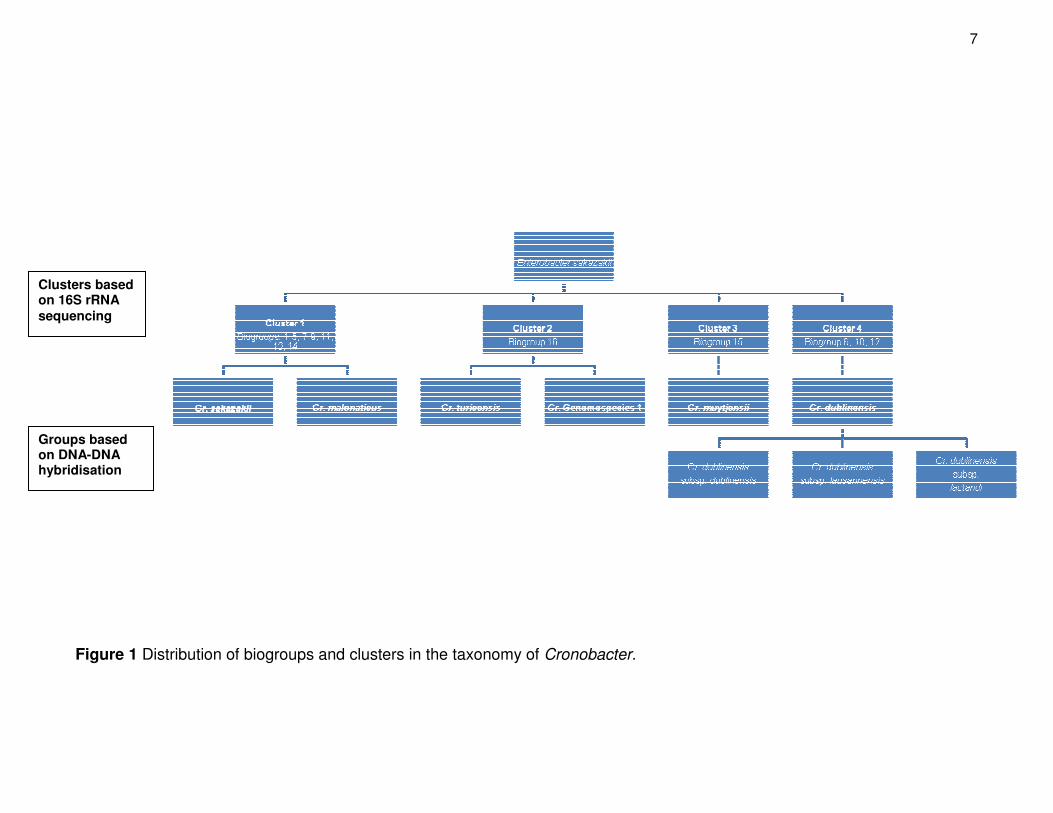
During the first classification of E. sakazakii it was proposed that the isolates may
comprise of more than one species. The biochemical evaluation of 57 strains resulted in
15 different biogroups and it was suggested that biogroup 15 may be grouped in a
separate species from biogroups 1 – 14 (Fig. 1) (Farmer et al., 1980). These findings were
compared to the partial 16S ribosomal RNA (rRNA) genotypes of 189 different strains with
corresponding biochemical traits (Iversen et al., 2006). The molecular analysis gave rise to
an additional biogroup (biogroup 16) and the 189 strains were divided into four clusters.
7
Figure 1 Distribution of biogroups and clusters in the taxonomy of Cronobacter.
Clusters based on 16S rRNA
sequencing
Groups based on DNA-DNA hybridisation
8
These four clusters could be distinguished with inositol, dulcitol and indole tests (Iversen
et al., 2006). The divergent biogroups reinforced the hypothesis that there could be more
than one species and it was proposed to divide E. sakazakii into five separate
genomospecies (Iversen et al., 2007a). Finally in 2008 Enterobacter sakazakii was
reclassified as a member of the novel genus, Cronobacter that contains five species and
three subspecies (Iversen et al., 2008a) (Fig. 1).
B. Characteristics of Cronobacter
Species of the genus Cronobacter consists of Gram-negative rods that are 1 µm x 3 µm in
size. These bacteria are facultative anaerobic and generally peritrichous. Enzymatic
profiles confirmed the absence of phosphoamidase and indicated that α-glucosidase
activity is unique to Cronobacter species (Muytjens et al., 1984). The activity of
α-glucosidase has been implemented as a selective marker in differential chromogenic
agar (Iversen et al., 2004a).
Species of this genus can grow at temperatures between 6° - 47 °C with an
optimum growth temperature of 39 °C (Iversen & Forsythe, 2003). However, some strains
are inhibited at temperatures above 44 °C (Nazarowec-White & Farber, 1997a; Iversen
et al., 2004a) and some strains are capable of growing at 5 °C (Nazarowec-White &
Farber, 1997b). Pigment production and colony size on tryptone soy agar (TSA) are
greatly influenced by the incubation temperature. The yellow pigment production after 24 h
of incubation is more pronounced at 25 °C than at 36 °C, with colony sizes of 1 – 2 mm
and 2 – 3 mm, respectively (Farmer et al., 1980).
Two colony types have been observed when isolates were streaked on TSA. Type
A (Farmer et al., 1980) or matt (Iversen & Forsythe, 2003) is dry or mucoid, scalloped and
rubbery when touched with a loop. Type B (Farmer et al., 1980) or glossy (Iversen &
Forsythe, 2003) is smooth and often exhibits little pigment production. Sub-culturing of the
different colony types showed that the matt colonies may spontaneously change to glossy
colonies and it is very common to find both colony types in one culture (Farmer et al.,
1980). Differences between environmental strains and clinical strains were also observed
(Beuchat et al., 2009). The clinical strain produced mucoidal colonies on violet red bile
glucose agar (VRBGA), whereas the environmental strains produced crinkled, matt
colonies with a rubbery texture.
9
Biofilm formation
Members of the Cronobacter genus have been reported to form biofilms on a range of
surfaces including silicon, glass, stainless steel, latex and polycarbonate (Iversen et al.,
2004b). Attachment of the bacteria occurs more rapidly to hydrophobic substances such
as plastic than to hydrophilic materials (Lehner et al., 2005). The production of biofilms can
be influenced by the nutrient availability, as well as the temperature of the growth medium
(Kim et al., 2006). The importance of nutrient availability was demonstrated by the
formation of biofilms on plastic in different media. The highest number of strains (77 %)
produced biofilms in infant formula milk (IFM), whereas only 16 % produced biofilms in
diluted tryptone soy broth (TSB) (Oh et al., 2007). The influence of temperature was
illustrated as no biofilms were formed in any of the media at 12 °C (Kim et al., 2006).
The disinfectants used in hospitals, day-care centres and food service kitchens are
inefficient in eliminating cells embedded in organic matrices (Kim et al., 2007). Therefore,
formation of biofilms in hospital environments and on equipment may increase the risk of
infections in infants and immuno-compromised adults. In addition, high density
(107 cfu.cm-1) biofilm formation in IFM was observed after 24 h on enteral feeding tubes.
This greatly increases the risk for neonatal infection specifically in hospitals as enteral
feeding tubes can remain in situ at 37 °C for several days and nutrients are administered to
the infants every 2 - 3 h (Hurelle et al., 2009).
Thermal resistance
Thermal resistance studies indicate contradicting results for species of the genus
Cronobacter. There is a general agreement that Cronobacter spp. are thermo-tolerant
bacteria (Chenu & Cox, 2009), although D-values (the time required for a 10-fold reduction
in the viable numbers of a bacterium at a given temperature) of 0.4 min (Breeuwer et al.,
2003), 2.6 min (Iversen et al., 2004b) and 4.2 min (Nazarowec-White & Farber, 1997a)
have been reported at 58 °C. The thermal resistance of Cronobacter spp. has been shown
to be strain dependent (Breeuwer et al., 2003; Edelson-Mammel & Buchanan, 2004;
Arroyo et al., 2009), although some studies did not report any significant differences
between different strains (Nazarowec-White & Farber, 1997a; Iversen et al., 2004b).
10
Heat shock, pH and water activity influence the thermal resistance of Cronobacter
(Arroy et al., 2009). Contradicting results have been published regarding the effect that
heat shock has on the thermal resistance of Cronobacter strains. After subjection of
Cronobacter cells to heat shock for 15 min at 47 °C, the thermal resistance at 47 °C was
enhanced (Chang et al., 2009). However, the sensitivity of Cronobacter cells to heat
treatment increased after a heat shock for 1 h at 42.5 ° and 45 °C, respectively. The
maximum heat resistance of these Cronobacter cells was observed after incubation at
20 °C. The thermal resistance of Cronobacter was approximately 10 times more at pH 7
than at pH 4. However, when the water activity was lowered from more than 0.99 to 0.96
the bacteria showed a 32-fold increase in thermal resistance at 4 °C and pH 4 (Arroyo
et al., 2009).
The heat treatment applied during commercial pasteurisation is sufficient to destroy
Cronobacter in IFM as no contamination was observed directly after pasteurisation in an
IFM manufacturing facility (Proudy et al., 2008). It was, therefore, suggested that
contamination occurs after the pasteurisation step, as Cronobacter have been isolated
from the final products. This can possibly occur during the production process of IFM
before or during spray drying (Iversen & Forsythe, 2003; Proudy et al., 2008). Cronobacter
strains can survive spray drying at inlet temperatures of 160 °C and outlet temperatures of
90 °C (Arku et al., 2008), confirming that this may be the point of contamination in an IFM
processing facility.
Osmotic and desiccation tolerance
Members of Cronobacter have shown greater osmotic and desiccation tolerance than other
bacteria like E. coli and species of Salmonella (Breeuwer et al., 2003). Cronobacter strains
can survive in substances with a low water activity, such as infant rice cereals
(0.3 - 0.69 aw) and IFM (0.25 - 0.5 aw). The survival of Cronobacter spp. at lower
temperatures seems better as higher numbers of the bacteria survived in cereals
(0.63 - 0.83 aw) at 4 °C as opposed to 21° or 31 °C (Gurtler & Beuchat, 2007; Lin &
Beuchat, 2007).
Given that Cronobacter cells can survive for up to 2 years in a desiccated
environment and multiply rapidly after hydration, it is important to understand the stress
responses of Cronobacter in order to control contamination (Osaili & Forsythe, 2009). Cell
11
metabolism studies indicate the accumulation of trehalose in Cronobacter cells during the
stationary phase. Trehalose is a highly soluble disaccharide of glucose which can possibly
stabilise proteins and phospholipid membranes, thereby protecting the bacteria from
dehydration (Breeuwer et al., 2003). Another stress response is the modification of
proteins produced in Cronobacter cells during desiccation which can lead to oxidation of
DNA and membrane components. Several proteins like the Dps and Hns proteins were
shown to be expressed in Cronobacter cells exposed to desiccation. These proteins are
involved in DNA repair and protection of proteins against oxidation. The production of
higher levels of superoxide dismutase and alkylhydroperoxide reductase may also
participate in the protection against oxidation (Riedel & Lehner, 2007).
Acid tolerance
Cronobacter has been described as moderately acid resistant enteric bacteria, as the acid
resistance correlates with that of salmonellae (Gorden & Small, 1993). Substantial
diversity in the acid resistance among 12 Cronobacter strains was observed, particularly
when the strains were exposed to a very low pH (3.0) for 1 h. Despite this diversity, all the
strains were inactivated when exposed to pH 3 for 6 h at 36 °C, while the strains had a
higher resistance at pH 3.5 at 36 °C (Edelson-Mammel et al., 2006).
The acid resistance of Cronobacter in food products may vary. Cronobacter cells
survived in acidic juices such as watermelon (pH 5.0), cantaloupe (pH 6.8) and tomato
juices (pH 4.4) (Kim & Beuchat, 2005). However, these bacteria did not survive in
strawberry and apple juices inoculated with 1000 cfu.mL-1 Cronobacter cells. These juices
have acidity of pH 3.6 and pH 3.9, respectively and were incubated at 25 °C. In contrast
Cronobacter strains have been isolated from fermented products with a higher acidity such
as sobia, a fermented food product made from wheat and malt, with an average
pH of 3.4 - 4.0 (Gassem, 2002). Cronobacter has also been isolated from cheese, such as
domiati with a pH between 4.9 and 6.4 and ras with a pH of 5.8 (El Sharoud et al., 2009).
Antibiotic susceptibility
Members of the genus Cronobacter appear to differ considerably in terms of their
susceptibility to various antibiotics. All the Cronobacter strains tested by Farmer et al.
12
(1980) were resistant to penicillin, whereas some strains were susceptible to
chloramphenicol and ampicillin and only 13 % of the strains were susceptible to
cephalothin. In contrast Cronobacter strains were subsequently identified that were
resistant to cephalothin and chloramphenicol, as well as ampicillin and tetracycline
(Muytjens & van der Ros-van de Repe, 1982; Nazarowec-White & Farber, 1999). Recently
the resistance of Cronobacter species to ampicillin, cephalothin and extended spectrum
penicillin have been confirmed (Kuzina et al., 2001; Lai, 2001). A recent clinical case has
been reported in which multiple antibiotics including ampicillin, gentamicin and
cefotaxamine were ineffective in the treatment of a Cronobacter infection (Dennison &
Morris, 2002). Ampicillin in combination with chloramphenicol or gentamicin is also
inefficient in the treatment of Cronobacter infections as the pathogen seems to be
increasingly resistant to these antibiotics (Lai, 2001).
Virulence
The genus Cronobacter contains opportunistic pathogens, causing illness in immuno-
compromised individuals (Pagotto et al., 2003). The strains in this genus display
differences in pathogenicity and may have different virulence factors (Pagotto et al., 2003;
Healy et al., 2009; MacLean et al., 2009). Cronobacter sakazakii, Cr. turicensis and
Cr. malonaticus are the only species which have been isolated from cases of neonatal
meningitis (Kucerova et al., 2010). However, a strain belonging to Cr. muytjensii has been
isolated from human bone marrow which would normally be sterile (Farmer et al., 1980).
Since the bacterium was reclassified in 2008 as Cronobacter (Iversen et al., 2008a), the
World Health Organisation (WHO) classified all six species as pathogens (FAO/WHO,
2008). Little is known about the mechanism of infection or the different virulence factors of
Cronobacter spp.
One virulence factor of Cronobacter is the O-antigen. These polysaccharide side
chains are variable and are responsible for serological diversity among bacteria. Two
serotypes of the rfb locus which are implicated in the synthesis of the O-antigen were
identified in Cronobacter strains. This has important implications for the virulence of
Cronobacter since the O-antigen is a major surface antigen present in Gram-negative
bacteria (Mullane et al., 2008a). The structure of the O-antigen in the endotoxin of
Cr. muytjensii strain 3270 has recently been described. The O-polysaccharide produced
13
by this strain is a linear unbranched polymer consisting of a repeating pentasaccharide
unit. The structure of this O-polysaccharide differs in size according to sugar composition
and complexity of the structure when compared to the O-polysaccharide structures of other
Cronobacter sakazakii strains. These differences create diversity between serotypes
(Healy et al., 2009) and may possibly reveal that Cronobacter is serologically
heterogeneous with respect to the O-antigens (MacLean et al., 2009). Microarray analysis
supports the observations that there are multiple O-antigen serotypes, not only between
Cronobacter species, but also within Cr. sakazakii.
Another virulence factor of the Cronobacter species is the production of proteolytic
enzymes. Cell deformation, particularly “rounding” of cells, is a result of the action of
various proteases (Lockwood et al., 1982) and Cronobacter strains have been found to
cause this type of deformation of the tissue cells of mice (Pagotto et al., 2003). In
particular a zinc-containing metalloprotease were identified in Cronobacter cells which
caused rounding of Chinese hamster ovary cells. This enzyme had collagenolytic (lysis of
collagen) activity which may allow the pathogen to cross the blood-brain barrier or cause
the extensive cell damage found in neonates with necrotising enterocolitis. It was found
that all of the strains tested possessed the zpx gene which codes for this proteolytic
enzyme (Kothary et al., 2007).
Additionally, Cronobacter strains have been found to produce an enterotoxin
(Pagotto et al., 2003). Purification and characterisation of this enterotoxin indicated its
molecular mass as 66 kDA and that it is most active at pH 6. The enterotoxin proved to be
highly stable as it was unaffected after incubation at 70 °C for 30 min and showed only a
decrease in activity after 30 min incubation at 90 °C (Rhagav & Aggrawal, 2007). However,
the importance of the enterotoxin is still unclear as the genes encoding the putative toxin
and the protein itself remain unidentified (Chenu & Cox, 2009).
C. Disease report
Since the case of a Cronobacter infection in Tennessee was reported in 2002 (CDC, 2002),
the number of well documented cases worldwide has increased. However, the surveillance
of the infections and number of incidences in different age groups are not sufficient to
provide the exact number of infections attributed to this pathogen. At least 111 cases have
been reported in infants and children of which 26 were fatal (Nazarowec-White & Farber,
14
1997a; Iversen & Forsythe, 2003; Gurtler et al., 2005; Drudy et al., 2006; Mullane et al.,
2008b). Only a few of these cases are well described and most of them occurred
sporadically, making epidemiological investigations impossible (FAO/WHO, 2008).
In the case of IFM, the bacteria may exist in clumps rather than be spread out
evenly throughout the product (Witthuhn et al., 2007). This may also lead to false negative
results causing underestimation of contamination and the retail of contaminated products
(FAO/WHO, 2008). The accurate determination of the occurrence of Cronobacter species
is also greatly influenced by the low sensitivity and specificity of the detection methods for
this genus. However, improvements have been made in the detection and identification
methods that would aid in the accurate estimation of Cronobacter contamination (Druggan
& Iversen, 2009).
These contributions will assist in the development of a reasonable risk assessment
and consequent control of Cronobacter. While England, Wales, Scotland and Ireland has
the most information about Cronobacter infections (FAO/WHO, 2008), countries such as
Canada, Argentina and the Netherlands are making remarkable efforts to evaluate the risk
and characteristics of Cronobacter spp. (Pagotto & Farber, 2009; Reij et al., 2009;
Terragno et al., 2009).
Characteristics of disease
The genus Cronobacter has been associated with sporadic infections and outbreaks
(FAO/WHO, 2004). The first known cases of Cronobacter infections were two cases of
meningitis in neonates (Urmenyi & Franklin, 1961). These pathogens have been shown to
cause a severe form of neonatal meningitis which is an acute inflammation of the
membranes surrounding the brain and spinal cord (Nazarowec-White & Faber, 1997c).
Cronobacter strains have also been isolated from infants associated with necrotising
enterocolitis which is caused by infection of the intestines. Other symptoms of infections
include septicaemia (Nazarowec-White & Faber, 1997c), bloody diarrhoea (Simmons et al.,
1989) and brain abscess (Naqvi et al., 1990). The mortality rate has been reported to vary
from 10 % to 80 % with fatalities occurring just days after symptoms developed (Iversen
et al., 2004a). Generally Cronobacter affects the central nervous system (Gallagher & Ball,
1991) and survivors often suffer from severe neurological problems after the infections
(Muytjens et al., 1983; Naqvi et al., 1990; Lai, 2001).
15
Risk groups
Infections of Cronobacter have been reported in all age groups, though immuno-
compromised and very young individuals are more susceptible to infection. Neonates with
a low birth weight or who are immuno-compromised are at the greatest risk (FAO/WHO,
2008). Infections in healthy infants have also been reported (Adamson & Rogers, 1981)
and it may be that the immature immune system and gastro-intestinal tract contribute to the
high susceptibility of this group. Since IFM is a liquid it passes quickly through the stomach
to the small intestines and, therefore, the pathogen is not as stressed as it would be in the
case of a mature adult (Iversen & Forsythe, 2003). Reports of infections in adults are
mostly in combination with other diseases such as the isolation of Cronobacter strains from
a foot ulcer (Pribyl et al., 1985) and an infected wound (Dennison & Morris, 2002).
Cronobacter species pose a significant threat to immuno-compromised individuals,
especially HIV/AIDS patients. This is even more intensified as HIV-positive mothers are
encouraged to give IFM to their babies to prevent transmission of the HI-virus via breast-
feeding (FAO/WHO, 2004). Developing countries that have a high number of premature
babies and HIV-infected individuals are, therefore, likely to have more Cronobacter
infections. High ambient temperatures in developing countries increase the risk of rapid
growth of Cronobacter and insufficient surveillance systems prevent the documentation of
infections (FAO/WHO, 2008).
Infectious dose
The infectious dose for Cronobacter has not been determined, although Health Canada is
working on a dose-response relationship (Pagotto & Farber, 2009). The infectious dose
will be influenced by the state of the bacteria, the immune system of the host and the
environment in which the bacteria grew before infection. The proposed infectious dose
value is 1 000 cfu.100 g-1 although Pagotto et al. (2003) found that 10 000 cfu per mouse
was the lowest count to be lethal in a suckling mouse assay. Nevertheless, it will take up
to 9 days at 8 °C in reconstituted IFM for the pathogen to reach 1 000 cfu.g-1, whereas it
may take only 17.9 h at room temperature with a contamination level of 0.36 cfu.100 g-1.
This model shows that it is unlikely that normal contamination levels would cause infection.
16
The more likely possibility is temperature abuse and/or cross contamination from
preparation utensils (Iversen & Forsythe, 2003).
D. Importance in the food industry
Sources of Cronobacter
Species of Cronobacter have been described as ubiquitous bacteria (Cawthorn et al.,
2008; El Sharoud et al., 2009). These pathogens have been isolated from a range of
environmental, clinical, food and beverage sources. IFM is the only source that has been
epidemiologically linked to disease outbreaks caused by Cronobacter (WHO/FAO, 2004)
and research has specifically focused on the presence of Cronobacter strains in IFM
processing facilities (Mullane et al., 2007; Proudy et al., 2008), raw materials (El-Sharoud
et al., 2009) and final products (Mullane et al., 2008b). Contamination of IFM can occur via
an intrinsic route through the addition of contaminated raw materials after pasteurisation or
the processing facility environment during packaging. External contamination can occur
during reconstitution by using contaminated utensils (Mullane et al., 2008b).
In addition to IFM, Cronobacter has been isolated from numerous dry environments
including dust, soil, grains, tea, herbs and spices. Cronobacter has also been isolated from
water, vegetables, cheese and meat (Kahn et al., 1998; Iversen & Forsythe, 2003; Kim &
Beuchat, 2005; Beuchat et al., 2009; El Sharoud et al., 2009). Additionally, Cronobacter
has been isolated from ready-to-eat foods (Kandhai et al., 2004). The presence of
Cronobacter in these products and subsequent contamination of households increase the
potential risks for infections in immuno-compromised adults (Baumgartner et al., 2009).
Cronobacter strains have not been isolated from cattle faeces, indicating that the pathogen
is not carried by beef cattle (Molloy et al., 2009).
Clinical sources of Cronobacter are diverse and include blood, nose, throat, sputum,
and bone marrow. Apart from isolation from infected patients, Cronobacter strains have
also been isolated from the hospital environment, including a physician’s stethoscope
(Farmer et al., 1980) and utensils used to prepare IFM in a hospital nursery (Simmons
et al., 1989; Bar-Oz et al., 2001). The possibility of cross-contamination in hospitals is
supported by evidence that a contaminated brush used to clean feeding bottles were the
source of three cases of infections (Smeets et al., 1998).
17
The original habitat and mode of transmission of Cronobacter are still unknown
(Nazarowec-White & Farber, 1997b), however, it has been suggested that since the
pathogen does not occur naturally in animals and humans, the principal sources of food
contamination is soil, water and vegetables (Iversen & Forsythe, 2003). The yellow
pigment production, production of a gum-like extracellular polysaccharide and the ability to
persist in a desiccated state suggests an environmental niche associated with plants.
Cronobacter strains that were evaluated for root colonising properties showed solubilisation
of mineral phosphate and the production of indole acetic acid. These characteristics are
often found in plant-associated bacteria and rhizosphere microorganisms indicating that the
natural habitat of Cronobacter may be of plant origin (Schmid et al., 2009).
Inactivation and inhibition in IFM
The inactivation and inhibition of Cronobacter has been predominantly studied in IFM since
the product was identified as the primary vehicle for infection (Muytjens et al., 1983). IFM
is not a sterile product and it is recommended to keep the reconstituted milk at low
temperatures before consumption. Sterilisation of IFM is only possible by irradiation, but
the high levels required to inactivate Cronobacter influence the organoleptic properties of
the product (FAO/WHO, 2004).
Natural antimicrobial agents may be used as additional control measures in the final
products (Al-Holy et al., 2009). The addition of caprylic acid to IFM was proposed as this
antimicrobial agent was found to inhibit Cronobacter strains at lower temperatures. After
treatment with 10 mM caprylic acid for 20 min at 55 °C and 30 mM caprylic acid for 10 min
at 55 °C no Cronobacter cells were recovered (Jang & Rhee, 2009). Lactic acid (0.2 %) in
combination with copper (II) sulphate (50 µg ml-1) resulted in the complete elimination of
Cronobacter strains after a 6 h treatment at 21 °C. The lactic acid chelates the copper
ions, allowing it to penetrate the cytoplasmic membrane and become toxic to the bacteria.
Unfortunately the organoleptic and nutritional properties of IFM using these compounds
were not evaluated (Al Holy et al., 2009). Bovine lactoferrin related compounds may be
useful for the inhibition of Cronobacter spp. in foods as growth was inhibited by lactoferrin
at 1 mg.ml-1 (Wakabayashi et al., 2008). In contrast, stressed Cronobacter cells were not
affected by bovine lactoferrin. Cells were grown in 0.2 % peptone water and reconstituted
IFM and challenged with bovine lactoferrin and nisin. Desiccated cells in peptone water
18
were more susceptible to lactoferrin than undesiccated cells, whereas the undesiccated
cells were more susceptible to nisin. Both antimicrobial agents had no inhibitory effect on
Cronobacter strains in reconstituted IFM at 37 °C. Bovine lactoferrin may, therefore, not be
a suitable antimicrobial agent in IFM for the inhibition of Cronobacter (Al-Nabulsi et al.,
2009).
Regulatory aspects
The Codex Alimentarius Commission (CAC) provides regulations relative to IFM in the
Recommended International Code for Hygienic Practice for Foods for Infants and Children
(CAC, 1979) stating that good manufacturing practices should be followed and clear
labelling should be applied. IFM is not required to be sterile and previously Cronobacter
spp. fell under the specifications for coliforms. These specifications stipulated that four out
of five control samples should contain less than 3 cfu.g-1 and a maximum of one out of five
samples should contain between 3 and 20 cfu.g-1. Since infections occurred under these
limitations the CAC determined new specifications for Cronobacter spp. The revised
regulations require that the product should specifically be tested for the presence of
species of Cronobacter. A test should comprise of 30 samples of which none may be
positive for Cronobacter strains, where a positive result is seen as 1 cfu.100 g-1 sample
(CAC, 2008). The European Union set microbiological standards for Cronobacter as
negative in 30 x 10 g samples. These guidelines are more stringent and will influence the
import of products into European countries (EC, 2005).
E. Detection of Cronobacter
Isolation
The rapid isolation and identification of Cronobacter is important for the appropriate
response to cases of contamination or infection. The detection protocols for Cronobacter
strains should have a high specificity and sensitivity to prevent false positive and false
negative results (Druggan & Iversen, 2009). The current Food and Drug Administration
(FDA) culturing method for the detection of Cronobacter is based on the enumeration of
Enterobacteriaceae in dried foods and IFM. It includes pre-enrichment in a non-selective
19
broth, enrichment in Enterobacteriaceae enrichment broth (EE), culturing on VRBGA, and
sub-culturing onto TSA. Confirmation of presumptive Cronobacter strains are done with an
oxidase test (oxidase negative) (FDA, 2002). This method has been found to have low
sensitivity and relatively low specificity (Cawthorn et al., 2008; Iversen et al., 2008b).
Alternative methods were developed for the detection and enumeration of members
of the genus Cronobacter (Iversen et al., 2004a; Druggan & Iversen, 2009; Lampel & Chen,
2009). The pre-enrichment step included in these methods recovers injured cells and
should increase the sensitivity of the detection method. Buffered peptone water, proposed
by the International Organisation for Standardisation (ISO) appear to be more effective
than distilled water prescribed in the FDA method (Druggan & Iversen, 2009). Selective
enrichment should increase the number of Cronobacter cells relatively to the competitive
bacteria. In some cases this step can reduce the sensitivity of the method as some
Cronobacter strains have been found to be sensitive to selective agents including dyes,
bile salts, detergents and some antibiotics (Joosten et al., 2008; Druggan & Iversen, 2009).
The use of antibiotics to increase specificity of detection methods have also been
discarded by Druggan & Iversen (2009) and presently vancomycin is the only antibiotic that
is used to inhibit Gram-positive bacteria. These findings led to the development of a
screening broth, Cronobacter screening broth (CSB) that does not inhibit the growth of
other Enterobacteriaceae (Iversen et al., 2008b). However, the incubation temperature of
42 °C and inclusion of sucrose give Cronobacter strains advantage over competitors that
cannot grow at this temperature or utilise sucrose. The combination of this broth and
chromogenic agar based on 5-bromo-4-chloro-3-indolyl-α,D-glugopyranoside (XαGlc) has
superior specificity and sensitivity compared to other existing methods (Druggan & Iversen,
2009).
Media that have been proposed as alternatives for VRBGA and TSA include
fluorogenic, chromogenic and dual chromogenic media. Enterobacter sakazakii Isolation
Agar (ESIA®) for example has very good specificity, however, the incubation of 45 °C and
inclusion of crystal violet reduces the sensitivity. Modified Druggan-Forsythe-Iversen Agar
(mDFI), is an improvement on Druggan-Forsythe-Iversen Agar (DFI) and address the
limitations of both DFI and ESIA® (Iversen et al., 2008b). A higher concentration of XαGlc,
a reduced concentration of sodium deoxycholate and incubation at 42 °C increased the
sensitivity of mDFI for the detection of Cronobacter.
20
The FDA is currently in the process of validating a new detection protocol for
Cronobacter that will subsequently be adopted into the FDA’s Bacteriological Analytical
Manual (Druggan & Iversen, 2009; Lampel & Chen, 2009). The proposed protocol includes
incubation at 36 ± 1 °C in buffered peptone water after which the culture is streaked on DFI
and R&F agar (R&F Laboratories, Downers Grove, IL: http://wwwrf-labs.com/). Single
colonies are subjected to a real-time PCR assay developed by Seo & Brackett (2005).
RAPID ID 32E is also used as a confirmation step and the complete analysis can be
accomplished in 24 to 48 h.
Identification
Colonies on isolation media that are presumed to be Cronobacter are traditionally
confirmed with biochemical galleries (Iversen et al., 2004a; Drudy et al., 2006; Fanjat et al.,
2007). Biochemical galleries such as the API 20E are regularly used to identify organisms.
However, numerous reports have indicated that identification of Cronobacter strains with
biochemical galleries was often inaccurate. Iversen et al. (2004a) reported false-negative
and false-positive results with ID 32E, whereas Drudy et al. (2006) identified 98 % of 57
isolates correctly. More recently the VITEK 2, ID 32E version 2.0 and ID 32E version 3.0
systems were evaluated for the identification of Cronobacter. It was found that the newest
version of the ID 32E 3.0 and VITEK 2 systems had a 100 % sensitivity and the ID 32E
version 2.0 only had a 71.4 % sensitivity (Fanjat et al., 2007).
Alternative methods for the identification of Cronobacter have also been explored
(Hoffman et al., 2008; Lin et al., 2009). Gas chromatography with flame ionisation
detection was used to analyse the cellular fatty acid methyl esters of 30 Cronobacter
strains. The resulting fatty acid profiles had good repeatability and could be useful for
identification purposes (Hoffmann et al., 2008). Detection by Fourier transform infrared
(FTI) spectroscopy was also used to discriminate Cronobacter from Enterobacter cloacae,
Escherichia coli and Klebsiella pneumoniae. Subtle compositional differences were
detected with FTI in the carbohydrates of the cell membranes between the Cronobacter
strains and other species (Lin et al., 2009).
Molecular methods for the identification of Cronobacter have been extensively
researched (Malorny & Wagner, 2005; Seo & Brackett, 2005; Lehner et al., 2006; Liu
et al., 2006a; 2006b; Mohan Nair & Venkitanarayanan, 2006; Mullane et al., 2006;
21
Cawthorn et al., 2008). Iversen et al. (2007b) developed dnaG based reverse
transcriptase-PCR (RT-PCR) and 1,6-α-glucosidase (gluA) based conventional PCR
assays. These PCR systems were evaluated with 312 Enterobacteriaceae strains,
including 210 Cronobacter strains, making it the most extensively evaluated methods
(Iversen et al., 2007b). All the Cronobacter strains tested positive with both PCR assays.
F. Phylogeny
The reclassification of Enterobacter sakazakii to Cronobacter was based on a polyphasic
approach that included DNA-DNA hybridisation, amplified fragment length polymorphisms
(AFLP), automated ribotyping, full length 16S rRNA gene sequencing and phenotypic
analysis (Iversen et al., 2007a; 2008a). A total of 210 strains, previously described as
Enterobacter sakazakii were divided into 16 biogroups based on indole production, methyl
red test, Voges-Proskauer, ornithine decarboxylase motility, malonate utilisation and acid
production from i-inositol, dulcitol and methylglucoside. Defining characteristics of each
biogroup corresponded with previous findings and included indole, dulcitol and inositol
tests (Iversen et al., 2006; 2007a). Sequence analysis based on 16S rRNA of these strains
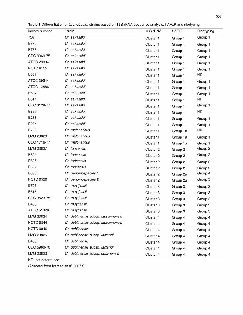
resulted in four clusters (Table 1). The majority of strains were grouped in cluster 1
together with the Enterobacter sakazakii type strain, ATCC 29544T. Automated ribotyping
of the 210 strains resulted in four groups largely corresponding with the four 16S rRNA
clusters. The ribotyping results showed a similarity pattern of less than 62 % between the
Enterobacter sakazakii strains and other Enterobacteriaceae. Subsequent
fluorescent-AFLP (f-AFLP) analysis divided the strains into 6 groups that corresponded
with the 16S rRNA clusters, as clusters 1 and 2 were each divided into two groups
(Table 1) (Iversen et al., 2007a).
DNA-DNA hybridisation is considered to be the “gold standard” method to evaluate
relatedness between bacterial species (Stackebrandt et al., 2002). The recommended
cut-off point for species delineation is regarded at a DNA homology of more than or equal
to 70 % between two strains (Wayne et al., 1987). Representative strains of each of the
four 16S rRNA clusters were subjected to DNA-DNA hybridisation and these strains were
divided into five groups which had DNA-homology values of less than 70 %. Based on
the combination of the genetic and phenotypic data, four Cronobacter species were
proposed, Cronobacter sakazakii, Cronobacter turicensis, Cronobacter dublinensis and
22
Cronobacter muytjensii, an additional Cronobacter genomospecies 1 and a subspecies
namely Cr. sakazakii subsp. malonaticus. This subspecies grouped separately from
Cr. sakazakii strains with f-AFLP and ribotyping analysis, but had a 99.6 % similarity based
on 16S rRNA with the Cr. sakazakii type strain, ATCC 29544T (Iversen et al., 2007a). This
subspecies was accepted as a distinct species, namely Cronobacter malonaticus after
subsequent DNA-DNA hybridisation indicated that the Cr. malonaticus strains had DNA
homology values of less than 70 % with the other Cronobacter species (Iversen et al.,
2008a).
There is a high level of similarity between Cr. sakazakii and Cr. malonaticus and
sequence analysis based on 16S rRNA is not sufficient to distinguish between these two
species (Iversen et al., 2007a; 2008a; Kuhnert et al., 2009). Biochemical differentiation
between the two species can be accomplished by testing for the utilisation of malonate,
although a small number of Cr. sakazakii strains does utilise malonate (Iversen et al.,
2008a). Controversial results regarding these two species have been found when the
phenotypic data of 150 isolates was compared to ribotyping results (Miled-Bennour et al.,
2010). According to biochemical analysis strain 05CHPL02 was identified as Cr. sakazakii
and strain 05CPL53 as Cr. malonaticus. However, the ribotyping results placed the
Cr. sakazakii strain closer to the non-sakazakii strains and the Cr. malonaticus strain was
grouped with the Cr. sakazakii strains (Miled-Bennour et al., 2010). A higher resolution
between Cr. sakazakii and Cr. malonaticus were obtained with multilocus sequence typing
(MLST) based on seven genes. The strains of these two species were clearly phylogenetic
distinct, supporting the organisation of Cr. sakazakii and Cr. malonaticus in two distinct
species (Baldwin et al., 2009).
Species description
The genus Cronobacter consists of five distinct species and Cr. sakazakii is the type
species of the genus. Cronobacter sakazakii was named in honour of the renowned
microbiologist Riichi Sakazaki and has been the dominant species in terms of isolation
frequency. The strains of Cr. sakazakii are allocated in biogroups 1-4, 7, 8, 11 and 13
(Iversen et al., 2007a) and the type strain of this species (DSM 4485T; ATCC 29544T;
NCTC 11467T) was isolated from a child’s throat (Farmer et al., 1980).
23
Table 1 Differentiation of Cronobacter strains based on 16S rRNA sequence analysis, f-AFLP and ribotyping
Isolate number Strain 16S rRNA f-AFLP Ribotyping
756 Cr. sakazakii Cluster 1 Group 1 Group 1
E775 Cr. sakazakii Cluster 1 Group 1 Group 1
E768 Cr. sakazakii Cluster 1 Group 1 Group 1
CDC 9369-75 Cr. sakazakii Cluster 1 Group 1 Group 1
ATCC 29004 Cr. sakazakii Cluster 1 Group 1 Group 1
NCTC 8155 Cr. sakazakii Cluster 1 Group 1 Group 1
E807 Cr. sakazakii Cluster 1 Group 1 ND
ATCC 29544 Cr. sakazakii Cluster 1 Group 1 Group 1
ATCC 12868 Cr. sakazakii Cluster 1 Group 1 Group 1
E607 Cr. sakazakii Cluster 1 Group 1 Group 1
E811 Cr. sakazakii Cluster 1 Group 1 ND
CDC 3128-77 Cr. sakazakii Cluster 1 Group 1 Group 1
E327 Cr. sakazakii Cluster 1 Group 1 ND
E266 Cr. sakazakii Cluster 1 Group 1 Group 1
E274 Cr. sakazakii Cluster 1 Group 1 Group 1
E765 Cr. malonaticus Cluster 1 Group 1a ND
LMG 23826 Cr. malonaticus Cluster 1 Group 1a Group 1
CDC 1716-77 Cr. malonaticus Cluster 1 Group 1a Group 1
LMG 23827 Cr. turicensis Cluster 2 Group 2 Group 2
E694 Cr. turicensis Cluster 2 Group 2 Group 2
E625 Cr. turicensis Cluster 2 Group 2 Group 2
E609 Cr. turicensis Cluster 2 Group 2 Group 2
E680 Cr. genomospecies 1 Cluster 2 Group 2a Group 4
NCTC 9529 Cr. genomospecies 2 Cluster 2 Group 2a Group 3
E769 Cr. muytjensii Cluster 3 Group 3 Group 3
E616 Cr. muytjensii Cluster 3 Group 3 Group 3
CDC 3523-75 Cr. muytjensii Cluster 3 Group 3 Group 3
E488 Cr. muytjensii Cluster 3 Group 3 Group 3
ATCC 51329 Cr. muytjensii Cluster 3 Group 3 Group 3
LMG 23824 Cr. dublinensis subsp. lausannensis Cluster 4 Group 4 Group 4
NCTC 9844 Cr. dublinensis subsp. lausannensis Cluster 4 Group 4 Group 4
NCTC 9846 Cr. dublinensis Cluster 4 Group 4 Group 4
LMG 23825 Cr. dublinensis subsp. lactaridi Cluster 4 Group 4 Group 4
E465 Cr. dublinensis Cluster 4 Group 4 Group 4
CDC 5960-70 Cr. dublinensis subsp. lactaridi Cluster 4 Group 4 Group 4
LMG 23823 Cr. dublinensis subsp. dublinensis Cluster 4 Group 4 Group 4
ND: not determined
(Adapted from Iversen et al. 2007a)
24
Cronobacter sakazakii is generally indole, dulcitol and malonate negative, but methyl-α-D-
glucopyranoside positive (Iversen et al., 2007a). DNA-DNA hybridisation analysis of Cr.
sakazakii with the other Cronobacter species resulted in a maximum similarity of 61 % with
strains of Cr. malonaticus and a minimum value of 16 % with Cr. dublinensis (Iversen et al.,
2008a).
Cronobacter malonaticus is the closest related to Cr. sakazakii of all the
Cronobacter species (Kuhnert et al., 2009) and is characterised by the utilisation of
malonate (Iversen et al., 2008a). This species is negative for indole and dulcitol utilisation
and include biogroups 5, 9 and 14. The type strain (DSM 18702T; CDC 1058-77T; LMG
23826T) was originally isolated from a breast abscess (Iversen et al., 2007a). Sequence
analysis based on 16S rRNA showed no distinction between Cr. sakazakii and
Cr. malonaticus, but DNA-DNA hybridisation resulted in DNA homology values between 54
and 60 %. DNA-homology between two Cr. malonaticus strains was found to be 95.6 %,
confirming that the strains from Cr. malonaticus form a separate species (Iversen et al.,
2008a).
Cronobacter turicensis is derived from biogroup 16 identified by Iversen et al.
(2006). The type strain (DSM 18703T; LMG 23827T; z3032T) was isolated from an
individual suffering from neonatal meningitis in 2005 (Mange et al., 2006). The strains from
this species are indole negative, but malonate and dulcitol positive. It is interesting to note
that this species consists of all the strains in biogroup 16, except the strains NCTC 9529
and E680. DNA-DNA hybridisation of NCTC 9529 with the Cr. turicensis type strain
resulted in DNA homology of 55 % and, therefore, these two strains (NCTC 9529 and
E680) are currently designated as Cronobacter genomospecies 1 (Iversen et al., 2008a).
However, no distinction can be made with phenotypic analysis and the two strains have a
16S rRNA similarity of 99.6 % with the Cr. turicensis strains. Analysis of these two strains
with f-AFLP resulted in patterns that had less than 50 % similarity with the Cr. turicensis
strains. Additionally, ribotyping did not group these two strains with the other Cr. turicensis
strains (Table 1) (Iversen et al., 2007a).
Cronobacter muytjensii is the only Cronobacter species that is negative for the
utilisation of 1-0-methyl-α-D-glucopyranoside. This species is also positive for indole,
dulcitol and malonate utilisation. Cronobacter muytjensii consists of strains in biogroup 15
(Farmer et al., 1980) and is represented by the type strain ATCC 51329T (CIP 103581T)
(Iversen et al., 2007a). Sequencing based on 16S rRNA resulted in similarity values above
25
97 % between Cr. muytjensii and the other Cronobacter species. Automated ribotyping
and f-AFLP, however, showed these strains to be separate from the other Cronobacter
species (Table 1). DNA-DNA hybridisation of Cr. muytjensii and Cr. sakazakii resulted in
31 to 53 % DNA homology values.
Cronobacter dublinensis consists of three subspecies, namely Cr. dublinensis
subsp. dublinensis, Cr. dublinensis subsp. lausannensis and Cr. dublinensis subsp.
lactaridi. Cronobacter dublinensis subsp. dublinensis (DSM 18707T LMG 23825T; E464T)
contains strains from biogroup 6 and the type strain was isolated from an environmental
sample in a milk processing facility. Similarly the type strain of Cr. dublinensis subsp.
lactaridi (DSM 18705T; LMG 23823T; DES 187T) was isolated from a dried milk product
manufacturing facility (biogroup 12). Cronobacter dublinensis subsp. lausannensis
(DSM 18706T; LMG 23824T; E515T) consists of strains in biogroup 10 and the type strain of
this species was isolated from the basin of a water fountain (Iversen et al., 2008a). The
strains from these three subspecies were grouped together with sequence analysis based
on 16S rRNA, f-AFLP and ribotyping (Iversen et al., 2007a). The DNA-DNA relatedness
between Cr. sakazakii ATCC 29544T and strains from all of the Cr. dublinensis subspecies
were less than 55 %. Cronobacter dublinensis subsp. dublinensis DSM 18705T and
Cr. dublinensis subsp. lausannensis NCTC 9844 had a DNA-DNA hybridisation value of
95.2 %, whereas Cr. dublinensis subsp. lausannensis NCTC 9844 had a value of 77.4 %
when compared with Cr. dublinensis subsp. lactaridi CDC 5960-70. Based on these
results the three biogroups were designated as three subspecies (Iversen et al., 2008a).
These subspecies are generally dulcitol negative and indole production is variable (Iversen
et al., 2007a).
Typing of Cronobacter
Recent studies on the identification of the five Cronobacter species is based on
biochemical tests to differentiate between the strains, as well as identification based on a
molecular approach (Healy et al., 2009; Terragno et al., 2009). Extensive phenotypic
analysis was done on 23 isolates originating from three brands of IFM in Argentina. The
biochemical tests included the production of acid from carbohydrates, gas production from
glucose and the Voges-Proskauer test. The utilisation of malonate, as well as citrate as a
sole carbon source was also evaluated. Based on the phenotypic analysis 22 isolates
26
were identified as Cr. sakazakii and one isolate as Cr. malonaticus. The strains were also
subjected to pulsed field gel electrophoresis (PFGE) analysis using the restriction enzyme
XbaI. The phenotypic analysis corresponded with the PFGE results as the single
Cr. malonaticus strain had a distinct XbaI pattern and 7 different patterns could be
distinguished for the Cr. sakazakii strains (Terragno et al., 2009).
Phenotypic analysis was used to classify a collection of 150 Enterobacter sakazakii
strains. These strains originated from IFM production facilities and final products, as well
as clinical samples. The phenotypic analysis included indole production, malonate
utilisation and acid production from methyl-α-D-glucopyranoside and dulcitol. The majority
of strains were identified as Cr. sakazakii (82.5 %), with 8 % of the strains designated as
Cr. malonaticus, 5 % as Cr. muytjensii, 3 % as Cr. dublinensis and 1.5 % as Cr. turicensis.
Genetic typing data for these strains based on PFGE, ribotyping and 16S rRNA
sequencing indicated a high level of diversity between the 150 strains and compared well
with the phenotypic results. However, discrepancies were found between the phenotypic
groupings and the 16S rRNA data of Cr. malonaticus and Cr. sakazakii strains. This was
expected as limited resolution of the 16S rRNA gene between these two species is not
uncommon (Miled-Bennour et al., 2010).
Genetic characterisation of Cronobacter
Microarray-based comparative genomic indexing (CGI) was used to analyse 78
Cronobacter strains isolated from food, environmental and clinical samples. The array
consisted of 276 open reading frames, targeting most of the functional gene categories in
the genome of Cr. sakazakii strain BAA-894. In total, 200 of the 276 DNA coding
sequences were present in all the Cronobacter strains. The gene categories with the most
variable genes were the extracellular structures and the cell wall/membrane biogenesis.
Species-specific genes which were present in all the Cr. sakazakii strains, but variable in
the other species, were identified and can possibly serve as molecular markers for the
identification of particular species in the genus Cronobacter (Healy et al., 2009).
The genome of Cr. sakazakii strain BAA-894 was sequenced and described as a
4.4 Mb chromosome with two plasmids of 31 kb and 131 kb. Comparative genomic
hybridisation (CGH) was undertaken on representatives of the five Cronobacter species,
excluding Cronobacter genomospecies 1. In total, 4 382 genes were examined of which
27
43 % were common to all the Cronobacter strains. Genes that are associated with
virulence factors were of particular interest. All the species had the ompA gene which is
associated with the invasive ability of the E. coli strains responsible for neonatal meningitis
(Prasadarao et al., 1996). However, the genes encoding for a cation efflux system which
allows bacteria to invade brain microvascular endothelial cells (Franke et al., 2003) were
only present in Cronobacter species associated with neonatal infections. In addition,
15 gene clusters including putative prophages and prophage fragments were absent in
more than half of the tested strains. Putative virulence genes were identified in most of
these clusters. This indicates that the acquisition of genes via integration or phages, as
well as specific gene loss played a major role in the evolution of Cronobacter and diversity
among the species (Kucerova et al., 2010).
Multilocus sequence analysis (MLSA) was used to evaluate the similarity of the
recN, rpoA and thdF genes of different Enterobacteriaceae focussing on the genus
Cronobacter. Based on the recN gene, Cr. malonaticus and Cr. sakazakii showed the
highest similarity and Cr. dublinensis and Cr. muytjensii was shown to be least similar with
respect to the rest of the genus. The rpoA gene proved to be useful in the identification
and differentiation of species in the Enterobacteriaceae family. The generation of
phylogenetic data with these genes supported the reclassification of the Cronobacter spp.
and provided data for phylogenetic and taxonomic analysis and identification (Kuhnert
et al., 2009).
The use of genes other than the 16S rRNA is common in PCR assays for the typing
of bacterial strains. The rpoB gene which encodes for the bacterial RNA polymerase
β-subunit has been evaluated for its suitability to distinguish between species (Mollet et al.,
1997). The levels of divergence in Enterobacteriaceae strains between the sequences of
the rpoB gene were significantly higher than the levels of divergence between the
16S rRNA sequences. A conventional PCR assay targeting the rpoB gene was the first
species-specific PCR assay for the Cronobacter genus is (Stoop et al., 2009). In this
assay six primer pairs, namely Cturf/Cturr, Cdubf/Cdubr, Cmuyf/Cmuyr,
Cgenomof/Cgenomor, Cmalf/Cmalr and Csakf/Csakr were used to differentiate between 57
strains. The first four primer pairs had a 100 % specificity, but the Cmalf/Cmalr and
Csakf/Csakr primer pairs had to be used in a two-step procedure since the rpoB gene
sequences of Cr. malonaticus and Cr. sakazakii are very closely related. The strains that
tested positive with the Csakf/Csakr primers were tested in a follow-up PCR with the

28
Cmalf/Cmalr primer pair. Cronobacter malonaticus strains would test positive with the
latter PCR (Stoop et al., 2009). The rpoB gene is, therefore, useful in the differentiation
between Cronobacter species.
G. Conclusion
Differentiation between the various Cronobacter species has been the focus of research
since these bacteria have been identified as opportunistic pathogens readily isolated from
IFM. The inactivation, inhibition, thermal, osmotic and desiccation tolerance of these
bacteria have been characterised to create risk management strategies for the production
of hygienic products. This was aided by the improvements made in the isolation and
detection of Cronobacter strains. Furthermore, the reclassification of this genus has played
an important role in understanding the phylogeny of these pathogens.
However, there still remain many questions regarding differences in the
characteristics of the five species. Although all five Cronobacter species are classified as
pathogens it has yet to be shown that they are all virulent. The differentiation on a species
level has become more important for this reason. The few typing methods available for the
Cronobacter species are either time consuming or inaccurate and, therefore, other genetic
typing methods should be evaluated for the accurate distinction between the species of
Cronobacter.
References
Adamson, D. H., & Rogers, J. R. (1981). Enterobacter sakazakii meningitis with sepsis.
Clinical Microbiology Newsletter, 3, 19-20.
Al-Holy, M., Castro, L.F. & Al-Qadiri, H. (2009). Inactivation of Cronobacter spp.
(Enterobacter sakazakii) in infant formula using lactic acid, copper sulfate and
monolaurin. Letters in Applied Microbiology, 50 (3), 246-251.
Al-Nabulsi, A., Osaili, T.M., Al-Holy, M., Shaker, R.R., Ayyash, M.M., Olaimat, A.N. &
Holley, R.A. (2009). Influence of desiccation on the sensitivity of Cronobacter spp.
to lactoferrin or nisin in broth and powdered infant formula. International Journal of
Food Microbiology, 136 (2), 221-226.
Arku, B., Mullane, N., Fox, E., Fanning, S. & Jordan, K. (2008). Enterobacter sakazakii
survives spray drying. International Journal of Dairy Technology, 61 (1), 102-108.
29
Arroyo, C., Condon, S. & Pagan, R. (2009). Thermobacteriological characterisation of
Enterobacter sakazakii. International Journal of Food Microbiology, 136 (1), 110-
118.
Baldwin, A., Loughlin, M., Caubilla-Barron, J., Kucerova, E., Manning, G., Dowson, C. &
Forsythe, S. (2009). Multilocus sequence typing of Cronobacter sakazakii and
Cronobacter malonaticus reveals stable clonal structures with clinical significance
which do not correlate with biotypes. BMC Microbiology, 9, 223.
Bar-Oz, B., Preminger, A., Peleg, O., Block, C. & Arad, I. (2001). Enterobacter sakazakii
infection in the newborn. Acta Paediatrica, 90, 356-358.
Baumgartner, A., Grand, M., Liniger, M. & Iversen, C. (2009). Detection and frequency of
Cronobacter spp. (Enterobacter sakazakii) in different categories of ready-to-eat
foods other than infant formula. International Journal of Food Microbiology, 136 (2),
189-192.
Beuchat, L.R., Kim, H., Gurtler, J.B., Lin, L.C., Ryu, J.H. & Richards, G.M. (2009).
Cronobacter sakazakii in foods and factors affecting its survival, growth, and
inactivation. International Journal of Food Microbiology, 136 (2), 204-213.
Breeuwer, P., Lardeau, A., Peterz, M. & Joosten, H.M. (2003). Desiccation and heat
tolerance of Enterobacter sakazakii. Journal of Applied Microbiology, 95 (5), 967-
973.
Brenner, D.J. (1974). DNA reassociation for the clinical differentiation of enteric bacteria,
Public Health Lab., 118–130.
Cawthorn, D.M., Botha, S. & Witthuhn, R.C. (2008). Evaluation of different methods for the
detection and identification of Enterobacter sakazakii isolated from South African
infant formula milks and the processing environment. International Journal of Food
Microbiology, 127 (1-2), 129-138.
Chenu, J.W. & Cox, J.M. (2009). Cronobacter ("Enterobacter sakazakii"): current status
and future prospects. Letters in Applied Microbiology, 49 (2), 153-159.
Chang, C., Chiang, M., Chou, C. (2009). The effect of temperature and length of heat
shock treatment on the thermal tolerance and cell leakage of Cronobacter sakazakii
BCRC 13988. International Journal of Food Microbiology, 134 (3), 184-189.
CDC (Centers for Disease Control and Prevention). 2002. Enterobacter sakazakii
infections associated with the use of powdered infant formula - Tennessee, 2001.
Morbidity and Mortality Weekly Report, 51: 297-300.
30
Codex Alimentarius Commission (CAC) (1979). Recommended international code of
hygienic practice for foods for infants and children (CAC/RCP 21-1979). [www
document]. URL http://fao.org/codex/standard/en/CXP021e.pdf. 16 September
2006.
Codex Alimentarius Commission (CAC) (2008). Code of Hygienic Practice for Powdered
Formulae for Infants and Young Children (Cac/Rcp 66-2008). [Www Document]. Url
http://fao.org/codex/standard/en/cxp066e.pdf. 19 February 2009.
Dennison S.K. & Morris J. (2002). Multiresistant Enterobacter sakazakii wound infection in
an adult, Infect. Med.. 533–535.
Drudy, D., O'Rourke, M., Murphy, M., Mullane, N.R., O'Mahony, R., Kelly, L., Fischer, M.,
Sanjaq, S., Shannon, P., Wall, P., O'Mahony, M., Whyte, P. & Fanning, S. (2006).
Characterization of a collection of Enterobacter sakazakii isolates from
environmental and food sources. International Journal of Food Microbiology, 110
(2), 127-134.
Druggan, P. & Iversen, C. (2009). Culture media for the isolation of Cronobacter spp.
International Journal of Food Microbiology, 136 (2), 169-178.
Edelson-Mammel, S. & Buchanan, R.L. (2004). Thermal inactivation of Enterobacter
sakazakii in rehydrated infant formula. Journal of Food Protection, 67 (1), 60-63.
Edelson-Mammel, S., Porteous, M.K. & Buchanan, R.L. (2006). Acid Resistance of Twelve
Strains of Enterobacter sakazakii, and the Impact of Habituating the Cells to an
Acidic Environment. Journal of Food Science, 71 (6), 201-207.
El-Sharoud, W., O'Brien, S., Negredo, C., Iversen, C., Fanning, S. & Healy, B. (2009).
Characterization of Cronobacter recovered from dried milk and related products.
BMC Microbiology, 9, 9.
European Commission (EC) (2005). Commission regulation (EC) number 2073/2005 of 15
November 2005 on microbiological criteria for foodstuffs. Official Journal of the
European Union, L338, 1-26.
Fanjat, N., Leclercq, A., Joosten, H. & Robichon, D. (2007). Comparison of the
phenotyping methods ID 32E and VITEK 2 compact GN with 16S rRNA gene
sequencing for the identification of Enterobacter sakazakii. Journal of Clinical
Microbiology, 45 (6), 2048-2050.
Farmer, J.J., Asbury, M.A., Hickman, F.W., Brenner, D.J. & the Enterobacteriaceae Study
Group (1980). Enterobacter sakazakii, new species of Enterobacteriaceae isolated
31
from clinical specimens. International Journal of Systematic Bacteriology, 30, 569-
584.
Felske, A.,Franke, S., Grass, G., Rensing, C. & Nies, D.H. (2003). Molecular analysis of
the copper-transporting efflux system CusCFBA of Escherichia coli. Journal of
Bacteriology 185, 3804–12.
Food and Agriculture Organization / World Health Organization (FAO/WHO) (2004).
Enterobacter sakazakii and other microorganisms in powdered infant formula:
Meeting Report, MRA series 6. Geneva, Switzerland. [www document]. URL
http://www.who.int/foodsafety/micro/meetings/feb2004/en/. 6 March 2009.
Food and Agriculture Organization / World Health Organization (FAO/WHO) (2008).
Enterobacter sakazakii (Cronobacter spp.) in powdered follow-up formulae: Meeting
Report, MRA series 15. Rome. [www document]. URL
http://www.who.int/foodsafety/micro/meetings/2008/en/. 11 May 2009.
Food and Drug Administration (FDA) (2002). Isolation and enumeration of Enterobacter
sakazakii from dehydrated powdered infant formula. [www document]. URL
http://www.cfsan.fda.gov/~comm/mmesakaz.html. 29 July 2006.
Franke, S., Grass, G., Rensing, C. & Nies, D. H. (2003). Molecular analysis of the copper-
transporting efflux system CusCFBA of Escherichia coli. Journal of Bacteriology,
185, 3804–12.
Gallagher, P.G. & Ball, W.S. (1991). Cerebral infarctions due to CNS infection with
Enterobacter sakazakii. Pediatric Radiology, 21, 135-136.
Gassem, M.A.A. (2002). A microbiological study of Sobia: a fermented beverage in the
Western province of Saudi Arabia. World Journal of Microbiology & Biotechnology
18: 173–177.
Gorden, J. & Small, P.L.C. (1993). Acid resistance in enteric bacteria. Infection and
Immunity, 61, 364-367.
Gurtler, J.B. & Beuchat, L.R. (2007). Survival of Enterobacter sakazakii in powdered infant
formula as affected by composition, water activity, and temperature. Journal of
Food Protection, 70 (7), 1579-1586.
Gurtler, J.B., Kornacki, J.L. & Beuchat, L.R. (2005). Enterobacter sakazakii: a coliform of
increased concern to infant health. International Journal of Food Microbiology, 104,
1-34.
32
Healy, B., Huynh, S., Mullane, N., O'Brien, S., Iversen, C., Lehner, A., Stephan, R., Parker,
C.T. & Fanning, S. (2009). Microarray-based comparative genomic indexing of the
Cronobacter genus (Enterobacter sakazakii). International Journal of Food
Microbiology, 136 (2), 159-164.
Hoffmann, M., Keys, C.E., Song, K.Y., Brown, E.W., Fry, F.S. & Whittaker, P. (2008).
Evaluation of multiple strains of Enterobacter sakazakii using fatty acid profiles.
Food Chemistry, 107 (4), 1623-1628.
Hurrelle, E., Kucerova, E., Loughlin, M., Caubilla-Barron, S., Forsythe, S.J. (2009). Biofilm
formation on enteral feeding tubes by Cronobacter sakazakii, Salmonella serovars
and other Enterobacteriaceae. International Journal of Food Microbiology, 136 (2),
227-31.
Iversen, C. & Forsythe, S. (2003). Risk profile of Enterobacter sakazakii, an emergent
pathogen associated with infant milk formula. Trends in Food Science &
Technology, 14 (11), 443-454.
Iversen, C., Druggan, P. & Forsythe, S. (2004a). A selective differential medium for
Enterobacter sakazakii, a preliminary study. International Journal of Food
Microbiology, 96 (2), 133-139.
Iversen, C., Lane, M. & Forsythe, S. J. (2004b). The growth profile, thermotolerance and
biofilm formation of Enterobacter sakazakii grown in infant formula milk. Letter
Applied Microbiology, 38, 378–382.
Iversen, C., Waddington, M., Farmer, J. J. & Forsythe, S. J. (2006). The biochemical
differentiation of Enterobacter sakazakii genotypes. BMC Microbiology, 6 (94).
Iversen, C., Lehner, A., Mullane, N., Bidlas, E., Cleenwerck, I., Marugg, J., Fanning, S.,
Stephan, R. & Joosten, H. (2007a). The taxonomy of Enterobacter sakazakii:
proposal of a new genus Cronobacter gen. nov. and descriptions of Cronobacter
sakazakii comb. nov. Cronobacter sakazakii subsp. sakazakii, comb. nov.,
Cronobacter sakazakii subsp. malonaticus subsp. nov., Cronobacter turicensis sp.
nov., Cronobacter muytjensii sp. nov., Cronobacter dublinensis sp. nov.,
Cronobacter genomospecies 1. BMC Evolutionary Biology, 64 (7), 1471-2148.
Iversen, C., Lehner, A., Mullane, N., Marugg, J., Fanning, S., Stephan, R. & Joosten, H.
(2007b). Identification of ‘Cronobacter’ spp. [Enterobacter sakazakii]. Journal of
Clinical Microbiology, 45 (11), 3814-3816.
33
Iversen, C., Mullane, N., McCardell, B., Tall, B.D., Lehner, A., Fanning, S., Stephan, R. &
Joosten, H. (2008a). Cronobacter gen. nov., a new genus to accommodate the
biogroups of Enterobacter sakazakii, and proposal of Cronobacter sakazakii gen.
nov., comb. nov., Cronobacter malonaticus sp. nov., Cronobacter turicensis sp. nov.,
Cronobacter muytjensii sp. nov., Cronobacter dublinensis sp. nov., Cronobacter
genomospecies 1, and of three subspecies, Cronobacter dublinensis subsp.
dublinensis subsp. nov., Cronobacter dublinensis subsp. lausannensis subsp. nov.
and Cronobacter dublinensis subsp. lactardi subsp. nov.. International Journal of
Systematic and Evolutionary Microbiology, 58 (6), 1442-1447.
Iversen, C., Druggan, P., Schumacher, S., Lehner, A., Feer, C., Gschwend, K., Joosten, H.
& Stephan, R. (2008b). Development of a novel screening method for the isolation
of ‘Cronobacter’ spp. [Enterobacter sakazakii]. Applied and Environmental
Microbiology, 74 (8), 2550-2553.
Jang, H.I. & Rhee, S.M. (2009). Inhibitory effect of caprylic acid and mild heat on
Cronobacter spp. (Enterobacter sakazakii) in reconstituted infant formula and
determination of injury by flow cytometry. International Journal of Food
Microbiology, 133 (1), 113-120.
Joker, R. N., Norholm, T. & Siboni, K. E. (1965). A case of neonatal meningitis caused by
a yellow Enterobacter. Danish Medical Bulletin, 128–130.
Joosten, H., Marugg, J., Stephan, R., Klijn, A., Jackson, T. & Iversen, C. (2008). A rapid
and reliable alternative to ISO 21528-1:2004 for detection of Enterobacteriaceae.
International Journal of Food Microbiology, 125 (3), 344-346.
Kandhai, C., Reij, M.W., Gorris, L.G.M., Guillaume-Gentil, O. & Schothorst, M. (2004).
Occurrence of Enterobacter sakazakii in food production environments and
households. Lancet, 363 (9402), 39-40.
Khan, A.A, Jones, R.A., Cerniglia, C.E. (1998). Rapid method for the detection of
genetically engineered microorganisms by polymerase chain reaction from soil and
sediments. Journal of Industrial Microbiology & Biotechnology, 20, 90-94.
Kim, H. & Beuchat, L.R. (2005). Survival and growth of Enterobacter sakazakii on fresh-
cut fruits and vegetables and in unpasteurized juices as affected by storage
temperature. Journal of Food Protection, 68 (12), 2541-2552.
34
Kim, H., Ryu, J.-H., Beuchat, L.R., (2006). Attachment and biofilm formation by
Enterobacter sakazakii on stainless steel and enteral feeding tubes. Applied
Environmental Microbiology, 73, 5846–5856.
Kim, H., Ryu, J.H. and Beuchat, L.R. (2007). Effectiveness of disinfectants in killing
Enterobacter sakazakii in suspension, dried on the surface of stainless steel, and in
a biofilm. Applied Environmental Microbiology, 73, 1256–1265.
Kothary, M.H., McCardell, B.A., Frazar, C.D., Deer, D. & Tall, B.D. (2007).
Characterization of the zinc-containing metalloprotease encoded by zpx and
development of a species-specific detection method for Enterobacter sakazakii.
Applied and Environmental Microbiology, 73 (13), 4142-4151.
Kucerova, E., Clifton, S.W., Xia, X., Long, F., Porwollik, S., Fulton, L., Fronick, C., Minx, P.,
Kyung, K.,Warren, W., Fulton, R., Feng, D., Wollam, A., Shah, N., Bhonagiri, V.,
Nash, W.E., Hallsworth-Pepin, K., Wilson, R.K., McClelland, M. & Forsythe, S.J.
(2010). Genome sequence of Cronobacter sakazakii BAA-894 and comparative
genomic hybridisation analysis with other Cronobacter species. PLoS ONE. 5 (3)
e9556.
Kuhnert, P., Korczak, B.M., Stephan, R., Joosten, H. & Iversen, C. (2009). Phylogeny and
prediction of genetic similarity of Cronobacter and related taxa by multilocus
sequence analysis (MLSA). International Journal of Food Microbiology, 136 (2),
152-158.
Kuzina, L.V., Peloquin, J.J., Vacek, D.C. & Miller, T.A. (2001). Isolation and identification
of bacteria associated with adult laboratory Mexican fruit flies,Anastrepha ludens.
Current Microbiology, 42, 290-294.
Lai, K.K. (2001). Enterobacter sakazakii infections among neonates, infants, children, and
adults: case reports and a review of the literature. Medicine Baltimore, 80, 113–122.
Lampel, K.A. & Chen, Y. (2009). Method for the isolation and detection of Enterobacter
sakazakii (Cronobacter) from powdered infant formula. International Journal of Food
Microbiology, 136 (2), 179-184.
Lehner, A., Riedel, K., Eberl, L., Breeuwer, P., Diep, B. & Stephan, R. (2005). Biofilm
formation, extracellular polysaccharide production, and cell-to-cell signaling in
various Enterobacter sakazakii strains: aspects promoting environmental
persistence. Journal of Food Protection, 68 (11), 2287-2294.
35
Lehner, A., Riedel, K., Rattei, T., Ruepp, A., Frishman, D., Breeuwer, P., Diep, B., Eberl, L.
& Stephan, R. (2006). Molecular characterization of the α-glucosidase activity in
Enterobacter sakazakii reveals the presence of a putative gene cluster for palatinose
metabolism. Systematic and Applied Microbiology, 29 (8), 609-625.
Lin, L.C. & Beuchat, L.R. (2007). Survival and growth of Enterobacter sakazakii in infant
cereal as affected by composition, reconstitution liquid, and storage temperature.
Journal of Food Protection, 70 (6), 1410-1422.
Lin, M., Al-Holy, M., Al-Qadiri, H., Kong, F., Rasco, B.A. & Setiady, D. (2009). Detection
and discrimination of Enterobacter sakazakii (Cronobacter spp.) by mid-infrared
spectroscopy and multivariate statistical analyses. Journal of Food Safety, 29 (4),
531-545.
Liu, Y., Cai, X., Zhang, X., Gao, Q., Yang, X., Zheng, Z., Luo, M. & Huang, X. (2006a).
Real time PCR using TaqMan and SYBR Green for detection of Enterobacter
sakazakii in infant formula. Journal of Microbiological Methods, 65 (1), 21-31.
Liu, Y., Gao, Q., Zhang, X., Hou, Y., Yang, J. & Huang, X. (2006b). PCR and
oligonucleotide array for detection of Enterobacter sakazakii in infant formula.
Molecular and Cellular Probes, 20 (1), 11-17.
Lockwood, D.E.; Kreger, A.S.; Richardson, S.H. Detection of toxins produced by Vibrio
fluvialis (1982). Infect. Immun., 35, 702-708.
MacLean, L.L., Pagotto, F., Farber, J.M. & Perry, M.B. (2009). The structure of the O-
antigen in the endotoxin of the emerging food pathogen Cronobacter (Enterobacter)
muytjensii strain 3270. Carbohydrate Research, 344 (5), 667-671.
Malorny, B. & Wagner, M. (2005). Detection of Enterobacter sakazakii strains by real-time
PCR. Journal of Food Protection, 68 (8), 1623-1627.
Mange, J.P., Stephan, R., Borel, N., Wild, P., Kim, K.S., Pospischil, A. & Lehner, A. (2006).
Adhesive properties of Enterobacter sakazakii to human epithelial and brain
microvascular endothelial cells. BMS Microbiology. 6 (58).
Miled-Bennour, R., Ells, C., Pagotto, F.J., Farber, J.M., Kerouanton, A., Meheut, T., Colin,
P., Joosten, H., Leclercq, A. & Besse, N.G. (2010). Genotypic and phenotypic
characterisation of a collection of Cronobacter (Enterobacter sakazakii) isolates.
International Journal of Food Microbiology, 139, 116–125.
Mohan Nair, M.K. & Venkitanarayanan, K.S. (2006). Cloning and sequencing of the ompA
gene of Enterobacter sakazakii and development of an ompA-targeted PCR for rapid
36
detection of Enterobacter sakazakii in infant formula. Applied and Environmental
Microbiology, 72 (4), 2539-2546.
Mollet, C., Drancourt, M. & Raoult, D. (1997). rpoB sequence analysis as a novel basis for
bacterial identification. Molecular Microbiology. 25 (5), 1005-1011.
Molloy, C., Cagney, C., O'Brien, S., Iversen, C., Fanning, S. & Duffy, G. (2009).
Surveillance and characterisation by Pulsed-Field Gel Electrophoresis of
Cronobacter spp. in farming and domestic environments, food production animals
and retail foods. International Journal of Food Microbiology, 136 (2), 198-203.
Mullane, N.R., Murray, J., Drudy, D., Prentice, N., Whyte, P., Wall, P.G., Parton, A. &
Fanning, S. (2006). Detection of Enterobacter sakazakii in dried infant milk formula
by cationic-magnetic-bead capture. Applied and Environmental Microbiology, 72 (9),
6325-6330.
Mullane, N.R., Whyte, P., Wall, P.G., Quinn, T. & Fanning, S. (2007). Application of
pulsed-field gel electrophoresis to characterise and trace the prevalence of
Enterobacter sakazakii in an infant formula processing facility. International Journal
of Food Microbiology, 116 (1), 73-81.
Mullane, N., O'Gaora, P., Nally, J.E., Iversen, C., Whyte, P., Wall, P.G. & Fanning, S.
(2008a). Molecular analysis of the Enterobacter sakazakii O-antigen gene locus.
Applied and Environmental Microbiology, 74 (12), 3783-3794.
Mullane, N., Healy, B., Meade, J., Whyte, P., Wall, P.G. & Fanning, S. (2008b).
Dissemination of Cronobacter spp. [Enterobacter sakazakii] in a powdered milk
protein manufacturing facility. Applied and Environmental Microbiology, 74 (19),
5913-5917.
Muytjens, H.L. & van der Ros-van der Repe, J. (1982). Comparative studies of 13 β-
Lactam Antibiotics. Antimicrobial Agents and Chemotherapy, 21 (6), 6925-934.
Muytjens, H.L., Zanen, C.H., Sonderkamp, J.H., Kollee, L.A., Wachsmuth, K.,Farmer, J.J.
(1983). Analysis of Eight Cases of Neonatal Meningitis and Sepsis. Journal of
Clinical Microbiology, 18 (1), 115-120.
Muytjens, H.L., van der Ros-van de Repe, J., van Druten, H. (1984). Enzymatic Profiles of
Enterobacter sakazakii and related species with Special Reference to the ox-
Glucosidase Reaction and Reproducibility of the Test System. Journal of Clinical
Microbiology, 20 (4), 684-686.
37
Naqvi, S.H., Maxwell, M.A. & Schwalbe, R.S. (1990). Nosocomial bacteremia caused by
Enterobacter sakazakii and Leuconostoc mesenteroides resulting from extrinsic
contamination of infant formula. Journal of Pediatric Infectious Diseases, 9, 447–
449.
Nazarowec-White, M. & Farber, J.M. 1995, Biological characterization of Enterobacter
sakazakii, Microbiol. Res. Div., Banting Building, Health Canada, Ottawa, Ont. K1A
0L2, Canada.
Nazarowec-White, M. & Farber, J.M. (1997a). Thermal resistance of Enterobacter
sakazakii in reconstituted dried-infant formula. Letters in Applied Microbiology, 24
(1), 9-13.
Nazarowec-White, M. & Farber, J.M. (1997b). Incidence, survival, and growth of
Enterobacter sakazakii in infant formula. Journal of Food Protection, 60 (3), 226-
230.
Nazarowec-White, M. & Farber, J.M. (1997c). Enterobacter sakazakii: a review.
International Journal of Food Microbiology, 34 (2), 103-113.
Nazarowec-White, M., and J. M. Farber. (1999). Phenotypic and genotypic typing of food
and clinical isolates of Enterobacter sakazakii. Journal of Medical Microbiology, 48,
559–567.
Oh, S.W., Chen, P.C., & Kang, D.H. (2007). Biofilm formation by Enterobacter sakazakii
grown in artificial broth and infant milk formula on plastic surface. Journal of Rapid
Methods and Automation in Microbiology, 15 (4), 311-319.
Osaili, T. & Forsythe, S. (2009). Desiccation resistance and persistence of Cronobacter
species in infant formula. International Journal of Food Microbiology, 136 (2), 214-
220.
Pagotto, F.J. & Farber, J.M. (2009). Cronobacter spp. (Enterobacter sakazakii): Advice,
policy and research in Canada. International Journal of Food Microbiology, 136 (2),
238-245.
Pagotto, F.J., Nazarowec-White, M., Bidawid, S. & Farber, J.M. (2003). Enterobacter
sakazakii: infectivity and enterotoxin production in vitro and in vivo. Journal of Food
Protection, 66 (3), 370-375.
Prasadarao, N.V., Wass, C.A., Weiser, J.N., Stins, M.F., & Huang. S.H.. (1996). Outer
membrane protein A of Escherichia coli contributes to invasion of brain
microvascular endothelial cells. Infection and Immunity, 64, 146–53.
38
Pribyl, C., Salzer, R., Beskin, J., Haddad, R.J., Pollock, B., Beville, R., Holmes. B.,
Mogabgab, W.J. (1985). Aztreozam in the Treatment of Serious Orthopedic
Infections. The American Journal of Medicine, 78, 51-56.
Proudy, I., Bougle, D., Leclercq, R. & Vergnaud, M. (2008). Tracing of Enterobacter
sakazakii isolates in infant milk formula processing by BOX-PCR genotyping.
Journal of Applied Microbiology, 105 (2), 550-558.
Raghav, M. & Aggarwal, P.K. (2007). Purification and characterisation of Enterobacter
sakazakii enterotoxin. Canadian Journal of Microbiology, 53, 750-755.
Reij, M.W., Jongenburger, I., Gkogka, E., Gorris, L.G.M. & Zwietering, M.H. (2009).
Perspective on the risk to infants in the Netherlands associated with Cronobacter
spp. occurring in powdered infant formula. International Journal of Food
Microbiology, 136 (2), 232-237.
Riedel, K. & Lehner, A. (2007). Identification of proteins involved in osmotic stress
response of Enterobacter sakazakii by proteonomics. Proteonomics, 7, 1217-1231.
Schmid, M., Iversen, C., Gontia, I., Stephan, R., Hofmann, A., Hartmann, A., Jha, B., Eberl,
L., Riedel, K. & Lehner, A. (2009). Evidence for a plant-associated natural habitat
for Cronobacter spp. Research in Microbiology, 160 (8), 608-614.
Seo, K.H. & Brackett, R.E. (2005). Rapid, specific detection of Enterobacter sakazakii in
infant formula using a real-time PCR assay. Journal of Food Protection, 68 (1), 59-
63.
Simmons, B.P., Gelfand, M.S., Haas, M., Metts, L. & Ferguson, J. (1989). Enterobacter
sakazakii infections in neonates associated with intrinsic contamination of a
powdered infant formula. Infection Control and Hospital Epidemiology, 10, 398-401.
Smeets, L. C., A. Voss, H. L. Muytjens, J. F. G. M. Meis, and W. J. G. Melchers. 1998.
Genetische karakterisatie van Enterobacter sakazakii-isolaten van Nederlandse
patienten met neonatale meningitis. Ned. Tijdschr. Med. Microbiol. 6, 113–115.
Stackebrandt, E., Frederiksen, W., Garrity, G. M. & 10 other authors (2002). Report of the
ad hoc committee for the re-evaluation of the species definition in bacteriology.
International Journal of Systematic Evolutionary Microbiology 52, 1043–1047.
Stoop, B., Lehner, A., Iversen, C., Fanning, S. & Stephan, R. (2009). Development and
evaluation of rpoB based PCR systems to differentiate the six proposed species
within the genus Cronobacter. International Journal of Food Microbiology, 136 (2),
165-168.
39
Terragno, R., Salve, A., Pichel, M., Epszteyn, S., Brengi, S. & Binsztein, N. (2009).
Characterization and subtyping of Cronobacter spp. from imported powdered infant
formulae in Argentina. International Journal of Food Microbiology, 136 (2), 193-197.
Urmenyi, A.M.C. & Franklin, A.W. (1961). Neonatal death from pigmented coliform
infection. The Lancet, 1, 313-315.
Wakabayashi, H., Yamauchi, K. & Takase, M. (2008). Inhibitory effects of bovine
lactoferrin and lactoferricin B on Enterobacter sakazakii. Biocontrol Science, 13 (1),
29-32.
Wayne, L. G., Brenner, D. J., Colwell, R. R. & 9 other authors (1987). International
Committee on Systematic Bacteriology. Report of the ad hoc committee on
reconciliation of approaches to bacterial systematics. International Journal of
Systematic Bacteriology 37, 463–464.
Witthuhn, R.C., Kemp, F. & Britz, T.J. (2007). Isolation and PCR detection of Enterobacter
sakazakii in South African food products, specifically infant formula milks. World
Journal of Microbiology & Biotechnology, 23 (2), 151-157.
40
CHAPTER 3
PHYLOGENETIC ANALYSIS OF CRONOBACTER ISOLATES BASED ON THE rpoA
AND 16S rRNA GENES
Abstract
The reclassification of the genus Cronobacter (previously known as Enterobacter
sakazakii) was based on a polyphasic analysis that led to the description of five species.
These bacteria are opportunistic pathogens that can cause neonatal meningitis and other
infections in immuno-compromised individuals. Cronobacter strains have been reported to
show differences in sensitivity to antibiotics, heat and chemicals, as well as differences in
virulence. The aim of this study was to classify Cronobacter strains isolated from infant
formula milk, the food processing environment and fresh produce in South Africa and to
evaluate the phylogenetic placement of these isolates based on the rpoA and 16S
ribosomal RNA (rRNA) gene sequences. All the South African strains were identified as
Cr. sakazakii despite the wide variety of isolation sources. No relation between the
phylogenetic placement and strain origin could be determined. Strains from Cr. sakazakii,
Cr. dublinensis, Cr. turicensis and Cr. muytjensii could be differentiated from each other,
but it was not possible to differentiate between Cr. sakazakii and Cr. malonaticus based on
the rpoA and 16S rRNA gene sequences. However, sequence data of these two genes
can be used to differentiate between the members of the genus Cronobacter when used in
combination with biochemical analysis based on the utilisation of malonate.
Introduction
The genus Cronobacter (previously known as Enterobacter sakazakii) consists of five
species and three subspecies, namely Cronobacter sakazakii, Cronobacter malonaticus,
Cronobacter muytjensii, Cronobacter dublinensis subsp. dublinensis, Cr. dublinensis
subsp. lausannensis, Cr. dublinensis subsp. lactaridi and Cronobacter turicensis.
Additionally, three Cronobacter strains were grouped together as Cronobacter
genomospecies 1 (Iversen et al., 2008). The existence of multiple species in the E.
sakazakii group had already been suggested in 1980, as 15 different biogroups were
identified among these strains (Farmer et al., 1980). The reclassification of Cronobacter
was based on a polyphasic approach which included DNA-DNA hybridisation, amplified
41
fragment length polymorphisms (AFLP), automated ribotyping, full length 16S ribosomal
RNA (rRNA) gene sequencing and phenotypic analysis (Iversen et al., 2007; 2008). The
description of the five new Cronobacter species was supported by two independent multi-
locus sequencing data sets based on seven and three genes, respectively (Baldwin et al.,
2009; Kuhnert et al., 2009). However, some discrepancies have been reported concerning
the identification of some Cronobacter strains at the species level, where the results of
phenotypic analysis differed from that obtained with sequencing based on the 16S rRNA
gene (Miled-Bennour et al., 2010).
Cronobacter strains have been reported to show differences in sensitivity to
chemical reagents, heat, antibiotics (Kuzina et al., 2001; Lai, 2001; Arroyo et al., 2009), as
well as differences in virulence factors (Pagotto et al., 2003; Healy et al., 2009; MacLean et
al., 2009). Recently, Kucerova et al. (2010) reported that only strains from Cr. sakazakii,
Cr. malonaticus and Cr. turicensis have been associated with neonatal infections.
Additionally, only these three species possess a gene cluster encoding for a cation efflux
system which allows bacteria to invade brain microvascular endothelial cells (Franke et al.,
2003). Cronobacter sakazakii strains isolated from a hospital in France showed that all the
strains associated with fatal Cronobacter infections were grouped together in one
pulsetype (Caubilla-Barron et al., 2007). This indicates that there may be variation
between the Cr. sakazakii strains regarding virulence potential.
Members of the genus Cronobacter can cause a severe form of neonatal meningitis,
necrotising enterocolitis and septicaemia (Nazorowec-White & Farber, 1997; Van Acker et
al., 2001). These opportunistic pathogens have been associated with sporadic infections
and disease outbreaks (FAO/WHO, 2004; 2008). The number of Cronobacter related
infections in South Africa is unknown, but these bacteria may pose a significant risk to the
high number of HIV-positive individuals. Between 2005 and 2008 Cronobacter strains (E.
sakazakii) have been isolated from an infant formula milk (IFM) processing facility and final
products, as well as fresh produce in South Africa (Cawthorn et al., 2008; Mofokeng et al.,
2010). These results and the recent reclassification of the genus have led to the present
study in which the aim is to identify the different Cronobacter spp. and to determine the
phylogenetic relationship between the recently isolated strains.
Materials and methods
42
Bacterial isolates
Bacterial isolates previously identified as E. sakazakii (Cronobacter) were evaluated in this
study. The 24 isolates were recently isolated from various food and environmental sources
in South Africa, including IFM products, the environment of an IFM production facility and
fresh produce. The fresh produce included raw milk, potatoes, beetroot, carrots and
mincemeat. The type strains Cr. malonaticus DSM 18702T, Cr. sakazakii DSM 4485T, Cr.
turicensis DSM 18703T, Cr. dublinensis DSM 18705T (obtained from the Deutche
Sammeling von Microorganismen und Zelkulturen) and Cr. muytjensii ATCC 51329T
(obtained from the American Type Culture Collection) were also included in this study.
Bacteria were inoculated in tryptone soy broth (TSB) (Merck, Cape Town South Africa) and
incubated at 37 °C for 24 h. Confirmation of Cronobacter spp. was done by streaking the
incubated broth on Chromocult® agar, followed by incubation at 42 °C for 24 h. Blue-green
colonies were regarded as Cronobacter positive.
DNA isolations
DNA was isolated using either the TZ-method described by Wang & Levin (2006) or a
modified phenol-chloroform method described by van Elsas et al. (1997). For the TZ-
extraction method cells were harvested by centrifuging 250 µL of the 24 h TSB cultures at
8 000 g for 5 min. The pellets were suspended in 250 µL double strength TZ (2 x TZ)
(Wang & Levin, 2006), which consisted of 5 mg.mL-1 sodium azide (Merck) and 4 % (v/v)
Triton X-100 (Merck) in 0.1 M Tris-HCl (Fluka) at pH 8. Cells were lysed by placing
samples in a water bath at 100 °C for 10 min. The debris was pelleted by centrifugation at
10 000 g for 5 min and the supernatant was used as DNA template.
For the phenol-chloroform method bacterial samples were prepared by centrifuging
(Eppendorf Centrifuge 5415D, Germany) 2 ml of the 24 h TSB cultures at 8 000 g for 10
min. Glass beads (0.6 g) were added to the cell pellets and 800 µL phosphate buffer (1
part 120 mM NaH2PO4 (Merck) to 9 parts 120 mM Na2HPO4 (Merck); pH 8), 700 µL phenol
(Fluka, supplied by Sigma-Aldrich) and 100 µL of 20 % (m/v) sodium dodecyl sulphate
(SDS) (Merck) were added to the samples. The samples were then vortexed for 1 to 2 min
followed by incubation for 20 min at 60 °C. The vortexing and incubation step was
repeated three times, after which the samples were centrifuged for 5 min at 4000 g.
Phenol (600 µL) was added to each supernatant, the samples were vortexed and
centrifuged for 5 min at 8 000 g. The water phase was removed and 600 µL
43
phenol:chloroform:isoamylalcohol (25:24:1) was added and the samples were centrifuged
(5 min at 8 000 g) until the interphase was clean. The waterphase was removed, the
volume of the supernatant noted and DNA was precipitated by adding 0.1 volume 3 M
sodium acetate (NaOAc) (pH 5.5) (Saarchem, supplied by Merck) and 0.6 volume
isopropanol (Saarchem) to the samples. The samples were stored overnight at -20 °C
followed by centrifugation for 10 min at 12 000 g. The resulting pellet was washed with 70
% (v/v) ethanol after which the samples were centrifuged for 5 min at 12 000 g. The pellet
was air-dried and 100 µL of 1 x TE (10 mM Tris (Fluka), 1 mM EDTA (Merck); pH 8) buffer
was added. Samples were treated with 1 µL RNase for 1 h at 37 °C and stored at -20 °C.
Phylogenetic analysis
Phylogenetic evaluation of Cronobacter strains was based on two genes, 16S rRNA and
rpoA. In order to compare the South African strains to strains from various sources and
origin, 15 Cronobacter strains representing Cr. sakazakii, Cr. muytjensii, Cr. dublinensis
and Cr. turicensis were obtained from GenBank (Table 1). Apart from the Cr. malonaticus
type strain DSM 18702T, no other strain from this species had both the rpoA and 16S rRNA
gene sequences available and no additional Cr. malonaticus strains could be included in
this study.
16S rRNA gene sequencing
The 16S rRNA gene was amplified with the primers F8 (5’- CAC GGA TCC AGA CTT TGA
TYM TGG CTC AG -3’) and R1512 (5’- GTG AAG CTT ACG GYT AGC TTG TTA CGA
CTT -3’) (Felske et al., 1997), resulting in a 1.5 kilobase (kb) fragment. The PCR reaction
mixture (50 µL total volume) contained 1 µL (5 U) Taq DNA polymerase (Super-Therm,
supplied by Southern Cross Biotechnologies, Cape Town, South Africa), 5 µL 10 X buffer
(Super-Therm), 3 µL (1.5 mM) MgCl2 (Super-Therm), 2 µL (400 nM) of each primer, 2 µL
(400 nM) dNTPs (AB gene, supplied by Southern Cross Biotechnologies), 2 µL 99% (v/v)
DMSO (Merck) and 2 µL DNA template. Thermal cycling parameters (Eppendorf
mastercycler personal, Eppendorf, Germany) were as follows: initial denaturation at 92 °C
for 3 min; 35 cycles of denaturation at 92 °C for 30 s, annealing at 54 °C for 30 s, and
elongation at 68 °C for 60 s; and final elongation at 72 ºC for 7 min (Felske et al., 1997).
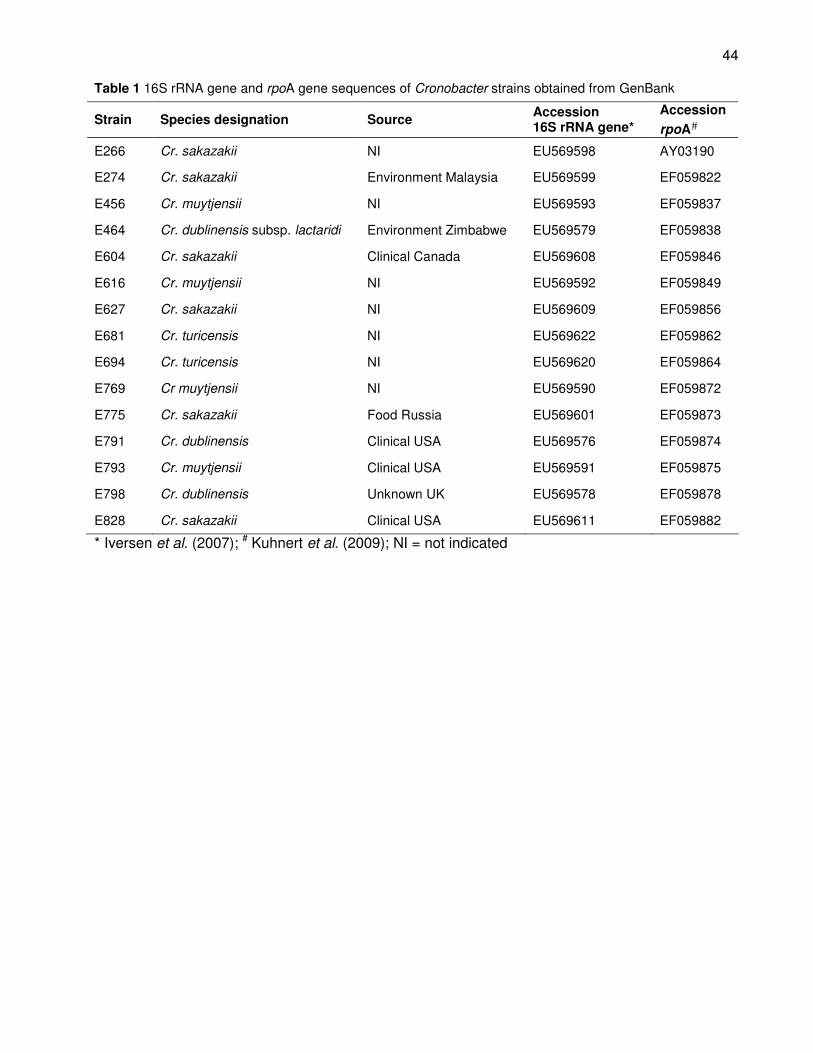
44
Table 1 16S rRNA gene and rpoA gene sequences of Cronobacter strains obtained from GenBank
Strain Species designation Source Accession 16S rRNA gene*
Accession
rpoA#
E266 Cr. sakazakii NI EU569598 AY03190
E274 Cr. sakazakii Environment Malaysia EU569599 EF059822
E456 Cr. muytjensii NI EU569593 EF059837
E464 Cr. dublinensis subsp. lactaridi Environment Zimbabwe EU569579 EF059838
E604 Cr. sakazakii Clinical Canada EU569608 EF059846
E616 Cr. muytjensii NI EU569592 EF059849
E627 Cr. sakazakii NI EU569609 EF059856
E681 Cr. turicensis NI EU569622 EF059862
E694 Cr. turicensis NI EU569620 EF059864
E769 Cr muytjensii NI EU569590 EF059872
E775 Cr. sakazakii Food Russia EU569601 EF059873
E791 Cr. dublinensis Clinical USA EU569576 EF059874
E793 Cr. muytjensii Clinical USA EU569591 EF059875
E798 Cr. dublinensis Unknown UK EU569578 EF059878
E828 Cr. sakazakii Clinical USA EU569611 EF059882
* Iversen et al. (2007); # Kuhnert et al. (2009); NI = not indicated
45
rpoA gene sequencing
The rpoA gene was amplified using the primers rpoA_entero-L (ATG CAG GGT TCT GTG
ACA GAG) and rpoA_entero-R (GGT GGC CAR TTT TCY AGG CGC) (Kuhnert et al.,
2010), resulting in a 960 basepair (bp) fragment. The PCR reaction mixture (30 µL total
volume) contained 0.5 µL (2.5 U) Taq DNA polymerase (Super-Therm), 3 µL 10 X buffer
(Super-Therm), 1.5 µL (1.25 mM) MgCl2 (Super-Therm), 0.5 µL (167 nM) of each primer, 2
µL (667 mM) dNTPs (AB gene) and 1 µL DNA template. Thermal cycling parameters
(Eppendorf mastercycler) were as follows: initial denaturation at 92 °C for 3 min; 35 cycles
of denaturation at 92 °C for 30 s, annealing at 54 °C for 30 s, and elongation at 68 °C for
60 s; and final elongation at 72 ºC for 7 min.
Sequence analysis
Sequencing of the PCR products was performed using the ABI 3100 Genetic Analyser
(Applied Biosystems, Foster City, USA). The closest recognised relatives of the isolates
were determined by comparing the generated sequences to sequences available in
GenBank using the BLASTn search option (Altschul et al., 1997).
The rpoA and 16S rRNA gene sequences of Cronobacter strains obtained from
GenBank were added to the sequences generated for the South African strains and
aligned with MAFFT (http://mafft.cbrc.jp/alignment/software/). Phylogenetic analysis was
done on each set of sequences using the phylogenetic analysis using parsimony (PAUP)
software with 1000 bootstraps and the neighbour-joining algorithm. Phylograms were
constructed from the sequences of each gene, as well as from a combination of the two
genes. A Templeton Nonparametric Wilcoxon Signed Ranked Test (Kellogg et al., 1996;
Swofford, 2001) was performed to determine whether the two data sets could be
combined. The type strain of Enterobacter aerogenes, DSM 30053T, was used as an
outgroup in the analysis since the rpoA and 16S rRNA gene sequences of this strain were
related, but genetically distinct from the Cronobacter species.
Results and discussion
Reclassification of the South African strains
All the South African Cronobacter isolates showed blue-green colonies on the Chromocult®
agar, testing positive for Cronobacter. The rpoA and 16S rRNA genes of the 24 South
African strains were successfully amplified resulting in fragments of 960 bp and 1.5 kb,
46
respectively. Furthermore, the South African strains were all identified as Cr. sakazakii
based on both the 16S rRNA gene and rpoA gene sequences with the BLASTn search
function in Genbank (Table 2). This study supports the finding that Cr. sakazakii is the
dominant species in terms of isolation frequency (Baldwin et al., 2009; Kuhnert et al.,
2009). Iversen et al. (2007) also reported on the dominance of this species as Cr sakazakii
consists of 12 of the 16 biogroups identified among the Cronobacter strains. Furthermore,
in a separate study 82 % of 150 isolates were identified as Cr. sakazakii (Miled-Benour et
al., 2010). The absence of the four other Cronobacter species amongst the South African
isolates was not expected as the strains were isolated from a wide variety of sources.
Since only 24 isolates were used in this study, it is possible that strains from the other
Cronobacter species will be identified when more isolates of these bacteria are obtained in
South Africa.
Phylogeny and genetic relatedness
The phylogeny of the genus Cronobacter was evaluated based on the two genes, 16S
rRNA and rpoA. The 16S rRNA gene is most frequently used in phylogenetic studies but
has been found to be too conserved to differentiate between some strains in the
Enterobacteriaceae family (Mollet et al., 1997). The 16S rRNA gene sequences of many
Cronobacter strains have been deposited in GenBank and were, therefore, available for
comparisons in this study. The rpoA gene encodes for the α-subunit of the RNA
polymerase and has proved to be useful in the identification and differentiation of species
in the Enterobacteriaceae family (Kuhnert et al., 2009). This gene has also been
sequenced for numerous Cronobacter strains and these sequences have also been made
available on GenBank.
Phylogeny based on the 16S rRNA gene
The phylogram based on the 16S rRNA gene sequences showed clear separation between
the Cronobacter isolates and the outgroup strain, E. aerogenes (Fig. 1). All the South
African strains were grouped amongst the Cr. sakazakii strains from GenBank in two
clusters (clusters 1 and 2). A total of 10 strains isolated from South Africa were grouped
with the Cr. sakazakii type strain DSM 4485T in cluster 1. All the strains that were isolated
from fresh produce were grouped in this cluster, as well as four strains that were isolated
from dried milk related sources (IFM and powdered milk). Cluster 2 contained all the
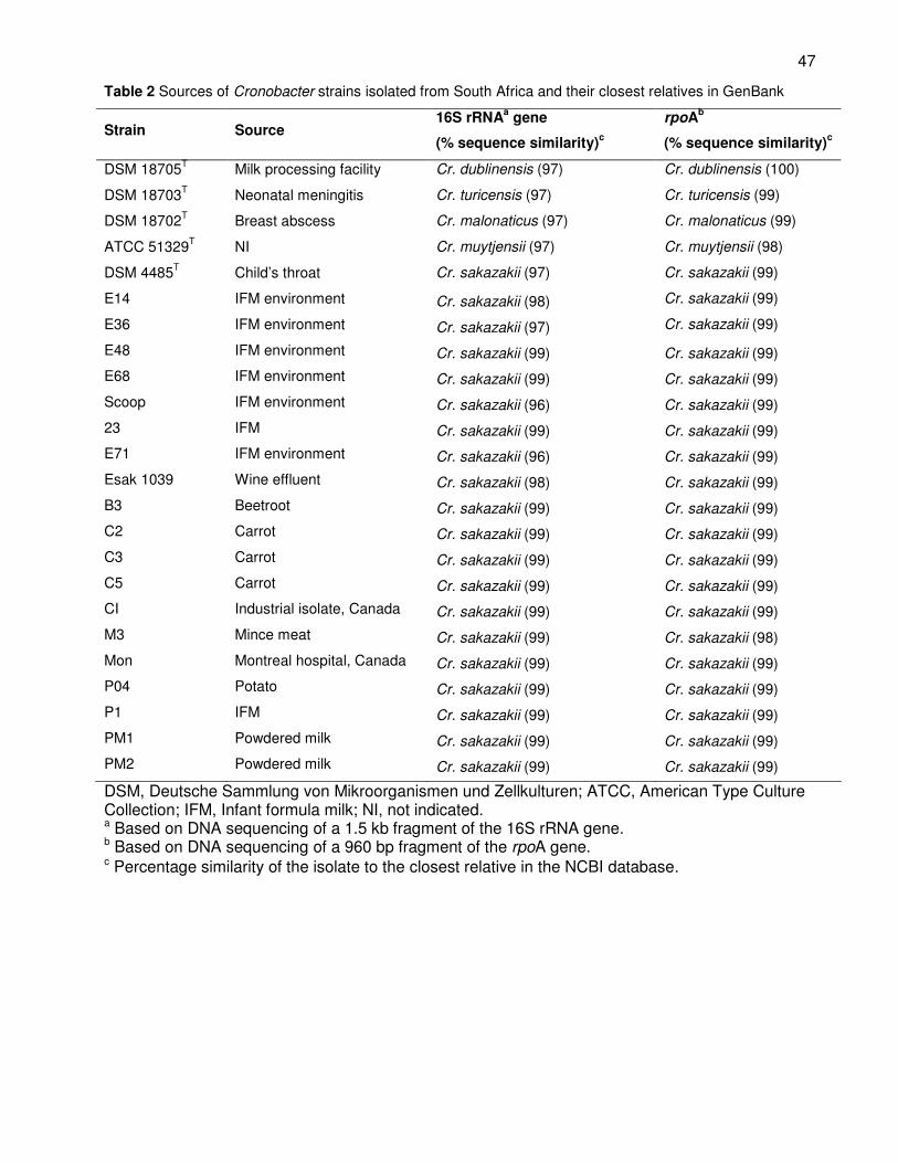
47
Table 2 Sources of Cronobacter strains isolated from South Africa and their closest relatives in GenBank
Strain Source 16S rRNA
a gene
(% sequence similarity)c
rpoAb
(% sequence similarity)c
DSM 18705T
Milk processing facility Cr. dublinensis (97) Cr. dublinensis (100)
DSM 18703T
Neonatal meningitis Cr. turicensis (97) Cr. turicensis (99)
DSM 18702T
Breast abscess Cr. malonaticus (97) Cr. malonaticus (99)
ATCC 51329T
NI Cr. muytjensii (97) Cr. muytjensii (98)
DSM 4485T
Child’s throat Cr. sakazakii (97) Cr. sakazakii (99)
E14 IFM environment Cr. sakazakii (98) Cr. sakazakii (99)
E36 IFM environment Cr. sakazakii (97) Cr. sakazakii (99)
E48 IFM environment Cr. sakazakii (99) Cr. sakazakii (99)
E68 IFM environment Cr. sakazakii (99) Cr. sakazakii (99)
Scoop IFM environment Cr. sakazakii (96) Cr. sakazakii (99)
23 IFM Cr. sakazakii (99) Cr. sakazakii (99)
E71 IFM environment Cr. sakazakii (96) Cr. sakazakii (99)
Esak 1039 Wine effluent Cr. sakazakii (98) Cr. sakazakii (99)
B3 Beetroot Cr. sakazakii (99) Cr. sakazakii (99)
C2 Carrot Cr. sakazakii (99) Cr. sakazakii (99)
C3 Carrot Cr. sakazakii (99) Cr. sakazakii (99)
C5 Carrot Cr. sakazakii (99) Cr. sakazakii (99)
CI Industrial isolate, Canada Cr. sakazakii (99) Cr. sakazakii (99)
M3 Mince meat Cr. sakazakii (99) Cr. sakazakii (98)
Mon Montreal hospital, Canada Cr. sakazakii (99) Cr. sakazakii (99)
P04 Potato Cr. sakazakii (99) Cr. sakazakii (99)
P1 IFM Cr. sakazakii (99) Cr. sakazakii (99)
PM1 Powdered milk Cr. sakazakii (99) Cr. sakazakii (99)
PM2 Powdered milk Cr. sakazakii (99) Cr. sakazakii (99)
DSM, Deutsche Sammlung von Mikroorganismen und Zellkulturen; ATCC, American Type Culture Collection; IFM, Infant formula milk; NI, not indicated. a Based on DNA sequencing of a 1.5 kb fragment of the 16S rRNA gene. b Based on DNA sequencing of a 960 bp fragment of the rpoA gene. c Percentage similarity of the isolate to the closest relative in the NCBI database.
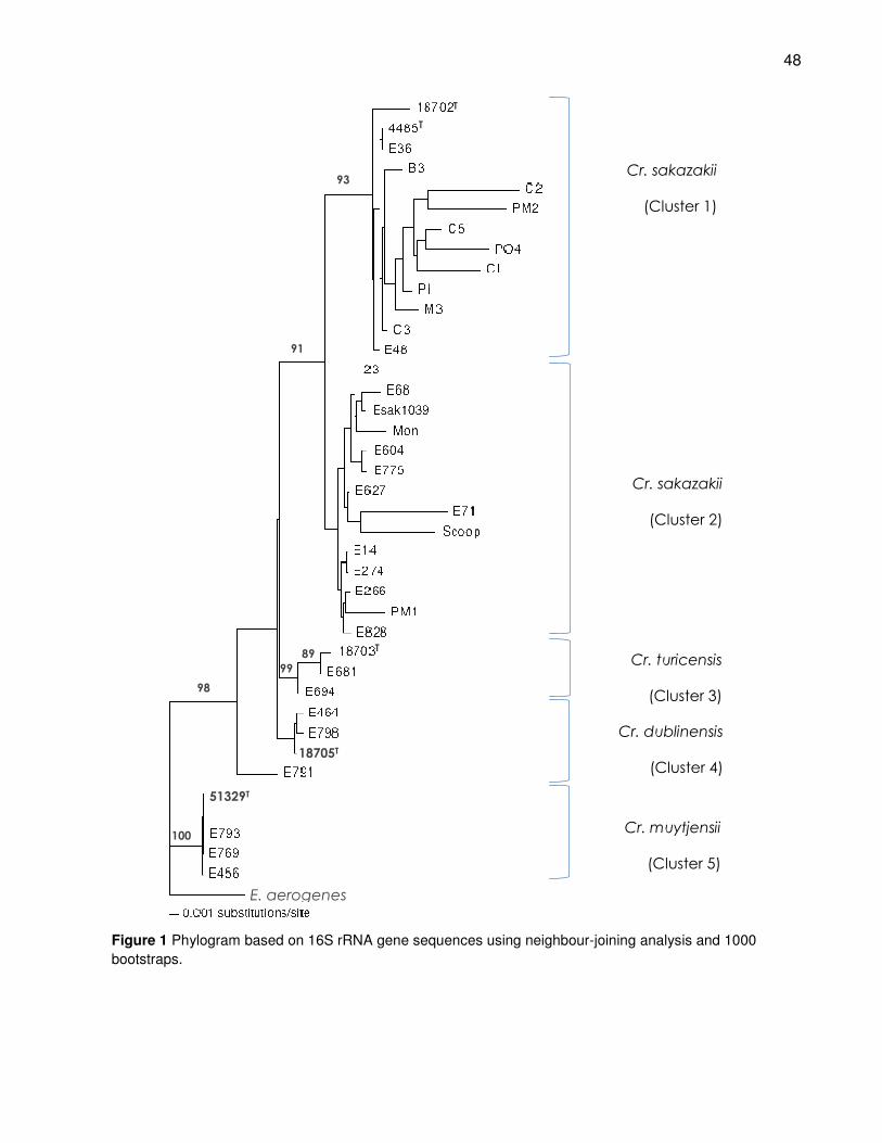
48
T
93
91
98
100
99
89
Figure 1 Phylogram based on 16S rRNA gene sequences using neighbour-joining analysis and 1000
bootstraps.
Cr. sakazakii
(Cluster 1)
Cr. sakazakii
(Cluster 2)
Cr. turicensis
(Cluster 3)
Cr. dublinensis
(Cluster 4)
Cr. muytjensii
(Cluster 5)
E. aerogenes
51329T
18705T
T
T
49
strains from GenBank and the remaining eight South African strains. The strains in cluster
2 were isolated from clinical, environmental and food sources, including wine effluent and
IFM. It is interesting to note that all the fresh produce strains were grouped with the Cr.
sakazakii type strain, which is a clinical strain. This supports the idea that fresh produce
can be a source of infection for immuno-compromised adults, especially since Cronobacter
has been shown to survive refrigeration temperatures (Nazarowec-White & Farber, 1997).
No correlation could be made between strain grouping and origin as the strains from
different sources were distributed throughout the two clusters.
These results confirm findings from other studies that evaluated the phylogenetic
relationship of Cronobacter isolates in showing that there are differences between the Cr.
sakazakii strains as confirmed by the formation of clusters 1 and 2 (Fig. 1) and that these
strains do not form clear groups based on isolation source. Miled-Bennour et al. (2010)
reported similar results after characterising of 64 Cronobacter isolates, using 16S rRNA
gene sequencing, ribotyping and pulsed field gel electrophoresis. Three subgroups
showing diversity in the genus were identified for the Cr. sakazakii strains in this study.
Although these 64 Cronobacter isolates had a high genetic diversity it was not possible to
trace a route of contamination. This indicates how difficult it can be to develop and
maintain surveillance systems for these pathogens (Miled-Bennour et al., 2010). A
phylogram from a study by Kuhnert et al. (2009) based on the recN gene sequences of
Cronobacter isolates indicated significant differences between Cr. sakazakii strains. Three
Cr. sakazakii strains only had a 0.88 % similarity with the rest of this species compared to
the more than 0.9 % similarity between strains from the rest of the Cronobacter genus.
Based on multi-locus sequence analysis of seven loci, 9 sequence types were assigned
to 60 Cr. sakazakii strains (Baldwin et al., 2009). The sequence analysis showed that Cr.
sakazakii strains isolated from different sources and countries did grouped together. One
sequence type was dominated by clinical strains (7 out of 8 strains) that were isolated from
four different countries. This merits further investigation as there was strong evidence that
this group may represent a specific virulence type of Cr. sakazakii (Baldwin et al., 2009).
No differentiation was observed in cluster 1 between the Cr. malonaticus type strain
and the Cr. sakazakii strains. The high level of similarity between Cr. sakazakii and Cr.
malonaticus have been noted on numerous occasions and sequence analysis based on
the 16S rRNA gene have been found unsuitable for differentiation between these two
species (Iversen et al., 2007; 2008; Kuhnert et al., 2009). Malonate utilisation can be used
50
for biochemical differentiation between the two species, although controversial results have
been reported when biochemical analyses of 150 isolates were compared to ribotyping
(Miled-Bennour et al., 2010). Since no differentiation between these two species was
accomplished in this study there may be isolates that have been incorrectly identified as
Cr. sakazakii and not Cr. malonaticus.
All the isolates of Cr. muytjensii formed a distinct cluster (cluster 5) from all the other
Cronobacter species with a bootstrap value of 100 %. The strains in the Cr. muytjensii
cluster did not show a high level of divergence, although they were isolated from different
countries and sources.
The Cr. turicensis strains also formed a distinct group in cluster 3, separate from all
the other isolates with a confidence value of 99 %. In the Cr. turicensis cluster strain E694
grouped separately from the type strain (DSM 18703T) and strain E681 with a bootstrap
value of 89 %.
Two isolates of Cr. dublinensis formed a distinct cluster (cluster 4) with the Cr.
dublinensis type strain (Cr. dublinensis subsp. dublinensis DSM 18705T), basal to the Cr.
sakazakii and Cr. turicensis grouping. One isolate (E791) of Cr. dublinensis was basal to
the groupings of Cr. sakazakii, Cr. turicensis and Cr. dublinensis. The different subspecies
of Cr. dublinensis may have been represented by this grouping as strain E464 was
identified as Cr. dublinensis subsp. lactaridi by Iversen et al. (2008). Strains E791 and
E798 may represent the other subspecies Cr. dublinensis subsp. lausannensis or Cr.
dublinensis subsp. dublinensis.
Phylogeny based on rpoA
In the phylogram based on the rpoA gene, the sequence of E. aerogenes had sufficient
genetic differences to distinguish this strain from the Cronobacter isolates (Fig. 2). Unlike
the analysis based on the 16S rRNA gene, the majority of the Cr. sakazakii strains all
grouped in cluster 1 with a 93 % confidence. The South African strains C3 and C5 isolated
from carrots were grouped with strains E68 and M3 that were isolated from the
environment of an IFM production facility and mincemeat, respectively. A clinical strain
from Canada (E604) and a strain isolated from a food sample in Russia (E775) grouped
together with two strains from South Africa that were isolated from IFM and the IFM
production environment. The remaining 16 Cr. sakazakii strains, with the exception of
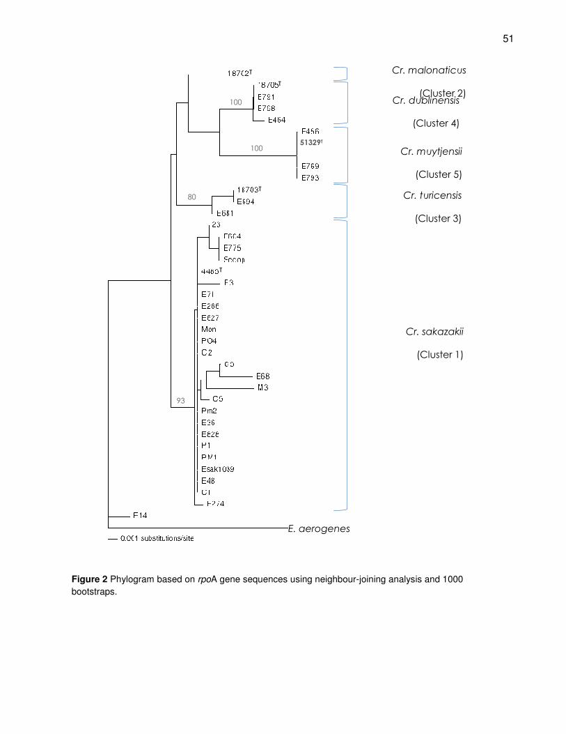
51
Figure 2 Phylogram based on rpoA gene sequences using neighbour-joining analysis and 1000
bootstraps.
93
80
100
100
Cr. sakazakii
(Cluster 1)
Cr. turicensis
(Cluster 3)
Cr. dublinensis
(Cluster 4)
Cr. malonaticus
(Cluster 2)
51329T
Cr. muytjensii
(Cluster 5)
E. aerogenes
T
T
T
T
52
strains E274 and E14, showed no differences in their rpoA sequences. Strain E274, from
the environment in Malaysia, was grouped basal to the other Cr. sakazakii strains with a
low confidence percentage of 55 %. Strain E14 showed the highest genetic diversity
based on the rpoA gene as it was separate from all the other Cronobacter strains. Based
on the phylogenetic analysis this strain does not seem to be Cronobacter, although it was
identified as Cr. sakazakii based on both the rpoA and 16S rRNA gene sequences using
the BLASTn search option in GenBank. Strains E274 and E14 were grouped together in
the 16S rRNA phylogram in cluster 2 (Fig. 1) and seem to be distant from the other Cr.
sakazakii strains.
The Cr. malonaticus type strain grouped separately from the Cr. sakazakii strains in
cluster 2, but with a low bootstrap value of 70 %. The absence of additional Cr.
malonaticus strains may have influenced this lack of differentiation between Cr. sakazakii
and Cr. malonaticus. The rpoA sequences of 48 Cronobacter isolates have also been
used to evaluate the genetic relatedness of this genus (Kuhnert et al., 2009). The results
from this study indicated that the rpoA gene sequences were divergent enough to enable
differentiation between all five Cronobacter species.
Cronobacter muytjensii strains were grouped in cluster 5 distinct from the other
Cronobacter isolates with 100 % bootstrap value. No genetic variation for the rpoA gene
sequences for these Cr. muytjensii strains was observed.
The strains belonging to Cr. turicensis were grouped separately from the rest of the
Cronobacter isolates in cluster 3 with a bootstrap value of 80 %. The Cr. turicensis type
strain (DSM 18703T) and strain E694 was separate from strain E681 as in the case of the
16S rRNA gene phylogram analysis. The recN gene sequences analysed by Kuhnert et al.
(2009) also indicated the separation of strain E681 from the Cr. turicensis type strain and
strain E694. These results indicate that there may be differences between the Cr.
turicensis strains, especially since the differences are displayed by multiple genes. This
might be related to the distinction of some strains from biogroup 16 (identified as Cr.
turicensis) which were classified as Cronobacter genomospecies 1. However, no
distinction can be made with phenotypic analysis between strains from Cr. turicensis and
strains classified as Cronobacter genomospecies 1 (Iversen et al., 2008).
The strains identified as Cr. dublinensis were clearly separated in cluster 4 from the
other Cronobacter isolates with a 100 % confidence. This grouping of the Cr. dublinensis
strains based on the rpoA gene was more distinct than in the 16S rRNA gene phylogram.
53
Additionally, strain E791 that was separated from cluster 4 in Fig.1, grouped with the rest of
the Cr. dublinensis strains. No genetic variation was observed based on the rpoA gene
between this strain and the other Cr. dublinensis strains in cluster 4 (Fig 2). This indicates
that the differences between these Cr. dublinensis strains are located in the 16S rRNA
gene and this could be the reason why the rpoA gene showed a more distinct Cr.
dublinensis cluster.
Phylogeny based on rpoA and 16S rRNA genes
The tree topologies resulting from the analysis of the rpoA and 16S rRNA gene datasets
appeared to be similar to each other. This was confirmed with the Templeton
Nonparametric Wilcoxon Signed Ranked Test (Kellogg et al., 1996) and the datasets were
combined for further phylogenetic analysis. The resulting phylogram based on the
combined sequences of the rpoA and 16S rRNA gene sequences served as a good
indication of the genetic relatedness between the Cronobacter species, relatively to the
outgroup E. aerogenes (Fig 3). All the species were clustered separately with high
bootstrap values except for Cr. malonaticus (DSM 18702T), which could not be
differentiated from Cr. sakazakii in cluster 1. The Cr. sakazakii strains were divided into
two clusters (cluster 1 and 2), although both contained strains from clinical, environmental
and food sources. The strains identified as Cr. muytjensii was shown to be least similar to
the rest of the species in cluster 5 with a bootstrap value of 100 %. This confirms findings
of a study based on the rpoA, recN and thdF genes (Kuhnert et al., 2009). Cronobacter
turicensis strains formed a group basal to Cr. sakazakii in cluster 3 with a confidence level
of 99 %. Additionally, Cr. dublinensis strains (cluster 4) were separated from the rest of the
Cronobacter isolates with a bootstrap value of only 79 %.
The phylogeny of the Cronobacter genus based on the rpoA and 16S rRNA genes
indicated that the five species were genetically closely related. However, the genus seems
to be in the process of speciation as some strains show genetic variation in specific genes,
but similarity in other genes. This was found in the case of strain E791, which was
separated from the other Cr. dublinensis strains using the 16S rRNA gene analysis, but
grouped with the Cr. dublinensis species in the rpoA based phylogram. Furthermore, strain
E274 grouped basal to the rest of the Cr. sakazakii strains and strain E14 was separated
from all the other Cronobacter isolates based on the phylogram using the rpoA gene. Both
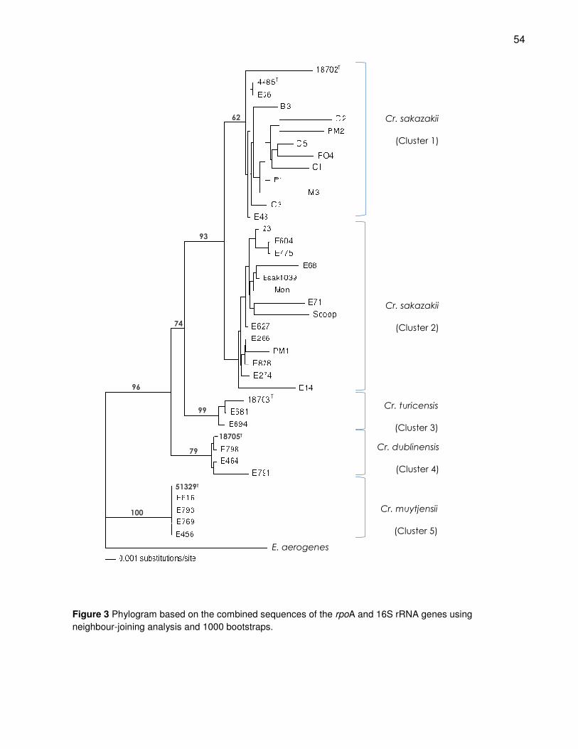
54
Figure 3 Phylogram based on the combined sequences of the rpoA and 16S rRNA genes using
neighbour-joining analysis and 1000 bootstraps.
100
96
99
93
79
74
62 Cr. sakazakii
(Cluster 1)
Cr. sakazakii
(Cluster 2)
Cr. turicensis
(Cluster 3)
Cr. dublinensis
(Cluster 4)
Cr. muytjensii
(Cluster 5)
18705T
51329T
E. aerogenes
T
T
T
55
of these strains were grouped with the other Cr. sakazakii strains in the phylograms based
on the 16S rRNA gene and the combined sequences of the rpoA and 16S rRNA genes.
Baldwin et al. (2009) evaluated the relationship between Cr. sakazakii and Cr.
malonaticus sequences of seven loci. The results of this multi-locus sequencing analysis
indicated that Cr. sakazakii showed a more clonal recombinating population. This type of
recombination is an indication of bacteria that evolve through accumulation of point
mutations. The speciation in the Cronobacter genus can also partially be clarified by
comparative genomic hybridisation (CGH) on more than 4000 genes of Cronobacter
species (Kucerova et al., 2010). A total of 15 gene clusters including putative prophages
and prophage fragments were absent in more than half of the tested strains. Additionally,
putative virulence genes were identified in most of these clusters. This indicates that the
acquisition of genes via integration or phages, as well as specific gene loss played a major
role in the evolution of Cronobacter and diversity among its species (Kucerova et al.,
2010).
Conclusions
Insight into different characteristics of pathogens is a prerequisite for improving the
reliability of risk assessment and the acquisition of data regarding the diversity of strains is
essential for the development of risk management strategies. The recent reclassification of
Cronobacter has involved the characterisation of more than 150 Cronobacter isolates and
aided in the understanding of the phylogenetic relationship of these pathogens. The
presence of these pathogens in South Africa is a cause of concern for both immuno-
compromised adults and infants as South Africa has considerable population of HIV-
infected individuals.
Phylogenetically, the species of Cronobacter are very closely related based on the
rpoA and 16S rRNA genes. These two genes were not divergent enough to sufficiently
distinguish between all the Cronobacter species and it is, therefore, advisable to use
multiple genes when further evaluating the phylogeny of this genus. When used in
combination with a biochemical analysis based on malonate utilisation, these two genes
can be used to differentiate between the Cronobacter species. It is, however, necessary to
develop a rapid and accurate typing method for the five different Cronobacter species.
References
56
Altschul, S.F., Madden, T.L., Schaffer, A.A., Zhang, Z., Miller, W. & Lipman, D.J. (1997).
Gapped BLAST and PSI-BLAST: a new generation of protein database search
programs. Nucleic Acid Research, 25, 3389-3402.
Arroyo, C., Condon, S. & Pagan, R. (2009). Thermobacteriological characterization of
Enterobacter sakazakii. International Journal of Food Microbiology, 136 (1), 110-
118.
Baldwin, A., Loughlin, M., Caubilla-Barron, J., Kucerova, E., Manning, G., Dowson, C. &
Forsythe, S. (2009). Multilocus sequence typing of Cronobacter sakazakii and
Cronobacter malonaticus reveals stable clonal structures with clinical significance
which do not correlate with biotypes. BMC Microbiology, 9, 223.
Caubilla-Barron J., Hurrell E., Townsend S., Cheetham P., Loc-Carrillo C., Fayet O., Prere
M. F., Forsythe S.J. (2007). Genotypic and phenotypic analysis of Enterobacter
sakazakii strains from an outbreak resulting in fatalities in a neonatal intensive care
unit in France. Journal of Clinical Microbiology, 45, 3979-3985.
Cawthorn, D.M., Botha, S. & Witthuhn, R.C. (2008). Evaluation of different methods for the
detection and identification of Enterobacter sakazakii isolated from South African
infant formula milks and the processing environment. International Journal of Food
Microbiology, 127 (1-2), 129-138.
Farmer, J.J., Asbury, M.A., Hickman, F.W., Brenner, D.J. & the Enterobacteriaceae Study
Group (1980). Enterobacter sakazakii, new species of Enterobacteriaceae isolated
from clinical specimens. International Journal of Systematic Bacteriology, 30, 569-
584.
Felske, A., Rheims, H., Wolterink, A., Stackebrandt, E. & Akkermans, A.D.L. (1997).
Ribosome analysis reveals prominent activity of an uncultured member of the class
Acinetobacteria in grassland soils. Microbiology, 143, 2983-2989.
Food and Agriculture Organization / World Health Organization (FAO/WHO) (2004).
Enterobacter sakazakii and other microorganisms in powdered infant formula:
Meeting Report, MRA series 6. Geneva, Switzerland. [www document]. URL
http://www.who.int/foodsafety/micro/meetings/feb2004/en/. 6 March 2009.
Food and Agriculture Organization / World Health Organization (FAO/WHO) (2008).
Enterobacter sakazakii (Cronobacter spp.) in powdered follow-up formulae: Meeting
Report, MRA series 15. Rome. [www document]. URL
http://www.who.int/foodsafety/micro/meetings/2008/en/. 11 May 2009.
57
Franke, S., Grass, G., Rensing, C. & Nies, D. H. (2003). Molecular analysis of the copper-
transporting efflux system CusCFBA of Escherichia coli. Journal of Bacteriology,
185, 3804–12.
Healy, B., Huynh, S., Mullane, N., O'Brien, S., Iversen, C., Lehner, A., Stephan, R., Parker,
C.T. & Fanning, S. (2009). Microarray-based comparative genomic indexing of the
Cronobacter genus (Enterobacter sakazakii). International Journal of Food
Microbiology, 136 (2), 159-164.
Iversen, C., Lehner, A., Mullane, N., Bidlas, E., Cleenwerck, I., Marugg, J., Fanning, S.,
Stephan, R. & Joosten, H. (2007). The taxonomy of Enterobacter sakazakii:
proposal of a new genus Cronobacter gen. nov. and descriptions of Cronobacter
sakazakii comb. nov. Cronobacter sakazakii subsp. sakazakii, comb. nov.,
Cronobacter sakazakii subsp. malonaticus subsp. nov., Cronobacter turicensis sp.
nov., Cronobacter muytjensii sp. nov., Cronobacter dublinensis sp. nov.,
Cronobacter genomospecies 1. BMC Evolutionary Biology, 64 (7), 1471-2148.
Iversen, C., Mullane, N., McCardell, B., Tall, B.D., Lehner, A., Fanning, S., Stephan, R. &
Joosten, H. (2008). Cronobacter gen. nov., a new genus to accommodate the
biogroups of Enterobacter sakazakii, and proposal of Cronobacter sakazakii gen.
nov., comb. nov., Cronobacter malonaticus sp. nov., Cronobacter turicensis sp. nov.,
Cronobacter muytjensii sp. nov., Cronobacter dublinensis sp. nov., Cronobacter
genomospecies 1, and of three subspecies, Cronobacter dublinensis subsp.
dublinensis subsp. nov., Cronobacter dublinensis subsp. lausannensis subsp. nov.
and Cronobacter dublinensis subsp. lactardi subsp. nov.. International Journal of
Systematic and Evolutionary Microbiology, 58 (6), 1442-1447.
Kellogg, E.A., Appels, R. & Mason-Gamer, R.J. (1996). When genes tell different stories:
the diploid genera of Triticeae. Systematic Bot, 21, 321-347.
Kucerova, E., Clifton, S.W., Xia, X., Long, F., Porwollik, S., Fulton, L., Fronick, C., Minx, P.,
Kyung, K.,Warren, W., Fulton, R., Feng, D., Wollam, A., Shah, N., Bhonagiri, V.,
Nash, W.E., Hallsworth-Pepin, K., Wilson, R.K., McClelland, M. & Forsythe, S.J.
(2010). Genome sequence of Cronobacter sakazakii BAA-894 and comparative
genomic hybridisation analysis with other Cronobacter species. PLoS ONE. 5 (3)
e9556. Doi: 10.1371/ journal.pone0009556.
Kuhnert, P., Korczak, B.M., Stephan, R., Joosten, H. & Iversen, C. (2009). Phylogeny and
prediction of genetic similarity of Cronobacter and related taxa by multilocus
58
sequence analysis (MLSA). International Journal of Food Microbiology, 136 (2),
152-158.
Kuzina, L.V., Peloquin, J.J., Vacek, D.C. & Miller, T.A. (2001). Isolation and identification
of bacteria associated with adult laboratory Mexican fruit flies, Anastrepha ludens.
Current Microbiology, 42, 290-294.
Lai, K.K. (2001). Enterobacter sakazakii infections among neonates, infants, children, and
adults: case reports and a review of the literature. Medicine Baltimore, 80, 113–122.
MacLean, L.L., Pagotto, F., Farber, J.M. & Perry, M.B. (2009). The structure of the O-
antigen in the endotoxin of the emerging food pathogen Cronobacter (Enterobacter)
muytjensii strain 3270. Carbohydrate Research, 344 (5), 667-671.
Miled-Bennour, R., Ells, C., Pagotto, F.J., Farber, J.M., Kerouanton, A., Meheut, T., Colin,
P., Joosten, H., Leclercq, A. & Besse, N.G. (2010). Genotypic and phenotypic
characterisation of a collection of Cronobacter (Enterobacter sakazakii) isolates.
International Journal of Food Microbiology, 139, 116–125.
Mofokeng, L., Cawthorn, D. M., Witthuhn, C.R., Anelich, L. E. M. & Jooste, P. J. (2010).
Characterization of Cronobacter species (Enterobacter Sakazakii) isolated from
various South African food sources. Journal of Food Safety, 272, 1745-4565.
Mollet, C., Drancourt, M. & Raoult, D. (1997). rpoB sequence analysis as a novel basis for
bacterial identification. Molecular Microbiology. 25 (5), 1005-1011.
Nazarowec-White, M. & Farber, J.M. (1997). Enterobacter sakazakii: a review.
International Journal of Food Microbiology, 34 (2), 103-113.
Pagotto, F.J., Nazarowec-White, M., Bidawid, S. & Farber, J.M. (2003). Enterobacter
sakazakii: infectivity and enterotoxin production in vitro and in vivo. Journal of Food
Protection, 66 (3), 370-375.
Swofford, D.L. (2001). PAUP: phylogenetic analysis using parsimony (and other methods).
Version 4.0. Sinauer Associates, Sunderland, MA, USA
Van Elsas, J. D.; Mäntynen, V.; Wolters, A.C. (1997). Soil DNA extraction and assessment
of the fate of Mycobacterium chlorophenolicum strain PCP-1 in different soil by 16S
ribosomal RNA gene sequence based most-probable-number PCR and
immunofluorescence. Biol Fertil Soils. 24,188-195.
Van Acker, J., Smet, F., Muyldermans, G., Bougatef, A., Naessens, A. & Lauwers, S.
(2001). Outbreak of necrotizing enterocolitis associated with Enterobacter sakazakii
in powdered milk formula. Journal of Clinical Microbiology, 39 (1), 293-297.

59
Wang, S. & Levin, R.E. (2006). Discrimination of viable Vibrio vulnificus cells from dead
cells in real-time PCR. Journal of Microbiological Methods, 64, 1-8.
60
CHAPTER 4
PCR-RFLP ANALYSIS OF THE rpoB GENE TO DISTINGUISH THE FIVE SPECIES OF
CRONOBACTER
Abstract
Members of the genus Cronobacter (previously known as Enterobacter sakazakii) are
opportunistic pathogens associated with life-threatening infections in immuno-compromised
individuals. Polyphasic analysis has facilitated the classification of the novel genus
Cronobacter containing five species. However, since this recent reclassification there are
not many typing methods optimised for differentiation between the five Cronobacter
species. This differentiation between the species is of importance as there are indications
that the species may be diverse regarding their virulence. The aim of this study was to
develop a PCR-RFLP protocol to differentiate between the five Cronobacter species. The
rpoB gene of 49 Enterobacteriaceae strains, including 33 Cronobacter strains was
amplified using conventional PCR, followed by digestion of these PCR products with
restriction endonucleases MboI, HinP1I and Csp6I. The PCR-RFLP analysis with single
digestions of each of the restriction endonucleases did not distinguish between all five
Cronobacter species. This study describes the successful differentiation of the five
Cronobacter species based on the amplification of the rpoB gene followed by the combined
digestion with restriction endonucleases Csp6I and HinP1I. Unique profiles for each of the
five Cronobacter species were obtained and it was also possible to differentiate between
Enterobacteriaceae and Cronobacter strains. This PCR-RFLP assay is an accurate typing
method that ensures rapid differentiation between the five species of Cronobacter.
Introduction
The genus Cronobacter contains facultative anaerobic, Gram-negative rods that belong to
the family Enterobacteriaceae (Nazarowec-White & Farber, 1997). Previously known as
Enterobacter sakazakii this genus currently consists of five species, namely Cronobacter
sakazakii, Cronobacter malonaticus, Cronobacter muytjensii, Cronobacter dublinensis and
Cronobacter turicensis. Additionally, three Cronobacter strains were grouped together and
designated as Cronobacter genomospecies 1 (Iversen et al., 2008). Cronobacter species
are opportunistic pathogens that have been associated with sporadic infections and
61
outbreaks (FAO/WHO, 2004; 2008). These bacteria may cause a severe form of neonatal
meningitis, necrotising enterocolitis and septicaemia (Nazarowec-White & Farber, 1997;
Van Acker et al., 2001). The mortality rates of these infections can vary between 10 and
80 % with fatalities occurring just days after infection (Iversen et al., 2004a).
Species of the genus Cronobacter have been reported to differ in sensitivity to
chemical reagents, heat and antibiotics (Farmer et al., 1980; Kuzina et al., 2001; Lai, 2001;
Arroyo et al., 2009) and specifically vary in the type of virulence factors expressed by these
bacteria (Pagotto et al., 2003; Healy et al., 2009; MacLean et al., 2009). Recently,
Kucerova et al. (2010) reported that only strains from three of the Cronobacter species,
namely Cr. sakazakii, Cr. malonaticus and Cr. turicensis have been associated with
neonatal infections. For these reasons, it is imperative to distinguish between the different
species of Cronobacter.
Typing methods for differentiation between the members of Cronobacter are not
readily reported in literature and isolates are identified with direct sequence analysis of the
16S ribosomal RNA (rRNA) gene. However, due to the close relationship between
Cr. sakazakii and Cr. malonaticus it is not possible to distinguish between these two
species using 16S rRNA sequence data (Iversen et al., 2007). Alternative genes such as
the rpoB gene can be used for the identification of Cronobacter species (Stoop et al.,
2009). This universal gene encodes for the bacterial RNA polymerase β-subunit and has
been evaluated for its suitability in species identification. Sequence analysis of strains
belonging to the Enterobacteriaceae family indicated that the rpoB gene was more diverse
than the 16S rRNA gene (Mollet et al., 1997). The rpoB gene can, therefore, potentially be
used as a diagnostic tool to identify and differentiate between Cronobacter strains and may
provide distinction between Cr. sakazakii and Cr. malonaticus, the two most closely related
species in this genus (Kuhnert et al., 2009).
The use of conventional PCR assays for the differentiation between bacterial strains
can be enhanced by using restriction endonucleases to digest the PCR fragments, followed
by electrophoretic analysis of the restriction fragment length polymorphisms (RFLP). The
aim of this study was to develop a PCR-RFLP assay based on the rpoB gene to
differentiate between the five Cronobacter species. Three restriction endonucleases,
namely MboI, Csp6I and HinP1I were used singly to digest the rpoB PCR fragments.
Additionally, Csp6I and HinP1I were used in a combined digestion reaction. The specificity
62
of the newly developed PCR-RFLP method was evaluated by typing Cronobacter isolates,
obtained from various environmental and food sources in South Africa.
Materials and methods
Bacterial isolates
Bacterial isolates from the Enterobacteriaceae family, including 28 Enterobacter sakazakii
(Cronobacter) strains and 16 closely related strains were evaluated in this study. Isolates
were obtained from various food and environmental sources in South Africa, including
infant formula milk (IFM) products, the IFM production facility and fresh produce. The fresh
produce strains were isolated from beetroot, carrots, potatoes, raw milk and mincemeat.
The type strains Cr. sakazakii DSM 4485T, Cr. malonaticus DSM 18702T, Cr. turicensis
DSM 18703T, Cr. dublinensis DSM 18705T (obtained from the Deutsche Sammeling von
Mikroorganismen und Zelkulturen) and Cr. muytjensii ATCC 51329T (obtained from the
American Type Culture Collection) were also included in this study. Bacterial cultures were
inoculated into tryptone soy broth (TSB) (Merck, Cape Town, South Africa) and incubated
at 37 °C for 24 h. Identification of Cronobacter spp. was done by streaking the incubated
broth on Chromocult® (Merck) followed by incubation at 42 °C for 24 h. Blue-green
colonies were regarded as Cronobacter positive. Additionally, malonate broth (Sigma-
Aldrich, Kempton Park, South Africa) was used to differentiate between Cr. sakazakii and
Cr. malonaticus isolates. The 49 isolates were inoculated into the malonate broth and
incubated at 37 °C for 24 h. A colour change in the broth from green to blue indicated a
positive result for the utilisation of malonate and these strains were recorded as
Cr. malonaticus.
DNA isolations
DNA was isolated using either a modified phenol-chloroform DNA-extraction method (van
Elsas et al., 1997) or the TZ-method described by Wang & Levin (2006). For the phenol-
chloroform method bacterial samples were prepared by centrifuging (Eppendorf Centrifuge
5415D) 2 ml of the 24 h TSB cultures at 8 000 g for 10 min. The supernatants were
discarded and 0.6 g glass beads were added to the cell pellets. Subsequently 800 µL
phosphate buffer (1 part 120 mM NaH2PO4 (Merck) to 9 parts 120 mM Na2HPO4 (Merck),
pH 8), 700 µL phenol (Fluka, supplied by Sigma-Aldrich) and 100 µL 20 % (m/v) sodium
dodecyl sulphate (SDS) (Merck) were added to the samples. The samples were then
63
subjected to vortexing for 1 to 2 min followed by incubation for 20 min at 60 °C, and this
was repeated three times. After the third incubation step the samples were centrifuged for
5 min at 4 000 g. Phenol (600 µL) was added to each supernatant, the samples were
vortexed and centrifuged for 5 min at 8 000 g. The water phase was removed and 600 µL
phenol:chloroform:isoamylalcohol (25:24:1) was added. These samples were mixed and
centrifuged for 5 min at 8 000 g, the water phase removed and repeated until the
interphase was clean. The volume of supernatant was noted and the DNA was
precipitated by adding 0.1 volume 3 M sodium acetate (NaOAc) (pH 5.5) (Saarchem,
supplied by Merck) and 0.6 volume isopropanol (Saarchem). The samples were stored
overnight at -20 °C. After centrifugation (10 min at 12 000 g), the resulting pellet was
washed with 70 % (v/v) ethanol, followed by centrifugation for 5 min at 12 000 g. The pellet
was air-dried and 100 µL of 1 x TE (10 mM Tris (Fluka), 1 mM EDTA (Merck), pH 8) buffer
was added. Samples were treated with 1 µL RNase for 1 h at 37 °C and stored at -20 °C.
The TZ-method was performed on 250 µL of the 24 h TSB cultures after the cells
were harvested by centrifugation at 8 000 g for 5 min. The pellets were resuspended in
250 µL double strength TZ (2 x TZ) (Wang & Levin, 2006) which consisted of 5 mg.mL-1
sodium azide (Merck) and 4 % (v/v) Triton X-100 (Merck) in 0.1 M Tris-HCl (Fluka) at pH 8.
Samples were boiled for 10 min to lyse the cells after which it was left to cool. The debris
was pelleted by centrifugation at 10 000 g for 5 min and 200 µL of the supernatant was
used as DNA template.
Sequencing
The identities of 28 presumptive Cronobacter isolates and 16 non-Cronobacter isolates
were determined with sequence data based on the 16S rRNA gene. The PCR
amplification of a 1.5 kilobase (kb) fragment of the 16S rRNA gene was performed using
the primers F8 (5’- CAC GGA TCC AGA CTT TGA TYM TGG CTC AG -3’) and R1512
(5’- GTG AAG CTT ACG GYT AGC TTG TTA CGA CTT -3’) (Felske et al., 1997). The PCR
reaction mixture (50 µL total volume) contained 1 µL (5 U) Taq DNA polymerase (Super-
Therm, supplied by Southern Cross Biotechnologies, Cape Town, South Africa), 5 µL 10 X
buffer (Super-Therm), 3 µL (1.5 mM) MgCl2 (Super-Therm), 2 µL (400 nM) of each primer,
2 µL (400 nM) dNTPs (AB gene, supplied by Southern Cross Biotechnologies), 2 µL 99 %
(v/v) DMSO (Merck) and 2 µL DNA template. Thermal cycling parameters (Eppendorf
mastercycler personal, Eppendorf, Germany) were as follows: initial denaturation at 92 °C
64
for 3 min; 35 cycles of denaturation at 92 °C for 30 s, annealing at 54 °C for 30 s, and
elongation at 68 °C for 60 s; and final elongation at 72 °C for 7 min (Felske et al., 1997).
Sequencing of the PCR products was performed using the ABI 3100 Genetic
Analyser (Applied Biosystems, Foster City, USA). The closest recognised relatives of the
isolates were determined by comparing the generated sequences to sequences in
GenBank, using the BLASTn search option (Altschul et al., 1997).
Amplification of the rpoB gene
Primer design
The rpoB gene of the five Cronobacter type strains, Cr. sakazakii DSM 4485T,
Cr. malonaticus DSM 18702T, Cr. turicensis DSM 18703T, Cr. dublinensis DSM 18705T and
Cr. muytjensii ATCC 51329T were used to design the primers. The rpoB sequences of
these type strains were obtained from GenBank and aligned using Multalin
(http://multalin.toulouse.inra.fr/multalin/). The conserved regions in this gene were
evaluated for suitability to be used as primers, including the affinity for dimer and hairpin
formation with Oligocal (http://www.basic.northwestern.edu/biotools/oligocalc.html).
PCR assay
Amplification of the rpoB fragment was performed using the newly designed primers CroF2
(5’- TCT CTG GGC GAT CTG GATA -3’) and CroR (5’- TGC GCG CTG ATA AGC CGCT -
3’). The PCR reaction mixture (50 µL total volume) contained 0.5 µL (2.5 U) Taq
polymerase (Super-Therm), 1 µL 10 X buffer (Super-Therm), 2.5 µL (1.25 mM) MgCl2
(Super-Therm), 1 µL (200 nM) of each primer, 1 µL (200 nM) dNTPs (AB gene) and 2.5 µL
DNA template. Thermal cycling parameters (Eppendorf mastercycler) were as follows:
initial denaturation at 92 °C for 3 min; 50 cycles of denaturation at 92 °C for 30 s, annealing
at 61.5 °C for 30 s and elongation at 68 °C for 45 s; and final elongation at 72 °C for 7 min.
PCR products were separated with electrophoresis at 90 V for 50 min on a 1.5 %
(m/v) agarose gel (Merck) containing 0.02 µl.ml-1 ethidium bromide in 0.5 X TBE
electrophoresis buffer ((5.4 g.l-1 Tris (Fluka), 2.7 g.l-1 boric acid (Merck), 2.0 ml.l-1 0.5 M
EDTA (Merck)). The amplicons were visualised under UV light (Vilber Lourmat, Marne La
Vallee, France) and a 100 bp ladder (Fermentas, supplied by Inqaba, Pretoria, South
Africa) was used as a size indicator.
65
RFLP
The type strains, Cr. sakazakii DSM 4485T, Cr. malonaticus DSM 18702T, Cr. turicensis
18703T, Cr. dublinensis DSM 18705T and Cr. muytjensii ATCC 51329T were used in
preliminary PCR-RFLP analysis in which each restriction endonuclease was tested.
Enterobacter hormaechei (strain 38) was used as a negative control as 4 of the 16 non-
Cronobacter strains were identified as E. hormaechei. Enterobacter helveticus DSM
18963T was used as an additional negative control as this species has been shown to be
closely related to Cronobacter (El-Sharoud et al., 2009). The aligned rpoB gene
sequences of the Cronobacter type strains were used to select restriction endonucleases
for the PCR-RFLP analysis. Based on the variable regions in this gene, restriction
endonucleases MboI (GATC·), Csp6I (G·TAC) and HinP1I (G·CGC) (Fast Digest®,
Fermentas) were selected.
PCR products were digested with each endonuclease separately and with a
combination of Csp6I and HinP1I. When investigating the restriction endonucleases as
individual treatments, 10 µL PCR product was added to 1 µL endonuclease and 2 µL Fast
Digest Universal Buffer® (Fermentas) in a 30 µL reaction volume. The reaction mixture of
two endonucleases included 1 µL of each endonuclease, 2 µL Fast Digest Universal
Buffer® (Fermentas) and 10 µL PCR product in a 30 µL reaction volume. Digestion took
place at 37 °C for 10 min followed by electrophoresis at 100 V for 110 min. The fragments
were separated on a 5 % (m/v) agarose gel containing 0.02 µl.ml-1 ethidium bromide in 1 X
TBE electrophoresis buffer. The PCR-RFLP profiles were visualised under UV light (Vilber
Lourmat) and an Ultra-Low Range Ladder (Fermentas) was used as a size indicator.
Results and discussion
Bacterial isolates
After 24 h incubation all the Cronobacter strains produced blue-green colonies on the
Chromocult® agar (Merck), whereas the other related Enterobacteriaceae strains produced
white colonies (Table 1, Fig. 1). The colonies of the E. helveticus strain DSM 18963T,
however, turned green after three days incubation. Strains of E. helveticus, Enterobacter
pulveris and Enterobacter turicensis are reportedly responsible for the majority of false
positive results on chromogenic agar containing the chromogen 5-bromo-4-chloro-3-
indolyl-α-D-glycopyranoside (Stephan et al., 2007). These false positive results can be
attributed to different transport mechanisms of the chromogen which is induced by sugars
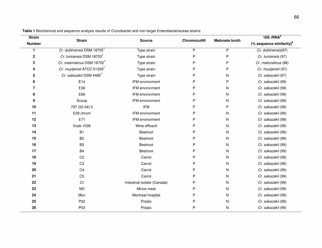
66
Table 1 Biochemical and sequence analysis results of Cronobacter and non-target Enterobacteriaceae strains
Strain
Number Strain Source Chromocult® Malonate broth
16S rRNAa
(% sequence similarity)b
1 Cr. dublinensis DSM 18705T
Type strain P P Cr. dublinensis(97)
2 Cr. turicensis DSM 18703T
Type strain P P Cr. turicensis (97)
3 Cr. malonaticus DSM 18702T
Type strain P P Cr. malonaticus (98)
4 Cr. muytjensii ATCC 51329T
Type strain P P Cr. muytjensii (97)
5 Cr. sakazakii DSM 4485T
Type strain P N Cr. sakazakii (97)
6 E14 IFM environment P P Cr. sakazakii (99)
7 E36 IFM environment P N Cr. sakazakii (99)
8 E68 IFM environment P N Cr. sakazakii (99)
9 Scoop IFM environment P N Cr. sakazakii (99)
10 797 (52-54) 5 IFM P P Cr. sakazakii (99)
11 E39 chrom IFM environment P N Cr. sakazakii (99)
12 E71 IFM environment P N Cr. sakazakii (99)
13 Esak 1039 Wine effluent P N Cr. sakazakii (99)
14 B1 Beetroot P N Cr. sakazakii (99)
15 B2 Beetroot P N Cr. sakazakii (99)
16 B3 Beetroot P N Cr. sakazakii (99)
17 B4 Beetroot P N Cr. sakazakii (99)
18 C2 Carrot P N Cr. sakazakii (99)
19 C3 Carrot P N Cr. sakazakii (99)
20 C4 Carrot P N Cr. sakazakii (99)
21 C5 Carrot P N Cr. sakazakii (99)
22 CI Industrial isolate (Canada) P N Cr. sakazakii (99)
23 M3 Mince meat P N Cr. sakazakii (99)
24 Mon Montreal hospital P N Cr. sakazakii (99)
25 P02 Potato P N Cr. sakazakii (99)
26 P03 Potato P N Cr. sakazakii (99)
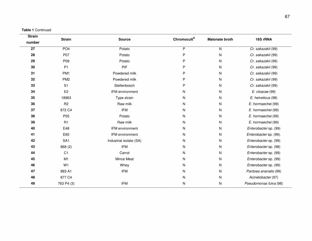
67
Table 1 Continued
Strain
number Strain Source Chromocult
® Malonate broth 16S rRNA
27 PO4 Potato P N Cr. sakazakii (99)
28 P07 Potato P N Cr. sakazakii (99)
29 P08 Potato P N Cr. sakazakii (99)
30 P1 PIF P N Cr. sakazakii (99)
31 PM1 Powdered milk P N Cr. sakazakii (99)
32 PM2 Powdered milk P N Cr. sakazakii (99)
33 S1 Stellenbosch P N Cr. sakazakii (99)
34 E2 IFM environment N N E. cloacae (99)
35 18963 Type strain N N E. helveticus (98)
36 R2 Raw milk N N E. hormaechei (99)
37 872 C4 IFM N N E. hormaechei (99)
38 P05 Potato N N E. hormaechei (99)
39 R1 Raw milk N N E. hormaechei (99)
40 E48 IFM environment N N Enterobacter sp. (99)
41 E65 IFM environment N N Enterobacter sp. (99).
42 SA1 Industrial isolate (SA) N N Enterobacter sp. (99)
43 868 (2) IFM N N Enterobacter sp. (98)
44 C1 Carrot N N Enterobacter sp. (99)
45 M1 Mince Meat N N Enterobacter sp. (99)
46 W1 Whey N N Enterobacter sp. (99)
47 883 A1 IFM N N Pantoea ananatis (99)
48 877 C4 N N Acinetobacter (97)
49 763 P4 (3) IFM N N Pseudomonas fulva (98)
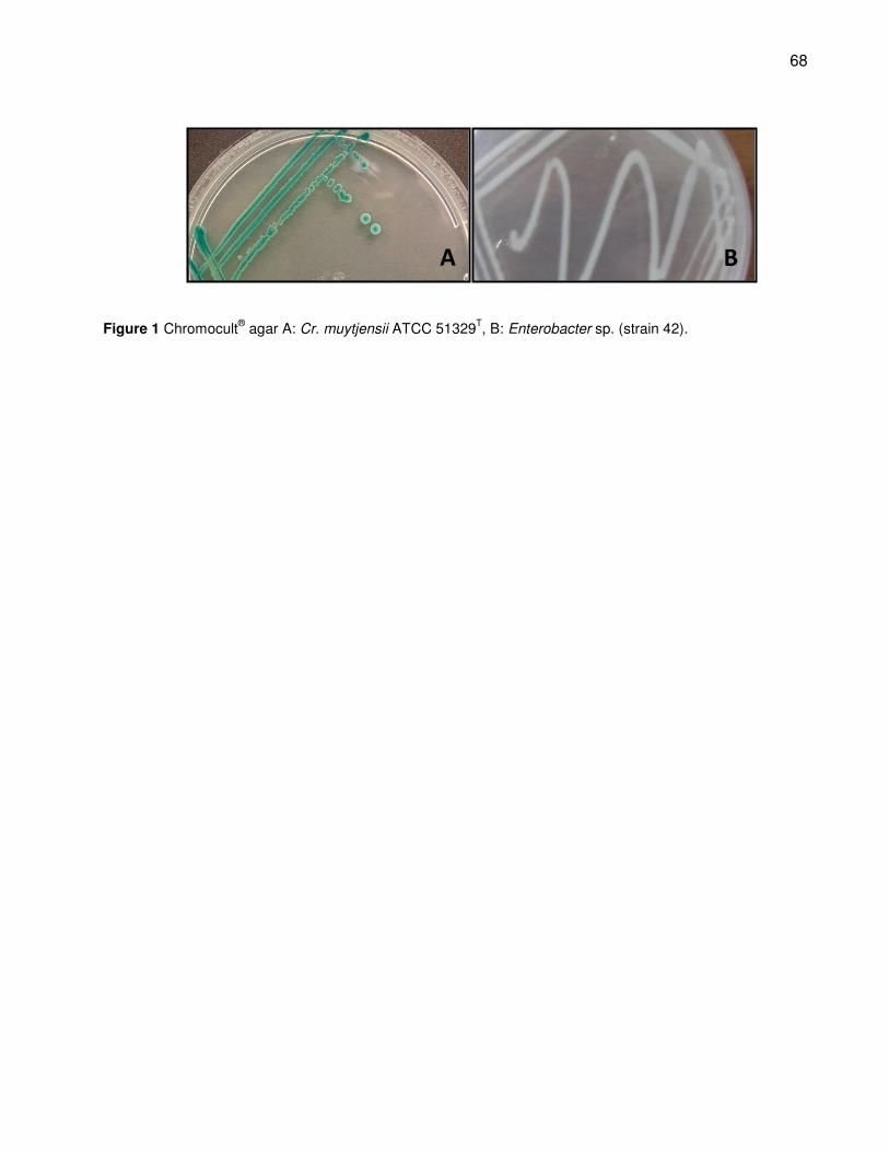
68
Figure 1 Chromocult® agar A: Cr. muytjensii ATCC 51329
T, B: Enterobacter sp. (strain 42).
A B
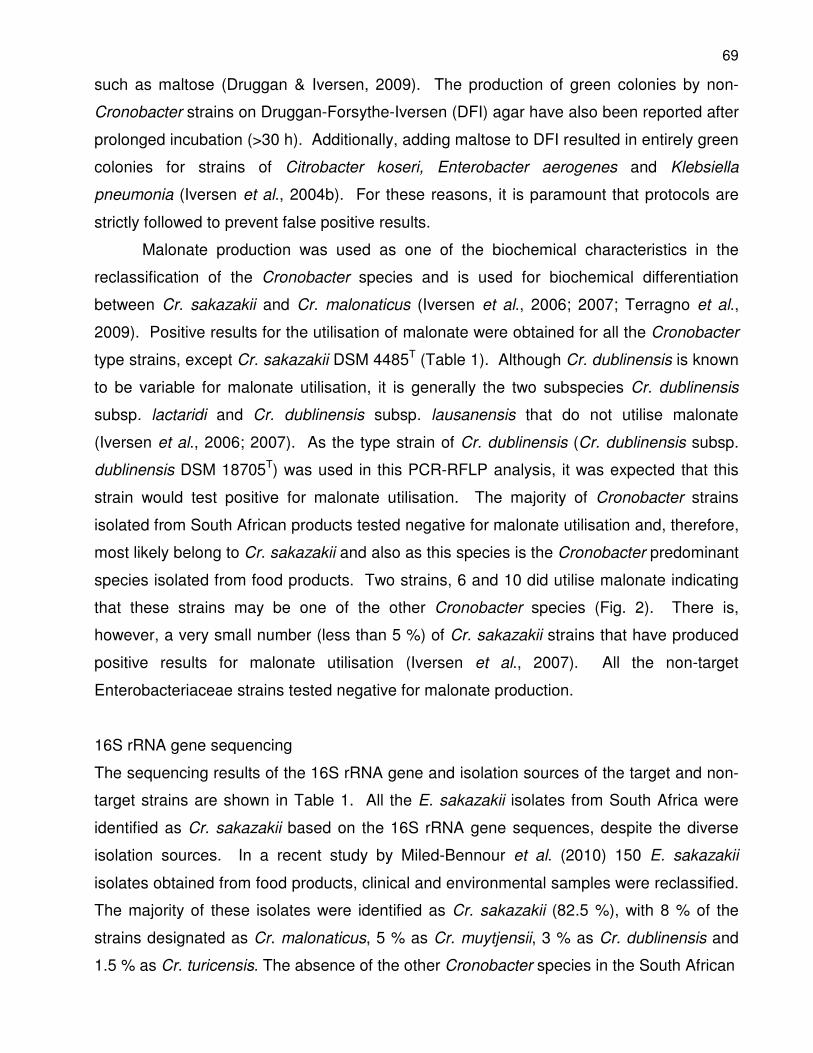
69
such as maltose (Druggan & Iversen, 2009). The production of green colonies by non-
Cronobacter strains on Druggan-Forsythe-Iversen (DFI) agar have also been reported after
prolonged incubation (>30 h). Additionally, adding maltose to DFI resulted in entirely green
colonies for strains of Citrobacter koseri, Enterobacter aerogenes and Klebsiella
pneumonia (Iversen et al., 2004b). For these reasons, it is paramount that protocols are
strictly followed to prevent false positive results.
Malonate production was used as one of the biochemical characteristics in the
reclassification of the Cronobacter species and is used for biochemical differentiation
between Cr. sakazakii and Cr. malonaticus (Iversen et al., 2006; 2007; Terragno et al.,
2009). Positive results for the utilisation of malonate were obtained for all the Cronobacter
type strains, except Cr. sakazakii DSM 4485T (Table 1). Although Cr. dublinensis is known
to be variable for malonate utilisation, it is generally the two subspecies Cr. dublinensis
subsp. lactaridi and Cr. dublinensis subsp. lausanensis that do not utilise malonate
(Iversen et al., 2006; 2007). As the type strain of Cr. dublinensis (Cr. dublinensis subsp.
dublinensis DSM 18705T) was used in this PCR-RFLP analysis, it was expected that this
strain would test positive for malonate utilisation. The majority of Cronobacter strains
isolated from South African products tested negative for malonate utilisation and, therefore,
most likely belong to Cr. sakazakii and also as this species is the Cronobacter predominant
species isolated from food products. Two strains, 6 and 10 did utilise malonate indicating
that these strains may be one of the other Cronobacter species (Fig. 2). There is,
however, a very small number (less than 5 %) of Cr. sakazakii strains that have produced
positive results for malonate utilisation (Iversen et al., 2007). All the non-target
Enterobacteriaceae strains tested negative for malonate production.
16S rRNA gene sequencing
The sequencing results of the 16S rRNA gene and isolation sources of the target and non-
target strains are shown in Table 1. All the E. sakazakii isolates from South Africa were
identified as Cr. sakazakii based on the 16S rRNA gene sequences, despite the diverse
isolation sources. In a recent study by Miled-Bennour et al. (2010) 150 E. sakazakii
isolates obtained from food products, clinical and environmental samples were reclassified.
The majority of these isolates were identified as Cr. sakazakii (82.5 %), with 8 % of the
strains designated as Cr. malonaticus, 5 % as Cr. muytjensii, 3 % as Cr. dublinensis and
1.5 % as Cr. turicensis. The absence of the other Cronobacter species in the South African
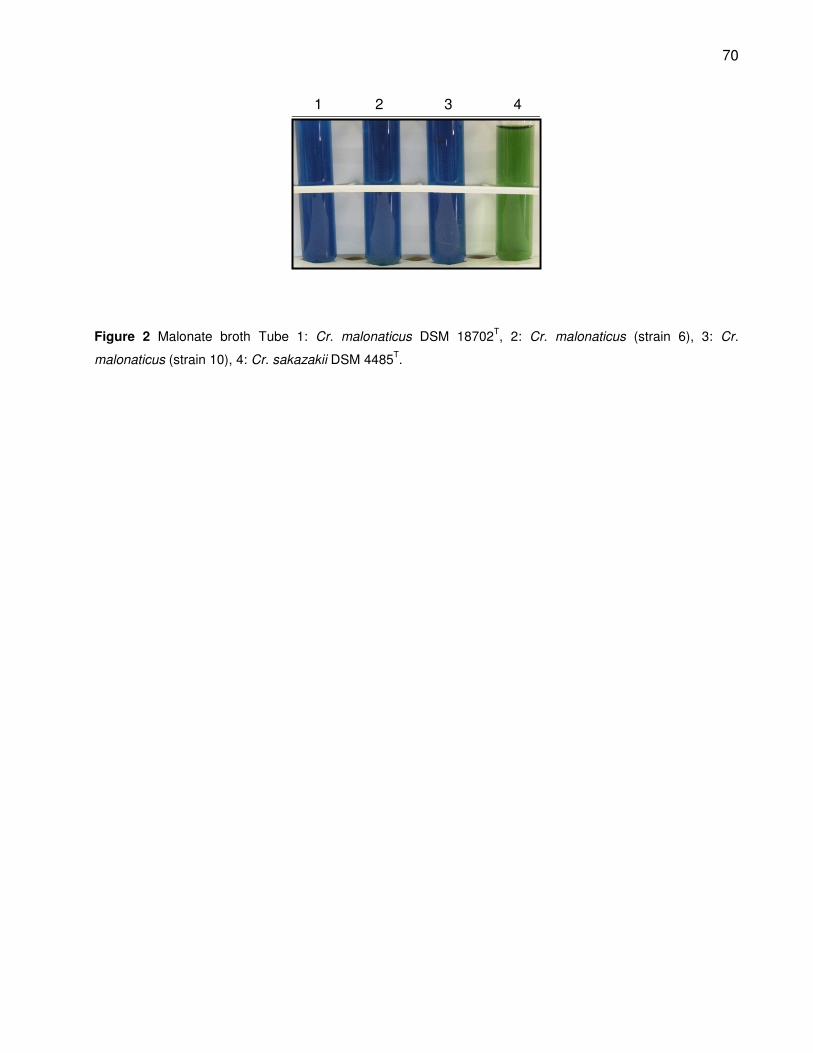
70
1 2 3 4
Figure 2 Malonate broth Tube 1: Cr. malonaticus DSM 18702T, 2: Cr. malonaticus (strain 6), 3: Cr.
malonaticus (strain 10), 4: Cr. sakazakii DSM 4485T.
71
isolates may, therefore, be contributed to the smaller number of isolates evaluated in this
study and the fact that Cr. sakazakii is the dominant species in terms of isolation
frequency. The majority of the 16 non-target strains were identified as Enterobacter spp.
Strains 36 to 39 that were isolated from milk products and fresh produce were identified as
E. hormaechei. Another three of the non-target strains isolated from IFM were identified as
Pantoea ananatis, Acinetobacter sp. and Pseudomonas fulva.
PCR-RFLP
The aligned sequences of the Cronobacter type strains indicated several conserved
regions within the rpoB gene (Fig. 3). The blocks highlighted in Fig. 3 indicate the
conserved regions that were used for the design of the primers to ensure amplification of a
660 bp fragment of all five Cronobacter species. The oligonucleotide used as forward
primer CroF2 is 19 bp long and has a 53 % GC content. The reverse primer, CroR, was
also 19 bp long with a GC % content of 63 %. Both primers had no affinity for secondary
structure formation such as dimers and hairpins. Amplification of the partial region of the
rpoB gene between basepairs 328247 and 328907 in the genome was successful for all
the target and non-target strains (Fig. 4). An unusually large number of cycles (50 cycles)
during PCR amplification provided sufficient amplicons for digestion with the restriction
endonucleases. The fragments in the profiles ranged from 20 to 300 bp. Fragments
smaller than 50 bp were not always clearly visible and, therefore, not included in defining
profiles.
Single restriction endonuclease digestion
Digestion with MboI resulted in unique profiles for only Cr. dublinensis, Cr. muytjensii and
Cr. turicensis as identical profiles were obtained for Cr. sakazakii and Cr. malonaticus (Fig.
5). Unlike the theoretical profiles based on the sequences available in GenBank,
Cr. muytjensii had an identical profile to E. hormaechei. The PCR-RFLP assay with MboI
was, therefore, unsuitable for differentiation between the Cronobacter species, as well as
differentiating between Cronobacter and Enterobacteriaceae strains.
Restriction digestion of the type strains and two negative controls with Csp61I
resulted in unique profiles for Cr. sakazakii and E. helveticus (Fig. 6). Cronobacter
muytjensii and Cr. dublinensis had identical profiles as predicted by the theoretical profiles.
These two profiles were, however, also similar to the profile of Cr. malonaticus due to the

72
Figure 3 Alignment of rpoB gene sequences of five Cronobacter strains (Block A indicates the forward primer (Crof2) and B the reverse primer (CroR)).
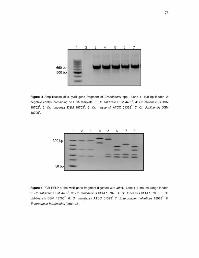
73
1 2 3 4 5 6 7
660 bp
500 bp
Figure 4 Amplification of a rpoB gene fragment of Cronobacter spp. Lane 1: 100 bp ladder, 2:
negative control containing no DNA template, 3: Cr. sakazakii DSM 4485T, 4: Cr. malonaticus DSM
18702T, 5: Cr. turicensis DSM 18703
T, 6: Cr. muytjensii ATCC 51329
T, 7: Cr. dublinensis DSM
18705T.
Figure 5 PCR-RFLP of the rpoB gene fragment digested with MboI. Lane 1: Ultra low range ladder,
2: Cr. sakazakii DSM 4485T, 3: Cr. malonaticus DSM 18702
T, 4: Cr. turicensis DSM 18703
T, 5: Cr.
dublinensis DSM 18705T, 6: Cr. muytjensii ATCC 51329
T 7: Enterobacter helveticus 18963
T, 8:
Enterobacter hormaechei (strain 38).
1 2 3 4 5 6 7 8
50 bp
300 bp
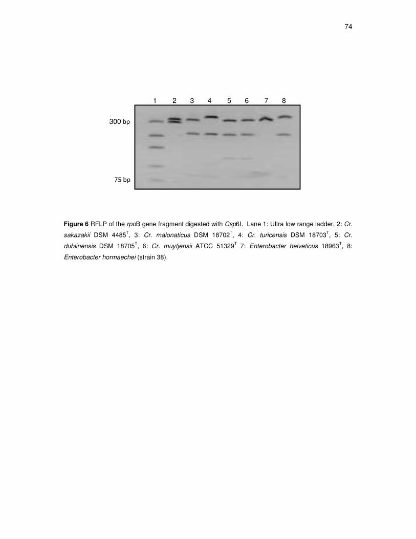
74
1 2 3 4 5 6 7 8
75 bp
300 bp
Figure 6 RFLP of the rpoB gene fragment digested with Csp6I. Lane 1: Ultra low range ladder, 2: Cr.
sakazakii DSM 4485T, 3: Cr. malonaticus DSM 18702
T, 4: Cr. turicensis DSM 18703
T, 5: Cr.
dublinensis DSM 18705T, 6: Cr. muytjensii ATCC 51329
T 7: Enterobacter helveticus 18963
T, 8:
Enterobacter hormaechei (strain 38).
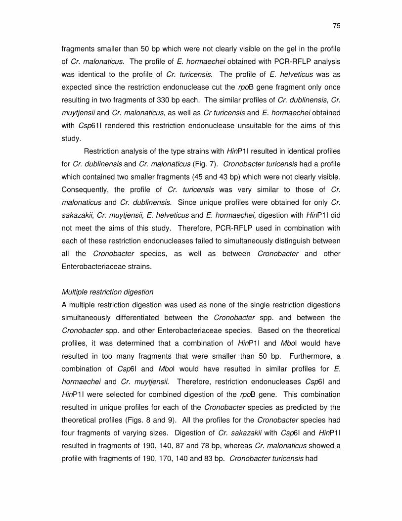
75
fragments smaller than 50 bp which were not clearly visible on the gel in the profile
of Cr. malonaticus. The profile of E. hormaechei obtained with PCR-RFLP analysis
was identical to the profile of Cr. turicensis. The profile of E. helveticus was as
expected since the restriction endonuclease cut the rpoB gene fragment only once
resulting in two fragments of 330 bp each. The similar profiles of Cr. dublinensis, Cr.
muytjensii and Cr. malonaticus, as well as Cr turicensis and E. hormaechei obtained
with Csp61I rendered this restriction endonuclease unsuitable for the aims of this
study.
Restriction analysis of the type strains with HinP1I resulted in identical profiles
for Cr. dublinensis and Cr. malonaticus (Fig. 7). Cronobacter turicensis had a profile
which contained two smaller fragments (45 and 43 bp) which were not clearly visible.
Consequently, the profile of Cr. turicensis was very similar to those of Cr.
malonaticus and Cr. dublinensis. Since unique profiles were obtained for only Cr.
sakazakii, Cr. muytjensii, E. helveticus and E. hormaechei, digestion with HinP1I did
not meet the aims of this study. Therefore, PCR-RFLP used in combination with
each of these restriction endonucleases failed to simultaneously distinguish between
all the Cronobacter species, as well as between Cronobacter and other
Enterobacteriaceae strains.
Multiple restriction digestion
A multiple restriction digestion was used as none of the single restriction digestions
simultaneously differentiated between the Cronobacter spp. and between the
Cronobacter spp. and other Enterobacteriaceae species. Based on the theoretical
profiles, it was determined that a combination of HinP1I and MboI would have
resulted in too many fragments that were smaller than 50 bp. Furthermore, a
combination of Csp6I and MboI would have resulted in similar profiles for E.
hormaechei and Cr. muytjensii. Therefore, restriction endonucleases Csp6I and
HinP1I were selected for combined digestion of the rpoB gene. This combination
resulted in unique profiles for each of the Cronobacter species as predicted by the
theoretical profiles (Figs. 8 and 9). All the profiles for the Cronobacter species had
four fragments of varying sizes. Digestion of Cr. sakazakii with Csp6I and HinP1I
resulted in fragments of 190, 140, 87 and 78 bp, whereas Cr. malonaticus showed a
profile with fragments of 190, 170, 140 and 83 bp. Cronobacter turicensis had
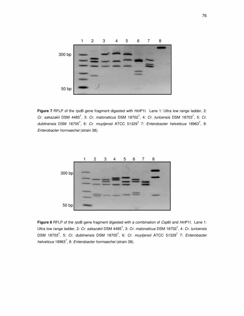
76
1 2 3 4 5 6 7 8
1 2 3 4 5 6 7 8
50 bp
300 bp
50 bp
300 bp
Figure 7 RFLP of the rpoB gene fragment digested with HinP1I. Lane 1: Ultra low range ladder, 2:
Cr. sakazakii DSM 4485T, 3: Cr. malonaticus DSM 18702
T, 4: Cr. turicensis DSM 18703
T, 5: Cr.
dublinensis DSM 18705T, 6: Cr. muytjensii ATCC 51329
T 7: Enterobacter helveticus 18963
T, 8:
Enterobacter hormaechei (strain 38).
Figure 8 RFLP of the rpoB gene fragment digested with a combination of Csp6I and HinP1I. Lane 1:
Ultra low range ladder, 2: Cr. sakazakii DSM 4485T, 3: Cr. malonaticus DSM 18702
T, 4: Cr. turicensis
DSM 18703T, 5: Cr. dublinensis DSM 18705
T, 6: Cr. muytjensii ATCC 51329
T 7: Enterobacter
helveticus 18963T, 8: Enterobacter hormaechei (strain 38).
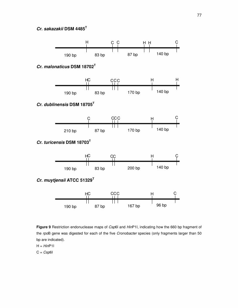
77
Cr. sakazakii DSM 4485T
Cr. malonaticus DSM 18702T
Cr. dublinensis DSM 18705T
Cr. turicensis DSM 18703T
Cr. muytjensii ATCC 51329T
Figure 9 Restriction endonuclease maps of Csp6I and HinP1I, indicating how the 660 bp fragment of
the rpoB gene was digested for each of the five Cronobacter species (only fragments larger than 50
bp are indicated).
H = HinP1I
C = Csp6I
140 bp 170 bp 190 bp 83 bp
H C H H C C C
140 bp 200 bp 190 bp 83 bp
H C H C C C
140 bp 170 bp 210 bp 87 bp
C H C C C C
96 bp 167 bp 190 bp 87 bp
H C H C C C C
140 bp 87 bp 190 bp 83 bp
H C H H C C
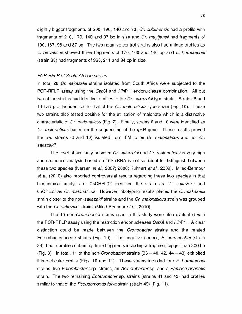
78
slightly bigger fragments of 200, 190, 140 and 83, Cr. dublinensis had a profile with
fragments of 210, 170, 140 and 87 bp in size and Cr. muytjensii had fragments of
190, 167, 96 and 87 bp. The two negative control strains also had unique profiles as
E. helveticus showed three fragments of 170, 160 and 140 bp and E. hormaechei
(strain 38) had fragments of 365, 211 and 84 bp in size.
PCR-RFLP of South African strains
In total 28 Cr. sakazakii strains isolated from South Africa were subjected to the
PCR-RFLP assay using the Csp6I and HinP1I endonuclease combination. All but
two of the strains had identical profiles to the Cr. sakazakii type strain. Strains 6 and
10 had profiles identical to that of the Cr. malonaticus type strain (Fig. 10). These
two strains also tested positive for the utilisation of malonate which is a distinctive
characteristic of Cr. malonaticus (Fig. 2). Finally, strains 6 and 10 were identified as
Cr. malonaticus based on the sequencing of the rpoB gene. These results proved
the two strains (6 and 10) isolated from IFM to be Cr. malonaticus and not Cr.
sakazakii.
The level of similarity between Cr. sakazakii and Cr. malonaticus is very high
and sequence analysis based on 16S rRNA is not sufficient to distinguish between
these two species (Iversen et al., 2007; 2008; Kuhnert et al., 2009). Miled-Bennour
et al. (2010) also reported controversial results regarding these two species in that
biochemical analysis of 05CHPL02 identified the strain as Cr. sakazakii and
05CPL53 as Cr. malonaticus. However, ribotyping results placed the Cr. sakazakii
strain closer to the non-sakazakii strains and the Cr. malonaticus strain was grouped
with the Cr. sakazakii strains (Miled-Bennour et al., 2010).
The 15 non-Cronobacter stains used in this study were also evaluated with
the PCR-RFLP assay using the restriction endonucleases Csp6I and HinP1I. A clear
distinction could be made between the Cronobacter strains and the related
Enterobacteriaceae strains (Fig. 10). The negative control, E. hormaechei (strain
38), had a profile containing three fragments including a fragment bigger than 300 bp
(Fig. 8). In total, 11 of the non-Cronobacter strains (36 – 40, 42, 44 – 48) exhibited
this particular profile (Figs. 10 and 11). These strains included four E. hormaechei
strains, five Enterobacter spp. strains, an Acinetobacter sp. and a Pantoea ananatis
strain. The two remaining Enterobacter sp. strains (strains 41 and 43) had profiles
similar to that of the Pseudomonas fulva strain (strain 49) (Fig. 11).
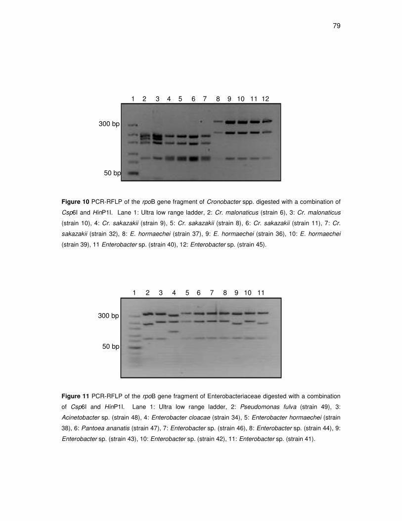
79
50 bp
300 bp
1 2 3 4 5 6 7 8 9 10 11 12
50 bp
1 2 3 4 5 6 7 8 9 10 11
300 bp
Figure 10 PCR-RFLP of the rpoB gene fragment of Cronobacter spp. digested with a combination of
Csp6I and HinP1I. Lane 1: Ultra low range ladder, 2: Cr. malonaticus (strain 6), 3: Cr. malonaticus
(strain 10), 4: Cr. sakazakii (strain 9), 5: Cr. sakazakii (strain 8), 6: Cr. sakazakii (strain 11), 7: Cr.
sakazakii (strain 32), 8: E. hormaechei (strain 37), 9: E. hormaechei (strain 36), 10: E. hormaechei
(strain 39), 11 Enterobacter sp. (strain 40), 12: Enterobacter sp. (strain 45).
Figure 11 PCR-RFLP of the rpoB gene fragment of Enterobacteriaceae digested with a combination
of Csp6I and HinP1I. Lane 1: Ultra low range ladder, 2: Pseudomonas fulva (strain 49), 3:
Acinetobacter sp. (strain 48), 4: Enterobacter cloacae (strain 34), 5: Enterobacter hormaechei (strain
38), 6: Pantoea ananatis (strain 47), 7: Enterobacter sp. (strain 46), 8: Enterobacter sp. (strain 44), 9:
Enterobacter sp. (strain 43), 10: Enterobacter sp. (strain 42), 11: Enterobacter sp. (strain 41).
80
Lastly the E. cloacae strain had an unique profile with fragments of 300, 210 and 140 bp
(Fig. 11). Although there were differences between the non-Cronobacter strains, there was
not enough evidence to support any conclusions. This particular gene and endonuclease
combination is not sufficient to distinguish between non-Cronobacter strains as some of
these species had similar profiles.
Conclusions
The formation of risk management strategies is essential to produce food products that are
safe for consumption. The recent reclassification of Enterobacter sakazakii to Cronobacter
simplified the characterisation of these pathogens and, therefore, assisted in the formation
of effective risk management strategies. The definition of the five species has, however,
led to the challenge of designing rapid and reliable typing methods that can differentiate
between these species. The ability to differentiate between the Cronobacter species is
particularly important since there are indications that these species do not display the same
virulence traits. This study described the development of a novel PCR-RFLP typing
method for the Cronobacter species that can be used as a reliable alternative to
sequencing based on 16S rRNA gene sequences. The PCR-RFLP assay is based on the
rpoB gene and used in combination with restriction endonucleases Csp6I and HinP1I. The
results from this study indicated that the rpoB gene of the Cronobacter spp. is divergent
enough to be used in typing methods. This assay can also be used to differentiate
between Cronobacter and other Enterobacteriaceae strains, however, the PCR-RFLP
assay was not optimised for species other than Cronobacter.
References
Altschul, S.F., Madden, T.L., Schaffer, A.A., Zhang, Z., Miller, W. & Lipman, D.J. (1997).
Gapped BLAST and PSI-BLAST: a new generation of protein database search
programs. Nucleic Acid Research, 25, 3389-3402.
Arroyo, C., Condon, S. & Pagan, R. (2009). Thermobacteriological characterization of
Enterobacter sakazakii. International Journal of Food Microbiology, 136 (1), 110-
118.
Druggan, P. & Iversen, C. (2009). Culture media for the isolation of Cronobacter spp.
International Journal of Food Microbiology, 136 (2), 169-178.
81
El-Sharoud, W., O'Brien, S., Negredo, C., Iversen, C., Fanning, S. & Healy, B. (2009).
Characterization of Cronobacter recovered from dried milk and related products.
BMC Microbiology, 9, 9.
Farmer, J.J., Asbury, M.A., Hickman, F.W., Brenner, D.J. & the Enterobacteriaceae Study
Group (1980). Enterobacter sakazakii, new species of Enterobacteriaceae isolated
from clinical specimens. International Journal of Systematic Bacteriology, 30, 569-
584.
Felske, A., Rheims, H., Wolterink, A., Stackebrandt, E. & Akkermans, A.D.L. (1997).
Ribosome analysis reveals prominent activity of an uncultured member of the class
Acinetobacteria in grassland soils. Microbiology, 143, 2983-2989.
Food and Agriculture Organization / World Health Organization (FAO/WHO) (2004).
Enterobacter sakazakii and other microorganisms in powdered infant formula:
Meeting Report, MRA series 6. Geneva, Switzerland. [www document]. URL
http://www.who.int/foodsafety/micro/meetings/feb2004/en/. 6 March 2009.
Food and Agriculture Organization / World Health Organization (FAO/WHO) (2008).
Enterobacter sakazakii (Cronobacter spp.) in powdered follow-up formulae: Meeting
Report, MRA series 15. Rome. [www document]. URL
http://www.who.int/foodsafety/micro/meetings/2008/en/. 11 May 2009.
Healy, B., Huynh, S., Mullane, N., O'Brien, S., Iversen, C., Lehner, A., Stephan, R., Parker,
C.T. & Fanning, S. (2009). Microarray-based comparative genomic indexing of the
Cronobacter genus (Enterobacter sakazakii). International Journal of Food
Microbiology, 136 (2), 159-164.
Iversen, C., Druggan, P. & Forsythe, S. (2004a). A selective differential medium for
Enterobacter sakazakii, a preliminary study. International Journal of Food
Microbiology, 96 (2), 133-139.
Iversen, C., Waddington, M., On, S.L.W. & Forsythe, S. (2004b). Identification and
phylogeny of Enterobacter sakazakii relative to Enterobacter and Citrobacter
species. Journal of Clinical Microbiology, 42 (11), 5368-5370.
Iversen, C., Waddington, M., Farmer, J. J. & Forsythe, S. J. (2006). The biochemical
differentiation of Enterobacter sakazakii genotypes. BMC Microbiology 6 (94).
Iversen, C., Lehner, A., Mullane, N., Bidlas, E., Cleenwerck, I., Marugg, J., Fanning, S.,
Stephan, R. & Joosten, H. (2007). The taxonomy of Enterobacter sakazakii:
proposal of a new genus Cronobacter gen. nov. and descriptions of Cronobacter
82
sakazakii comb. nov. Cronobacter sakazakii subsp. sakazakii, comb. nov.,
Cronobacter sakazakii subsp. malonaticus subsp. nov., Cronobacter turicensis sp.
nov., Cronobacter muytjensii sp. nov., Cronobacter dublinensis sp. nov.,
Cronobacter genomospecies 1. BMC Evolutionary Biology, 64 (7), 1471-2148.
Iversen, C., Mullane, N., McCardell, B., Tall, B.D., Lehner, A., Fanning, S., Stephan, R. &
Joosten, H. (2008). Cronobacter gen. nov., a new genus to accommodate the
biogroups of Enterobacter sakazakii, and proposal of Cronobacter sakazakii gen.
nov., comb. nov., Cronobacter malonaticus sp. nov., Cronobacter turicensis sp. nov.,
Cronobacter muytjensii sp. nov., Cronobacter dublinensis sp. nov., Cronobacter
genomospecies 1, and of three subspecies, Cronobacter dublinensis subsp.
dublinensis subsp. nov., Cronobacter dublinensis subsp. lausannensis subsp. nov.
and Cronobacter dublinensis subsp. lactaridi subsp. nov. International Journal of
Systematic and Evolutionary Microbiology, 58 (6), 1442-1447.
Kucerova, E., Clifton, S.W., Xia, X., Long, F., Porwollik, S., Fulton, L., Fronick, C., Minx, P.,
Kyung, K.,Warren, W., Fulton, R., Feng, D., Wollam, A., Shah, N., Bhonagiri, V.,
Nash, W.E., Hallsworth-Pepin, K., Wilson, R.K., McClelland, M. & Forsythe, S.J.
(2010). Genome sequence of Cronobacter sakazakii BAA-894 and comparative
genomic hybridisation analysis with other Cronobacter species. PLoS ONE. 5 (3)
e9556. Doi: 10.1371/ journal.pone0009556.
Kuhnert, P., Korczak, B.M., Stephan, R., Joosten, H. & Iversen, C. (2009). Phylogeny and
prediction of genetic similarity of Cronobacter and related taxa by multilocus
sequence analysis (MLSA). International Journal of Food Microbiology, 136 (2),
152-158.
Kuzina, L.V., Peloquin, J.J., Vacek, D.C. & Miller, T.A. (2001). Isolation and identification
of bacteria associated with adult laboratory Mexican fruit flies, Anastrepha ludens.
Current Microbiology, 42, 290-294.
Lai, K.K. (2001). Enterobacter sakazakii infections among neonates, infants, children, and
adults: case reports and a review of the literature. Medicine Baltimore, 80, 113–122.
MacLean, L.L., Pagotto, F., Farber, J.M. & Perry, M.B. (2009). The structure of the O-
antigen in the endotoxin of the emerging food pathogen Cronobacter (Enterobacter)
muytjensii strain 3270. Carbohydrate Research, 344 (5), 667-671.
Miled-Bennour, R., Ells, C., Pagotto, F.J., Farber, J.M., Kerouanton, A., Meheut, T., Colin,
P., Joosten, H., Leclercq, A. & Besse, N.G. (2010). Genotypic and phenotypic
83
characterisation of a collection of Cronobacter (Enterobacter sakazakii) isolates.
International Journal of Food Microbiology, 139, 116–125.
Mollet, C., Drancourt, M. & Raoult, D. (1997). rpoB sequence analysis as a novel basis for
bacterial identification. Molecular Microbiology. 25 (5), 1005-1011.
Nazarowec-White, M. & Farber, J.M. (1997). Enterobacter sakazakii: a review.
International Journal of Food Microbiology, 34 (2), 103-113.
Pagotto, F.J., Nazarowec-White, M., Bidawid, S. & Farber, J.M. (2003). Enterobacter
sakazakii: infectivity and enterotoxin production in vitro and in vivo. Journal of Food
Protection, 66 (3), 370-375.
Stephan,R., Van Trappen, S., Cleenwerck, I., Vancanneyt, M., De Vos, P. & Lehner, A.
(2007). Enterobacter turicensis sp. nov. and Enterobacter helveticus sp. nov.,
isolated from fruit powder. International Journal of Systematic and Evolutionary
Microbiology, 57, 820–826.
Stoop, B., Lehner, A., Iversen, C., Fanning, S. & Stephan, R. (2009). Development and
evaluation of rpoB based PCR systems to differentiate the six proposed species
within the genus Cronobacter. International Journal of Food Microbiology, 136 (2),
165-168.
Terragno, R., Salve, A., Pichel, M., Epszteyn, S., Brengi, S. & Binsztein, N. (2009).
Characterization and subtyping of Cronobacter spp. from imported powdered infant
formulae in Argentina. International Journal of Food Microbiology, 136 (2), 193-197.
Van Acker, J., Smet, F., Muyldermans, G., Bougatef, A., Naessens, A. & Lauwers, S.
(2001). Outbreak of necrotizing enterocolitis associated with Enterobacter sakazakii
in powdered milk formula. Journal of Clinical Microbiology, 39 (1), 293-297.
Van Elsas, J. D.; Mäntynen, V.; Wolters, A.C. (1997). Soil DNA extraction and assessment
of the fate of Mycobacterium chlorophenolicum strain PCP-1 in different soil by 16S
ribosomal RNA gene sequence based most-probable-number PCR and
immunofluorescence. Biol Fertil Soils. 24,188-195.
Wang, S. & Levin, R.E. (2006). Discrimination of viable Vibrio vulnificus cells from dead
cells in real-time PCR. Journal of Microbiological Methods, 64, 1-8.
84
CHAPTER 5
GENERAL DISCUSSION AND CONCLUSIONS
The genus Cronobacter (previously known as Enterobacter sakazakii) can cause
foodborne infections such as neonatal meningitis, septicaemia and necrotising enterocolitis
(Nazarowec-White & Farber, 1997; Van Acker et al., 2001). These pathogens have been
associated with disease outbreaks, as well as infections and are a health concern for
immuno-compromised individuals, especially premature infants (FAO/WHO, 2004). The
number of Cronobacter related infections in South Africa (SA) is unknown although these
pathogens have been isolated from environmental sources and food produced in SA.
Given the risk posed by these pathogens a comprehensive understanding of Cronobacter
is necessary.
This study describes the evaluation of 24 isolates previously identified as E.
sakazakii. The isolates originated from IFM processing facilities and final products, as well
as fresh produce from SA (Cawthorn et al., 2008; Mofokeng et al., 2010). All the strains
isolated from SA were reclassified as Cr. sakazakii despite the wide variety of isolation
sources. Since Cr. sakazakii is always the dominant species in terms of isolation
frequency (Iversen et al., 2007; 2008; Baldwin et al., 2009; Kuhnert et al., 2009) the
absence of the other Cronobacter species can be attributed to the small number of isolates
used in this study. It should, however, not be concluded that the other Cronobacter
species do not occur in SA as they could be identified when more isolates are evaluated.
Furthermore, the phylogram based on the 16S rRNA gene showed that the Cr. sakazakii
strains were divided into two significantly different clusters, separated by a 93 % bootstrap
value.
The phylogram based on the combined sequences of the rpoA and 16S rRNA genes
showed distinct clusters for Cr. dublinensis, Cr. muytjensii, Cr. turicensis and Cr. sakazakii.
Cronobacter malonaticus was not phylogenetically distinct from Cr. sakazakii since the type
strain, Cr. malonaticus DSM 18702T grouped in a cluster with Cr. sakazakii strains. The
phylogenetic analysis in this study showed that these two species are very closely related
and it is, therefore, not advisable to use the rpoA and 16S rRNA genes as a basis for
differentiation between these two Cronobacter species. If these two genes are used in
85
future studies an additional test such as malonate utilisation should be included to
distinguish between Cr. sakazakii and Cr. malonaticus.
The reclassification of the five Cronobacter species has led to the challenge of
designing rapid and reliable typing methods that can differentiate between these species.
The ability to differentiate between the Cronobacter species is particularly important since
there are indications that these species do not display the same virulence traits. Recently,
Kucerova et al. (2010) reported that only Cr. sakazakii, Cr. malonaticus and Cr. turicensis
have been associated with neonatal infections. Since it is still unclear whether all of the
species are virulent, the whole genus is currently classified as pathogenic (FAO/WHO,
2008). The few typing methods available for Cronobacter species such as sequencing
based on the 16S rRNA gene and biochemical analysis are either time consuming or
inaccurate and other molecular typing methods should be evaluated for the accurate
differentiation between the species of Cronobacter.
This study describes the development of a novel PCR-RFLP assay based on the
rpoB gene that facilitates differentiation between the five Cronobacter species. Single
digestions with the restriction endonucleases, MboI, Csp6I and HinP1I did not
simultaneously differentiate between all the Cronobacter species and, therefore, the PCR-
RFLP assay was combined with a multiple restriction digestion that included Csp6I and
HinP1I. With this optimised PCR-RFLP assay it was possible to differentiate between
Cronobacter strains and other related Enterobacteriaceae. Additionally, two strains
isolated from SA that were reclassified as Cr. sakazakii based on sequence data of the
rpoA and 16S rRNA genes, were identified as Cr. malonaticus with the PCR-RFLP assay.
Sequence data based on the rpoB gene of these two strains and a biochemical test of
malonate utilisation confirmed the PCR-RFLP results. The newly developed PCR-RFLP
assay may, therefore, serve as an accurate and rapid typing method for the genus
Cronobacter.
Recommendations
Currently the most significant challenge regarding the Cronobacter genus is the
determination of the virulence status of the five species. This study addresses the problem
of differentiation between the species by evaluating Cronobacter strains isolated from SA.
The data from this study showed that the rpoA and 16S rRNA genes were highly
conserved and did not provide enough genetic variation to simultaneously differentiate
86
between the Cronobacter species. It is, therefore, advisable to use multiple genes when
evaluating the phylogeny of this genus. Future phylogenetic studies should focus on the
genetic variation of virulence genes such as ompA, cusA, cusB, cusC, cusF, cusR, ibeA
and yijP. These genes are all associated with the invasion of brain microvascular
endothelial cells and should aid in the understanding of the virulence characteristics of the
Cronobacter genus (Kucerova et al., 2010). Furthermore, evaluation of clinical
Cronobacter isolates from SA should also shed light on the virulence of these pathogens.
The inability to differentiate between all five Cronobacter species based on the rpoA
and 16S rRNA genes led to the development of a PCR-RFLP typing method that can
simultaneously differentiate between all the Cronobacter species. This PCR-RFLP method
can be used in combination with a chromogenic agar such as Chromocult® to identify
Cronobacter isolates. The outcome of this study increases the understanding of the
phylogeny of these pathogens and aid in the development of risk management strategies
for Cronobacter contamination.
References
Baldwin, A., Loughlin, M., Caubilla-Barron, J., Kucerova, E., Manning, G., Dowson, C. &
Forsythe, S. (2009). Multilocus sequence typing of Cronobacter sakazakii and
Cronobacter malonaticus reveals stable clonal structures with clinical significance
which do not correlate with biotypes. BMC Microbiology, 9, 223.
Cawthorn, D.M., Botha, S. & Witthuhn, R.C. (2008). Evaluation of different methods for the
detection and identification of Enterobacter sakazakii isolated from South African
infant formula milks and the processing environment. International Journal of Food
Microbiology, 127 (1-2), 129-138.
Food and Agriculture Organization / World Health Organization (FAO/WHO) (2004).
Enterobacter sakazakii and other microorganisms in powdered infant formula:
Meeting Report, MRA series 6. Geneva, Switzerland. [www document]. URL
http://www.who.int/foodsafety/micro/meetings/feb2004/en/. 6 March 2009.
Food and Agriculture Organization / World Health Organization (FAO/WHO) (2008).
Enterobacter sakazakii (Cronobacter spp.) in powdered follow-up formulae: Meeting
Report, MRA series 15. Rome. [www document]. URL
http://www.who.int/foodsafety/micro/meetings/2008/en/. 11 May 2009.
87
Iversen, C., Lehner, A., Mullane, N., Bidlas, E., Cleenwerck, I., Marugg, J., Fanning, S.,
Stephan, R. & Joosten, H. (2007). The taxonomy of Enterobacter sakazakii:
proposal of a new genus Cronobacter gen. nov. and descriptions of Cronobacter
sakazakii comb. nov. Cronobacter sakazakii subsp. sakazakii, comb. nov.,
Cronobacter sakazakii subsp. malonaticus subsp. nov., Cronobacter turicensis sp.
nov., Cronobacter muytjensii sp. nov., Cronobacter dublinensis sp. nov.,
Cronobacter genomospecies 1. BMC Evolutionary Biology, 64 (7), 1471-2148.
Iversen, C., Mullane, N., McCardell, B., Tall, B.D., Lehner, A., Fanning, S., Stephan, R. &
Joosten, H. (2008). Cronobacter gen. nov., a new genus to accommodate the
biogroups of Enterobacter sakazakii, and proposal of Cronobacter sakazakii gen.
nov., comb. nov., Cronobacter malonaticus sp. nov., Cronobacter turicensis sp. nov.,
Cronobacter muytjensii sp. nov., Cronobacter dublinensis sp. nov., Cronobacter
genomospecies 1, and of three subspecies, Cronobacter dublinensis subsp.
dublinensis subsp. nov., Cronobacter dublinensis subsp. lausannensis subsp. nov.
and Cronobacter dublinensis subsp. lactardi subsp. nov.. International Journal of
Systematic and Evolutionary Microbiology, 58 (6), 1442-1447.
Kuhnert, P., Korczak, B.M., Stephan, R., Joosten, H. & Iversen, C. (2009). Phylogeny and
prediction of genetic similarity of Cronobacter and related taxa by multilocus
sequence analysis (MLSA). International Journal of Food Microbiology, 136 (2),
152-158.
Kucerova, E., Clifton, S.W., Xia, X., Long, F., Porwollik, S., Fulton, L., Fronick, C., Minx, P.,
Kyung, K.,Warren, W., Fulton, R., Feng, D., Wollam, A., Shah, N., Bhonagiri, V.,
Nash, W.E., Hallsworth-Pepin, K., Wilson, R.K., McClelland, M. & Forsythe, S.J.
(2010). Genome sequence of Cronobacter sakazakii BAA-894 and comparative
genomic hybridisation analysis with other Cronobacter species. PLoS ONE. 5 (3)
e9556. Doi: 10.1371/ journal.pone0009556.
Mofokeng, L., Cawthorn, D. M., Witthuhn, C.R., Anelich, L. E. M. & Jooste, P. J. (2010).
Characterization of Cronobacter species (Enterobacter Sakazakii) isolated from
various South African food sources. Journal of Food Safety, 272, 1745-4565.
Nazarowec-White, M. & Farber, J.M. (1997). Enterobacter sakazakii: a review.
International Journal of Food Microbiology, 34 (2), 103-113.
Van Acker, J., de Smet, F., Muyldermans, G., Bougatef, A., Naessens, A. & Lauwers, S.

88
(2001). Outbreak of necrotizing enterocolitis associated with Enterobacter sakazakii
in powdered milk formula. Journal of Clinical Microbiology, 39, 293-297.
Dennison & Morris 2002